Note : Les descriptions sont présentées dans la langue officielle dans laquelle elles ont été soumises.
CA 02990395 2017-12-20
WO 2017/007592 PCT/US2016/038203
PERCUTANEOUS SYSTEM AND METHODS FOR
ENHANCED EPIDURAL ACCESS FOR SPINE SURGERY
[0001] This application claims the benefit of and priority to U.S.
Provisional
Patent Application Serial No. 62/021,637, filed July 7, 2014, and U.S. Non-
Provisional
Patent Application Serial No. 14/791,864, filed July 6, 2015, the contents of
each are
incorporated herein by reference. This application is also a continuation-in-
part of U.S. Pat.
Appl. Ser. No. 13/551,166 filed July 17, 2012, now pending, the entire
disclosure of which is
incorporated by reference herein.
TECHNICAL FIELD & BACKGROUND
[0002] Spinal canal stenosis and foraminal stenosis are very common
diseases
of the spine affecting a relatively significant number of people involving all
age groups.
Spinal stenosis is a disease of the spinal column that is caused by a
progressive narrowing of
the spinal canal and/or neuroforaminal space thus limiting and restricting the
space or room
for neural elements. Canal stenosis can be due to the hypertrophy of both
posterior elements
and or anterior elements within the spinal canal. Canal stenosis can also
occur due to
overgrowth of bone tissue, ligamentum flavum, soft tissue or tumor inside the
canal. Mostly a
disease of the elderly, as life expectancy increases so does the incidence of
spinal canal
stenosis. In younger populations it can be seen with congenital anomalies such
as associated
canal stenosis secondary to short pedicles, trauma or other factors. As
symptoms and disease
progress the neural elements are compressed further typically resulting in
pain, weakness,
numbness, burning sensations, tingling and/or in severe cases can cause
bladder and bowel
instability, bladder or bowel failure and/or paralysis of the upper body
and/or lower body
depending on which levels of the spine are affected. Additionally, foraminal
stenosis is a
narrowing of the spinal foramen that pathologically compresses a spinal nerve
as it exits the
spine. Additionally, foraminal stenosis can be associated with central canal
stenosis or can be
an independent pathology.
[0003] The intervertebral foramen provides a protective exit tunnel
for the
spinal nerve to leave the spinal canal. The intervertebral foramen is formed
posteriorly by the
superior articular process of the vertebra below and the inferior articular
process of the
vertebra above, anteriorly by the vertebral bodies and the intervening
intervertebral disc, and
superiorly and inferiorly by the respective vertebral pedicles. Foraminal
stenosis refers to
narrowing of the intervertebral foramina. It is commonly caused by a
degenerative articular
-1-
CA 02990395 2017-12-20
WO 2017/007592 PCT/US2016/038203
process enlargement posteriorly, anteriorly by posterolateral intervertebral
disc bulging and
posterolateral vertebral body lipping (osteophytes), and superiorly by the
vertebral pedicle
that moves inferiorly with intervertebral disc dehydration and collapse during
degenerative
disc disease.
[0004] As the result of canal and or foraminal stenosis, nerves
and/or spinal
cord are compressed resulting in pain, tingling, numbness and weakness in the
muscles of the
affected area. Current medical practice regarding central stenosis and
foraminal stenosis has
afforded limited viable minimally invasive choices to both practitioners and
patients. In mild
cases, canal stenosis and foraminal stenosis can be treated with rest,
rehabilitation,
strengthening, oral analgesics, anti-inflammatory drugs and/or other
conservative measures.
Moderate cases can be treated temporarily with corticosteroids generally in
the form of
epidural steroid injections for canal stenosis or transforaminal epidural
steroid injections for
foraminal stenosis in combination with conservative measures typically with
limited or mixed
results. Open surgeries are reserved for progressive cases of foraminal
stenosis and canal
stenosis with variable results. Results depend on the cause of the patient's
lower back pain
and most patients can expect considerable relief from pain and some
improvement in
functioning. However there is some disagreement among surgeons about the
success rate of
open spine surgeries, which appears to be due to the several factors most
notably failed back
syndrome (scar tissue from post open surgery). Minimally invasive surgical
procedures and
devices have been developed over the years to treat spinal stenosis but with
limited success.
Typically these devices have only treated these symptoms by restricting
movement and
according to some reports with less than 50% of patients reporting some pain
relief
[0005] As surgical techniques, procedures and devices have
progressed and
improved the trend for less invasive and minimally invasive procedures and
devices has
become desired by both practitioners and patients. There are many benefits
associated with
minimally invasive procedures as seen in many surgical specialties and
subspecialties
including less invasive arthroscopic procedures, laparoscopic procedures and
minimally
invasive spinal procedures. Several newer spinal related surgical procedures
claim to be
minimally invasive but in actuality are open or partial open techniques and
require general
anesthesia and carry the same or similar intraoperative risks in regards to
general anesthesia
as general open procedures. This has been a major problem affecting both
practitioners and
patients in respect to the void of truly viable minimally invasive approaches
to spinal stenosis
and foraminal stenosis.
-2-
CA 02990395 2017-12-20
WO 2017/007592 PCT/US2016/038203
[0006] One aspect of the present disclosure generally relates to a
plurality of
methods for treating one or more spinal conditions particularly for spinal
stenosis, spinal
compression, foraminal compression and foraminal stenosis that utilizes a
plurality of
exclusively percutaneous methods using a plurality of T-techniques. The T-
techniques are
minimally invasive techniques to treat spinal stenosis and foraminal stenosis.
The present
invention achieves decompression of the spinal canal and the neuroforamen
through
percutaneous techniques and methods where a cutting instrument or tissue
modifying tool are
in the form of a wire tool which is made to pass through an epidural needle
tool (introducer
needle) and made to exit through another epidural needle tool (exit needle)
with the help of a
grasper like tool such that the tissue modifying wire tool remains behind
(inferior to) the
target lamina or roof of the foramen while the two ends (a proximal and distal
portion) of the
tissue modifying wire tool remain outside the patient's skin. In carrying out
the objectives of
the T- techniques, several additional benefits will accompany these methods
which include
the use of a minimally invasive procedure and experience, minimal or no scar
post-op,
minimal or no bleeding during or post-op, minimal or no failed back surgery
syndrome,
minimal or no scar tissue, using a procedure being performed under local
anesthesia with no
added potential complications from general anesthesia, less pain following the
procedure, less
time in the operating room and less time spent in a recovery phase. Patients
will be awake
during the procedure and will be able to feel an immediate relief. As only a
minimally
invasive modification is used, mainly the diseased anatomy is manipulated
and/or
maneuvered thus allowing for a quicker and more natural healing.
[0007] One aspect of the present disclosure results in less time
spent in the
hospital as compared to more invasive procedures especially for elderly or
relatively more
complicated cases and can be performed in an outpatient setting in younger
patients or on a
case by case basis. Unfortunately, as a person ages the risk of complications
increase during
prolonged intraoperative procedures under general anesthesia. The
complications associated
with general anesthesia are well known and documented. The present invention
is unlike
other procedures, techniques or devices that have preceded it in respect to
spinal stenosis and
foraminal stenosis in that it is the only procedure that provides a truly
minimally invasive
percutaneous laminoplasty or foraminoplasty that manipulates and corrects the
diseased
anatomy while the patient is awake and not under general anesthesia. Thus the
complications
inherent of general anesthesia are avoided. Furthermore, as the patient is
awake during the
procedure the possibility of getting a nerve injury is lessened and almost
negligible as the
-3-
CA 02990395 2017-12-20
WO 2017/007592 PCT/US2016/038203
patient will get paresthesia even with a slight touch of the wire tool with
the spinal cord or a
nerve root. The paresthesia is accepted as an initial safety gauge in many
performed
minimally invasive percutaneous spinal procedures today such as lumbar
epidural injections,
transforaminal epidural steroid injections and other similar procedures. The
paresthesia
allows a practitioner to know that he is in a sensitive area and to modify his
or her approach.
This is only possible if the patient is awake as in the present invention.
Open techniques
and/or partially open techniques do not have this level of safety because
patients are under
general anesthesia. Added measures of safety can be provided that also include
patient
feedback devices such as nerve stimulators, electromyography (EMG), evoked
muscle action
potentials, epiduroscopes and other commonly accepted methods for determining
early injury
to nerve or dura.
[0008] The present disclosure at its most basic description is the
simple idea
of passing a wire tool through two needle tools as described herein as the T-
technique and
method. The T-technique is a minimally invasive method for the treatment of
spinal stenosis
and foraminal stenosis. In the scope of medical practice there have been
limited choices for
both patients and physicians in regards to minimally invasive procedures for
treatment of
spinal stenosis and foraminal stenosis. The traditional methods of
laminoplasty, laminectomy,
foraminoplasty and other suitable methods of treatment are open procedures and
carry the
inherent risks of general anesthesia, prolonged operating time and other well-
documented
complications. An X-STOPTm titanium implant made by Medtronic Inc. is an
implanted
device that only treats symptomology mainly by restricting extension of the
stenotic segment
of the lumbar spine. The Baxano technique or i0FLEXTM system is described as
a system
that utilizes thin, flexible instruments to provide precision lumbar
decompression from the
"inside out". The Baxano technique in practicality is an open or partially
open technique
that requires full general anesthesia and thus when examining the safety
profile of the
Baxano technique the complications associated with general anesthesia must be
included.
In contrast, the present invention known as the T-technique is a truly
percutaneous minimally
invasive method for treating spinal stenosis and foraminal stenosis that is
performed under
local anesthesia that corrects and treats both pathology and symptomology.
[0009] The present disclosure described herein as the T-Technique
is
completely percutaneous and does not utilize open technique. This is unlike
other techniques
such as the Baxano Corporation technique where the exit of a surgical tool-
like wire is not
-4-
CA 02990395 2017-12-20
WO 2017/007592 PCT/US2016/038203
clear and/or is continuously pushed through tissue dangerously and is
practically not possible
and/or where exit cannot be possible without an open technique.
[0010] The present disclosure utilizes the idea of percutaneously
being able to
connect one epidural space to another epidural space by passing any conjoining
tool
including a guide wire tool, a cutting tool, a hollow tube with a lumen
capable of allowing
additional guide wire tools to be passed through it, or any other suitable
tissue modifying
device or wire by using any tool or tools including a pair of epidural
needles. Furthermore the
T-Technique may be used in this method as described herein to connect one or
multiple
epidural interlaminar spaces with one or multiple other epidural interlaminar
spaces at the
same level and/or different levels of the spine.
[0011] The present disclosure utilizes the idea of percutaneously
being able to
connect one epidural space to an intervertebral foraminal space through
passing any
conjoining tool including a guide wire tool, a cutting tool, a hollow tube
with a lumen capable
of allowing additional guide wire tools to be passed through it, or any other
suitable tissue
modifying device or wire by using any tool or tools including a pair of
epidural needles.
Furthermore the T-Technique may be used in this method as described herein to
connect one
or multiple epidural interlaminar spaces with one or multiple other
intervertebral foraminal
spaces at the same level and/or different levels of the spine.
[0012] The present disclosure also utilizes the idea of
percutaneously being
able to connect from one intervertebral foraminal space to another
intervertebral foraminal
space by passing any conjoining tool including a guide wire tool, a cutting
tool, a hollow tube
with a lumen capable of allowing additional guide wire tools to be passed
through it, or any
other suitable tissue modifying device or wire by using any tool or tools
including a pair of
epidural needles. Furthermore the T-Technique may be used in this method as
described
herein to connect one or multiple intervertebral foraminal spaces with one or
multiple other
intervertebral foraminal spaces at the same level and/or different levels of
the spine.
[0013] The present disclosure can be performed for any combination
of
percutaneous laminoplasty and percutaneous foraminoplasty. The idea of a third
needle tool,
a fourth needle tool, a fifth needle tool and additional consecutive needle
tools can be added
on such that instead of using just (two) 2 epidural needle tools where the
first would be an
introducer needle tool and the second an exit needle tool, that some other
combination of
similar needle tools could perform the same function as utilized with the
previously
mentioned methods described herein. In regards to the term needle, it is
defined as any tool or
-5 -
CA 02990395 2017-12-20
WO 2017/007592 PCT/US2016/038203
tools that are used to puncture or enter an epidural space or a neuroforaminal
space through a
percutaneous technique in contrast to open technique and as described for
purposes and
intentions herein described as the T-Technique. The T-Technique can include in
its
description the passing of any conjoining tool including a guide wire tool, a
cutting tool, a
hollow tube with a lumen capable of allowing additional guide wire tools to be
passed
through its lumen, or any other suitable tissue modifying device that can
transport similar
tools to connect interlaminar epidural spaces with other interlaminar epidural
spaces and/or to
connect interlaminar epidural spaces with intervertebral foraminal spaces
and/or to connect
intervertebral foraminal spaces with other intervertebral foraminal spaces
using any suitable
tool or tools including a pair of epidural needles. These needle tools will
include an
introducer and exit needle tool and can allow other medical tools such as
forceps, graspers,
wires and other medical tools to pass through the needle tools and be able to
function and
perform as a medical instrument, tool or device inside the patient's body in
the epidural space
or neuroforaminal space. A medical tool for example like a grasper tool can be
used
functionally to catch a guide wire tool that is passed through the introducer
needle tool.
Furthermore, other functions of the medical tools passed through the
introducer or exit
epidural needle tools inside the patient's body can include the ability to
deliver medicines,
irrigate fluids and suction fluids as well as the ability to maneuver and
place other medical
surgical tools and devices including surgical cutting wire and abrasive tissue
modifying tools
in desired target areas.
[0014] The present disclosure is a method performed percutaneously
which
will increase the anteroposterior (AP) diameter of the spinal canal for canal
stenosis as well
create increased foraminal space to relieve pressure on compressed exiting
spinal nerves in
foraminal stenosis. This resultant space creation and pressure relief of
neural elements will be
resultant of the abrasive and cutting nature of the percutaneous T-techniques
and methods
described herein. The T-technique's abrasive and cutting action applied to
target segments of
vertebral bone including lamina, spinous process, superior articular process,
inferior articular
process, pedicle and other desired target tissue will heal with or without
percutaneous fusion
though a natural healing process. A major benefit for a patient who
experiences the
percutaneous T-Technique for spinal stenosis or foraminal stenosis is
decreased healing time
as the adjacent structures will remain intact as compared to open and
partially open
techniques that require substantial tissue modification and dissection and
thus prolonged
healing times.
-6-
CA 02990395 2017-12-20
WO 2017/007592 PCT/US2016/038203
[0015] The present disclosure utilizes a plurality of T-technique
methods that
are percutaneous minimally invasive techniques that provide anatomical change
in context to
laminoplasty and foraminoplasty. The T-techniques do not require open
technique or partially
open technique as required by traditional laminoplasty or foraminoplasty. The
T-technique
for percutaneous laminoplasty will potentially replace a large portion of the
open surgical
methods in current practice by a simple percutaneous procedure for cutting
lamina and other
desired bones. Additionally the T-technique for percutaneous foraminoplasty
will also
potentially replace a large portion of the open surgical methods in current
practice by a
simple percutaneous procedure that allows for partial cutting through one or
more superior
and/or inferior articular processes and/or pedicle. This relief of pressure
and space creating
will cause the patient to feel a reduction of pain immediately following T-
Technique. The
present invention also includes a T-technique percutaneous laminoplasty with
percutaneous
foraminoplasty that is a combination of both previously described techniques
herein. The T-
techniques do not require any general anesthesia and can be completely done
under local and
or segmental regional anesthesia avoiding the risk of general anesthesia
especially in an
elderly population. The T-techniques can be used to treat radiculopathy and
can be used to
achieve decompression due to cord (neural ailment) compression, where the
compression is
due to one or more posterior overgrown structures. The T-techniques can be a
procedure of
choice for one or more syndromes where younger patients develop canal stenosis
due to short
pedicles and other congenital anomalies. Because of its simplicity and ease,
the T-technique
can give practitioners the ability to treat developing cases and earlier
staged cases in canal
and foraminal stenosis to avoid the complications of chronic disease. The T-
technique will be
used for central canal stenosis and for lateral canal stenosis (foraminal
stenosis). The T-
techniques may be a procedure of choice for all ages especially patients
categorized as high
risk for intraoperative procedures. The technical aspects of performing the
described T-
technique will be no more difficult than that of procedures performed in
common pain
management practice today. The percutaneous T-Technique will provide a patient
with
desired modification of the diseased anatomical structures including
ligamentum flavum,
pedicle, lamina and articular processes. This will occur by application of the
present
invention's cutting and abrasive properties, and subsequent stretching,
pulling and mobilizing
of loose bone followed by stabilization and natural boney healing with fusion
resulting in an
increase of space for neural elements and pain relief
-7-
CA 02990395 2017-12-20
WO 2017/007592 PCT/US2016/038203
[0016] The present disclosure will increase AP diameter of the
spinal canal by
a percutaneous (through the skin) procedure that does not require vertical or
horizontal
incisions as do traditional open surgeries such as laminectomy, laminoplasty,
foraminoplasty
and foraminotomy. This incision for traditional open surgeries has to be made
through many
layers of tissue including skin, fat and muscle that must be dissected and
retracted. The
trauma inflicted to the muscle and surrounding tissue requires significant
time to heal after
surgery. Because this is a percutaneous technique there are no long incisions
during T-
technique. Practitioners do not have to cut through muscle or surrounding
tissue to complete
the procedure, leading to less tissue damage and quicker recovery. The present
invention is a
percutaneous technique described for laminoplasty and foraminoplasty patients
that will
experience minimal or no scarring of skin as well as less or negligible scar
tissue and surgical
adhesions which is a common cause of failed back syndrome related to open
techniques.
[0017] The T-techniques can be performed in a more efficient and
safer
manner when compared to open procedures resulting in less time in the
operating room for
the patient. The patient will not have to undergo general anesthesia as the T-
technique is
performed under local anesthesia, thus avoiding the risks and complications
that accompany
general anesthesia. Under the T-techniques there will be less blood loss as
compared to
traditional open techniques. The patient will suffer less pain with the T-
techniques when
compared to traditional open surgeries. The T-techniques can reduce the
overall hospital stay
and T-technique patients will be able to start mobilization earlier than
patients that have
traditional open technique methods. The present invention is a minimally
invasive procedure
with minimal or no bleeding during procedure or post-op, minimal or no failed
back surgery
incidence (scar tissue) and is performed under local anesthesia without added
complications
from general anesthesia. The present invention involves less pain following
the procedure,
less time in an operating room, less time spent in the recovery phase and
patients will be
awake during the procedure and will be able to feel relative immediate relief
As only a
minimally invasive modification is used, mainly the diseased anatomy is
maneuvered thus
allowing for a relative quicker and more natural healing process. The present
invention also
allows for less time spent in the hospital and can be performed in an
outpatient setting on
relatively younger patients or on a case by case basis.
BRIEF DESCRIPTION OF THE DRAWINGS
-8-
CA 02990395 2017-12-20
WO 2017/007592 PCT/US2016/038203
[0018] The technology disclosed herein, in accordance with one or
more
various embodiments, is described in detail with reference to the following
figures. The
drawings are provided for purposes of illustration only and merely depict
typical or example
embodiments of the disclosed technology. These drawings are provided to
facilitate the
reader's understanding of the disclosed technology and shall not be considered
limiting of the
breadth, scope, or applicability thereof. It should be noted that for clarity
and ease of
illustration these drawings are not necessarily made to scale:
[0019] Figure 1 is a front perspective view of a cutting wire
utilized during
cutting of a lamina on a left side or a right side of a spinous process, in
accordance with one
embodiment of the present invention.
[0020] Figure 2 is a front perspective view of a metallic wire
utilized during
cutting a left lamina on a left side of a spinous process, in accordance with
one embodiment
of the present invention.
[0021] Figure 3 is a front view of 4 needles in two epidural spaces
keeping a
target lamina in a center area, in accordance with one embodiment of the
present invention.
[0022] Figure 4 is a front view of 4 needles with a pair of cutting
wires and a
pair of graspers, in accordance with one embodiment of the present invention.
[0023] Figure 5 is a front view of an exit needle and an introducer
needle in an
epidural space on a left side of a spinous process targeting a L5 lamina, in
accordance with
one embodiment of the present invention.
[0024] Figure 6 is a front perspective view of a pair of wires
cutting a lamina
on a left side and a right side of a spinous process during a percutaneous
laminoplasty by a T-
technique, in accordance with one embodiment of the present invention.
[0025] Figure 7 is a front view of two cutting wires placed under a
right target
lamina and a left target lamina through an epidural space, in accordance with
one
embodiment of the present invention.
[0026] Figure 8 is a front view model of a patient's spine that
includes a pair
of interchangeable exit needles and a pair of introducer needles, in
accordance with one
embodiment of the present invention.
[0027] Figure 9 illustrates a front view of a percutaneous
foraminoplasty
through a T-itechnique using an introducer interlaminar epidural needle tool
and an exit
needle tool in a neuroforaminal space, in accordance with one embodiment of
the present
invention.
-9-
CA 02990395 2017-12-20
WO 2017/007592 PCT/US2016/038203
[0028] Figure 10 is front view of a final position of a cutting
wire after a
plurality of needles are removed in a right side percutaneous foraminoplasty,
in accordance
with one embodiment of the present invention.
[0029] Figures 11A, 11B, 11C and 11D illustrate a flowchart of a
method for
performing a percutaneous laminoplasty, in accordance with one embodiment of
the present
invention.
[0030] Figures 12A and 12B illustrate a flowchart of a method 1500
for
performing a percutaneous foraminoplasty, in accordance with one embodiment of
the
present invention.
[0031] Figure 13 illustrates a systematic representation of an
epidural scope
visualization system with a light source not attached to the epiduroscope or
visualization
system but seeking the light source in the epidural space of the spine.
[0032] Figure 14 illustrates a complete loop circuit in the spine
where the
touching of two medical tools in the spine form a circuit that creates an
alert that the loop has
been formed.
[0033] The figures are not intended to be exhaustive or to limit
the invention
to the precise form disclosed. The figures are not drawn to scale. It should
be understood that
the disclosed technology can be practiced with modification and alteration,
and that the
disclosed technology be limited only by the claims and the equivalents thereof
DETAILED DESCRIPTION OF ILLUSTRATIVE EMBODIMENTS
[0034] Various aspects of the illustrative embodiments will be
described using
terms commonly employed by those skilled in the art to convey the substance of
their work to
others skilled in the art. However, it will be apparent to those skilled in
the art that the present
invention may be practiced with only some of the described aspects. For
purposes of
explanation, specific numbers, materials and configurations are set forth in
order to provide a
thorough understanding of the illustrative embodiments. However, it will be
apparent to one
skilled in the art that the present invention may be practiced without the
specific details. In
other instances, well-known features are omitted or simplified in order not to
obscure the
illustrative embodiments.
[0035] Various operations will be described as multiple discrete
operations, in
turn, in a manner that is most helpful in understanding the present invention.
However, the
order of description should not be construed as to imply that these operations
are necessarily
-10-
CA 02990395 2017-12-20
WO 2017/007592 PCT/US2016/038203
order dependent. In particular, these operations need not be performed in the
order of
presentation.
[0036] The phrase in one embodiment is utilized repeatedly. The
phrase
generally does not refer to the same embodiment, however, it may. The terms
comprising,
having and including are synonymous, unless the context dictates otherwise.
[0037] Figure 1 is a front perspective view of a pair of cutting
wires
performing percutaneous laminoplasty by T-Technique. The left cutting wire 110
is on the
left side of the body and lies to the left of the spinous process 120 inferior
to the left L5
lamina 113. The right cutting wire 100 is on the right side of the body and
lies to the right of
the spinous process120 inferior to the right L5 lamina 103. The right cutting
wire 100 is on
the right side of the body and lies to the right of the spinous process 120
and has a proximal
end 101 and a distal end 102 that are illustrated in Figure 1 outside of the
body. The left
cutting wire 110 on the left side has a proximal end 111 and a distal end 112
that are
illustrated in Figure 1 outside of the body.
[0038] Figure 2 is a front perspective view of a left cutting wire
110 with a
proximal end 111 and a distal end 112 utilized during a percutaneous
laminoplasty by a T-
technique process, in accordance with one embodiment of the present invention.
The left
cutting wire 110 is positioned across the left side of L5 Lamina 113 on left
side of spinous
process 120. The proximal end 111 and distal end 112 of the left cutting wire
110 remain
outside of the body.
[0039] Figure 3 is a front view of 4 needles in two epidural spaces
keeping a
target lamina in a center area, in accordance with one embodiment of the
present invention.
Figure 3 includes a left lamina 305, a right lamina 310 and a spinous process
315 which
divides the right lamina 310 and the left lamina 305 of the target vertebra
302. Figure 3 also
includes a left lamina 205, a spinous process 215 and a right lamina 210 of a
vertebra one
level above target vertebra 302. Figure 3 also demonstrates left lamina 105,
spinous process
215' and right lamina 210' of the vertebra one level below target vertebra
302. Figure 3 also
illustrates a left introducer epidural needle 320, a right introducer epidural
needle 321, a left
exit epidural needle 322 and a right exit epidural needle 323. The left
introducer needle 320
has a proximal end 324 and a distal end 326. The right introducer needle 321
has a proximal
end 325 and a distal end 327. The left exit needle 322 has a proximal end 330
and a distal end
328. The right exit needle 323 has a proximal end 331 and a distal end 329.
The proximal
ends of introducer needles 324,325 and the proximal ends of the exit needles
330,331 remain
-11-
CA 02990395 2017-12-20
WO 2017/007592 PCT/US2016/038203
outside of the patient's body. The distal ends of the introducer needles
326,327 enter the
epidural space 399 above target vertebra 302. The distal ends of the exit
needles 328,329
enter the epidural space 398 below the target vertebra 302. The left
introducer needle 320 and
its distal end 326 is placed and introduced in the epidural space 399 above
the target vertebra
302 to the left of the spinous process 315. The right introducer needle 321
and its distal end
327 are introduced in epidural space 399 on the right side of spinous process
315. The left
exit epidural needle 322 and its distal end 328 enter the epidural space 398
below target
vertebra 302 to the left of the spinous process 315. The right exit needle 323
and its distal end
329 enter the epidural space 398 below target vertebra 302 to the left of the
spinous process
315. Figure 3 illustrates that the left distal end 326 of the introducer
needle 320 and the left
distal end 328 of the exit needle 322 are facing each other. Figure 3 further
illustrates that the
right distal end 327 of the introducer needle 321 and the right distal end 329
of the exit needle
323 are facing each other.
[0040] Figure 4 is a front view of 4 needles with a pair of cutting
wires and a
pair of graspers, in accordance with one embodiment of the present invention.
Figure 4
includes a pair of introducer needles 410, a pair of exit needles 420, a pair
of cutting wires
430, a pair of grasper tools 440, a left lamina 452 of target vertebra 460 and
a right lamina
454 of target vertebra 460, a spinous process 455 of target vertebra 460, a
spinous process
455' of vertebra one level above target vertebra and a spinous process 455" of
vertebra one
level below target vertebra, a pair of distal ends 442 of the pair of grasper
tools 440, a pair of
traversing distal ends 432 of the pair of cutting wires 430 through an
epidural space and a
pair of distal ends 434 of the pair of cutting wires 430 through a target
vertebra 460.
[0041] Illustrating T- technique percutaneous laminoplasty is done
through
Figure 4. A pair of exit epidural needles 420 and a pair of introducer
epidural needles 410 are
illustrated in Figure 4. The left introducer epidural needle 410 distal end
will enter into the
epidural space 456 above target vertebra 460 to left of spinous process 455.
The right
introducer epidural needle 410 distal end will enter into the epidural space
456 above target
vertebra 460 to right of spinous process 455. The left exit needle 420 distal
end will enter the
epidural space 466 below target vertebra 460 to the left of the spinous
process 455. The right
exit needle 420 distal end will enter the epidural space 466 below target
vertebra 460 to the
right of the spinous process 455.
[0042] The pair of cutting wires 430 is passed through and exits
the pair of
introducer epidural needles 410 and enters the epidural space 456 on each
respective side of
-12-
CA 02990395 2017-12-20
WO 2017/007592 PCT/US2016/038203
the spinous process 455. The left cutting wire 430 can be any suitable tissue
modifying wire
and is pushed manually or with the aid of a mechanical or electronic device
through the distal
end of the left introducer epidural needle 410 to cross through the epidural
space 456 and go
behind (inferior to) the left target lamina 452 on left side of spinous
process 455. Similarly
the right cutting wire 430 can be any suitable tissue modifying wire and is
pushed manually
or with the aid of a mechanic or electronic device through the distal end of
the right
introducer needle 410 to cross through the epidural space 456 and go behind
(inferior to) the
right target lamina 454 on right side of spinous process 455. The cutting wire
430 (which is
one continuous wire) as described and illustrated in Figure 4 as having a
proximal end 430
(outside of the body that enters introducer epidural needle 410), a middle
part 432 (describes
the part of the cutting wire 430 that is immediately exiting the introducer
epidural needle 410
inside epidural space 456 and continuing to reach the epidural space 466 one
level below)
and at this position in the T-technique is labeled as the distal end of the
guide wire 434. (In
subsequent stages of T-technique the distal end of the guide wire 434 will be
located outside
of the body.)
[0043] The grasper tool 440 (the proximal end that is outside the
body) is
introduced through the pair of exit needles 420. The distal end of the grasper
442 is illustrated
in Figure 4 and is seen immediately exiting the exit needle and placed in the
epidural space
466. The distal ends of the grasper tool 442 will catch the distal ends of
wire 434 in the
epidural space 466. The distal portion of the grasper 442 now controlling the
distal portion of
the cutting wire 434 will proceed to exit the epidural space and retreat in
the opposite
direction from which it came from to exit the body though the exit needle 420
and pull the
distal wire 434 it has captured out through the exit needles 420. The distal
end of the cutting
wire 435 is seen once it has exited the body after being pulled by the grasper
tool 440 through
the exit needle 420.
[0044] Figure 5 is a front perspective view of an exit needle 500
and an
introducer needle 510 in an epidural space 520 under target vertebra 599, in
accordance with
one embodiment of the present invention. The exit needle 500 has a distal tip
502 and a
proximal head 504 and the introducer needle 510 has a distal tip 512 and a
proximal head 514
as well. The distal tips 502,512 point toward and face each other allowing a
grasping tool (not
shown) that is passed through the exit needle 500 that will catch a guide wire
(not shown) in
the epidural space 520. The guide wire will be passed through an introducer
needle 510. The
grasping tool will pull the guide wire out through the exit needle 500.
-13-
CA 02990395 2017-12-20
WO 2017/007592 PCT/US2016/038203
[0045] Figure 6 is a front perspective view of a pair of guide
wires illustrating
a percutaneous laminoplasty by T-technique, in accordance with one embodiment
of the
present invention. The pair of guide wires includes a left guide wire 600 and
a right guide
wire 610. The left guide wire 600 is a bone cutting wire placed inferior to
(behind) the left
lamina 623 to the left of the spinous process 620. The right guide wire 610 is
a bone cutting
wire placed inferior to (behind) the right lamina 624 to the right of the
spinous process 620.
The left guide wire 600 and the right guide wire 610 are inserted through the
patient's body
having a proximal end and distal end that extend outside of the patient's
body. The left guide
wire 600 and the right guide wire 610 can be utilized on any vertebrae along a
patient's spinal
column. Cutting motion or abrasive action is commenced as the distal ends and
proximal
ends of the left guide wire 600 and the right guide wire 610 are pushed and
pulled with
tension, force and/or vibration as the target tissue (right lamina 624 and
left lamina 623) are
cut in an abrasive manner from an anterior to a posterior direction (inside to
out) on both
sides of spinous process 620 through percutaneous method.
[0046] Figure 7 is front perspective view of a right cutting wire
and a left
cutting wire in a final position behind a target lamina performing a
percutaneous
laminoplasty by a T-technique, in accordance with one embodiment of the
present invention.
Figure 7 includes a right cutting or tissue modifying wire 810 with a proximal
end 812
located outside of the patient's body and a distal end 814 located outside of
the patient's body.
Figure 7 demonstrates a desired positioning of the guide wire 810,810' in
accordance with the
steps and methods described herein as the T-technique. The left guide wire
810' in a desired
position behind (inferior) to left lamina 820 in relation to spinous process
825 and the right
guide wire 810 in a desired position (inferior) to right lamina 815 in
relation to spinous
process 825. Figure 7 also includes a left cutting or tissue modifying wire
810' with a
proximal end 812' located outside of the patient's body and a distal end 814'
also located
outside of the patient's body. Three vertebrae bodies are illustrated in
Figure 7 including the
target vertebra 832. A first vertebra 830 not involved in cutting is above
target vertebra 832
and a second vertebra 834 not involved in cutting is below target vertebra
832. The
connecting epidural space 840 extends above and below the target vertebra 832.
Dotted lines
of left cutting wire 810' illustrate the left cutting wire 810' to be in a
desired cutting position
lying adjacent to the inferior aspect of left target lamina 820 to the left of
the spinous process
825. Dotted lines of right cutting wire 810 illustrate the right cutting wire
810 to be in a
-14-
CA 02990395 2017-12-20
WO 2017/007592 PCT/US2016/038203
desired cutting position lying adjacent to the inferior aspect of right target
lamina 815 to the
right of the spinous process 825.
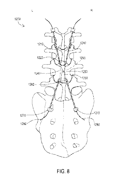
[0047] Figure 8 is a front view model of a patient's spine 1200
that includes a
pair of interchangeable exit needles and a pair of introducer needles, in
accordance with one
embodiment of the present invention. Figure 8 includes a pair of exit needles
1210, a first
epidural space 1220, a spinous process 1230, a left target lamina 1240, a
right target lamina
1250, a second epidural space 1260, a pair of introducer needles 1270 and a
pair of thread
wire 1280.
[0048] The pair of exit needles 1210 and the pair of introducer
needles 1270
are interchangeable. The pair of thread wire 1280 is passed through the pair
of introducer
needles 1270 and exits from the pair of exit needles 1210 such that the pair
of thread wire
1280 remains behind (inferior to) the right target lamina 1250 and left target
lamina 1240 on
either side of the spinous process 1230.
[0049] The pair of exit needles 1210 and the pair of introducer
needles 1270
are removed leaving the pair of thread wires 1280 in respective desired
positions behind the
target lamina 1240, 1250 with applied tension and pressure are moved back and
forth
resulting in a cutting motion from inside out through the right target lamina
1250 and the left
target lamina 1240 thereby relieving pressure on a plurality of underlying
neural tissue 1290
(not visibly seen in this diagram).
[0050] Figure 9 is a front view of a right sided percutaneous
foraminoplasty
performed by T-technique, where an introducer epidural needle is placed in an
epidural space
and an exit needle is placed in a neuroforaminal space, in accordance with one
embodiment
of the present invention.
[0051] Figure 9 illustrates a percutaneous foraminoplasty that
includes an
introducer epidural needle 910, an exit needle 920, a proximal end of a
catcher or forceps tool
930, a guide wire 944 (dotted lines) made of cutting wire or abrasive
material, a right
transverse process 916, a right lamina 999 of target vertebra 998 and a distal
end 935 of the
grasper tool 930 that is able to catch and secure the guide wire 944 in either
an epidural space
913 or a neuroforaminal space 934. Once the distal end 935 of grasper tool 930
secures the
guide wire 944, the grasper tool 930 will reverse and exit the exit needle 920
and pull the
guide wire 944 with it outside the patient's body.
[0052] Figure 10 is front view of a final position of a cutting
wire or an
abrasive wire 1100 after a plurality of needles (not shown) are removed in a
right side
-15-
CA 02990395 2017-12-20
WO 2017/007592 PCT/US2016/038203
percutaneous foraminoplasty, in accordance with one embodiment of the present
invention.
Figure 10 illustrates the cutting or the abrasive wire 1100 in a final
position after a pair of
introducer needles (not shown) and the exit needles (not shown) are taken out.
[0053] The percutaneous foraminoplasty illustrated in Figure 10 has
the
cutting wire or the abrasive wire 1100 that includes a proximal end 1102
(located outside of
body) and a distal end 1104 (located outside of body). Figure 10 also
illustrates a right
transverse process 1110, an epidural space 1120, a target vertebra 1130
possessing a
neuroforaminal space 1199, a right lamina 1140 and a target tissue 1150
(shaded area)
including right superior articular process (not shown) and right inferior
articular process (not
shown) and neuroforaminal canal (not shown). The cutting wire or the abrasive
wire 1100
having a proximal end 1102 (located outside the patient's body) a distal end
1104 of the
cutting wire or the abrasive wire 1100 (located outside the patient's body)
and the middle
portion 1198 adjacent to target tissue 1150 (shaded area) including right
superior articular
process (not shown) and right inferior articular process (not shown) and right
neuroforaminal
canal (not shown). The distal ends 1104 and proximal ends 1102 of the cutting
and the
abrasive wire 1100 has tension applied in a pulling and pushing motion that is
either
manually or electronically controlled with the middle portion 1198 of the
cutting or the
abrasive wire 1100 lying adjacent to target tissue 1150 (shaded area)
including right superior
articular process (not shown) and right inferior articular process (not shown)
and right
neuroforaminal canal (not shown).
[0054] Figures 11A, 11B, 11C and 11D illustrate a flowchart of a
method
1400 for performing a percutaneous laminoplasty, in accordance with one
embodiment of the
present invention. The method 1400 for performing percutaneous laminoplasty
utilizes a
selected one of a local anesthesia and a segmental anesthesia while a patient
is awake and in a
prone position.
[0055] The steps of the method 1400 include entering a first
introducer
epidural needle that includes a proximal end outside of the patient, a distal
end, a first hollow
internal diameter and a first penetrating perforating tip, the first
penetrating perforating tip is
disposed on the distal end, the first hollow internal diameter allows one or
more first wire
tools to pass through the introducer epidural needle, the first penetrating
perforating tip is
percutaneously placed into an epidural space of a spine on a first side
allowing the one or
more first wire tools to be introduced and entered into the epidural space of
a selected right
lamina of the spine above a targeted vertebra with a side, where a spinous
process divides a
-16-
CA 02990395 2017-12-20
WO 2017/007592 PCT/US2016/038203
right lamina and a left lamina of the target vertebra 1410, entering a first
exit epidural needle
that includes a proximal end outside of the patient, a distal end, a second
hollow internal
diameter and a second penetrating perforating tip, the second penetrating
perforating tip is
disposed on the distal end, the second hollow internal diameter allows a one
or more second
wire tools to pass through the exit epidural needle, the second penetrating
perforating tip is
percutaneously placed into the epidural space of the spine that introduces and
enters the
second wire tools below the selected right lamina of the side of the targeted
vertebra where
the first introducer epidural needle is entered in the epidural space of the
spine below the
selected right lamina, the first penetrating perforating tip and the second
penetrating
perforating tip in the epidural space resulting in the first penetrating
perforating tip and the
second penetrating perforating tip facing each other, the first penetrating
perforating tip and
the second penetrating perforating tip centering the right lamina 1420,
introducing a first
hook-like grasper tool with a distal end and a proximal end outside of the
patient, the distal
end of the first hook-like grasper tool is a selected one of manually extended
and
mechanically extended through the first hollow internal diameter of the first
exit epidural
needle, the distal end of the first hook-like grasper tool attaches the one or
more first wire
tools introduced through the first introducer epidural needle within the
epidural space, the one
or more first wire tools and the first hook-like grasper tool are pulled
through the first exit
epidural needle and out of a patient body, the attached first hook-like
grasper tool and the one
or more first wire tools engaging below the selected right lamina of the
target vertebra, where
a spinous process divides the right lamina and the left lamina, the one or
more first wire tools
having a curved middle portion, the curved middle portion lying adjacent to
the inferior
aspect (behind) of right lamina, the curved middle portion cuts the right
lamina of the target
vertebra in an anterior to posterior direction 1430, entering a second
introducer epidural
needle that includes a proximal end outside of the patient, a distal end, a
third hollow internal
diameter and a third penetrating perforating tip disposed on the distal end,
the third hollow
internal diameter allows one or more third wire tools to pass through the
second introducer
epidural needle, the third penetrating perforating tip is percutaneously
placed into the
epidural space of the spine allowing the one or more third wire tools to be
introduced and
entered into the epidural space of a selected left lamina of the spine above a
targeted vertebra
with a side, where a spinous process divides the right lamina and the left
lamina 1440,
entering a second exit epidural needle that includes a proximal end outside of
the patient, a
distal end, a fourth hollow internal diameter, a fourth penetrating
perforating tip disposed on
-17-
CA 02990395 2017-12-20
WO 2017/007592 PCT/US2016/038203
the distal end that is a selected one of manually extended and mechanically
extended, the
fourth hollow internal diameter allows a selected one or more fourth wire
tools to pass
through the second exit epidural needle, the fourth penetrating perforating
tip is
percutaneously placed into the epidural space of the spine that introduces and
enters the one
or more fourth wire tools below the side of the targeted vertebra where the
second introducer
epidural needle is entered into the epidural space of the spine of the
selected left lamina, the
third penetrating perforating tip and the fourth penetrating perforating tip
in the epidural
space resulting in the third penetrating perforating tip and the fourth
penetrating perforating
tip facing each other, the third penetrating perforating tip and the fourth
penetrating
perforating tip centering the left lamina 1450, introducing a second hook-like
grasper tool
with a distal end and a proximal end outside of the patient, the distal end of
the second hook-
like grasper tool is a selected one of manually extended and mechanically
extended through
the fourth hollow internal diameter of the second exit epidural needle, the
second hook-like
grasper tool attaches the selected one or more third wire tools introduced
through the second
introducer epidural needle within the epidural space, the selected one or more
third wire tools
is pulled through the second exit needle and out of a patient body, the
attached second hook-
like grasper tool and the one or more third wire tools engaging a selected
left lamina, the one
or more third wire tools having a curved middle portion lying adjacent to the
inferior aspect
(behind) of left lamina, the curved middle portion cuts the left lamina of the
target vertebra in
an anterior to posterior direction 1460 and implementing a plurality of safety
mechanisms
that include an intraoperative electromyogram, a plurality of nerve conduction
studies and
one or more nerve sensors to achieve a safe percutaneous environment 1470.
[0056] The third hollow internal diameter allows a selected one or
more first
fluids and first medicines to pass through the second introducer epidural
needle. The fourth
hollow internal diameter allows a selected one or more second fluids and
second medicines to
pass through the second exit epidural needle. The introducer epidural needles
are a selected
one of a flat tipped introducer epidural needle, a curved introducer epidural
needle, a rigid
introducer epidural needle, a c-shaped introducer epidural needle, an
expandable introducer
epidural needle and a flexible introducer epidural needle. The introducer
epidural needles
have a selected one of a curved penetrating perforating tip and a penetrating
perforating
straight tip. The introducer epidural needles have a hollow tube that is a
protective sheath.
The one or more wire tools are a selected one from the group of a guide wire,
a thread wire, a
bone temperature sensor and a twisted wire. The one or more wire tools are
made of a
-18-
CA 02990395 2017-12-20
WO 2017/007592 PCT/US2016/038203
selected one of metal, plastic, nylon and rubber. The one or more wire tools
have a selected
one of bone cutting and one or more abrasive properties that spare nerves and
dura when
cutting. The one or more wire tools are utilized to modify tissue, to cut
tissue and to cut bone.
The one or more wire tools are a selected one of one or more bone-cutting
devices, one or
more t-saw (Tomita saw) wires, one or more bone cutting wires and a saw
device. The one or
more wire tools includes an expanding hollow lumen that allows one or more
wires, fluids,
and medical devices to pass through the expanding hollow lumen. The one or
more wire tools
includes a plurality of channels and a plurality of apertures to be passed
through the
expanding hollow lumen to irrigate one or more anatomical areas of the spine.
The one or
more anatomical areas of the spine are irrigated with cold water. The
expanding hollow
lumen is made of a selected one of a plastic and a malleable polymer. The one
or more wire
tools can provide suction. The one or more wire tools are a selected one of
left in the epidural
space, removed immediately from the epidural space and removed at a later date
from the
epidural space. The one or more wire tools have a plurality of grooves that
pick-up bone
debris osteophytes and carry the bone debris osteophytes outside the patient's
body by a
selected one of pushing and pulling of the one or more wire tools. The one or
more wire tools
can be an expanding balloon. The expanding balloon is a selected one of radio-
opaque and
radiolucent, the expanding balloon provides a larger target to the exit
epidural needle. The
one or more wire tools are a selected one of a plurality of pieces and one
continuous piece.
The one or more wire tools are a selected one of radiolucent and radiopaque.
The one or more
wire tools are a selected one or more of being magnetic, having one or more
electromagnetic
capabilities, generating heat, being coupled to a medical device that has a
laser eliciting
capability, producing a laser, being motorized, vibrating independently and
vibrating at one
or more calculated rhythms. The epiduroscope has an ultrasound guided
capability and a
wireless capability to transmit data. The hook-like grasper tools is a pair of
grasping forceps.
The hook-like grasper tools is a selected one or more of having a fork-shape,
having one or
more apertures, having a locking device, having a selected one of a closing
door and a
pinching door, having a sticky substance and having a selected one of magnetic
properties
and electromagnetic properties. The hook-like grasper tools can suture a
selected one of a
wire, a lead and a tool at more than one level along the spinal cord
accommodates a pain
pump lead and accommodates a spinal cord stimulator lead. The hook-like
grasper tools
attaches a selected one or more of one or more wires, leads, medical devices
and desired
target tissue by using a selected one of suture wire, one or more buttons, one
or more bolsters,
-19-
CA 02990395 2017-12-20
WO 2017/007592 PCT/US2016/038203
one or more bridges and thread. The method is replicated on one or more spinal
cord levels
that include cervical, thoracic, lumbar and sacral regions on the patient
body. The method is
performed under a selected one of X-Ray, fluoroscopy, ultrasound, CT, Mill,
and 3D- MRI.
In the method, the spinous process is cut to replace a selected one of the
left lamina and the
right lamina.
[0057] Figures 12A and 12B illustrate a flowchart of a method 1500
for
performing a percutaneous foraminoplasty, in accordance with one embodiment of
the
present invention. The method 1500 for performing percutaneous foraminoplasty
that utilizes
a selected one of a local anesthesia and a segmental anesthesia while a
patient is awake and in
a prone position, the method for performing percutaneous laminoplasty is
performed on a
selected one of a first side and a second side of a spine.
[0058] The method 1500 comprises the steps of entering a first
introducer
epidural needle that includes a proximal end outside of the patient, a distal
end, a first hollow
internal diameter and a first penetrating perforating tip, the first
penetrating perforating tip is
disposed on the distal end, the first hollow internal diameter allows one or
more first wire
tools to pass through the first introducer epidural needle, the first
penetrating perforating tip
is percutaneously placed into the epidural space of a spine on a first side
allowing the one or
more first wire tools to be introduced and entered into the epidural space of
a selected right
lamina of the spine above a targeted vertebra with a side, where a spinous
process divides a
right lamina and a left lamina 1510, entering a first exit epidural needle
that includes a
proximal end outside of the patient, a distal end, a second hollow internal
diameter and a
second penetrating perforating tip, the second penetrating perforating tip is
disposed on the
distal end, the second hollow internal diameter allows one or more second wire
tools to pass
through the exit epidural needle, the second penetrating perforating tip is
percutaneously
placed into the neuroforaminal space of the spine a selected one level above,
one level below
and at an adjacent level to the selected right lamina of a targeted vertebra,
the second
penetrating perforating tip introduces and enters the second wire tools into
the
neuroforaminal space of the spine a selected one level above, one level below
and at an
adjacent level to the selected right lamina of a targeted vertebra, the first
penetrating
perforating tip in the epidural space of a selected right lamina of the spine
above a targeted
vertebra with a side, where a spinous process divides a right lamina and left
lamina and the
second penetrating perforating tip in the neuroforaminal space of the spine a
selected one
level above, one level below and at an adjacent level to the selected right
lamina of a targeted
-20-
CA 02990395 2017-12-20
WO 2017/007592 PCT/US2016/038203
vertebra, resulting in the first penetrating perforating tip and the second
Penetrating
perforating tip facing each other, the first penetrating perforating tip and
the second
penetrating perforating tip centering the neuroforaminal canal of the right
side of the target
vertebra 1520, introducing a first hook-like grasper tool with a distal end
and a proximal end
outside of the patient, the distal end of the first hook-like grasper tool is
a selected one of
manually extended and mechanically extended through the first hollow internal
diameter of
the first exit epidural needle, the distal end of the first hook-like grasper
wire tool attaches the
one or more first wire tools introduced through the first introducer epidural
needle within the
neuroforaminal space of the spine a selected one level above, one level below
and at an
adjacent level to the selected right lamina of a targeted vertebra , the one
or more first wire
tools and the first hook-like grasper tool are pulled through the first exit
epidural needle and
out of a patient's body, the one or more first wire tools having a curved
middle portion, the
curved middle portion lying adjacent to the neuroforamen and neuroforaminal
canal , the
curved middle portion cuts one or more boney structures of the neuroforamen
and the
neuroforaminal canal 1530 and implementing a plurality of safety mechanisms
that include
an intraoperative electromyogram, a plurality of nerve conduction studies and
one or more
nerve sensors to achieve a safe percutaneous environment 1540.
[0059] The first hollow internal diameter allows a selected one or
more first
fluids and first medicines to pass through the first introducer epidural
needle. The second
hollow internal diameter allows a selected one or more second fluids and
second medicines to
pass through the second exit epidural needle. The introducer epidural needles
are a selected
one of a flat tipped introducer epidural needle, a curved introducer epidural
needle, a rigid
introducer epidural needle, a c-shaped introducer epidural needle, an
expandable introducer
epidural needle and a flexible introducer epidural needle. The introducer
epidural needles
have a selected one of a curved penetrating perforating tip and a penetrating
perforating
straight tip. The introducer epidural needles have a hollow tube that is a
protective sheath.
The one or more wire tools are a selected one from the group of a guide wire,
a thread wire, a
bone temperature sensor and a twisted wire. The one or more wire tools are
made of a
selected one of metal, plastic, nylon and rubber. The one or more wire tools
have a selected
one of bone cutting and one or more abrasive properties that spare nerves and
dura when
cutting. The one or more wire tools are utilized to modify tissue, to cut
tissue and to cut bone.
The one or more wire tools are a selected one of one or more bone-cutting
devices, one or
more t-saw (Tomita saw) wires, one or more bone cutting wires and a saw
device. The one or
-21-
CA 02990395 2017-12-20
WO 2017/007592 PCT/US2016/038203
more wire tools includes an expanding hollow lumen that allows one or more
wires, fluids,
and medical devices to pass through the expanding hollow lumen. The one or
more wire tools
includes a plurality of channels and a plurality of apertures to be passed
through the
expanding hollow lumen to irrigate one or more anatomical areas of the spine.
The one or
more anatomical areas of the spine are irrigated with cold water. The
expanding hollow
lumen is made of a selected one of a plastic and a malleable polymer. The one
or more wire
tools can provide suction. The one or more wire tools are a selected one of
left in the epidural
space, removed immediately from the epidural space and removed at a later date
from the
epidural space. The one or more wire tools have a plurality of grooves that
pick-up bone
debris osteophytes and carry the bone debris osteophytes outside the patient's
body by a
selected one of pushing and pulling of the one or more wire tools. The one or
more wire tools
are an expanding balloon. The expanding balloon is a selected one of radio-
opaque and
radiolucent, the expanding balloon provides a larger target to the exit
epidural needle. The
one or more wire tools are a selected one of a plurality of pieces and one
continuous piece.
The one or more wire tools are a selected one of radiolucent and radiopaque.
The one or more
wire tools are a selected one or more of being magnetic, having one or more
electromagnetic
capabilities, generating heat, being coupled to a medical device that has a
laser eliciting
capability, producing a laser, being motorized, vibrating independently and
vibrating at one
or more calculated rhythms. The epiduroscope has an ultrasound guided
capability and a
wireless capability to transmit data. The hook-like grasper tools is a pair of
grasping forceps.
The hook-like grasper tools is a selected one or more of having a fork-shape,
having one or
more apertures, having a locking device, having a selected one of a closing
door and a
pinching door, having a sticky substance and having a selected one of magnetic
properties
and electromagnetic properties. The hook-like grasper tools sutures a selected
one of a wire, a
lead and a tool at more than one level along the spinal cord, accommodates a
pain pump lead
and accommodates a spinal cord stimulator lead. The hook-like grasper tools
attaches a
selected one or more of one or more wires, leads, medical devices and desired
target tissue by
using a selected one of suture wire, one or more buttons, one or more
bolsters, one or more
bridges and thread. The method is replicated on one or more spinal cord levels
that include
cervical, thoracic, lumbar and sacral regions on the patient body. The method
is performed
under a selected one of X-Ray, fluoroscopy, ultrasound, CT, Mill, and 3D- MRI.
[0060] The present invention is a method for performing a
percutaneous
laminoplasty and a method for performing a percutaneous foraminoplasty. The
one or more
-22-
CA 02990395 2017-12-20
WO 2017/007592 PCT/US2016/038203
components and one or more tools utilized for these methods include an
introducer needle
tool, an exit needle tool, a guide wire tool and a grasper tool. In regards to
the term needle, it
is defined as any tool or tools that are used to puncture or enter an epidural
space or a
neuroforaminal space through a percutaneous technique in contrast to open
technique and as
described for purposes and intentions herein as the T-Technique. The
introducer needle tool
has an internal diameter that is capable of introducing a guide wire or a
thread wire into an
epidural space. The introducer needle tool can be rigid, flat tipped, curved,
c-shaped,
expandable or flexible. The introducer needle has the ability to be inserted,
left in during
procedure, removed and reinserted into a desired epidural space as a
practitioner deems
necessary when performing T-Technique. The exit needle can be rigid, flat
tipped, curved, c-
shaped, expandable or flexible. The terms exit needle or introducer needle can
be used
interchangeably as it pertains to T-technique described herein. The exit
needle tool has an
internal diameter that is capable of introducing a grasper catcher tool or
other suitable
medical tools that may be used to catch guide wire tools as described herein
as the T-
technique. The term wire can be known interchangeably as a guide wire, a
cutting wire, a t-
saw or a thread wire, can be rigid, flexible or fluid that has a plurality of
functions including
navigating inside the patient's body through the epidural space and can be
passed to help
navigate further into the desired direction towards a desired epidural space
or neuroforaminal
space where the exit needle is waiting with a grasper tool. The wire can
possess tissue
modifying capability as well the capability to transport similar tools by
coupling and either
pulling or pushing medical tools or medical devices to a desired position as
well as
navigation capability that allows to connect interlaminar epidural spaces with
other
interlaminar epidural spaces, to connect interlaminar epidural spaces with
intervertebral
foramen and intervertebral foramen with other intervertebral foramen as
described herein in
methods known as the T-technique. Furthermore the term wire can represent a
tool that can
be a hollow tube with holes with an abrasive exterior that allows for air, gas
or fluid to be
released or removed by vacuum potential, that can be plastic, rubber, non-
metallic or metallic
and can vary in size. The wire can be further described and function as a
guide wire, thread
saw, a connecting device that allows other tools to be pulled into a desired
location, cutting
wire, or can represent any suitable tissue modifying tool utilized during T-
technique process
and methods described herein in accordance with one embodiment of the present
invention.
[0061] The wire has bone and target tissue cutting and molding
capabilities or
can connect to a bone-cutting device or saw device through its coupling
capability. The wire
-23 -
CA 02990395 2017-12-20
WO 2017/007592 PCT/US2016/038203
can be hollow to allow the passage of another material or guide wire through
it. The distal
end or proximal end of the guide wire can have magnetic properties to attract
one or more
forceps and grasping tools with similar attracting magnetic properties. The
wire can made of
any number of suitable materials including plastic, metal, minerals, rubber,
and allow for the
passage of fluids or gases through it. The wire can have apertures that allow
leakage of fluid
or gas for irrigation. The wire can also have suctioning capability and have
grooves that can
pick-up bone debris osteophytes and bring the debris osteophytes outside of
the patient's body
following pulling or pushing of the wire. The wire or guide wire can also be a
hollow tube
made of a malleable plastic like material that can permit other guide wires or
wires or
medical tools or devices to pass through it. The cutting wire tool device can
have access to
heat and can be construed to allow a laser to be attached or be capable of
producing a laser. It
can be motorized, have the ability to vibrate, and can be encapsulated in
order to protect vital
structures from damage from sharp edges because of poor placement, unforeseen
movements
or malfunction of the device. The thread wire can have a protective covering
that can be used
to preserve tissue where cutting is not desired during sawing action. The
protective covering
can be a plastic covering that allows for guide wires to move freely within
it. The protective
covering can be absorbed into the patient's body or be manually removed, and
can be rubbed
off with friction. The protective covering can be disposed on the entire
thread wire or in a
plurality of desired locations along the thread wire such as over the cutting
portion of the
wire. The encapsulation on the wire saw can be rubbed off with friction as the
wire comes in
contact with bone or target tissue during cutting. Furthermore, the
encapsulation can
manually be removed at an optimal position and time during the procedure, can
expand
manually and independently, can be removed independently and manually, can
shrink or
decrease in size manually, independently or with applied force, or absorb into
a body system
without damage or disintegrate with time. The encapsulation can be made to
have one or
more hooks or magnets attached to a pulley device to be removed.
[0062] The guide wires include a plurality of cutting and abrasive
components
and can be made of an expanding lumen, can be radiolucent or radiopaque, can
be magnetic
or have electromagnetic capabilities and can have a tip at a proximal end or a
distal end that
can have multiple purposes including a balloon that can expand once placed in
a desired
location. The balloon can be radiopaque or radiolucent, and can be expanded in
a desired
location to create a larger target for an exit epidural needle catcher grasper
tool to be located
while under fluoroscopy or other imaging study that can assist a practitioner
in locating and
-24-
CA 02990395 2017-12-20
WO 2017/007592 PCT/US2016/038203
performing a task. The balloon can be retracted, expanded, have several lumens
for utility,
can have a plurality of different levels of opacity or lucency to help
identify the depth of a
balloon when inside the body, epidural space or neuroforaminal space. The
balloon can have
different radiolucent or opaque shapes and designs engraved on its exterior
and can absorb a
grasper tool such as a hook and bring an attached absorbed thread wire
exteriorly out of the
patient's body. The tip of the guide wire can be a balloon or another
similarly expanding tool
that can conjoin to a catcher tool or can be caught by a grasper tool that has
been passed
through an exit needle. After the wire has been caught by a grasper tool, the
grasper tool is
now in control of the wire and can reverse in direction and exit the exit
needle tool which it
came into the patient's body from and pull the wire that it has secured out of
the patient's
body through the exit needle. The exit needle or capturing needle has an
internal diameter
capable to allow epiduroscopes, catchers, grasper tools, forceps, flexible
graspers and/or one
or more hook like devices or a bone temperature sensor to pass within and
through into
epidural space or neuroforaminal space where described medical tools and
devices can catch
the wire, guide wire or cutting wire or tissue modifying wire and pull it
outside the body.
Optionally grasping forceps, a holder tool or a hook can be passed through an
epiduroscope
that can be passed through the exit needle. The terms and functionality of the
exit needle or
the introducer needle can be used interchangeably and can possesses a US
(ultrasound)
guided tip that can define structures while in the epidural space and the
transforaminal space.
Furthermore image enhancing tools such as x-ray imaging, fluoroscopy, CT, MRI,
and US
technology can assist a practitioner to perform such tasks as required by one
or more epidural
introducer tools and exit needle tools for methods described herein as the T-
technique.
[0063] A hollow tube known as a vessel tool can be passed between
the
introducer and the exit epidural needles with the T-Technique. The hollow tube
can be made
of rubber or plastic, can be flexible or rigid, contouring, absorbable,
penetrable, have a
plurality of apertures, can be a plurality of pieces or one continuous piece,
can allow for
passage and placement of one or more guide wires, can act as a protective
sheath for a guide
wire, can allow fluid to pass though, can have suctioning capability, can
allow one or more
gases to pass through and can be used as a medium for transfer of medical
tools and devices.
The hollow tube can allow fluid to pass through in an effort to cool the
thread wire while
cutting. The hollow tube can have suction applied on either end to remove
fluid from a field
environment during the procedure. An epidural drain catheter can be passed in
one space
above or below the procedure site through an epidural needle tool and the
catheter can be
-25 -
CA 02990395 2017-12-20
WO 2017/007592 PCT/US2016/038203
attached to negative pressure suction located outside of the body so as to
drain any possible
blood or fluid collected in the epidural space. The drain catheter can be left
in the space after
the procedure, removed immediately or removed at a later date.
[0064] The methods also utilize a grasping forceps tool that can be
in any
suitable form and can be flexible or non-flexible. The grasping forceps tool
is used to
interlock or connect while positioned in an epidural space or neuroforaminal
space to a
related wire that is passed from an introducer needle by the T-technique. Once
the grasper
tool has made the catch or connection with the guide wire it can now be pulled
through the
exit needle outside the patient's body. The grasping forceps can be a hook
mechanism, with a
fork-shape, have an aperture, a locking device, a closing or pinching door,
can be a sticky
substance, can have magnetic or electromagnetic properties and/or have an
attractive force
that can attract the distal end of the guide wire to the grasping forceps. The
grasping forceps
can have a coating where an US (ultrasound machine outside the body) could be
used to
determine the distance between the grasping forceps and the guide wire.
Locking or catching
the guide wire can also be done under Fluoroscopy, with an US, a CT, an MRI, a
3-
Dimensional MRI or other suitable imaging studies that can assist the
practitioner in
completing such a task.
[0065] Alternatively, the grasper tool can also possess the ability
to suture a
wire, a lead or a tool at more than one level along the spinal cord such as
leads for a pain
pump or leads for a spinal cord stimulator and tie them to other wires, leads,
medical devices
or desired target tissue by using suture wire, buttons, bolsters, bridges,
thread or similar
surgical tools and devices. This is one of the most common causes of failure
of spinal cord
stimulators that include lead breakage and lead migration. In application of
the T-technique,
the grasper tool can be used for lead placement and fastening of both the
distal and proximal
ends of the leads. This process will allow a practitioner to access both the
distal and proximal
ends of the leads or wires as well as at any point along the spine where there
is access to
wires, leads, devices and target tissue through the described T-technique
where the grasper
tool enters the exit needle or the introducer needle to access the
neuroforaminal space and/or
epidural space at any target level of the spine.
[0066] Figure 13 illustrates a systematic representation of an
epidural scope
visualization system lens 1662 with a light source 1672 that may not be
attached to the
epiduroscope visualization system lens 1662 but may seek the light source 1672
in the
epidural space 399 at any level of the spine. As depicted in Fig. 13, four
needles in two
-26-
CA 02990395 2017-12-20
WO 2017/007592 PCT/US2016/038203
epidural spaces keep a target lamina in a center area. As explained with
reference to Fig. 3,
above, Fig. 13 also depicts a left lamina 305, a right lamina 310 and a
spinous process 315
which divides the right lamina 310 and the left lamina 305 of the target
vertebra 302. Fig. 13
also includes a left lamina 205, a spinous process 215 and a right lamina 210
of a vertebra
one level above target vertebra 302. Also shown is a left lamina 105, spinous
process 215'
and right lamina 210' of the vertebra one level below target vertebra 302. As
one of ordinary
skill in the art may appreciate, a left introducer epidural needle 320, a
right introducer
epidural needle 321, a left exit epidural needle 322 and a right exit epidural
needle 323 are
also shown. The left introducer needle 320 may have a proximal end 324 and a
distal end
326. The right introducer needle 321 may have a proximal end 325 and a distal
end 327. The
left exit needle 322 may have a proximal end 330 and a distal end 328. The
right exit needle
323 may have a proximal end 331 and a distal end 329.
[0067] The proximal ends of introducer needles 324,325 and the
proximal
ends of the exit needles 330,331 may remain outside of a patient's body while
the distal ends
of the introducer needles 326,327 may enter the epidural space 399 above
target vertebra 302.
The distal ends of the exit needles 328,329 enter the epidural space 398 below
the target
vertebra 302. The left introducer needle 320 and its distal end 326 may be
placed and
introduced in the epidural space 399 above the target vertebra 302 to the left
of the spinous
process 315. The right introducer needle 321 and its distal end 327 may be
introduced in
epidural space 399 on the right side of spinous process 315. The left exit
epidural needle 322
and its distal end 328 may enter the epidural space 398 below target vertebra
302 to the left of
the spinous process 315. The right exit needle 323 and its distal end 329 may
enter the
epidural space 398 below target vertebra 302 to the left of the spinous
process 315. As
previously discussed, the left distal end 326 of the introducer needle 320 and
the left distal
end 328 of the exit needle 322 may be facing each other, and, correspondingly,
the right
distal end 327 of the introducer needle 321 and the right distal end 329 of
the exit needle 323
may be facing each other.
[0068] The epiduroscope visualization system 1660 may be located
outside
the patient's body to allow visualization of a light source 1672 that is
independent of the
epiduroscope visualization system 1660. The middle attached scope portion 1661
that may
enter into the epidural space 399 through the left introducer needle 320 may
have an
epiduroscope visualization system lens 1662 at the distal end. The
epiduroscope visualization
system lens 1662 may have fibers that allow it in a co-axial plane to
visualize light source
-27-
CA 02990395 2017-12-20
WO 2017/007592 PCT/US2016/038203
1672 and other structures outside the patient's body by seeking the light
source 1672 that is
not otherwise attached to it.
[0069] The power source 1670 for the distal light source 1672 that
may shine
in the epidural space 399 that allows the epiduroscope visualization system
lens 1662 to seek
light to allow visualization may enter the left exit needle 322 through its
middle component
1671 to connect to the distal light source 1672 used to allow the epiduroscope
visualization
system lens 1662 to seek and visualize the light. Ultimately, the epiduroscope
visualization
system lens 1662 seeking the light source 1672 will help facilitate the
creation of a
mechanical or electromagnetic coupling within the epidural space as described
earlier.
[0070] As a person of ordinary skill may appreciate, the
representations
disclosed herein with respect to Fig. 13 may be performed at any level and
either side of the
spine. Likewise, although reference is made herein that the light source 1672
is routed
through an exit needle 322, a person of ordinary skill would not be so limited
and may find
other suitable arrangements of advancing the light source 1672 and lens 1662
through various
combinations of exit and entrance needles.
[0071] Figure 14 represents a completed loop electrical circuit
1780 which
may be percutaneously formed with part of the completed circuit 1780 outside
the patient's
body and part of the completed loop circuit 1780 may reside in any portion of
the spine in the
epidural space 399. The completed loop circuit 1780 is formed in the spine
where two distal
ends of two medical tools 1771,1775 in co-axial plane in the spine have a
point of contact
1776 in the epidural space 399.
[0072] The completed loop circuit 1780 is formed once an electrical
current
from a power source 1749 is connected and forms a circuit with conjoining wire
1770 that
may be partially outside a patient's body and partially inside the patient's
body by entering or
exiting percutaneously through the left introducer needle 320 distally into
the epidural space
399 where the two medical tools 1771,1775 make physical contact and create a
point of
electromagnetic contact 1176 that can be used to verify if a catch is made
along a co-axial
plane. The completed loop circuit 1780 is further described of a conjoining
wire 1774 that
exits or enters the epidural space 399 through left exit needle 322 where the
proximal tip
1773 of the conjoining wire 1774 outside the patient's body enters the
proximal portion of the
other medical device 1769 that forms this particular completed loop circuit
1780. Once the
completed loop circuit 1780 is formed, the practitioner will be aware that the
catch has been
made as previously described. This awareness may be surmised by way of a
visual or audio
-28 -
CA 02990395 2017-12-20
WO 2017/007592 PCT/US2016/038203
cue that would indicate the creation of a closed loop circuit 1780. Similarly,
and although
described across a single level of vertebra, this system may be used at any
level and either
side of the spine.
[0073] An epiduroscope or a fiberscope with fiber optic capability,
can be
passed through epidural needles and placed in the epidural space, the extra
epidural space or
the neuroforaminal space and can be left in place for direct vision while
utilizing the T-
Technique. The epiduroscope or fiberscope can be one continuous piece or a
plurality of
pieces working together from an introducer needle to an exit needle with one
continuous
point of visualization, a single point of visualization or a plurality of
points of visualization.
The epiduroscope or fiberscope can also have one or more ultrasound guided
capabilities and
one or more wireless capabilities for data transmission. The scope can have an
option to
allow a cutting instrument to pass through to perform cutting or the scope can
itself be used
as a cutting device by using a saw, a blade, a laser, heat energy or other
suitable cutting
device. The scope can have the ability to pass fluids, medical tools or
materials, medically
useful gases or substances with medicinal benefit in desired target areas. The
scope can have
a light source in many locations, a single location or a continuous location.
The scope can
have a lumen or a plurality of lumens to allow materials such as gas, fluids,
or medical tools
such as guide wires, grasper tools or probes to pass through and position them
in the desired
target areas.
[0074] Additionally a catheter with an inflatable tip balloon like
structure can
be passed through an epiduroscope or fiberscope through its working channel or
lumen or
through one or more introducer or exit epidural needles. The balloon structure
can expand
from addition of gas or liquids. Furthermore the inflatable balloon can be
placed in such a
position that it remains as a shield between the cutting wire and vital
anterior structures like
exiting nerves and dura. The inflatable balloon can be designed to expand
first laterally and
then posteriorly so that it does not exert more pressure on dura and may help
to push cut
lamina posteriorly (outwards) following cuts from the T-Technique. The
inflated balloon can
be deflated and taken out after the procedure or left in the epidural space as
a support
structure or other suitable utility or be absorbed by the patient's body. The
inflated balloon
can have a plurality of grooves on its posterior surface to accommodate a
cutting wire to have
better control during cutting. The inflated balloon can have radiopaque
properties or can be
injected with contrast material so that its placement is well visualized under
fluoroscopy.
Additionally the patient will be awake during T-Technique percutaneous
procedures giving a
-29-
CA 02990395 2017-12-20
WO 2017/007592 PCT/US2016/038203
practitioner immediate awareness if neural structures are being encroached
upon by
immediate paresthesia felt and reported by a patient, which would prompt
immediate
cessation and an alternative approach which is a common practice in the field
of pain
management. The application of current safety mechanisms such as
intraoperative EMG
(Electromyography), NCS (Nerve conduction studies) and nerve sensors can be
used to
achieve a desired safe procedural environment. Ultrasound technology,
radiofrequency, CT,
MRI, 3-dimensional MRI, C-Arm or other suitable instruments can be used in
assistance to
complete the task to identify surface anatomy and distance between neural
structures, thread
wire and other medical tools.
[0075] The present invention also includes a method for fixing,
fusing and
lifting loose bone following an applied T-Technique. The method includes a
technique to
secure the spinous process of a target vertebra with a modified spinous
process screw tool.
The spinous process is a relatively superficial bony structure in the spine
and can be easily
felt under the skin. The spinous process can easily be approached
percutaneously with a
modified percutaneous spinous process screw tool, an epiduroscope or other
similar
percutaneous drilling devices. The modified spinous process screw tool that is
percutaneously
inserted into a spinous process and then fixated with a locking, rotating
screwing motion,
where a plurality of teeth like protrusions, insertions or hooks attach the
screw tool to the
spinous process. The spinous process modified screw and a plurality of other
suitable types of
screw tools regarding the T-Technique can also be made of implantable material
such as
stainless steel, titanium and other suitable biocompatible materials. The
spinous process
screw tool is attached to a gauge tool outside of the patient's body that can
adjust the desired
outward (posterior) pressure on loose bone manually or automatically and can
adjust and
assist in maneuvering a cut portion of the bone into a desired position. The
screw tool can
have one or multiple apertures, one or multiple lumens, hooks or ports that
can attach to one
or more wires, bars, needles, other screws or tools for anchoring or other
utility. The
modified spinous process screw tool allows a practitioner to maneuver, move
and adjust loose
bone that has been cut by the T-Technique. The modified spinous process method
for
modeling and maneuvering loose bone can be equally applied by both
percutaneous T-
technique laminoplasty and foraminoplasty. An example of loose bone in a case
of
percutaneous laminoplasty by T-Technique would be defined as target vertebral
bone medial
to the cuts of its right lamina and its left lamina. In this example the loose
bone would include
the right lamina, spinous process and left lamina of the target vertebral
bone. Following cuts
-30-
CA 02990395 2017-12-20
WO 2017/007592 PCT/US2016/038203
to the target lamina, the loose bone is no longer attached continuously with
the original
anatomy of the target vertebra and is now fully free to be mobilized by
application of
posterior (outward) force and pressure by a modified spinous process screw
tool attached to a
gauge tool outside of the patient's body. Following fixation of a spinous
process screw tool
into spinous process by methods described herein, posterior (outwards)
pressure is applied
that can allow loose bone to be placed in a desired position that will allow
for expansion of
the spinal canal and neuroforamen by the T-Techniques. The maneuvering will
achieve
decompression by creating space for the neural elements. The loose bone now in
place will be
secured using the subsequent tools, percutaneous fusion and methods that will
lead to
osteogenesis between the cut ends of lamina where healing and fusion will take
place.
[0076] There is also an optional percutaneous method for loose bone
lifting
and fixation that utilizes a plurality of M Technique steps. The M Technique
requires the use
of a plurality of modified pedicle screws and a plurality of modified fixing
screws.
Following T-Technique laminoplasty or foraminoplasty, a percutaneous modified
spinous
process screw will be placed into target tissue where the distal end of the
screw will be
inserted into spinous process and the proximal end will protrude and have an
exit from the
skin of the patient. Pressure will be applied in a posterior direction with
the use of a pressure
gauge tool that is positioned outside the body that is attached to the spinous
process screw.
The posterior pressure placed on the modified spinous process screw though
utility of the
gauge tool will be sufficient to protect the canal from anterior drift as well
as properly place
the cut lamina in a desired position to alleviate foraminal and/or canal
stenosis. Subsequently
the M technique follows by percutaneously inserting one modified pedicle screw
through
each pedicle of a chosen target vertebra in the AP position, (for example:
vertebral level 5 has
one modified pedicle screw in a right pedicle and one modified pedicle screw
in a left
pedicle). The modified pedicle screw can have the possibility of angulation or
curvature
where the distal end of the screw will be inserted into target tissue and
fastened into the
pedicle by percutaneous methods and the proximal end can have the capability
to interlock
into one or more other screws or tools. The length of the modified pedicle
screw can be
variable and can be increased through an interlocking feature that will extend
to a desired
needed length. The proximal end of the modified pedicle screw will have one or
more
openings through which a fusing screw tool can be passed through and interlock
with it. The
fusing screw can be expandable by both automatic, or manual technique, can
decrease in size,
can vibrate, can contain fluid, can absorb fluid, can have drilling or
puncturing capability
-3 1 -
CA 02990395 2017-12-20
WO 2017/007592 PCT/US2016/038203
independently or with a practitioner's assistance such as a screwing rotation
that can elicit a
drilling or a puncturing capability or a plurality of teeth-like, nail-like
projections to enter the
touching structures and attach to them, can have one or more rotational
capabilities and will
not only separate target tissue but likewise keep it in a secured position.
The fusing screw
tool can use the modified pedicle screw for support as it interlocks with the
proximal end of
the modified pedicle screw percutaneously. The fusing screw can interlock with
the modified
pedicle screw at any point along the fusing screw. The distal end of the
fusing screw will
target the loose bone seen following cutting as described in T-technique
foraminoplasty
and/or laminoplasty. The fusing screws can be angled towards the loose bone
and
subsequently fastened into target tissue to secure the lamina and loose bone
into its new
position. After several weeks following fusion and healing, the screws can be
removed as
needed.
[0077] In an alternative embodiment, a spinal guidance system light
seeking
fiberscope is disclosed. As one of ordinary skill in the art may appreciate,
fiberscopes and/or
epiduroscopes, typically have light to help guide them in body cavities. In
such a process, one
of ordinary skill may think of the light as going in the direction of the
fiberscope or being
pushed toward the desired target as is typical of any directional lighting
fixture. One
disadvantage of adding light to scopes that are designed to operate in
miniature scale so as to
be inserted in body cavities is that the respective diameter of the scope
increases to
compensate for the addition of light emitting elements. This disadvantage is
exponentially
worsened when attempting to operate within the confines of the epidural space.
Accordingly,
there is a need to have a fiberscope operate without light, thereby
maintaining its slim profile
within the epidural space. In addition, to the smaller diameter, this design
may help the
guidewire find out where the catcher tool is located because the guidewire or
the catch tool
may be also a fiberscope or have fiberscope capabilities; and once in coaxial
plane, the
guidewire can be pushed to find the light, that will be attached to the head
of the grasper /
catcher tool, and thus the guidewire will be steered directly into the light
of the catcher/
grasper tool.
[0078] Unlike other scopes, there is no need for visual access,
rather just the
ability to see light or some level of brightness. In other words, if there is
no visible light
viewable by the practitioner, then the appropriate placement within the
epidural space has not
been attained. Conversely, if the practitioner is able to see light, then the
correct direction and
placement has been established.
-32-
CA 02990395 2017-12-20
WO 2017/007592 PCT/US2016/038203
[0079] In yet another embodiment, a complete and closed circuit is
created
while having a portion of the circuit within the epidural space itself. The
spinal guidance
system visual engagement indication system (VEIS) percutaneous epidural
complete circuit
may create a circuit in the form of a loop. Once the circuit is complete an
LED light or audio
sound or some sort of alert or other indication is generated where the
practitioner realizes that
the guidewire has actually touched the grasper tool. By way of non-limiting
example, a
battery with a positive and negative side, is outside the patient's body. A
negative current is
passed along a wire that is attached from the negative side of the battery to
the LED light.
The negative charge is then continued though the LED light and then extended
by wire and
through the grasper tool. A positive current is then passed along a wire from
the other side of
the battery where current is then applied to the a guidewire. The catcher tool
is thus placed
into the epidural space and the guidewire is then placed in the epidural space
in coaxial plane
and advanced toward each other. Once the guidewire tool touches the grasper
tool/ catcher
tool, the circuit is complete and the LED illuminates outside the patient's
body. (The voltage
of the circuit may be as minimal as needed not to be adverse to normal bodily
functions,
which may be less than .5 V).
[0080] By way of further example, the guidewire or the
catcher/grasper tool
can be either positive or negatively charged, depending on the orientation of
the battery. The
indication mechanism of the circuit may be an LED diode that can be any color,
it can reflect
a screen that expresses words or other relevant means of expression. An
alarming mechanism
can be a sound or other means of alerting the practitioner that the circuit
has been created.
There can be one, two, three circuits or more created at time or at any one
particular catch.
Alternatively, each circuit and indication mechanism can relate a different
portion of the
catch tool or guidewire tool, for instance the inner aspect of the catch tool
can be one color
and the outer aspect of the catch grasper tool can be another, each
independent circuits of
each other, giving the practitioner the ability to know where the exactly the
to two tools are in
relation to each other.
[0081] While various embodiments of the disclosed technology have
been
described above, it should be understood that they have been presented by way
of example
only, and not of limitation. Likewise, the various diagrams may depict an
example
architectural or other configuration for the disclosed technology, which is
done to aid in
understanding the features and functionality that can be included in the
disclosed technology.
The disclosed technology is not restricted to the illustrated example
architectures or
-33 -
CA 02990395 2017-12-20
WO 2017/007592 PCT/US2016/038203
configurations, but the desired features can be implemented using a variety of
alternative
architectures and configurations. Indeed, it will be apparent to one of skill
in the art how
alternative functional, logical or physical partitioning and configurations
can be implemented
to implement the desired features of the technology disclosed herein. Also, a
multitude of
different constituent module names other than those depicted herein can be
applied to the
various partitions. Additionally, with regard to flow diagrams, operational
descriptions and
method claims, the order in which the steps are presented herein shall not
mandate that
various embodiments be implemented to perform the recited functionality in the
same order
unless the context dictates otherwise.
[0082] Although the disclosed technology is described above in
terms of
various exemplary embodiments and implementations, it should be understood
that the
various features, aspects and functionality described in one or more of the
individual
embodiments are not limited in their applicability to the particular
embodiment with which
they are described, but instead can be applied, alone or in various
combinations, to one or
more of the other embodiments of the disclosed technology, whether or not such
embodiments are described and whether or not such features are presented as
being a part of a
described embodiment. Thus, the breadth and scope of the technology disclosed
herein
should not be limited by any of the above-described exemplary embodiments.
[0083] Terms and phrases used in this document, and variations
thereof,
unless otherwise expressly stated, should be construed as open ended as
opposed to limiting.
As examples of the foregoing: the term "including" should be read as meaning
"including,
without limitation" or the like; the term "example" is used to provide
exemplary instances of
the item in discussion, not an exhaustive or limiting list thereof; the terms
"a" or "an" should
be read as meaning "at least one," "one or more" or the like; and adjectives
such as
"conventional," "traditional," "normal," "standard," "known" and terms of
similar meaning
should not be construed as limiting the item described to a given time period
or to an item
available as of a given time, but instead should be read to encompass
conventional,
traditional, normal, or standard technologies that may be available or known
now or at any
time in the future. Likewise, where this document refers to technologies that
would be
apparent or known to one of ordinary skill in the art, such technologies
encompass those
apparent or known to the skilled artisan now or at any time in the future.
[0084] The presence of broadening words and phrases such as "one or
more,"
"at least," "but not limited to" or other like phrases in some instances shall
not be read to
-34-
CA 02990395 2017-12-20
WO 2017/007592 PCT/US2016/038203
mean that the narrower case is intended or required in instances where such
broadening
phrases may be absent. Additionally, the various embodiments set forth herein
are described
in terms of exemplary block diagrams, flow charts and other illustrations. As
will become
apparent to one of ordinary skill in the art after reading this document, the
illustrated
embodiments and their various alternatives can be implemented without
confinement to the
illustrated examples. For example, block diagrams and their accompanying
description
should not be construed as mandating a particular architecture or
configuration.
-35 -