Note : Les descriptions sont présentées dans la langue officielle dans laquelle elles ont été soumises.
Systems And Methods For Immobilizing Extracellular Matrix Material
on Organ On Chip, Multilayer Microfluidics Microdevices, and Three-dimensional
Cell Culture Systems
BACKGROUND
Recapitulating native three-dimensional (3D) organ microenvironments is a
fundamental
challenge in the development of biomimetic models of human physiology and
disease.
Microenvironmental cues such as local architecture, mechanical forces, and
biochemical signals can
define the physiological, or pathological situation in vivo. The extracellular
matrix (ECM), serving as
.. both a structural scaffold and cell adhesion substrate, possesses a tissue-
specific composition and
topology that can instruct diverse processes including growth,
differentiation, and tissue
morphogenesis/remodeling. In polarized tissues, such as the epithelium and
endothelium, cells can
interact with a planar layer of ECM called the basement membrane, while in
tissues such as muscle
and connective tissue/stroma, the cells reside in a truly 3D milieu of ECM and
surrounding cells. By
.. reconstituting the microenvironment, 3D models can facilitate investigation
of-relevant human
physiological and pathophysiological processes involving tissue elements such
as stroma and their
interfaces with epithelial and vascular components.
3D culture platforms have been developed that aim to model the native tissue
microenvironment. Specific examples include hydrogels derived from native ECM
or synthetic
.. materials, solid state polymeric scaffolds, and matrix/scaffold-free
systems such as spheroid cultures,
with ECM gels being the most commonly employed. While such 3D hydrogel-based
models can
reconstitute the composition and mechanical properties of the native tissue
microenvironment, critical
aspects of organ
1
Date Regue/Date Received 2023-02-21
CA 02993943 2018-01-26
WO 2017/019799 PCT/US2016/044321
structure such as patterned tissue-tissue interfaces and dynamic mass
transport are
absent in these models.
One approach to meet these challenges is to leverage biomimetic
microengineering techniques to develop microphysiological models of human
tissues
and organs, termed "organs-on-chips." Comprised of 3D arrangements of
perfusable
microchannels, human organs-on-chips can allow for precise control of dynamic
flow
conditions and application of physical stimuli to cells and engineered tissues
equivalents. Researchers have sought to create systems that incorporate 3D ECM
gels, which mimic the in vivo stromal compartment to facilitate the study of
angiogenesis, tumor cell invasion, and metastasis.
There remains a need to be able to control the spatial geometry and
microarchitecture of the ECM hydrogel in these 3D culture organ-on-a-chip
systems
in order to develop improved microphysiological models. Furthermore, there
remains
a need to be able to prevent detachment of the ECM gel from the anchoring
substrate
due to cell-mediated contraction, resulting in the loss of the originally
defined
construct geometry and limited timeframes of experimentation.
The problem of cellular contraction of hydrogel matrices and detachment
during 3D culture is a common obstacle to hydrogel anchorage in traditional
cell
culture models. A technique is needed that enables culture and maintenance of
living
cells in a 3D ECM environment for prolonged periods of time without causing
significant changes to the volume of hydrogel and preventing loss to its
structural
integrity.
SUMMARY
The presently disclosed subject matter provides a method for anchoring of
protein-composed 3D cell culture substrates in biomimetic microdevices and/or
organ-on-a-chip platforms and/or general 3D cell culture systems. The
presently
disclosed subject matter can control the location at which a hydrogel
construct
detaches from the substrate, thereby creating a predictable geometric change.
The
presently disclosed subject matter can have specific applications beyond
establishing
construct stability, including but not limited to shaping the geometry of
living tissues
in vitro by harnessing cell-mediated contractile forces to contract the 3D
tissue in a
2
CA 02993943 2018-01-26
WO 2017/019799 PCT/US2016/044321
rationally designed manner by patterning the locations of tissue anchorage. In
certain
embodiments, the method can include selecting one or more substrates for
tissue
growth. In certain embodiments, the method can further include identifying
desired
tissue anchorage points on the one or more selected substrates to facilitate
creation of
rationally designed tissue geometries by allowing the natural process of
cellular
contraction to occur. In certain embodiments, the method can further include
coating
the one or more selected substrates with a heterobifunctional crosslinker at
the
identified desired tissue anchorage points. In certain embodiments, the method
can
further include curing the heterobifunctional crosslinker to the one or more
substrates.
In certain embodiments, the method can further include adding a gel layer
embedded
with at least one of tissue and cells to the one or more substrates. In
certain
embodiments, the method can further include allowing cell-mediated contractile
forces to shape tissue geometry as the gel layer contracts between the fixed
anchorage
points.
In certain embodiments, the heterobifunctional crosslinker can be sulfo-
SANPAH.
In certain embodiments, the substrate can be poly-di-methyl-siloxane (PDMS).
In certain embodiments, the substrate can be sulfo-SANPAH. In
certain
embodiments, the substrate can be composed of different build materials that
can be
coated with a heterobifunctional crosslinker at the identified desired tissue
anchorage
points. In certain embodiments, the substrate on which the disclosed methods
can be
performed can be any polymeric, glass, and metal surfaces that are compatible
with
sulfo-SANPAH and/or any heterobifunctional crosslinkers that would serve the
same
function as sulfo-SANPAH.
In certain embodiments, the gel layer can include extracellular matrix
proteins.
In certain embodiments, the extracellular matrix proteins can be selected from
the
group consisting of, but not limited to, collagen, fibronectin, laminin,
hyaluaronic
acid, and mixtures thereof.
In certain embodiments, tissue and cells embedded within the gel layer can be
fibroblasts. In certain embodiments, the tissue and cells embedded within the
gel
layer can be at stromal tissue and stromal cells. In certain embodiments, the
tissue
and cells embedded within the gel layer can be myoblasts. In certain
embodiments,
the tissue and cells embedded within the gel layer can be mesenchymal stem
cells. In
certain embodiments, the tissue and cells embedded within the gel layer can be
3
CA 02993943 2018-01-26
WO 2017/019799 PCT/US2016/044321
vascular cells. In certain embodiments, the tissue and cells embedded within
the gel
layer can be epithelial cells. In certain embodiments, any other type of
tissue and
cells can be embedded within the gel layer.
In certain embodiments, the desired tissue anchorage points can be within a
single horizontal plane. In certain embodiments, the desired tissue anchorage
points
can be within a plurality of horizontal planes. In certain embodiments, the
desired
tissue anchorage can be within a single vertical plane. In certain
embodiments, the
desired tissue anchorage can be within a plurality of vertical planes. In
certain
embodiments, the desired tissue anchorage can be within a single angled plane.
In
certain embodiments, the desired tissue anchorage can be within a plurality of
angled
planes. In certain embodiments, the desired tissue anchorage can be within a
plurality
of horizontal, vertical, and angled planes.
In certain embodiments, a first biopsy punch can be used to create a cell
culture chamber in the substrate, and a second biopsy punch can be used to
create
outer nodes in the substrate that overlaps with a portion of the cell culture
chamber.
In certain embodiments, the outer nodes can be used as tissue anchorage
points. In
certain embodiments, a mold can be generated by 3D printing, photolithography,
stereolithography, or other similar method(s). In certain embodiments, the
mold can
be used to create a cell culture chamber and anchorage points. In certain
embodiments, a substrate can be directly etched and/or ablated using etchants,
laser,
and/or similar method(s) to create a cell culture chamber and anchorage
points.
In certain embodiments, the presently disclosed subject matter further
provides
a technique to form and maintain 3D tissue in a microengineered cell culture
device.
In certain embodiments, the microengineered device can include a body having
one or
.. more cell culture chambers. In certain embodiments, the walls of the
chamber can be
treated with a heterobifunctional crosslinker and the chamber walls can form a
substrate for hydrogel attachment and tissue growth. In certain embodiments, a
hydrogel can be formed in the chamber and anchored to the walls of the
chamber. In
certain embodiments, the hydrogel can contain cells. In certain embodiments, a
first
microfluidic channel can be disposed above the gel layer. In certain
embodiments, a
second microchannel can be disposed under the gel layer. In certain
embodiments,
the microchannels can be perfused with culture media, blood, artificial blood,
and
other fluids to maintain and/or stimulate the cells embedded in the gel.
In certain embodiments, the presently disclosed subject matter further
provides
4
CA 02993943 2018-01-26
WO 2017/019799 PCT/US2016/044321
a microengineered perfusable lumen sculpted from engineered tissue. In certain
embodiments, the perfusable lumen can include a body having a microchannel. In
certain embodiments, the walls of the microchannel can form a substrate for
tissue
growth. In certain embodiments, the perfusable lumen can include a tissue
embedded
in a gel layer adhered to each of three different walls of the microchannel.
In certain
embodiments, the tissue can be unconnected to a fourth wall of the
microchannel such
that the tissue is shaped to create a semicircular opening within the
microchannel. In
certain embodiments, the semicircular opening can extends through a length of
the
microchannel and forms a conduit.
In certain embodiments, the conduit formed can be injected with a gel
containing tissue, resulting in a tissue-to-tissue interface without using an
intervening
membrane.
In certain embodiments, the presently disclosed subject matter further
provides
methods of fabricating a microengineered perfusable lumen sculpted from
tissue. In
certain embodiments, the method can include fabricating a microchannel in a
first
body. In certain embodiments, the first body can form a substrate for tissue
growth
such that the microchannel is fabricated by bonding a second body to the first
body.
In certain embodiments, the method can further include injecting the
microchannel
with a heterobifunctional crosslinker. In certain embodiments, the method can
further
include curing and/or activating the heterobifunctional crosslinker. In
certain
embodiments, the method can further include replacing the second body with a
third
body to form a four-sided microchannel having three different sides treated
with the
heterobifunctional crosslinker. In certain embodiments, the method can further
include injecting a gel layer embedded with at least one of tissues and cells
into the
microchannel. In certain embodiments, the method can further include allowing
cell-
mediated contractile forces to shape tissue geometry as the gel layer
contracts,
forming a semicircular conduit along a length of the microchannel.
In certain embodiments, the microchannel can be formed using
photolithography.
In certain embodiments, upon formation of the conduit, the method can further
include injecting the conduit with a gel containing tissue, resulting in a
tissue-to-tissue
interface without use of an intervening membrane.
5
The presently disclosed subject matter provides a method of shaping the
geometry of living
tissues in vitro, the method comprising: a) selecting one or more substrates
for tissue growth; b)
identifying fixed tissue anchorage points on the one or more selected
substrates; c) coating the one or
more selected substrates with a heterobifunctional crosslinker at the
identified fixed tissue anchorage
points; d) curing the heterobifunctional crosslinker to the one or more
selected substrates; e) adding an
extracellular matrix gel layer embedded with contractile tissue to the one or
more selected substrates;
and 0 allowing cell-mediated contractile forces to shape a geometry of the
extracellular matrix gel
layer as the contractile tissue within the extracellular matrix gel layer
contracts the extracellular matrix
gel layer between the fixed anchorage points.
The presently disclosed subject matter also provides a microengineered
perfusable lumen
sculpted from tissue comprising: a) a microdevice having at least one
microchannel, wherein walls of
the at least one microchannel form a substrate for tissue growth; and b) a
contractile tissue embedded
in an extracellular matrix gel layer adhered to each of three different walls
of the microchannel, wherein
the contractile tissue is not connected to a fourth wall of the microchannel,
wherein the contractile
tissue contracts to create a semicircular opening within the microchannel, and
wherein the semicircular
opening extends through a length of the microchannel and forms a conduit.
The presently disclosed subject matter also provides a method of fabricating a
microengineered
perfusable lumen sculpted from tissue comprising: a) fabricating a
microchannel in a first body, said
first body forming a substrate for tissue growth, wherein the microchannel is
fabricated by bonding a
second body to the first body; b) injecting the microchannel with a
heterobifunctional crosslinker; c)
curing the heterobifunctional crosslinker; d) replacing the second body with a
third body to form a
four-sided microchannel having three different sides treated with the
heterobifunctional crosslinker;
e) injecting an extracellular matrix gel layer embedded with contractile
tissue into the microchannel;
and 0 allowing cell-mediated contractile forces to shape tissue geometry as
the extracellular matrix gel
layer contracts, forming a semicircular conduit along a length of the
microchannel.
5a
Date Regue/Date Received 2023-02-21
CA 02993943 2018-01-26
WO 2017/019799 PCT/US2016/044321
BRIEF DESCRIPTION OF THE FIGURES
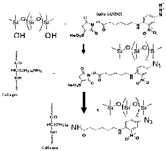
Figure 1 is a diagram illustrating the chemical structure of sulfo-SANPAH and
a two-step reaction scheme depicted illustrating collagen being chemically
tethered to
the surface of PDMS.
Figures 2A and 2B are diagrams illustrating schematics of 2-D patterning for
microtissue sculpting. Figure 2C is a diagram illustrating a computational
model
predictions of gel detachment from untreated surfaces due to cell-mediated
contraction.
Figure 3 illustrates a schematic of 3-D patterning for microtissue sculpting.
Figure 4 illustrates a photograph in which the sulfo-SANPAH solution is seen
pipetted into each node.
Figures 5A-5D are photographs illustrating the collagen gel layer being
sandwiched between two PDMS layers.
Figures 6A and 6B show images in which a droplet of collagen gel was cast on
PDMS that was either untreated (Figure 6A) or sulfo-SANPAH-treated (Figure
6B).
Figure 6C illustrates a chart depicting the mean fluorescence intensity over
the entire
original surface area of gel anchorage.
Figures 7A and 7B, which illustrate the results of an experiment investigating
the effect of continuous mechanical strain on the collagen-to-PDMS anchorage.
Figures 8A and 8B are photographs depicting the time course of patterned
microtissue sculpting by embryonic mouse fibroblasts in multiple geometries.
Figure 9 illustrates results of the experiment in which connective tissue
microtissue was sculpted using human fibroblasts.
Figure 10A illustrates exemplary images of dense regular connective tissue
and Figure 10B illustrates exemplary images of microengineered connective
tissues
that have been in vitro for nine to ten days.
Figure 11 is a table illustrating orientation data for coherency and dominant
direction of fibronectin and nuclei alignment.
Figures 12A and 12B illustrate examples of cell nuclei orientation analysis
for
unpatterned and/or contracted samples (Figure 12A) and for 2-node aligned
samples
(Figure 12B).
Figures 13A and 13B illustrate examples of fibronectin orientation analysis
for
unpatterned and/or contracted samples (Figure 13A) and for 2-node aligned
samples
(Figure 13B).
6
CA 02993943 2018-01-26
WO 2017/019799 PCT/US2016/044321
Figure 14A and 14B illustrates parallel cellular cytoskeleton and ECM in
sculpted 2-node microtissues.
Figures 15A, 15B, and 15C illustrate images depicting sculpted connective
tissue morphogenesis.
Figures 16A and 16B illustrate images depicting the SMA distribution for
sculpted samples without any growth factors (16A) and plate-bound samples with
growth factors (Figure 16B).
Figures 17A-17D are images that illustrate mesenchymal stem cell
differentiation to a contractile phenotype in aligned microtissues.
Figure 18 is an image illustrating fibronectin receptor phenotypes of MSC-
derived 'sculpting cells' on construct boundaries near a node insertion point.
Figure 19A and 19B are images illustrating collagen skeletal muscle-like
microtissues sculpted in sulfo-SANPAH treated PDMS devices. ]
Figures 20A and 20B are Z-stack images generated from F-actin staining the
control sample (Figure 20A) and the sulfo-SANPAH treated sample (Figure 20B).
Figures 21A and 21B illustrate microscopic images of C2C12/collagen
cultured in sulfo-SANPAH treated PDMS over 30 days using phase contrast
microscope and confocal microscope.
Figure 22 illustrates an experiment timeline for myogenic differentiation.
Figure 23A illustrates the myotube after being stained with a myogenic
marker. Figure 23B illustrates results of the samples being stained with the
Myosin
Heavy Chain marker at 18 days. Figure 23C illustrates the multi-nucleated
myotube
after being stained with a myogenic marker.
Figure 24A-24C illustrate results for myogenic differentiation without
exogenous stimulating factors.
Figure 25A-25D illustrate the method (Figure 25A) for fabricating perfusable
sculpted lumens and measurement images of the lumen (Figures 25B-D).
Figure 26 depicts a schematic of Unfolded Protein Response (UPR) stress
response.
Figure 27 depicts the cellular physiology of the biomimetic model before the
exposure to an agent.
Figure 28 depicts UPR induction via the staining of AFT6.
Figure 29 depicts UPR induction via the staining of phosphorylated EIF2a
(pEIF2a).
7
CA 02993943 2018-01-26
WO 2017/019799 PCT/US2016/044321
Figure 30A-30B depicts UPR induction via staining of AFT6 and pEIF2a in
(Figure 30A) control/ air treated cells and (Figure 30B) smoke exposed cells.
Figures 31A-31B depict cellular injury via staining of viable cells with
calcein
AM (green) and labeling of dead/dying cells with ethidium bromide (red) in
(Figure
31A) cells exposed to smoke for 4 hours and (Figure 31B) cells exposed to air
for 4
hours.
Figure 32 depicts cell morphology in cells exposed to either air or smoke for
12 hours.
Figure 33A-33B depict UPR induction via staining of AFT6 and pEIF2a in
(Figure 33A) cells exposed to air for 16 hours and (Figure 33B) cells exposed
to
smoke for 16 hours.
Figure 34 depicts UPR induction via staining of AFT6 and pE1F2a in COPD
cells exposed to smoke.
Figure 35 depicts the cellular physiology of the biomimetic model according
to certain embodiments, wherein the model incorporates the gel layer.
Figure 36 depicts the cell viability of the biomimetic model after 72 hours of
incorporating the gel layer.
Figure 37 depicts the incorporation of macrophages among the airway
epithelial layer.
Figure 38 depicts an exemplary clamp apparatus for mechanically bonding the
different layers of the biomimetic organ model together.
Figure 39 depicts the cellular physiology of the stromal cells after 5 days in
culture. The arrows denote dead cells.
Figure 40 depicts one embodiment of the cellular physiology of the cell-lined
fluidic channels with the gel layer of the 5-layer model.
Figure 41 depicts the effect of serum concentrations on cell viability and
density.
Figure 42 depicts fibroblast proliferation induced by varying the serum
concentration and culturing for 12 days or 16 days via staining of fibronectin
(FN)
and smooth muscle actin (SMA).
Figure 43 depicts fibroblast proliferation induced by varying the serum
concentration and culturing for 12, 16, or 28 days via staining of fibronectin
(FN) and
smooth muscle actin (SMA).
8
CA 02993943 2018-01-26
WO 2017/019799 PCT/US2016/044321
Figure 44 depicts detachment of the gel layer from the chamber induced by
varying the serum concentration and culturing for 16 days.
Figure 45 depicts distinct stromal cell subsets and emergent fibrotic foci
following culturing the gel layer in 0.2% serum for 16 days.
Figure 46 depicts live/dead staining after long periods of culture. The arrows
denote the few dead cells.
Figure 47 depicts the presence of Gli-1 in the stromal layer of the five-layer
model.
Figure 48 depicts the use of a gel immobilization technique in connection with
sonic hedgehog-driven (SHH) fibrosis, including sonic hedgehog, a pro-fibrotic
signaling protein.
Figure 49 depicts SRC kinase inhibition induced reduction in serum-induce
fibrosis.
Figure 50 depicts retinoic acid induced reduction in serum-induce fibrosis.
Figure 51 depicts the presence of CD 1 lb and CD206 in the stromal layer of
the five-layer model.
Figure 52 depicts the effect of M2 microenvironment promotion of fibrosis.
Figure 53 depicts the presence of Gli-1 in the stromal layer of the five-layer
model in M2 conditioned media.
Figure 54 depicts fibroblast proliferation in a five-layer liver model.
DETAILED DESCRIPTION
The presently disclosed subject matter provides systems and methods to form
and maintain cell-laden 3D hydrogel constructs without gel detachment and
contraction. The presently disclosed subject matter further enables cell laden
3D
hydrogel constructs to be firmly anchored to the substrate, allowing for
tissue
patterning and shaping using such hydrogel constructs. The presently disclosed
subject matter further allows the 3D hydrogel constructs to have long-term
stability
without significant deformation in shape and/or binding to the substrate. The
presently
disclosed subject matter further provides an approach to address the needs for
microscale control in shaping the spatial geometry and microarchitecture of 3D
collagen hydrogels. In certain embodiments, the disclosed subject matter
provides
for methods and systems that use N-sulfosuccinimidy1-6-(4'-azido-2'-nitro-
phenylamino)hexanoate, hereinafter also referred to as sulfo-SANPAH, as a
covalent
9
CA 02993943 2018-01-26
WO 2017/019799 PCT/US2016/044321
crosslinker between collagen type I hydrogels and poly(dimethylsiloxane),
hereinafter
referred to as PDMS, a commonly used building material for organ-on-a-chip
devices.
Figure 1 is a diagram illustrating the chemical structure of sulfo-SANPAH.
Sulfo-SANPAH is a heterobifunctional cross-linker that contains an amine-
reactive
NHS ester and a photoactivatable nitrophenylazide group. It is water soluble
and
reactive to amine groups and nucleophiles. In certain embodiments, the
disclosed
subject matter provides a simple and rapid means of improving ECM hydrogel
anchorage to PDMS surfaces (e.g., substrates), thereby allowing researchers to
curtail
gel contraction and/or detachment in certain applications, or by patterning
differential
anchorage strength in innovative ways. For example, the two step reaction
scheme
depicted in Figure 1 illustrates how the collagen is chemically tethered to
the surface
of PDMS.
Figures 2A and 2B are diagrams illustrating schematics of 2-D patterning for
microtissue sculpting. While the patterning shown in Figures 2A and 2B are in
a 2-D
plane, the construct disclosed can fit the definition of a 3-D tissue,
commonly referred
to as 3-D patterning, which entails anchoring points in different horizontal
planes,
even though both situations involve culture of cells within a 3-D gel matrix.
Figure
2A illustrates the sample at 0 days when the gel 220 is in contact with all
surfaces in
the PDMS well 210, but only the semi-circular nodes 230 have been sulfo-SANPAH
treated. Figure 2B illustrates the sample at 2-3 days of being cultured, when
the
stromal cells have spread in the gel and have generated traction forces via
their
adhesions to the collagen matrix. This cell-generated force can lead to
detachment of
the gel matrix from the untreated surfaces, followed by contraction and
compaction to
align along the axis created by the two anchoring nodes. Figure 2C is a
diagram
illustrating a computational model predictions of gel detachment from
untreated
surfaces due to cell-mediated contraction. The diagram of Figure 2C
illustrates the
correlation of experimental results with mathematical theory.
Simulation results
illustrated in Figure 2C depict that summed cellular traction forces in the
two-node
configuration can drive alignment (x-direction in Figure 2C) and compaction (y-
direction in Figure 2C). The rim along the top indicates space where the gel
has
detached from the PDMS wall and contracted after 2 days in the simulations.
In certain embodiments, cellular contraction of hydrogel matrices, typically
considered to be an obstacle to hydrogel anchorage in conventional methods and
CA 02993943 2018-01-26
WO 2017/019799 PCT/US2016/044321
systems, can be used by the presently disclosed subject matter to control the
location
at which a hydrogel construct detaches, thereby creating a predictable
geometric
change. In certain embodiments, collagen-to-PDMS anchorage can provide
improved
increases in mechanical integrity over conventional methods, which can be used
to
'sculpt' diverse microtissue geometries. In certain embodiments, the presently
disclosed subject matter facilitates engineering of various shapes including
aligned
microtissues with skeletal muscle-like cellular architecture and
ultrastructure by
patterning nodes of increased local surface area for anchorage into the
initial construct
geometry. By selectively patterning sulfo-SANPAH on surfaces of collagen-
filled
microchannels while allowing cell-mediated contraction to detach collagen from
others, perfusable lumens can be created within stromal microtissues.
In certain embodiments, the presently disclosed subject matter can mimic the
contraction and movements of embryonic tissues, following similar tissue
patterning
as those in organismal development during the embryonic stage. For example,
contractile forces generated by cells can have a pivotal role in the formation
of
specialized tissue patterns and structures during embryonic development (e.g.,
the
development of aligned tissues such as musculoskeletal and connective
tissues). In
certain embodiments, the presently disclosed subject matter provides methods
inspired by this biomechanical process of tissue morphogenesis to pattern
three-
dimensional (3D) living tissues in vitro.
In certain embodiments, mechanical forces can control biological processes
that drive tissue and organ development during embryogenesis. For example,
various
different types of forces can act in concert with genes and soluble morphogens
to
induce the transformation of cellular aggregates in an early embryo into
complex 3D
tissues having unique architectures and specialized functions. In
particular,
intracellular mechanical forces generated by actin-myosin contraction can be
transmitted to neighboring cells and the extracellular environment to drive
tissue
assembly and pattern formation during morphogenesis. For example, traction
forces
exerted by mesenchymal cells can induce contraction and reorientation of the
ECM,
leading to tissue compaction and alignment that typically occur during the
development of certain types of connective tissues.
In certain embodiments, fibroblast-generated traction forces can wrinkle
underlying silicon substrates and deform collagen gels, causing morphogenesis
of
11
CA 02993943 2018-01-26
WO 2017/019799 PCT/US2016/044321
aligned tissue structures including tendons, ligaments, and muscles. These
dynamic
morphological changes due to cell-generated forces can occur while contractile
tissues
are mechanically constrained. These constraints often arise from the geometry
and
physical properties of adjacent tissues, and have a profound influence on
morphogenesis by creating spatial variations in traction forces. Studies have
shown
that this type of non-uniform, multiaxial mechanical loading due to boundary
constraints gives rise to various modes of structural deformation such as
folding,
extension, and contraction that sculpt living tissues into different shapes.
This
geometric modulation of multicellular contractility through mechanical
boundary
constraints represents a key biophysical mechanism underlying the emergence of
distinct tissue morphologies during the development of complex living
organisms.
In certain embodiments, the disclosed subject matter provides a novel 3D cell
culture strategy inspired by this fundamental principle of morphogenesis to
engineer
the shape of 3D living tissues in vitro. In certain embodiments, this strategy
is based
on the use of heterobifunctional crosslinking chemistry to spatially pattern
surface
anchorage of cell-laden ECM hydrogel scaffolds. The well-defined and readily
adjustable boundary constraints attainable in this approach facilitate
variation in the
spatial distribution of cell contractility and therefore allow control over
the change
and evolution of tissue morphology due to traction force-induced hydrogel
contraction
and detachment. In certain embodiments, the disclosed surface engineering
techniques also provide for stable tethering and long-term maintenance of 3D
tissue
constructs, providing capacity for direct visualization and morphometric
analysis
during the course of tissue pattern formation. For example using collagen
hydrogels
that encapsulate stromal fibroblasts or myoblasts, spatially guiding
contractive
deformation of ECM scaffolds can be used to sculpt 3D tissues into various
simple
shapes. In certain embodiments, muscle constructs can be formed to exhibit
morphological properties that closely match those of native tissue.
Additionally, In
certain embodiments, the disclosed subject matter provides microengineered
systems
that model vascular perfusion of stromal tissue to enable physiological tissue
microarchitecture in tissue- and organ-on-a-chip microdevices.
In certain embodiments, engineering the surface anchorage of cell-laden
extracellular matrix (ECM) hydrogels can be used to control the spatial
distribution of
cellular traction forces and the resultant matrix contraction. In certain
embodiments,
12
CA 02993943 2018-01-26
WO 2017/019799 PCT/US2016/044321
3D tissue constructs can be actively shaped and maintained long term by
culturing
contractile cells such as fibroblasts and myoblasts in collagen scaffolds. In
certain
embodiments, 3D tissues can additionally or alternatively be sculpted with
physiological microarchitecture by creating microengineered stroma that
includes
perfusable lumen-like structures. In certain embodiments, the presently
disclosed
subject matter can provide a simple yet robust 3D culture platform for the
development of cell-based screening assays and physiological tissue models for
a
wide variety of applications.
Detachment and shrinkage of 3D tissue constructs have been a long-standing
.. obstacle to hydrogel anchorage in traditional cell culture models, making
it extremely
difficult, if not impossible, to recapitulate compaction of living tissue in
vivo. As
tissue develops, cellular constituents can proliferate and can secrete
extracellular
matrix proteins to remodel their matrices, and these natural development
processes
can lead to significant increases in the density of cells and matrices.
Mimicking this
physiological compaction process in vitro has not previously been possible in
conventional systems and technique due to the technical challenges of
culturing and
maintaining cells (e.g., contractile cells such as fibroblasts found in the
connective
tissue, muscle cells, etc.) in a 3D hydrogel environment for prolonged periods
without
gel shrinkage and detachment from culture substrates. However, the presently
disclosed subject matter can overcome this obstacle by controlling the
location at
which a hydrogel construct detaches, thereby creating a predictable geometric
change.
Sulfo-SANPAH protocol
For the purpose of illustration and not limitation, an exemplary method for
cross-linking sulfo-SANPAH to a PDMS substrate is provided herein. In certain
embodiments, the sulfo-SANPAH (hereinafter also referred to as ProteoChem) can
be
dissolved in deionized water at a concentration of 10 mM and then diluted in
deionized water to a desired working concentration (e.g., 1 mg/mL). In certain
embodiments, the sulfo-SANPAH solution can be placed on the PDMS substrate to
fully cover the contact surface for the ECM hydrogel and exposed to UV light
for 5
minutes. This solution can be aspirated and the previous step repeated for
another 5
13
CA 02993943 2018-01-26
WO 2017/019799 PCT/US2016/044321
minutes of UV exposure. The PDMS surface can then be thoroughly washed with a
phosphate buffered saline (PBS) solution and prepared for collagen deposition.
Figure 3 illustrates a schematic of 3-D patterning for microtissue sculpting.
In
certain embodiments, an additional layer of complexity for engineering complex
tissue and organ architecture can be incorporated by introducing anchorage
points in
multiple horizontal planes. Examples are illustrated of 1:1 and 1:3 designs
for 3-D
patterning. As shown in Figure 3, sulfo-SANPAH can be used to treat nodes in
upper
and lower PDMS slabs. In certain embodiments, the cell-laden collagen
precursor
solution can be placed on the lower slab and using needle supports, the upper
slab can
be placed in contact with the gel, after which the sample can be incubated at
37 C for
gelation. After gelation, the samples can be placed in a 6-well plate and
bathed in
media for culturing. Figure 4 illustrates a photograph in which the sulfo-
SANPAH
solution is seen pipetted into each node. In certain embodiments, after UV
treatment,
the collagen gel layer is sandwiched between the two PDMS layers as
illustrated in
Figures 5A-5D. Figure 5A is a photograph of the construct at the start of the
experiment at 0 hours. Figure 5B is a photograph of the construct after 72
hours.
Figure 5C and Figure 5D are zoomed in versions of Figures 5A and 5B,
respectively.
Collagen tear-off and fluorescence Quantification
In certain embodiments, in order to prepare bottom PDMS slabs for both
Sulfo-SANPAH treated and untreated groups, PDMS pre-polymer (e.g., Sylgard
184)
can be mixed at a 10:1 ratio with curing agent, poured into a petri dish, and
subsequently can be cured at 65 C. For the treated group, the PDMS surface
can be
covered with sulfo-SANPAH and treated following the steps outlined above. In
certain embodiments, the untreated group can be left as a control group
without any
surface treatments performed on the samples. To create circular PDMS wells, a
top
PDMS well with 2 mm hole punches can be confoimally bonded to the bottom PDMS
slab, followed by filling of the wells with a 2 mg/ml collagen type I
precursor
solution. In certain embodiments, after gelation, the top well layer can be
peeled
away and the molded collagen gel droplets can be manually detached from the
surface
using a Pasteur pipette. The resulting residual collagen layer, which can be
detectable
by immunohistochemistry, is an indicator that collagen to PDMS tethering has
successfully occurred.
14
CA 02993943 2018-01-26
WO 2017/019799 PCT/US2016/044321
Collagen droplet detachment assay
In certain embodiments, upon preparation of PDMS slabs and sulfo-SANPAH
treatment method described herein, a collagen type I solution with PBS and IN
NaOH
can be prepared and then deposited as 50 uL droplets in a 6x7 array on the
PDMS
surface of each dish. In certain embodiments, the droplets can be incubated at
37 C
in a cell culture incubator for 45 minutes for gelation. In certain
embodiments, each
dish can be filled with sufficient volume of canola oil, chosen due its
increased
viscosity, to fully cover the droplet surfaces and placed on an orbital shaker
set at 150
rpm. In certain embodiments, these samples can be exposed to rotation for 6
hours
and the detachment of droplets can be recorded.
Cell culture
In certain embodiments, mouse embryonic NIH/3T3 fibroblasts and mouse
C2C12 myoblasts can be employed in the cell shaping studies. For example, the
NIH/3T3 cells can be cultured in Dulbecco's Modified Eagle Medium (DMEM)
supplemented with 10% fetal bovine serum and 1% Penicillin-Streptomycin. In
certain embodiments, cultures can be maintained in a tissue culture incubator
at 37 C
and 5% CO2.
Cell-Mediated Collagen Shaping
In certain embodiments, scaffolds for collagen shaping experiments can be
prepared from a 1 mm thick PDMS slab, which can be cured in the manner
described
previously. For example, ell culture chambers can be cut into the PDMS slab
with
symmetrical outer nodes (number of nodes (n) = 1, 2, 3, 4 and 5). In certain
embodiments, the central portion of the chamber can be created using a 6 mm
biopsy
punch. In certain embodiments, outer nodes can be added in a symmetrical
fashion
using a 2.5 mm biopsy punch. In certain embodiments, the Sulfo-SANPAH solution
can be prepared at a concentration of 1 mg/mL in diH20 and pipetted into the
outer
nodes of each culture chamber in the treated group. In certain embodiments,
the
sulfo-SANPAH UV treatment can be performed as detailed above. In certain
embodiments, the untreated group of samples can be left without any surface
treatment.
In certain embodiments, the collagen precursor solution with a final
concentration of 2.0
mg/mi. can be prepared by mixing type I collagen, 10X DMEM, 1N NaOH, and PBS
at ratios specified
in the manufacturer's protocol. In certain embodiments, the collagen precursor
solution can be mixed
with mouse embryonic NIH/3T3 fibroblasts (e.g., having a concentration of 3
x106 cells/mL), loaded
into the PDMS culture chambers, and incubated for 1 hour at 37 C for gelation.
In certain
embodiments, cultures can be immersed in culture medium and maintained in a 12-
well plate within a
tissue culture incubator at 37C/5% CO2. In certain embodiments, cultures can
be imaged daily over the
course of seven days of culture using a Zeiss Axio Observer microscope.
Mvoblast Alienment in PDMS Wells
In certain embodiments, C2C12 myoblast embedded collagen gel can be aligned in
a PDMS
well. In certain embodiments, in order to form such an alignment, a set
concentration of sulfo-
SANPAH (e.g., 1.0mg/m1) can be selectively treated into a PDMS well containing
a central chamber
and two symmetric outer nodes. In certain embodiments, the PDMS well can be
fabricated in the same
manner with collagen shaping experiment. For example, the diameter of outer
nodes and central
chamber can be 2 mm and 6 mm, respectively. hi certain embodiments, in order
to initiate crosslinking
between PDMS and Sulfo-SANPAH, a high power UV lamp can be used to
photoactivate the
crosslinker. In certain embodiments, following sulfo-SANPAH treatment, the
PDMS chamber can be
filled with collagen I precursor solution (e.g., at a concentration of 2.0
mg/mL) containing C2C12
myoblasts (e.g., at a concentration of 3 x 10⁶ cells/mL) and then can be
incubated for 30 min at 37 C
for gelation. In certain embodiments, the cell-laden collagen gel can be
maintained at 37 and 5% CO2
in DMEM containing 10% FBS for 2-3 days. In certain embodiments, the culture
medium can then be
replaced with DMEM supplemented with 10% horse serum to induce myotube
differentiation over a
period of 7 days.
In certain embodiments, the C2C12 collagen constructs can be prepared for
immunostaining
to assess cell alignment. In certain embodiments, the constructs can be fixed
in 4% paraformaldehyde
for 15 minutes. In certain embodiments, after thorough washing in PBS, the
cells can be permeabilized
in 0.5% TritonTm-X and then blocked in 1% bovine serum albumin (BSA). In
certain embodiments,
the constructs can then be incubated with primary anti-alpha-actinin antibody
overnight at 4 C
16
Date Regue/Date Received 2023-02-21
CA 02993943 2018-01-26
WO 2017/019799 PCT/US2016/044321
followed by secondary antibody-fluorescein isothiocyanate (FITC), which can be
treated for overnight at 4 C. In certain embodiments, both antibodies can be
diluted
in 1% BSA (e.g., at a ratio of 1:200). In order to stain nuclei and F-actin,
the samples
were incubated with 4',6-diamidino-2-phenylindole (DAPI) (e.g., which can be
.. diluted at a ratio of 1:500) and phalloidin (e.g, which can be diluted at a
ratio of
1:200) for 2 hours at room temperature. To remove the remaining reagents in a
washing step, lx DPBS can be applied for 3 minutes for three times. In certain
embodiments, the samples can then be imaged using a LRSM confocal microscope.
In certain embodiments, the samples can be imaged using a scanning electron
microscopy (SEM). For example, in SEM imaging, aligned C2C12 collagen
constructs can be fixed in 1% glutaraldehyde in 0.1M cacodylate buffer (e.g.,
having
pH 7) for 5 minutes. In certain embodiments, dehydration can be gradually
conducted with 50%, 75%, 90%, and 99.9% ethanol for 5 minutes each and the
sample can be dried using critical point dryer. In certain embodiments, the
sample
.. can be sectioned longitudinally and imaged using the SEM.
Microfluidic lumen formation
In certain embodiments, a straight channel microdevice can be fabricated by
casting a PDMS pre-polymer against a photolithographically prepared master
that
contained a micropattern made of photoresist. In certain embodiments, the
microdevice can include a straight channel having dimensions of 1 mm (width) x
450
gm (height). In certain embodiments, in order to selectively treat three
surfaces of the
microchannel with sulfo-SANPAH, the microdevice can be conformally bonded onto
a temporary PDMS slab. The sulfo-SANPAH solution can be injected into the
microchannel and two side walls and a ceiling of the microchannel can be
treated with
the sulfo-SANPAH solution. Subsequently, the microchannel slab can then be
removed from the temporary PDMS slab and then transferred to a fresh PDMS
substrate, thus leaving only the bottom of the microchannel untreated. By
using such
a method, selective treatment of sulfo-SANPAH solution can be achieved inside
the
microchannel.
In certain embodiments, following sulfo-SANPAH treatment of the device, a
collagen gel precursor containing NIH3T3 fibroblasts can be injected into the
microchannel. Specifically, the collagen precursor solution can be prepared by
mixing
10X DMEM, rat tail collagen Type I, 0.2N NaOH, and 1X DMEM to achieve final
17
CA 02993943 2018-01-26
WO 2017/019799 PCT/US2016/044321
collagen concentration of 2.0 mg/mL and cell density of 3 million cells/mL. In
certain embodiments, after filling the microchannel with the precursor
solution, the
device can be placed in a cell culture incubator (e.g., at 37 C and 5% CO2)
for 1 hour
to allow for collagen polymerization. Once gelation is completed, the
fibroblast
culture can be maintained by diffusing medium for 3 days while gel contraction
is
monitored.
In certain embodiments, the presently disclosed subject matter provides for
visualizing lumen geometry and cellular distribution in the collagen gel. In
certain
embodiments, the collagen gel fibers and nuclei of the fibroblasts can be
fluorescently
stained. In certain embodiments, the collagen gel and fibroblasts in the
microdevice
can be fixed overnight by filling the lumen with 4% paraformaldehyde. In
certain
embodiments, after thoroughly rinsing with PBS, the cells can be permeabilized
with
0.25% Triton-X and blocked with 0.1% BSA in PBS. The sample can be incubated
with anti-collagen I primary antibody followed by secondary antibody staining
to
stain the collagen fibers. Cell nuclei can be labeled with DAPI prior to
mounting in
Fluoroshield medium.
In certain embodiments, in order to demonstrate the functionality of the
lumen, fluorescent microspheres can be perfused through the microchannel of a
device still in active culture. In certain embodiments, the microsphere
solution can be
manually injected into the microchannel and the microsphere movement can be
tracked by imaging using a microscope. Time lapse images can be recorded to
track
bead movement through the lumen that form within the device.
Statistical significance analysis of these samples can be performed using a
two-tailed Student's t-test. The results of such an analysis can be presented
as the
mean standard error of mean (S.E.M.). Differences can be considered
statistically
significant at a value of p <0.05 and/or of p < 0.01 although other
differences may
also be statistically significant as known in the art.
EXAMPLES
Example 1: Sulfo-SANPAH-mediated Collagen-to-PDMS Anchora2e
In certain embodiments, sulfo-SANPAH can be used to conjugate collagen to
poly(methyl methacrylate) and/or Arginylglycylaspartic acid (RGD) peptide to
PDMS. Sulfo-SANPAH is cross-linked to PDMS via its nitrophenylazide group.
18
CA 02993943 2018-01-26
WO 2017/019799 PCT/US2016/044321
During the UV treatment procedure described above (e.g., the sulfo-SANPAH
solution being placed on the PDMS substrate and exposed to UV light for 5
minutes),
a highly reactive nitrene can be formed from the nitrophenylazide group, which
can
then be cross-linked to double bonds on the PDMS surface. When a collagen
solution
is gelled in contact with a sulfo-SANPAH-coated surface, collagen fibers are
crosslinked to the PDMS surface via the open NHS ester, as shown in Figure 1.
In this experiment, the functional strength (e.g., resistance to mechanical
failure) of the interfacial bond between collagen and PDMS was assessed.
Figures 6A
and 6B show images in which a droplet of collagen gel was cast on PDMS that
was
either untreated (Figure 6A) or sulfo-SANPAH-treated (Figure 6B). The collagen
was
mechanically dislodged from the PDMS surface, followed by immunofluorescent
staining to confirm the presence of anchored collagen in the SS-treated group.
After gelation, collagen gel droplets can be mechanically dislodged on SS-
treated and untreated control PDMS surfaces, and then probed for the presence
of
residual collagen indicative of mechanical failure in the bulk gel and not at
the
interface as shown in Figure 6B. Low levels of faint fluorescence can be
observed
throughout untreated PDMS surfaces with no discernable layer of residual fiber
network except for sparse patches near the droplet boundaries (Figure 6A),
suggesting
that the gel detached at the collagen-to-PDMS interface. By contrast, SS-
treated
PDMS surfaces retained a macroscopically visible collagen film, which appeared
as a
thin layer of fibrous collagen type I network with a greater than 5-fold
increase in
mean fluorescence intensity over the entire original surface area of gel
anchorage (as
shown in Figure 6C). Thus, gel breakage can occur within the collagen fiber
matrix,
while the collagen-to-PDMS bonding at the interface can remain intact.
In certain embodiments, a similar approach can be used to investigate the
effect of continuous mechanical strain on the collagen-to-PDMS anchorage. In
certain embodiments, collagen droplets can be dropped on SS-treated and/or
control
PDMS substrates to continuous rotational shear stress over 6 hours in an oil
medium,
by being exposed to mechanical agitation using an orbital shaker
As observed from Figures 7A and 7B, which illustrate the results of this
experiment, no droplets detached in either group detached for the first 4
hours,
suggesting that simple absorptive bonding of collagen to PDMS can provide a
degree
of short-term resistance to mechanical failure. However, 98% of droplets
detached
from the control surfaces between 4 and 6 hours. By 6 hours, essentially 100%
of the
19
CA 02993943 2018-01-26
WO 2017/019799 PCT/US2016/044321
collagen droplets on untreated PDMS surfaces had detached, while sulfo-SANPAH
treatment successfully anchored collagen without any observable detachment. As
observed from Figures 7A and 7B, sulfo-SANPAH treatment of PDMS surfaces can
abrogate collagen droplet detachment in this assay, further confirming the
mechanical
integrity of collagen-to-PDMS anchorage using the disclosed method.
In certain embodiments, such functional demonstrations can extend to other
specific applications of microengineered collagen hydrogels (e.g., stabilizing
the bond
between collagen gels and microchannel walls when exposing an incorporated
lumen
or one side/surface of a gel to fluid shear forces). In addition, the improved
resistance
to mechanical failure at the interface can aide in preventing gel detachment
due to
cell-mediated contractile forces, which can be especially prominent under
pathological conditions required to model myriad fibrotic diseases.
Example 2: Harnessing Patterned Collagen Anchorage to Sculpt Microtissue
Geometry
Having confirmed the integrity of collagen anchorage to PDMS surfaces,
Figure 8 illustrates results of applying the above-described techniques to
sculpt the
form of microengineered tissue constructs. Such microengineered tissue
constructs
can be sculpted by fabricating circular PDMS wells with patterns of between 1
and 5
evenly spaced anchoring nodes that provide increased local surface area of
collagen
anchorage in those areas of the gel boundary, as shown in Figure 8. By
incorporating
contractile cells, 3T3 mouse embryonic fibroblasts can selectively detach the
collagen
gel from the PDMS surfaces between anchoring nodes due to decreased local
surface
area of anchorage, creating a tissue construct with axial connections between
neighboring nodes and a predictable resultant geometry (e.g., linear for 2-
node,
triangular for 3-node, etc). This approach can provide a novel paradigm for
tuning
cell-mediated sculpting of collagen gel-based microtissue constructs.
Figures 8A and 8B are photographs depicting the time course of patterned
microtissue sculpting by embryonic mouse fibroblasts in multiple geometries.
Figures 8A and 8B demonstrate that rationally designed geometries can be
engineered
via cell force-mediated gel sculpting. In certain embodiments, increasing the
number
of anchoring nodes delayed bulk gel detachment in untreated PDMS surfaces.
However, detachment from nodes and intervening boundaries was observed by day
5
for all designs tested (Figures 8A and 8B). By comparison, no gel detachment
from
CA 02993943 2018-01-26
WO 2017/019799 PCT/US2016/044321
anchoring node surfaces was observed in the SS-treated group for all designs
tested.
For the single node design, by day 5 gel was observed to be only attached to
the nodal
surface, with the remainder of the gel boundary completely detached and
contracted
to a small fraction of the original area/volume (Figures 8A & 8B, top row). As
illustrated in Figures 8A and 8B, the 2-node design can result in the
formation of an
aligned/linear construct, while the 3-, 4- and 5-node designs can produce
triangular,
diamond-shaped and pentagonal constructs, respectively. By introducing nodes
of
increased local surface area for anchorage that had been treated with sulfo-
SANPAH,
locations of gel detachment were able to be controlled in a predictable
fashion,
thereby using contractile stromal cells as a microengineering tool to sculpt
defined
microtissue construct geometries. This platform can also be used to sculpt
aligned
connective microtissues using human fibroblasts.
During tendon development, bone and muscle elongation can progressively
load tendons axially, parallel to the direction of tendon insertion, promoting
cell
elongation and/or alignment and increased production of ECM. Similarly,
experiments in chick embryos demonstrate that tendons fail to develop in
immobilized (e.g., mechanically isolated) tibiofemoral and tibiotarsal joints,
demonstrating the requirement of constant load from bone and muscle elongation
in
tendon organization. Using highly contractile human lung fibroblasts, axial
boundary
constraints in the two-node design can focus cellular traction forces,
promoting
fibroblast alignment, increased ECM synthesis and parallel alignment of newly
deposited ECM. Computational models developed for studying cell and ECM
alignment and contraction based on cell-generated traction forces can be used
to
simulate the time course of construct morphogenesis, as shown in Figure 2C.
Corroborating modeling simulations, detachment from untreated surfaces
occurred by
2-3 days of culture, with compaction and alignment along the central axis
progressing
over 9-11 days in culture, as shown in Figure 9. Figure 9 illustrates results
of the
experiment in which connective tissue microtissue was sculpted using human
fibroblasts. These constructs can be stable for up to thirty days in culture
depending
on the cell type used and the serum concentration of the cell culture medium.
Image
910 depicts the construct at 5 days, image 920 depicts the construct at 7
days, and
image 930 depicts the construct at 9 days. As Figure 9 illustrates, gel can
detach from
untreated surfaces within two to three days and cellular traction forces
acting on the
21
CA 02993943 2018-01-26
WO 2017/019799 PCT/US2016/044321
boundary anchors can create axial tension. Figure 9 also illustrates that over
time the
alignment and compaction changes.
Figure 10A illustrates exemplary images of dense regular connective tissue
and Figure 10B illustrates exemplary images of microengineered connective
tissues
that have been in vitro for nine to ten days. Figure 10A and 10B illustrate
that
microtissue organization resulting from autonomous cell-mediated collagen gel
sculpting yields structures highly reminiscent of dense regular connective
tissues in
vivo.
Figure 11 is a table illustrating orientation data for coherency and dominant
direction of fibronectin and nuclei alignment. The dominant direction of 0
degrees
indicates a horizontal alignment while a value of 45 degrees with low
coherency
indicates totally random orientation. Coherency (e.g., how similar the
orientation is
over the entire image) can have a value between 0 and 1, a value of 1
indicating
identical orientation throughout. Such data illustrated in Figure 11
provide
quantification of cell and extracellular matrix alignment in the sculpted two
node
configuration vs. un-patterned tissues. The data shown in the table of Figure
11 can
demonstrate alignment in the two-node configuration, an example of the
platform
described by the disclosed subject matter.
Figures 12A and 12B illustrate examples of cell nuclei orientation analysis
for
unpatterned and/or contracted samples (Figure 12A) and for 2-node aligned
samples
(Figure 12B). Photo 1210 illustrates an image of the unpatterned sample while
graph
1220 illustrates a graph plotting the distribution of orientation against the
orientation
of the cell nuclei for unpatterned samples. Photo 1230 illustrates an image of
the 2-
node aligned sample while graph 1240 illustrates a graph plotting the
distribution of
orientation against the orientation of the cell nuclei for 2-node aligned
samples.
Figures 13A and 13B illustrate examples of fibronectin orientation analysis
for
unpatterned and/or contracted samples (Figure 13A) and for 2-node aligned
samples
(Figure 13B). Photo 1310 illustrates an image of the unpatterned sample while
graph
1320 illustrates a graph plotting the distribution of orientation against the
orientation
of the fibronectin for unpatterned samples. Photo 1330 illustrates an image of
the 2-
node aligned sample while graph 1340 illustrates a graph plotting the
distribution of
orientation against the orientation of the fibronectin for 2-node aligned
samples.
22
CA 02993943 2018-01-26
WO 2017/019799 PCT/US2016/044321
In certain embodiments, fibroblast and fibronectin alignment in the axial
direction was accompanied by robust deposition of collagen type III, an
integral
fibrillar collagen in connective tissues that is not readily produced by
fibroblasts in
vitro. This can be of critical importance to connective tissue development and
maturation.
Figure 14A and 14B illustrates parallel cellular cytoskeleton and ECM in
sculpted 2-node connective microtissues. Figures 14A and 14B illustrate normal
fibroblasts and 2-node samples that have been cultured for 9 days. The
portions of
these images marked in green depict cytoskeleton stained with smooth muscle
actin
antibody and the portions of these images marked in red depict ECM labeled
with
fibronectin antibody. Figures 14A and 14B show that the gel can be anchored
and can
be cultured for 9 days or longer durations without tearing off
Figures 15A, 15B, and 15C illustrate images depicting sculpted connective
tissue morphogenesis. Figure 15A illustrates an unpatterned sample at 0 days.
Figure
15B illustrates a patterned sample at 5 days, and Figure 15C illustrates a
patterned
sample at 9 days. As shown by Figures 15A, 15B, and 15C, sculpted connective
tissue morphogenesis alone increases collagen type III production. Collagen
type III
production can be difficult to achieve in vitro. In certain embodiments,
sculpted
connective tissues can create a physiological environment that promotes
collagen type
III production and deposition, further illustrating the utility of the
disclosed subject
matter for engineering human connective tissues for a myriad of applications.
As shown in the previous examples for skeletal and connective tissues, the 2-
node anchoring configuration can result in a highly aligned tissue
architecture
characteristic of connective tissues such as tendons, ligaments and fascia, as
well as
muscle tissue and other examples. The integration of mesenchymal stem cells
into
healing and/or scarring tissue structures is an area of intense research
interest, both in
the context of regenerative medicine and fibrotic disease. The disclosed
subject matter
provides for integration of MSC into aligned tissues, where they can
differentiate to
acquire a more contractile and aligned morphology that is characteristic of
their
integration in the aforementioned tissue structures.
In certain embodiments, in addition to fibroblast cell and matrix alignment in
response to mechanical loading (e.g., the response of differentiated cells to
microenvironmental cues), the differentiation of human mesenchymal progenitor
cells
23
CA 02993943 2018-01-26
WO 2017/019799 PCT/US2016/044321
(e.g., cells that are isolated from specific tissues or derived from the bone
marrow) to
a contractile fibroblast phenotype can be examined in the context of
connective tissue
development and can also be relevant in adult wound healing and pathological
fibrosis. To test the utility of the disclosed methods and systems for
inducing
differentiation of mesenchymal stem cells (MSCs), lung fibroblasts can be
replaced
with human MSC in the 2-node design. Directed cellular traction forces can
drive
MSC alignment and differentiation to a more contractile phenotype. MSC can be
derived from healthy individuals and/or from patients with various diseases to
create
connective and musculoskeletal tissue disease models.
Figures 16A and 16B illustrate images depicting the SMA distribution for
sculpted samples without any growth factors (16A) and plate-bound samples with
growth factors (Figure 16B). As can be observed by comparing Figures 16A and
16B, the dynamic mechanical environment during sculpting can drive a
contractile
phenotype (e.g., SMA) in MSC, even more potently than a static culture with
growth
factor-based approaches. However, the dynamic mechanical environment can drive
the contractile phenotype in a more natural, autonomous fashion based on
initial
geometry and patterned boundary constraints than a static culture with growth
factor-
based approaches. In certain embodiments, such as the ones depicted in Figures
16A
and 16B, efficient alignment and markedly increased numbers of SMA+ cells can
be
observed in aligned sculpted microtissues versus plate bound control tissues
with
amorphous geometry.
Figures 17A-17D are images that illustrate mesenchymal stem cell
differentiation to a contractile phenotype in aligned microtissues. Figure 17A
illustrates MSC two-node samples at 6 days at the center of the construct and
Figure
17B illustrates MSC two-node samples at 6 days near the boundary. Figure 17C
illustrates MSC two-node samples at 11 days at the center of the construct and
Figure
17D illustrates MSC two-node samples at 11 days near the boundary. As shown in
the
previous examples for skeletal and connective tissues, the two-node anchoring
configuration can result in a highly aligned tissue architecture
characteristic of
connective tissues such as tendons, ligaments and fascia, as well as muscle
tissue and
other examples. The disclosed methods and systems can provide for integration
for
MSC into aligned tissues, where they differentiate to acquire a more
contractile and
aligned morphology that is characteristic of their integration in the
aforementioned
tissue structures.
24
CA 02993943 2018-01-26
WO 2017/019799 PCT/US2016/044321
Due to the observed spatial heterogeneity of smooth muscle actin expression
in differentiating MSC, with much lower levels of expression along the center
region
of constructs as shown in Figures 17A-D, cells along the detached boundaries
can be
determined to be the most active 'sculptors' (e.g., the most contractile cells
primarily
responsible for contracting the edges). In certain embodiments, increases in
integrin
expression can be a molecular output reflective of mechanosensitive, activated
phenotypes in a myriad of cell types. In certain embodiments, increased alpha-
5
integrin (fibronectin receptor) expression at construct boundaries can be a
driving
force of the increased abundance of SMA+ cells in these regions.
Figure 18 is an image illustrating fibronectin receptor (e.g., SMA/a5-
integrin) phenotypes of MSC-derived 'sculpting cells' on construct boundaries
near a
node insertion point. Figure 18 illustrates the construct at 3 days total,
approximately
24 hours after the construct has fully detached from the un-treated side
walls, during
early compaction and alignment. As expected based on the observed contraction
behavior, several layers of MSC along the tissue border can co-express high
levels of
SMA and alpha-5 integrin, suggestive of the aforementioned mechanosensitive,
highly contractile phenotype. In certain embodiments, mechanical cues during
compaction and alignment can instruct mesenchymal stem cells to adopt a
contractile
(e.g., SMA) and adhesive (e.g., alpha-5) phenotype required to generate
traction
forces and sculpt the tissue. Based on the SMA staining for later time points
shown
previously, expression of SMA can progress inward from construct boundaries
due to
the buildup of tension making its way to the central axis as the compaction
progresses. For example, by 9-11 days, SMA cells can be more evenly
distributed,
but at ¨ 3 days they can be restricted mostly to the boundaries.
In certain embodiments, in order to expand upon the paradigm of cell-
mediated microtissue sculpting in a more application-specific context, the two-
node
design can be utilized to generate an aligned skeletal myotube construct.
Using the
C2C12 myoblast cell line, a formation of aligned microtissue constructs can be
observed having a cellular architecture and ECM ultrastructure reminiscent of
skeletal
myotubes as illustrated in Figure 19A, 19B, 20A, 20B, 21A, and 21B.
Figure 19A and 19B illustrate collagen skeletal muscle-like microtissues
sculpted in sulfo-SANPAH treated PDMS devices. In particular, Figures 19A and
19B illustrate myotube differentiation and/or maturation as the sulfo-SANPAH
treated PDMS devices were monitored over 2 weeks. As illustrated in Figures
19A
CA 02993943 2018-01-26
WO 2017/019799 PCT/US2016/044321
and 19B, some of samples were immunestained to investigate intracellular
cellular
behavior, specifically a-actinin expression. As illustrated in Figures 19A and
19B, in
control samples, complete gel detachment and contraction were observed to a
small
volume by day 4, with embedded myoblasts exhibiting a randomly oriented
stellate
appearance, while the aligned microtissues in the SS-treated group were
comprised of
uniformly aligned C2C12 cells.
Figures 20A and 20B are Z-stack images generated from F-actin staining the
control sample (Figure 20A) and the sulfo-SANPAH treated sample (Figure 20B)
that
have been generated by confocal imaging and image processing the constructs.
Figures 20A and 20B illustrate confocal imaging results that can be used to
investigate the 3D morphology of cell and/or collagen complex.
Figures 21A and 21B illustrate microscopic images of C2C12/collagen
cultured in sulfo-SANPAH treated PDMS over 30 days using phase contrast
microscope and confocal microscope. Figure 21B is zoomed in portion of the cut-
section of Figure 21A. As can be seen in Figures 21A and 21B, the sulfo-SANPAH
treated PDMS surface can retain its binding characteristics, even in long term
cell
culture processes. Figures 21A and 21B exhibit its long term cell anchoring
performance and confirm the binding stability of Sulfo SANPAH between collagen
and PDMS.
In certain embodiments, organotypic alignment of skeletal myoblasts in the
disclosed sculpted microtissues can promote differentiation toward a more
mature
myotube-like phenotype. For example, in the examples discussed above, C2C12
cells
in aligned microtissues of varying thickness consistently expressed a-actinin,
a
marker of muscle cell differentiation, while randomly oriented C2C12 in
control
group constructs (e.g., completely detached from all PDMS surfaces) did not
express
a-actinin. With otherwise equivalent culture conditions, the muscle-like
aligned
geometry and resultant axial strains created by patterning of cell-mediated
microtissue
sculpting can induce a-actinin expression. Examining the ultrastructure of
aligned
skeletal muscle-like microtissues by SEM can confirm the presence of aligned
extracellular fibers, as illustrated in Figures 20A and 20B. The presence of
collagen
fiber bundles from the original gel, as observed in Figures 20A and 20B can
confirm
that cell-mediated microtissue sculpting confers defined geometries from the
macro
(e.g., the whole construct) down to the micro (e.g., the cells and ECM) scale.
26
CA 02993943 2018-01-26
WO 2017/019799 PCT/US2016/044321
Figures 22-24 illustrate results for myogenic differentiation of MSCs. Figure
22 illustrates an experiment timeline for myogenic differentiation. As
illustrated in
Figure 22, over time, the percentage of serum decreases from 10% serum to 0.2%
serum (e.g., over a time period of 18 days). At 9-11 days into the experiment,
detachment from the anchor points can be observed if the serum is not reduced.
At 18
days, experiment is stopped and samples are stained for the myogenic marker:
Myosin
Heavy Chain (MyHC). Figure 23B illustrates results of the samples being
stained
with the Myosin Heavy Chain marker at 18 days. Figure 23A illustrates the
myotube
and Figure 23C illustrates the multi-nucleated myotube.
Figure 24A-24C illustrate results for myogenic differentiation without
exogenous stimulating factors. As the timeline (Figure 24A) illustrates,
myogenic
differentiation without exogenous stimulating factors requires extended
culture and is
ongoing with 18 days. Longer culture periods can lead to further maturation.
Comparing the results observed at 9 days (Figure 24B) and at 18 days (Figure
24C), it
can be observed that increase in the level of green fluorescence from 9 to 18
days
indicates myogenic differentiation.
In certain embodiments, the microengineering of the pattern of collagen gel
anchorage to a PDMS substrate, cell-mediated contractile forces which have
typically
been viewed as an impediment to engineering tissues with predefined
geometries, can
actually be harnessed to sculpt desired shapes. The disclosed subject matter
has
extended and improved upon previous efforts aimed at modifying surface
interactions
with cells to apply similar technology toward engineered 3D microtissue
architectures.
Example 3: Sculpting Perfusable Microfluidic Lumens in Microengineered
Stromal Tissues
The ability to generate a perfusable microfluidic lumen within a stromal
microtissue can have a variety of applications, such as engineering direct,
membrane-
free endothelial-stromal or epithelial-stromal interfaces. Lumens within ECM
gel-
filled microchannels have been reported using methods such as needle
withdrawal and
on a larger scale using sacrificial materials such as water-soluble printed
polysaccharides. The presently disclosed subject matter provides a system in
which
three walls of a rectangular microchannel can be sulfo-SANPAH treated.
Selective
cell-mediated detachment of the untreated wall can be allowed, which can
result in the
formation of a longitudinal, perfusable channel within the gel.
27
CA 02993943 2018-01-26
WO 2017/019799 PCT/US2016/044321
Figure 25A-25D illustrate the method for fabricating perfusable sculpted
lumens (Figure 25A) and measurement images of the lumen (Figures 25B-D).
In certain embodiments, the SS method of gel anchoring can be leveraged to
sculpt tissue geometries within the spatially constrained environment of a
PDMS
microchannel. Filling microchannels with a cell and gel mixture can require
interfacing with a parallel "feeding channel" via a porous membrane. However,
by
using the SS-mediated collagen anchoring approach, a perfusable lumen space
within
the gel itself can be created by harnessing the sculpting phenomena. Three out
of the
4 walls of a PDMS channel can be treated with sulfo-SANPAH according to the
embodiments above, allowing the cells to detach the gel from the untreated
surface,
which upon contraction results in the formation of perfusable semi-circular
lumen
between the gel and the untreated PDMS surface.
In certain embodiments, stromal microtissues can be generated using 3T3 cells
to fill the entire volume of a rectangular microchannel, where only three of
the four
channel walls had been treated with sulfo-SANPAH. Following 72 hours of
culture,
gel detachment can be observed from the untreated wall only, resulting in the
formation of a semi-circular lumen, as illustrated in Figure 25B. This lumen
can be
visualized in cross-sectional planes of 3D z-projections in samples labeled
with anti-
collagen I antibody, staining collagen fibers, and DAPI, which stained
fibroblast cell
nuclei, as seen in Figure 22B.
Figure 25C illustrates microscopy images 1910, 1920, and 1930 depicting
directional changes of five different microspheres introduced into the
microfluidic
lumen. In the certain embodiment illustrated in Figure 25C, the functionality
of cell-
sculpted lumens for perfusion applications can be determined by tracking the
position
of microspheres suspended in cell culture medium introduced to the cell-
sculpted
microfluidic lumen. In three sequential time-lapsed images 1910, 1920, and
1930,
which were still frames captured from a video file, directional changes of
five
different spheres were tracked, as illustrated in Figure 25C, confirming free
movement of the microspheres with the bulk fluid through the lumen space. This
functional demonstration that the cell-sculpted lumen is perfusable can
indicate the
utility of the lumen as a conduit to deliver nutrients and oxygen to cells
embedded in
the collagen gel within the confined geometry of a microfluidic device. This
lumen
sculpting technique can be used to engineer stromal interfaces with an
epithelium or
endothelium without the use of an intervening membrane, which is commonly
28
CA 02993943 2018-01-26
WO 2017/019799 PCT/US2016/044321
employed in organ-on-a-chip microsystems. To engineer such stromal interfaces,
a
second cells of a desired cell type can be injected into the lumen space
subsequent to
its cell-mediated formation. The cells can then adhere to the ECM hydrogel
surface,
creating a direct tissue-tissue interface.
Figure 25D illustrates top-down images in three different focal planes (e.g.,
top (T) 1940, middle (M) 1950, and bottom (B) 1960) to illustrate that
collagen fibers
and cells can uniformly fill the space near the top plane and cover half of
the space,
leaving the center portion empty. Additionally, Figure 25D illustrates that in
the
bottom-most plane, the fibers and cells can cover only the narrow side edges.
Lung Disease Models
The following examples relate to microengineered biomimetic lung models
that can use the disclosed methods to provide for stable tethering and long-
term
maintenance of 3D tissue constructs in the biomimetic lung models. Sulfo
SANPAH
treatment of the entire surface of the gel compartment of the biomimetic lung
model
can anchor the original geometry of the 3D tissue constructs in the biomimetic
lung
model. By harnessing cell-mediated traction, the 3D tissue constructs of
biomimetic
lung models can be shaped. For example, the geometry and microarchitecture of
3D
collagen hydrogels and living tissues can be shaped in vitro by harnessing
cell-
mediated contractile forces described above. The following examples are
offered to
more fully illustrate the disclosure, but are not to be construed as limiting
the scope
thereof.
Example 4: Smoking-induced disease model of a human small airway
Cigarette smoking-induced pathology involves induction of cellular stress
responses in the epithelial cells lining the airways of human lungs, including
activation of endoplasmic reticulum (ER) stress responses which result from
the cell's
inability to cope with its protein production demands. Acute smoke exposure
causes
oxidative stress, a consequence of which is disrupted proteostasis. Cells have
evolved
various mechanisms for coping with disrupted proteostasis, one of which is the
Unfolded Protein Response (UPR) (Figure 26). Stress tolerance leads to the
return to
homeostasis (proteostasis). Failure to restore homeostasis prompts a cell
death
program. Typically the apoptosis is immunologically silent; however, during
heavy
stress proinflammatory necrosis is prevalent. Thus, the cells either recover,
or they
29
CA 02993943 2018-01-26
WO 2017/019799 PCT/US2016/044321
don't and die, which is part of the beginning of the disease process that
leads to
chronic obstructive pulmonary disease (COPD), fibrosis or other lung diseases.
It has been shown that the UPR is activated in the lungs of smokers with
COPD (Jorgensen et al., 2008; Kelsen et al., 2008) and in the lungs of
laboratory
animals after exposure to the smoke of a single cigarette (Kenche et al.,
2013).
The body of the model was formed using soft lithography techniques, in which
the PDMF mixture was poured over the mold, and the body was allowed to cure.
The
microchannels were etched into the body, with the dimensions of 1 mm x 1 mm x
1
mm.
In this example, non-diseased small airway epithelial cells from Lonza were
used. These are from healthy people, human small airway cells. Fibronectin was
applied to the membrane prior to seeding of the cells. A 2-8 million cells/ml
density
cell suspension was introduced to the channel and allowed to incubate under
static
conditions for 2-4 hours. After the period of attachment, flow was initiated
to wash
away unattached cells. After cell proliferation was allowed to occur for 1-3
days, the
medium was removed to initiate air-liquid interface culture.
The device delivering the cell culture medium to the microchannels was
disconnected from the body of the model before smoke was delivered to the
microchannel above the membrane. Cell culture media remained in the lower
.. microchannel to nourish the cells. A picture of the membrane was taken and
shown in
Figure 27.
A lit cigarette was placed into a chamber to allow the smoke to accumulate.
The air with smoke was channeled over the cells by pulling the smoke from the
chamber through the upper microchannel of the model via a syringe device
attached
to the body of the model via a connecting tube.
UPR activation was measured by examining biomarkers ATF6 and EIF2a via
immunohistochemistry and fluorescence microscopy.
Up-regulation and nuclear translocation of ATF6 was observed (Figure 28).
Phosphorylation of EIF2a was also observed (Figure 29) following exposure to
smoke
for approximately 2-3 minutes.
Figures 30A-30B depicts UPR induction via staining of AFT6 and pEIF2a in
(Figure 30A) control/ air treated cells and (Figure 30B) smoke exposed cells.
CA 02993943 2018-01-26
WO 2017/019799 PCT/US2016/044321
Even after exposure to tine amounts of cigarette smoke (fractions of
individual puffs), an increase in UPR protein staining (AFT6 green, pEIF2a
red) was
induced (Figure 31B).
After 4 hours of smoke exposure at a dilution ration of 1-10% an increase in
cellular injury was observed (Figure 31A) as compared to cells exposed only to
air
(Figure 31 B).
After 12 hours of smoke exposure at a dilution ration of 1-10% there was a
dramatic change in the cellular morphology of the airway epithelial cells. In
particular, a greater percentage of the cells were rounded, which indicated
that the
cells were undergoing apoptosis (Figure 32).
After 16 hours, very low levels of UPR activation (i.e., stress response) is
seen
in the control, air treated, cells (Figure 33A). On the other hand, after 16
hours of
exposure to smoke there was robust UPR activation in the exposed bronchial
epithelial cells (Figure 33B).
Single smoke exposure induced acute injury of human bronchial epithelial
cells and small airway epithelial cells, leading to significant loss of
epithelial integrity
and barrier function. This injurious response was accompanied by increased
stress in
the endoplasmic reticulum, as manifested by robust activation of the unfolded
protein
response.
Example 5: COPD disease model
A biomimetic lung model was fabricated to mimic COPD in small airway
cells. This model can be used to study modulation of the dysfunctional state
in the
epithelial cells, and to potentially discover/develop new therapeutics.
The body of the model was foimed using soft lithography techniques, in which
the PDMF mixture was poured over the mold, and the body was allowed to cure.
The
microchannels were etched into the body, with the dimensions of 1 mm x 1 mm x
1
mm.
Cells isolated from the lungs of smokers with COPD small airway cells were
obtained from Lonza. A 2-8 million cells/ml density cell suspension was
introduced
to the channel and allowed to incubate under static conditions for 2-4 hours.
After the
period of attachment, flow was initiated to wash away unattached cells. After
cell
proliferation was allowed to occur for 1-3 days, the medium was removed to
initiate
air-liquid interface culture.
31
CA 02993943 2018-01-26
WO 2017/019799 PCT/US2016/044321
A lit cigarette was placed into a chamber to allow the smoke to accumulate.
The air with smoke was channeled over the cells by pulling the smoke from the
chamber through the upper microchannel of the model via a syringe device
attached
to the body of the model via a connecting tube.
Cells were stained for expression of ATF6 and pE1F2a, which are markers of
the UPR response. They show high levels of activation in all conditions, which
is
indicative of their pathology. When smoked was delivered to the regular/normal
airway cells, they started to express these same proteins found constitutively
in the
COPD cells.
The cells were examined by immunohistochemistry and fluorescence
microscopy. The similarity of the staining in both the control and the smoke
exposed
COPD cells demonstrated that the COPD cells have the disease characteristics
regardless of in vitro smoke exposure (Figure 34).
Example 6: Biomimetic lung model with basal stromal tissue and airway lumen
macrophages
A biomimetic lung model was fabricated to include both basal stromal tissue
and airway lumen macrophages (Figure 35).
The body of the model was formed using soft lithography techniques, in which
the PDMF mixture was poured over the mold, and the body was allowed to cure.
The
microchannels were etched into the body, with the dimensions of 1 mm x 1 mm x
1
mm.
In this example, non-diseased small airway epithelial cells from Lonza were
used. These are from healthy people, human small airway cells. A 2-8 million
cells/ml density cell suspension was introduced to the channel and allowed to
incubate
under static conditions for 2-4 hours. After the period of attachment, flow
was
initiated to wash away unattached cells. After cell proliferation was allowed
to occur
for 1-3 days, the medium was removed to initiate air-liquid interface culture.
The gel was created by adding 1-8 mg of collegen to water, depending on the
desired thickness, and the liquid gel was kept at 4 C. In stanances when cells
were
added to the gel, they were added during this liquid phase. The membrane was
treated with sulfo-sanpah to promote collagen/ECM anchorage. The gel was
pipetted
onto the underside of a membrane that had been stamped to the microchannel
while
the device was flipped upside down. Once the gel layer solidified by
incubating at
32
CA 02993943 2018-01-26
WO 2017/019799 PCT/US2016/044321
37 C, the upper channel portion ¨ now with a cast gel under the membrane ¨ was
flipped back over and placed over the reservoir layer to complete the device
assembly.
The epithelial cells remained viable once the gel layer was attached to the
underside of the membrane. In particular, Figure 36 shows that after 72 hours
after
the attachment of the gel layer, the epithelial cells and stromal cells
(fibroblasts) in the
air-liquid interface remained viable. Viability studies were conducted with an
alive/dead stain (calcein-AM and ethidium bromide) for simultaneous
fluorescence
staining of viable and dead cells.
A THP-1 monocyte/macrophage cell line was also seeded onto the bronchial
epithelial cell-lined channel. Staining with cell tracker die indicated
that
adherent/crawling macrophage-like cells were present on the surface of the
airway
epithelium, mimicking the multicellular complexity of the in vivo airway niche
(Figure 37).
Fibrosis Disease Models
The following examples related to organ models that are fabricated to mimic
fibrosis. In certain embodiments, such models can be used to study modulation
of the
dysfunctional state in the fibroblasts and epithelial cells, and to
potentially
discover/develop new therapeutics. In such models, the epithelial cells can be
seeded
onto the first side of the membrane within the upper microchannel. Fibroblasts
can be
added to the gel layer prior to being cast and set upon the second side of the
membrane as described above. Furthermore, the following examples are offered
to
more fully illustrate the disclosure, but are not to be construed as limiting
the scope
thereof.
Example 7: Five-layer organ model
The first and second channel slabs and the chamber slab of the model were
formed using soft lithography techniques, in which the PDMS mixture was poured
over the mold, and the slabs were allowed to cure. The microchannels were
etched
into each of the channel slabs, with the dimensions of 10 mm x 1 mm x 0.15 mm
(length x width x height). The chamber was etched into the chamber slab, with
the
dimensions of 6 mm x 3 mm x 1 mm (length x width x height). See Figure 38 for
a
picture of the five-layer model.
33
CA 02993943 2018-01-26
WO 2017/019799 PCT/US2016/044321
In order to test whether the cells in the gel layer can be fed via the
channels,
one experiment was conducted with only cells in the gel layer (Figure 39). In
particular, human lung fibroblasts and THP-1 macrophage cells were included in
the
gel layer. The gel was created by adding collegen to physiological aqueous
buffer.
Additionally or alternatively, any aqueous buffer (e.g., phosphate buffered
saline
(PBS) buffer) can be used. The aqueous buffer can be kept at a concentration
between
0.1 and 2 mg/ml and kept at 4 C. Human lung fibroblasts (100 K cells/nil) and
THP-1
macrophage (50 K cells/ml) cells were added to the gel during the liquid
phase. The
side of the membranes facing the chamber slab (e.g. 242 and 251) were treated
with
sulfo-sanpah to promote collagen/ECM anchorage. The lower channel slab, lower
membrane, and chamber slab were stacked. The gel was then pipetted into the
chamber (e.g., 231). After the upper membrane and upper channel slab was
placed on
top, the biomimetic organ model was clamped and the biomimetic organ model was
placed in the incubator at 37 C. A picture of the clamp apparatus is shown in
Figure
38. The biomimetic organ model was incubated for five days. For continuous
perfusion of culture medium at 200 L/hr in each channel, FGM-2 can be used as
the
medium having a reduced serum (e.g., between 0-2% and 2%). The stromal cells
in
the gel layer of the five-layer model exhibited greater than 99% viability
(Figure 39).
Thus, it was demonstrated that the cells in the gel layer can be fed via the
channels in
the full five-layer assembly.
Next, the cellular physiology of the cell-lined fluidic channels with the gel
layer of the five-layer model was examined (Figure 40). In this instance, the
upper
channel contained human lung endothelial cells cultured with commercially
available
medium from the supplying vendor of the cells. The lower channel contained
small
airway epithelial cells with similar specific medium, both from same vendor.
The gel
was created by adding collagen to physiological aqueous buffer at a
concentration of
2 mg/ml and kept at 4 C. Human lung fibroblasts (100 K cells/ml) were added to
the
gel during the liquid phase. The side of the membranes facing the chamber slab
were
treated with sulfo-sanpah to promote collagen/ECM anchorage. The lower channel
slab, lower membrane, and chamber slab were stacked. The gel was then pipetted
into the chamber. After the upper membrane and upper channel slab was placed
on
top, the biomimetic organ model was clamped. The endothelial and epithelial
cells
were then introduced via injection into the channel after presoaking with
medium and
ECM coating. Figure 40 is a phase contrast image, taking during the culture
period,
34
CA 02993943 2018-01-26
WO 2017/019799 PCT/US2016/044321
depicting the interface between the gel and two membranes. The culture period,
in
this example, was 1 week. However, the culture period can have a longer
duration
(e.g., several weeks).
Example 8: Five-laver lung fibrosis model
This example presents a microengineered modular platform that leverages
three-dimensional cell culture in a compartmentalized microdevice to replicate
organ-
specific alterations in the micromechanics of stromal tissue during fibrosis.
This
system combines tissue-engineered hydrogel constructs impregnated with human
fibroblasts with perfusable microchannels to mimic the stromal-vascular and
stromal-
.. epithelial interface.
The ability to tune fibrotic responses using this model was demonstrated by
varying the microenvironment to form a normal stroma consisting of quiescent
human
lung fibroblasts (HLFs) or to induce the development of fibrotic foci
comprised of
proliferating HLFs and a dense ECM. Furthermore, this example demonstrated the
potential of this system for therapeutic screening by showing attenuated
fibrotic
responses via inhibition of integrin-mediated signaling known to promote organ
fibrosis in vivo.
The first and second channel slabs and the chamber slab of the model was
formed using soft lithography techniques, in which the PDMF mixture was poured
over the mold, and the slabs were allowed to cure. The microchannels were
etched
into each of the channel slabs, with the dimensions of 10 mm x 1 mm x 0.15 mm
(length x width x height). The chamber was etched into the chamber slab, with
the
dimensions of 6 mm x 3 mm x 1 mm (length x width x height).
Fibrosis was induced by varying the serum concentration of the serum in the
.. culture media. Incubating the gel layer containing the NHFL cells for 12
days in 2%
serum lead to a fibrotic change as indicated by live/dead staining (Figure 41)
This
was a marginal change. 2% serum lead to fibrotic cells, as the cells are very
dense
relatively and the gel has detached and begun to contract and fold over. By
day 16,
treatment with 0.2% serum lead to fibrotic changes and treatment with 2% serum
lead
to fibrotic stroma (Figures 42 and 43; stained for fibronectin (FN) and smooth
muscle
actin (SMA)). Changes with the 0.2% at day 16 were minor in comparison to 2%
serum but more fibrotic than 0% serum. Detachment of the gel layer from the
chamber was observed in most constructs cultured with 2% serum for 16 days
(Figure
CA 02993943 2018-01-26
WO 2017/019799 PCT/US2016/044321
44). Fibrotic foci-like structures with dense fibronectin matrix and
collections of
polygonal cells appeared in constructs cultured with 0.2% serum for 16 days
(Figure
45; stained for FN and SMA).
When the serum concentration was reduced from 0.2% to 0%, after 28 days
the cells were quiescent and no contraction of the gel layer occurred (Figure
46;
arrows denote the few dead cells). Here, the cells were cultured in 0.2% in a
2D
culture prior to use in the 3D model. The cells are normally grown in 2% serum
but in
this experiment they were cultured in 0.2% serum to slow down their rate of
growth.
They were placed in 0% serum concentration in the model, and they stay at 0%
serum
for up to 28 days as shown in figure with high viability. The live/dead
staining in
Figure 46 demonstrated quiescence based on low cell density after a long
period of
culture.
The presence of glioblastoma-1 (Gli-1), a marker of myofibroblast cells,
present in fibrotic lesions, indicated that this is a valid fibrosis model as
activity of
Gli-expressing cells is a relevant pathological feature of the in vivo disease
(Figure
47). In the gold standard model mouse bleomycin model depicted in Figure 47,
the
staining pattern observed in the mouse model of lung fibrosis is similar to
what is
observed in the disclosed engineered human model.
Using different serum concentrations demonstrated the ability to measure
increased in fibrotic outputs including cell proliferation, extracellular
matrix ECM
production, and changes in stromal cell shape in areas of intense ECM
production.
The 3D nature of the cell culture was important to modeling fibrosis.
Example 9: Five-layer Injury Model
In this study, the development of the organ injury model was examined. A
biomimetic lung model was fabricated as indicated in Example 7.
The serum concentration studies above in Example 8 is one example of
tunable fibrosis in the model. In this study, we study an agent induced injury
model.
Injured epithelial cells release sonic hedgehog (SHH), so SHH was added
exogenously to determine if a fibrotic response can be induced. The initial
conditions (e.g., cell density, gel concentration, etc.) did not change from
the previous
examples (Examples 7 and 8). However, the agent used is different in Example 9
from Examples 7 and 8. For example, SHH was added at 500 ng/ml to produce the
pro-fibrotic effect.
36
CA 02993943 2018-01-26
WO 2017/019799 PCT/US2016/044321
As demonstrated in Figure 48, a fibrotic response can be induced by treating
the cells with SHH.
Example 10: Regulation of the fibrotic pathway using the biomimetic five-layer
lung fibrosis model
This example examined the regulation of the fibrotic pathway using inhibitors
to reduce serum-induced fibrosis. In order to investigate this, PP2 and
separately
retinoic acid (RA) were added to the cell culture media. PP2 is a non-
selective proto-
oncogene tyrosine-protein kinase Src (SRC kinase) inhibitor. Src kinases
transduce
signals that control normal cellular processes such as cell proliferation,
adhesion and
motility. PP2 is known to promote a deactivated/quiescent state of cultured
(myo)fibroblasts by inhibiting activation pathways. These kinases are found on
integrin signaling complexes and have been shown to regulate integrin signals.
Therefore, blocking SRC kinases effectively blocks integrin signaling
intracelluarly
without directly interfering with cell adhesion.
Retinoic acid is involved in
extracellular matrix biosynthesis.
A biomimetic lung model was fabricated as indicated in Example 7.
The cells for the PP2 study were cultured in 2D with 2% serum and then
switched to 0.2% in the model. 2 1i.A4 of PP2 was added to the medium 24 hours
after
assembly of the model and maintained for the duration of the study. PP2
reduced
fibrosis, demonstrating this could be used as a screening platform for
inhibitors of
fibrosis (Figure 49). The initial conditions (e.g., cell density, gel
concentration, etc.)
did not change from the previous examples (Examples 7-9). However, the agent
used
is different in Example 10 from Examples 7-9.
For retinoic acid treatment, the cells and densities were the same as
indicated
for Figure 40 of Example 7. The cells were cultured in 0.2 or 2 /0 serum with
or
without 2 1.1A4 RA (0.2% serum) or 10 [.IM RA (2% serum), following similar
steps as
the previous examples (e.g., Examples 7-9), Fig. 50 depicts the RA inhibited
serum-
induced fibrotic response.
Example 11: Modeling injury induced fibrosis using the biomimetic five-laver
lung fibrosis model
Typically organ fibrosis occurs secondarily to an organ injury. The
37
CA 02993943 2018-01-26
WO 2017/019799 PCT/US2016/044321
biomimetic five-layer lung fibrosis model was used to examine injury induced
fibrosis, including macrophage differentiation.
A biomimetic lung model was fabricated as indicated in Example 7, and the
cells were plated and cultured as indicated for Figure 40.
In the absence of NHLF, monocytes did not proliferate and were not viable
(Figure 51). After the addition of NHLF and culturing for 7 days without
serum, the
cells began to differentiate and express CD1 lb (Figure 51), which is an
integrin
complex that the cells use to adhere and migrate through the tissue. Under the
same
conditions, the cells also began to differentiate and express CD206 (Figure
51), which
is a marker of differentiated tissue macrophages.
M2 is a phenotype of tissue macrophages, and can be further elevated by IL-4
and produce high levels of IL-10, TGF-beta and low levels of IL-12. M2
macrophages
are known to decrease inflammation, and would be present post tissue injury.
Culturing the cells in the presence of M2-polarized macrophages promoted
fibrosis in
the microengineered stromal tissue gel layer, while Ml-polarized macrophages
did
not (Figure 52). Culturing the cells for 13 days in the M2 conditioned media
(contains the natural mixture of factors secreted by M2 macrophases cells)
induced
the presence of Gli- I marker of myofibroblast cells (Figure 53). The arrow
indicates a
cluster of cells that co-express SMA and Gli-1 at high levels. These would be
the cells
that are found in fibrotic foci in vivo and serves as a validation of the
model compared
to what is known from organ fibrosis models in mice.
Example 12: Biomimetic five-laver liver fibrosis model
The first and second channel slabs and the chamber slab of the model was
formed using soft lithography techniques, in which the PDMF mixture was poured
over the mold, and the slabs were allowed to cure. The microchannels were
etched
into each of the channel slabs, with the dimensions of 10 mm x 1 mm x 0.15 mm
(length x width x height). The chamber was etched into the chamber slab, with
the
dimensions of 6 mm x 3 mm x 1 mm (length x width x height).
Increased levels of serum also induced fibrosis in the liver model (Figure
53).
.. Figure 54 depicts fibroblast proliferation in a five-layer liver model.
The present disclosure is well adapted to attain the ends and advantages
mentioned as well as those that are inherent therein. The particular
embodiments
disclosed above are illustrative only, as the present disclosure can be
modified and
38
practiced in different but equivalent manners apparent to those skilled in the
art having the benefit of
the teachings herein. Furthermore, no limitations are intended to the details
of construction or design
herein shown, other than as described in the claims below. It is therefore
evident that the particular
illustrative embodiments disclosed above can be altered or modified and all
such variations are
considered within the scope and spirit of the present disclosure.
39
Date Regue/Date Received 2023-02-21
CA 02993943 2018-01-26
WO 2017/019799 PCT/US2016/044321
REFERENCES
1. Jorgensen E et al., Cigarette smoke induces endoplasmic reticulum stress
and
the unfolded protein response in normal and malignant human lung cells. BMC
cancer 8:229. (2008)
2. Kelsen et al., Cigarette smoke induces an unfolded protein response in
the
human lung: a proteomic approach. American journal of respiratory cell and
molecular biology 38:541-550. (2008).
3. Kenche et al. Cigarette smoking affects oxidative protein folding in
endoplasmic reticulum by modifying protein disulfide isomerase. FASEB J
27:965-977. (2013)