Note : Les descriptions sont présentées dans la langue officielle dans laquelle elles ont été soumises.
CA 02995088 2018-02-07
WO 2017/027390
PCT/US2016/045813
FLUIDIC DEVICES INCORPORATING FUNCTIONAL MUSCLE TISSUE AND
METHODS OF USE
RELATED APPLICATIONS
This application is related to U.S. provisional patent application serial
number
62/202,213, filed on August 7, 2015, the entire contents of which are
incorporated herein by
reference in their entirety.
GOVERNMENT SUPPORT
This invention was made with government support under grant number UH3-
TR000522, awarded by the National Institute of Health (NIH); and under grant
number
W911NF-12-2-0036 awarded by the Defense Advanced Research Projects Agency
(DARPA). The government has certain rights in the invention.
BACKGROUND OF THE INVENTION
Identification and evaluation of new therapeutic agents or identification of
suspect
disease associated targets typically employ animal models which are expensive,
time
consuming, require skilled animal-trained staff, and utilize large numbers of
animals. In vitro
alternatives have relied on the use of conventional cell culture systems which
are limited in
that they do not allow the three-dimensional interactions that occur between
cells and their
surrounding tissue. This is a considerable disadvantage as such interactions
are well
documented as having a significant influence on the growth and activity of
cells in vivo
because in vivo cells divide and interconnect in the formation of complex
biological systems
creating structure-function hierarchies that range from the nanometer to meter
scales.
Efforts to build biosynthetic materials or engineered tissues that
recapitulate these
structure-function relationships often fail because of the inability to
replicate the in vivo
conditions that coax this behavior from ensembles of cells. For example,
engineering a
functional muscle tissue requires that the sarcomere and myofibrillogenesis be
controlled at
the micron length scale, while cellular alignment and formation of the
continuous tissue
require organizational cues over the millimeter to centimeter length scale.
Thus, to build a
functional biosynthetic material, the biotic-abiotic interface must contain
the chemical and
mechanical properties that support multiscale coupling.
Accordingly, there is a need in the art for improved methods and systems that
are less
1
CA 02995088 2018-02-07
WO 2017/027390
PCT/US2016/045813
expensive, time efficient, reproducible, and that permit cell adhesion and
tissue
morphogenesis in order to recapitulate in vivo structure-function hierarchies
for use, e.g., in
determining the effect of a test compound on biologically relevant parameters
in order to
enhance and speed-up the drug discovery and development process.
SUMMARY
In accordance with some embodiments of the present disclosure, a fluidic
device is
disclosed. The device includes a porous membrane, a solid support structure,
and a flexible
substrate. The solid support structure includes a first chamber, a second
chamber, and a base.
The second chamber is separated from the first chamber by the porous membrane
and is in
fluid communication with the first chamber via the porous membrane. The base
is disposed at
the second chamber opposite the porous membrane. The base includes a cyclic
olefin
copolymer (COC) and a surface. The device further includes a flexible
substrate. The flexible
substrate includes a polymer layer and/or a hydrogel layer disposed on the
surface of the
base. The flexible substrate supports growth of a functional muscle tissue.
In some embodiments, a functional muscle tissue is disposed on the flexible
substrate.
In some embodiments, a first portion of the surface of the base adjacent to
the flexible
substrate has a modified surface energy relative to a surface energy of the
rest of the surface
of the base material to inhibit cell adhesion to the surface of the base.
In some embodiments, the surface energy of the first portion of the surface of
the base
adjacent to the flexible substrate may be modified by laser etching. In some
embodiments, a
surface energy of a second portion of the surface of the base underlying the
flexible substrate
is modified relative to a surface energy of the rest of the surface of the
base material to
promote adhesion with the flexible substrate. In further embodiments, the
surface energy of
the second portion of the surface of the base may be modified by oxygen plasma
treatment.
In some embodiments, the flexible substrate covers the second portion of the
surface
of the base and a third portion of the surface of the base. The third portion
does not have a
modified surface energy to promote adhesion with the flexible substrate. In
some
embodiments, the flexible substrate is attached to the second portion of the
surface of the
base material and is not attached to the third portion of the surface of the
base.
In some embodiments, the device includes a functional muscle tissue disposed
on the
flexible substrate. In some embodiments, the functional muscle tissue and the
flexible
substrate form a functional muscle tissue strip having one or two cantilevered
portions.
2
CA 02995088 2018-02-07
WO 2017/027390
PCT/US2016/045813
In some embodiments, a portion or portions of the flexible substrate that are
not
attached to the surface of the base are configured to deflect away from the
surface of the base
in response to forces exerted by the functional muscle tissue.
In some embodiments, the flexible substrate has an elongate shape. In a
further
embodiment, a first end of the flexible substrate and a second end of the
flexible substrate
opposite the first end are not attached to the surface of the base and a
middle portion of the
flexible substrate is attached to the second portion of the surface of the
base.
In some embodiments, the flexible substrate includes a gelatin layer. In some
embodiments, the gelatin layer has an average height in a range of 165 [tm to
225 pm.
In some embodiments, a surface of the flexible substrate facing away from the
base
comprises micro-scale topological features to promote growth of a functional
muscle tissue.
In a further embodiment, the micro-scale topological features on the surface
of the flexible
substrate are micromolded features.
In some embodiments, the device further includes a flexible electrode array at
least
partially disposed between the flexible substrate and the base. In some
embodiments, the
flexible electrode array is bonded to the surface of the base. In some
embodiments, the
flexible substrate adheres to the flexible electrode array and to the surface
of the base.
In some embodiments, the flexible substrate includes gelatin. In some
embodiments,
the flexible substrate has an average height in a range of 55 [tm to 115 pm.
In a further
embodiment, the flexible substrate has an average height in a range of 75 [tm
to 95 pm.
In some embodiments, a surface of the flexible substrate facing away from the
base
includes micro-scale topological features to promote growth of a functional
muscle tissue. In
a further embodiment, the micro-scale topological features on the surface of
the flexible
substrate are micromolded features.
In some embodiments, the device includes a second flexible substrate. The
second
flexible substrate includes a polymer layer and/or a hydrogel layer disposed
on the surface of
the base. The second flexible substrate is configured to support growth of a
functional muscle
tissue. The second flexible substrate is spaced from the flexible substrate by
at least 1.5 mm.
In some embodiments, the porous membrane and at least a portion of the first
chamber define a first fluid channel. The porous membrane and at least a
portion of the
second chamber define a second fluid channel. In some embodiments, the porous
membrane
3
CA 02995088 2018-02-07
WO 2017/027390
PCT/US2016/045813
has a proximal (upstream) end and the surface of the base has a leading
portion between the
proximal end of the porous membrane and the portion of the surface of the base
covered by
the flexible substrate. In further embodiments, a length of the leading
portion is selected to
achieve sufficient uniformity in a drug concentration profile across the
flexible substrate for a
drug flowing through the first fluid channel at a first rate and diffusing
through the porous
membrane into a liquid flowing through the second fluid channel at a second
rate. The
sufficient uniformity is a difference in a drug concentration of less than 50%
between an
upstream end and a downstream end of the flexible substrate.
In some embodiments, a length of the leading portion is at least 4 mm. In some
embodiments, a porosity of the porous membrane is between 5% and 11%. In
further
embodiments, a porosity of the porous membrane is between 6% and 9%.
In some embodiments, the device includes a growth promoting layer disposed at
least
partially on the porous membrane in the first chamber. The growth promoting
layer is
configured to promote adhesion and growth of cells. In some embodiments, the
devices
further includes a plurality of cells adhered to the growth promoting layer
and disposed in the
first chamber. In some embodiments, the plurality of cells are selected from
the group
consisting of epithelial cells, endothelial cells, sensory transducer cells,
neuronal cells,
hormone-secreting/endocrine cells, glial cells and/or adipocytes.
In some embodiments, the porous membrane and at least a portion of the first
chamber define a first fluid channel having a surface opposite the porous
membrane. the
device further includes a first electrode, a second electrode, and a growth
promoting layer.
The first electrode is disposed in the first fluid channel at least partially
overlying the porous
membrane. The second electrode is disposed on the surface of the first fluid
channel opposite
the first electrode. The growth promoting layer is disposed in the first fluid
channel overlying
at least a portion of the first electrode and overlying at least a portion of
the porous
membrane. The growth promoting layer is configured to promote adhesion and
growth of
epithelial cells, endothelial cells, sensory transducer cells, neuronal cells,
hormone-
secreting/endocrine cells, glial cells and/or adipocytes.
In some embodiments, the first fluid channel has a proximal end defined near
an
inflow portion of the first fluid channel and a distal end defined near an
outflow portion of
the first fluid channel. The first electrode and the second electrode are
disposed at the
proximal end or at the distal end of the first fluid channel. In further
embodiments, the first
4
CA 02995088 2018-02-07
WO 2017/027390
PCT/US2016/045813
electrode and the second electrode are disposed away from the flexible
substrate. In some
embodiments, the first electrode and the second electrode comprise gold. In
some
embodiments, the first electrode has a thickness in a range of 20 nm to 400
nm. In further
embodiments, the first electrode has a thickness in an range of 20 nm to 200
nm.
In some embodiments, each of the first electrode and the second electrode
include an
adhesion layer including titanium and an overlying layer comprising gold. In
some
embodiments, the adhesion layer has a thickness in a range of 3 nm and 10 nm.
In some embodiments, the device further includes a third electrode disposed in
the
first fluid channel at least partially overlying the porous membrane and a
fourth electrode
disposed on the surface of the first fluid channel opposite the third
electrode.
In some embodiments, the porous membrane comprises polycarbonate.
In accordance with embodiments of the present disclosure, a fluidic device is
disclosed. The device includes a porous membrane. The device further includes
a first
channel defining member disposed on the porous membrane. The porous membrane
and the
first channel defining member define a first fluidic channel. The device
further includes a
support member providing mechanical support for the fluidic device. The device
further
includes a base disposed on the support member. The device further includes a
second
channel defining member disposed on the base. The porous membrane is disposed
on the
second channel defining member. The device further includes a gasket disposed
between the
base and the second channel defining member. The base, the second channel
defining
member, the gasket, and the porous membrane define a second fluidic channel.
The device
further includes a flexible substrate. The flexible substrate includes a
polymer layer and/or a
hydrogel layer disposed at least partially on the surface of the base. The
flexible substrate is
configured to support growth of a functional muscle tissue. The device further
includes one
or more securing elements that releasably secure the first channel defining
member, the
porous membrane, the second channel defining member and the base to the
support member.
In some embodiments, the fluid device is configured to be disassembled into a
first
portion including the first channel defining member and the porous membrane
and a second
portion including the base and the support member.
In some embodiments, the device further includes a growth promoting layer
disposed
on the porous membrane within the first fluidic channel. The growth promoting
layer is
configured to promote adhesion and growth of cells.
5
CA 02995088 2018-02-07
WO 2017/027390
PCT/US2016/045813
In some embodiments, the base comprises a cyclic olefin copolymer (COC).
In some embodiments, the device includes a flexible electrode array at least
partially
disposed between the substrate and the base. In further embodiments, the
device further
includes a functional muscle tissue disposed on the flexible substrate. In
some embodiments,
the functional muscle tissue and the flexible substrate form a functional
muscle tissue strip
having one or two cantilevered portions.
In some embodiments, the functional muscle tissue includes cells selected from
the
group consisting of cardiac muscle cells, ventricular cardiac muscle cells,
atrial cardiac
muscle cells, striated muscle cells, smooth muscle cells, vascular smooth
muscle cells and
combinations thereof.
In accordance with embodiments of the present disclosure, a kit is disclosed.
The kit
includes any of the devices described herein and a cell seeding well. The well
includes a well
body having a first surface and a second surface. The well body defines an
aperture extending
from the first surface to the second surface. The shape of the aperture at the
second surface
corresponds to a shape of the flexible substrate of the fluidic device. In
some embodiments,
the aperture tapers from a first cross-sectional area at the first surface to
a smaller second
cross-sectional area at the second surface.
In accordance with embodiments of the present disclosure, a method is
disclosed. The
method includes providing a fluidic device. The device further includes a
functional muscle
tissue disposed on the flexible substrate, a growth promoting layer disposed
on the porous
membrane, and a plurality of epithelial cells, endothelial cells, sensory
transducer cells,
neuronal cells, hormone-secreting/endocrine cells, glial cells and/or
adipocytes disposed on
the growth promoting layer.
In accordance with embodiments of the present disclosure, a method is
disclosed. The
method includes providing a fluidic device with the first portion separated
from the second
portion. The method further includes seeding a plurality of muscle cells onto
the flexible
substrate of the second portion of the fluidic device. The method further
includes culturing
the plurality of muscle cells to form a functional muscle tissue. The method
further includes
seeding a plurality of epithelial cells, endothelial cells, sensory transducer
cells, neuronal
cells, hormone-secreting/endocrine cells, glial cells and/or adipocytes onto
the growth
promoting layer of the first portion of the fluidic device. The method further
includes
culturing the plurality of cells on the growth promoting layer. The method
further includes
6
CA 02995088 2018-02-07
WO 2017/027390
PCT/US2016/045813
assembling the fluidic device thereby forming the first fluidic channel and
the second fluidic
channel.
In some embodiments, assembling the fluidic device includes positioning the
first
portion in contact with the second portion. Assembling the fluidic device
further includes
securing the first portion to the second portion using the one or more
securing elements.
In some embodiments, the method includes determining an electrical property of
the
epithelial cells, endothelial cells, sensory transducer cells, neuronal cells,
hormone-
secreting/endocrine cells, glial cells and/or adipocytes and determining a
contractile function
of the functional muscle tissue. In some embodiments, the contractile function
is a
biomechanical activity. In further embodiments, the biomechanical activity is
selected from
the group consisting of contractility, cell stress, cell swelling, and
rigidity. In some
embodiments, the contractile function is an electrophysiological activity. In
further
embodiments, the electrophysiological activity is a voltage parameter selected
from the group
including action potential, action potential duration (APD), conduction
velocity (CV),
refractory period, wavelength, restitution, bradycardia, tachycardia, and
reentrant arrhythmia.
In some embodiments, the electrophysiological activity is a calcium flux
parameter selected
from the group consisting of intracellular calcium transient, transient
amplitude, rise time
(contraction), decay time (relaxation), total area under the transient
(force), restitution, focal
and spontaneous calcium release.
In some embodiments, the method includes applying a stimulus.
In accordance with embodiments of the present disclosure, a method for
identifying a
compound that modulates a contractile function of a functional muscle tissue
is disclosed.
The method includes providing a fluidic device as described herein. The
fluidic device further
includes a functional muscle tissue disposed on the flexible substrate, a
growth promoting
layer disposed on the porous membrane, and a plurality of epithelial cells,
endothelial cells,
sensory transducer cells, neuronal cells, hormone-secreting/endocrine cells,
glial cells and/or
adipocytes disposed on the growth promoting layer. The method further includes
determining
the effect of a test compound on a contractile function of the functional
muscle tissue in the
presence and absence of the test compound. A modulation of the contractile
function of the
functional muscle tissue in the presence of said test compound as compared to
the contractile
function in the absence of the test compound indicates that the test compound
modulates a
contractile function of a functional muscle tissue, thereby identifying a
compound that
7
CA 02995088 2018-02-07
WO 2017/027390
PCT/US2016/045813
modulates a contractile function of a functional muscle tissue.
In accordance with embodiments of the present disclosure, a method for
identifying a
compound useful for treating or preventing a muscle disease is disclosed. The
method
includes providing a fluidic device as described above. The fluidic device
further includes a
functional muscle tissue disposed on the flexible substrate, a growth
promoting layer
disposed on the porous membrane, and a plurality of epithelial cells,
endothelial cells,
sensory transducer cells, neuronal cells, hormone-secreting/endocrine cells,
glial cells and/or
adipocytes disposed on the growth promoting layer. The method further includes
contacting
the functional muscle tissue with a test compound. The method further includes
determining
the effect of the test compound on a contractile function of the functional
muscle tissue in
the presence and absence of the test compound. A modulation of the contractile
function of
the functional muscle tissue in the presence of said test compound as compared
to the
contractile function in the absence of said test compound indicates that the
test compound
modulates a contractile function the functional muscle tissue, thereby
identifying a compound
useful for treating or preventing a muscle disease.
In accordance with embodiments of the present disclosure, a fluidic device is
disclosed. The device includes a solid support structure having a first
chamber and a second
chamber operably connected to the first chamber via a porous membrane. At
least a portion
of the first chamber and the porous membrane defines a fluid channel having a
surface
opposite the porous membrane.. The device further includes a first electrode
disposed in the
fluid channel at least partially overlying the porous membrane. The device
further includes a
second electrode disposed on the surface of the fluid channel opposite the
first electrode. The
device further includes a growth promoting layer disposed in the fluid channel
overlying at
least a portion of the first electrode and overlying at least a portion of the
porous membrane.
The growth promoting layer is configured to promote adhesion and growth of
epithelial cells,
endothelial cells, sensory transducer cells, neuronal cells, hormone-
secreting/endocrine cells,
glial cells and/or adipocytes.
In some embodiments, the fluid channel has a proximal end defined near an
inflow
portion of the fluid channel and a distal end defined near an outflow portion
of the fluid
channel. The first electrode and the second electrode are disposed at the
proximal end or at
the distal end of the fluid channel.
In some embodiments, the second chamber contains muscle cells.
8
CA 02995088 2018-02-07
WO 2017/027390
PCT/US2016/045813
In some embodiments, the first electrode and the second electrode are disposed
away
from the muscle cells. In some embodiments, the first electrode and the second
electrode
comprise gold. In some embodiments, the first electrode has a thickness
between 20 nm to
400 nm. In further embodiments, the first electrode has a thickness between 20
nm to 200
nm. In some embodiments, each of the first electrode and the second electrode
include an
adhesion layer including titanium and an overlying layer comprising gold. In
some
embodiments, the adhesion layer has a thickness between 3 nm and 10 nm.
In some embodiments, the device includes a third electrode disposed in the
fluid
channel at least partially overlying the porous membrane. The device further
includes a
fourth electrode disposed on the surface of the fluid channel opposite the
third electrode.
In some embodiments, the device includes endothelial cells cultured on the
growth
promoting layer.
In some embodiments, the device includes a plurality of cantilevered
functional
muscle tissue strips disposed in the second chamber.
In accordance with embodiments of the present disclosure, a method of
producing a
system for determining an electrical property of epithelial cells, endothelial
cells, sensory
transducer cells, neuronal cells, hormone-secreting/endocrine cells, glial
cells and/or
adipocytes and determining a muscle tissue function of a functional muscle
tissue is
disclosed. The method includes providing a fluidic device as previously
described. The
device further includes a plurality of cantilevered functional muscle tissue
strips disposed in
the second chamber. The method further includes culturing a layer of
epithelial cells,
endothelial cells, sensory transducer cells, neuronal cells, hormone-
secreting/endocrine cells,
glial cells and/or adipocytes on the growth promoting layer.
In accordance with embodiments of the present disclosure, a method for
measuring
impedance of epithelial cells, endothelial cells, sensory transducer cells,
neuronal cells,
hormone-secreting/endocrine cells, glial cells and/or adipocytes in a fluidic
device is
disclosed. The method includes providing a fluidic device as described above.
The method
further includes providing data regarding a measured baseline frequency-
dependent electrical
impedance across the fluid channel of the device. The method further includes
culturing a
layer of endothelial and/or epithelial cells on the growth promoting layer.
The method further
includes stimulating the fluidic device with an electrical current. The method
further includes
measuring electrical data from the first, second, third, and fourth
electrodes. The method
9
CA 02995088 2018-02-07
WO 2017/027390
PCT/US2016/045813
further includes calculating impedance caused by the epithelial cells,
endothelial cells,
sensory transducer cells, neuronal cells, hormone-secreting/endocrine cells,
glial cells and/or
adipocytes by subtracting the measured baseline frequency-dependent electrical
impedance
across the fluid channel from the measured electrical data.
In some embodiments, measuring impedance data includes measuring current via
the
first and third electrodes, and measuring voltage via the second and fourth
electrodes.
In some embodiments, the method includes providing a plurality of
cardiomyocyte
muscle thin films in the second chamber of the fluidic device.
In some embodiments, providing data regarding the measured baseline frequency-
dependent electrical impedance across the fluid channel of the device includes
measuring
electrical data from the first, second, third, and fourth electrodes prior to
culturing the layer of
endothelial cells on the growth promoting layer to obtain the measured
frequency-dependent
baseline electrical impedance across the fluid channel for the fluidic device.
In some embodiments, the fluidic device is simulated with an alternating
current of
10 [LA.
In accordance with embodiments of the present disclosure, a method of making a
fluidic device is disclosed. The method includes providing a base material
having a surface
with the surface including an area on which a flexible substrate will be
formed, a first area
adjacent to the area on which the flexible substrate will be formed and a
second area within
the area on which the flexible substrate will be formed. The method also
includes modifying
a surface energy of the first area of the surface of the base material
relative to a surface
energy of a reminder of the surface of the base material to inhibit cell
adhesion to the surface
of the base in the first area. The method also includes modifying a surface
energy of the
second area of the surface of the base material relative to a surface energy
of a remainder of
the surface of the base material to promote bonding between the base and the
flexible
substrate. The method includes forming the flexible substrate on the surface
of the base and
providing a solid support structure having one or more chambers in which the
base and the
flexible substrate are disposed. In some embodiments, the base includes a
cyclic olefin
copolymer and the flexible substrate includes gelatin. In some embodiments,
the surface
energy of the first area is modified by laser etching. In some embodiments,
the surface
energy of the second area is modified by oxygen plasma treatment. In some
embodiments,
the method further includes culturing functional muscle tissue on the flexible
substrate. In
CA 02995088 2018-02-07
WO 2017/027390
PCT/US2016/045813
some embodiments, the area on which the flexible substrate will be formed
includes the
second area and a third area in which the surface energy is not modified to
promote bonding
between the base and the flexible substrate. In some embodiments, the method
also includes
culturing a functional muscle tissue on the flexible substrate to form a
muscle tissue strip
including one or more cantilever portions unattached to the base without
manual peeling of
the flexible substrate.
Additional features and advantages are realized through the techniques of the
present
disclosure. Other embodiments and aspects of the disclosure are described in
detail herein
and are considered part of the invention. The recitation herein of desirable
objects, which are
met by various embodiments of the present disclosure, is not meant to imply or
suggest that
any or all of these objects are present as essential features, either
individually or collectively,
in the most general embodiment of the present disclosure, or in any of its
more specific
embodiments.
BRIEF DESCRIPTION OF THE DRAWINGS
The features and advantages of the present disclosure will be more fully
understood
from the following description of exemplary embodiments when read together
with the
accompanying drawings. The drawings are intended to illustrate the teachings
taught herein
and are not intended to show relative sizes and dimensions, or to limit the
scope of examples
or embodiments. In the drawings, the same numbers are used throughout the
drawings to
reference like features and components of like function.
Figure 1 schematically depicts a cross-sectional view of a fluidic device
taken across
a direction of flow according to an embodiment.
Figure 2 schematically depicts a cross-sectional view of a fluidic device
taken along a
direction of flow according to an embodiment.
Figure 3 schematically depicts a top view of a surface of a base having a
portion with
a modified surface energy to inhibit cell attachment and a portion with a
modified surface
energy to promote bonding with a flexible substrate according to an
embodiment.
Figure 4 schematically depicts a top view of a surface of a base having a
portion with
a modified surface energy to inhibit cell attachment and a portion with a
modified surface
energy to promote bonding with only a portion of a flexible substrate
according to an
embodiment.
11
CA 02995088 2018-02-07
WO 2017/027390
PCT/US2016/045813
Figure 5 depicts an exploded perspective view of a fluidic device according to
an
embodiment.
Figure 6 is an image of a top view of the fluidic device of Figure 5 as
assembled with
a flexible substrate having multiple cantilever portions according to an
embodiment.
Figure 7 is an exploded view of a fluidic device including a flexible
electrode array
with a first portion of the fluidic device separated from a second portion of
the fluidic device
according to an embodiment.
Figure 8 is an image of the fluidic device of Figure 7 as assembled according
to an
embodiment.
Figure 9 is perspective view of the second portion of the fluidic device of
Figures 7
and 8 showing the gasket in relation to the flexible electrode array according
to an
embodiment.
Figure 10 schematically depicts a cross-sectional view taken along a flow
direction of
a fluidic device including a surface of the base having a leading portion
upstream of the
flexible substrate that is configured to facilitate more uniform delivery of a
drug through the
porous membrane to a functional muscle tissue on the flexible substrate
according to an
embodiment.
Figure 11 schematically depicts a cross-sectional view taken across a
direction of flow
of a fluidic device including electrodes to measure electrical properties of
cells disposed on a
porous membrane in accordance with an embodiment.
Figure 12 schematically depicts a cross-sectional view taken along a direction
of flow
of a fluidic device of Figure 11.
Figure 13 is an image and a detail of a fluidic device including electrodes to
measure
electrical properties of cells disposed on a porous membrane in accordance
with an
embodiment..
Figure 14 includes images of the fluidic device of Figure 13 prior to assembly
(A), as
assembled (B), as assembled and mounted to a carrier (C), and during use (D).
Figure 15 depicts a perspective view of a cell seeding well according to an
embodiment.
Figure 16 is an image of a batch of gaskets to be used with cell seeding wells
12
CA 02995088 2018-02-07
WO 2017/027390
PCT/US2016/045813
according to an embodiment.
Figure 17A is an image of a cell seeding well mounted on a second portion of a
fluidic device including a flexible substrate, a base and a support member
according to an
embodiment.
Figure 17B is an image of another view of the cell seeding well of Figure 17A.
Figure 18A schematically depicts a seeding well of a cell seeding system in
according
to an embodiment.
Figure 18B schematically depicts a ring to hold media for the cell seeding
system
according to an embodiment.
Figure 18C is an image of the cell seeding well affixed to the ring of the
cell seeding
system.
Figure 18D is an image of a gasket of the cell seeding system according to an
embodiment.
Figure 18E is an image of the cell seeding system mounted to a second portion
of a
fluidic device being used for cell seeding according to an embodiment.
Figure 19 depicts an overview of a process for making a flexible substrate on
a base
with a flexible electrode array probe disposed between the flexible substrate
and the base
according to an embodiment.
Figure 20 is an image of a micropatterned surface of a flexible substrate made
using
the process depicted in Figure 19.
Figure 21 is an image of functional muscle tissue grown on the flexible
substrate
made using the process depicted in Figure 19.
Figure 22 is an overview of a process of making a flexible substrate on a base
with the
flexible substrate having two cantilevers unattached to a surface of the base
according to
some embodiments.
Figure 23 is an image of a flexible substrate having cantilever portions made
using the
process of Figure 22.
Figure 24 is a detail view of a corner of a flexible substrate made using the
process of
Figure 22 and the underlying base showing where the base was laser etched
around the
flexible substrate in accordance with an embodiment.
13
CA 02995088 2018-02-07
WO 2017/027390
PCT/US2016/045813
Figure 25A is a view of an end of a flexible substrate made using the process
of
Figure 22 with a functional muscle tissue disposed on the flexible substrate
forming a muscle
tissue strip with the functional muscle tissue in an uncontracted state.
Figure 25B is a view of the same end of the muscle tissue strip with the
functional
muscle tissue in a contracted state causing the end of the muscle tissue strip
to deflect away
from the underlying base.
Figure 26 is an image of a system for recording electrical data during seeding
and
culturing of cells on a flexible substrate using a cell seeding well attached
to a second portion
of a fluidic device including a flexible electrode array according to some
embodiments.
Figure 27 is a graph of measured cardiac field potentials as a function of
time detected
by the microelectrode array of the system of Figure 26 for various
concentrations of applied
isoproterenol.
Figure 28 is a graph of QT intervals as a function of applied isoproterenol
dose for the
data in Figure 27 showing QT shortening with increased doses consistent with
predictions.
Figure 29 is a graph of measure FITC-Inulin transport across a permeable
membrane
having an endothelial cell layer as a function of time after cell seeding in a
fluidic device
including a first channel and a second channel separated by a permeable
membrane. The
graph indicates development of the endothelial cells into a confluent layer of
cells.
Figures 30A schematically depicts a first design of a fluidic device having a
first
channel and a second channel separated by a porous membrane with an
endothelial layer
according to an embodiment.
Figure 30B depicts results of simulation of diffusion of a drug from the first
channel
through the endothelial cell layer and the porous membrane and into the second
channel for
fluidic device of Figure 30A.
Figures 31A schematically depicts a second design of a fluidic device having a
first
channel and a second channel separated by a porous membrane with an
endothelial layer
according to an embodiment.
Figure 31B depicts results of simulation of diffusion of a drug from the first
channel
through the endothelial cell layer and the porous membrane and into the second
channel for
fluidic device of Figure 31A.
Figure 32 schematically depicts an experimental setup for measuring electrical
14
CA 02995088 2018-02-07
WO 2017/027390
PCT/US2016/045813
properties across a fluidic channel using the devices depicted in Figures 11
through 14.
Figure 33 includes graphs of baseline measurements of impedance across a
channel as
a function of frequency for various individual fluidic devices prior to cell
seeding.
DETAILED DESCRIPTION
Described herein are fluidic devices, methods of producing the fluidic
devices, and
methods of use of the fluidic devices.
In some embodiments, the fluidic devices include a porous membrane, a solid
support
structure, and a flexible substrate configured to support growth of a
functional muscle tissue.
The solid support structure includes a first chamber, a second chamber
separated from the
first chamber by a porous membrane and a base disposed at or in the second
chamber
opposite the porous member. The base includes a cyclic olefin copolymer (COC)
and has a
surface on which the flexible substrate is disposed. The base including a COC
may be
advantageous because COCs are chemically resistant to organic solvents, highly
biocompatible, easily cut and machined with lasers and a mill, and have low
autofluorescence. As described below, a surface energy of the COC base over
one or more
selected areas of the base may be modified to inhibit adhesion of cells to the
base and in other
selected areas may be modified to enhance bonding between the flexible
substrate and the
base. For example, laser etching may be employed to modify a surface energy of
part of the
base to inhibit cell attachment. As another example, in embodiments in which
the flexible
substrate comprises a gelatin, a portion of the surface of the base may be
modified with an
oxygen plasma treatment to enhance bonding of part or all of the gelatin
flexible substrate
with the COC base. Modification of the surface energy of the base to promote
bonding may
also promote bonding with other elements that may be included in the fluidic
device, such as
a flexible microelectrode array (MEA) disposed at least partially between the
flexible
substrate and the base in some embodiments. In some embodiments in which only
a portion
of the flexible substrate is to be attached to the underlying base such that
the flexible
substrate or a muscle tissue strip formed of the flexible substrate and a
functional muscle
tissue has cantilevered portions, modification of the surface energy of the
underlying base
may facilitate production of the device without manual peeling or after cell
seeding of
cantilever portions of the flexible substrate or the muscle tissue strip.
In some embodiments, the fluidic devices include a porous membrane, a first
channel
defining member disposed on the porous membrane, a support member that
provides
CA 02995088 2018-02-07
WO 2017/027390
PCT/US2016/045813
mechanical support for the fluidic device, a base disposed on the support
member, a second
channel defining member disposed on the base, a gasket, a flexible substrate
configured to
support growth of a functional muscle tissue, and one or more securing
elements that
releasably secure the first channel defining member, the porous membrane, the
second
channel defining member and the base to the support member. The modular nature
of some
fluidic devices described herein is convenient for seeding and growing
functional muscle
tissue on the flexible substrate with the fluidic device partially
disassembled and then easily
completing assembly of the fluidic device after the functional muscle tissue
is grown. In
embodiments that include a growth supporting layer configured to support cells
on the porous
membrane, the modular nature may be particularly advantageous if the cells be
seeded and
grown on the growth supporting layer on the porous membrane require different
culturing
conditions than those grown on the flexible membrane. The modular nature
enables separate
culturing of cells on the growth supporting layer and cells on the flexible
substrate, and then
easy assembly of the portions including the cultured cells into the fluidic
device. In some
embodiments, the modular nature enables electrical measurements of the cells
on the flexible
substrate during seeding and culturing to assess development of the functional
muscle tissue
prior to assembly of the full fluidic device.
In some embodiments fluidic devices include a solid support structure having a
first
chamber and a second chamber operably connected to the first chamber via a
porous
membrane. At least a portion of the first chamber and the porous membrane
define a fluid
channel having a surface opposite the porous membrane. The devices also
include a first
electrode disposed in the fluid channel at least partially overlying the
porous membrane and
a second electrode disposed on a surface of the fluid channel opposite the
first electrode. The
devices also include a growth promoting layer disposed in the fluid channel
overlying at least
a portion of the first electrode and overlying at least a portion of the
porous membrane. The
growth promoting layer is configured to promote adhesion of cells such as
epithelial cells,
endothelial cells, sensory transducer cells, neuronal cells, hormone-
secreting/endocrine cells,
glial cells and adipocytes. The first and second electrodes on opposing
surfaces of the fluid
channel provide quantitative data regarding changes in electrical properties
of cells attached
to the porous substrate.
Fluidic devices in accordance with various embodiments and method of using the
fluidic devices are described in further detail below.
16
CA 02995088 2018-02-07
WO 2017/027390
PCT/US2016/045813
Devices of the Invention
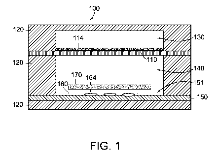
Figure 1 schematically depicts a fluidic device 100 in accordance with some
embodiments. The fluidic device 100 includes a porous membrane 110, a solid
support
structure 120, a base 150, and a flexible substrate 160. The solid support
structure 120
includes a first chamber 130 and a second chamber 140 separated from the first
chamber by
the porous membrane 100. The second chamber 140 is in fluid communication with
the first
chamber 130 via the porous membrane 110. The base 150 is disposed at least
partially in the
second chamber 140 opposite the porous membrane 110. The base 150 includes a
cyclic
olefin copolymer (COC).
The flexible substrate 160 includes a polymer layer and/or a hydrogel layer
disposed
on a surface 151 of the base 150. In some embodiments, the flexible substrate
160 comprises
a gelatin layer. Additional and alternative polymers and hydrogels that may be
included in
the flexible substrate are described below.
Hydrogels that can be included in the flexible substrate include, for example,
polyacrylamide gels, poly(N-isopropylacrylamide), pHEMA, collagen, fibrin,
gelatin,
alginate, and dextran. In one embodiment the hydrogel is alginate. In another
embodiment,
the hydrogel is gelatin. In one embodiment, the stiffness of the hydrogel is
tuned to mimic
the mechanical properties of healthy muscle tissue, e.g., cardiac tissue in
vivo, e.g., to have a
Young's modulus of about 10, 11, 12, 13, 14, 15, 16, 17, 18, 19, or about 20
kPa. In another
embodiment, the stiffness of the hydrogel is tuned to mimic the mechanical
properties of
diseased muscle tissue, e.g., cardiac tissue in vivo, e.g., to have a Young's
modulus of greater
than about 45, 46, 47, 48, 49, 50, 51, 52, 53, 54, or about 55 kPa.
Examples of the elastomers that can be used to form a polymer layer of the
flexible
substrate include polydimethylsiloxane (PDMS) and polyurethane. In one
embodiment, the
PDMS, once cured is opaque (e.g., light-absorbing). In other embodiments,
thermoplastic or
thermosetting polymers are used to form the flexible polymer layer.
Alternative non-
degradable polymers include polyurethanes, silicone-urethane copolymers,
carbonate-
urethane copolymers, polyisoprene, polybutadiene, copolymer of polystyrene and
polybutadiene, chloroprene rubber, Polyacrylic rubber (ACM, AB R), Fluoro
silicone Rubber
(FVMQ), Fluoroelastomers, Perfluoroelastomers, Tetrafluoro ethylene/propylene
rubbers
(FEPM) and Ethylene vinyl acetate (EVA).
In still other embodiments, biopolymers, such as collagens, elastins,
polysaccharides,
17
CA 02995088 2018-02-07
WO 2017/027390
PCT/US2016/045813
and other extracellular matrix proteins, are included in the flexible
substrate. Suitable
biodegradable elastomers include hydrogels, e.g., alginate and gelatin,
elastin-like peptides,
polyhydroxyalkanoates and poly(glycerol-sebecate). Suitable non-elastomer,
biodegrable
polymers include polylactic acid, polyglycolic acid, poly lactic glycolic acid
copolymers.
In one embodiment, a polymer layer included in the flexible substrate
comprises
polydimethylsiloxane (PDMS). Thickness of the PDMS layer can be controlled by
the
viscosity of the prepolymer and by the spin-coating speed (if spin coated),
ranging from 14 to
60 [tm thick after cure. The viscosity of the prepolymer increases as the
cross-link density
increases. This change in viscosity between mixing and gelation can be
utilized to spin-coat
different thicknesses of polymer layers. Alternatively the spin-coating speed
can be
increased to create thinner polymer layers. After spin-coating, the resulting
polymer
scaffolds are either fully cured at room temperature (generally, about 22 C)
or at 65 C. In
some embodiments, the polymer or hydrogel is deposited and molded, but not
spin coated.
In one embodiment, polymeric fibers prepared as described in U.S. Patent
Publication
No. 2012/0135448, (the entire contents of which are incorporated herein by
reference) may
be used in the polymer layer for the flexible substrate.
In one embodiment, e.g., nanoparticles and/or fluorescent beads, e.g.,
fluorospheres,
are mixed with the hydrogel prior to cross-linking and/or the flexible polymer
layer prior to
depositing (e.g., spin coating) the polymer layer onto the base.
The flexible substrate 160 is configured to support growth of a functional
muscle
tissue 170 disposed on the flexible substrate 160.
In some embodiments, a surface of the flexible substrate 160 facing away from
the
base 150 includes micro-scale topological features to promote growth of the
functional
muscle tissue 170. In some embodiments, the micro-scale topological features
on the surface
of the flexible substrate 160 are micromolded features. In other embodiments,
the micro-
scale topological features may be optically patterned into the hydrogel, e.g.,
gelatin, as
described in U.S. Provisional Application No. 62/371,385, filed on even date
herewith
(Attorney Docket No.: 117823-14001), the entire contents of which are
incorporated herein
by reference). The micro-scale topological features enable long-term culture
of aligned cells
on the flexible substrate 160.
In some embodiments, the functional muscle tissue comprises cells including
cardiac
muscle cells, ventricular cardiac muscle cells, atrial cardiac muscle cells,
striated muscle
18
CA 02995088 2018-02-07
WO 2017/027390
PCT/US2016/045813
cells, smooth muscle cells, vascular smooth muscle cells and combinations
thereof.
As used herein, a "functional muscle tissue" refers to a muscle tissue
prepared in vitro
which displays at least one physical characteristic typical of the muscle
tissue in vivo; and/or
at least one functional characteristic typical of the muscle tissue in vivo,
i.e., is functionally
active.
For example, a physical characteristic of a functional muscle tissue may
include the
presence of parallel (to the long axis of the cells) myofibrils with or
without sarcomeres
aligned in z-lines, and/or that the myofibrils cross cell-to-cell junctions,
and/or that the cells
maintain a registered array or sarcomeres, and/or that the cells form cell-to-
cell gap junctions
and/or cell-to-cell adherens junctions. Methods to determine such physical
characteristics
include, for example, microscopic analyses, such as, fluorescent microscopy,
confocal
microscopy, two-photon microscopy, and the like, immunohistochemical analyses,
e.g.,
staining for connexin 43 to determine if the cells have formed electrically-
competent
junctions, staining for 13-catenin to determine if the cells have formed
mechanically-
competent junctions, staining for 13-actin and determining, e.g., the
orientational order
parameter (00P) of the networks to determine if the cells have formed
registered myofibrils.
A functional characteristic of a functional muscle tissue may include an
electrophysiological activity, such as an action potential, or biomechanical
activity, such as
contraction. For example, the cells of a functional muscle tissue may be
mechanically and
electrically integrated, e.g., the cells synchronously contract, and/or the
cells generate a
contractile force, and/or the contractions of the cells are in phase, and/or
the contractile force
at the medial cell-to-cell junctions of the cells are about the same, and/or
the cells exhibit
synchronous Ca+2 transients, and/or the cells exhibit substantially the same
Ca+2 levels,
and/or the cells exhibit peak systolic and/or diastolic forces that are about
the same.
Methods to determine such functional characteristics include, for example,
microscopic analyses, such as fluorescent microscopy, confocal microscopy, two-
photon
microscopy, optical detection of deflection of the underlying flexible
substrate due to
contraction of the tissue and the like, immunohistochemical analyses, e.g.,
vinculin staining,
traction force microscopy, ratiometric Ca+2 imaging, optical mapping of the
action
potentials.
In some embodiments, most or all of the flexible substrate 160 adheres to or
is bonded
19
CA 02995088 2018-02-07
WO 2017/027390
PCT/US2016/045813
to the surface 151 of the base 150, as shown in Figure 1. In some embodiments,
at least a
portion of the surface 151 of the base is modified to promote adhesion or
bonding between
the flexible substrate 160 and the base 150 as described in more detail below
with respect to
Figures 3 and 4.
In some embodiments the fluidic device 100 includes an electrode array (e.g.,
a
microelectrode array (MEA), a flexible MEA) to measure electrical properties
of the
functional muscle tissue 170 on the flexible substrate 160. In some
embodiments, the fluidic
device 100 also includes a flexible electrode array 164 disposed between the
flexible
substrate 160 and the surface 151 of the base 150. In some embodiments, the
flexible
electrode array 164 is bonded to the surface 151 of the base 150. In some
embodiments, the
flexible substrate 160 adheres to the flexible electrode array 164 and to the
surface 151 of the
base 150. In some embodiments, a surface energy of the surface 151 of the base
150 in
selected area may be modified to promote bonding between the flexible
electrode layer 164
and the base 150.
A thickness or height of the flexible substrate 160 may be selected such that
it
provides sufficient height to support the desired micro-scale topological
features while
remaining sufficiently short/thin to obtain reliable electrical measurements
of the functional
muscle tissue 170 through the flexible substrate 160 using the flexible
electrode array 164.
In some embodiments, the flexible substrate 160 includes a gelatin layer
having a thickness
in a range of about 55 [tm to about 115 [tm or a range of about 75 [tm to
about 95 [tm.
In some embodiments, the porous membrane 110 is composed of a polycarbonate
material. In some embodiments, the porosity of the porous membrane 110 is
between 5%
and 11%. In further embodiments, the porosity of the porous membrane 110 is
between 6%
and 9%.
In some embodiments, the fluidic device 100 also includes a growth promoting
layer
114 to promote adhesion and growth of cells on the porous membrane 110. The
growth
promoting layer 114 is disposed at least partially on the porous membrane 110
in the first
chamber 130 In some embodiments, the cells grown on the porous membrane
include, but
are not limited to, epithelial cells, endothelial cells, sensory transducer
cells, neuronal cells,
hormone-secreting/endocrine cells, glial cells and/or adipocytes. In some
embodiments, the
cells grown on the porous membrane and the porous membrane 110 act as a
vascular-like
barrier between chambers of the fluidic device 100, e.g., exposing the muscle
tissue in the
CA 02995088 2018-02-07
WO 2017/027390
PCT/US2016/045813
second chamber to, e.g., 02, CO2, small molecules that can diffuse through the
porous
membrane and cells thereon..
In some embodiments, the growth promoting layer is a coating on the porous
membrane. In some embodiments, the growth promoting layer includes
extracellular matrix
molecules (ECM), or other proteins such as growth factors or ligands. In some
embodiments,
the surface of the porous membrane can be activated with any art-recognized
reactions, such
that ECM molecules, proteins such as growth factors or ligands, can be
attached to it.
In some embodiments, the porous membrane is not seeded with cells. In other
embodiments, the porous membrane is seeded with cells. In some embodiments
where cells
are seeded on the porous membrane, cells can be seeded on one side or both
sides of the
porous membrane. In some embodiments, both sides of the porous membrane can be
seeded
with the same cell types. In other embodiments, both sides of the porous
membrane can be
seeded with different cell types.
In some embodiments, the porous membrane can be seeded with at least one layer
of
cells, including, at least 2 layers of cells or more. Each layer of cells can
be the same or
different.
Figure 2 schematically depicts a fluidic device 102 in accordance with some
embodiments. Similar to fluidic device 100 described above, the fluidic device
102 includes
a porous membrane 110, a solid support structure 120 having a first chamber
130 and a
second chamber 140, a base 150, and a flexible substrate 160. However, as
depicted in
Figure 2, in fluidic device 102, one or more portions 162a, 162b of the
flexible substrate 160
are not adhered or attached to the surface 151 of the base. Modification of
the surface energy
of a portion of the surface 161 of the base for attachment of only a portion
of the flexible
substrate 160 to the underlying surface 151 of the base is described in more
detail below with
respect to Figure 4.
A portion or portions 162a, 162b of the flexible substrate 160 that are not
attached to
the surface 151 of the base 150 are configured to deflect away from the
surface of the base
150 in response to forces exerted by a functional muscle tissue 170 on the
flexible substrate
160. The deflection of portions of the flexible substrate 160 can be detected
or measured
(e.g., optically) to obtain measurements of contractile forces exerted on the
flexible substrate
by the functional muscle tissue. As used herein, a functional muscle tissue on
a flexible
substrate in which one or more portions of the flexible substrate are not
attached to the
21
CA 02995088 2018-02-07
WO 2017/027390
PCT/US2016/045813
underlying base and are free to deflect away from a surface of the base in
response to
contraction of the functional muscle tissue is referred to herein as a muscle
tissue strip. In
some embodiments, the functional muscle tissue 170 is disposed on the flexible
substrate 160
to form a functional muscle tissue strip having one or two cantilevered
portions 162a, 162b.
In some embodiments, the flexible substrate 160 has an elongate shape and a
first end 162a of
the flexible substrate and a second end 162b of the flexible substrate
opposite the first end are
not attached to the surface 151 of the base and a middle portion 162c of the
flexible substrate
is attached to the surface of the base.
Similar to flexible substrate 160 of fluidic device 100, a surface of the
flexible
substrate 160 facing away from the 151 of the base includes micro-scale
topological features
to promote growth of the functional muscle tissue 170. In some embodiments,
the flexible
substrate 160 comprises gelatin and has has an average height in a range of
about 165 [tm to
about 225 [tm. The average height or thickness of the flexible substrate 160
may be selected
to obtain a desired range of deflections in the one or more cantilevered
portions in response to
contractile forces exerted by the functional muscle tissue 170 on the flexible
substrate 160.
In some embodiments, the fluidic device 100, 102 may include a second flexible
substrate (not shown) comprising a polymer layer and/or a hydrogel layer
disposed on the
surface 151 of the base 150. The second flexible substrate may be configured
to support
growth of a second functional muscle tissue (not shown). In some embodiments,
the second
flexible substrate may be spaced from the first flexible substrate by at least
about 1.5 mm to
prevent cells growing on one flexible substrate from "bridging" the gap with
cells growing on
the second flexible substrate.
Figures 3 and 4 each show a top view of the base 150 having areas of modified
surface energy in accordance with some embodiments. Dotted line 161 indicates
the area of
the surface 151 of the base that would be covered by the flexible substrate
160. In some
embodiments, a first portion 152 of the surface 151 of the base adjacent to
the flexible
substrate 161 has a modified surface energy relative to a surface energy of
the rest of the
surface 151 of the base 150 material to inhibit cell adhesion to the surface
of the base 150
(see dotted area 152 identifying the first portion of the surface of the
base). For example, in
some embodiments, the surface energy of the first portion 152 of the surface
of the base 150
adjacent to the flexible substrate 160 is modified by laser etching. Laser
etching changes the
surface chemistry of the portion of the base 150 surrounding the flexible
substrate 160 to
inhibit cell adhesion. In some embodiments, the laser etching is carbon
dioxide laser-etching.
22
CA 02995088 2018-02-07
WO 2017/027390
PCT/US2016/045813
In some embodiments, a surface energy of a second portion 154, 154' of the
surface
of the base 150 underlying the flexible substrate 160 is modified relative to
a surface energy
of the rest of the surface of the base 150 material to promote adhesion with
or bonding to the
flexible substrate 160 (see striped areas 154, 154' identifying the second
portion of the
surface of the base). For example, the surface energy of the second portion
154, 154' of the
surface of the base 150 may be modified by oxygen plasma treatment. In some
embodiments, the second portion 154 of the surface of the base includes most
or all of the
area under the flexible substrate (i.e., most or all of the area within dotted
line 161) as shown
in Figure 3. Modifying the surface energy of most or all of the area of the
base that will be
covered by the flexible substrate 160 is particularly useful in embodiments
such as that
schematically depicted in Figure 1 in which a flexible electrode array is
employed to measure
electrical changes in functional muscle tissue disposed on the flexible
substrate.
In some embodiments, only some of the area of the surface of the base 151 that
will
be covered by the flexible substrate is modified to promote adhesion between
the base and
the flexible substrate. In some embodiments, the area of the base surface 151
covered by the
flexible substrate 161 includes the second portion 154' that has a modified
surface energy to
promote adhesion and a third portion 156 of the surface of the base that does
not have a
modified surface energy to promote adhesion with the flexible substrate as
shown in Figure 4.
In such an embodiment, the flexible substrate 160 attaches to the second
portion 154 of the
surface of the base 150 material and but does not attach to the third portion
156 of the surface
of the base 150. Using a base having areas of modified surface energy as shown
in Figure 4
for the fluidic device would result in the portions of the flexible substrate
that overlay the
third portion 156 of the area of the base surface being unattached to the
underlying substrate
and free to deflect away from the underlying substrate in response to forces
exerted by
functional muscle tissue as shown in the device of Figure 2 (see portions
162a, and 162b of
the flexible substrate).
Some techniques for forming functional muscle tissue strips require the manual
peeling or manual separation of a cantilevered portion of the muscle tissue
strip from the
underlying layer (e.g., the base) and from cells in the functional muscle
tissue that also adhere
to the underlying layer (e.g., the base). In embodiments that rely on
deflection of portions of
the flexible substrate away from the surface of the base, modification of the
surface energy of
portions of the base to resist cell adhesion (e.g., in first portion 152) and
modification of the
surface energy of the a portion of the base to promote adhesion over only a
portion of the area
23
CA 02995088 2018-02-07
WO 2017/027390
PCT/US2016/045813
that will be covered by the flexible substrate (e.g., in second portion 154',
but not third
portion 156) both limits cell adhesion and enables free motion of the
cantilever portion or
portions (162a, 162b) of the flexible substrate without the use of manual
peeling. Avoiding
manual peeling during manufacture of fluidic devices with flexible substrates
having one or
more cantilevered portions simplifies the manufacturing process and can reduce
errors and
potential damage to tissue and/or devices in manufacturing.
Figure 5 is an exploded perspective view of elements of a fluidic device 104
having a
modular structure according to an embodiment. The fluidic device 104 includes
a porous
membrane 110 and a first channel defining member 180. When the fluidic device
104 is
assembled with the first channel defining member 180 disposed on the porous
membrane
110, the porous membrane 110 and the first channel defining member 180 define
a first
fluidic channel 181. The fluidic device 104 also includes a support member 182
that provides
mechanical support for the fluidic device 104 and a base 150 disposed on the
support member
182 when the fluidic device 104 is assembled. The fluidic device 104 also
includes a second
channel defining member 184. In some embodiments, the fluidic device 104
further includes
a gasket 186. When the fluidic device 104 is assembled, the second channel
defining
member 184 is disposed on the base 150, and the porous membrane 110 is
disposed on the
second channel defining member 184. In embodiments that include a gasket 186,
the gasket
186 is disposed between the second channel defining member 184 and the base
150. When
the fluidic device 104 is assembled, the base 150, the second channel defining
member 184,
the gasket 186 (if used), and the porous membrane 110 define a second fluidic
channel 182.
The fluidic device 104 also include a flexible substrate 160 that is disposed
on the base 150.
The fluidic device 104 includes one or more securing elements 188 that
releasably secure the
first channel defining member 180, the porous membrane 110, the second channel
defining
member 184 and the base 150 to the support member 182. In some embodiments,
the fluidic
device 104 includes a growth promoting layer 114 disposed on the porous
membrane within
the first fluidic channel 181.
The flexible substrate 160 includes a polymer layer and/or a hydrogel layer
disposed
on the surface of the base as described above with respect to Figures 1 and 2.
The flexible
substrate is configured to support growth of a functional muscle tissue as
described above.
The function of the gasket 180 is discussed in further detail below with
respect to
Figure 9. In some embodiments, the gasket 180 is composed of
polydimethylsiloxilane
(PDMS). The gasket 180 may have various different shapes and is not limited by
its depiction
24
CA 02995088 2018-02-07
WO 2017/027390
PCT/US2016/045813
in the figures.
The elements of the fluidic device 100 are secured using one or more securing
elements 188a, 188b. The securing elements 188a, 188b can be screws, nuts and
bolts, snaps,
straps, clips, bands, or any other suitable elements for releasably securing
the components of
the fluidic device 100. In an embodiment, the securing elements are screws
188a and
threaded inserts 188b embedded in support member 182.
In the fluidic device 104, the first channel defining member 180, the second
channel
defining member 184, and the support member 182 are each part of the solid
support
structure of the fluidic device. In some embodiments, at least portions of the
solid support
structure 120 are made of polycarbonate material or an acrylic material. When
assembled,
the first channel defining member 180 and the porous membrane 110 define the
first chamber
of the fluidic device 104. When assembled, the second channel defining member
184, the
porous membrane 110 and the support member 182 define the second chamber of
the fluidic
device 104. In some embodiments, the base 150 comprises a COC.
Figure 6 is top view of the assembled fluid device 104 showing the first
channel 181
partially overlaying the second channel 182. In some embodiments, the fluidic
device may
include a flexible substrate 160 with multiple cantilever portions 163 that
are not attached to
the underlying base as depicted in Figure 6.
The fluidic device 104 is configured to be easily disassembled into a first
portion,
which includes the first channel defining member 180 and the porous membrane
110, and a
second portion, which includes the base 150 and the support member 120, and
then easily
reassembled. In some embodiments, the first portion also includes the second
channel
defining member 184. The securing elements 188a, 188b can be used to secure
the first
portion to the second portion. In some embodiments, the fluidic device 110 may
be provided
in a disassembled state with the first portion separate from the second
portion. Separating the
fluidic device 104 into a first portion and a second portion can facilitate
seeding and growth
of cells on the flexible substrate 160 and on the growth promoting layer 114
on the porous
membrane 110. For example, with the fluidic device separated into two or more
portions, the
cells on the flexible substrate 160 can be seeded and cultured separately from
the cells on the
porous membrane 110 and different culturing conditions can be used for each.
Additional
description of cell seeding and culturing is provided below with respect to
Figures 15 through
18E and in the methods section.
CA 02995088 2018-02-07
WO 2017/027390
PCT/US2016/045813
In some embodiments, the first channel defining member 180 and the porous
membrane 110 are bonded to each other (e.g., using an adhesive or another type
of permanent
or semi-permanent bond). In some embodiments, the porous membrane 110 and the
second
channel defining member 184 are bonded to each other (e.g., using an adhesive
or another
type of permanent or semi-permanent bond). In some embodiments, porous
membrane 110 is
bonded to both the first channel defining member 180 and to the second channel
defining
member 184 (e.g., via adhesive-free bonding, using an adhesive or another type
of
permanent or semi-permanent bond). For example, for a porous membrane of
polycarbonate
and first and second channel defining members of polycarbonate, adhesive-free
bonding may
be achieved by vaporizing a polycarbonate solvent (e.g., Dichloromethane
(DCM)) onto
relevant surfaces of the channel defining members, followed by aligning the
channel defining
members and the porous membranes and bringing them into contact with each
other, heating
all three to near the glass transition temperature of polycarbonate (Tg-150
C), and applying a
pressure about 135 lbslin2 (93 lkPa) for 1 hour. A similar procedure may be
employed for
adhesive-free bonding of a porous membrane and first and second channel
members made
from a polymer other than polycarbonate with the solvent, heating temperature
and pressure
applied adjusted accordingly. For a porous membrane made from a different
polymer than
that of the first or second channel defining member, an adhesive may be
employed.
In some embodiments, the fluidic device 104 includes a flexible electrode
array
between the flexible substrate 160 and the base 150. Figure 7 is an exploded
view of the
elements of the fluidic device 104 including a flexible electrode array 164
according to one
embodiment. Figure 8 is an image of the fluidic device 104 in Figure 7 fully
assembled. In
Figure 7, the first channel defining member 180, the porous membrane 110 and
the second
channel defining member 184 are bonded together to form a first portion 190 of
the fluidic
device 104. The first portion 190 of the fluidic device is separate from the
base 150 and
support member 182 of the second portion 192 of the fluidic device. In some
embodiments,
the flexible electrode array 164 is at least partially disposed between the
flexible substrate
160 and the base 150. The flexible substrate is not shown in Figure 7, however
a location for
placement of the flexible substrate is shown with dotted line 161. The
flexible electrode
array 164 may extend from the fluidic device 104 a sufficient distance to
contact
measurement devices external to the fluidic device 104.
In some embodiments, the flexible electrode array 164 is bonded to the surface
of the
base 150. In some embodiments, the flexible substrate 161 adheres to the
flexible electrode
26
CA 02995088 2018-02-07
WO 2017/027390
PCT/US2016/045813
array 164 and the base 150. In these embodiments, the gasket 180 may be
employed to aid in
hold the flexible electrode array 164 in place. In some embodiments, the
flexible electrode
array 164 is secured to the base 150 by pressure from the gasket 180. For
example, Figure 9
depicts the flexible electrode array 164 disposed on the base 160 of a fluidic
device 104 with
the gasket 186 over a portion of the flexible electrode array 164. For ease in
visualization,
the first portion 190 of the fluidic device is not shown. The first portion of
the fluidic device
exerts pressure on the gasket 186, which, exerts pressure on the flexible
electrode array 164
that aids in securing the flexible electrode array to the base 160. The
flexible substrate is not
shown in Figure 9, however a location for placement of the flexible substrate
is shown with
dotted line 161.
In some embodiments, a fluidic device has a configuration that facilitates
achieving a
specified level of uniformity of concentration of a drug across a functional
muscle tissue. For
example, Figure 10 schematically depicts a cross-sectional view of a fluidic
device 108
including a porous membrane 110, a solid support structure 120, a base 150,
and a flexible
substrate 160. In some embodiments, a functional muscle tissue 170 is disposed
on the
flexible substrate 160. The solid support structure 120 includes a first
chamber 130 and a
second chamber 140 separated from the first chamber by the porous membrane
100. The
second chamber 140 is in fluid communication with the first chamber 130 via
the porous
membrane 110. The porous membrane 110 and at least a portion of the first
chamber 130
define a first fluid channel 196 as shown. The porous membrane 110 and at
least a portion of
the second chamber 140 define a second fluid channel 198 as shown with the
proximal end
110p of the porous membrane being on the upstream end of the second fluid
channel. The
surface 151 of the base 150 has a leading portion 200 corresponding the
distance along
second fluid channel between the proximal end 110p of the porous membrane and
the portion
of the surface of the base 150 covered by the flexible substrate 160. A length
of the leading
portion 200 is selected to achieve sufficient uniformity in a drug
concentration profile across
the flexible substrate 160 for a drug flowing through the first fluid channel
196 at a first rate
and diffusing through the porous membrane 150 into a liquid flowing through
the second
fluid channel 198 at a second rate. In some embodiments, a sufficient
uniformity is a
difference in a drug concentration of less than 50% between an upstream end
and a
downstream end of the flexible substrate 160. In an embodiment, the leading
portion 192 is at
least about 4 mm long and the portion of the base 150 covered by the flexible
substrate 160 is
at least about 8 mm long. In some embodiment, a length of a leading portion is
in a range of
27
CA 02995088 2018-02-07
WO 2017/027390
PCT/US2016/045813
about 2 mm to about 6 mm. In some embodiments, a length of a leading portion
is in a range
of about 3 mm to about 5 mm. In some embodiments, the porosity of the porous
membrane
110 is between about 5% and about 11%. In some embodiments, the porosity of
the porous
membrane 110 is between about 6% and about 9%. Computer simulation of a drug
concentration at the flexible substrate for different configuration of a fluid
device are
described below in the examples with respect to Figure 31A through 32B.
In some embodiments, a fluidic device also includes electrodes that measure
electrical
properties of cells disposed on the porous membrane (e.g., an impedance of a
cell layer on the
porous membrane). For example, in some embodiments, a fluidic device also
includes a first
electrode disposed in the first fluid channel at least partially overlying the
porous membrane,
a second electrode disposed on a surface of the fluid channel opposite the
first electrode, and
a growth promoting layer disposed in the first fluid channel overlying at
least a portion of the
first electrode. Further description of embodiments including electrodes that
measure
electrical properties of cells disposed on the porous membrane are provided
below with
respect to Figures 11 through 14. Aspects of embodiments depicted in Figures
11-14 may be
combined with or incorporated into any other fluidic devices disclosed herein.
Figure 11 is a schematic cross-sectional view of a fluidic device 300 taken
across a
direction of flow and Figure 12 is a schematic cross-sectional view of the
fluidic device 330
taken along a direction of flow. Figures 13 and 14 are images of the fluidic
device 300.
Turning again to Figures 11 and 12, the fluidic device 300 includes a solid
support structure
320 having a first chamber 330 separated from a second chamber 340 by a porous
membrane
310. At least a portion of the first chamber 330 and the porous membrane 310
define a fluid
channel 332 having a surface 333 opposite the porous membrane 310. The fluidic
device 300
also includes a first electrode 342 disposed in the fluid channel 332 at least
partially
overlying the porous membrane 310 and a second electrode 344 disposed on the
surface 333
of the fluid channel opposite the first electrode 342. The fluidic device 300
also includes a
growth promoting layer 314 disposed in the fluid channel 332 and overlying at
least a portion
of the first electrode 342 and overlying at least a portion of the porous
membrane 310. The
growth promoting layer 314 is configured to promote adhesion and growth of
epithelial cells,
endothelial cells, sensory transducer cells, neuronal cells, hormone-
secreting/endocrine cells,
glial cells and/or adipocytes. In some embodiments, the fluidic device 310
also includes the
cells 316 cultured on the growth promoting layer 314.
28
CA 02995088 2018-02-07
WO 2017/027390
PCT/US2016/045813
In some embodiments, the fluidic device 300 also includes a third electrode
346
disposed in the first fluid channel 332 at least partially overlying the
porous membrane 310
and a fourth electrode 348 disposed on the surface 333 of the first fluid
channel opposite the
third electrode 336 (see Figure 12). The electrodes are configured for
measurement of
electrical properties such as impedance across the first fluid channel 332 or
impedance of the
cells 316 attached to the porous membrane 310. In some embodiments the second
chamber
includes muscle cells, such as a functional muscle tissue 370. In some
embodiments, the
second chamber includes a flexible substrate 360 that supports the functional
muscle tissue
370. In some embodiments, the functional muscle tissue 370 and flexible
substrate 360 are in
the form of a muscle tissue strip having a cantilever portion that is free to
deflect in response
to contractive force exerted by the functional muscle tissue 370. In some
embodiments, the
second chamber 340 includes a plurality of flexible substrates 360 each
supporting a
functional muscle tissue 370 as shown in Figure 12.
In some embodiments, the first fluid channel 332 has a proximal end 334a
defined
near an inflow portion of the first fluid channel and a distal end 334b
defined near an outflow
portion of the first fluid channel, and wherein the first electrode 342 and
the second electrode
344 are disposed at the proximal end 334a or at the distal end 334b of the
first fluid channel
332. In some embodiments, the fluidic device 300 has an upstream end and a
downstream
end and the first electrode 342 and the second electrode 344 are disposed
upstream or
downstream of the flexible substrate 360. In some embodiments, the third
electrode 346 and
the fourth electrode 348 are also disposed upstream or downstream of the
flexible substrate
360.
In some embodiments, a thickness of electrodes that at least partially
underlie the
growth promoting layer (e.g., the first electrode 342 and the third electrode
346) is selected to
achieve desired electrical properties without interfering with growth of the
cells over the
electrode. In some embodiments, the first electrode 342 and the third
electrode 346 (if
included) each have a thickness in a range of about 20 nm to about 400 nm. In
some
embodiments, the first electrode 342 and the third electrode 346 (if included)
each have a
thickness in a range of about 20 nm to about 200 nm. In some embodiments, the
first
electrode 156 and the second electrode 224 include gold.
In some embodiments, the first electrode 342 and the second electrode 344
include an
adhesion layer and an overlying layer. In some embodiments the adhesion layer
includes
titanium. In some embodiments the overlying layer includes gold. In some
embodiments,
29
CA 02995088 2018-02-07
WO 2017/027390
PCT/US2016/045813
the adhesion layer has a thickness in a range of about 3 nm to about 10 nm.
In some embodiments, the porous membrane 310 and the cells cultured on growth
promoting layer 314 act as a vascular-like barrier between channels of the
fluidic device 300.
In some embodiments, the porous membrane 310 is composed of or includes a
polycarbonate
material.
A description of how to obtain impedance measurements using fluidic device 300
is
provided below in the examples section below with respect to Figures 32 and
33.
Some embodiments include a kit including a fluidic device as described herein
(e.g.,
fluidic device 104, fluidic device 102) and a cell seeding well. Figure 15 is
a perspective
view of a cell seeding well 400 according to an embodiment. The cell seeding
well 400
includes a well body 410 having a first surface 412 and a second surface bad
414, the well
body 410 defining an aperture 416 extending from the first surface 412 to the
second surface
414. The shape of the aperture 416 at the second surface corresponds to a
shape of the
flexible substrate of a fluidic device. The cell seeding well 400 is
configured to achieve high
densities of cell seeding. For example, in some embodiments, a shape of the
aperture 416
tapers from a first cross-sectional area at the first surface 412 to a smaller
second cross-
sectional area at the second surface 414. This tapering of the aperture 416
produces a funnel
effect that can facilitate a high density of cell seeding. The shape of the
cell seeding well 410
distributes cells evenly at the bottom of the well 410. The small area of the
aperture 416 at
the second surface 414 at the bottom of the well enables a high seeding
density using a
relatively small number of cells. For example, in one embodiment, the well 410
is capable of
seeding ten thousand human cardiomyocytes per fluidic device for an aperture
416 having the
dimensions of about 5 mm x about 2.5 mm at the second surface 414.
In some embodiments, the seeding well comprises polytetrafluoroethylene (PTFE)
(e.g., TEFLON from Chemours Co.), which is non-cytotoxic, autoclavable and
easily
handled. In some embodiments, the seeding well is machined out of a piece of
PTFE.
In some embodiments, the cell seeding well 410 is configured to be attached to
a
second portion 192 of a fluidic device 104 including a base 150, a flexible
substrate 160 and a
support member 182 when the fluidic device is in a partially disassembled
state. For
example, cell seeding well 400 has holes 420 through which securing elements
(e.g., securing
elements 188a) can extend. In some embodiments, a gasket is employed between
the seeding
well 400 and the base 150. Figure 16 includes an image of a batch molded set
of such
CA 02995088 2018-02-07
WO 2017/027390
PCT/US2016/045813
gaskets in accordance with an embodiment. In some embodiments, the gasket
comprises
polydimethylsiloxane (PDMS) (e.g., silicone). Figures 17A an 17B are images of
a cell
seeding well 410 mounted on the second portion 192 of the fluidic device,
which includes a
flexible electrode array 164 between the flexible substrate and the base 150,
being used for
cell seeding. In Figure 17B, dotted line 424 indicates the position of the
gasket.
Figures 18A through 18E depict another embodiment of a cell seeding system
that
includes a seeding well 402 (e.g., an acrylic seeding well) defining an
aperture 418
corresponding to an area of a flexible substrate, a ring 426 to hold media,
and a gasket 428
that may comprise PDMS. Figure 18C is an image of the ring 426 affixed to the
seeding well
402 using epoxy. Figure 18D is an image of the gasket 428 (e.g., a PDMS
gasket), which is
configured to be disposed under the seeding well 402. Figure 18E is an image
of the cell
seeding well system positioned on the second portion of a fluidic device 164
with the gasket
428 between the second portion of the fluidic device 164 and the seeding well
402.
Methods of Use of the Devices of the Invention
The devices of the present invention are useful for, among other things,
measuring
muscle cell activities and functions, investigating muscle developmental
biology and disease
pathology, drug delivery, as well as in drug discovery and toxicity testing.
To prepare a functional muscle tissue, a flexible substrate comprising a
polymer
and/or hydrogel layer disposed on the surface of the base of the device is
placed in culture
with a myocyte suspension, the cells are allowed to settle and adhere to the
substrate. In the
case of an adhesive surface treatment, cells bind to the flexible substrate in
a manner dictated
by the micro-scale topological features on a surface of the flexible substrate
facing away from
the base and the cells respond to the features in terms of maturation, growth
and function.
The cells on the substrates may be cultured, e.g., in an incubator, under
physiologic
conditions (e.g., at 37 C) until the cells form a two-dimensional (2D) tissue
(i.e., a layer of
cells that is less than about 200 microns thick, or, in particular
embodiments, less than about
100 microns thick, less than about 50 microns thick, or even just a monolayer
of cells), the
anisotropy or isotropy of which is determined by the micro-scale topological
features. In one
embodiment, the micro-scale topological features are isotropic. In another
embodiment, the
micro-scale topological features are anisotropic.
Any appropriate cell culture method may be used to establish the tissue on the
flexible
substrate. The seeding density of the cells will vary depending on the cell
size and cell type,
31
CA 02995088 2018-02-07
WO 2017/027390
PCT/US2016/045813
but can easily be determined by methods known in the art. In some embodiments,
the
myocytes are cultured in the presence of, e.g., a fluorophore, nanoparticles
and/or fluorescent
beads, e.g., fluoro spheres. In one embodiment, a fluorophore, a nanoparticle
and/or a
fluorescent bead, e.g., a fluoro sphere, is mixed with the gelatin prior to
cross-linking and/or
the flexible substrate.
The myocytes may be normal myocytes, abnormal myocytes (e.g., those derived
from
a diseased tissue, or those that are physically or genetically altered to
achieve an abnormal or
pathological phenotype or function), normal or diseased myocytes derived from
embryonic
stem cells or induced pluripotent stem cells, or myocytes comprising a genetic
construct,
such as an expression vector expressing an optogenetic gene, e.g., a light
sensitive ion
channel (e.g., channelrhodopsin (ChR2), C1V1, Chrimson, Chronos, SSFO, ArchT,
ChETA,
NpHR, SwiChR, iC1C2, or the like). Myocytes can be cultured in vitro, derived
from a
natural source, genetically engineered, or produced by any other means. Any
natural source
of myocytes may be used, including from neonates, e.g., mouse and rat
neonates.
Suitable myocytes for the preparation of a functional muscle tissue include,
cardiomyocytes, such as ventricular or atrial cardiac cells vascular smooth
muscle cells,
airway smooth muscle cells, and striated muscle cells, such as skeletal muscle
cells, and
combinations thereof.
The term "stem cell" as used herein, refers to an undifferentiated cell which
is capable of
proliferation and giving rise to more progenitor cells having the ability to
generate a large
number of mother cells that can in turn give rise to differentiated, or
differentiable daughter
cells. The daughter cells themselves can be induced to proliferate and produce
progeny that
subsequently differentiate into one or more mature cell types, while also
retaining one or
more cells with parental developmental potential. The term "stem cell" refers
to a subset of
progenitors that have the capacity or potential, under particular
circumstances, to differentiate
to a more specialized or differentiated phenotype, and which retains the
capacity, under
certain circumstances, to proliferate without substantially differentiating.
In one
embodiment, the term stem cell refers generally to a naturally occurring
mother cell whose
descendants (progeny) specialize, often in different directions, by
differentiation, e.g., by
acquiring completely individual characters, as occurs in progressive
diversification of
embryonic cells and tissues. Cellular differentiation is a complex process
typically occurring
through many cell divisions. A differentiated cell may derive from a
multipotent cell which
itself is derived from a multipotent cell, and so on. While each of these
multipotent cells may
32
CA 02995088 2018-02-07
WO 2017/027390
PCT/US2016/045813
be considered stem cells, the range of cell types each can give rise to may
vary considerably.
Some differentiated cells also have the capacity to give rise to cells of
greater developmental
potential. Such capacity may be natural or may be induced artificially upon
treatment with
various factors. In many biological instances, stem cells are also
"multipotent" because they
can produce progeny of more than one distinct cell type, but this is not
required for "stem-
ness." Self-renewal is the other classical part of the stem cell definition.
In theory, self-
renewal can occur by either of two major mechanisms. Stem cells may divide
asymmetrically, with one daughter retaining the stem state and the other
daughter expressing
some distinct other specific function and phenotype. Alternatively, some of
the stem cells in a
population can divide symmetrically into two stems, thus maintaining some stem
cells in the
population as a whole, while other cells in the population give rise to
differentiated progeny
only. Formally, it is possible that cells that begin as stem cells might
proceed toward a
differentiated phenotype, but then "reverse" and re-express the stem cell
phenotype, a term
often referred to as "dedifferentiation" or "reprogramming" or
"retrodifferentiation".
The term "embryonic stem cell" is used to refer to the pluripotent stem cells
of the
inner cell mass of the embryonic blastocyst (see, e.g., U.S. Patent Nos.
5,843,780, 6,200,806,
the entire contents of each of which are incorporated herein by reference).
Such cells can
similarly be obtained from the inner cell mass of blastocysts derived from
somatic cell
nuclear transfer (see, for example, U.S. Patent Nos. 5,945,577, 5,994,619,
6,235,970, the
entire contents of each of which are incorporated herein by reference). The
distinguishing
characteristics of an embryonic stem cell define an embryonic stem cell
phenotype.
Accordingly, a cell has the phenotype of an embryonic stem cell if it
possesses one or more
of the unique characteristics of an embryonic stem cell such that that cell
can be distinguished
from other cells. Exemplary distinguishing embryonic stem cell characteristics
include,
without limitation, gene expression profile, proliferative capacity,
differentiation capacity,
karyotype, responsiveness to particular culture conditions, and the like.
The term "adult stem cell" or "ASC" is used to refer to any multipotent stem
cell
derived from non-embryonic tissue, including fetal, juvenile, and adult
tissue. Stem cells have
been isolated from a wide variety of adult tissues including blood, bone
marrow, brain,
olfactory epithelium, skin, pancreas, skeletal muscle, and cardiac muscle.
Each of these stem
cells can be characterized based on gene expression, factor responsiveness,
and morphology
in culture. Exemplary adult stem cells include neural stem cells, neural crest
stem cells,
mesenchymal stem cells, hematopoietic stem cells, and pancreatic stem cells.
33
CA 02995088 2018-02-07
WO 2017/027390
PCT/US2016/045813
The term "progenitor cell" is used herein to refer to cells that have a
cellular
phenotype that is more primitive (e.g., is at an earlier step along a
developmental pathway or
progression than is a fully differentiated cell) relative to a cell which it
can give rise to by
differentiation. Often, progenitor cells also have significant or very high
proliferative
potential. Progenitor cells can give rise to multiple distinct differentiated
cell types or to a
single differentiated cell type, depending on the developmental pathway and on
the
environment in which the cells develop and differentiate. Furthermore, the
term "progenitor
cell" is used herein synonymously with "stem cell."
In one embodiment, progenitor cells suitable for use in the claimed devices
and
methods are Committed Ventricular Progenitor (CVP) cells as described in PCT
Application
No. WO 2010/042856, entitled "Tissue Engineered Mycocardium and Methods of
Productions and Uses Thereof," filed October 9, 2009, the entire contents of
which are
incorporated herein by reference.
Suitable stem cells for use in the present invention are stem cells that will
differentiate
into a myocyte, the differentiated progeny of such stem cells, and the
dedifferentiated
progeny of myocytes, and include embryonic (primary and cell lines), fetal
(primary and cell
lines), adult (primary and cell lines) and iPS (induced pluripotent stem
cells). The stem cells
may be normal stem cells, abnormal stem cells (e.g., those derived from a
diseased tissue, or
those that are physically or genetically altered to achieve an abnormal or
pathological
phenotype or function), normal or diseased cells derived from embryonic stem
cells or
induced pluripotent stem cells, or cells comprising a genetic construct, such
as an expression
vector expressing an optogenetic gene, e.g., a light sensitive ion channel
(e.g.,
channelrhodopsin (ChR2), C1V1, Chrimson, Chronos, SSFO, ArchT, ChETA, NpHR,
SwiChR, iC1C2, or the like).
Stem cells can be cultured in vitro, derived from a natural source,
genetically engineered, or
produced by any other means. Any natural source of cells may be used. For
example, in one
embodiment the stem cells suitable for use in the present invention, e.g.,
stem cells that give
rise to myocytes, may be selected from the group consisting of a primary
embryonic stem
cell, a stem cell from an embryonic stem cell line, a primary fetal stem cell,
a stem cell from a
fetal stem cell line, a primary adult stem cell, a stem cell from an adult
stem cell line, a stem
cell de-differentiated from an adult cell, and an induced pluripotent stem
cell (iPS).
In some embodiments, a growth promoting layer is disposed at least partially
on a
34
CA 02995088 2018-02-07
WO 2017/027390
PCT/US2016/045813
porous membrane and configured to promote adhesion and growth of cells to,
e.g., further
mimic the in vivo milieu of a functional muscle tissue which includes, among
others, blood
vessels, nerve cells, fat cells, etc. Thus, the growth promoting layer may be
seeded with, for
example, epithelial cells, endothelial cells (e.g., vascular endothelial
cells), sensory
transducer cells, neuronal cells, hormone-secreting/endocrine cells, glial
cells, and
adipocytes, or combinations thereof.
As discussed above with reference to the seeding of mycoytes, cells may be
seeded on
a growth promoting layer by placing the growth promoting layer in culture with
the cells,
allowing the cells to settle and adhere to the growth promoting layer, and
culturing the cells,
e.g., in an incubator, under physiologic conditions (e.g., at 37 C) until the
cells form a
substantially confluent layer.
Any appropriate cell culture method may be used. The seeding density of the
cells
will vary depending on the cell size and cell type, but can easily be
determined by methods
known in the art.
The cells seeded on the growth promoting layer may be normal cells, abnormal
cells (e.g.,
those derived from a diseased tissue, or those that are physically or
genetically altered to
achieve an abnormal or pathological phenotype or function), normal or diseased
cells derived
from embryonic stem cells or induced pluripotent stem cells, or cells
comprising a genetic
construct, such as an expression vector expressing an optogenetic gene, e.g.,
a light sensitive
ion channel (e.g., channelrhodopsin (ChR2), C1V1, Chrimson, Chronos, SSFO,
ArchT,
ChETA, NpHR, SwiChR, iC1C2, or the like). Such cells can be cultured in vitro,
derived
from a natural source, genetically engineered, or produced by any other means.
Any natural
source of cells may be used, including from neonates, e.g., mouse and rat
neonates.
In some embodiments the devices of the invention include both a functional
muscle
tissue on a flexible substrate comprising a polymer and/or hydrogel layer
disposed on a
surface of a base of the device with a functional muscle tissue and cells such
as epithelial
cells, endothelial cells, sensory transducer cells, neuronal cells, hormone-
secreting/endocrine
cells, glial cells, and adipocytes, or combinations thereof cultured on a
growth a growth
promoting layer disposed at least partially on a porous membrane of the
device. In such
devices, the seeding and culturing of cells to form a functional muscle tissue
and the culturing
of the cells on the growth promoting layer may be performed on separated
portions of the
fluidic device prior to assembly of the fluidic device to form the first
fluidic channel and the
CA 02995088 2018-02-07
WO 2017/027390
PCT/US2016/045813
second fluidic channel. Assembling the fluidic device after culturing the
functional muscle
tissue and the cells on the porous membrane may include positioning the first
portion of the
device in contact with the second portion of the device and securing the first
portion of the
device to the second portion of the device using one or more securing
elements.
In some embodiments, the fluidic device including functional muscle tissue and
cells
cultured on the porous membrane is used to determine an electrical property of
the epithelial
cells, endothelial cells, sensory transducer cells, neuronal cells, hormone-
secreting/endocrine
cells, glial cells and/or adipocytes and to determine a contractile function
of the functional
muscle tissue. In some embodiments, the contractile function is a
biomechanical activity
(e.g., contractility, cell stress, cell swelling, and rigidity).
In some embodiments, the contractile function is an electrophysiological
activity such
as a voltage parameter (e.g., action potential, action potential duration
(APD), conduction
velocity (CV), refractory period, wavelength, restitution, bradycardia,
tachycardia, and
reentrant arrhythmia). In some embodiment the contractile function is a
calcium flux
parameter (e.g., intracellular calcium transient, transient amplitude, rise
time (contraction),
decay time (relaxation), total area under the transient (force), restitution,
focal and
spontaneous calcium release).
In some embodiments, a stimulus (e.g. an electrical stimulus and/or a
pharmacological
stimulus) is applied before or during measurement of the electrical property
of the epithelial
cells, endothelial cells, sensory transducer cells, neuronal cells, hormone-
secreting/endocrine
cells, glial cells and/or adipocytes or before or during measurement of the
contractile function
of the functional muscle tissue.
Numerous physiologically relevant parameters, e.g., muscle activities, e.g.,
biomechanical and electrophysiological activities, can be evaluated using the
methods and
devices of the invention. For example, in one embodiment, the devices of the
present
invention can be used in contractility assays for contractile cells, such as
muscular cells or
tissues, such as chemically and/or electrically stimulated contraction of
vascular, airway or
gut smooth muscle, cardiac muscle, vascular endothelial tissue, or skeletal
muscle. In
addition, the differential contractility of different muscle cell types to the
same stimulus (e.g.,
pharmacological and/or electrical) can be studied.
In another embodiment, the devices of the present invention can be used for
measurements of solid stress due to osmotic swelling of cells. For example, as
the cells swell
36
CA 02995088 2018-02-07
WO 2017/027390
PCT/US2016/045813
the muscle tissue will contract/bend and as a result, volume changes, force
and points of
rupture due to cell swelling can be measured.
In another embodiment, the devices of the present invention can be used for
pre-stress
or residual stress measurements in cells. For example, vascular smooth muscle
cell
remodeling due to long term contraction in the presence of endothelin-1 can be
studied.
Further still, the devices of the present invention can be used to study the
loss of
rigidity in tissue structure after traumatic injury, e.g., traumatic brain
injury. Traumatic stress
can be applied to vascular smooth muscle thin films as a model of vaso spasm.
These devices
can be used to determine what forces are necessary to cause vascular smooth
muscle to enter
a hyper-contracted state. These devices can also be used to test drugs
suitable for minimizing
vaso spasm response or improving post-injury response and returning vascular
smooth muscle
contractility to normal levels more rapidly.
In other embodiments, the devices of the present invention can be used to
study
biomechanical responses to paracrine released factors (e.g., vascular smooth
muscle dilation
due to release of nitric oxide from vascular endothelial cells, or cardiac
myocyte dilation due
to release of nitric oxide).
In still other embodiments, the devices of the present invention can be used
to
measure the influence of biomaterials on a biomechanical response. For
example, differential
contraction of vascular smooth muscle remodeling due to variation in material
properties
(e.g., stiffness, surface topography, surface chemistry or geometric
patterning) of, e.g., a
gelatin layer, can be studied.
In further embodiments, the devices of the present invention can be used to
study
functional differentiation of stem cells (e.g., pluripotent stem cells,
multipotent stem cells,
induced pluripotent stem cells, and progenitor cells of embryonic, fetal,
neonatal, juvenile
and adult origin) into contractile phenotypes. For example, undifferentiated
cells, e.g., stem
cells, are seeded on the devices of the invention and differentiation into a
contractile
phenotype is observed by observing contraction/bending. Differentiation into
an anisotropic
tissue may also be observed by quantifying the degree of alignment of
sarcomeres and/or
quantifying the orientational order parameter (00P). Differentiation can be
observed as a
function of: co-culture (e.g., co-culture with differentiated cells),
paracrine signaling,
pharmacology, electrical stimulation, magnetic stimulation, thermal
fluctuation, transfection
with specific genes, chemical and/or biomechanical perturbation (e.g., cyclic
and/or static
37
CA 02995088 2018-02-07
WO 2017/027390
PCT/US2016/045813
strains).
In one embodiment a biomechanical perturbation is stretching of, e.g., the
flexible
substrate during tissue formation. In one embodiment, the stretching is cyclic
stretching. In
another embodiment, the stretching is sustained stretching.
The devices of the invention are also useful for evaluating the effects of
particular
delivery vehicles for therapeutic agents e.g., to compare the effects of the
same agent
administered via different delivery systems, or simply to assess whether a
delivery vehicle
itself (e.g., a viral vector or a liposome) is capable of affecting the
biological activity of the
muscle tissue. These delivery vehicles may be of any form, from conventional
pharmaceutical formulations, to gene delivery vehicles. For example, the
devices of the
invention may be used to compare the therapeutic effect of the same agent
administered by
two or more different delivery systems (e.g., a depot formulation and a
controlled release
formulation). The devices and methods of the invention may also be used to
investigate
whether a particular vehicle may have effects of itself on the tissue. As the
use of gene-based
therapeutics increases, the safety issues associated with the various possible
delivery systems
become increasingly important. Thus, the devices of the present invention may
be used to
investigate the properties of delivery systems for nucleic acid therapeutics,
such as naked
DNA or RNA, viral vectors (e.g., retroviral or adenoviral vectors), liposomes
and the like.
Thus, the test compound may be a delivery vehicle of any appropriate type with
or without
any associated therapeutic agent.
In other embodiments, the devices of the invention can be used to evaluate the
effects
of a test compound on a contractile function of a functional muscle tissue.
Accordingly, in
one aspect, the present invention provides methods for identifying a compound
that
modulates a contractile function of a functional muscle tissue. The methods
include
providing any one of the devices disclosed herein comprising a functional
muscle tissue, e.g.,
a functional muscle tissue comprising a substantially confluent layer of
muscle cells and/or a
functional muscle tissue strip, contacting the functional muscle tissue with a
test compound;
and determining the effect of the test compound on a contractile function in
the presence and
absence of the test compound, wherein a modulation of the contractile function
in the
presence of the test compound as compared to the contractile function in the
absence of the
test compound indicates that the test compound modulates a contractile
function, thereby
identifying a compound that modulates a contractile function.
38
CA 02995088 2018-02-07
WO 2017/027390
PCT/US2016/045813
In one embodiment, the contractile function is a biomechanical activity, e.g.,
contractility, cell stress, cell swelling, and/or rigidity. In some
embodiment, fluorescent
beads are included in the gelatin layer and the biomechanical activity is
determined using
traction force microscopy.
In some embodiments, e.g., when the device include a functional muscle tissue
strip
or a plurality of functional muscle tissue strips, determining a biomechanical
activity includes
determining the degree of contraction, i.e., the degree of curvature or bend
of the tissue strip,
and the rate or frequency of contraction/rate of relaxation compared to a
normal control or
control strip in the absence of the test compound (see, e.g., U.S. Patent No.
9,012,172 and
U.S. Patent Publication No. 20140342394, the entire contents of each of which
are
incorporated herein by reference).
In another embodiment, the contractile function is an electrophysiological
activity,
e.g., an electrophysiological profile comprising a voltage parameter selected
from the group
consisting of action potential, action potential morphology, action potential
duration (APD),
conduction velocity (CV), refractory period, wavelength, restitution,
bradycardia,
tachycardia, reentrant arrhythmia, and/or a calcium flux parameter, e.g.,
intracellular calcium
transient, transient amplitude, rise time (contraction), decay time
(relaxation), total area under
the transient (force), restitution, focal and spontaneous calcium release, and
wave propagation
velocity. For example, a decrease in a voltage or calcium flux parameter of a
muscle tissue
comprising cardiomyocytes upon contraction of the tissue in the presence of a
test compound
would be an indication that the test compound is cardiotoxic.
In yet another embodiment, the devices of the present invention can be used in
pharmacological assays for measuring the effect of a test compound on the
stress state of a
tissue. For example, the assays may involve determining the effect of a drug
on tissue stress
and structural remodeling of the muscle tissue. In addition, the assays may
involve
determining the effect of a drug on cyto skeletal structure (e.g., sarcomere
alignment) and,
thus, the contractility of the muscle tissue.
In another embodiment, the devices of the invention may be used to determine
the
toxicity of a test compound by evaluating, e.g., the effect of the compound on
an
electrophysiological response of a muscle tissue. For example, opening of
calcium channels
results in influx of calcium ions into the cell, which plays an important role
in excitation-
contraction coupling in cardiac and skeletal muscle fibers. The reversal
potential for calcium
39
CA 02995088 2018-02-07
WO 2017/027390
PCT/US2016/045813
is positive, so calcium current is almost always inward, resulting in an
action potential
plateau in many excitable cells. These channels are the target of therapeutic
intervention, e.g.,
calcium channel blocker sub-type of anti-hypertensive drugs. Candidate drugs
may be tested
in the electrophysiological characterization assays described herein to
identify those
compounds that may potentially cause adverse clinical effects, e.g.,
unacceptable changes in
cardiac excitation, that may lead to arrhythmia.
For example, unacceptable changes in cardiac excitation that may lead to
arrhythmia
include, e.g., blockage of ion channel requisite for normal action potential
conduction, e.g., a
drug that blocks Na + channel would block the action potential and no upstroke
would be
visible; a drug that blocks Ca2+ channels would prolong repolarization and
increase the
refractory period; blockage of 1( channels would block rapid repolarization,
and, thus, would
be dominated by slower Ca2+ channel mediated repolarization.
In addition, metabolic changes may be assessed to determine whether a test
compound is toxic by determining, e.g., whether contacting with a test
compound results in a
decrease in metabolic activity and/or cell death. For example, detection of
metabolic
changes may be measured using a variety of detectable label systems such as
fluormetric/chrmogenic detection or detection of bioluminescence using, e.g.,
AlamarBlue
fluorescent/chromogenic determination of REDOX activity (Invitrogen), REDOX
indicator
changes from oxidized (non-fluorescent, blue) state to reduced
state(fluorescent, red) in
metabolically active cells; Vybrant MTT chromogenic determination of metabolic
activity
(Invitrogen), water soluble MTT reduced to insoluble formazan in metabolically
active cells;
and Cyquant NF fluorescent measurement of cellular DNA content (Invitrogen),
fluorescent
DNA dye enters cell with assistance from permeation agent and binds nuclear
chromatin. For
bioluminescent assays, the following exemplary reagents may be used: Cell-
Titer Glo
luciferase-based ATP measurement (Promega), a thermally stable firefly
luciferase glows in
the presence of soluble ATP released from metabolically active cells.
In another aspect, the present invention provides methods for identifying a
compound
useful for treating or preventing a muscle disease. The methods include
providing any one of
the devices disclosed herein comprising a functional muscle tissue, e.g., a
functional muscle
tissue comprising a substantially confluent layer of muscle cells and/or a
functional muscle
tissue strip; contacting a plurality of the muscle tissues with a test
compound; and
determining the effect of the test compound on a contractile function in the
presence and
absence of the test compound, wherein a modulation of the contractile function
in the
CA 02995088 2018-02-07
WO 2017/027390
PCT/US2016/045813
presence of the test compound as compared to the contractile function in the
absence of the
test compound indicates that the test compound modulates a contractile
function, thereby
identifying a compound useful for treating or preventing a muscle disease. For
example, by
determining a biomechanical activity of the functional muscle tissue in the
presence and
absence of a test compound, an increase in the degree of contraction or rate
of contraction
indicates, e.g., that the compound is useful in treatment or amelioration of
pathologies
associated with myopathies such as muscle weakness or muscular wasting. Such a
profile
also indicates that the test compound is useful as a vasocontractor. A
decrease in the degree
of contraction or rate of contraction is an indication that the compound is
useful as a
vasodilator and as a therapeutic agent for muscle or neuromuscular disorders
characterized by
excessive contraction or muscle thickening that impairs contractile function.
Compounds evaluated in this manner are useful in treatment or amelioration of
the
symptoms of muscular and neuromuscular pathologies such as those described
below.
Muscular Dystrophies include Duchenne Muscular Dystrophy (DMD) (also known as
Pseudohypertrophic), Becker Muscular Dystrophy (BMD), Emery-Dreifuss Muscular
Dystrophy (EDMD), Limb-Girdle Muscular Dystrophy (LGMD), Facioscapulohumeral
Muscular Dystrophy (FSH or FSHD) (Also known as Landouzy-Dejerine), Myotonic
Dystrophy (MMD) (Also known as Steinert's Disease), Oculopharyngeal Muscular
Dystrophy (OPMD), Distal Muscular Dystrophy (DD), and Congenital Muscular
Dystrophy
(CMD). Motor Neuron Diseases include Amyotrophic Lateral Sclerosis (ALS) (Also
known
as Lou Gehrig's Disease), Infantile Progressive Spinal Muscular Atrophy (SMA,
SMA1 or
WH) (also known as SMA Type 1, Werdnig-Hoffman), Intermediate Spinal Muscular
Atrophy (SMA or SMA2) (also known as SMA Type 2), Juvenile Spinal Muscular
Atrophy
(SMA, SMA3 or KW) (also known as SMA Type 3, Kugelberg-Welander), Spinal
Bulbar
Muscular Atrophy (SBMA) (also known as Kennedy's Disease and X-Linked SBMA),
Adult
Spinal Muscular Atrophy (SMA). Inflammatory Myopathies include Dermatomyositis
(PM/DM), Polymyositis (PM/DM), Inclusion Body Myositis (IBM). Neuromuscular
junction
pathologies include Myasthenia Gravis (MG), Lambert-Eaton Syndrome (LES), and
Congenital Myasthenic Syndrome (CMS). Myopathies due to endocrine
abnormalities
include Hyperthyroid Myopathy (HYPTM), and Hypothyroid Myopathy (HYPOTM).
Diseases of peripheral nerves include Charcot-Marie-Tooth Disease (CMT) (Also
known as
Hereditary Motor and Sensory Neuropathy (HMSN) or Peroneal Muscular Atrophy
(PMA)),
Dejerine-Sottas Disease (DS) (Also known as CMT Type 3 or Progressive
Hypertrophic
41
CA 02995088 2018-02-07
WO 2017/027390
PCT/US2016/045813
Interstitial Neuropathy), and Friedreich's Ataxia (FA). Other Myopathies
include Myotonia
Congenita (MC) (Two forms: Thomsen's and Becker's Disease), Paramyotonia
Congenita
(PC), Central Core Disease (CCD), Nemaline Myopathy (NM), Myotubular Myopathy
(MTM or MM), Periodic Paralysis (PP) (Two forms: Hypokalemic - HYPOP - and
Hyperkalemic - HYPP) as well as myopathies associated with HIV/AIDS.
The methods and devices of the present invention are also useful for
identifying
therapeutic agents suitable for treating or ameliorating the symptoms of
metabolic muscle
disorders such as Phosphorylase Deficiency (MPD or PYGM) (Also known as
McArdle's
Disease), Acid Maltase Deficiency (AMD) (Also known as Pompe's Disease),
Phosphofructokinase Deficiency (PFKM) (Also known as Tarui's Disease),
Debrancher
Enzyme Deficiency (DBD) (Also known as Con's or Forbes' Disease),
Mitochondrial
Myopathy (MITO), Carnitine Deficiency (CD), Carnitine Palmityl Transferase
Deficiency
(CPT), Phosphoglycerate Kinase Deficiency (PGK), Phosphoglycerate Mutase
Deficiency
(PGAM or PGAMM), Lactate Dehydrogenase Deficiency (LDHA), and Myoadenylate
Deaminase Deficiency (MAD).
In addition to the disorders listed above, the screening methods described
herein are
useful for identifying agents suitable for reducing vasospasms, heart
arrhythmias, and
cardiomyopathies.
Vasodilators identified as described above are used to reduce hypertension and
compromised muscular function associated with atherosclerotic plaques. Smooth
muscle
cells associated with atherosclerotic plaques are characterized by an altered
cell shape and
aberrant contractile function. Such cells are used to prepare a functional
muscle tissue on a
device of the invention, exposed to candidate compounds as described above,
and a
contractile function evaluated as described above. Those agents that improve
cell shape and
function are useful for treating or reducing the symptoms of such disorders.
Smooth muscle cells and/or striated muscle cells line a number of lumen
structures in
the body, such as uterine tissues, airways, gastrointestinal tissues (e.g.,
esophagus, intestines)
and urinary tissues, e.g., bladder. The function of smooth muscle cells on
thin films in the
presence and absence of a candidate compound may be evaluated as described
above to
identify agents that increase or decrease the degree or rate of muscle
contraction to treat or
reduce the symptoms associated with a pathological degree or rate of
contraction. For
example, such agents are used to treat gastrointestinal motility disorders,
e.g., irritable bowel
42
CA 02995088 2018-02-07
WO 2017/027390
PCT/US2016/045813
syndrome, esophageal spasms, achalasia, Hirschsprung's disease, or chronic
intestinal
pseudo-obstruction.
Any of the screening methods of the invention generally comprise determining
the
effect of a test compound on a functional muscle tissue as a whole, however,
the methods of
the invention may comprise further evaluating the effect of a test compound on
an individual
cell type(s) of the muscle tissue.
In some aspects of the methods of the invention, such as when the devices of
the
invention include a growth promoting layer disposed at least partially on a
porous membrane
with cells cultured on the growth promoting layer, (e.g., epithelial cells,
endothelial cells,
sensory transducer cells, neuronal cells, hormone-secreting/endocrine cells,
glial cells, and
adipocytes, or combinations thereof,) the methods of the invention may include
evaluating
the health and/or integrity of the cells cultured on the growth promoting
layer. In other
aspects of the methods of the invention, such as when the devices of the
invention include
both a functional muscle tissue cultured on a flexible substrate, which
includes a polymer
and/or hydrogel layer disposed on the surface of the base of the device, and
cells (e.g.,
epithelial cells, endothelial cells, sensory transducer cells, neuronal cells,
hormone-
secreting/endocrine cells, glial cells, and adipocytes, or combinations
thereof) cultured on a
growth promoting layer disposed at least partially on a porous membrane, the
methods of the
invention may further include evaluating the health and/or integrity of the
functional muscle
tissue.
For example, in one embodiment, an electrical property of the cells on the
growth
promoting layer may be determined by contacting the cells with a test
compound; and
determining the effect of the test compound on an electrical property in the
presence and
absence of the test compound, wherein a modulation of the electrical property
in the presence
of the test compound as compared to the electrical property in the absence of
the test
compound indicates that the test compound modulates an electrical property of
the cells.
In one embodiment, the electrical property of the cells is impedance of the
cells. In
one embodiment, the cells on the growth promoting layer are epithelial cells.
In another
embodiment, the cells on the growth promoting layer are endothelial cells,
e.g., vascular
endothelial cells.
In some embodiments, the impedance of the cells on the growth promoting layer
is
determined by methods which include providing data regarding a measured
baseline
43
CA 02995088 2018-02-07
WO 2017/027390
PCT/US2016/045813
frequency-dependent electrical impedance across the fluid channel of the
devices of the
invention. The methods include providing a device with a growth promoting
layer disposed
on a porous membrane of the fluidic device; culturing a layer of cells, e.g.,
endothelial cells,
on the growth promoting layer; stimulating the fluidic device with an
electrical current;
measuring electrical data from, e.g., a first, second, third, and/or fourth
electrodes; and
calculating impedance of the cells, e.g., endothelial cells, by subtracting a
measured baseline
frequency-dependent electrical impedance across the fluid channel from the
measured
electrical data.
In some embodiment, determining impedance includes measuring current via first
and
third electrodes, and measuring voltage via second and fourth electrodes.
In some embodiments, providing data regarding the measured baseline frequency-
dependent electrical impedance across the fluid channel of the device
comprises measuring
electrical data from a first, second, third, and fourth electrodes prior to
culturing the layer of
cells, e.g., endothelial cells, on the growth promoting layer to obtain the
measured frequency-
dependent baseline electrical impedance across the fluid channel for the
fluidic device.
In one embodiment, the fluidic device is simulated with an alternating current
of 10
[LA.
As used herein, the various forms of the term "modulate" are intended to
include
stimulation (e.g., increasing or upregulating a particular response or
activity) and inhibition
(e.g., decreasing or downregulating a particular response or activity).
As used herein, the term "contacting" (e.g., contacting a functional muscle
tissue with
a test compound) is intended to include any form of interaction (e.g., direct
or indirect
interaction) of a test compound and a functional muscle tissue. The term
contacting includes
incubating a compound and a functional muscle tissue together (e.g., adding
the test
compound to a functional muscle tissue in culture).
Test compounds, may be any agents including chemical agents (such as toxins),
small
molecules, pharmaceuticals, peptides, proteins (such as antibodies, cytokines,
enzymes, and
the like), nanoparticles, and nucleic acids, including gene medicines and
introduced genes,
which may encode therapeutic agents, such as proteins, antisense agents (i.e.,
nucleic acids
comprising a sequence complementary to a target RNA expressed in a target cell
type, such
as RNAi or siRNA), ribozymes, and the like.
The test compound may be added to a tissue by any suitable means. For example,
the
44
CA 02995088 2018-02-07
WO 2017/027390
PCT/US2016/045813
test compound may be added drop-wise onto the surface of a device of the
invention and
allowed to diffuse into or otherwise enter the device, or it can be added to
the nutrient
medium and allowed to diffuse through the medium. In one embodiment, the
screening
platform includes a microfluidics handling system to deliver a test compound
and simulate
exposure of the microvasculature to drug delivery. In one embodiment, the test
compound is
added to the first fluidic channel comprising a porous membrane and a growth
promoting
layer comprising cells cultured, e.g., endothelial cells, and the test
compound diffuses
through the porous membrane in order to contact a functional muscle tissue in
a second
chamber of the device. In one embodiment, a solution comprising the test
compound may
also comprise fluorescent particles, and a muscle cell function may be
monitored using
Particle Image Velocimetry (PIV).
In certain embodiments, the methods of the invention are high throughput
methods,
where a plurality of test compositions or conditions are screened. For
example, in certain
embodiments, a library of compositions are screened, where each composition of
the library
is individually contacted to the co-cultures in order to identify which agents
suitable for use
as described herein.
In one aspect, any of the methods of the invention may further include
applying a
stimulus, such as an electrical stimulus or a chemical stimulus, or, in the
case of cells
expressing an optogenetic gene, a light stimulus, to the cells. In one
embodiment, the cells
are simulated with an alternating current of 10 [LA.
EXAMPLES
Development of Techniques and Materials for Inhibiting Cell Adhesion to Base
Before developing the method of inhibiting cell adhesion on a portion of the
base by
modifying the surface energy of the base using laser etching, the inventors
explored many
different methods for preventing cells from adhering to the base material
around the gelatin
flexible substrate. The methods and techniques tried including use of an
acrylic seeding
mask which was removed after seeding to remove the cells that were not on the
flexible
substrate. The manual placement of the acrylic mask in sterile conditions was
difficult and
the acrylic floated requiring vacuum grease to adhere the mask to the base
material, which
was messy and may prevent penetration of sterilizing light. Although the
problem of
difficulty in placement was solved with a mask having a placement holder, the
acrylic mask
still required adhesive to stick to the base. The inventors also tried using a
polyimide film
CA 02995088 2018-02-07
WO 2017/027390
PCT/US2016/045813
(specifically, KAPTON tape fromE. I. du Pont de Nemours and Company) as a
seeding mask
to prevent cells from sticking to the base around the flexible substrate.
Unfortunately,
removal of the KAPTON tape mask after seeding damaged the functional muscle
tissues.
The inventors also tried using of a laser cut gold foil seeding mask, which
did not work
because it was brittle and deformed easily causing leaks or misalignment. A
machined
combined mask and seeding well did not work because the mask alignment was not
perfect
due to tolerances in the machined mask and misalignment caused the gelatin
flexible
substrate to be damaged.
Eventually the inventors developed methods described herein which rely on
modification of a surface energy of an areas of the base adjacent to the
flexible substrate to
inhibit cell attachment to the base.
Development of Material for the Base
The inventors had previously used glass as a base when forming functional
muscle
tissues on flexible substrates; however, due to some disadvantages of glass
(e.g., fragility,
difficulty in machining, and the complexity of activating a glass surface to
facilitate bonding
with the flexible substrate) the inventors explored a variety of materials as
candidate
materials for the base. The criteria for the base material included
machinability, the ability to
activate the surface by oxygen plasma treatment, biocompatibility, and optical
properties.
The inventors determined that a suitable base material should facilitate
bonding of selective
portions of the gelatin flexible substrate to the base and for embodiments
that incorporate
commercially available flexible electrode array, should facilitate bonding
between the base
and the flexible electrode array. The inventors were also interested in
materials that could be
cut with a laser.
Several initially tested materials were ruled out. For example polymethyl
methacrylate (PMMA, acrylic) had unstable surface activation and hence
unstable adhesion
to the gelatin. Polycarbonate (PC) released a toxic chlorine gas when cut with
a carbon
dioxide laser, may discolor when cut, and exhibits autofluoresence that may
hinder imaging.
Polymethuylpentene (e.g., PERMANOX from Thermo Scientific) was a soft material
that
scratched easily, melted when cut with a carbon dioxide laser, and commercial
available
sheets appears to be opaque.
Additional materials evaluated for the base included polyester, specifically
THERMANOX
from Nunc, Inc., TOPAS COC from TOPAS Advanced Polymers, ZENOR COC from ZEON
46
CA 02995088 2018-02-07
WO 2017/027390
PCT/US2016/045813
Corp., and polyimide. The table below includes results of experimental
evaluations of the
various materials.
Slide Material Machinability Micro electrode array Gelatin
Adhesion
bonding
THERMANOX Good No Excellent
(Polyester) (No burning/ (Cultured
cells over
melting, clean cut with CO2 days on
chip)
laser)
TOPAS Good Good (Can be Excellent
(Cyclo Olefin (Can be cut w/ CO2 and UV reversibly bonded by
(Cultured cells max.
Copolymer) laser) plasma treating) weeks on
chip)
ZEONOR Good Not determined Not
determined
(Cyclo Olefin (Can be cut w/ CO2 laser)
Polymer)
Polyimide Poor No Not
determined
(Edges burn with CO2 laser) (material
not tested
to poor
"processability")
The inventors determined that COC materials such as TOPAS and ZEONOR were
particularly well suited as base materials due to the ability to modify
surface energies of the
material to inhibit cell attachment using a laser, the ability to modify the
surface energy to
promote selective attachment and bonding with a flexible electrode array
coated in polyimide
and with a gelatin flexible substrate using oxygen plasma treatment, and the
ability to
machine the material using a laser without burning or melting.
Manufacture of Flexible Substrate on Base with Microelectrode Array
The inventors developed a manufacturing method for forming a micropatterned
gelatin flexible substrate on a COC base with a micro electrode array (MEA)
probe at least
partially disposed between the flexible substrate and the base with the MEA
bonded to the
base and the gelatin flexible substrate bonded to the MEA and the base. The
height of the
gelatin substrate needed to be precisely controlled to have sufficiently large
height to
accommodate the micropatterning of the top surface needed for cell adhesion
and to have a
sufficiently small height to obtain high quality electrical recordings from
the MEA.
A gelatin flexible substrate was formed on a COC base layer with a
microelectrode
array disposed at least partially between flexible substrate and the COC base
layer. The
47
CA 02995088 2018-02-07
WO 2017/027390
PCT/US2016/045813
manufacturing steps are illustrated in Figure 19. Initially, an adhesive mask
was applied to a
base in the form of a COC slide and areas to be exposed to oxygen plasma
treatment were
laser cut and subsequently peeled to expose adhesion zones. The exposed
portions of the
surface of the base (i.e., the adhesion zones) were plasma treated to modify a
surface energy
and to promote bonding, and the flexible multiple electrode array (flex MEA)
probe was
attached to one of the adhesion zone. The gelatin flexible substrate was
micromolded on the
base over the flex MEA probe and over the other adhesion zone. A
polydimethylacrylamide
(PDMA) stamp was used to micromold the surface of the gelatin flexible
substrate. The
adhesive mask acted as a spacer during deposition and of the gelatin such that
the height of
the gelatin flexible substrate was the thickness of the adhesive mask, which,
in this example,
was about 54 microns. The adhesive mask was removed leaving the gelatin
flexible substrate
having a micropatterned surface and the flex MEA probe bonded to the COC base.
Figure 20
is an image of the micromolded surface of the gelatin flexible substrate over
the embedded
flex MEA probe. Figure 21 is an image of human cardiomyocytes seeded and
cultured 14
days on the gelatin flexible substrate to form a functional muscle tissue over
the flex MEA
probe.
Manufacture of Muscle Tissue Strips on a Base
Some methods for forming a functional muscle tissue on an flexible layer
having a
cantilever portion that deflects away from an underlying base due to
contractile forces in the
functional muscle tissue require that the cantilever portion be mechanically
freed from the
underlying base (e.g., through mechanical peeling) prior to use of the device.
The inventors
developed a method of forming a muscle tissue strip having one or more
cantilever portions
in which the cantilever portions are free to bend away from the underlying
base without
having to peel the cantilever portions of the muscle tissue strip away from
the underlying
base. Figure 22 illustrates steps in forming a "winged" muscle tissue strip
having a
cantilevered portion at each end of the strip. Initially, the COC base is
masked with an
adhesive mask having a cutout that defines an adhesion zone on the surface the
base (step
610). The masked base and the exposed adhesion zone are exposed to an oxygen
plasma to
change the surface energy of the base in the adhesion zone (e.g., to activate
the surface of the
base in the adhesion zone) (step 612). A portion of the mask is removed to
expose a gelatin
casting zone (step 614). Gelatin is cast and micromolded on the masked base
using a PDMS
stamp (step 616). The shape of the gelatin flexible substrate is laser cut
from the
micromolded gelatin and the COC surface is laser etched around the flexible
substrate to
48
CA 02995088 2018-02-07
WO 2017/027390
PCT/US2016/045813
prevent cell adhesion (step 618). The metallic-looking shine on the surface of
the gelatin in
the image for step 618 is merely a lighting artifact. The extra gelatin and
adhesive mask are
removed leaving the flexible substrate with two cantilever end portions
attached to the base
(step 620). In the image for step 620, the cantilever end portion of the
flexible substrate is
being mechanically lifted to show that it is not attached to the underlying
base. Figure 23 is
an image of a resulting flexible substrate with dotted and dashed lines
indicated the central
portion attached to the underlying base and the end cantilever portions.
Figure 24 is detail
view of a corner of a resulting flexible substrate showing the etching of the
base adjacent to
the flexible substrate, which inhibits cell adhesion to the surface of the
base. Cells were
seeded on the flexible substrate and cultured to form a muscle tissue strip
and when the
functional muscle tissue was sufficiently developed, contractile forces in the
functional
muscle tissue caused deflection of the ends of the muscle tissue strip without
the muscle
tissue strip having been peeled from the underlying base. Figure 25A is an
image of the end
of the muscle tissue strip during diastole (relaxed) and Figure 25B is an
image of same end of
the muscle tissue strip deflected away from the underlying base during systole
(contracted).
Development of Cell Seeding Well
Prior to the development of the cell seeding wells described herein for
seeding of cells
on flexible substrates, the inventors explored many other methods and
techniques to facilitate
high density cell seeding of the flexible substrate with a relatively small
amount of wasted
cells. Initially, the inventors developed a silicon seeding gasket that sealed
to the base and
provided openings to place a droplet with cells on the seeding area. The
silicon seeding
gasket did not work for long term culture because it developed leaks over time
and did not
work with embodiments that included MEAs due to leakage. The inventors also
developed a
silicone gasket that was magnetically clamped to the base layer to prevent
leakage, however,
the magnetic field appeared to be toxic to immature cardiac cells. An 0-ring
gasket and a
seeding chamber clamped with screws for cell seeding provided too small a
volume and the
0-ring was potentially toxic to the cells over time.
The inventors initially developed the cell seeding well system shown in
Figures 18A-
18E that included a flat cell seeding well 402 of laser-cut acrylic, a ring
426 to hold cells and
media attached to the flat cell seeding well 402 using epoxy and a gasket 428
made of PDMS.
The cells seeding well system shown in Figures 18A-18E was tested and found to
be leak
proof when used with a bottom device portion including a flexible electrode
array. The
inventors further developed the cell seeding well system resulting in the
TEFLON cell
49
CA 02995088 2018-02-07
WO 2017/027390
PCT/US2016/045813
seeding well 400 with a tapered geometry that improves the density and
uniformity of cell
seeding and accompanying PDMS gasket as described above with respect to
Figures 15 to
17B.
Testing of Functional Muscle Tissue Using Seeding Well and Second Portion of
Fluidic
Device
A cell seeding well attached to a second portion of a fluidic device including
a gelatin
flexible substrate with a micropatterned top surface on a COC base was used to
culture a
functional muscle tissue of human cardiac tissue on the flexible substrate and
to separately
evaluate the functional muscle tissue prior to assembling the rest of the
fluidic device. Figure
26 is an image of the cell seeding well 400 attached to the second portion 192
of the fluidic
device. The cell seeding well itself is shown in Figure 15, 17 and 18. The top
of the cell
seeding well 400 was wrapped in a gas permeable membrane. A flexible electrode
array 164
disposed at least partially between the flexible substrate and the base was
used to measure
electrical behavior of the cardiac tissue in response to stimulation. The
flexible electrode
array 164 was attached to an adapter 510 and a preamplifier 520, which were
used to obtain
electrical signals from the flexible electrode array 164. The cultured
functional muscle tissue
on the flexible substrate was exposed to escalating doses of isoproterenol, a
beta-adrenergic
agonist, and resulting cardiac field potentials were measured using the
flexible electrode
array. Figure 27 is a graph of the measured cardiac field potentials 610, 620,
330, 640, 650,
660, 670 as a function of time detected by the microelectrode array for
various concentrations
of isoproterenol. Figure 28 is a graph of the measured QT interval shortening
as a function of
the dose of isoproterenol. The shortening QT interval in response to
increasing isoproterenol
concentrations demonstrates the physiological sensitivity of the lower portion
of the fluidic
device including the muscle thin film on the flexible substrate and the
flexible electrode
array. Thus, the modular nature of the fluidic device enabled the flexible
substrate on the
second portion of the device to be easily seeded and cultured to form the
functional muscle
tissue, and enabled the functional muscle tissue to be evaluated prior to
assembly of the full
fluidic device.
Measurement of Solute Transport Across Endothelial Membrane
A fluidic device was made that included a porous membrane separating a first
fluidic
channel from a second fluidic channel with endothelial cells on the porous
membrane in the
first fluidic channel. Transport of FITC-Inulin across the endothelial cells
from the first
CA 02995088 2018-02-07
WO 2017/027390
PCT/US2016/045813
fluidic channel to the second fluid channel was measured as a function of time
after cell
seeding with the results shown in a graph in Figure 29. Transport of FITC-
Inulin was
established by measuring FITC fluorescence using optical density (OD); because
the
fluorescent signal strength is proportional to FITC-Inulin concentration, this
enabled
measurement of the FITC-Inulin concentration in flow inlet and outlet of both
channels and
hence establish tracer transport across the channels. The measured barrier
function of the
endothelial cell layer and the porous membrane with regard to solutes was
predictable and
consistent with computer modeling. After about 130 hours, the measured Inulin-
FITC
transport rate of 15%, matched the rate predicted for a confluent layer by
computer modeling.
The expedite data established that the endothelial layer on the porous
membrane functioned
as a vascular-like barrier to drugs.
Simulation of Fluid Device Configuration on Drug Uniformity at Functional
Muscle
Tissue
Simulations were performed for fluidic devices having different geometries to
improve a uniformity of a drug concentration across the functional muscle
tissue. Figure 30A
schematically depicts a first design of a fluidic device. The first design
includes a relatively
short leading portion between the proximal upstream end of the porous membrane
and the
portion of the surface of the base covered by the cardiac tissue. The porosity
of the porous
membrane is 3.1 % and the overall device is 8 mm long. Figure 30B shows
results of a
simulation of the first fluidic device in which a drug in the first channel
diffusing is through
an endothelial layer and into the second channel. As shown in Figure 30B,
there is a
relatively large gradient in the concentration of the drug along the length of
the cardiac tissue.
Figure 31A schematically depicts a second design of the fluidic device. In the
second
design, there is a substantial (e.g., 4 mm long) leading portion between the
proximal upstream
end of the porous membrane and the portion of the surface of the base covered
by the cardiac
tissue. The height of the second fluidic channel is reduced from 1.8 mm to 1.2
mm and the
membrane porosity is increased from 3.1% to 8%. Figure 31B shows results of a
simulation
of the second fluidic device in which a drug in the first channel is diffusing
through an
endothelial layer and into the second channel. As shown in Figure 31B, there
is a relatively
smaller gradient in the concentration of the drug along the length of the
cardiac tissue as
compared to the gradient experience by the cardiac tissue in the simulation of
the first design.
51
CA 02995088 2018-02-07
WO 2017/027390
PCT/US2016/045813
Device for Measurement of Impedance of Cells on Porous Membrane
The inventors made a fluidic device 300 as described above with respect to
Figures 11
through 14 for obtaining impedance measurements of cells attached to a porous
membrane.
To obtain impedance measurements using fluidic device 300, alternating current
(AC) is
applied using an AC frequency sweep and the resulting voltage change as a
function of
frequency is used to determine the impedance. Figure 32 identifies how the
various
electrodes in the fluidic device were used. Specifically the second electrode
344 (the
working electrode) and the third electrode 346 (the counter electrode)
provided current while
the first electrode 342 (the reference electrode) the and the fourth electrode
348 (the sense
electrode) were used to measure voltage. Real time impedance measurements were
made
using a galvanostat 710. A baseline frequency dependent impedance measurement
is needed
for each fluidic device prior to cell seeding, and then the baseline is
subtracted from the
measurements taken using the devices after the cells are cultured to obtain
the impedance
contribution from the cultured cells on the porous membrane. Despite a uniform
manufacture
process, the baseline frequency dependent impedance appeared to differ for
each device
requiring that a baseline frequency dependent impedance measurement be
obtained for the
specific device prior to seeding and culturing of cells on the porous
membrane. Figure 33
includes four graphs of baseline frequency dependent impedance measurements
for four
different devices showing the variability from device to device.
The techniques, methods and materials disclosed herein for cell seeding and
preventing attachment of cells to the base around the flexible substrate and
formation of
muscle tissue strips without manual peeling enable semi-automated production
of fluidic
devices including functional muscle tissues.
In describing exemplary embodiments, specific terminology is used for the sake
of
clarity. For purposes of description, each specific term is intended to at
least include all
technical and functional equivalents that operate in a similar manner to
accomplish a similar
purpose. Additionally, in some instances where a particular exemplary
embodiment includes
a plurality of system elements or method steps, those elements or steps may be
replaced with
a single element or step. Likewise, a single element or step may be replaced
with a plurality
of elements or steps that serve the same purpose. Further, where parameters
for various
properties are specified herein for exemplary embodiments, those parameters
may be adjusted
52
CA 02995088 2018-02-07
WO 2017/027390
PCT/US2016/045813
up or down by 1/20th, 1/10th, 1/5th, 1/3rd, 1/2, etc., or by rounded-off
approximations thereof,
unless otherwise specified. Moreover, while exemplary embodiments have been
shown and
described with references to particular embodiments thereof, those of ordinary
skill in the art
will understand that various substitutions and alterations in form and details
may be made
therein without departing from the scope of the invention. Further still,
other aspects,
functions and advantages are also within the scope of the invention.
The contents of all references, including patents and patent applications,
cited
throughout this application are hereby incorporated herein by reference in
their entirety. The
appropriate components and methods of those references may be selected for the
invention
and embodiments thereof. Still further, the components and methods identified
in the
Background section are integral to this disclosure and can be used in
conjunction with or
substituted for components and methods described elsewhere in the disclosure
within the
scope of the invention.
As may be recognized by those of ordinary skill in the pertinent art based on
the teachings
herein, numerous changes and modifications may be made to the above-described
and other
embodiments of the present disclosure without departing from the spirit of the
invention as
defined in the appended claims. Accordingly, this detailed description of
embodiments is to
be taken in an illustrative, as opposed to a limiting, sense. Those skilled in
the art will
recognize, or be able to ascertain using no more than routine experimentation,
many
equivalents to the specific embodiments of the described herein. Such
equivalents are
intended to be encompassed by the following claims.
53