Note : Les descriptions sont présentées dans la langue officielle dans laquelle elles ont été soumises.
CA 03000912 2018-04-04
WO 2017/068140
PCT/EP2016/075407
1
STEM CELL THERAPY BASED ON ADIPOSE-DERIVED STEM CELLS
FIELD OF THE INVENTION
The present invention relates to adipose-derived stem cells (ASCs) and
compositions, as well
as methods for preparing and using such ASCs and compositions for therapy.
BACKGROUND OF THE INVENTION
A multitude of preclinical studies have established that mesenchymal stromal
cells from bone
marrow (MSCs) as well as adipose tissue have profound regenerative capacities.
Mesenchymal stromal cells from both tissue origins improve regeneration
through paracrine
mechanisms, releasing extracellular substances promoting natural endogenous
repair
mechanisms including matrix remodelling, revascularisation and immune
modulation.
MSCs have proven to be safe and effective in the treatment of severe stable
coronary artery
disease and refractory angina and chronic ischemic heart failure (e.g.,
Mathiasen et al.,
2012; Mathiasen et al., 2013). The clinical safety of treating chronic
myocardial ischemia with
ASCs has likewise been documented (Qayyum et al., 2012; Ekblond, 2015). MSCs
also have
immunosuppressive properties, deriving from their ability to inhibit or halt
maturation of
dendritic cells and proliferation of T cells, B cells and NK cells, and are
being explored for
treatment of a variety of autoimmune or other inflammatory disorders (Gebler
etal., 2012;
Wang et al., 2014).
However, current ASC production methods and clinical logistics are less than
optimal,
preventing wide dissemination of this type of treatment. So, there is a need
for safe and
efficient methods for producing and preserving high-quality allogenic ASC
preparations
suitable for a wide range of therapeutic applications.
WO 2014/203267 (Kaziak Research PVT Ltd.) relates to a method for isolation,
purification
and industrial scale expansion of human adipose tissue derived MSCs and their
use in
treating type-1 diabetes mellitus, critical limb ischemia and other disorders.
WO 2006/037649 (Cellerix S.L. and Universidad Autonoma de Madrid) relates to
the
identification and isolation of multipotent cells from non-osteochondral
mesenchymal tissue,
characterized by certain markers.
CA 03000912 2018-04-04
WO 2017/068140
PCT/EP2016/075407
2
Despite these and other progresses in the art, there is still a need for new
manufacturing and
formulation technologies for ASCs.
SUMMARY OF THE INVENTION
It has been found by the present inventors that high-quality "off-the-shelf"
preparations of
ASCs can be efficiently produced and frozen at a high concentration in a
protein-free
cryoprotectant. The frozen ASC preparations are, when thawed, ready for
clinical use. In
addition, the ASC preparations have immunosuppressive properties, making them
suitable for
both autologous and allogeneic use, e.g., in immunosuppressive therapy.
So, in a first aspect the present invention relates to a process preparing a
composition
comprising a substantially homogenous adult human stem cell population,
comprising one or
more of the following steps:
(i) adding the stromal vascular fraction (SVF) of a lipoaspirate collected
from a donor to a
bioreactor, optionally wherein a surface is pre-treated to promote adhesion of
ASCs;
(ii) in the bioreactor, cultivating adherent cells to confluence in a serum-
free culture
medium supplemented with human platelet lysate;
(iii) detaching the adherent cells;
(iv) freezing the detached cells in a cryoprotectant at a concentration of at
least 1 x 10⁶
cells/mL;
(v) thawing the frozen cells and repeating steps (ii) and (iii), and
optionally (iv), at least
once,
(vi) freezing the detached cells at a concentration of at least 1 x 10⁷
cells/mL; and
(vii) optionally, thawing the frozen composition.
In a second aspect, the invention relates to a composition, such as a
pharmaceutical
composition, comprising a suspension of a substantially homogenous adult human
stern cell
population, isolated from adipose tissue collected from a donor, in a protein-
free
cryoprotectant, wherein the cell concentration is at least 1 x 10⁷ cells per
mL. The
composition is optionally frozen. In one embodiment, the composition is
prepared using the
process of the first aspect.
In a third aspect, the invention relates to the use of such a composition as a
medicament,
e.g., for immunosuppression, for treatment of an autoimmune or other
inflammatory
disorders, and for treatment of ischemic disorders or other disorders
characterized by
destruction of tissue. In one embodiment, the composition is used in a method
for treating
ischemic heart disease, typically administering the composition by direct
intra-myocardial
CA 03000912 2018-04-04
WO 2017/068140
PCT/EP2016/075407
3
injection. Particularly contemplated is allogeneic therapy, i.e., where the
donor of the ASCs is
not the patient to whom the composition is to be administered.
In a fourth aspect, the invention relates to a substantially homogenous human
stem cell
population, isolated from adipose tissue collected from a donor, wherein at
least about 80%
of the ASC population express CD90, CD73, CD13, CD105, CD29, CD166, CD10,
CD140b,
CD160, CD204, CD272, CD44, CD49a, CD54, CD9, Galectin 3, Galectin 9, HLA-G and
LT8R
and at most about 15% of the ASC population express CD45, CD19, CD14, CD106,
CD31 and
CD36. In one embodiment, the stem cell population is prepared using the
process of the first
aspect.
These and other aspects and embodiments are explained in more detail below.
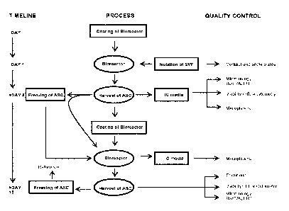
LEGENDS TO THE FIGURES
Fig. 1 depicts ASC production processes according to some embodiments of the
invention.
DETAILED DISCLOSURE OF THE INVENTION
The present invention relates to a stem cell product based on ASCs isolated
from healthy
donors, typically by two rounds of expanding the ASCs in a bioreactor
separated by a
cryopreservation step, resulting in a composition suitable for
cryopreservation in a cell bank.
The product is useful as an allogeneic therapeutic drug, e.g., for
regenerative therapy in
disorders or diseases characterized by ischemia or other tissue destruction,
such as heart
disease with and without heart failure, for immunosuppression of autoimmune
reactions or
transplant rejection, or anti-inflammatory therapy of inflammatory diseases.
In particular, the
ASC composition can be used as an off-the-shelf cryopreserved product, stored
in e.g., liquid
nitrogen, and ready for use directly after thawing. The ASC composition can
then be
administered intravenously, intra-arterially or by direct injection or
infusion into a tissue,
e.g., myocardium.
Moreover, a cell bank comprising multiple ASC preparations from different
donors according
to the invention can provide for personalized treatment by, e.g., allowing for
tissue matching
between donor and recipient prior to treatment, several treatments of the
recipient, and, in
case the recipient needs several treatments, the possibility to switch ASCs
from one donor to
another. The latter is particularly useful in case the recipient developed an
allo-antibody
response to ASCs from an earlier-administered ASC preparation.
4
Definitions
"ASCs," "Adipose-tissue derived stem cells," "adipose tissue-derived stromal
cells" and the
like, refer to multipotent stromal stem cells, also known as mesenchymal stem
cells,
multipotent stromal cells, multipotent stem cells, and mesenchymal
stromal/stem cells, which
are derived from adipose tissue. Certain criteria for identifying ASCs are
known in the art and
are described in, for example, Bourin et al. (2013).
In some embodiments, ASCs are characterized by their ability to differentiate
along
adipocytic, chondroblastic and osteoblastic lineages under appropriate
conditions. ASCs in
culture may be characterized by expression of one or more of the following
cell-surface
markers: CD90, CD73, CD105 and lack of expression of CD45 and CD31. In some
embodiments, they can be distinguished from bone-marrow-derived MSCs by their
positivity
for CD36 and negativity for CD106.
The stem cell population prepared according to the inventive method described
herein is
"substantially homogenous", meaning that the majority of the cells comply with
ASC
standards. Typically, a substantially homogenous ASC population according to
the present
invention is characterized by at least about 80% of the ASC population
expressing CD90,
CD105, CD13, CD73, CD166, CD29, and, optionally, CD10, CD140b, CD160, CD204,
CD272,
CD44, CD49a, CD54, CD9, Galectin 3, Galectin 9, HLA-G and LTI3R; and by at
most about
15% of the ASC population expressing CD45, CD31, CD14, and CD19. In some ASC
populations of the invention, one or more of CD90, CD73, CD13, CD105, CD29,
CD166,
CD10, CD140b, CD160, CD204, CD272, CD44, CD49a, CD54, CD9, Galectin 3,
Galectin 9,
HLA-G and LTBR can be expressed by at least about 80%, such as at least about
85%, such
as at least about 90%, such as at least about 95%, such as at least about 97%
or more of
the ASC population. Likewise, in some ASC populations of the invention, one or
more of
CD45, CD19, CD14, CD106, CD31 and CD36 can be expressed by at most about 15%,
such
as at most about 12%, such as at most about 10%, such as at most about 7%,
such as at
most about 5%, such as at most about 3% or less of the ASC population.
Specific ranges
contemplated for these and other markers are those defined by defined by the
minimum and
maximum expression percentages of an ASC population as shown in Tables 15 and
20.
By "adipose" is meant any fat tissue. The adipose tissue may be brown or white
adipose
tissue, derived from the abdominal area or other adipose tissue site. In
certain embodiments
the adipose is subcutaneous white adipose tissue or visceral adipose tissue or
any other
tissue containing adipose cells. The adipose tissue may be from any mammal.
Preferably, the
adipose tissue is human, most preferably from an adult human. A convenient
source of
adipose tissue is from liposuction surgery.
Date Regue/Date Received 2023-03-01
CA 03000912 2018-04-04
WO 2017/068140
PCT/EP2016/075407
A "lipoaspirate", as used herein, refers to the material removed during
liposuction via an
aspirator, i.e., a suction device. The lipoaspirate comprises adipocytes, fat,
connective tissue,
blood vessels, and a stromal vascular fraction. Any type of liposuction method
known in the
art can be used, including, but not limited to, suction-assisted, ultrasound-
assisted, power-
5 assisted, twin-cannula assisted, laser-assisted and water-assisted
liposuction (WAL). WAL is,
however, among the preferred options. The "stromal vascular fraction" or "SVF"
can then be
isolated from the lipoaspirate using methods known in the art, and exemplified
below.
As used herein, the term "bioreactor" refers to any device in which biological
and/or
biochemical processes develop under monitored and controlled environmental and
operating
conditions, for example, pH, temperature, supply of gas/air and nutrients and
waste removal.
The term "cryopreserve" or its various grammatical forms as used herein refers
to preserving
cells for storage in a cryoprotectant at subzero temperatures. For long-term
storage,
cryovials containing the cells and cryoprotectant are usually placed in liquid
nitrogen.
The term "cryoprotectant" as used herein refers to an agent that minimizes ice
crystal
formation in a cell or tissue, when the cell or tissue is cooled to subzero
temperatures and
results in substantially less damage to the cell or tissue after warming in
comparison to the
effect of cooling without cryoprotectant.
"Viability" as used herein refers to the feature of cells of not taking up
membrane
impermeant dye (e.g., Trypan Blue, FVS-780, SYTOX blue, propidium iodide),
thereby
demonstrating cell membrane integrity.
"Proliferative capacity" as used herein refers to the ability of cells to
multiply in a suitable
cultivation medium. Proliferative capacity can, for example, be represented by
the relative
number of cells after a 24h, 48h or 72h cultivation period as compared to the
number of cells
initially plated. This can also be expressed as "population doublings" during
a certain period.
For example, a population doubling of at least 1 during 48h in cell culture
means that the
number of cells seeded have doubled at least once during that period.
As used herein, the term 'donor' refers to the human or mammal from which the
adipose
tissue is retrieved, typically by liposuction. Preferably, the human is an
adult.
The terms "treatment," "therapy" and the like are used herein to generally
refer to obtaining
a desired pharmacologic and/or physiologic effect. The effect may be
prophylactic in terms of
completely or partially preventing a disease or symptom thereof and/or may be
therapeutic
in terms of a partial or complete stabilization or cure for a disease and/or
adverse effect
CA 03000912 2018-04-04
WO 2017/068140
PCT/EP2016/075407
6
attributable to the disease. "Treatment" as used herein covers any treatment
of a disease in
a mammal, particularly a human or veterinary subject, and includes: (a)
preventing the
disease or symptom from occurring in a subject which may be predisposed to the
disease or
symptom but has not yet been diagnosed as having it; (b) inhibiting the
disease symptom,
i.e., arresting its development; or (c) relieving the disease symptom, i.e.,
causing regression
of the disease or symptom.
In the context of therapeutic use of the disclosed pharmaceutical
compositions, in 'allogeneic'
therapy, the donor and the recipient are genetically different individuals of
the same species,
whereas in 'autologous' therapy, the donor and the recipient is the same
individual.
The terms "recipient", "subject" and "patient" are used interchangeably herein
and refer to
the mammalian subject for whom treatment or therapy is desired, particularly
humans.
Specific embodiments of the invention
Process:
The process according to the invention offers a safe and effective
manufacturing technology
based on, e.g., the combination of human platelet lysate as a growth
supplement for ASCs,
expansion in a closed bioreactor system and final formulation of ASCs as an
allogeneic
cryopreserved ready-to-use product with high-quality ASCs.
A general overview of the process according to some different embodiments is
shown in
Figure 1.
Typically, using the process of the invention, the preparation of a batch of
ASC product from
an SVF only takes from about 15 to about 18 days, excluding the time in
cryostorage. The
expansion efficiency is particularly surprising, considering that from one
bioreactor SVF run
an average yield of 11 5 intermediate product vials can be obtained (mean of 3
SVF runs),
each giving rise to an average batch yield of 5 2 ampoules of final product
(based on an
average of 8 bioreactor ASC runs). Thus an average yield of 55 cryovials with
about 110
million ASCs in each may be obtained from about 100 million mononuclear cells
(MNCs) in
the SVF. Furthermore, as described in the Examples, the ASC product is
characterized by a
high viability (average 90 2%), as determined immediately after thawing of the
second
passage ASC product.
In one embodiment, the process for preparing a composition comprising a
substantially
homogenous adult human stem cell population, comprises the steps of
CA 03000912 2018-04-04
WO 2017/068140
PCT/EP2016/075407
7
(i) adding the SVF of a lipoaspirate collected from a donor to a bioreactor
wherein at least
one surface is pre-treated to promote adhesion of adult human stem cells;
(ii) in the bioreactor, cultivating adherent cells to confluence in a serum-
free culture
medium supplemented with human platelet lysate;
(iii) detaching the adherent cells;
(iv) freezing the detached cells in a cryoprotectant at a concentration of at
least 1 x 10⁶
cells/mL;
(v) thawing the frozen cells and repeating steps (ii) to (iii) at least once,
(vi) freezing the detached cells at a concentration of at least 1 x 10⁷
cells/mL; and
(vii) optionally, thawing the frozen composition.
In one embodiment, the process for preparing a composition comprising a
substantially
homogenous adult human stem cell population, comprises the steps of
(i) adding the SVF of a lipoaspirate collected from a donor to a bioreactor
wherein at least
one surface is pre-treated to promote adhesion of adult human stem cells;
(ii) in the bioreactor, cultivating adherent cells to confluence in a serum-
free culture
medium supplemented with human platelet lysate;
(iii) detaching the adherent cells;
(iv) repeating steps (ii) and (iii) at least once;
(v) freezing the detached cells at a concentration of at least 1 x 10⁷
cells/mL; and,
optionally,
(vi) thawing the frozen composition.
In one embodiment, the process for preparing a composition comprising a
substantially
homogenous adult human stem cell population comprises the steps of
(i) adding the stromal vascular fraction (SVF) of a lipoaspirate collected
from a donor to a
bioreactor wherein at least one surface is pre-treated to promote adhesion of
adult
human stem cells;
(ii) cultivating adherent cells of the SVF to confluence in a serum-free
culture medium
supplemented with human platelet lysate;
(iii) detaching the adherent cells;
(iv) freezing the detached cells in a cryoprotectant at a concentration of at
least 1 x 10⁶
million cells/mL;
(v) thawing the frozen cells and repeating steps (ii) to (iv), freezing the
detached cells at a
concentration of at least 1 x 10⁷ cells/mL; and, optionally,
(vi) thawing the frozen composition.
CA 03000912 2018-04-04
WO 2017/068140
PCT/EP2016/075407
8
The SVF is isolated from a lipoaspirate obtained from a healthy donor, e.g.,
50 mL, 100 mL,
200 mL, 300mL or 500 mL, such as between 100-300 mL, lipoaspirate. Typically,
the adipose
tissue is first separated from non-adipose tissue using a tissue collection
container that
utilizes decantation, sedimentation, or centrifugation techniques to separate
the materials.
The adipose tissue can then disaggregated using methods such as mechanical
force (mincing
or shear forces), enzymatic digestion with one or more proteolytic enzymes,
such as
collagenase, trypsin, TrypLe Select, lipase, liberase HI, pepsin, or a
combination of
mechanical and enzymatic methods. Thereafter, the remaining cells can be
retrieved by
filtration, centrifugation or the like. Examples of methods to retrieve the
SVF from a
.. lipoaspirate are described in Godthardt, etal. (2008) MACS Miltenyi Biotec
Information
Pamphlet and WO 2014/138383.
In one embodiment, approximately 100 ml lipoaspirate is obtained from a donor
by
liposuction from the abdomen under local anesthesia. The lipoaspirate is
washed twice with
phosphate buffered saline (PBS) pH 7.4 to remove residual blood. The adipose
tissue is then
.. digested by incubation with collagenase dissolved in a balanced salt
solution at 37 C for 45
min. under constant rotation. The collagenase is neutralised with medium
holding 5% human
platelet lysate and 1% Penicillin/Streptomycin and is filtered through a 100pm
filter. The
remaining cells are centrifuged at 1200 x g for 10 min at room temperature, re-
suspended
and counted using a cell counter according to manufacturer's instructions.
Typically, at least one surface of the bioreactor is pre-treated to facilitate
or promote
adhesion of ASCs, either by the manufacturer of the bioreactor or at some
chosen point of
time before initiating the production process. Various types of treatments to
promote cell
adhesion are known in the art and include, e.g., tissue culture treatment and
coating with
synthetic charged polymers, nanofibers, glycosaminoglycans and various protein
compositions. For tissue culture treatment, a polystyrene-based surface in the
bioreactor is
modified with plasma gas, resulting in the hydrophobic plastic surface
becoming more
hydrophilic, the net negative charge promoting cell attachment. As for pre-
coating the
surface, protein compositions useful for this purpose may comprise one or more
plasma
proteins such as, e.g., fibrinogen, fibronectin, Factor VIII, von Willebrand
factor and Factor
XIII; one or more extracellular matrix proteins such as, e.g., collagens and
laminins; and/or
one or more proteoglycans. In one embodiment, the protein composition
comprises or
consists of one or both of fibrinogen and fibronectin. In one embodiment, the
protein
composition comprises or consists of cryoprecipitate. Cryoprecipitate is a
well-known blood
product prepared from plasma, e.g., where fresh plasma is frozen and thawed
and the
precipitate collected. The product typically contains fibrinogen and Factor
VIII, as well as e.g.
von Willebrand factor, Factor XIII and fibronectin. In some embodiments, the
cryoprecipitate
contains at least 140 mg or more of fibrinogen per 70 IU of Factor VIII,
optionally prepared
CA 03000912 2018-04-04
WO 2017/068140
PCT/EP2016/075407
9
from either AB or low-titer A blood donors. In another embodiment, the protein
composition
comprises or consists of human platelet lysate, described below.
The basic classes of bioreactors suitable for use with the present invention
include hollow-
fiber bioreactors and rocking, spinning or rotating perfusion systems, with or
without micro-
.. carriers or discs, suitable for anchorage-dependent cell expansion.
Preferably, the bioreactor
is a functionally closed system or protected by sterile barrier filters,
capable of providing for a
continuous supply of culture medium and a continuous removal of waste during
cell culture.
Most preferable are disposable hollow-fiber bioreactors enclosed in an
incubator, providing a
surface area for cell attachment of at least, 0.5 m2, such as at least 1 m2,
such as at least 1.5
.. m2, such as at least 2 m2, such as between 1 to 3 m2. Preferably, the
surface area is at least
2 m2, such as about 2.1 m2. One example of such a bioreactor is the Quantum
Cell Expansion
System (herein also referred to as "Quantum bioreactor") which is fed through
two circulation
loops with inlets for media and reagents or cells, waste being removed into a
waste bag. As
shown in Example 7, expanding the ASCs in a bioreactor significantly increased
the expansion
rate and yield relative to manual processing in standard tissue culture
flasks.
Prior to loading of the SVF (or first passage ASCs) into the bioreactors, the
system can be
primed with a buffer, e.g., phosphate buffered saline, and subsequently loaded
with a protein
or other composition for coating. Before loading the cells, the buffer can
then be washed out
of the system and replaced with complete medium.
.. The cells can then be added to the bioreactor. For example, approximately
10, 20, 50, 100,
200 or 500 million mononuclear cells (MNCs) from the SVF preparation or
approximately 5,
10, 20, 50 or 100 million first-passage ASCs can be loaded into the primed and
coated
bioreactor through the inlet, optionally via a filter. Preferably, for MNCs,
about 100 million
cells from the SVF are loaded. For the second passage, preferably, about 5 to
50 million cells
from the first-passage ASCs are loaded, such as between 5 and 30, 5 and 25, 10
and 30 or
between 15 and 25 million cells. For any higher passage of the cells (i.e.,
3'd passage, 4th
passage etc.), cells can be added in amounts similar to the second passage.
The cells are
then allowed to attach for a sufficient period of time, such as for at least
5h and/or up to
about 24 h, after which continuous feeding with media is activated. For
example, the media
feeding rate may start at about 0.1 mL/min, and then adjusted based on glucose
and/or
lactate measurements and/or cell expansion.
Any standard cell culture medium can be used, such as, e.g., Dulbecco's
Modified Eagle's
Medium (DMEM), alpha-Minimum Essential Medium (a-MEM). As shown in Example 6,
however, the use of a human plasma lysate (HLP) product was clearly a more
effective
.. growth supplement in terms of proliferative capacity than the use of FBS,
without
CA 03000912 2018-04-04
WO 2017/068140
PCT/EP2016/075407
compromising genomic stability. HPL is typically a turbid, light-yellow liquid
that is obtained
from human blood platelets after one, two, three or more freeze/thaw cycles.
These cycles
cause the platelets to lyse, releasing their intracellular contents, including
growth factors and
the like, into the surrounding medium. Some HPL preparations include blood
clotting factors,
5 in which case it may be advantageous to add an anti-coagulant such as
heparin to prevent
coagulation. Other HPL preparations can be processed to remove, or otherwise
inhibit the
effect of, the clotting factors. HPL preparations, some of which GMP grade,
are available
commercially from, e.g., Compass Biomedical, Inc., Cook General
BIotechnologyl,
Macopharma SA, Cook Regentech, Mill Creek, iBiologics and Trinova Biochem GmbH
under
10 the product lines PLUS, Stemulate, Human Platelet Lysate, PLTMax,
XcytePlus and CRUX
RUFA Media Supplements. Preferably, the culture medium for the ASCs comprise
from about
1% to about 20%, such as from about 2% to about 15%, such as from about 3% to
about
12%, such as from about 5% to about 10%, such as about 5%, 8% or 10% HPL.
Preferably,
the culture medium comprises from about 2% to about 15% HPL in, e.g., MEM. A
preferred
HPL preparation is Stemulate, which does not require the addition of heparin
(WO
2015031465 Al).
Once cell growth has reached or nearly reached its stationary phase, as
determined by, e.g.,
stagnation in glucose consumption and/or lactate production, the ASCs are
harvested,
typically by loading TrypLe Select into the system. Harvested cells can then
be washed and
transferred into centrifuge tubes, pelleted and counted.
First-passage ("intermediate") ASCs can then be cryopreserved or,
alternatively, directly
loaded into the pre-coated bioreactor for a second (or 3`d, 4th, etc.) round
of expansion. For
cryopreservation, the intermediate ASCs are suspended in cryoprotectant at a
concentration
of at least about 1 x 10⁶ cells per mL, such as at least about 2 x 10⁶ cells
per mL, at least
about 5 x 10⁶ cells per mL, at least about 10 x 106 cells per mL, at least
about 15 x 106 cells
per mL, at least about 20 x 106 cells per mL, or at least about 50 x 106 cells
per mL, such as
between 1 x 10⁶ and 50 x 106 cells per mL, such as between 1 x 10⁶ cells and
20 x 106 cells
per mL.
Second (or higher, such as 3rd, 4th etc.) passage ASCs can be cryopreserved.
For
cryopreservation, the second (or higher) passage ASCs are suspended in
cryoprotectant at a
concentration of at least about 1 x 10⁷ cells per mL, such as at least about
1.5 x 10⁷ cells per
mL, at least about 2 x 10⁷ cells per mL, at least about 2.5 x 10⁷ cells per
mL, at least about 3
x 107 cells per mL, at least about 5 x 10⁷ cells per mL, or at least about 10
x 107 cells per
mL, such as between 1 x 10⁷ and 5 x 10⁷ cells per mL, such as between 2 x 10⁷
cells and 3 x
107 cells per mL. In one embodiment, the second (or higher, such as 3'd, 4th
etc.) passage
ASCs are suspended in cryoprotectant at a concentration of about 2 x 10⁷ cells
per mL
11
cryoprotectant, such as about 2.2 x 10⁷ cells per mL cryoprotectant. As used
herein, unless
contradicted by context, 2 x 10⁷ cells per mL includes or corresponds to from
1.6 x 10⁷ to 2.4
x 107 cells per mL and about 2.2 x 10⁷ cells per mL includes or corresponds to
from 2.0 to
2.4 cells per mL.
.. The cryoprotectant is preferably protein-free, endotoxin-free and sterile.
While several
suitable cryoprotectants are available, non-limiting examples of
cryoprotectants
contemplated for the ASC compositions of the present invention are CryoStor
(BioLife
Solutions), including CryoStor CS2, CryoStor CS5 and CryoStor CS10; and
ProFreeze
(Lonza). CryoStor freeze media are sterile serum-free, protein-free and animal
component
free, having a pH 7.5 - 7.7, and an endotoxin level under 1 EU/mL. In one
embodiment, the
cryoprotectant is Hypothermosol (CMS, Rockville, Md.) plus 10% DMSO (WO
2000/002572
Al). Hypothermosol comprises Trolox (6-hydroxy-2,5,7,8-tetramethylchroman-2-
carboxylic
acid), Nat, K+, ca2+, mg2+1.cra, H2PO4-, HEPES, lactobionate, sucrose,
mannitol, glucose,
Dextran-40 (i.e., dextran with an average MW of 40,000 Da), adenosine and
glutathione (WO
.. 2010/064054 Al). According to the manufacturer, ProFreeze should be
supplemented with
10% DMSO at time of use. WO 2000/002572 Al and WO 2010/064054 Al.
In any embodiment herein where DMSO is used, the DMSO can be replaced by a
glucan such
as, for examples dextran, having an average molecular weight in the range of
35000 to
45000 Da, such as, e.g., Dextran-40.
In one embodiment, the cryoprotectant comprises between 5% and 15% DMSO, such
as
about 5%, about 6%, about 8%, about 10%, about 12% or about 15% DMSO, and
Trolox,
Nat, K+, Ca2+, Mg2+1CI-, H2PO4-, HEPES, lactobionate, sucrose, mannitol,
glucose, Dextran-
40, adenosine and glutathione. Preferably, the cryoprotectant comprises about
10% DMSO.
.. In one embodiment, the cryoprotectant comprises a 1:10 to about 1:20
mixture of DMSO
and an aqueous solution comprising
(a) one or more electrolytes selected from the group consisting of potassium
ions at a
concentration ranging from about 35-45 mM, sodium ions ranging from about 80-
120
mM, magnesium ions ranging from about 2-10 mM, and calcium ions ranging from
about 0.01-0.1 mM;
(b) a macromolecular oncotic agent having a size sufficiently large to limit
escape from
the circulation system and effective to maintain oncotic pressure equivalent
to that of
blood plasma and selected from the group consisting of human serum albumin,
polysaccharide and colloidal starch;
Date Regue/Date Received 2023-03-01
CA 03000912 2018-04-04
WO 2017/068140
PCT/EP2016/075407
12
(c) a biological pH buffer effective under physiological and hypothermic
conditions;
(d) a nutritive effective amount of at least one simple sugar;
(e) an impermeant and hydroxyl radical scavenging effective amount of
mannitol;
(f) an impermeant anion impermeable to cell membranes and effective to
counteract cell
swelling during cold exposure, said impermeant ion being at least one member
selected from the group consisting of lactobionate, gluconate, citrate and
glycerophosphate;
(g) a substrate effective for the regeneration of ATP, said substrate being at
least one
member selected from the group consisting of adenosine, fructose, ribose and
adenine; and
(h) glutathione.
In one embodiment, the cryoprotectant comprises a 1:10 to about 1:20 mixture
of DMSO
and an aqueous solution comprising
a). one or more electrolytes selected from the group consisting of potassium
ions at a
concentration ranging from 35-45 mM, sodium ions ranging from 80-120 mM,
magnesium ions ranging from 2-10 mM, and calcium ions ranging from 0.01-0.1
mM;
b). a macromolecular oncotic agent having a size sufficiently large to limit
escape from
the circulation system and effective to maintain oncotic pressure equivalent
to that of
blood plasma and selected from the group consisting of human serum albumin,
polysaccharide and colloidal starch;
c). a biological pH buffer effective under physiological and hypothermic
conditions;
d). a nutritive effective amount of at least one simple sugar;
e). an impermeant and hydroxyl radical scavenging effective amount of
mannitol;
f). an impermeant anion impermeable to cell membranes and effective to
counteract cell
swelling during cold exposure, said impermeant ion being at least one member
selected from the group consisting of lactobionate, gluconate, citrate and
glycerophosphate;
g). a substrate effective for the regeneration of ATP, said substrate being at
least one
member selected from the group consisting of adenosine, fructose, ribose and
adenine, and
h). at least one agent which regulates apoptotic induced cell death.
The suspension of first, second (or higher) passage ASCs in cryoprotectant is
then added to
cryovials. For first-passage (intermediate) ASCs, each cryovial may contain
about 1, about 2,
about 3, about 4, about 5, about 6, about 7, about 8, about 9, about 10, about
15, or about
20 mL ASC suspension, corresponding to a number of ASCs in the range of 5
million cells to
250 million cells, such as, e.g., about 20, about 30, about 40, about 50,
about 60, about 80
CA 03000912 2018-04-04
WO 2017/068140
PCT/EP2016/075407
13
or about 100 million cells. Preferably, cryovials with first-passage ASCs
contain about 50
million cells in 5 mL.
For the final passage ASCs, i.e., the 2nd or higher passage ASCs, each
cryovial may contain
about 1, about 2, about 3, about 4, about 5, about 6, about 7, about 8, about
9, about 10,
about 15, or about 20 mL ASC suspension, corresponding to a number of ASCs in
the range
of 10 million cells to 500 million cells, such as, e.g., about 40, about 60,
about 80, about
100, about 110, about 120, about 160 or about 200 million cells. Preferably,
cryovials with
second-passage ASCs contain from about 100 million to about 120 million cells
in about 5
mL, such as about 110 million cells in about 5 mL. In the compositions of the
invention,
unless contradicted by context, about 110 million cells includes from 100
million to 120
million cells.
Freezing can advantageously be performed by automated freezing with the use of
a
controlled rate freezer. After freezing vials can be transferred and stored at
a temperature in
the range of -70 C to -196 C, such as between -150 C to -190 C, such as in the
-180 C-
range. Various means for freezing and maintaining vials under such freezing
conditions are
known, many of which involving liquid nitrogen. They include, for example,
immersion of the
vials in liquid nitrogen, storing the vials in the vapour phase of liquid
nitrogen, and placing
the vials in so-called dry storage. In the later type of storage, the vials
are not in direct
contact with liquid or vapour phase nitrogen since the nitrogen is contained
in the shell of the
container, resulting in a storage temperature in the -180 C-range.
In one aspect, a two-tiered cell banking system is established for the vials,
constituting a
working cell bank holding intermediate ASCs from first passage bioreactor
expansions and a
product cell bank holding finally formulated and packaged ASC compositions. In
the cell bank,
the first passage intermediate ASCs are typically cryopreserved at about 50
million cells in
about 5 ml CryoStor10 in cryovials until loading for second bioreactor
expansion. The final
ASC product is typically cryopreserved at about 110 million cells in about 5
ml CryoStor10 in
cryovials until right before use. This product can also be designated "CSCC
ASC". In one
embodiment, the cell bank comprises a plurality of vials stored under freezing
conditions,
each vial comprising about 5 mL of the second-passage ASCs, wherein the cell
concentration
is 2 x 10⁷ cells per mL, e.g., in the range of 1.6 x 10⁷ to 2.4 x 10⁷ cells
per mL, or about 2.2
x 107 cells per mL, e.g., in the range of 2.0 x 10⁷ to 2.4 x 10 cells per mL.
ASCs:
The ASCs of the invention are characterized by their multipotent capacity,
marker profile,
and/or and by functional characteristics of the ASCs, such as proliferation
capacity, viability,
CA 03000912 2018-04-04
WO 2017/068140
PCT/EP2016/075407
14
recovery and immunosuppressive capability, even after cryopreservation. These
ASC
characteristics, detailed below, each applies equally to the ASCs obtained
according to the
process of the invention. Marker profiles can, for example, be conveniently
determined by
flow cytometry using fluorescence-labelled antibodies against each marker,
e.g., as described
in Example 2, 10, 11 or 14.
The ASCs are, in particular, characterized by their ability to differentiate
along adipocytic,
chondroblastic and osteoblastic lineages under appropriate conditions. As
shown in Example
4, ASCs prepared according to the manufacturing methods described in Example 1
differentiated into adipocytes, chondrocytes and osteoblasts when cultured in
differentiation
medium.
The ASCs can also or alternatively be characterized according to their
phenotype, i.e., marker
profile, regarding their expression of markers in common with other
mesenchymal
stromal/stem cells, including CD90, CD73, CD105, and CD44, and maintaining low
or
negligible expression levels of CD45 and CD31 (Bourin etal., 2013).
In some embodiments, a substantially homogenous ASC population is one wherein
at least
80%, such as at least 85%, such as at least 90%, such as at least 95%, such as
at least
98% of the stem cell population express CD105, CD90, CD73, CD29 and CD13, and
at most
10%, such as at most 5%, such as at most 3%, such as at most 2% express CD45,
CD34,
HLA-DR, CD19 and CD14. Preferably, at least 95% express CD90, CD73 and CD13
and at
most 5% express CD45, CD34, HLA-DR, CD19 and CD14.
In some embodiments, a substantially homogenous ASC population is one wherein
at least
90% express CD90, CD73, CD13, CD29 and CD166; at most 5% express CD45, CD19,
CD14
and CD31; at most 10% express CD106; between 2 and 15% express CD36; at least
10%
express CD146; at least 80% express CD105 and at most 40% express CD34. In
some
embodiments, at least 95% express CD90, CD73, CD13, CD29 and CD166. In
addition, at
least 80%, such as at least 85%, such as at least 90%, such as at least 95%,
such as at
least 98% may express CD44.
In some embodiments, a substantially homogenous ASC population is one wherein
at least
about 80%, such as at least about 85% express CD105, CD90, CD73, CD13, CD29
and
CD166, such as at least about 90%, such as at least about 95% express CD90,
CD73, CD13,
CD29 and CD166; at most about 15%, such as at most about 10% express CD45,
CD19,
CD14, CD106, CD31 and CD36, such as at most about 7%, such as at most about
5%, such
as at most about 3% express CD45, CD19, CD14, CD106 and CD31; at least about
2%, such
as at least about 5%, such as between about 1% and about 20%, such as between
about 2%
CA 03000912 2018-04-04
WO 2017/068140
PCT/EP2016/075407
and about 15% or between about 5% and about 15% express CD36; at most about
50%,
such as at most about 40%, such as at most about 20%, such as at most about
10% express
CD34; and at least about 10%, such as at last about 12% express CD146. In one
embodiment, at least 95% express CD90, CD73, CD13, CD29 and CD166; at least
85%
5 express CD105, at most 2% express CD45, HLA-DR, CD19, CD14 and CD31;
between 2%
and 15% express CD36; and between 10% and 60% express CD146. In one
embodiment, a
substantially homogenous ASC population is one wherein the ranges of the
minimum and
maximum expression percentages of markers are those shown in Table 20.
When determined immediately after thawing of the cryopreserved 2nd passage
cells, at least
10 about 80%, such as at least 85%, such as at least 90%, such as at least
95%, such as at
least 98% of the cells are viable, as determined by dye exclusion (see, e.g.,
Example 3).
Preferably, at least 90% of the cells are viable.
As for proliferation capacity, when placed in culture immediately after
thawing, the ASCs are
characterized by a population doubling (PD) of at least 1, such as at least
1.3, such as at
15 least 1.5, such as at least 1.7, such as at least 2, when cultured in
tissue culture flasks for
48h (e.g., according to the method in Example 3). Preferably, the ASCs have a
PD of at least
1, such as at least 1.5. PD is calculated as Ln (N)/Ln 2, where N =
Cell harvested/Cell seeded.
As shown in the Examples, the ASCs of the invention are further characterized
by their
immunosuppressive properties. For example, the ASCs may be characterized by
one or more
or all of the following: suppressing activation of dendritic cells (DCs),
suppressing
proliferation of peripheral blood mononuclear cells (PBMCs), cell surface
markers indicative of
immunomodulation, especially immunosuppression, or by a change in one or more
cell
surface markers in response to a cytokine such as interferon-gamma.
In one embodiment, the ASCs of the invention suppress activation of DCs, e.g.,
reducing the
expression of CD40, CD80, CD86 and HLA-DR by DCs mixed with ASCs as compared
to DCs
not mixed with ASCs (i.e., a positive control). In a specific embodiment, the
assay of
Example 9 is used, wherein ASCs and DCs are seeded to result in approximately
a 1:1 ratio;
the DCs being stimulated with 1 pg/mL lipopolysaccharide (LPS) and 20 ng/mL
interferon-
gamma and incubated for 24 h; and the respective expression level of CD40,
CD80, CD86
and HLA-DR is reduced, in average, to at most 80%, 65%, 70% and 80%,
respectively, of
the positive control.
In one embodiment, the ASCs of the invention suppress the proliferation of
PBMCs, e.g., as
determined in a Mixed Lymphocyte Reaction (MLR). This type of assay is well-
known in the
CA 03000912 2018-04-04
WO 2017/068140
PCT/EP2016/075407
16
art, and may comprise mixing ASCs with stimulated PBMCs from an allogeneic
donor in
different ratios, e.g., in the range 1:20 to 1:1, using PBMCs without ASCs as
positive
controls, and measuring after a 4-day co-culture period, the PBMC
incorporation of 3H-
thymidine (25 pSi/m1) during an 18-20 h incubation period. Using this type of
assay, as
compared to the positive control, a 1:20, 1:10, 1:5 and 1:1 ratio of ASCs to
PBMCs may
result in an average 3H-thymidine incorporation of at most about 80%, 75%,
55%, and 25%,
respectively, of the positive control.
In some embodiments, the ASCs are also or alternatively characterized by
specific markers
indicative of immunomodulation, especially immunosuppression, such as CD10,
CD140a,
CD160, CD204, CD258, CD270, CD272, CD44, CD49a, CD54, CD9, Galectin 3,
Galectin 9,
HLA-G, LTPR and combinations thereof. Without being limited to theory, these
markers are
associated with immune signalling, cell-cell and cell-ECM adhesion, homing,
pattern
recognition, T cell inhibition, up-regulation of growth factor receptors and
inactivation of pro-
inflammatory proteins.
In particular, in one embodiment, a substantially homogenous ASC population is
one wherein
at least about 80%, such as at least about 85%, express CD10, CD140b, CD160,
CD204,
CD272, CD44, CD49a, CD54, CD9, Galectin 3, Galectin 9, HLA-G and/or LTPR, and,
optionally, HLA-ABC, such as at least about 90% or in some cases at least
about 95% or
more express CD10, CD140b, CD160, CD204, CD272, CD44, CD54, CD9, Galectin 3,
Galectin
9, HLA-G and LT13R. In some embodiments, the ASCs may further be characterized
by
expressing no more than about 20%, such as no more than about 15%, or in some
cases no
more than about 10%, of CD152, CD274 and/or CD86; and/or optionally, at least
about 70%
CD258, at least about 55% CD270, at least about 80% CD49a, up to about 30%
CXCR4
and/or between about 5% to about 35% CD200. In some embodiments, the ASC
population
may also be one wherein at most about 15% of the ASCs express CD15, CD152,
CD163,
CD18, CD274, CD39, CD40, CD62L, CD80, CD86, and, optionally HLA-DR, -DQ, -DP.
In one embodiment, a substantially homogenous ASC population is one wherein at
least 90%
express CD10, CD140b, CD160, CD204, CD272, CD44, CD54, CD9, Galectin 3,
Galectin 9,
HLA-G and LTPR; at least 80% express CD49a; at least 60% express CD258 and
CD270 and
at least 5% express CD200; at most 15% express CD15, CD152, CD163, CD18,
CD274,
CD39, CD40, CD62L, CD80 and CD86; and at most 30% express CXCR4.
In one embodiment, a substantially homogenous ASC population is one wherein at
least 95% of
the ASC population express CD10, CD140b, CD160, CD204, CD272, CD44, CD54, CD9,
Galectin 3, Galectin 9, HLA-G and LTpR; at least 85% express CD49a, at least
65% express
CD258 and CD270 and at least 10% of the population express CD200, and at most
15% of
CA 03000912 2018-04-04
WO 2017/068140
PCT/EP2016/075407
17
the population express CD15, CD152, CD163, CD18, CD274, CD39, CD40, CD62L,
CD80,
CD86, and HLA-DR, -DQ, and - DP, and at most 25% express CXCR4.
In one embodiment, the ASCs are further characterized by less than 20%, such
as less than
about 15%, such as less than about 10% of the ASCs expressing CD274.
Optionally, the
ASCs are also characterized by at least about 90%, such as at least about 95%,
such as at
least about 98% of the ASCs expressing CD54. In another specific embodiment,
the ASCs are
characterized by each marker in Table 15 in Example 10 at a percentage of
population in the
range from the minimum to the maximum value shown in Table 15.
The ASCs may also be characterized by their expression percentages of
stromal/stem cell
markers according to one embodiment herein and their expression percentages of
immunomodulation markers according to one embodiment herein. As a non-limiting
example,
a substantially homogenous ASC population is one wherein
- at least 90% express CD90, CD73, CD13, CD29 and CD166; at most 5% express
CD45, CD19, CD14 and CD31; at most 10% express CD106; between 2 and 15%
express CD36; at least 10% express CD146; at least 80% express CD105 and at
most 40% express CD34; and
- at least 90% express CD10, CD140b, CD160, CD204, CD272, CD44, CD54, CD9,
Galectin 3, Galectin 9, HLA-G and LT13R; at least 80% express CD49a; at least
60%
express CD258 and CD270 and at least 5% express CD200; at most 15% express
CD15, CD152, CD163, CD18, CD274, CD39, CD40, CD62L, CD80 and CD86; and at
most 30% express CXCR4.
In a further embodiment, the ASCs of the invention are also or alternatively
characterized by
a change in one or more cell surface markers in response to a pro-inflammatory
cytokine
such as interferon-gamma. This may advantageously be tested according to the
assay of
Example 11, typically measuring a change in one or more ASC markers in Table
16 and 17
showing
- a positive or negative change in the percentage of the ASC population
expressing the
marker in at least 5% of the ASC population, or
- a positive- or negative change in the expression level of the marker on
the portion of
cells expressing the marker of at least 0.5-fold,
when cultivated for 3 days in the presence of 50 ng/ml IFN-gamma, as compared
to a
control, such as cells from the same ASCs which have not been stimulated with
IFN-gamma.
CA 03000912 2018-04-04
WO 2017/068140
PCT/EP2016/075407
18
For example, in some embodiments, upon INF-gamma stimulation, the percentages
of the
ASC population expressing CD200, CD270, CD9, CXCR4 are reduced; the
percentages of the
ASC population expressing CD274 and CD49a are increased, and the expression
level of
CD54 on CD54-positive cells is increased.
In some embodiments, the change is one or more or all of
- the percentage of the ASC population expressing CD274õ CD106 and/or CD49a
being increased by at least 5%, such as the percentage of CD274 being
increased by
at least 40%, such as at least 60%;
- the percentage of the ASC population expressing CD200, CD270, CD9 and/or
CXCR4
being reduced by at least 5%, such as the percentage of the ASC population
expressing being reduced by at least 10%;
- the expression level of CD10, CD54, HLA-ABC and/or HLA-DR/DQ/DP
increasing on
marker-positive cells, such as the expression level of CD54 on CD54-expressing
cells
increasing by at least 20-fold, such as at least 35-fold, such as at least 30-
fold;
and/or
- the expression level of L113R decreasing, e.g., by at least _2-fold.
In a specific embodiment, at most about 30%, such as at most about 20%, such
as at most
about 15%, such as at most about 10% of the ASC population expresses CD274
whereas
upon interferon-gamma stimulation, at least 70%, such as at least about 80%,
such as at
least about 85%, such as at least about 90%, such as at least about 95% of the
ASC
population expresses CD274, e.g., when cultivating the ASCs for 3 days in the
absence and
presence of 50 ng/ml IFN-gamma, respectively.
Also of note is an increase in MFI from 3 to 97 for the marker CD54 (ICAM-1)
which
illustrates the mobilisation of an intercellular adhesion molecule necessary
for the
stabilisation of ASC-leukocyte interactions and signal transduction. ICAM-1 is
a ligand for
LEA-1 (integrin), a receptor found on leukocytes. So, in one embodiment, at
least 95% of the
ASC population expresses CD54 and upon interferon-gamma stimulation, the
expression level
of CD54 on CD54-expressing cells is increased by at least 20-fold, such as at
least 30-fold.
In a particular embodiment, upon interferon-gamma stimulation, the percentage
of the ASC
population expressing CD274 is increased to at least 80% and the expression
level of CD54
on CD54-positive cells is increased at least 25-fold.
CA 03000912 2018-04-04
WO 2017/068140
PCT/EP2016/075407
19
Compositions:
Also provided by the present invention are compositions of each ASC population
detailed in
the aspects and embodiments herein, including those in the preceding "ASCs"
section and
those obtained by each process in the "Process" section. Each such ASC
population is also
provided in the format of a composition of the invention.
In particular, it has been found that ASCs, particularly ASCs obtained
according to the
process of the invention, can be cryopreserved at high concentrations in
protein-free
cryoprotectant; at least about 1 x 10⁷ cells per mL, without compromising
viability,
proliferation capacity, immunosupressive properties, or recovery. It has also
been found that
CryoStor-based cryoprotectant may provide for a higher proliferation capacity
than a human
serum albumin (HSA)-based cryoprotectant.
In particular, the compositions of the invention may comprise a suspension of
a substantially
homogenous adult human stem cell population, isolated from adipose tissue
collected from a
donor, in a protein-free cryoprotectant, wherein the cell concentration is at
least 1 x 10⁷
cells, such as at least 1.5 x 10⁷ cells, such as at least 2 x 10⁷ cells, such
as at least 3 x 10⁷
cells, such as at least 5 x 10⁷ cells, per mL added cryoprotectant.
Preferably, the cell
concentration is at least 2 x 10⁷ cells, such as about 2.2 x 10⁷ cells per mL
added
cryoprotectant.
As described above, the protein-free cryoprotectant typically comprises from
about 5% to
about 15% DMSO and Trolox, Nat, K+, c.a2+, mg2-Fla-, H2p04-, HEPES,
lactobionate, sucrose,
mannitol, glucose, dextran-40, adenosine and glutathione. Preferably, the
cryoprotectant
comprises about 10% DMSO. Other protein-free cryoprotectants known in the art
may also
be used. Those already described in the "Process" section are particularly
contemplated.
Also provided are the compositions obtained when thawing the frozen ASC
compositions. The
frozen ASC compositions may, for example, be thawed in a 37 C water bath or
thawed/stored in room temperature in the operation room. Preferred are
compositions
where, immediately after thawing
(a) at least 85% of the ASC population are viable cells, and the viability
after storage in
room temperature for 2 hours is at least 80%;
(b) the ASC population has a proliferation capacity providing for a PD of at
least 1 when
cultured for 48 hours;
(c) the ASC population is capable of suppressing dendritic cell maturation and
activation;
CA 03000912 2018-04-04
WO 2017/068140
PCT/EP2016/075407
(d) the recovery after thawing is over 95%, and the recovery of cells after
storage at
room temperature for 2 h after thawing is at least 85%
(e) the ASC population has an in vitro cell adherence such that at least 60%,
such as at
least 65%, such as at least 70% of the total number of cells are adherent
after 5h in
5 cultivation.
Also preferred are compositions, where, after storage at room temperature for
2h after
thawing,
(a) at least 80% of the stem cell population are viable cells;
(b) the stem cell population has a PD of at least 1 when cultured for 48
hours;
10 (c) the stem cell population is capable of suppressing dendritic cell
maturation and
activation;
(d) the recovery is at least 85%, and
(e) the ASC population has an in vitro cell adherence such that at least 60%,
such as at
least 70% of the total number of cells are adherent after 5h in cultivation.
15 Also provided by the present invention are pharmaceutical compositions.
The compositions of
the invention are sterile and free of endotoxins and mycoplasms, endotoxin
levels typically
determined with an analytical method providing for a minimum detection level
of 10 IF per
ml. Since the compositions of the invention are ready for clinical use, the
pharmaceutical
compositions typically only comprise the ASCs and the protein-free
cryoprotectant. However,
20 additional components are also contemplated. Specifically, the
pharmaceutical composition
may further comprise a soluble biomaterial or hydrogel containing natural or
synthetic
biopolymers such as extracellular matrix proteins, -peptides or -
glycosaminoglycans and/or
alginate. For example, the pharmaceutical composition may comprise sterile and
endotoxin
free Alginate (Sodium alginate VLVG, Novamatrix, FMC Biopolymers, Norway),
partially
.. calcium cross-linked with D-gluconic acid and hemicalcium salt (Follin
etal., 2015). In one
embodiment, the alginate is mixed with ASCs and cryoprotectant to a final
concentration of 1
% (w/v) partially cross-linked alginate before the final cryopreservation
step. In another
embodiment partially cross-linked alginate is stored at RT and mixed with the
final product to
a final concentration of 1% (w/v) alginate, e.g., by injecting the ASC
preparation into the
alginate container before the final suspension is aspirated and connected with
the injection
catheter.
Also provided are syringes or other means for injection or infusion of the ASC
compositions,
which contain the ASC compositions. Typically, the ampoule holding the ASC
composition is
sterilised with an alcohol swab (82% Ethanol and 0.5% Chlorhexidin, Mediq,
Denmark) and
the cell suspension aspirated with a needle into a sterile syringe. The
syringe is then
CA 03000912 2018-04-04
WO 2017/068140
PCT/EP2016/075407
21
connected to an injection catheter, e.g., a MYOSTAR injection catheter
(Biological Delivery
System, Cordis, Johnson & Johnson, USA) for injection. Injection is
recommended within 3
hours of thawing. Preferably, the syringe or other means for injection or
infusion contain
about 5 mL of the composition, comprising from about 100 million to about 120
million cells,
such as about 110 million ASCs.
Therapeutic use:
In one embodiment, there is provided an ASC composition for use as a
medicament, e.g., for
tissue regeneration, immunosuppression and/or as an anti-inflammatory drug.
Indeed, the invention provides for various therapeutic uses of the ASCs and
ASC
compositions of the invention, which are "off-the-shelf" and ready for
clinical use. Because of
their high concentration of ASCs, typically at least 1 x 10⁷ cells, preferably
at least 2 x 10⁷
cells, such as about 2.2 x 10⁷ cells, per mL cryoprotectant, the compositions
can be used for
applications where a small injection volume is essential as well as for
applications where the
high-concentration compositions are diluted before administration and
administered, e.g., by
infusion. Moreover, the ASCs from several different donors can be stored in a
cell bank,
allowing for repeated and/or more versatile treatment options.
Without being limited to theory, after administration, the ASCs stimulate and
improve
regeneration through paracrine mechanisms releasing extracellular substances
promoting
natural endogenous repair mechanisms including matrix remodelling,
revascularisation and
immune modulation. Another inherent part of ASC immune modulation is active
immunosuppression preventing immunogenicity and thereby rejection of the
allogeneic ASC
graft. This may be an inborn ASC characteristic distinguishing these cells
from other somatic
cells.
So, in one embodiment, there is provided an allogenic therapeutic product for
use in tissue
regeneration, e.g., in treating or preventing an ischemic tissue disorder or
other tissue
dysfunction or destruction disorder. Non-limiting examples of ischemic tissue
disorders
include ischemic heart disease (with and without heart failure), acute
myocardial infarction,
ischemic cerebral stroke, critical limb ischemia, ischemic wound and ischemic
reperfusion-
injury/primary organ graft dysfunction. Non-limiting examples of tissue
dysfunction or
destruction disorders include intervertebral disc repair, non-ischemic dilated
cardiomyopathy
and joint cartilage disorders. In some embodiments, from about 5 x 10⁷ to
about 5 x 10⁸ cells
in at most about 5 mL of a composition according to the invention are
administered directly
to the ischemic tissue. In a specific embodiment, from about 5 x 10⁷ to about
5 x 10⁸ cells in
at most about 5 mL of a composition according to the invention are
administered by intra-
CA 03000912 2018-04-04
WO 2017/068140
PCT/EP2016/075407
22
myocardial injection to a patient with reduced left ventricular ejection
fraction (EF) and heart
failure. For example, a CSCC_ASC product comprising about 110 million cells in
5 mL
cryoprotectant can be thawed and administered within at most 1, at most 2 or
at most 3
hours by direct intra-myocardial injection in a patient with reduced left
ventricular EF and
heart failure. In another specific embodiment, from about 5 x 10⁷ to about 5 x
108 cells in at
most about 1 to about 5 mL of a composition according to the invention are
administered by
direct injection into a joint of a patient, e.g., suffering from joint
cartilage disorder.
In one embodiment, there is provided an allogenic therapeutic product for use
as an
immunosuppressant, e.g., for treating or preventing an autoimmune disease or
disorder or
transplant rejection. Non-limiting examples of autoimmune diseases and
disorders include
Crohn's disease, multiple sclerosis, type 1 diabetes, kidney disease,
rheumatic arthritis and
rejection of a transplanted organ, including but not limited to bone marrow,
heart, lung and
kidney transplants. In a specific embodiment, from about 10 x 106 to about 0.5
x 106 cells
per mL in about 200 ml sterile infusion liquid of a composition according to
the invention can
be administered by intravenous or intra-arterial injection or by infusion to a
patient, e.g., a
patient suffering from Crohn's disease.
In one embodiment, there is provided an allogenic therapeutic product for use
in treating or
preventing inflammation, i.e., as an anti-inflammatory drug. Non-limiting
examples of
inflammatory disorders include type 2 diabetes, kidney disease, non-ischemic
dilated
cardiomyopathy, pulmonal arterial hypertension and sepsis. In a specific
embodiment, from
about 10 x 106 to about 0.5 x 10⁶ cells per mL in about 200 ml sterile
infusion liquid of a
composition according to the invention can be administered by intravenous or
intra-arterial
injection or by infusion to a patient, e.g., a patient suffering from pulmonal
arterial
hypertension.
For example, a CSCC_ASC product can be thawed, optionally diluted in 5 to
about 200 ml
sterile infusion liquid to a concentration from about 10 x 106 to about 0.5 x
106 cells per mL,
e.g., 1, 2, 3, 4, 5, 6, 7, 8, 9 or 10 million cells per mL; and administered
within at most 1, at
most 2 or at most 3 hours by injection or infusion in a patient suffering from
or at risk for an
autoimmune or inflammatory disorder or transplant rejection. Alternatively, a
CSCC_ASC
.. product can be thawed and diluted in sterile liquid to a concentration of
about 12, 13, 14, 15,
16, 17, 18, 19 or 20 million cells per mL, and administered by intravenous or
intra-arterial
injection or infusion, or administered directly into a diseased tissue.
In some embodiments, multiple ASC preparations according to the invention can
provide for
personalized treatment by, e.g., allowing for tissue matching between donor
and recipient
prior to treatment, several treatments of the recipient, and, in case the
recipient needs
CA 03000912 2018-04-04
WO 2017/068140
PCT/EP2016/075407
23
several treatments, the possibility to switch ASCs from one donor to another.
The latter is
particularly useful in case the recipient has developed an allo-antibody
response to ASCs
from an earlier-administered ASC preparation, in which case it may not be
possible to
continue using ASCs from the same donor. Specifically, a cell bank with ASCs
from multiple
donors permits recipient-donor tissue matching, which may improve clinical
efficacy.
The present invention is illustrated by the following Examples, which are not
intended to be
limiting.
EXAMPLE 1
Manufacture of ASC product
The following procedure has been used:
Isolation of stromal vascular fraction:
Lipoaspirate is obtained from healthy donors. Donor eligibility is determined
based on a donor
interview, a questionnaire and testing for infectious disease markers HIV,
hepatitis B and C,
syphilis and HTLV. Liposuction of subcutaneous abdominal fat is performed
under local
anesthesia and provides approximately 100 ml to 300 mL lipoaspirate from each
donor. Local
anesthesia is placed in the abdominal skin, 2-4 places for subsequent
liposuction plug in.
Through these holes the infusion cannula (thin needle) is introduced while
injecting liquid
holding, e.g., anaesthetics and reagents to loosen up fat tissue, such as
lactated buffer,
sodium bicarbonate, adrenalin and lidocaine. Bodyjet EVO (water assisted
liposuction)
(Human med, Germany) is used . Lipoaspirate is removed through the Bodyjet
suction
cannula and into the sterile collection chamber. Lipoaspirate is transferred
into a sterile flask
/ bottle with a sterile syringe.
Stromal vascular fraction is isolated from lipoaspirate by enzymatic digestion
of adipose
tissue prior to culture expansion in bioreactors. The lipoaspirate is washed
twice with
phosphate buffered saline (PBS) pH 7.4 to remove residual blood. The adipose
tissue is
digested by incubation with 0.6 PZ Wm! collagenase NB6 (Serva GmbH, Germany)
dissolved
in HBSS (+CaCl2 +MgC12) (Gibco, Life Technologies) diluted to a concentration
of 2mM Ca2+
at 37 C for 45 minutes under constant rotation. The collagenase is neutralised
with medium
holding 5% human platelet lysate and 1% Penicillin/Streptomycin and is
filtered through a
100pm filter (Steriflip , Millipore ). The remaining cells are centrifuged at
1200g for 10 min.
at room temperature, re-suspended and counted using a NucleoCounter0 NC-100TM
according to manufacturer's instructions.
CA 03000912 2018-04-04
WO 2017/068140
PCT/EP2016/075407
24
First Passage expansion:
Approximately 100 million SVF cells are loaded into a bioreactor (Quantum Cell
Expansion
System, Terumo, Belgium) for expansion of ASCs. The Quantum bioreactor is a
functionally
closed system consisting of a disposable hollow-fiber bioreactor enclosed in a
stand-alone
incubator. It is fed through two circulation loops with inlets for media and
reagents or cells.
Waste is removed into a waste bag.
The entire process is computerised and controlled by a touch screen interface,
allowing
control of medium perfusion rate, harvest time, media washouts and other tasks
associated
with the growth of ASCs. With the exception of filling inlet bags with
cryoprecipitate; adding
enzymes for detachment of cells; or transferring harvested cells from the
harvest bag to the
cell inlet bag (all of which are done prior to loading onto the Bioreactor),
all procedures
associated with the bioreactor are closed or protected by a 0.2-mm sterile
barrier filter.
Four to 24 hours prior to loading of SVF into a bioreactor, the system is
primed with
phosphate buffered saline (PBS) and subsequently loaded with approximately 30
ml
cryoprecipitate (Blood Bank, Rigshospitalet, Denmark) for coating of the
bioreactor. The
cryoprecipitate is used as is or diluted, e.g., by 1:3 or 1:4, such as to a
total of 100 ml, with
PBS. Before loading the cells, the PBS and cryoprecipitate is washed out of
the system and
replaced with complete medium (Minimum Essential Medium, MEM Alpha (aMEM)
without
Ribonucleosides and Deoxyribonucleosides, (Gibco, Life Technologies), 1%
Penicillin/Streptomycin (Gibco, Life Technologies), 5% human platelet lysate
(Stemulate,
Cook General Biotechnology). Gas is provided to the system as a pre-mixed
supply of 20%
02, 5% CO2 and balanced with N2 (Strandm011en, Denmark).
100 million SVF diluted in 100 ml complete medium are transferred to a cell
inlet bag using a
60 ml syringe and loaded into the Quantum bioreactor. Cells are allowed to
attach for 24 h,
after which continuous feeding with media is activated. Media feeding rate
starts at 0.1
mL/min. Based on glucose and lactate measurements feeding rate is adjusted and
expansion
of cells identified. Glucose and lactate levels are monitored by removing
samples from the
Quantum bioreactor sample port and taking measurements on a blood gas analyzer
ABL 835
FLEX (Radiometer, Denmark). Approximately 9 days after loading of cells ASCs
are harvested
by loading a TrypLE Select (Gibco, Life Technologies) into the system. The
harvest process
includes a washout of the system with PBS, the addition of 180 mL TrypLE
Select, and 20
minutes incubation. Harvested cells are washed into the cell harvest bag.
Cells from the
harvest bag are transferred into centrifuge tubes in Laminar air flow, washed,
pelleted and
counted and tested for viability with a NucleoCounter.
CA 03000912 2018-04-04
WO 2017/068140
PCT/EP2016/075407
First passage intermediate ASCs are pelleted and re-suspended in CryoStor10
(BioLifeSolutions) in a concentration of 10 million cells per ml. This
solution is aliquoted into
CellSeal cryo vials (Cook General Biotechnology) in a total volume of 5 ml per
vial.
The cryo vials are then frozen in a controlled rate freezer (Kryo 560-16,
Planer) to attain -
5 80 C and are transferred on dry ice to a liquid nitrogen dry-storage
Isothermal Liquid
Nitrogen Freezer (V1500-AB, CBS) which provides uniform temperatures in the -
180 C-
range, with no liquid nitrogen in the sample storage space. This system
minimises the risk of
cross contamination.
Cryostored first passage intermediate products constitute the working cell
bank and are
10 stored until loading for second bioreactor expansion.
Second passage expansion
Four to 24 hours prior to loading of first passage intermediate ASCs for
second passage
expansion, a new bioreactor is primed and coated as described for first
passage expansion.
The system is loaded with complete medium and gas is provided to the system as
a pre-
15 mixed supply of 20% 02, 5% CO2 and balanced with N2. One CellSeal vial
holding 50 million
first passage ASCs is removed from the nitrogen tank. The ampoule of cells is
transferred to
a "zip bag" and thawed in a 37 C water bath. The seal from the bottom of the
vial is removed
and disinfected with alcohol. A piece of tubing is cut from the air vent, to
avoid too much
pressure on the cells. With a 10 ml syringe and a 16G needle, a predetermined
amount of the
20 cells (e.g., 1 quarter of the ampoule, 1 half of the ampoule or the
entire ampoule) are drawn
and transferred to a 10 ml centrifuge tube. Cells are counted and viability is
determined with
a NucleoCounter. Cells are diluted with 100 ml complete medium and are
transferred to the
Cell Inlet using a 60 ml syringe. Expansion and harvest is performed as
described for first
passage expansion.
After approximately 7 days of second passage expansion ASCs are harvested,
cells are
pelleted and re-suspended in CryoStor10 at a concentration of about 22 million
cells per ml,
typically in the range of about 20 to about 24 million cells per mL. This
solution is aliquoted
into CellSeal cryovials in a total volume of 5 ml per vial, holding about 110
million cells per
vial, typically in the range of about 100 to about 120 million cells per vial.
The cryovials are then frozen in a controlled-rate freezer to attain -80 C and
are transferred
on dry ice to a liquid nitrogen dry-storage container which provides uniform
temperatures in
the -180 C-range, with no liquid nitrogen in the sample storage space. Cryo
stored second
CA 03000912 2018-04-04
WO 2017/068140
PCT/EP2016/075407
26
passage ASCs constitute the investigational medicinal product CSCC_ASC and are
stored in
the product cell bank until shipment for clinical use.
The product is ready for use and requires only minimal handling at the bed
side. A frozen vial
is thawed in a 37 C water bath in/near the operation room. The vial is
sterilised (typically
.. with an alcohol swab) and the cell suspension is aspirated with a needle
into a sterile syringe.
The syringe is then connected to the injection catheter and administered to
the patient.
EXAMPLE 2
Characterisation of ASC product -cell surface markers
Immunophenotyping was used to identify cell quality and was performed on the
first passage
.. intermediate product and the second passage final product before and after
cryopreservation
for ASC product prepared according to Example 1.
Harvested cells were washed, filtered, and distributed to tubes with or
without antibodies.
The cells were incubated for 30 min. at room temperature with antibodies shown
in Table 1.
After incubation, the cells were washed, centrifuged, and re-suspended in PBS
for flow
.. cytometry using a six-colour protocol.
According to the European Pharmacopeia, flow cytometry is a suitable method
for
identification of cell surface markers. Fluorescence-labelled antibodies
against surface
markers recommended by ISCT/IFATS (International Federation for Adipose
Therapeutics and
Science and the International Society for Cellular Therapy) for identification
of ASCs were
added to the ASCs and cell-associated fluorescence was measured using a flow
cytometer.
For characterisation of given product, a compensated 6 colour protocol
labelling cells with
fluorophores Phycoerythrin, Flourescein Isothicyanate, Phycoerythrin-Texas
Red,
Phycoerythrin-cyanin and Allophycocyanin is used. Viability was determined by
SYTOX blue
staining. The protocol was developed with manual compensation, isotypic
controls and
.. Fluorescence Minus One controls. Dead cells and doublets were excluded from
the final
analysis. Data was collected and analysed using a GMP compliant Navios
(Beckman Coulter,
Germany Data was analysed using Navios software and Kaluza (Beckman Coulter,
Germany).
CA 03000912 2018-04-04
WO 2017/068140
PCT/EP2016/075407
27
TABLE 1
Surface markers on first passage intermediate ASCs from 5 different donors.
Donor Donor Donor Donor Donor Mean of 5
1 2 3 4 5 donors
Primary surface markers
(percentage of total population)
Viability 96 92 98 98 97 96
C045 1 1 1 1 0 1
C034 13 4 5 8 0 6
CD105 100 100 - 100 - 100 100 100
CD90 100 100 100 100 100 100
CD73 100 100 100 99 100 100
CD13 99 100 100 100 100 100
HLA-DR 3 1 4 2 1 2
CD19 1 0 0 0 0 0
CD14 1 0 3 0 1 1
Secondary surface markers
(percentage of total population)
CD29 100 100 ' 100 100 99 100
CD166 99 100 100 99 100 100
CD146 30 15 34 43 81 41
CD106 3 1 4 2 2 2
CD31 2 2 3 2 0 2
CD36 14 10 17 8 12 12
TABLE 2
Surface markers on second passage final ASCs from 3 different donors before
cryopreservation
Donor 1 Donor 2 Donor 3 Mean of
3
donors
Primary surface markers
(percentage of total population)
Viability 96 97 98 97
CD45 0 1 1 1
CD34 1 0 1 1
CD105 100 100 100 100
CD90 100 100 100 100
CD73 100 100 100 100
CD13 100 100 100 100
HLA-DR 0 1 3 1
CA 03000912 2018-04-04
WO 2017/068140
PCT/EP2016/075407
28
CD19 0 0 0 0
CD14 0 0 0 0
Secondary surface markers
(percentage of total population)
CD29 100 100 100 100
CD166 100 100 100 100
CD146 25 15 23 21
CD106 1 1 1 1
CD31 1 1 1 1
CD36 3 3 8 5
TABLE 3
Surface markers on second passage final ASCs from 3 different donors directly
after
cryopreseryation
Donor 1 Donor 2 Donor 3 Mean of
3
donors
Primary surface markers
(percentage of total population)
Viability 95 96 95 95
CD45 0 0 0 0
C1534 1 0 0 0
CD105 100 100 100 100
CD90 100 100 100 100
CD73 100 100 100 100
CD13 100 100 100 100
HLA-DR 0 0 0 0
CD19 0 0 0 0
CD14 0 0 0 0
Secondary surface markers
(percentage of total population)
CD29 100 100 100 100
CD166 100 100 100 100
CD146 29 40 51
CD106 0 0 0 0
CD31 0 0 0 0
CD36 3 3 3 3
CA 03000912 2018-04-04
WO 2017/068140
PCT/EP2016/075407
29
EXAMPLE 3
Characterisation of ASC product - cell viability
Product quality and the effect of the final product are subject to viability
of ASCs. Thus
viability is significant throughout the manufacturing process.
Viability was determined several times during the production process of
Example 1; viability
of SVF was determined before loading into the bioreactor for first expansion;
viability of first
passage expanded ASC was determined after harvest and after thawing and load
into second
passage; viability of final product was determined after harvest and after
cryopreservation
and thaw. Percentage viability was determined with a NucleoCounterC) NC-lOUTM.
The
NucleoCounter is an image cytometer based on detection of fluorescence from
the DNA
binding fluorescent dye, Propidium Iodide (PI).
TABLE 4
Viability of SVF and ASC during the manufacturing process
Production step Viability
(oh)
SVF (n=3) 86 3
First passage ASC After harvest (n=3 ) 89 2
First passage ASC After cryopreservation (n=10) 90 2
Second passage ASC After harvest (n=10) 90 3
Second passage ASC After cryopreservation (n=2) 91 2
EXAMPLE 4
Differentiation assays
The osteogenic, adipogenic, and chondrogenic differentiation capacity of
second passage
ASCs expanded with Stemulate as a growth supplement in Quantum bioreactors was
determined using StemPro differentiation kit (Gibco, Life Technology),
according to the
.. manufacturer's protocols.
For osteogenic differentiation, 10,000 ASCs/well in 12-well plates were
incubated in
osteogenic induction medium (StemPro Osteocyte/Chondrocyte Differentiation
Basal Medium,
StemPro Osteogenesis Supplement, Penicillin/Streptomycin). For adipogenic
differentiation,
20,000 ASCs/well in 12-well plates were incubated in adipogenic induction
medium (StemPro
CA 03000912 2018-04-04
WO 2017/068140
PCT/EP2016/075407
Adipocyte Differentiation Basal Medium, StemPro Adipocyte Supplement,
Penicillin/Streptomycin). For chondrogenic differentiation, multiple 5 pl
drops of 80,000 ASCs
were incubated in chondrogenic induction medium (StemPro Osteocyte/Chondrocyte
Differentiation Basal Medium, StemPro Chondrogenesis Supplement,
Penicillin/Streptomycin).
5 Cells were induced for 21 days, with medium changed every 3-4 days.
Control cells were
incubated with complete medium without supplement until confluent.
Osteogenic differentiation was documented as cells showed calcium deposits
with Alizarin Red
S stain (Sigma-Aldrich). Adipogenic differentiation was documented through the
morphological appearance of fat droplets stained with Oil Red 0 (Sigma-
Aldrich).
10 Chondrogenic differentiation was documented as cells were stained with
Alcian Blue 8GX
(Sigma-Aldrich). Control cells maintained in complete media were all negative.
EXAMPLE 5
Comparison of cryoprotectant formulations
Viability and function of ASCs in the final cryopreserved product upon thaw at
the bed side is
15 of primary importance for product efficacy. Viability and function was
determined for
cryopreserved ASC with the use of different cryoprotectant formulations.
First passage intermediate ASCs and second passage final ASC products were
manufactured
as described in Example 1. Intermediate ASCs were frozen in different
cryoprotectant
formulations 50 x 106 cells per 5mL in cryo vials.
20 Second passage final ASCs were frozen in different cryo formulations 100
x 106 cells per 5mL
in cryo vials. Cells were frozen in an automated freezer to -80 C, stored in
liquid nitrogen
dry-storage and thawed in a 37 C water bath; viability and recovery was
determined
immediately upon thawing and up till 3 hours after thawing while kept in cryo
formulation at
room temperature.
25 Immediately after thawing and up till 3 hours after thawing, while kept
in cryo formulation at
room temperature, final ASC cell product was washed and put in culture for
identification of
cell function as determined by in vitro morphology, adherence and
proliferation. Cultures
were established with 1x10⁶ cells per T75 flask with 20 ml complete medium, 37
C, 5% CO2.
Viability and recovery was determined with a Nucleocounter. Morphology and
adhesion was
30 determined by microscopy 24 h after cells were put in culture.
Proliferation was determined
by detachment and counting of cells with a Nucleocounter 48 h after they were
put in culture.
CA 03000912 2018-04-04
WO 2017/068140
PCT/EP2016/075407
31
Cryoformulations containing 5% or 10% HPL or human albumin (HA) and DMSO in
isotonic
saline, CryoStor10 and CryoStor5 were tested.
TABLE 5
Recovery and viability immediately after thawing of intermediate first passage
ASCs (50 x 106
cells/5m1) in different cryo formulations
50 x 106 ¨ 10% HA 5% HA CryoStor10
N=3
% recovery 97% 96% 94%
% viability 93% 90% 90%
TABLE 6
Recovery and viability immediately after thawing of second passage final ASCs
(100 x 106
cells / 5 mL) in different cryo formulations.
100 x 106 (N=1) 10% HA 5% HA CryoStor10
Immediately after thaw % recovery,Ohr 107% 104% 108%
% viability, Ohr 91% 92% 91%
3h after thaw % recovery,3hr 106% 105% 950/s
% viability,3hr 87% 87% 82%
TABLE 7
Viability and recovery of second passage ASC final product cells (100 x 106
ASC / 5 m1).
Three different donor cryopreserved in CryoStor10. Viability and recovery
measured
immediately after thawing (Oh) and 1, 2 and 3 h after thawing and storage in
cryoprotectant
formulation at RT.
0 hr 1 hr 2 hr 3 hr % 0 hr 1 hr 2
hr 3 hr
Viability Recovery
Donorl 91% 82% D1 108% 95%
Donor2 88% 84% 82% 82% D2 100% 90% 85% 92%
Donor3 90% 87% 85% 83% D3 99% 106% 105% 113%
Average 90% 86% 84% 82% Average 102% 98% 95% 100% -
CA 03000912 2018-04-04
WO 2017/068140
PCT/EP2016/075407
32
Function and potency of cells are the best predictors of clinical efficacy.
With present
available in vitro methods, morphology, attachment and proliferation are the
best overall
indicators of cell function. Microscopic examination revealed that cell
function (morphology,
adhesion and proliferation) was superior for cells formulated in CryoStor10.
TABLE 8
Proliferation of intermediate first passage ASCs frozen in different
cryoprotectant
formulations. 1 x 10⁶ Cells were put in culture immediately after thawing and
proliferation
was determined after 48 h (n=3).
50 x 106 10% HA 5% HA CryoStor10
Cell number: 4,94 x 10⁵ 6,49 x 10⁵ 1,61 x 10⁶
TABLE 9
Proliferation of second passage final ASCs frozen in different cryo
formulations. 1 x 10⁶ Cells
were put in culture immediately after thawing and proliferation was determined
after 48 h
(n=1).
100 x 106 10% HA 5% HA CryoStor10
Cell number Ohr 1,17 x 10⁶ 7,96 x lOs 1,73 x 10⁶
Cell number 3hr 3,65 x lOs ' 2,11 x 10s 7,05 x 10⁵
TABLE 10
Proliferation of second passage ASC final product cells (100 x 106 ASC /5 m1).
ASCs from
three different donor cryopreserved in CryoStor10. Proliferation measured with
cells put in
culture immediately after thawing (Oh) and 1, 2 and 3 h after thawing and
storage in cryo
formulation at RT. 1 x10⁶ ASCs were plated in T75 flasks and counted after 48
hr.
0 hr 1 hr 2 hr 3 hr
Donorl 1,73 x 10⁶ 7,05 x 10⁵
Donor2 2,72 x 10⁶ 2,15 x 10⁶ 1,92 x 10⁶ 1,49 x 10⁶
Donor3 3,59 x 10⁶ 2,14 x 10⁶
2,08 x 10⁶ 1,89 x 10⁶
Average 2,68 x 10⁶ 2,15 x 10⁶ 2,00 x 10⁶ 1,36 x 10⁶
CA 03000912 2018-04-04
WO 2017/068140
PCT/EP2016/075407
33
EXAMPLE 6
Comparison of growth supplements
SVF was isolated from lipoaspirate obtained from three healthy female donors
(age between
32-47 years; mean age 40 years) as described in example 1. The SVF was
cultured in four
different GMP-compliant media containing 5% hPL or 10% FBS. ASCs PO, P1 and P5
were
characterised and used for analysis.
Primary cell cultures of ASCs were established seeding 4.5x10⁶ SVF/T75-flask
in complete
medium containing Minimum Essential Medium, MEM Alpha (aMEM), 1%
Penicillin/Streptomycin and with four different growth supplements:
1. 5% Human platelet lysate (PLTMax, Mill Creek Life Sciences), 10 IU heparin
2. 5% Human platelet lysate (Stemulate, hPL-S, COOK General Biotechnology), 10
IU
heparin
3. 5% Human platelet lysate (Stemulate, hPL-SP, COOK General Biotechnology)
4. 10% Fetal Bovine Serum (FBS, Gibco, Life Technologies)
HPL is comprised of plasma with fibrinogen and other clotting factors,
therefore heparin must
be added to prevent gelatinization. COOK General Biotechnology produces
StemulateTM
pooled human platelet lysate in two different versions, heparin-requiring PL-S
and the non-
heparin-requiring PL-SP, where some of the clotting factors have been removed
and addition
of heparin is not required.
The cells were incubated under standard conditions at 37 C in humid air with
5% CO2. The
medium of ASCs was changed after 2 days to discard of non-adherent cells, and
subsequently every 3-4 days. When the culture reached a confluence level of
approximately
90%, cells were washed with PBS, detached and passaged for experimental
setups.
Proliferation was determined day 1, 2, 3, 5 and 7 by manual counting in a
BOrker-Turk
chamber. Population doublings (PD), were calculated as PD = In (N/NO)/In 2,
where N is the
harvested cell number at day 7 and NO is the seeded cell number. Osteogenic,
adipogenic,
and chondrogenic differentiation capacity of ASCs was determined using Stem
Pro
differentiation kit. Immunophenotyping was determined as described in Example
2. Genomic
stability was determined by Comparative Genomic Hybridization.
CA 03000912 2018-04-04
WO 2017/068140
PCT/EP2016/075407
34
TABLE 11
Proliferation of ASCs in media with different growth supplements.
N = 3 SVF ASC PO Mean ASC:SVF ASC PO
ASC P1 Mean PD
seeded harvested Days in ratio seeded harvested
days in
culture culture
PLTMax 4.5E+06 6.8E+06 7 0 1.51
3.5E+05 5.0E+06 7 0 3.8
3.7E+05 5.1E+05
hPL-S 4.5E+06 6.6E+06 7 0 1.47
3.5E+05 4,6E+06 7 0 3.7
1.1E+06 7.4E+05
hPL-SP 4.5E+06 5.2E+06 7 0 1.16
3.5E+05 4.1E+06 7 0 3.5
8.1E+05 3.4E+05
FBS 4.5E+06 5.1E+06 7 0 1.13
3.5E+05 1.3E+06 23 2.2 1.9
1.1E+06 3.7E+05
As illustrated in Table 11, the use of any HPL product is clearly a more
effective growth
supplement in terms of proliferative capacity than the use of FBS.
ASCs cultured with all tested growth supplements at passage one proved to have
comparable
and ASC characteristic immunophenotypic profiles and maintained their tri-
lineage
differentiation capacity. Genomic stability as identified by Comparative
Genomic Hybridization
(CGH) shows that ASCs expanded in vitro, in the presence of PLTMax, Stem ulate
and FBS did
not show any imbalanced chromosomal rearrangements when cultured until fifth
passage.
Thus the higher proliferative capacity of ASCs cultured in PLTMax or Stemulate
did not
compromise genomic stability.
EXAMPLE 7
Comparison of manual and automated expansion in bioreactors
SVF was isolated from abdominal fat, suspended in basal medium supplemented
with
Penicillin/Streptomycin and a serum-containing growth supplement and seeded
onto either
T75 flasks or in a Quantum bioreactor that had been coated with
cryoprecipitate. The
cultivation of ASCs passaged from SVF was performed via three methods: flask
to flask; flask
to bioreactor; and bioreactor to bioreactor. In all cases, quality controls
were conducted by
CA 03000912 2018-04-04
WO 2017/068140
PCT/EP2016/075407
testing sterility, mycoplasma, and endotoxin, in addition to the assessment of
cell counts,
viability, and immunophenotype as determined by flow cytometry.
Primary flask cultures were established by seeding 4.5x10⁶ SVF cells per T75-
flask incubated
under standard conditions at 37 C, 5% CO2. The culture medium was changed
after 3 days
5 removing non-adherent cells. Subsequently, the medium was changed every 3-
4 days
throughout the remainder of the culture. Reaching a confluence level of 90%
cells were
harvested. Cells were re-seeded at 3.5 x10⁵ cells/T75-flask. Viability and
yield for ASCs at
passage 0 (PO) and passage 1 (P1) was determined with a NucleoCounter NC-
100TM and
calculated as means of three T75 flasks.
10 .. For primary expansion of SVF in the Quantum bioreactor, the system was
primed and coated
with cryoprecipitate as described in example 1. 100x106 cells from SVF were
loaded and
allowed to attach for 24 hours, before feeding at 0.1 mL/min started
automatically. After 3
days cultivation, a washout task was made to remove the non-adherent cells.
For second
passage expansion of pre-cultured ASCs (i.e., those resulting from the primary
expansion),
15 the Quantum bioreactor was seeded with 30x106 ASC. Expansion of SVF and
ASC performed
in the Quantum bioreactor used media and reagents that were identical with
those used for
flask culture.
TABLE 12
Comparison of SVF and ASC yield in manual flask culture and automated
bioreactor
20 expansion
N=3 SVF seeded SVF seeded Mean ASC harvested Viability
ASC:SVF
* STD per cm2 days in from
first ratio
culture passage
*STD *STD
Quantum 5.56 x 10⁷ 2.66 x 10³ 15 9 8,98 x 10⁷ 96% 1.7
1,76 x 10⁷ +4.88 x 10⁷
Flasks 4.50 x 10⁶ 6.00 x 10 9 2 1.75 x 10⁶ 97% 0.4
0 1.08106
N=3 ASC seeded ASC seeded Mean ASC harvested Viability PD
for second per cm2 days in from second
expansion culture passage
*STD *STD
Q-Q 2.10 x 10⁷ 0 1.00 x 10³ 18 +6 9.53 x 10⁷ 96% 2.13
5.77 x 10⁶
F-F 3.50 x 10⁵ 4.67 x 10³ 17 +6 7.53 x 10⁵ 98% 1.08
+ 0 +7.71 x10⁴
F-Q 2.10 x 10⁷ 1.00 x 10³ 17 6 9.91 x 10⁷ 97% 2.23
0 1.28 x 10⁷
CA 03000912 2018-04-04
WO 2017/068140
PCT/EP2016/075407
36
The viability of ASCs PO and P1 was above 96%, regardless of cultivation in
flask or
bioreactor. ASCs expanded under all conditions proved to have comparable and
ASC
characteristic immunophenotypic profiles. Sterility, mycoplasma and endotoxin
tests were
.. consistently negative.
However, an average of 55x106 SVF cells loaded into a bioreactor yielded 89 x
106 ASCs PO
(1.6 times higher number of ASCs relative to the number of SVF cells seeded),
while 4.5x10⁶
SVF seeded per T75 flask yielded an average of 1.75x10⁶ ASCs (0.4 times the
number of
ASCs relative to the number of SVF cells seeded). ASCs P1 expanded in the
Quantum
bioreactor demonstrated a population doubling (PD) around 2.1 regardless of
whether PO was
cultured in flasks or in the Quantum bioreactor, while ASCs P1 in flasks only
reached a PD of
1Ø
In conclusion, manufacturing of ASCs in a bioreactor enhances ASC expansion
rate and yield
significantly relative to manual processing in T-flasks, while maintaining the
purity and
quality essential to safe and robust cell production.
EXAMPLE 8
Comparison of growth supplements in Quantum Bioreactors
The use of HPL (Stemulate, hPL-SP, heparin-free, COOK General Biotechnology)
and Fetal
Bovine Serum (FBS) (Gibco, Life Technology) as growth supplements for
expansion of ASCs
in a Quantum bioreactor were compared.
From three-donor lipoaspirations, isolation of SVF from abdominal fat and
first passage
expansion was performed according to Example 1. From these passages 30x106
ASCs were
reloaded to a new Quantum bioreactor for second expansion. In all passages
metabolic
monitoring (glucose and lactate) guided feeding rate and time of harvest.
Viability, sterility,
purity, differentiation capacity and genomic stability of ASCs were
determined. Microbial
quality control, flow cytometry, triple differentiation and genomic stability
as determined by
comparative genomic hybridization array assays were performed.
Each passage of this two-passage process demonstrated that HPL supported
proliferation of
ASCs to a greater extent than FBS. As an average of 3 donors, cultivation of
SVF in HPL-
media for 9 (7-11) days yielded in average 546x106 ASCs. Cultivation of the
SVF in FBS-
media required 8 days more to yield 111 x 106 ASCs. Second passage ASCs
yielded in
CA 03000912 2018-04-04
WO 2017/068140
PCT/EP2016/075407
37
average 800x106 cells (PD 5.2) after 6 days in hPL-media compared to 100x106
(PD: 2.2)
cells in FBS-media after 21 days. ASCs fulfilled ISCT criteria in both media
types
(immunophenotype and triple differentiation). Comparative genomic
hybridization
demonstrated genomic stability. Sterility, mycoplasma and endotoxin tests were
all negative.
Combining the use of Quantum bioreactors and hPL, 5 times more first passage
ASCs and 8
times more second passage ASCs were retrieved as compared to the use of
Quantum
bioreactors and FBS.
EXAMPLE 9
Immunosuppressive activity of final ASC product
A manifest during tissue damage of any origin, be it ischemic, traumatic or
autoimmune by
nature, is the prompt appearance of inflammatory cells, slowly diminishing
during the
successful process of regeneration but persisting during chronic
ischemic/traumatic wound
healing and the autoimmune reaction. This infiltrate consists of an initial
accumulation of
monocytes /macrophages followed by lymphocytes. As such immunosuppression
exerted by
an ASC product on especially the monocyte/macrophage population is expected to
change
the balance in favour of regeneration. ASC immunosuppressive activity is
likewise imperative
for allograft survival and thereby a multitude of regenerative mechanisms.
As immunosuppression is an inherent ASC characteristic of interest for both
allogeneic use
and efficacy, in vitro cell models which address innate and cellular immunity,
such as human
dendritic cell assays and human mixed lymphocyte reactions were used to
examine the
immunosuppressive activity.
Co-cultures with ASCs and dendritic cells (DCs) derived from circulating
monocytes were
established according to method by Jensen and Gad (2010). Peripheral blood
mononuclear
cells were isolated from buffy coats from healthy donors below age 50 years,
by
centrifugation over a Ficoll-Paque gradient (GE Healthcare) and isolation of
the cellular
interface. The cells were washed in RPMI 1640 (Roswell Park Memorial
Institute) medium
(Sigma) with 1% pen/strep, and CD14+ monocytes were isolated by positive
selection using
a MACS separation column and magnetic beads (CD14 MACS microbeads, human,
Miltenyi
Biotec). The column was washed with degassed PBSE buffer (PBS, 0.5 mol/L EDTA,
2% FBS),
before and after applying the cell suspension. CD14+ monocytes were seeded in
six-well
plates at a density of 2 x 10⁶ cells/mL in RPMI 1640 medium with 1% pen/strep,
2% human
AB serum, human recombinant granulocyte macrophage colony stimulating factor
(20 ng/mL)
and human interleukin (IL)-4 (20 ng/mL; PeproTech). The medium was changed
every 2-3
CA 03000912 2018-04-04
WO 2017/068140
PCT/EP2016/075407
38
days. After 6 days of differentiation the cells were harvested, and prepared
for DC:ASC co-
culture. The cell product CSCC_ASC; second passage cryopreserved ASCs (in
CryoStor10)
was thawed and cells were put in flask culture (aMEM, 5% Stemulate, 1%
pen/strep) for a
week before DC:ASC co-culture was initiated (culture rehabilitated ASCs). On
the day of
DC:ASC co-culture a vial of cell product CSCC_ASC; second passage
cryopreserved ASCs (in
CryoStor10) was thawed for direct use in co-cultures. ASCs were seeded in a 48-
well plate in
a concentration of 1 x 10⁶ per well, resulting in a ratio of DC to ASC of 1:1.
DCs were
activated by stimulation with 1 pg/mL lipopolysaccharide (LPS; Sigma) and 20
ng/mL
interferon-gamma (IFN-g; Peprotech). As a positive control for DC activation,
one well
containing DCs only was equally stimulated with 1 pg/mL lipopolysaccharide
(LPS; Sigma)
and 20 ng/mL interferon-gamma (IFN-g; Peprotech) and incubated for 24 h.
Flow cytometry of dendritic cell maturation markers
Co-cultures were harvested by resuspending and removing supernatant and
scraping the
bottom of the wells with a bended pipette tip. The harvested cells were washed
twice with
FACS wash buffer (PBS supplemented with 1.5% NaN3 and 1% heat-inactivated
FBS). The
samples were incubated with human IgG (Sigma) on ice for 15 min to block Fc
receptors. The
primary antibodies used were IgG-APC, CD11c-APC, IgGlk-PE, CD4O-PE, CD8O-PE,
CD86-PE
(BD Bioscience) and HLA-DR-FITC (Beckman Coulter). The samples were incubated
with
antibodies for 30 min on ice and washed, and data were acquired on a FACS
Accuri (BD
Bioscience). Data were acquired on events gated as the DC population by CD11c
positivity
and size and analysed in Flowlogic. Mean fluorescent intensity (MFI) was
measured, and
compared to the MFI of positive control DCs.
TABLE 13
Marker CD40 CD80 CD86 HLA-dr
Treatment Expression in percentage of stimulated
DCs
STD
N=5
DC unstimulated (control) 66 21 16 6 65 31 71 27
DC stimulated (positive control) 100 100 100 100
DC stimulated 78 17 59 9 65 24 78 20
Co-cultured w. ASCs directly from
cryostorage
DC stimulated 67 18 42 8 50 21 60 33
Co-cultured w. culture
rehabilitated ASCs
CA 03000912 2018-04-04
WO 2017/068140
PCT/EP2016/075407
39
Human dendritic cell assays showed that second passage final and cryo
formulated ASC
product suppressed human dendritic cells maturation/activation. This
designated that the
cell product was actively immunosuppressive and would escape rejection after
in vivo
injection. Experiments were performed with the final product directly after
thawing and with
ASCs from the final and thawed product that had been put in culture to
rehabilitate. The
influence of ASC formulation and cryogenic storage with regard to activation
of dendritic cells
had thereby been addressed.
Immunosuppressive activity of the second passage final ASC product was further
examined in
an in vitro cellular model based on a mixed lymphocyte reaction (MLR). In this
model,
circulating peripheral blood mononuclear cells (PBMCs) were stimulated by
irradiated PBMCs
from an allogeneic donor and different ratios of final ASC product was added.
Briefly, ASCs
were cultured in 96-well plates in ratios corresponding to 1:20 to 1:1 of the
following
responder PBMCs. Wells without ASCs (medium only) were used as positive
controls. The day
after peripheral blood mononuclear cells (PBMC) were purified from buffy coats
by means of
density gradient centrifugation using Lymphoprep. Half of each PBMC pool was
irradiated with
a gamma radiation source (3000 RAD). PBMCs were combined in 96-well plates
(100p1 +
100p1 per well) to make a number of unique MLRs; proliferative PBMC pools were
challenged
by irradiated pools from other donors. After a 4-day co-culture period, 3H-
thymidine (25
pSi/m1) was added and incubated for 18-20 hours. Using an automated harvester,
cells were
harvested onto filter plates, put in scintillation fluid and counts per
minute, visualizing PBMC
proliferative responses, were determined with a scintillation counter
(Topcount).
Addition of second passage final ASC product to MLR demonstrated that ASCs
exert
suppressive effects on the rapid lymphocyte proliferation that normally
occurs.
TABLE 14
MLR with escalating doses of second passage ASCs, starting
MLR with 1 ASC per 20th PBMC responder.
DATA Data normalised to MLR with no ASCs present.
N=5 ASC donors
N
Ratio Mean Std. Error of Mean
MLR reactions
No ASC 100% 0 30
ASC 1:20 75% 0,09 30
_ _
ASC 1:10 72% 0,07 30
ASC 1:5 53% 0,09 30
,
ASC 1:1 21% 0,05 30
CA 03000912 2018-04-04
WO 2017/068140
PCT/EP2016/075407
EXAMPLE 10
Immunomodulatory phenotyping of final product / second passage ASCs
Phenotypic characterisation of final product CSCC ASC, prepared according to
Example 1,
5 based on irnmunomodulatory surface markers was performed by flow
cytometry. ASCs were
harvested with TrypleSelect, washed, filtered, and distributed to tubes with
or without
antibodies. The cells were incubated for 30 min. at room temperature with
antibodies shown
in Table 15. After incubation, the cells were washed, centrifuged, and re-
suspended in PBS
for flow cytornetry.
10 A single colour protocol labelling cells with antibodies conjugated to
fluorophores like
Phycoerythrin, Flourescein Isothicyanate, Phycoerythrin-Texas Red,
Phycoerythrin-cyanin,
Allophycocyanin and Brilliant Violet (conducting polymers) were used.
Viability was
determined by FVS-780 staining (Becton Dickinson). Dead cells and doublets
were excluded
from the final analysis. Data was collected and analysed using a GMP compliant
Navios
15 (Beckman Coulter, Germany) Data were analysed using Navios software and
Kaluza
(Beckman Coulter, Germany).
TABLE 15
Minimum expression Maximum expression
Marker Percentage of population
Percentage of population
N=3 N=3
CD10 99 100
CD140b 99 100
CD15 0 3
CD152 0 9
CD160 99 100
CD163 0 2
CD18 0 1
CD200 10 28
CD204 99 100
CD258 80 100
CD270 65 90
CD272 99 100
CD274 2 13
CD39 0 1
CD40 0 1
CD44 99 100
CA 03000912 2018-04-04
WO 2017/068140
PCT/EP2016/075407
41
CD49a 88 100
CD54 98 100
CD62L 0 1
CD80 0 1
CD86 0 7
CD9 94 100
CXCR4 1 20
Galectin 3 99 100
Galectin 9 99 100
HLA-ABC 99 100
HLA-DR, DQ, DP 0 1
HLA-G 99 100
LTBR 99 100
CXCR4: C-X-C chemokine receptor type 4
LTBR: Lymphotoxin Beta Receptor
The CD markers shown in Table 15 have all been chosen for their relevance in
immunomodulatory and especially immunosuppressive functions. The ASC
characterisation
as shown in Table 15 is in accordance with a considerable immunosuppressive
potential
(Krampera etal., 2013). Especially the strong positive markers CD10, CD140a,
CD160,
CD204, CD258, CD270, CD272, CD44, CD49a, CD54, CD9, Galectin 3 and Galectin 9,
HLA-G
and LT8R reflects ASC immunomodulatory capacities through mechanisms like
immune
signalling, cell-cell and cell-ECM adhesion, homing, pattern recognition, T
cell inhibition, up
regulation of growth factor receptors and inactivation of pro-inflammatory
proteins.
EXAMPLE 11
Immunomodulatory functional capacities; phenotypical adaptations to pro-
inflammatory
environment
In order to examine whether the immunomodulatory and immunosuppressive
phenotype
illustrated in Example 10 complies with actual immunosuppressive
responsiveness, ASCs
from the product CSCC ASC, prepared according to Example 1, were challenged
with a pro-
inflammatory cytokine in vitro and phenotypical changes were examined.
Final product ASCs from different donors were cultured in vitro in standard
medium (aMEM,
Pen/Strep, 5% hPL). At about 80% confluence half of the cultures from each
donor were
stimulated with 50 ng/ml IFN-gamma, 25 ml medium per culture. After 3 days
cultures were
harvested with TrypleSelect and prepared for flow cytometry as described in
Example 10.
CA 03000912 2018-04-04
WO 2017/068140
PCT/EP2016/075407
42
TABLE 16
Unstimulated ASCs IFN-y stimulated ASCs
Minimum Maximum Minimum Maximum
expression expression expression expression
Marker Percentage Percentage
of Percentage of Percentage of
of population population population population
N=3 N=3 N=3 N=3
CD10 99 100 99 100
CD140b 99 100 99 100
CD15 0 3 0 1
CD152 0 9 1 8
CD160 99 100 99 100
CD163 0 2 0 1
CD18 0 1 0 1
CD200 10 28 12 20
CD204 99 100 99 100
CD258 80 100 86 90
CD270 65 90 52 88
CD272 99 100 93 99
CD274 2 13 89 99
CD39 0 1 0 1
CD40 0 1 3 15
CD44 99 100 99 100
CD49a 88 100 99 100
CD54 98 100 99 100
CD62L 0 1 0 1
CD80 0 1 0 1
CD86 0 7 1 4
CD9 94 100 86 94
CXCR4 1 20 1 3
Galectin 3 99 100 98 100
Galectin 9 99 100 99 100
HLA-ABC 99 100 99 100
HLA-DR, DQ, DP 0 1 90 93
HLA-G 99 100 99 100
LTBR 99 100 3 5
CA 03000912 2018-04-04
WO 2017/068140
PCT/EP2016/075407
43
TABLE 17
Percent of population Mean Flourescent Intensity
(MFI)
Mean (n=3) Mean (n=3)
IFN-y
Unstimu- stimu- Unstimu- IFN-y
Fold
lated lated lated stimulated
change
Marker ASC ASC ivy, ASC ASC AMFI MFI
CD10 100 100 0 28 43 15
1,5
CD140b 100 99 -1 13 10 -3
0,8
CD15 2 1 -1 1 2 1 2
CD152 4 3 -1 2 3 1
1,5
CD160 100 100 0 4 4 0 1
CD163 1 1 -1 1 3 2 3
CD18 0 1 0 4 4 0 1
CD200 19 13 -7 1 2 1 2
CD204 100 100 0 4 4 0 1
CD258 90 88 -3 4 3 -1
0,8
CD270 78 70 -8 3 3 0 1
CD272 99 96 -4 5 4 -2
0,8
CD274 8 94 87 2 3 1
1,5
CD39 0 1 0 3 3 1 1
CD40 0 8 8 3 2 -1
0,7
CD44 100 100 0 61 63 2 1
CD49a 93 100 6 3 8 5
2,7
CD54 99 100 1 3 97 94
32
CD62L 0 0 0 2 5 3
2,5
CD80 0 1 0 3 4 0
1,3
CD86 4 2 -2 2 2 1 1
CD9 96 89 -7 7 4 -3
0,6
CXCR4 8 2 -6 2 2 1 1
Galectin 3 100 99 -1 6 5 -1
0,8
Galectin 9 100 100 0 4 3 0
0,8
HLA-ABC 100 100 0 14 181 167
13
HLA-DR, DQ, DP 0 92 91 6 24 18 4
HLA-G 100 100 0 4 5 1
1,25
LT13R 100 100 0 9 4 -5
0,5
As shown in Table 16 and 17 final product ASCs are able to respond to an
inflammatory
environment. Responses are found on a population level as well as on
individual cell level.
Percentage of population means the percentage of cells in the entire
population that displays
the mentioned surface marker. Mean Fluorescence Intensity numbers correspond
to up- or
down-regulation of the number of markers on each individual cell.
CA 03000912 2018-04-04
WO 2017/068140
PCT/EP2016/075407
44
Of particular importance is that an 87% upregulation of CD274 (PD-L1) on a
population basis
illustrates the ability to initiate immunosuppressive actions. CD274, known
for its expression
on macrophages, plays a major role in suppressing the immune system during
particular
events such as tissue allografts and autoimmune diseases (Camillieri et al,
2016; Krampera
etal., 2013). PD-L1 binds to its receptor, PD-1, found on activated T cells, B
cells, and
myeloid cells, and modulate activation or inhibit T cell proliferation. PD-L1
acts at least partly
through induction of apoptosis. The precise role of CD274 on second passage
ASC has yet to
be determined, but CD274-positive bone marrow MSCs, manually expanded with
FBS,
regulate T-cell proliferation and Th17 polarization, and have demonstrated
comparable
effects upon INFgamma licensing.
Also of note is an increase in MFI from 3 to 97 for the marker CD54 (ICAM-1)
which
illustrates the mobilisation of an intercellular adhesion molecule necessary
for the
stabilisation of ASC-leukocyte interactions and signal transduction. ICAM-1 is
a ligand for
LFA-1 (integrin), a receptor found on leukocytes.
The significant upregulation of HLA DR/DP/DQ is expected, but in context of a
lack of
upregulation of known co-stimulatory markers CD40, CD80 and CD86, an antigen-
presenting
phenotype is not obtained. Rather the simultaneous significant upregulation of
CD274 and
the persistent expression of other immunosuppressive markers is consistent
with the net
output being anti-inflammatory (Krampera etal., 2013; Galipeau etal., 2016)
The ability to phenotypically and functionally respond to the environment is
critical for the
products ability to interact with the in vivo environment into which cells are
injected upon
treatment. This inherent interactive trait of the product is significant for
its characteristics
and its clinical efficacy. The ability to take on an immunosuppressive
phenotype permits and
explains allogeneic use.
EXAMPLE 12
Clinical study
10 patients age 62.5 6.6 years (mean+SD) with chronic ischemic heart disease
and heart
failure, reduced left ventricular EF (450/0), New York Heart Failure (NYHA)
class II-III,
without any further revascularization options and on maximal tolerable medical
therapy have
been treated with the final product CSCC_ASC. With a NOGA Myostar catheter
(Biological
Delivery System, Cordis, Johnson & Johnson, USA) approximately 15 injections
of 0.3 mL
CSCC_ASC prepared according to Example 1, were injected into viable myocardium
in the
border zone of infarcted area.
CA 03000912 2018-04-04
WO 2017/068140
PCT/EP2016/075407
Patients had a cardiac ECHO and CT scan with contrast at baseline and after
six months.
A 320-multidetector CT scanner (Aquilion One, Toshiba Medical systems
Corporation,
Otawara, Japan) was used to perform a cardiac CT scans. The R-R interval and
multi-
segmental image reconstruction was performed with the scanner software. Images
were
5 reconstructed with 0.5 mm slice thickness and increments of 0.25 mm in 2
% interval in the
prospective window.
ECHO was measuring cardiac function and volumes in parasternal and apical
views.
All image data was analyzed with the cvi post-processing tool (Circle
Cardiovascular Imaging,
Calgary, Alberta, Canada). Endocardial and epicardial borders were traced
manually in end-
10 diastole and end-systole and the mitral plane set to define the basal
border of the LV.
The New York Heart Association (NYHA) Functional Classification was used. It
places patients
in one of four categories based on how much they are limited during physical
activity with
regard to shortness of breath.
The 6 minutes walking test (6MWT) was standardized according to the American
Thoracic
15 Society guideline March 2002 using a 30 m long (100 ft) hallway or
corridor. The Borg CR10
Scales was used for measuring intensity of experience.
TABLE 18
Clinical efficacy 95%
Confidence
Number Base 6 months Difference SD Interval
of Before After Lower Upper
patients treatment treatment
LVEF 9 28.8 % 31.7 % 2.9 % 4.1 0.2 6.1
0.065a
LVESV 9 205 mL 182 mL 23 mL 34 -3 49
0.073a
6MWT 8 460 m 495 m 35 m 14 24 47
<0.0001
NYHA 10 2.8 2.2 0.6 0.8 0 1.2
0.063b
a: Paired T-test
b: Wilcoxon Signed Ranks Test
Six months after treatment, Left Ventricular Ejection Fraction (LVEF) and 6MWT
was
improved while Left Ventricular End Systolic Volume (LVESV) and NYHA score had
decreased.
Measures reveal improved pump function of the heart, increased working
capacity and fewer
attacks of shortness of breath. Treatment was safe and efficacious.
CA 03000912 2018-04-04
WO 2017/068140
PCT/EP2016/075407
46
EXAMPLE 13
Surface adherence of ASCs
One inherent characteristic defining an ASC population it the ability of ASCs
to adhere. In
addition ability to adhere is the first of many traits demonstrating that ASCs
have maintained
normal cell function. In this Example, the ability to adhere of ASCs from the
product
CSCC_ASC, produced as described in Example 1 after storage in dry-storage
liquid nitrogen
containers at -180 C for up to 12 months was studied. Briefly, ASCs from
three donors,
prepared according to Example 1 were stored for 1, 3, 6 and 12 months, thawed,
counted
and put in to in vitro culture, 1 million viable cells per flask in aMEM with
5% HPL for 5 hours.
After 5 h cultures were washed, and the adherent population was detached with
TrypleSelect
and counted.
TABLE 19 A, B AND C
Table 19 a:
Percent ASCs attached after 5 h in vitro compared to number
seeded
(1 million viable freeze-thawed cells from CSCC_ASC seeded)
Donor/Months
storage 1 3 6 12
B003 77%
PEO4 80% 76%
LA09 84% 92% 74%
Table 19 b:
Viability of ASCs in CSCC_ASC vials before seeding
Donor/Months
storage 1 3 6 12
B003 93%
PEO4 95% 93%
LA09 92% 97% 96%
Table 19 c:
Percent ASCs in vials able to adhere within 5 hr in vitro
Donor/Months
storage . 1 3 6 12
B003 72%
PEO4 . 76% 71%
LA09 77% 89% 71%
CA 03000912 2018-04-04
WO 2017/068140
PCT/EP2016/075407
47
The ability to adhere to a surface is an important ASC characteristic. The
product CSCC_ASC
presents with a high degree of maintained ASC adhesiveness after freezing,
storage and
thawing.
If stored in N2 dry-storage containers at -180 C for up till 12 months,
between 71 and 89%
(mean: 76%) of cells maintain functional ability to adhere.
EXAMPLE 14
Surface markers
Cells produced as described in Example 1 were characterized as described in
Example 2,
except for using a single-color flow cytometry protocol, yielding the results
shown in the
Table 20 below for first passage ASCs (8 batches) and second passage ASCs (14
batches).
In short, cells were analyzed directly after harvest and before
cryopreservation. For flow
cytometry cells were washed with PBS, adjusted to 1 million cells per ml and
labelled with the
viability stain FVS-780 (Becton Dickinson) for 10 minutes at RT in the dark.
FVS-780 was
washed out with PBS containing FBS, cell suspension was filtered and
antibodies were added
for 30 min at RT in the dark and in volumes according to prior titration. All
antibodies used
were from Becton Dickinson and conjugated with fluorochromes PE
(Phycoerythrin), Brilliant
Violet 510, FITC (fluorescein isothiocyanate), APC (Allophycocyanin) and PerCp-
Cy5.5
(Peridinin-chlorophyll tandem). Dead cells and doublets were excluded from the
final
analysis. Data was collected and analysed using a GMP compliant Navios
(Beckman Coulter,
Germany Data was analysed using Navios software and Kaluza (Beckman Coulter,
Germany).
TABLE 20
Marker Percentage of ASC population
Percentage of ASC population
First passage Second passage
n=8 batches n=14 batches
Mean Minimum Maximum Mean Minimum Maximum
Viability
93,6 84,5 97,5 95,6 90,3 98,4
CD45 0,9 0,3 2,7 0,2 0,1 0,4
HLA-DR 0,4 0,2 0,8 0,1 0,0 0,2
CD14 1,1 0,2 2,9 0,3 0,1 1,3
CD31 1,0 0,3 1,6 0,2 0,1 0,6
CD34 8,0 3,0 15,8 9,5 0,1 34,5
CA 03000912 2018-04-04
WO 2017/068140
PCT/EP2016/075407
48
CD36 15,6 9,7 27,2 6,9 3,5 11,3
CD106 4,6 0,4 9,8 2,1 0,1 7,4
CD146 32,4 9,1 52,7 31,0 12,3 52,0
CD13 99,3 99,2 99,7 99,7 99,2 100,0
CD29 96,7 95,0 98,3 97,1 93,1 99,8
CD73 99,1 99,0 99,6 99,8 99,6 100,0
CD90 98,8 95,8 99,7 99,5 96,9 99,9
CD105 97,7 95,9 99,5 96,5 88,5 99,9
CD166 97,8 97,1 99,5 99,7 99,1 100,0
Table 20 shows percentages of ASCs after first and second passages expressing
named surface
markers used for characterization of ASC.s. Mean expression values are based
on 8 and 14 batches
for first passage and second passages, respectively. Minimum and maximum
expression levels are
also shown.
LIST OF REFERENCES
Bourin P. etal. Cytotherapy. 2013 June; 15(6): 641-648.
Camilleri et al. Stem Cell Research & Therapy (2016) 7:107
Dominici M, et al. Cytotherapy (2006) Vol. 8, No. 4, 313-317.
Ekblond A. Presentation at the International Congress on Adipose Stem Cell
Treatments 2015
(iCAST2015).
Follin B, etal. Cytotherapy. 2015 Aug;17(8):1104-18).
Gebler A, et al. Trends in Molecular Medicine 18.2 (2012): 128-134.
Jensen SS and Gad M.) Inflamm (Lond) 2010;7:37.
Krampera etal., Cytotherapy, 2013; 15:1054-1061
Mathiasen AB, etal. Am Heart J. 2012 164(3):285-91.
Mathiasen AB, etal. Int J Cardiol. 2013 170(2):246-51.
Qayyum AA, etal. Regen Med. 2012 7(3):421-8.
Wang Y, etal. Nature Immunology 15.11 (2014): 1009-1016.
WO 2014/203267 A2 (Kaziak Research PVT Ltd.)
WO 2006/037649 Al (Cellerix S.L. and Universidad Autonoma de Madrid)
WO 00/02572 Al (Baust ).G.)
WO 2010/064054 Al (Reneuron Ltd.)