Note : Les descriptions sont présentées dans la langue officielle dans laquelle elles ont été soumises.
CA 03002889 2018-04-23
1
Automated Generation of Bone Treatment Means
The invention relates to a method for producing bone treatment means, for
example
orthognathic osteosyntheses and/or templates.
Related methods are known, for example, from the US patents US 8 855 389 B1
and
US 2014/0094924 Al. US 8 855 389 B1 discloses a computer-implemented method
for employing a finite-element technique for bone implant systems. In this
context,
also a library including pre-constructed implant data is accessed. Said data
are
applied/morphed onto an intact bone, however. In US 2014/0094924 Al, on the
other
hand, a mirror image of an intact/undamaged contra-lateral bone is made use
of.
Further state of the art is known from US 2011/0 151 400 Al.
In the existing methods for producing bone treatment means, viz, either
implants or
bone resection/bone cut templates, the quality is not satisfying. In this
respect, an
improvement is to be provided. Furthermore, bone treatment means adapted to
the
individual patients are to be enabled to be produced and made available more
quickly, more inexpensively and more easily. Also, higher planning reliability
is to be
ensured. Planning is further intended to be facilitated. Finally, also the
user
friendliness of such method is to be enhanced.
In a method for producing bone treatment means this object is achieved by
using
different steps. In a first step, for example, (original) 3D data of a bone or
a bone
portion of a specific patient to be treated are to be
detected/retrieved/utilized, wherein
a site to be treated is present inside the bone or the bone portion. Said site
usually is
a defect or a bone material defect. Said (original) 3D data of a bone or a
bone portion
of the specific patient to be treated are based, for instance, on a data
collection step,
e.g. by means of CT, MRT, MRI, DICOM or similar methods and apparatuses. In a
third reconstructive step, the two sets of data are linked to each other so
that 3D data
are supplemented or completed for the reconstruction of the site to be
treated.
Accordingly, trimming of the original 3D data or of the obtained supplemented
data
on the basis of the 3D data of the selected reference patient is or may be
included,
thus allowing to obtain reconstructed and trimmed 3 data for bridging or
CA 03002889 2018-04-23
2
supplementing a bone material defect in the patient to be treated. This helps
to
achieve a substantial improvement as compared to previous methods. The data of
the "reference patient" may especially relate to a data set which has been
composed
of different individual patients. Accordingly, for example formations of mean
values,
formations of medians and/or other/similar algorithms may be used. Hence the
"reference patient" need not necessarily, but may be, understood to be an
"individual
person". It suggests itself to compose an "artificial" "reference patient"
from existing
data sets. Finally, a statistic model is employed. A patient is meant to be a
living or a
dead person or animal and/or parts thereof. A mirroring step is used to obtain
combined 3D data of the site to be treated by means of superposing 3D data of
the
specific patient to be treated onto the site concerned, wherein the superposed
3D
data have their origin on a mirror-symmetrical other side of the patient,
specifically at
a site corresponding to the bone or bone portion. While already involving
statistical
data, i.e. the data which have been made available by one or more reference
patients, shows an improvement, such improvement is significantly further
improved
by making use of a mirroring step and making use of the mirrored data of the
sound
side of the specific individual patient to be treated. Thus, also a step of
involving 3D
data is provided, wherein said 3D data correspond to the bone or the bone
portion
with the site to be treated (however on the sound side), and hence have their
origin
on a side that is mirror-symmetrical to the side to be treated.
Advantageous embodiments are claimed in the subclaims and shall be illustrated
in
detail in the following.
It is of advantage when the result of at least the three steps is used for
planning the
operation.
It is of further advantage when the three steps of making available the
original 3D
data of the patient to be treated, of involving the 3D data of the reference
patient and
of supplementing are run successively or at least partially in parallel. In
this way,
planning sections of 5 minutes to 10 minutes can be observed and even complete
manufacture of < 12 hours can be reached, when manufacture is carried out in
situ,
or of < 48 hours, when a medical engineering enterprise is employed at a
different
location.
CA 03002889 2018-04-23
3
When after the third step the bone treatment means is produced in a fourth
step in
the form of an implant or an osteotomy template, a component to be fastened to
the
bone can be made available relatively quickly.
It has also turned out to be advantageous when in a preparation step the
original 3D
data of the patient and/or the 3D data of one or more reference patients are
entered
into an e.g. web-based data base and/or are gathered therefrom.
io An advantageous embodiment is also characterized in that prior to the
mirroring step
and/or after the first step a computer-aided 20 or 3D visualization is
performed. In
this way, the user friendliness is increased.
In order to enable identification of the individual bones and, resp., bone
fragments to
be carried out efficiently and easily, it is of advantage when before or after
the
mirroring step, preferably after the step of visualizing, defined bone marker
points will
be/are selected, for example in the form of "landmarks" or markings. In order
to be
able to improve not only existing bones, but also to replace actually missing
material
it is of advantage when a bone material defect of the patient to be treated is
closed or
bridged or filled by type of a hole. In this way, the field of application of
the method
can be significantly broadened. Of course, it is also possible to utilize the
bone
treatment means so that, after being fastened to the bone/bone portion, it
serves as a
guiding and/or directing means for perforating, cutting and piercing the bone.
The patient can be provided with help more quickly than previously, when prior
to the
fourth step, i.e. manufacturing, in a generation step 3D data and/or
manufacturing
data for controlling production machines, e.g. NC or CNC data are generated
and
advantageously said NC or CNC data are directly or indirectly fed into a
production
device such as a control device of a milling, turning, sintering or welding
system.
Master-forming, reforming, especially machining and/or additive manufacturing
methods then can be used quickly and efficiently. Especially advantageous is
the use
of rapid prototyping techniques such as 3D print techniques, especially those
which
make use of a *.3mf data format. Apart from geometrical information, also
CA 03002889 2018-04-23
4
manufacturing information for additive and/or machining manufacture should be
included.
It is of further advantage when preferably directly after the third step
and/or prior to
the fourth step a modelling step for attaining surfaces, axes, localizations
and/or
deviation factors is carried out.
It is useful when an operation planning step is carried out prior to the
fourth step or
instead of the fourth step.
An advantageous embodiment is also characterized in that the 3D data of the
patient
to be treated and/or the 3D data of the reference patient(s) are stored in/on
a
database of a hospital or in an l-cloud server (or a similar unit) or a
database of a
medical engineering enterprise. Both in-hospital, out-hospital and all-
available data
then can be used. Especially by a web-based solution the acceptance of the
method
is improved and the use is facilitated.
When the 3D data of the reference patient(s) contain selection criteria such
as
information about smoker/non-smoker, sex, age, size, profession, ethnics
and/or
constitution physiology, the selection of the respective (individually)
matching data for
reconstructing the bone is facilitated. Concerning the constitution physiology
information, the classification according to Kretschmer is suited, although
his
classification is discussed in a controversial manner.
The invention also relates to an apparatus for carrying out a planning and/or
manufacturing method, wherein means for carrying out the method according to
the
invention are contained/established and prepared.
A development consists in the fact that a computer is comprised/contained
which is
prepared and established for automatically carrying out the steps of the
method.
Thus, interaction with an operating staff is minimized.
Use according to the invention consists in inserting irregularities in a bone
and thus
obtaining a better diagnosis.
CA 03002889 2018-04-23
In other words, a method or process is described in which, while utilizing
statistical
form models, a surface and/or a volume is/are generated on and/or in which the
implant reconstruction and the templates for osteotomy are deposited in a
database.
5 In this way, bone treatment steps and/or bone treatment means can be
automatically
adapted to and calculated for each individual. The bone treatment means can
also be
produced in individual adaptation and especially promptly.
Statistical models of anatomic regions are suitable for medical planning.
These are
virtual models that allow for supplementing or replacing missing or defective
regions
by way of existing individual form information.
It has turned out that the statistical models for reconstruction of the bone
supporting
apparatus of human beings enable/show higher accuracy than simple/singular
mirroring of the sound side to the defective side. It is of great advantage
that in
automated reconstruction of the pathologically or traumatologically modified
bone
merely an orientation by way of points or surfaces on local bones is required
for
applying the statistical form model and for obtaining a reconstruction
irrespective of
more complicated segmentation methods. In addition, the type and quality of
the
present 3D image information of the individual now is independent of the
result of
reconstruction by the statistical model. This also means that the presence of
artefacts, for example based on metal bolts, which cause blurred areas in
imaging
diagnosis methods can be segregated and thus can be removed.
When the statistical model is combined with implant constructions, this means
that
the latter can be adapted to the respective individual by an automated
procedure. By
selecting typical fracture localizations e.g. individual implants can be
generated by an
automated procedure in this way. It is a further idea to collect information
of the
individual reconstructions in order to thus obtain an implant optimization for
standardized average implants.
The same principle can also be applied to the so-called "cutting guides".
"Cutting
guides" are required for performing calculated osteotomies on the bone. For
example, in a mandibular reconstruction in which a bone transplantation from
the
CA 03002889 2018-04-23
6
fibula is to be inserted, it is calculated in advance in which way the raised
bone has
to be cut so that the anatomic shape of the mandible can be reconstructed.
When
said defects are deposited in a database, the "cutting guides" can be
calculated by
an automated procedure. In addition, by such method the additional X-ray
exposure
of the donor region can be dropped in the future, when the statistical model
is
adapted to provide said information as an average value in an automated
manner,
which is assumed at present.
The process chain for manufacturing implants is as follows:
1. data collection (CT, MRT, ultrasound, statistic pattern (sex, age, size,
profession...))
2. selection of the region and/or of the implant by points or surfaces
3. application of a statistical model to the selected region
4. deformation of the implant to the assigned region
5. export of the finished implant construction file.
The process chain for the "cutting guide" can be characterized as follows:
1. data collection
2. selection of the region to be reconstructed
3. application of a statistical model to the selected region
4. selection of the donor region and calculation of the osteotomies
required
5. representation of the required repositioning correction and automatic
construction
of the cutting guide
6. export of the finished construction file
Diverse advantages over other methods are resulting. For example, no mirroring
of
the side is necessarily used. In this way, the individual asymmetry can be
taken into
account. New construction of the implant is not required. Any number of "raw
implants" can be deposited. They can be retrieved depending on the indication
and
the operating surgeon. "Cutting guides" can also be calculated in the
operation
planning method. The process chain is significantly reduced in this way. The
required
examination by the physician is dropped, as it is carried out in the same
session of
CA 03002889 2018-04-23
7
the implant generation by the planning person. The web-based application
allows for
quick and efficient planning without any additional software. The software can
continuously improve the implants and the surfaces in the self-learning mode.
It
becomes possible to deposit "standard measures" and "standard axes" in order
to
detect pathological changes and to suggest the appropriate correction. An
additional
radiograph and a related radiation exposure of the donor region may be
dropped.
Hence it is the special feature that an automatic reconstruction of the bone
surface
by 3D data takes place, specifically using present data of the specific
individual
patient that are supplemented by data from a statistical model. The
combination of
the present (residual) data of the individual patient with the supplementary
3D data
from the statistical model therefore results in a pinpoint surface
reconstruction of the
bone to be treated.
The statistical form model serves for computer-aided planning. The shape model
is
integrated in the respective planning software (e.g. as STL data set) and may
be
used for "virtual reconstruction" in surgical navigation. It is the advantage
of this
method that mirroring need not, but can, be carried out for reconstruction. In
this way,
bilateral (two-sided) defects can be navigated. The simultaneous entraining of
the
virtual implants permits precise control of the surgical positioning by
navigation.
Furthermore, a special application consists in the fact that a standardized
implant is
already "constructed" for a region. I.e. an "average implant" was already
generated
by way of standard mean values. Said average implant is deposited in a
database.
By way of the construction points, it is anchored in the statistical model and
is
automatically placed at the appropriate site of the individual. In a second
step, the
surface of the implant area facing the bone then is adapted. The construction
file
varies when the statistical form model is adapted to the individual bone.
It is also a special feature, when a standard implant is supported on the
appropriate
site of the bone (best fit). By a trimming method material is filled between
the surface
of the implant facing the bone and the bone.
CA 03002889 2018-04-23
8
Hereinafter the invention shall be illustrated in detail by way of several
Figures,
wherein:
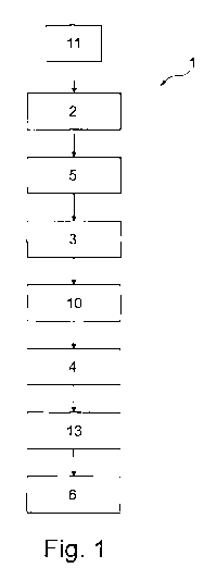
Fig. 1 shows a flow chart for carrying out a method according to the
invention,
Fig. 2 shows the course of remodeling on a bone,
Fig. 3 shows the position of an area to be treated on an exemplary skull and
Fig. 4 shows the mounting of bone fastening means, by type of an eye socket
implant and a maxilla implant.
1.0 The Figures are merely schematic and only serve for the comprehension
of the
invention. Like elements are provided with like reference numerals.
The invention is appropriate for use in the skull and face surgery, but it may
finally be
used on and/or for each osseous structure of a human being or a mammal.
In a method 1 according to the invention, there is a first step 2 of making
available
original 3D data of a bone or a bone portion of a specific patient to be
treated. This is
followed by a second step 3 in which involving of 3D data of a reference
patient who
has been selected according to predefined criteria takes place, namely the 3D
data
are gathered in a comparable region which is due to be treated. In a following
third
step 4 supplementing, possibly comprising trimming, of the 3D data combined of
step
2 and step 3 is performed, wherein combining of the data takes place in a
partial
step.
Between the first step 2 and the second step 3 also a mirroring step 5 may
take
place. In said mirroring step, 3D data which are opposed to the longitudinal
axis or a
plane of symmetry including the longitudinal axis of the body are gathered
from a
sound site on the ill (specific) patient to be treated and are superposed to
the 3D data
of the ill side to be treated. It is recommendable to make use of this step.
In a fourth step 6, also referred to as manufacturing step, a bone treatment
means 7
is manufactured for example by type of an implant or an osteotomy template.
Thus
also "virtual surgical planning" is possible. Such bone treatment means 7
which is
fastened to a bone 8 of a specific individual patient to be treated is shown
in Fig. 4.
CA 03002889 2018-04-23
9
Fig. 3 illustrates an area 9 to be treated on a skull including a bone 8.
While the eye
socket of said skull has a defect in the area of the region 9 to be treated on
the right
side when viewed from the patient, the eye socket has no defect on the left
side
when viewed from the patient.
The respective data of the sound side are transmitted to the defective site in
a
mirroring step 10 visualized in Fig. 1. They are morphed thereon/therein.
Preceding
the previous steps, there is a preparation step 11 in which the 3D data of the
patient
1.0 and/or the 3D data of one or more reference patients are entered into a
local or web-
based database and, resp., are gathered therefrom.
Fig. 2 depicts in which way, starting from a bone defect, landmarks are
created, then
an "adjustment" takes place in which a superposed form model is used which is
not
yet adapted to subsequently insert a statistical form model in a calculation
cut so as
to obtain an adapted model with a replaced bone defect. Markers 12 that form
the
"landmarks" are characterized by the reference numeral 12.
Hence, the point is that so far exclusively e.g. skull defects have been
reconstructed
in most cases by mirroring of the sound side to the defective side. This is
only
matching to a limited extent, however, or the results are not sufficient. In
the present
method, a plurality of skull models is evaluated to form a statistical model.
From the
statistical model the defective site now can be reconstructed on the defective
skull.
In the method 1 according to the invention a generation step 13 is used.
CA 03002889 2018-04-23
Reference numerals
1 method
5 2 first step (data for making available)
3 second step (involving a statistical model)
4 third step (supplementing plus trimming, where appropriate)
5 mirroring step
6 fourth step/manufacturing step
10 7 bone treatment means
8 bone
9 region to be treated
10 mirroring step
11 preparation step
12 marker
13 generation step