Note : Les descriptions sont présentées dans la langue officielle dans laquelle elles ont été soumises.
CA 03006055 2018-05-23
WO 2017/091662 PCT/US2016/063486
1
LARGE SCALE CELL MANUFACTURE SYSTEM
CROSS-REFERENCE TO RELATED APPLICATIONS
[0001] This application claims priority to U.S. Provisional Patent
Application No.
62/260,109 filed on November 25, 2015, the disclosure of which is hereby
expressly
incorporated by reference in its entirety.
BACKGROUND OF THE DISCLOSURE
[0002] The present disclosure relates generally to culturing and
manufacturing cells
in hollow hydrogel fibers made from alginate polymers. More particularly, the
present
disclosure relates to a manufacturing system and device for culturing cells at
various scales,
particularly on a large-scale level, the cells of which can be used for
various applications.
[0003] Mammalian cells have many applications. Stem cells, such as human
pluripotent stem cells (hPSCs), including human embryonic stem cells (hESCs)
and human
induced pluripotent stem cells (iPSCs), and their progenies (i.e., cells
differentiated from stem
cells) can be used for treating degenerative diseases, injuries and cancers.
They can also be used
to make artificial tissues and organs. In addition, stem cells and their
progenies can be used for
modeling diseases, screening drugs and testing efficacy and toxicity of
chemicals. Mammalian
cells are also widely used for expressing recombinant proteins and viruses
both in laboratories
and industry. Many of these proteins and viruses are used in clinics. These
applications require
large numbers of cells of high quality. For instance, -105 surviving
dopaminergic (DA) neurons,
-109 cardiomyocytes, or -10913 cells are required to treat a patient with
Parkinson's disease (PD),
myocardial infarction (MI), or Type 1 diabetes, respectively. Additionally,
far more cells are
needed initially because both in vitro cell culture yields and subsequent in
vivo survival of
transplanted cells are typically very low. As examples of the latter, only -6%
of transplanted
dopaminergic neurons or -1% of injected cardiomyocytes reportedly survived in
rodent models
several months after transplantation. Furthermore, there are large patient
populations with
degenerative diseases or organ failure, including over 1 million people with
PD, 1-2.5 million
with Type 1 diabetes, and -8 million with MI in the US alone. Large numbers of
cells are also
necessary for applications such as tissue engineering, where, for example, -
1019 hepatocytes or
cardiomyocytes would be required for an artificial human liver or heart,
respectively. Additionally,
-10b9 cells may be needed to screen a million-compound library once, and
advances in combinatorial
CA 03006055 2018-05-23
WO 2017/091662 PCT/US2016/063486
2
chemistry, noncoding RNAs, and investigations of complex signaling and
transcriptional networks
have given rise to large libraries that can be screened against many targets.
Large numbers of
mammalian cells, such as Chinese Hamster Ovary cells (CHO cells) and Human
Embryonic Kidney
293 cells (HEK293), are also needed for producing therapeutic biologics, such
as monoclonal
antibodies (mAbs), enzymes and viral particles.
[0004]
Currently, there are few methods that can cost-effectively manufacture stem
cells, and their progenies, and primary cells, especially in large scale. The
most widely used 2D
cell culture systems, in which cells are cultured on a 2D surface, are limited
by their low yield,
heterogeneity, scalability and reproducibility. For instance, only about
50,000 cardiomyocytes
can be cultured per cm2 of surface area.
[0005] Due
to the above drawbacks, three dimensional (3D) suspension cell culture
systems, such as spinner flasks and stirred-take bioreactors are being widely
studied to scale up
the production. However, cellular spheroids in suspension cultures frequently
aggregate to form
large cellular agglomerates. It is well known that the transport of nutrients,
oxygen and growth
factors to, and the metabolic waste from cells located at the center of
agglomerates (FIG. 10A)
with diameters larger than 500 um become insufficient, leading to slow cell
proliferation,
apoptosis, and uncontrolled differentiation. While stirring or shaking the
culture reduces cellular
agglomeration, they also generate hydrodynamic stress that negatively affects
cell viability,
proliferation and phenotype. High
cell density in the culture also promotes cellular
agglomeration. Considering all these factors, in current suspension culture
studies, cells are
generally seeded at low density (e.g., ¨3x10⁵ cells/mL) and stirred at 70 to
120 rotations-per-
minute (rpm). Under even these optimized conditions, slow cell growth,
significant cell death,
phenotype change, genomic mutations, and low volumetric yield are common. For
instance, it
has been shown that hPSCs typically expanded 4-fold per 4 days to yield around
2.0x10⁶
cells/mL. These cells merely occupy less than 0.4% of the bioreactor volume.
The low yield
leads to both economic and technical challenges for manufacturing large-scale
cells.
[0006] Based
on the foregoing, there is a need in the art for a robust cell culture
system that can cost-effectively manufacture different types of cells at
various scales,
particularly at large scale. This system would be useful in both research
laboratories and
industry.
CA 03006055 2018-05-23
WO 2017/091662 PCT/US2016/063486
3
BRIEF DESCRIPTION OF THE DISCLOSURE
[0007] The present disclosure is generally directed to culturing and
manufacturing
mammalian cells in hollow hydrogel fibers made from alginate polymers. More
particularly, the
present disclosure is directed to a culturing system and device capable of
manufacturing cells at
various scales, especially at large-scale levels, and to methods of using the
system and device for
culturing and manufacturing cells in hollow hydrogel fibers made from alginate
polymers.
[0008] It has been found that use of the hollow hydrogel fibers as a
cell culture
system promotes initial cellular clustering, ensures efficient mass transport
to cells and
eliminates hydrodynamic stress for cells, allows culturing cells with high
viability, high cell
growth rate and high volumetric yield (e.g. producing up to 5.0x10⁸ cells per
milliliter of
volume). These advantages dramatically reduce the bioreactor volume,
production time and cost.
Thus, this new culture system has potential to transform the cellular
manufacturing.
[0009] In one aspect, the present disclosure is directed to a method of
manufacturing
cells at various scales, the method comprising: suspending a cell solution
including cells in the
hollow space of alginate hydrogel fibers; and suspending the hollow fibers
including the cells in
cell culture medium; and culturing the cells.
[0010] In another aspect, the present disclosure is directed to a method
of
manufacturing cells at various scales, the method comprising: extruding a cell
solution and an
alginate solution into a cell compatible solution, the cell compatible
solution crosslinking the
alginate polymers within the alginate polymer solution to form hollow alginate
hydrogel fibers;
suspending the hollow fibers including the cells in cell culture medium or
cell compatible buffer;
and culturing the cells.
[0011] In another aspect, the present disclosure is directed to a system
for culturing
cells. The system comprising: a housing comprising a first inlet, a second
inlet, a core channel, a
shell channel, and an outlet, the first inlet operable for introducing a cell
solution into the core
channel, the second inlet operable for introducing an alginate solution into
the shell channel,
wherein the shell channel is in fluid contact with the core channel to allow
contact between the
cell solution and alginate solution, and a cell culture vessel in fluid
contact with the housing at
the outlet, wherein the cell culture vessel comprises a cell compatible
buffer.
CA 03006055 2018-05-23
WO 2017/091662 PCT/US2016/063486
4
[0012] In accordance with the present disclosure, methods have been
discovered that
surprisingly allow for culturing various types of cells on a large-scale
level. As used herein,
"large" or "large-scale" refers to a product of from about 107 to about 1030
cells, including from
about 107 to about 1015 cells, and including from about 107 to about 1012
cells. The methods and
manufacturing system of the present disclosure will have significant impact on
regenerative medicine
as they allow for sufficient, high quality and affordable cells. Further, the
system and methods
provide an advantageous impact on the biopharmaceutical industry by providing
more affordable
methods for manufacturing recombinant proteins and viruses.
BRIEF DESCRIPTION OF THE DRAWINGS
[0013] The disclosure will be better understood, and features, aspects
and advantages
other than those set forth above will become apparent when consideration is
given to the
following detailed description thereof. Such detailed description makes
reference to the
following drawings, wherein:
[0014] FIG. 1 is a schematic depicting a device of the present
disclosure for
processing hollow alginate hydrogel fibers.
[0015] FIG. 2 depicts an exemplary device of the present disclosure for
processing
hollow alginate hydrogel fibers.
[0016] FIGS. 3A-3E is a schematic depicting the method steps of the
present
disclosure for culturing cells within the hollow alginate hydrogel fibers.
FIG. 3A depicts cells
cultured in a medium-filled space of hollow alginate hydrogel fibers. The
fibers including the
cells are suspended in cell culture medium in cell culture vessels or
bioreactors. Cells are
expanded (FIG. 3B) and harvested (FIG. 3C) or differentiated (FIG. 3D) in the
hollow fibers.
Cells in the hollow fibers can also be used to produce recombinant proteins
and viruses (FIG.
3E).
[0017] FIG. 4 depicts hollow alginate hydrogel fibers including cells
suspended in a
cell culture medium as disclosed in the present disclosure.
[0018] FIGS. 5A-5C depict culturing stem cells in hollow hydrogel
fibers. H9
Human embryonic stem cells (FIG. 5A), induced human pluripotent stem cells:
MSC-iPSCs
(iPSCs made from human mesenchymal stem cells) (FIG. 5B) and Fib-iPSCs (iPSCs
made from
CA 03006055 2018-05-23
WO 2017/091662 PCT/US2016/063486
human fibroblasts) (FIG. 5C) are shown. Cells were cultured in the hollow
fibers for 8 days,
during which cells grew into large aggregates from single cells.
[0019] FIGS. 6A-6F depict human iPSCs differentiated into cortical
neurons (FIGS.
6A-6C) and dopaminergic neurons (FIGS. 6D-6F). FIGS. 6A and 6B depict phase
images of
cortical neurons within the hollow alginate hydrogel fibers at day 30. FIGS.
6D and 6E depict
phase images of dopaminergic neurons within the hollow fibers at day 30. FIGS.
6C and 6F
depict immunostaining at day 30 of human iPSCs differentiated into
corresponding neurons.
[0020] FIGS. 7A-7C depicts human glioblastoma stem cells cultured in
hollow
alginate hydrogel fibers. FIG. 7A depicts cell line LO cultured in the hollow
fibers over a period
of 7 days. FIG. 7B depicts cell line Li cultured in the hollow fibers over a
period of 7 days.
FIG. 7C depicts cell line L2 cultured in the hollow fibers over a period of 7
days. Cells grew
into large aggregates from single cells.
[0021] FIG. 8 depicts mouse L cells cultured in hollow alginate hydrogel
fibers for
producing recombinant Wnt 3A proteins. L cells stably expressing Wnt3A
proteins were
cultured in the hollow fibers for 6 days. Cells grew into high density
aggregates by day 6.
[0022] FIGS. 9A-9J depicts the hollow alginate hydrogel fiber cell
culture system
("cell culture system)" as analyzed in Example 5. FIGS. 9A & 9B show a home-
made micro-
extruder for processing one hollow fiber. A hyaluronic acid (HA) solution
containing single
cells and an alginate solution was pumped into the central and side channel of
the micro-
extruder, respectively, to form a coaxial core-shell flow that is extruded
into a 100 mM CaC12
buffer, which instantly crosslinks the alginates to form a hydrogel shell to
make one hollow
fiber. Subsequently, CaC12 buffer is replaced by cell culture medium and cells
are suspended and
grown in the core microspace of the hollow fiber. FIG. 9C depicts freshly
prepared hollow
fibers in the CaC12 buffer. FIGS. 9D-9F depict a micro-extruder with 9 nozzles
for
simultaneously processing 9 hollow fibers. FIG. 9G depicts that HAs are
required to process
defect-free hollow fibers. Without HAs (-HA), fibers with asymmetric shells or
beads are
formed. FIG. 9H is an illustration of a hollow alginate hydrogel fiber showing
cell growth in the
cell culture system. Within hours, single cells associate to form small cell
clusters (i.e. the initial
clustering). Subsequently, cells proliferate and the small cell clusters
expand as spheroids that
eventually merge to form a cylindrical cell mass. The diameter of the cell
mass is controlled to
be less than 500 um to ensure efficient mass transport in the cell mass. Two
vials of H9 hESCs,
CA 03006055 2018-05-23
WO 2017/091662 PCT/US2016/063486
6
stained with DIO and DID dyes appearing green and red fluorescence,
respectively, were mixed
at 1:1 and cultured in the cell culture system. Single cells (day 0), small
cell clusters (day 1), a
cylindrical cell mass (day 9) were clearly seen. FIG. 9J depicts ROCK
inhibitors (RIs) required
for the initial cell survival. Live/dead staining showed a majority of the
cells went apoptosis
after 24 hours without RIs (-RI). Cells survived and grew well with RIs (+RI).
Scale bar: (FIG.
9G, 91, and 9J) 200 um.
[0023] FIG. 10A depicts that in the current 3D suspension cultures (e.g.
spinner
flasks or stirred-tank bioreactors), single hPSCs associated to form small
cell clusters within 24
hours (i.e. the initial clustering phase) that subsequently expanded as
spheroids (i.e. the cell
expansion phase). Cells and spheroids frequently fused to each other to form
large agglomerates.
FIG. 10B confirms cellular agglomeration in experiment: two vials of H9 hESCs,
stained with
DIO and DID dyes respectively, were mixed at 1:1 and cultured in suspension.
The lipophilic
DIO and DID dyes stained cells to appear green and red, respectively, under
fluorescent
microscopy. Single cells (day 0), small clusters with both green and red cells
(day 1), spheroids
and agglomerates with both green and red cells (day 4) were clearly seen.
Scale bar: 100 um.
[0024] FIGS. 11A-11F depict the influence of alginate hydrogel
formulation on hPSC
culture in the cell culture system. H9 hESCs were cultured for 9 days in
hollow alginate
hydrogel fibers (inner diameter: ¨400 um; shell thickness: ¨40 um) processed
from 2% alginates
from Sigma (#A2033-100G) or Wako Chemicals with varied viscosity or molecular
weight
(500-600 cp; 300-400 cp and 80-120 cp). FIG. 11A depicts phase images showing
single H9s
on day 0 and H9 spheroids on day 4 in the hollow fibers. FIG. 11B depicts that
live/dead
staining revealed almost no dead cells in the hollow fibers. FIG. 11C depicts
Oct4 staining on
day 10 cells. H9s were released from hollow fibers on day 9 and plated on
Matrigel-coated plate
overnight before fixing and staining. Arrows point to the differentiated Oct4-
cells. FIGS. 11D
& 11E depict expansion fold and volumetric yield on day 5, 7 and 9. FIG. 11F
depicts the % of
Oct4+ cells after the 9-day culture. Error bars represent the standard
deviation (n=3). ***
indicates statistical significance at a level of p<0.001. Scale bar: (FIGS.
11A & 11B) 400 um;
(FIG. 11C) 50 um.
[0025] FIGS. 12A-12E depict the influence of alginate hydrogel
formulation on
hPSC culture in the cell culture system. H9s were cultured for 9 days in
hollow alginate
hydrogel fibers (inner diameter: ¨400 um; shell thickness: ¨40 um) processed
from 1%, 1.5% or
2% alginates from Wako Chemicals (80-120 cp). FIG. 12 A depicts phase images
of the day 0,
CA 03006055 2018-05-23
WO 2017/091662 PCT/US2016/063486
7
1 and 8 cells in hollow fibers. FIGS. 12B & 12C depict expansion fold and
volumetric yield on
day 5, 7 and 9. FIG. 12D depicts Oct4 staining on day 10 cells. H9s were
released from hollow
fibers on day 9 and plated on Matrigel-coated plate overnight before fixing
and staining. FIG.
12E depicts the % of Oct4+ cells after the 9-day culture. Error bars represent
the standard
deviation (n=3). Scale bar: (FIG. 12A) 400 um; (FIG. 12D) 50 um.
[0026] FIGS. 13A-13G depict the influence of hydrogel shell thickness on
hPSC
culture in the cell culture system. H9s were cultured for 9 days in hollow
alginate hydrogel
fibers with shell thickness of 30, 40, 70, or 90 um processed from 1.5%
alginates from Wako
Chemicals (80-120 cp). FIG. 13A gives the equation used to predict the shell
thickness based on
the volumetric flow rates of the cell solution and alginate solution and the
fiber outer diameter.
FIG. 13B depicts that the experimental shell thickness fit well with the
predicted data. FIG. 13C
depicts phase images of the cells in hollow fibers with varied shell thickness
on day 0. FIGS.
13D & 13E depict expansion fold and volumetric yield on day 5, 7 and 9. FIG.
13F depicts Oct4
staining on day 10 cells. H9s were released from hollow fibers on day 9 and
plated on Matrigel-
coated plate overnight before fixing and staining. FIG. 13G depicts the % of
Oct4+ cells after
the 9-day culture. Error bars represent the standard deviation (n=3). Scale
bar: (FIG. 13C) 200
um; (FIG. 13D) 50 um.
[0027] FIGS. 14A-14E depict the influence of hollow fiber inner diameter
on hPSC
culture in the cell culture system. H9s were cultured for 9 days in hollow
alginate hydrogel
fibers with inner diameter of 400, 250 or 120 um processed from 1.5% alginates
from Wako
Chemicals (80-120 cp). FIG. 14A depict phase images of the day 0, 1, 5 and 8
cells in hollow
fibers. FIGS. 14B & 14C depict expansion fold and volumetric yield on day 5, 7
and 9. FIG.
14D depicts Oct4 staining on day 10 cells. H9s were released from hollow
fibers on day 9 and
plated on Matrigel-coated plate overnight before fixing and staining. FIG. 14E
depicts the % of
Oct4+ cells after the 9-day culture. Error bars represent the standard
deviation (n=3). Scale bar:
(FIG. 14A) 400 pm; (FIG. 14D) 50 pm.
[0028] FIGS. 15A-15F depict the influence of the liquid core niche on
hPSC culture
in the cell culture system. H9s were cultured for 9 days in hollow alginate
hydrogel fibers
processed from 1.5% alginates from Wako Chemicals (80-120cp) with varied core
liquid
formulations including 3% methylcellulose (MC), 1% hyaluronic acid (HA), 2%
HA, 2% HA +
1 ug/mL fibronectin + 0.5 ug/mL laminin or 2% HA + StemBeads. FIG. 15A depicts
phase
CA 03006055 2018-05-23
WO 2017/091662 PCT/US2016/063486
8
images showing day 0 and day 3 cells. FIG. 15B depicts that live/dead staining
revealed almost
no dead cells in the hollow fibers. FIG. 15C depicts Oct4 staining on day 10
cells. H9s were
released from hollow fibers on day 9 and plated on Matrigel-coated plate
overnight before fixing
and staining. FIGS. 15D & 15E depict expansion fold and volumetric yield on
day 5, 7 and 9.
FIG. 15F depicts the % of Oct4+ cells after the 9-day culture. Error bars
represent the standard
deviation (n=3). Scale bar: (FIGS. 15A & 15B) 400 um; (FIG. 15C) 50 um.
[0029] FIGS. 16A-16E depict the influence of cell seeding density on
hPSC culture
in the cell culture system. H9s were cultured for 9 days in hollow alginate
hydrogel fibers
processed from 1.5% alginates from Wako Chemicals (80-120 cp). FIG. 16A
depicts phase
images of the cells in hollow fibers. After 24 hours, the cell clusters were
bigger at higher
seeding density, but the number of clusters were similar. FIG. 16B depicts
that the expansion
fold on day 5, 7 and 9 showed hPSCs grew faster at lower seeding density,
while the final
volumetric yields on day 9 were very close (FIG. 16C). FIG. 16D depicts Oct4
staining on day
cells. H9s were released from hollow fibers on day 9 and plated on Matrigel-
coated plate
overnight before fixing and staining. FIG. 16E depicts the % of Oct4+ cells
after the 9-day
culture. Error bars represent the standard deviation (n=3). *** indicates
statistical significance
at a level of p<0.001. Scale bar: (FIG. 16A) 400 um; (FIG. 16D) 50 um.
[0030] FIGS. 17A-17F depict culturing hPSCs in the cell culture system
with
ultralow seeding densities. H9s were seeded at 1.0x, 3.0x or 5.0x10⁵ cells/mL
in hollow alginate
hydrogel fibers processed from 1.5% alginates from Wako Chemicals (80-120 cp).
FIG. 17A
depicts phase images showing a few H9s grew into cylindrical cell mass in the
hollow fibers.
FIG. 17B depicts that live/dead staining revealed almost no dead cells. FIG.
17C are images
showing a single fiber with H9s on varied days along the culture. FIG. 17D
depicts that the final
volumetric yields were close at all seeding densities. FIG. 17E depicts Oct4
staining on day 10
cells. H9s were released from hollow fibers and plated on Matrigel-coated
plate overnight
before fixing and staining. FIG. 17F depicts the % of Oct4+ cells after 10-day
culture. Error
bars represent the standard deviation (n=3). Scale bar: (FIGS. 17A & 17B) 400
um; (FIG. 17E)
50 um.
[0031] FIGS. 18A-18D depict the passage 1 culturing of hPSCs in the cell
culture
system. H9s, MSC-iPSCs and Fib-iPSCs were cultured in hollow alginate hydrogel
fibers
processed from 1.5% alginates from Wako Chemicals (80-120 cp). FIG. 18A
depicts phase
images and live/dead staining of hPSCs in the cell culture system. FIGS. 18B &
18C depict
CA 03006055 2018-05-23
WO 2017/091662 PCT/US2016/063486
9
expansion fold and volumetric yield on day 5, 7 and 9. FIG. 18D depict that
the day 9 cell mass
was fixed and stained for the pluripotency markers: Nanog, Oct4, SSEA-4 and
alkaline
phosphatase (ALP). Images of varied slices of a cylindrical cell mass were
shown. Similar
results were obtained for MSC-iPSCs and Fib-iPSCs. Error bars represent the
standard deviation
(n=3). Scale bar: 400 um.
[0032] FIGS. 19A-19G depict long-term culturing of hPSCs in the cell
culture
system. H9s, Fib-iPSCs and MSC-iPSCs were cultured in hollow alginate hydrogel
fibers
processed from 1.5% alginates from Wako Chemicals (80-120 cp) for 10 passages.
FIG. 19A
depicts phase images of day 0, 3, and 5 cells in hollow fibers at passage 10.
FIG. 19B depicts
that live/dead staining revealed almost no dead cells in the hollow fibers at
passage 10. FIGS.
19C & 19D depict expansion fold and volumetric yield on day 5, 7 and 9 of
hPSCs at passage
10. FIG. 19E depicts the expression of the pluripotency markers: Nanog, Oct4,
SSEA-4 and
alkaline phosphatase (ALP) in the day-9 cell mass at passage 10. FIG. 19F
shows -95% of the
passage 10 cells expressed Oct4 and Nanog. FIG. 19G depicts that when seeded
at 1.0x10⁷
cells/mL, hPSCs consistently expanded -15-fold per passage per 5 days during
the long-term
culture. Error bars represent the standard deviation (n=3). Scale bar: (FIGS.
19A & 19B) 400
um; (FIG. 19E) 200 um.
[0033] FIGS. 20A-20F show that hPSCs retained pluripotency after long-
term
culturing in the cell culture system. H9s were cultured in hollow alginate
hydrogel fibers
processed from 1.5% alginates from Wako Chemicals (80-120 cp) for 10 passages.
Cells were
differentiated into the Nestin+ ectodermal, a-SMA+ mesodermal and FOXA2+
endodermal cells
in the embryoid assay (EB) assay (FIG. 20A), formed teratomas containing the
three germ layer
tissues (FIG. 20B) and had normal karyotypes (FIG. 20C). By further culturing
in a mesodermal
(FIG. 20D) or endodermal (FIG. 20E) or cardiomyocyte (FIG. 20F)
differentiation medium,
hPSCs in the hollow alginate hydrogel fibers could be differentiated into the
corresponding
Brachyury+ mesodermal cells or FOXA2+ endodermal cells or cTNT+ cardiomyocytes
at high
efficiency. Scale bar: (FIGS. 20A & 20B) 100 um; (FIGS. 20D-20F) 200 um.
[0034] FIGS. 21A-21F depict hPSCs retained pluripotency after long-term
culturing
in the cell culture system. MSC-iPSCs and Fib-iPSCs were cultured in hollow
alginate hydrogel
fibers processed from 1.5% alginates from Wako Chemicals (80-120 cp) for 10
passages. Both
cells were differentiated into the Nestin+ ectodermal, a-SMA+ mesodermal and
FOXA2+
endodermal cells in the EB assay (FIGS. 21A & 21B), formed teratomas
containing the three
CA 03006055 2018-05-23
WO 2017/091662 PCT/US2016/063486
germ layer tissues (FIGS. 21C & 21D) and had normal karyotypes (FIGS. 21E &
21F). Scale
bar: 100 um.
[0035] FIG. 22 depicts hPSCs retained pluripotency after long-term
culturing in the
cell culture system. H9s, MSC-iPSCs and Fib-iPSCs were cultured in hollow
fibers processed
from 1.5% alginates from Wako Chemicals (80-120 cp) for 10 passages. These
cells were
further cultured on Matrigel-coated plates. Images of the hPSC colonies
expressing the
pluripotency marker Oct4 after one passage on Matrigel-coated plates are
shown. Scale bar: 100
[0036] FIGS. 23A-23F depict a prototype bioreactor with the hollow
alginate
hydrogel fibers. FIG. 23A depicts hollow fibers with cells suspended in a
cylindrical container.
Medium was stored in a plastic bellow that could be pressed to flow the medium
into or released
to withdraw the medium from the container, respectively. FIG. 23B shows images
of the
mechanic stage for pressing and releasing the bellow; the controller that can
be programmed for
the pressing and releasing speed as well as the duration of the interval
between the pressing and
releasing; and the container and bellow. FIG. 23C is an image of the
cylindrical, white cell mass
in the bioreactor on day 10. FIG. 23D shows that 1.0x10⁹ cells were produced
with 2.0 mL
hollow fibers. FIGS. 23E & 23F show that these cells expressed the
pluripotency markers:
Nanog, Oct4, SSEA4 and ALP. Error bars represent the standard deviation (n=3).
Scale bar:
(FIG. 23C) 1 cm; (FIG. 23E) 200 um.
[0037] FIGS. 24A-24E depict culturing L-Wnt-3A-cells engineered to
express Wnt3a
proteins in the cell culture system. Cells were cultured in the cell culture
system with seeding
density at 1.0x or 2.0x10⁷ cells/mL. FIG. 24A depicts phase images of cells in
hollow fibers.
FIG. 24B depicts that live/dead staining revealed almost no dead cells. FIGS.
24C & 24D depict
expansion fold and volumetric yield on day 2 to 6. FIG. 24E shows that Wnt3a
proteins were
consistently expressed during a 16-day culture. Error bars represent the
standard deviation (n=3).
Scale bar: (FIGS. 24A & 24B) 400 um.
[0038] FIGS. 25A-25E depicts a prototype bioreactor for the cell culture
system.
Hollow fibers with cells were contained a closed cell culture chamber. Medium
was stored in a
flask and continuously perfused into the chamber. FIG. 25C is an image of the
cylindrical, white
cell mass in (harvested from the bioreactor on day 10) a 10 cm dish. FIGS. 25D
& 25E depicts
an extruder with 100 nozzles for simultaneously processing 100 hollow fibers.
CA 03006055 2018-05-23
WO 2017/091662 PCT/US2016/063486
11
[0039] While the disclosure is susceptible to various modifications and
alternative
forms, specific embodiments thereof have been shown by way of example in the
drawings and
are herein described below in detail. It should be understood, however, that
the description of
specific embodiments is not intended to limit the disclosure to cover all
modifications,
equivalents and alternatives falling within the spirit and scope of the
disclosure as defined by the
appended claims.
DETAILED DESCRIPTION OF THE DISCLOSURE
[0040] Unless defined otherwise, all technical and scientific terms used
herein have
the same meaning as commonly understood by one of ordinary skill in the art to
which the
disclosure belongs. Although any methods and materials similar to or
equivalent to those
described herein can be used in the practice or testing of the present
disclosure, the preferred
methods and materials are described below.
[0041] In accordance with the present disclosure, methods have been
discovered that
surprisingly allow for the culturing and manufacturing of cells on a large-
scale level.
Particularly, the present disclosure provides a manufacturing system and
device, and methods of
using the system and device for culturing and manufacturing cells in hollow
fibers made from
alginate polymers.
Methods of Manufacturing/Culturing Cells
[0042] The methods of the present disclosure may be used to culture and
manufacture
cells at various scales. The methods provide at least the following advantages
over conventional
cell culture methods: (1) allow for large-scale cell manufacture; (2) allow
for high density cell
culture, thereby reducing the space, labor, and materials of cell culture; (3)
allow for culturing
various types of cells; and (4) allow for manufacturing cells in a much
cheaper, more efficient
manner. Non-limiting examples of such cells that can be cultured and
manufactured using the
methods and systems described herein include mammalian cells, insert cells
(e.g., drosophila S2
cells), plant cells, yeast cells, and bacterial cells. While described more
fully using mammalian
cells, especially human pluripotent stem cells, it should be recognized that
the methods and
systems described herein can be used with any of the above-listed types of
cells without
departing from the scope of the present disclosure.
CA 03006055 2018-05-23
WO 2017/091662 PCT/US2016/063486
12
[0043] As used herein, "mammalian cells" refer to cells derived from
both humans
and animals. Particularly suitable mammalian cells for use in the methods and
systems of the
present disclosure include, mammalian embryonic stem cells, mammalian induced
pluripotent
stem cells, mammalian naive pluripotent stem cells, cells differentiated from
mammalian
embryonic stem cells, mammalian induced pluripotent stem cells and mammalian
naive
pluripotent stem cells, mammalian cells reprogrammed from other cell types
(e.g. human
neurons reprogrammed from human fibroblasts), mammalian primary cells (e.g.,
human
umbilical vein endothelial cells, cancer cells, T cells), mammalian tissue
stem cells (e.g.,
mesenchymal stem cells, fetal neural stem cells), mammalian cell lines (e.g.,
human embryonic
kidney (HEK293) cells, Chinese hamster ovary (CHO) cells).
[0044] In general, the method of the present disclosure includes:
suspending cells in a
liquid medium-filled space within hollow hydrogel fibers; suspending the
hollow fibers in a cell
culture medium to allow expansion and/or differentiation of the cells; and
harvesting the cells.
[0045] The cells are suspended in a cell culture medium or cell
compatible buffer to
form a cell solution. The cell culture medium is cell type dependent.
Suitably, cells are
suspended in medium at concentrations varying from 1 to a few billion cells
per cubic milliliter.
[0046] The hollow fibers are prepared from alginate polymer material.
Suitable
alginate polymer material for use in preparing the fibers include any
commercially available or
home-purified alginate polymer, such as alginate acid or sodium alginate from
Sigma
(+W201502), and modified alginate polymers, such as methacrylate modified
alginate, and
combinations thereof. As used herein, "combinations thereof refer to mixtures
of the polymers
as well as polymer blends. Further, in some embodiments, other polymers such
as hyaluronic
acids can be blended or incorporated into the alginate polymers to dope the
alginate hydrogel.
To form the fibers, alginate polymers are first dissolved in water or cell
compatible buffer to
form alginate solutions including from about 0.01% (w/v) to about 20% (w/v)
alginate. In
particularly suitable aspects, the fibers are then prepared and filled with
cells using an extruder.
Extrusion conditions will be those known in the art suitable for the
particular cell survival and
growth.
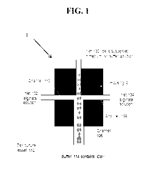
[0047] By way of example, as shown in FIGS. 1 and 2, a cell solution
including cells
is supplied via a first inlet 100 and the alginate solutions are supplied via
at least a second inlet
(shown in FIG. 1 as inlets 102, 104). Both the first stream including the cell
solution and the
CA 03006055 2018-05-23
WO 2017/091662 PCT/US2016/063486
13
second stream including the alginate solution are extruded into a cell
compatible solution
containing calcium ions or other ions or chemicals, such as barium ions, that
can crosslink the
alginate polymers in the alginate solution. The cell compatible solution
allows the alginate
polymers to instantly crosslink, thereby gelling the alginate solution and
forming the hollow
fibers. Typically, the fibers are sufficiently crosslinked in a time period of
from about one
minute to about 30 minutes.
[0048] Typically, as formed, the hollow fibers will be sized for the
particular cells
and amount of cell expansion desired. The fibers can have a length typically
ranging from
millimeters to meters. Additionally, the outer and inner diameters of the
hollow hydrogel fibers
can vary from micrometers to millimeters.
[0049] Once sufficiently crosslinked to form hollow fibers, the cell
compatible
solution is removed and cell culture medium is added to culture the cells now
within the
crosslinked hollow alginate hydrogel fibers. In some aspects, the fibers,
including cells, are
suspended in cell culture medium in cell culture vessels or bioreactors. The
cell culture medium
can be any medium known in the cell culture art that is suitable for
supporting cell survival,
growth and differentiation. Typically, the cell culture medium will include,
but is not limited to,
a carbon source, a nitrogen source, and growth factors. The specific cell
culture medium for use
in culturing the cells within the crosslinked hollow alginate hydrogel fibers
will depend on the
cell type to be cultured.
[0050] Cell culture conditions will vary depending on the type of cell,
the amount of
cell expansion, and the number of cells desired. Once sufficient cell
expansion and desired
numbers of cells are reached, the cells can be passaged and seeded into new
crosslinked hollow
alginate hydrogel fibers for a subsequent round of growth and expansion.
Alternatively, the
expanded cells can be differentiated into the final desired cell type within
the hollow space.
[0051] Cells are finally released from the hollow space of the fiber by
dissolving the
fiber chemically or physically. In one aspect, the fiber is dissolved using a
chemical dissolvent
such as ethylenediaminetetraacetic acid (EDTA), ethylene glycol tetraacetic
acid (EGTA), or an
alginate lyase solution (available from Sigma-Aldrich). In another aspect, the
fiber is dissolved
using a mechanical force. The duration of the cells within the hollow fiber
can typically vary
from days to months.
CA 03006055 2018-05-23
WO 2017/091662 PCT/US2016/063486
14
[0052] The cells are useful in both research laboratories and industry.
Small scale
and large scale of cells can be manufactured with the system for laboratorial
and industrial
applications, respectively. Cells can be efficiently and effectively prepared
in size and number
for use in degenerative disease and injury treatment, drug screening, for
expressing proteins and
the like. Additionally, the cells can be used to manufacture proteins and
vaccines. In yet other
aspects, the cells can be used for tissue engineering.
System/Device for Processing Alginate Hollow Fibers
[0053] In another aspect, the present disclosure is directed to a device
for processing
hollow fibers from alginate polymers with cells suspended in the hollow space.
Generally,
referring to FIG. 1, the device 1 includes a housing 2 including a core
channel 106 running down
the center of the housing 2. The core channel connects to the first inlet 100
for introducing cells
into the housing 2. The housing 2 of the device 1 further includes shell
channels 108, 110 for
flowing alginate solution introduced through the second inlets 104, 102 into
the housing 2.
Although shown with two shell channels, it should be understood that the
housing may include
less or more shell channels, such as a single shell channel, or three, four,
five or more shell
channels, without departing from the scope of the present disclosure. In some
particularly
suitable embodiments, pumps (not shown) are included at the inlets 100, 102,
104 for pushing
streams of cells and alginate solution into the housing 2 of the device 1.
[0054] The outlet of channel 106 of the device 1 is in contact with a
cell culture
vessel or bioreactor 112 including cell compatible buffer to form a system
including the housing
2 and the cell culture vessel or bioreactor 112. The vessel 112 includes a
buffer 114 as described
above including calcium ions or other ions or chemicals that can crosslink the
alginate polymers
within the alginate solution to gel the solution to form the fibers.
[0055] The disclosure will be more fully understood upon consideration
of the
following non-limiting Examples.
EXAMPLES
[0056] Unless otherwise indicated, the hollow fibers were prepared as
described
above.
CA 03006055 2018-05-23
WO 2017/091662 PCT/US2016/063486
EXAMPLE 1
[0057] In this Example, expansion and growth of human pluripotent stem
cells,
including human embryonic stem cells (hESCs) and human induced pluripotent
stem cells
(human iPSCs) in hollow fibers were analyzed over 8 days.
[0058] Single human embryonic stem cells (H9, WiCell) (FIG. 5A) or
induced
human pluripotent stem cells reprogrammed from human mesenchymal stem cells
(MSC-iPSCs)
(FIG. 5B) or from human skin fibroblasts (Fib-iPSCs) (FIG. 5C) were suspended
in Essential 8
Medium (Life Technology) containing 0.5% (w/v) hyaluronic acid (Lifecore
Biomedical) at a
density of 1x10⁶ cells/ml. Sodium alginate was dissolved in 0.9% (w/v) saline
to reach a
concentration of 1.2% (w/v) alginate and autoclaved. With an extruder (see
e.g., FIGS. 1 and 2),
10 ml of cell solution and 10 ml of alginate solution were extruded into the
100 ml of sterile
buffer containing 100 mM CaC12 at room temperature to form alginate hollow
fibers with cells
suspended in the hollow space. The fibers were crosslinked in the CaC12
solution for 5 minutes
at room temperature. The CaC12 solution was removed and replaced with
Essential 8 Medium.
Cells were cultured in the hollow fibers suspended in the medium in a regular
cell culture
incubator at 37 C, with 5% CO2, 95% air at 1 atm for 8 days. Single cells grew
into cell
aggregates. To harvest cells, the Essential 8 Medium was removed and replaced
with PBS
containing 100 mM EDTA (Sigma) or 40 mg/ml alginate lyase (Sigma) at 37 C for
10 minutes.
The alginate hydrogel fibers were dissolved and cells were harvested. These
cell aggregates can
be dissociated into single cells by treating them with Accutase (Life
Technology) at 37 C for 10
minutes. Cells can be processed into the alginate hollow fibers for a second
round of expansion.
As shown in FIGS. 5A-5C, the cells grew into large aggregates from single
cells effectively
using the hollow fibers.
EXAMPLE 2
[0059] In this Example, hollow alginate fibers including human iPSCs as
made in
Example 1 were differentiated into cortical neurons and dopaminergic neurons
within the fibers.
[0060] Human MSC-iPSCs were allowed to expand in the hollow fibers for 5
days.
The Essential 8 Medium was then replaced with home-made and chemically defined
neuronal
differentiation mediums and then differentiated into cortical and dopaminergic
neurons within
the alginate hollow fibers for 30 days. Results are shown in FIGS. 6A-6F. As
shown in FIGS.
CA 03006055 2018-05-23
WO 2017/091662 PCT/US2016/063486
16
6C and 6F, immunostaining on day 30 indicated that the majority of human iPSCs
were
differentiated into corresponding neurons.
EXAMPLE 3
[0061] In this Example, human glioblastoma stem cells were cultured in
hollow
fibers.
[0062] Three cancer stem cell lines, LO, Li and L2, isolated from human
glioblastoma were cultured in the hollow fibers. Single cells were suspended
in NeuroCult
medium (Stem Cell Technology) containing 0.8% (w/v) hyaluronic acid (Lifecore
Biomedical)
at a density of 0.5x10⁶ cells/ml. Sodium alginate was dissolved in 0.9% (w/v)
saline to reach a
concentration of 1.5% (w/v) alginate and autoclaved. With an extruder (see
e.g., FIGS. 1 and 2),
ml of cell solution and 10 ml of alginate solution were extruded into the 100
ml of sterile
buffer containing 100 mM CaC12 at room temperature to form alginate hollow
fibers with cells
suspended in the hollow space. The fibers were crosslinked in the CaC12
solution for 10 minutes
at room temperature. The CaC12 solution was removed and replaced with
NeuroCult medium.
Cells were cultured in the hollow fibers suspended in the medium in a regular
cell culture
incubator at 37 C, with 5% CO2 and 95% air at 1 atm for 7 days. Single cells
grew into
aggregates. To harvest cells, the NeuroCult Medium was removed and replaced
with PBS
containing 40 mg/ml alginate lyase (Sigma-Aldrich) at 37 C for 10 minutes. The
alginate fibers
were dissolved and cell aggregates were harvested. These aggregates can be
dissociated into
single cells by treating them with 0.05% trypsin (Life Technology) at 37 C for
10 minutes. Cells
can be processed into the alginate hollow fibers for a second round of
expansion. The cells grew
into large aggregates from single cells (see FIGS. 7A-7C).
EXAMPLE 4
[0063] In this Example, mouse L cells engineered to express Wnt 3A
proteins were
cultured for producing recombinant proteins in hollow fibers.
[0064] Mouse L cells stably expressing Wnt 3A proteins (ATCCO CRL-2647)
were
cultured in the hollow fibers for 20 days. Single cells were suspended in DMEM
medium (Stem
Cell Technology) containing 0.8% (w/v) hyaluronic acid (Lifecore Biomedical)
at a density of
1x10⁶ cells/ml. Sodium alginate was dissolved in 0.9% (w/v) saline to reach a
concentration of
1.2% (w/v) alginate and autoclaved. With an extruder (see FIGS. 1 and 2), 20
ml of cell solution
CA 03006055 2018-05-23
WO 2017/091662 PCT/US2016/063486
17
and 20 ml of alginate solution were extruded into the 200 ml of sterile buffer
containing 100 mM
CaC12 at room temperature to form alginate hollow fibers with cells suspended
in the hollow
space. The fibers were crosslinked in the CaC12 solution for 10 minutes at
room temperature.
The CaC12 solution was removed and replaced with DMEM medium containing 10%
FBS
(Atlanta Biologicals). Cells were cultured in the hollow fibers suspended in
the medium in a
regular cell culture incubator at 37 C, with 5% CO2 and 95% air at 1 atm for
20 days. Cells grew
into high density aggregates by day 6 (see FIG. 8).
EXAMPLE 5
[0065] In
this Example, various cells were suspended and grown in hollow alginate
hydrogel fibers (also referred to as the cell culture system or culture
system).
Materials and Methods
[0066]
Materials: Fib-iPSCs (iPSCs reprogrammed from human dermal fibroblasts)
and MSC-iPSCs (iPSCs reprogrammed from human mesenchymal stem cells) were
obtained
from George Q. Daley laboratory (Children's Hospital Boston, Boston). H9 hESCs
were
purchased from WiCell Research Institute. L Wnt-3A cells (ATCCO CRL2647TM)
were
acquired form ATCC. Reagents and their supplies: E8 medium (E8), Accutase and
Live/Dead
cell viability staining kit: Life Technologies; Y-27632: Selleckchem;
Matrigel: D Biosciences;
Sodium Hyaluronate (HA 700K-1): Lifecore Biomedical. Sodium alginates (500-600
cp;
300-400 cp and 80-120cp): Wako Chemicals. Sodium alginate (A2033-100G): Sigma.
Vybrant
cell-labeling solutions: Molecular Probes, Inc. DMEM: GE Healthcare Life
Sciences; FBS:
Atlanta biologicals; G418: Sigma.
Antibodies and their supplies: Oct4 (Santa Cruz
Biotechnology; 1:100); FOXA2 (Santa Cruz Biotechnology; 1:200); a-SMA (Abcam;
1:200);
Nestin (Millipore; 1:200). Nanog (10 mg/mL), Oct4 (10 mg/mL), SSEA-4 (10
mg/mL) and
alkaline phosphatase (10 mg/mL) and Brachyury (10 mg/mL) (R&D systems, Inc.).
Syringe
pump (New Era Pump System, Inc.); Disposable syringes (Henke sass wolf); Clear
acrylic
rectangular bar, steel tubes and plastics tubes (McMaster); Calcium chloride
(Acros Orcanics);
Sodium Chloride (Fisher scientific). Mechanical stage and controller (CESCO);
Bellows bottles
(Spectrum Chemical Mfg. Corp.); Luciferase assay kit (Biovision, K801-200).
[0067]
Processing alginate hollow fibers: a home-made micro-extruder was used to
process alginate hollow fibers. A hyaluronic acid (HA) solution containing
single cells and an
CA 03006055 2018-05-23
WO 2017/091662 PCT/US2016/063486
18
alginate solution was pumped into the central and side channel of the home-
made micro-
extruder, respectively, and extruded into a CaC12 buffer (100 mM) to make
hollow fibers.
Subsequently, CaC12 buffer was replaced by cell culture medium.
[0068] Culturing hPSCs in the hollow alginate hydrogel fibers: for a
typical cell
culture, 20 uL cell solution in alginate hollow fibers were suspended in 2 mL
E8 medium in a 6-
well plate and cultured in an incubator with 5% CO2, 21% 02 at 37 C. Medium
was changed
daily. To passage cells, medium was removed and alginate hydrogels were
dissolved with 0.5
mM EDTA for 5 minutes. Cell mass was collected by centrifuging at 100 g for 5
minutes,
treated with Accutase at 37 C for 12 minutes and dissociated into single cells
for following
culture.
[0069] Culturing L-Wnt3A-cells in the hollow alginate hydrogel fibers:
for a typical
cell culture, 20 uL cell solution in alginate hollow fibers were suspended in
2 mL DMEM
medium plus 10% FBS and 0.4 mg/mL G418 in a 6-well plate and cultured in an
incubator with
5% CO2, 21% 02 at 37 C. Medium was changed daily and collected for
quantifying Wnt3A
proteins. To quantify Wnt3A proteins, MDA-468 cells (ATCCO HTB-132Tm) were
stably
transfected with a luciferase reporter for the canonical Wnt signaling
(Addgene, #24308). These
MDA-468-TFP cells were plated in a 96-well plate (5000 cells/well/200 mL
medium). 24 hours
later, 150 mL fresh DMEM plus 10% FBS and 50 mL L-Wnt3A-cells conditioned
medium was
added and incubated for another 18 hours. Medium was then removed and cells
were washed
with PBS once before 200 mL cell lysis buffer was added and incubated for 10
minutes at room
temperature. 50 mL cell lysates, 50 mL substrate A and 50 mL substrate B from
the luciferase
assay kit were mixed and the light signals were immediately read with a
luminometer. The
quantity of Wnt3a protein was calculated with a standard curve.
[0070] Culturing hPSCs in the hollow alginate hydrogel fibers with
bioreactors: 2.0
mL cell solution in hollow fibers was suspended in a home-made bioreactor.
Cells were cultured
in an incubator with 5% CO2, 21% 02 at 37 C for 10 days. For bioreactor 1,
medium was stored
in a flask and continuously perfused into the bioreactor. For bioreactor 2,
medium was stored in
a bellow that was periodically pressed to flow the medium into or released to
withdraw the
medium from the container.
[0071] Staining and imaging: Cells cultured on 2D surfaces were fixed
with 4%
paraformaldehyde (PFA) at room temperature for 15 minutes, permeabilized with
0.25% Triton
CA 03006055 2018-05-23
WO 2017/091662 PCT/US2016/063486
19
X-100 for 15 minutes, and blocked with 5% donkey serum for 1 hour. Cells were
then incubated
with primary antibodies at 4 C overnight. After extensive washing, secondary
antibodies and
4',6-Diamidino-2-Phenylindole, Dihydrochloride (DAPI) were added and incubated
for another 1
hour at room temperature. Cells were washed with PBS for 3 times before
imaging with a Zeiss
Axio Observer Fluorescent Microscopy. To assess the pluripotency of cells,
hPSCs were plated
onto the Matrigel-coated plate overnight before fixation and staining. The
percentage of Oct4+
or Nanog+ nuclei was quantified with Image J software. At least 1000 nuclei
were analyzed. To
stain 3D cylindrical cell mass, the cell mass was harvested and fixed with 4%
PFA at room
temperature for 30 minutes, then incubated with PBS + 0.25% Triton X-100 + 5%
goat serum +
primary antibodies at 4 C for 48 hours. After extensive washing, secondary
antibodies in 2%
BSA were added and incubated at 4 C for 24 hours. Cells were washed with PBS
for 3 times
before imaging with Nikon Al Confocal Microscopy. LIVE/DEADO Cell Viability
staining was
used to assess live and dead cells, according to the product manual.
[0072] Embryoid body (EB) differentiation: hPSCs released from the
hollow alginate
hydrogel fibers were suspended in DMEM + 20% FBS + 10 uM 0-mercaptoethanol in
a low
adhesion plate for 6 days. The cell mass was then transferred onto plates
coated with 0.1%
gelatin and cultured in the same medium for another 6 days, followed by
fixation and staining as
above.
[0073] Teratoma formation in vivo: all animal protocols were approved by
the
Institutional Animal Care and Use Committee of the University of Nebraska-
Lincoln. All
experimental procedures involving animals were performed in accordance with
the guidelines of
the Institutional Animal Care and Use Committee of the University of Nebraska-
Lincoln. 2x10⁶
hPSCs were suspended in 25 uL PBS plus 25 uL Matrigel and injected
subcutaneously at the
back of the neck of the NOD-SCID mice (Charles River Laboratory). Tumors were
harvested
after 6-12 weeks. The tumors were fixed with 4% PFA for 48 hours and
sequentially dehydrated
with 70%, 95%, and 100% ethanol, and defatted with xylene for 2 hours before
embedding in
paraffin. Then 10 um thick sections were cut and stained with hematoxylin and
eosin.
[0074] Karyotype: Karyotyping was performed by WiCell Research
Institute.
[0075] Mesodermal induction: H9 cells in hollow fibers were cultured in
E8 medium
for 7 days, then in DMEM/F12 medium with 1% B27 minus insulin and 12 mM
CHIR99021 for
24 hours before fixation and staining.
CA 03006055 2018-05-23
WO 2017/091662 PCT/US2016/063486
[0076] Endodermal induction: H9 cells in hollow fibers were cultured in
E8 medium
for 7 days, then in RPMI 1640 medium with 1% GlutaMAX, 1% B27 minus insulin, 4
mM
CHIR99021 for 24 hours and in RPMI 1640 medium with 1% GlutaMAX, 1% B27 minus
insulin for additional 24 hours before fixation and staining.
[0077] Cardiomyocyte differentiation: H9 cells in hollow fibers were
cultured in E8
medium for 7 days, then in DMEM/F12 with 1% B27-insulin between for 6 days,
and
DMEM/F12 with 1% B27 for 9 days. The following small molecules were added
during the
differentiation: 12 mM CHIR99021 for days 0-1; 5 mM IWR1 for days 3-4. Cell
mass were
released on day 11 to gelatin coated plate. Beating cardiomyocytes were filmed
on day 15. Some
samples were fixed on day 11 for cTNT immunostaining.
[0078] Statistical analysis: Statistical analyses were done using the
statistical package
Instat (GraphPad Software, La Jolla, CA).
Results
[0079] A micro-extruder was made for processing hollow fibers with
alginate
hydrogels (FIGS. 9A & 9B). The extruder could have one or multiple nozzles for
simultaneously processing one or multiple hollow fibers (FIGS. 9A-9F). It was
found that the
viscosity of the cell solution and alginate solution should be close to
process defect-free hollow
fibers. Both hyaluronic acid (HA) and methylcellulose (MC) solutions could be
used to suspend
the cells for this purpose. Without HAs or MCs, hollow fibers with asymmetric
shells or beads
were frequently formed (FIG. 9G). Both defects could lead to cell culture
failure. Similar to
hPSCs in suspension cultures (FIGS. 10A & 10B). hPSCs in the hollow fiber cell
culture system
grew through an initial clustering phase and a subsequent cell expansion phase
(FIGS. 9H & 91)
and the ROCK inhibitor Y-27632 was required for the initial survival of the
dissociated hPSCs
(FIG. 9J).
[0080] It was found that the proliferation and pluripotency of hPSCs in
the fibers
were significantly influenced by the alginate hydrogel formulation. For
instance, when cultured
in hollow fibers processed from 2% alginates from Sigma (#A2033-100G) and Wako
Chemicals
(500-600 cp; 300-400 cp and 80-120cp) for 9 days, hPSCs expanded 27-, 51-, 51-
and 49-fold
to yield 2.7x, 5.1x, 5.1x and 4.9x10⁸ cells/mL with 47%, 76%, 80% and 89% of
the final cells
expressing the pluripotency marker Oct4, respectively (FIGS. 11A-11F).
Live/dead cell staining
CA 03006055 2018-05-23
WO 2017/091662 PCT/US2016/063486
21
revealed almost no cell death for all the cultures (FIG. 11B). Compared with
the alginate type,
the influence of alginate concentration in the range of 1.0% to 2.0% was much
less. For
instance, there was no significant difference in cell proliferation and
pluripotency for hPSCs
cultured for 9 days in hollow fibers processed with 1.0%, 1.5% and 2.0% Wako
Chemicals 80-
120 cp alginates (FIGS. 12A-12E). It was concluded that 1.5% Wako Chemicals 80-
120 cp
alginate hydrogel was appropriate for culturing hPSCs in the hollow alginate
hydrogel fiber cell
culture system.
[0081] The fiber geometry also influenced hPSC culture in the cell
culture system.
At a given fiber outer diameter, the hydrogel shell thickness can be
controlled by varying the
ratio of the cell solution and alginate solution flow rate and can be
predicted with the Equation
described in FIGS. 13A & 13B. The fiber outer diameter is roughly equal to the
inner diameter
of the extruder nozzle. When cultured in hollow fibers with 30, 40, 70 and 90
um shells for 9
days, 94%, 92%, 85% and 80% of the final cells retained the pluripotency
marker Oct4. There
was no significant difference in cell viability and expansion between the
different conditions
(FIGS. 13C-13E). When cultured in hollow fibers with inner diameter of 400 um,
250 um and
120 um for 9 days, 95% of the cells retained the Oct4 marker (FIGS. 14-14E).
It was concluded
that hollow fibers with shell thicknesses <70 um and inner diameters <400 um
were appropriate
for culturing hPSCs in the hollow alginate hydrogel fiber cell culture system.
[0082] Research showed adding extracellular matrix proteins such as
fibronectins and
laminins enhanced hPSC culture efficiency in suspension cultures. The results
of the instant
Example showed these proteins at the tested concentrations did not improve the
cell viability,
growth rate and pluripotency and were unnecessary with the cell culture system
(FIGS. 15A-
15F). Since both HAs and MCs could be used to cells, it was further analyzed
whether they
differentially influenced the cell culture. The results showed 1% HAs, 2% HAs
and 3% MCs
resulted in similar cell viability, expansion and pluripotency (FIGS. 15A-
15F). A main concern
with culturing hPSCs in alginate hollow fibers is that the large protein
factors (e.g. bFGFs,
insulins and transferrins) in the medium might not efficiently travel through
the hydrogel shell
and cell mass to feed the cells. When Poly Lactic-co-Glycolic Acid (PLGA)
microspheres
(StemBeads) containing and slowly releasing bFGFs were added to the liquid
core of the hollow
fibers, the cell viability, expansion and pluripotency were not improved,
indicating the transport
of proteins in the new culture system was efficient and sufficient (FIGS. 15A-
15F).
CA 03006055 2018-05-23
WO 2017/091662 PCT/US2016/063486
22
[0083] The influence of cell seeding density on hPSC culture in the cell
culture
system was also investigated. When seeded at 1.0x10⁶, 2.0x10⁶, 5.0x10⁶,
10.0x10⁶ cells/mL,
hPSCs expanded 433-, 196-, 104- and 46-fold on day 9, respectively, yielding
around 5.0x10⁸
cells/mL (FIG. 16B). For all conditions, cells grew through the aforementioned
two phases
(FIG. 16A). At 24 hours, the cell cluster size was larger for higher seeding
density, but the
number of cell cluster per volume was not significantly affected by the
seeding density (FIG.
16A, day 1, insert). These results showed hPSCs grew faster at lower seeding
density. However,
the seeding density did not influence the pluripotency (FIGS. 16D & 16E). It
was extremely
exciting that hPSCs seeded at ultralow densities could grow as well without
sacrificing cell
viability and pluripotency. When seeded at 1.0x, 3.0x and 5.0x10⁵ cells/mL,
hPSCs expanded
4000-, 1666- and 1000-fold to yield ¨4.2x, 5.1x, 4.8x10⁸ cells/mL on day 14,
12 and 10
respectively (FIG. 17D).
[0084] After optimization, the cell culture system was evaluated for
culturing
multiple hPSC lines for long term. All hPSCs grew well in the cell culture
system and there
were no significant difference in cell morphology, viability, growth rate and
pluripotency
between the hPSC lines (FIGS. 18A-18D). During a 10-passage culture in the
cell culture
system, when seeded at 1.0x10⁷ cells/mL, hPSCs consistently expanded ¨15-fold
per passage
per 5 days and >95% of the cells expressed Oct4 (FIG. 19G). The long-term
culture in the cell
culture system did not alter the cell phenotype as shown by the similar
morphology, viability,
growth kinetics and pluripotency to hPSCs at passage 1 (FIGS. 18A-18D and
FIGS. 19A-19G).
In vitro embryoid body (EB) differentiation and in vivo teratoma formation
confirmed their
pluripotency after the long-term culture. All hPSCs were successfully
differentiated into
FOXA2+ endodermal, a-SMA+ mesodermal and Nestin+ ectodermal cells in the EB
assay
(FIGS. 20A and 21A & 21 B). All hPSCs formed teratomas containing endodermal,
mesodermal
and ectodermal tissues when transplanted to immune-deficient mice (FIGS. 20B
and 21C &
21D). In addition, after the long-term culture, all hPSCs retained normal
karyotypes (FIGS. 20C
and 21E & 21F) and could be cryopreserved or further cultured on Matrigel-
coated 2D surface
(FIG. 22). After expansion and further culturing in a mesodermal or endodermal
or a
cardiomyocyte differentiation medium, hPSCs in the hollow fibers could be
differentiated to the
corresponding mesodermal cells or endodermal cells or cardiomyocytes at high
efficiency,
indicating the cell culture system supported hPSC differentiation (FIGS. 23D-
23F).
CA 03006055 2018-05-23
WO 2017/091662 PCT/US2016/063486
23
[0085] The culture system could be used to culture cells other than
hPSCs. For
instance, the murine L cells engineered to express Wnt3a proteins could be
efficiently cultured
without notable cell death, yielding around 6.0x10⁸ cells/mL. Importantly,
these cells
consistently expressed Wnt3a proteins during a 16-day culture at level similar
to this expressed
by L cells cultured in 2D dishes (FIGS. 24A-24E). This results demonstrated
the potential of the
hollow hydrogel fibers as a generally applicable system for culturing cells.
[0086] Two prototype bioreactors were designed and built for the cell
culture system.
Hollow fibers with cells were processed into a cylindrical container. In
Bioreactor 1, medium
stored in a flask was continuously perfused into the container (FIGS. 25A-
25C). In Bioreactor 2,
medium was stored in a plastic bellow that could be pressed to flow the medium
into or released
to withdraw the medium from the container, respectively (FIGS. 23A-23F). hPSCs
in both
bioreactors grew well and yielded ¨5.0x10⁸ cell/mL on day 10. >95% of cells
expressed the
pluripotency markers. These prototype bioreactors could be scaled up in the
future. To scale up
the processing of hollow alginate hydrogel fibers, an extruder was also made
with 100 nozzles
that could process 1 liter hollow fibers within 30 minutes (FIGS. 25D & 25E).
[0087] These results demonstrated that the methods and devices of the
present
disclosure can be used to culture and manufacture cells in hollow alginate
hydrogel fibers. It is
contemplated that the methods may be useful in both research laboratories and
industry for
preparing sufficient and high quality cells for disease and injury treatments,
screening libraries
for drugs, and manufacturing proteins and vaccines.