Note : Les descriptions sont présentées dans la langue officielle dans laquelle elles ont été soumises.
CA 03006302 2018-05-24
WO 2017/105993
PCMJS2016/065542
Method of Ex Vivo Enhancement of Immune Cell
Activity for Cancer Immunotherapy with a
Small Molecule Ablative Compound
TECHNICAL FIELD
The present invention relates to a method
of enhancing immune cell activity with intralesional
injection of a small molecule halogenated xanthene
tumor-ablative compound to induce immune system
components within the peripheral blood, tumor tissue
or lymphoid tissue of a mammal. Members of the
induced immune system components are collected,
expanded ex vivo and then reintroduced into the
mammal from which they originated as an enriched
tumor-specific immune anticancer agent composition.
BACKGROUND OF THE INVENTION
Immunotherapeutic strategies incorporating
intralesional (IL) therapy to elicit tumor specific
immune responses can serve as a non-surgical option
for cutaneous neoplasms. These strategies haven been
shown to induce both local and systemic tumor
regressions. Intratumoral injection of dendritic
cells (DCs), IL-2, and GM-CSE and treatment with
adjuvant Bacille Calmette-Guerin (BCC) or toll-like
receptor (TLR) agonists have been shown to enhance
systemic anti-tumor immunity in both melanoma tumor-
bearing mice and in patients with advanced melanoma
[Triozzi et al., Cancer 89:2646-2654 (2000); Pilon-
Thomas et al., J Immunother 29:381-387 (2006); Guo et
al., Int J Cancer 120:2418-2425 (2007); Kaufman et
al., Ann Surg Oncol 17:718-730 (2010); Kidner et al.,
J Immunother 35:716-720 (2012)]. Dendritic cells are
the most potent antigen presenting cells (APCs) and
-1-
CA 03006302 2018-05-24
WO 2017/105993
PCT/US2016/065542
can prime an immune response by T cells that have not
been exposed to the antigen previously [Small et al.,
J din Oncol 18:3894-3903 (2000)].
In the 20th century, several uses for
fluorescein analogs emerged. The compounds have been
used as textile dyes, biological stains, building
blocks for non-volatile memory devices, thermoimaging
substrates and food and cosmetics coloring. For
example, erythrosine (FD&C No.3) and partially
iodinated erythrosine (D&C Nos. 11 and 12) are used
as food, drug and cosmetic dyes. A particular
tetraiodo xanthene, rose bengal, has been used for
visualization of ocular disease and, in radiolabeled
form, as a medical diagnostic for liver function,
appearing in the United States Pharmacopeia in 1965.
Use of rose bengal (RB) as a biological
stain by ophthalmologists and in liver function
studies became common in the 20th century [Norn, Acta
Ophthalmol 48:546-559 (1970); Delprat Arch. Int. Med.
32:401-410 (1923)]. RB can kill both microorganisms
and cancer cells as a photodynamic sensitizer or even
without laser activation for metastatic melanoma and
ovarian cancer [Banks et al., J Appl Bact 58:391-400
(1985); Koevary, Int J Physiol Pathophysiol Pharmacol
4:99-107 (2012); Thompson et al., Melanoma Res
18:405-411 (2008); Wachter et al., Proc. SPIE
4620:143-147 (2002)].
RB can pass through the cell membrane and
accumulate in the lysosomes of tumor cells and
autolyse tumor cells within 30-60 minutes, while it
is excluded from normal cells [Wachter et al., Proc.
SPIE 4620:143-147 (2002)]. Notably, IL therapy of
PV-10 (10% rose bengal in PBS; Provectus
Biopharmaceuticals, Inc., Knoxville, TN) has been
-2-
CA 03006302 2018-05-24
WO 2017/105993
PCT/US2016/065542
shown to elicit tumor-specific immunity in human
studies [Thompson et al., Melanoma Res 18:405-411
(2008); and Thompson et al., Ann Surg Oncology
22:2135-2142 (2015)] manifested in un-injected
bystander lesion regression. Further studies
revealed that IL PV-10 treatment can induce T-cell
mediated tumor-specific immune responses in MT901
breast cancer and in B16 melanoma mouse models
[Toomey et al., PloS one 8:068561 (2013)]. However,
the underlying mechanisms remain unknown.
Thus, rose bengal has been disclosed as an
ablative agent patented for tumor destruction ([is
Patent No. 8,557,298). A novel action of post rose
bengal ablation is the ability of immune system
components to recognize tumor tissue in situ in the
treated mammal. These immune system components have
been found to induce a systemic immune system
response in the ablation-treated mammal.
A number of strategies have been proposed
to induce immune responses as a treatment strategy in
cancer. These approaches generally consist of
removing diseased tissue from the host and
manipulating that tissue to target, treat or expand
in size or number, useful immune system components
prior to readministration of the immune components to
the host.
For example, vaccines comprised of
dendritic cells pulsed ex vivo with tumor antigens
and expansion of natural killer cells are under
investigation (US Patent No. 8,597,946). Antigen-
and non-antigen-based antibodies have been
manufactured and manipulated ex vivo to target tumor
tissue (US Patent No. 8,153,120, US 2004/0161413 Al).
Notably, a combination of non-myeloablative
-3-
CA 03006302 2018-05-24
WO 2017/105993
PCT/US2016/065542
chemotherapy and adoptive transfer of expanded T-
cells has been reported to result in sustained
clinical responses in late-stage cancer patients
[Pilon-Thomas, J. Immunother 35(8):615-620 (2012)1.
These strategies are independent of endogenous immune
tissue and are manufactured ex vivo from extracted
tumor tissue or peripheral blood for administration
to patients. Additionally, the use of non-
myeloablative chemotherapy adds additional time and
cost to the treatment, and can enhance the
possibility of increased treatment-based morbidity.
Dying cancer cells can release soluble
molecules known as damage-associated molecular
pattern molecules (DAMPs), which are mainly
recognized by pattern recognition receptors (PRRs)
[Zitvoge] et al., Cell. 140:798-804 (2010)1.
Particular DAMPs can serve as powerful immunological
adjuvants for cancer therapy [Kroemer et al., Ann Rev
Immunol 31:51-72 (2013); Krysko et al., Nature Rev
Cancer 12:860-875 (2012)]. These DAMPs include
several members of the heat shock protein (HSP)
family, the S100 proteins, ATP, IL-la and high
mobility group box 1 (BMGB1), also known as
amphoterin [reviewed by Panzarini et al., PloS one
9:e105778 (2013)].
HMGB1 is an abundant protein bound to DNA
in almost all eukaryotic cells. Its putative
receptors include the receptor for advanced glycation
end-products (RAGE), Toll-like Receptor-2 (TLR2),
TLR4 and T cell immunoglobulin-3, (TIM-3) (Taguchi et
al., Nature 405:354-360 (2000); Park et al., J Biol
Chem 279:7370-7377 (2004); Chiba et al., Nature
Immunol 13:832-842 (2012)]. HMGB1 has a membrane-
bound form and also can be secreted into the
-4-
CA 03006302 2018-05-24
WO 2017/105993
PCT/US2016/065542
extracellular space as a cytokine-like factor. It is
secreted from activated immune cells such as
macrophages and dendritic cells (DCs) after its
acetylation, or can be released by necrotic,
apoptotic and autophagic cancer cells as a DAMP
[Scaffidi et al., Nature 418:191-195 (2002);Bonaldi
et al., EMBO J 22:5551-5560 (2003); Kazama et al.,
Immunity 29:21-32 (2008); Thorburn et al., Cell Death
Differ 16:175-183. (2009)].
HMGB1 plays an important role in the
activation of endothelial cells, promotion of
angiogenesis, immune cell migration, and initiation
of inflammation [Lotze et al., Nature Rev Immunol
5:331-342 (2005)]. Although HMGB1 has been shown to
contribute to tumor metastasis and neoangiogenesis,
its release by dying tumor cells can lead to the
activation of DCs to prevent tumor progression
[Apetoh et al., Nature Med 13:1050-1059 (2007);
Curtin et al., PLoS Med 6:e10 (2009)].
There are many ways known to isolate, bank,
expand, target, and retreat cancer patients with
tissues designed to stimulate immune system anti-
tumor activity. Some strategies such as PROVENCE
(SIPULEUCEL-T) and adoptive transfer start with
patient peripheral blood, lymphoid or tumor tissue.
However, none of these strategies uses an
intralesional ablation of tumor tissue to enhance the
quality of endogenous immune components in situ prior
to their removal from the patient. The post removal
treatment strategies can be patient-specific for
personalized therapies.
Immunoglobulins (antibodies) and sometimes
vaccines to common diseases have been used to enhance
an immune response for tumor treatment in a wider
-5-
CA 03006302 2018-05-24
WO 2017/105993
PCT/US2016/065542
patient population. Such antibodies and vaccines are
more generalizable to patients from whom they are not
necessarily isolated. For example, antibody design
using endogenous ligands is generally applicable to a
wide variety of patients and these ligands are often
discovered after probing a system with a stimulus to
elucidate an antibody that is more generalizable.
Similarly, antibodies can be engineered to
mimic naturally occurring events in endogenous immune
responses to cancer. For example, the anti-CTLA-4
(cytotoxic T lymphocyte-associated antigen 4)
monoclonal antibodies ipilimumab and tremelimumab are
designed to counter down-regulation of the immune
system by blocking CTLA-4 activity and thus augment
T-cell response against cancer. Similarly,
monoclonal antibodies such as pidilizumab, nivolumab,
]ambrolizumab and pembrolizumab bind to PD-1
(programmed death 1) receptor to counter down-
regulation of the immune system and augment T-cell
responses to cancerous tumors. Initial work with
antibodies to the PD-1 ligands, PD-Li and PD-L2, such
as BMS-936559, MEDI4736 and atezolizumab (MPDL3280A)
to PD-L1, also indicate inhibition of down-regulation
of the immune system and an augmented T-cell response
against cancer.
Alternative approaches utilize substances
that stimulate certain components of the immune
system (i.e., up-regulation or down-regulation),
including administering non-specific cytokines (such
as interleukin-1, -2, or -12; "IL-1", IL-2", or
"IL-12"; interferon-alpha or gamma, "IFN-a" and
"IFN-y"; granulocyte macrophage colony stimulating
factor, "GM-CSF"), or that attempt to provoke a
tumor-specific immune response.
-6-
CA 03006302 2018-05-24
WO 2017/105993
PCT/US2016/065542
As disclosed hereinafter, it is believed
that removal of tissue containing immune cells from a
host or tumor cells treated with a halogenated
xanthene, such as rose bengal in PV-10, can be used
to make general antibodies or personalized therapies.
These components could be isolated after exposure to
the ablative compound.
Intralesional administration of tumor
tissue with a halogenated xanthene (such as that of
PV-10) releases tumor antigens that stimulate tumor-
specific immune cells found in peripheral blood,
local lymphoid tissue or tumor tissue after ablation
but not in significant levels in these tissues of a
placebo-treated subject. The local antigen-
presenting cells are thereby pre-loaded with tumor
debris and can be valuable in the treatment of
disease on their own merit.
The disclosure that follows shows that IL
PV-10 injection can elicit tumor-specific immune
responses in illustrative patients with melanoma and
in melanoma-bearing mice. It is further found that
an underlying mechanism is that IL injection of PV-10
into melanoma tumors leads to the release of HMGB1
and activation of immune cells for the induction of
tumor-specific immunity. The induced tumor responses
in vivo and in vitro cascade in tumor and immune
system tissues whose activated cells can be banked
and reintroduced, or expanded and then reintroduced
using techniques known in the art to treat or inhibit
further episodes of the cancer.
Additionally these locally used ablative
agents can be used in mammalian or in vitro studies
as a tool to identify an antibody specific to the
ablated cells, or as a tool for identification prior
-7-
CA 03006302 2018-05-24
WO 2017/105993
PCT/US2016/065542
to cloning of useful biologic material for the
treatment of the cancer. These antibodies can be
useful as a more general treatment of cancer either
alone or after cloning and manufacturing according to
techniques known in the art. The immune components
so generated and isolated from peripheral blood,
spleen, tumor, or lymph nodes are capable of
responding to tumor both in mice and in people and
can be detected within about 1 day or more following
intralesional injection.
SUMMARY OF THE INVENTION
The present invention contemplates a method
of immunotherapy using an "enriched tumor-specific
immune anticancer agent composition" derived from
"induced immune anticancer components" such as one or
more types of immune cells, immunoglobulins, proteins
such as antigens, cytokines and other immune
components that are isolated after being induced in
an animal host by intralesional (IL) administration
of a halogenated xanthene tumor-ablative compound
such as rose bengal disodium into a solid cancerous
tumor of that host animal. A sample of the induced
immune anticancer components created by halogenated
xanthene tumor ablation is removed (collected) from
the tumor-bearing host, banked if desired, or
cultured and preferentially expanded to form an
enriched tumor-specific immune anticancer agent
composition. The enriched tumor-specific immune
anticancer agent composition can also be banked if
desired, or adjusted to form an immunologically-
effective enriched tumor-specific immune anticancer
agent composition and reintroduced in to the host
from which the predecessor induced immune anticancer
-8-
CA 03006302 2018-05-24
WO 2017/105993
PCT/US2016/065542
components were taken (autologous transfer), or into
another immunologically suitable host in need
(allogeneic transfer).
Those induced immune anticancer components
are collected by removing a sample of one or more of
an aliquot of peripheral blood, tumor tissue or
lymphoid (lymphocyte-containing) tissue, such as
draining lymph nodes, thymus cells and splenic cells.
The induced immune anticancer agents are preferably
collected about 1 to about 365 days, preferably about
4 to about 90 days and most preferably about 7 to
about 14 days, after IL ablation or after repeated
ablations. The induced immune anticancer components
can be banked by well-known blood banking techniques
such as but not limited to freezing, chilling to
about 1.0 to about 8.0 C or lyopnilization prior to
further action being taken.
The induced immune anticancer components
present in the sample of peripheral blood, tumor
tissue and/or lymphoid tissue are cultured in vitro
by known methods of cell culture to form an enriched
tumor-specific immune anticancer agent composition.
The enriched tumor-specific immune anticancer agent
composition can itself be frozen, lyophilized or
otherwise stored for later use (banked) as discussed
above.
In a preferred embodiment, the tumor-
specific immune anticancer agent composition is
adjusted to form an immunologically-effective
enriched tumor-specific immune anticancer agent
preparation that contains an immunologically-
effective concentration of enriched tumor-specific
immune anticancer agent dissolved or dispersed in a
pharmaceutically acceptable diluent, which
-9-
CA 03006302 2018-05-24
WO 2017/105993
PCT/US2016/065542
preparation also has a parental injection-appropriate
salt content, osmolality and pH value. That
preparation is parenterally reintroduced into the
host animal from which the induced immune anticancer
components were originally taken (autologous
administration) or administered to another
immunologically appropriate animal (allogeneic
administration), or further cultured to provide
further immune anticancer agents.
In a further embodiment, the induced immune
anticancer components are obtained and administered
to a tumor at a location different from the location
of the previously described, ablated tumor. This
administration can he carried out intralesionally, or
by other well-known means of parenteral
administration such as by intravenous, subcutaneous
or intraperitoneal administration.
This administration is carried out without
expansion of the induced immune anticancer components
into a tumor-specific immune anticancer agent
composition. The induced immune anticancer
components can be banked prior to their
reintroduction into the host animal.
In another embodiment, this method employs
intralesional injection of a halogenated xanthene
such as the illustrative rose bengal into tumor
tissue to induce interferon-positive T-cells targeted
to endogenous tumor tissue. In a further embodiment,
this method employs the same ablative exposure of
tumor tissue to rose bengal to induce dendritic
cells, expose tumor antigens, induce antibodies and
patient-specific or patient-independent cellular
therapeutics and cytokines.
-10-
CA 03006302 2018-05-24
WO 2017/105993
PCT/US2016/065542
The present invention has several benefits
and advantages.
One benefit of the invention is the
provision of an enriched tumor-specific immune
anticancer agent composition that can be used as a
source of such agents for subsequent use.
An advantage of the invention is that it
provides an augmented immunologically-based treatment
method for administration to various solid cancerous
tumors.
Still further benefits and advantages of
the invention will be apparent to those skilled in
the art from the disclosure that follows.
BRIEF DESCRIPTION OF THE DRAWINGS
In the drawings, forming a portion of this
disclosure,
Fig. 1A is a schematic that illustrates
clinical study design. Fig. 1B shows
photomicrographs of biopsy samples from a human
patient tumor before and 7-14 days after treatment
with IL PV-10 on the left, and biopsy samples from a
bystander tumor (not injected) at the same times.
The cells were stained with antibodies against melA
by immunohistochemistry (IHC) to determine the
presence of melanoma cells. Significantly reduced
tumor cells were noted in the treated and bystander
lesion, which were also confirmed by pathologist
examination. Fig. 1C shows graphs of tumor
regressions in injected tumors (inj) and in
bystander, non-injected tumors (uni). Figs. 1D, 1E
and 1F illustrate the increased CD8+ T (Fig. 1D),
CD4+ T (Fig. 1E), and NKT (Fig. 1F) cells in
peripheral blood mononuclear cells (PBMCs) of human
-11-
CA 03006302 2018-05-24
WO 2017/105993
PCT/US2016/065542
patients after treatment (n=14). Numbers in each
figure indicated p value along the centered bar. p
values were determined by Wilcoxon match-pairs signed
rank test. *, p<0.05 statistically significant
versus pre-treatment; **, TKO.01; ***, p<0.001; n.s.,
not significant. Figs. IG, 1H, and 11 illustrate
increased IFN-gamma (IFN-g) production from different
patients' CD8'T cells that were purified from PBMCs
and re-stimulated with autologous melanoma cells, 526
or 624 or both (Fig. 1G), HLA-matched 624 melanoma
cells (Fig. 1H), and HLA-matched melanoma cells
(Fig. 11). Data are shown for values pre-
administration (pre), at 7-14 days (D7-14) and 21-28
days (D21-28) post administration. P values were
determined by an unpaired Student's t-test. n.s. =
not significant.
Fig. 2A illustrates tumor growth following
injection of 3X10⁵ OVA-expressing melanoma B16 (M05)
cells into one flank of C57BL/6 mice on day O.
Thereafter, 50 1 of PV-10 or PBS were injected IL on
days 7 and 17 (n=4). Fig. 2B illustrates the
percentage of CD8+ and OVA tetramer-positive cells
from draining LNs 8 days after the injections as
measured by flow cytometry. Fig_ 2C is a graph
showing IFN-y (IFN-g) secretion from mice re-
challenged with 3X10⁵ M05 cells injected s.c. on the
opposite flank on day 7. On day 23, splenocytes were
expanded with 20 ng/ml IL-15, IL-21 and 1 g/ml OVA
257-264 peptide for 7 days and then challenged with
M05 cells. IFN-y (also IFN-g) production was
measured after 48 hours. Data are presented as mean
SEM from three independent studies. *, p< 0.05,
statistically significant versus control;**, p<0.01.
-12-
CA 03006302 2018-05-24
WO 2017/105993
PCT/US2016/065542
P values were determined by an unpaired Student's t-
test.
Fig. 3A illustrates that IL PV-10 elicits a
tumor-specific inhibiting immune response after 3X10⁵
OVA-expressing melanoma B16 (M05) cells were injected
into one flank of C57BL/6 mice on day 0, and on day
13, 50 1 PV-10 injected IL or PBS injected in the
opposite flank, and 2X10⁶ Celltracker violet-labeled
CD45.1 OT-1 T-cells were injected i.v. after 4 hours.
po< 0.05, the PV-10 + OT-1 group statistically
significant versus the PV-10 group on day 23,
unpaired student t-test. Fig. 3B illustrates the
survival of the monitored mice. *, p< 0.05, **,
p<0.01. P values were determined by a log-rank test.
After 4 days, cells from spleen (Fig. 3C, and Fig.
3D), the tumor (Fig. 3E) and distal LNs (Fig. 3F)
were stained with CD45.1 and CD45.2 antibodies.
Representative histograms of violet dye dilution show
the progenies (daughters) of CD45.1+ T-cells, which
have at least one division, after discrimination of
dead cells (Fig. 3C). Data are presented as mean
SEM from three independent experiments (n-5 mice /
group). *, p< 0.05, statistically significant versus
control;**, p<0.01.
Fig. 4A is a graph showing the number of
dendritic cells (DCs; CD11c+ MHC II+) from tumor
draining LNs (DLN) or non-draining LNs (NDLN) from
C57BL/6 mice into which 3X10⁵ M05 cells were injected
followed by IL injection of 50 1 PV-10 or PBS on day
7 as measured by flow cytometry 18 hours after IL
injection. Fig. 4B is a graph showing results for
FITC+ DCs from DLNs or NDLNs after FITC-OVA was
injected i.t. 4 hours after PV-10 treatment as
-13-
CA 03006302 2018-05-24
WO 2017/105993
PCT/US2016/065542
measured by flow cytometry after 18 hours. These
data are presented as mean SEM from three
independent studies (n=4 mice/group). Fig. 4C is a
graph showing OT-1 T-cell proliferation measured by
the [3-H]-thymidine incorporation during the last 16
hours of incubation of blood mononuclear cell-derived
(BM-derived) DCs that were incubated for 2 days with
complete medium (CM) or tumor supernatants (TS) from
B16 melanoma-bearing mice treated with IL PV-10 or
PBS, then pulsed with OVA protein and co-cultured
with OT-1 T-cells at different_ ratios for 3 days.
Fig. 4D is a graph comparing the numbers of BM-
derived DCs that were incubated with tumor lysate
from B16 cells pre-incubated with 100 M PV-10 or
PBS, pulsed with OVA protein and co-cultured with
OT-1 T-cells at the stated ratios for 3 days. Cell
proliferation was measured in triplicate by [3H]-
thymidine uptake in the last 16 hours of culture.
Data are shown as mean SEM and are representative
for two independent studies. *, p< 0.05,
statistically significant versus control; **, p<0.01;
***, p<0.001.
Fig. SA is a graph showing cell death
versus PV-10 concentration in mouse melanoma B16
cells, with an IC50 of 60 M after 48 hours, and also
in the 3T3 fibroblasts, with an IC50 of 110 M after
48 hours. Fig. 5B is a graph that shows a flow
cytometric analysis of 1316 cells, 3T3 fibroblasts,
human primary melanoma cells (P) and human embryonic
kidney 2931 cells, treated with 50 M PV-10 for 48
hours. Fig. 5C is a graph that shows a flow
cytometric analysis of significant increases in
necrosis (DAPII-) rather than early apoptosis (Annexin
-14-
CA 03006302 2018-05-24
WO 2017/105993
PCT/US2016/065542
DAPI-) were observed in melanoma cells treated
with PV-10 as compared to PBS.
Fig. 6A and Fig. 6B are graphs of
densitometric density of released HMGB1 in cell
supernatants (S) as detected by western blot after
treatment of 1316, 3T3 cells (Fig. EA ) or human 888
melanoma cells (Fig. 6B) with noted doses of PV-10
for 48 hours. Increased HMGB1 expression was
verified in cell lysates (L). Fig. 6C is a graph of
BM-derived DCs that were incubated with CM, or tumor
supernatants (TS) of B16 cells that were pre-
incubated with 100 M PV-10 or PBS in the presence of
HMGB1 neutralizing antibody or isotypc control for 2
days. Cells were stained with antibodies against
CD40 and CD11c and analyzed by flow cytometry. The
relative mean fluorescence intensity (MFI) of CD40 of
DCs is shown. Data are shown as mean SEM and are
representative for three independent studies. *I
p< 0.05, **, p < 0.01 Fig. 6D is a graph of counts
versus cell-to-cell culture ratios for BM-derived DCs
that were incubated with tumor supernatants from M05
melanoma-bearing mice treated with IL PV-10 for 2
days, pulsed with OVA protein and co-cultured with
OT-1 T-cells at stated ratios for 3 days. OT-1
T-cell proliferation was examined by the [3-H]-
thymidine incorporation in the last 16 hours of
culture. Data are shown as mean SEM and are
representative for two independent experiments. *,
p< 0.05, **, p< 0.01.
Fig. 7 is a graph showing HMGB1 levels
present in serum of melanoma patients at days 7-14
and 21-28 post intralesional PV-10 (10% aqueous rose
bengal; Provectus Biopharmaceuticals, Inc.,
Knoxville, TN) treatment therapy relative to the
-15-
CA 03006302 2018-05-24
WO 2017/105993
PCT/US2016/065542
serum amount present prior to treatment. Data are
shown as mean SEM (n=14). *, p<0.05; n.s., not
significant. P values were determined by a Wilcoxon
matched-pairs signed rank test.
DETAILED DESCRIPTION OF THE INVENTION
The present invention contemplates a method
of forming and using a preparation of enriched tumor-
specific immune anticancer components that can later
be used immunotherapeutically. The method comprises
the steps of (A) contacting tumor tissue in a host
animal with a chemoablating pharmaceutical
composition comprising a tumor-ablating amount of a
halogenated xanthene compound that is preferably
dissolved or dispersed in a pharmaceutical
composition. Specifics of a chemoablating
pharmaceutical composition are discussed separately
hereinafter. (B) The host animal is maintained for
a time period sufficient (about 1 to about 30 days)
for its immune system to produce tumor-specific
immune anticancer agents to that tumor; i.e., for
induction of tumor-specific immune anticancer agents.
(C) A sample comprising one or more of an aliquot of
peripheral blood, tumor tissue or lymphoid tissue
that contain tumor-specific immune anticancer
components is collected (removed) from the animal
host. (D) Tumor-specific immune anticancer
components present in that sample of peripheral
blood, tumor tissue and/or lymphoid tissue are
cultured and preferentially expanded in vitro to form
-16-
CA 03006302 2018-05-24
WO 2017/105993
PCT/US2016/065542
an enriched tumor-specific immune anticancer agent
composition.
Once the pharmaceutical composition
containing a tumor-ablating amount of a halogenated
xanthene is administered to a solid tumor of the host
animal to contact the cancer cells, the host animal
is maintained for at least a period of time
sufficient to induce the host animal's immune system
to produce tumor-specific immune anticancer agents to
said tumor. Typical maintenance times are about 1 to
about 30 days post ablating administration of the
pharmaceutical composition.
Maintenance in this circumstance is simply
permitting the animal to live its normal life, with
medical/veterinary observation and/or intervention as
is usual and needed following such treatments. For
example, phase 2 human clinical trials of 80 patients
with Stage IIIB-TV melanoma using an aqueous
composition of 10% rose bengal formulated for
intralesional injection (PV-10; Provectus
Biopharmaceuticals, Inc., Knoxville, TN) injected
into tumors found the treatment to be well tolerated,
with adverse events confined mainly to the injection
site, and no grade 4 or 5 adverse events associated
with use of PV-10.
Once the maintenance period is over, a body
sample comprising one or more of (1) an aliquot of
peripheral blood, (2) tumor tissue and (3) lymphoid
tissue that contains tumor-specific immune anticancer
components is collected (removed) from the animal
host. That sample can be banked (stored) as
discussed hereinafter prior to being cultured and
preferentially expanded. Alternatively, the tumor-
specific immune anticancer components can be
-17-
CA 03006302 2018-05-24
WO 2017/105993
PCT/US2016/065542
introduced into another tumor of the same host
animal, after appropriately sizing the solid
components and dispersion of those components in an
appropriate vehicle for parenteral administration.
As noted previously, such parenteral administration
can be carried out by intralesional, intravenous,
subcutaneous or intraperitoneal or similar
administration. Appropriate vehicles for such a
dispersion are discussed hereinafter.
Preferred tumor-specific immune anticancer
components are of two general types.
A first type of tumor-specific immune
anticancer component is an immune cell such as a T ,
cell, B cell, antigen-presenting cell (APC) such as a
dendritic cell, NK cell, or monocyte (macrophage).
Such cells recognize tumor cell antigens or ablated
tumor cell debris, and respond to that recognition by
binding to tumor cell antigens, proliferating,
secreting cytokines or otherwise becoming activated
in the presence of antigens/immunogens present on
intact cancer cells or debris from a halogenated
xanthene-ablated cancer cell.
A second type of tumor-specific immune
anticancer components are lymph-soluble cytokines or
other proteins such as antibodies that bind to an
antigen displayed on a whole tumor cell or
chemoablated cell debris or, peptides or sugars in
the host's plasma or lymph after halogenated xanthene
tumor ablation and maintenance time.
After the maintenance time, a tumor-
specific immune anticancer component is present in
the treated host in an amount that is significantly
greater than before administration of the
intralesional halogenated xanthene chemoablative
-18-
CA 03006302 2018-05-24
WO 2017/105993
PCT/US2016/065542
pharmaceutical composition to the tumor of the host
animal. Significantly enhanced concentrations of a
lymph-soluble cytokine such as IL-2, TNF-a,LT, GM-
CSF, IFN-y and HMGB1, of the immune cell types
discussed above or other tumor-specific immune
anticancer component after halogenated xanthene tumor
ablation and maintenance can be readily assayed in
blood or lymph or lymphoid tissue by standard
techniques reported in the literature and compared to
amounts present in the blood or lymph or lymphoid
tissue, respectively, prior to halogenated xanthene
tumor ablation. Significant enhancement of the
concentration of lymph-soluble cytokine or an immune
cell type is determined by an increase that is
statistically significant at least at the 90 percent
confidence level (p < 0.1), and preferably at the 95
percent confidence level (p < 0.05).
Peripheral whole blood is typically
utilized to provide white blood cells for culture and
preferential enrichment. Thus, peripheral whole
blood is typically separated into fractions by
Ficoll gradient centrifugation. Three layers are
typically formed with a top layer of plasma
(including cytokines and antibodies), followed by a
buffy coat layer of white blood cells and a bottom
fraction of polymorphonuclear cells (such as
neutrophils and eosinophils) and erythrocytes (red
blood cells; RBCs).
Of the white cells, peripheral blood
mononuclear cells (PBMCs), blood cells having a round
nucleus (as opposed to a lobed nucleus) or no nucleus
like a platelet, are of particular interest here.
Illustrative PBMCs include lymphocytes and monocytes
(macrophages). The lymphocyte population consists of
-19-
CA 03006302 2018-05-24
WO 2017/105993
PCT/US2016/065542
T cells (e.g., CD4/ and CD81 cells, about 75t), B
cells and NK cells (are about 25% combined). White
blood cells cryopreserved in 5% DMSO and 6%
pentastarch (about 50% hydroxyethylated starch) can
be stored for at least 7 years according to Stroncek
et al., Transfusion 51(12):2647-2655 (2011).
Following whole blood separation, PBMCs can
be collected, cultured and multiplied, and the plasma
can be used to provide an assay of antibody and/or
cytokine biomarker concentration relative to a pre-
ablation concentration of the selected biomarker.
Preferred lymphoid tissues are cells from one or more
lymph nodes that are preferably proximal to the site
of the chemoablated tumor, from splenic tissue or
tissue from the thymus or from ablated or untreated
tumor lesions.
In vitro culturing and preferential
expanding the induced immune anticancer components
present in a sample such as a lymphoid tissue sample
provides a composition of enriched tumor-specific
immune anticancer agents. Those enriched tumor-
specific immune anticancer agents can primarily
include enhanced numbers of PBMCs such as T cells
(e.g., CD8+ T cells and CD4+ T cells), monocytes
(macrophages) and NK cells that themselves can be
used to augment the host animal's immune response to
the ablated tumor tissue. Usual cell proliferation
techniques separate the cellular portions from the
growth medium-soluble portions that include cytokines
and antibodies, with the latter often being
discarded.
Cultivation and preferential expansion of B
cells, T cells, NK cells, dendritic cells and other
antigen-presenting cells (APCs) can lead to the
-20-
CA 03006302 2018-05-24
WO 2017/105993
PCT/US2016/065542
enhanced production of antibodies as well as
cytokines. Enhanced quantities of cytokines such as
IL-2, TNFa, LT, GM-CSF, and IFN-yand other proteins
such as HMGB1 that can augment the immune response
produced by the proliferating cells can be obtained
from the growth medium if desired by known separation
techniques such as column chromatography and/or
affinity chromatography.
Similar processes are applicable to in
vitro culturing and preferentially expanding an
equivalent group of tumor-specific immune anticancer
components present in a tumor tissue sample.
The phrase "preferentially expanding" and
its grammatically appropriate similar phrases are
used here to describe increasing the numbers of one
or more particular cell types relative to other cell
types whose numbers are not increased. That
preferential expansion (enrichment) is also used to
relate to an increase in relative concentration of an
anti-cancer proteinaceous compound such as a cytokine
or anti-tumor antibody secreted by the tumor-specific
immune anticancer cells. This expansion (enrichment)
in one or both of cell number and cytokine
concentration is carried out in vitro or ex vivo,
rather than in the body of the animal host (in vivo).
These increased number of immune cells
and/or concentration of anti-cancer proteinaceous
compound such as a cytokines or antibody can be
easily measured by well-known assays as is
illustrated herein. The enhancement in immune cell
numbers and/or cytokine concentration after the in
vitro (ex vivo) preferential expansion is
statistically significant relative to the
concentration in the removed body sample. The
-21-
CA 03006302 2018-05-24
WO 2017/105993
PCT/US2016/065542
composition resulting from the preferential expansion
is referred to herein as an enriched tumor-specific
immune anticancer agent composition.
A preparation of enriched tumor-specific
immune anticancer agent composition is usually not
useful "as is" for reintroduction into the original
host animal or introduction into another animal due
to an inappropriate concentration of the enriched
tumor-specific immune anticancer agents, the salt
content, osmolality, pH value or other factors such
as the presence of heterologous mitogens. As a
consequence, an enriched tumor-specific immune
anticancer agent composition is adjusted to form an
immunologically-effective enriched tumor-specific
immune anticancer agent preparation.
A contemplated immunologically-effective
enriched tumor-specific immune anticancer agent
preparation has an appropriate, immunologically-
effective concentration of enriched tumor-specific
immune anticancer agents dissolved or dispersed in a
pharmaceutically acceptable diluent. That
composition also contains a parental injection-
appropriate salt content, osmolality and pH value,
and is free of unwanted ingredients such as
heterologous mitogens. Specifics of such injectable
compositions can be found in the literature such as
Hoover, John E., Remington's Pharmaceutical Sciences,
Mack Publishing Co., Easton, Pennsylvania; 1975 and
Liberman, H.A. and Lachman, L., Eds., Pharmaceutical
Dosage Forms, Marcel Decker, New York, N.Y., 1980,
and include those described hereinafter for a
chemoablating pharmaceutical composition containing a
tumor-ablating effective amount of a contemplated
halogenated xanthene compound.
-22-
CA 03006302 2018-05-24
WO 2017/105993
PCT/US2016/065542
An immunologically-effective enriched
tumor-specific immune anticancer agent composition
can be administered back to the animal from which the
body sample was originally obtained (reintroduced) to
provide an immunological boost to an intralesional
treatment. Such an immunologically-effective
enriched tumor-specific immune anticancer agent
composition can also be administered to another
(second) immunologically appropriate syngeneic animal
host, usually after receiving myeloablative
chemotherapy.
Such an enriched tumor-specific immune
anticancer agent composition or an immunologically-
effective enriched tumor-specific immune anticancer
agent preparation can be stored (banked) for later
use as by refrigerating, or freezing, as is well-
known in the blood banking and cell preservation
arts. The tumor-specific immune anticancer agents
present in that composition or preparation can also
be further cultured to produce desired cytokines,
e.g., lymphokines, or antibodies or other immune
anticancer agents produced within the cell
preparation.
In the U.S., certain standards are set for
the collection and processing of each blood product.
"Whole blood" (WB) is the proper name for one defined
product, specifically unseparated venous blood with
an approved preservative added. Most blood for
transfusion is collected as whole blood. Autologous
donations are sometimes transfused without further
modification; however, whole blood is typically
separated (via centrifugation) into its components,
with red blood cells (RBC) suspended in solution
being the most commonly used product.
-23-
CA 03006302 2018-05-24
WO 2017/105993
PCT/US2016/065542
Units of WB and RBC are both kept
refrigerated at 33.8 to 42.8 F (1.0 to 6.0 C), with
maximum permitted storage periods (shelf lives) of 35
and 42 days respectively. RBC units can also be
frozen when buffered with glycerol, but is rarely
done. Frozen red cells are given an expiration date
of up to ten years and are stored at -85 F (-65 C).
The less-dense blood plasma is made into a
variety of frozen components, and is labeled
differently based on when it was frozen and what the
intended use of the product is. Plasma contains
antibodies and cytokines as well as other dispersed
proteins, peptides, sugars and salts.
If the plasma is frozen promptly and is
intended for transfusion, it is typically labeled as
fresh frozen plasma. If it is intended to be made
into other products, it is typically labeled as
recovered plasma or plasma for fractionation.
As noted in US Patent No. 8,153,120 to
Sheikh et al. and its division, US Patent No.
8,540,982, antigen presenting cells (APCs) and
dendritic cells (DCs) can be isolated by routine
methodologies that are readily available in the art.
An exemplary suitable methodology for isolation of
DCs is disclosed in US. Patents No. 5,976,546, No.
6,080,409, and No. 6,210,662.
Briefly, buffy coat cells can be prepared
from peripheral blood. Cells can be harvested from
leukopacs, layered over columns of organosilanized
colloidal silica (OCS) separation medium (prepared as
described by Dom in US. Pat. No. 4,927,749) at a
density 1.0770 g/ml, pH 7.4, 280 mOsm/kg H20) in
centrifuge tubes or devices. The OCS medium is
preferably prepared by reacting and thus blocking the
-24-
CA 03006302 2018-05-24
WO 2017/105993
PCT/US2016/065542
silanol groups of colloidal silica (approximately 10-
20 nm diameter particles) with an alkyl tri-methoxy
silane reagent.
Similarly, US Patent No. 8,597,946 to Mule
et al. teaches preparation of DCs from an apheresis
system followed by centrifugation on Ficoll-Hypaque0
gradients to provide peripheral blood mononuclear
cells (PBMCs). Those cells were then cryopreserved
in 70% human AB Serum, 20% X-VIVOTM 15 Hematopoietic
Media (Lonza America Inc., Allendale, NJ) and 10%
DMSO. Fresh or cyropreserved PBMCs were then
cultured in vitro in the presence of GM-CSF followed
by tumor lysate (prepared from irradiated tumor
cells) and anti-MARCO antibody.
In one embodiment of the present invention,
a contemplated method employs intralesional (IL)
injection of a halogenated xanthene into tumor tissue
to induce interferon-positive T-cells targeted to
endogenous tumor tissue. The induced T-cells are
preferably circulating CD4+ T-cells and/or CD8+ T-
cells. In another embodiment, the anticancer cells
are NK cells. In another embodiment, this method
employs the same exposure of tumor tissue to rose
bengal to induce dendritic cells, expose tumor
antigens, induce antibodies and patient-specific or
patient-independent therapeutics such as cytokines.
The peripheral blood aliquot, tumor tissue
and/or lymphoid tissue is removed (collected) after
the tumor ablation treatment so that the tumor-
ablation products and tumor cell antigens
(immunogens) migrate to the near-by (proximal),
draining lymph nodes (DLNs). Thus, the removal of
blood, tumor tissue and/or lymph tissue is preferably
carried out about 1 to about 30 days after ablative
-25-
CA 03006302 2018-05-24
WO 2017/105993
PCT/US2016/065542
dose administration, more preferably about 4 to about
90 days after tumor ablation, and most preferably
about 7 to about 14 days after ablative dose
administration to permit the migration and immune
response to the ablation.
In another preferred embodiment, one or
more systemic inhibitors of immune system down
regulation is administered to the host animal.
Preferably, the systemic inhibitor of immune system
down regulation is a monoclonal antibody that
immunoreacts with one or more of CTLA-4, PD-1, PD-Li
and PD-L2. These monoclonals are exemplified by
those named ipilimumab and tremelimumab that bind to
CTLA-4; pidilizumab, nivolumab, lambrolizumab and
pembrolizumab that bind to PD-1; and BMS-936559,
MEDI4736 and atezolizumab(MPDI,3280A) that bind to
PD-Li.
A systemic inhibitor of immune system down
regulation can be administered to the host animal
before, after or along with administration of a
tumor-ablating amount of a halogenated xanthene.
Preferably, that inhibitor administration is carried
out after tumor ablation and before collecting the
sample that contains induced immune anticancer
components from the host animal.
A plurality of immune system down
regulation inhibitor administrations can be provided
prior to collection of the sample. Two to four such
administrations have been successfully used in
initial studies. Those administrations are typically
separated by about 1 to about 7 days.
The dose of monoclonal antibody inhibitor
administered each time is that suggested by the
manufacturer for each particular product, as a
-26-
CA 03006302 2018-05-24
WO 2017/105993
PCT/US2016/065542
maximum dose, with doses of about 25 to about 75
percent of the maximum dose being more usually
administered. When administered separately, in the
absence of tumor-ablating xanthene compound, a
monoclonal antibody inhibitor is illustratively
administered as follows: ipilimumab (anti-CTLA-4) is
suggested in the Physicians' Desk Reference, 69 ed,
PDR Network, Montvale , NJ (2014) [PDR] to be
administered at 3 mg/kg every 3 weeks for a total of
four doses; pembrolizumab (anti-PD-1) is suggested to
be administered at 2 mg/kg every 3 weeks [PDR]; Phase
I/II dose-escalation studies using nivolumab (anti-
PD-1) administered at 0.1 to 10 mg/kg every two weeks
for 96 weeks [Topalian et al., J Clin Oncol 32:1020
(2014)]. Each administration was carried out
intravenously in an aqueous composition similar to
those discussed below.
Pharmaceutical Compositions
A pharmaceutical composition containing a
tumor-ablating effective amount of a contemplated
halogenated xanthene compound or a pharmaceutically
acceptable salt thereof dissolved or dispersed in a
pharmaceutically acceptable diluent is utilized in a
contemplated method. Such a composition, often
referred to herein as a chemoablative composition, is
administered in vivo into a tumor (intralesionally)
in a mammalian host animal to induce the animal
host's immune system to produce induced immune
anticancer components.
That pharmaceutical composition is
preferably an aqueous composition suitable for
intralesional injection that includes about 1% or
more of the halogenated xanthene such as rose bengal
-27-
CA 03006302 2018-05-24
WO 2017/105993
PCT/US2016/065542
(i.e., PV-10) or a similar solution of another
halogenated xanthene or mixtures thereof. A
pharmaceutically acceptable salt of the halogenated
xanthene such as the disodium or dipotassium salt can
be used in this composition.
An amount of halogenated xanthene greater
than about 1% (w/v) to about 3% (w/v) is particularly
useful for chemoablative use, because lower
concentrations are generally insufficient to directly
elicit destruction (ablation) of target tissues.
Consequently, in a preferred embodiment, the
concentration of halogenated xanthene is about 3%
(w/v) to about 20% (w/v), and more preferably about
3% (w/v) to about 10% (w/v). In another embodiment,
the concentration of halogenated xanthene is at about
10% (w/v) to about 20% (w/v). In still another
embodiment, the concentration of halogenated xanthene
is about 10% (w/v).
Typical dosages of a chemoablative
pharmaceutical composition administered by IL or
other parenteral administration are about 0.1 mL/cc
lesion volume to about 2 mL/cc lesion volume, more
preferably about 0.25 mL/cc to about 0.75 mL/cc
lesion volume, and most preferably about 0.5 mL/cc
lesion volume. Such doses typically correspond to a
patient dose of about 10 mg to about 1500 mg of
halogenated xanthene (which are significantly higher
than those doses used for diagnostic liver assays).
A contemplated pharmaceutical composition
is also highly stable and can be readily handled both
in manufacture and use. These preferred
concentrations are expressed in weight to volume
(w/v), however, concentration in weight to weight
(w/w) is substantially equivalent.
-28-
CA 03006302 2018-05-24
WO 2017/105993
PCT/US2016/065542
A preferred halogenated xanthene useful
herein is a compound of Formula 1, below, in which R1
2
is independently F, Cl, Br, I, H or C1-C4 alkyl; R ,
3 4 5
R, R, and R are independently Cl, H or I wherein at
2 3 4 5
least one substituent of R, R, R, and R is I and at
6
least one is Cl or H; R is independently H or C1-C4
11 12
alkyl; R is H or C1-C4 alkyl; R is H or C1-C7acyl;
and all (a) tautomeric forms; (b) atropisomers, (c)
closed lactone forms as depicted in Formula 2, below,
(d) enantiomers of lactone forms depicted in Formula
2, and (e) pharmaceutically acceptable salts thereof.
R1
R1 R1 R1
R1
e R1
0 R6 0 Re
R6 R6 R5 R2
R5 R2
R12 HO 0 OH
0 0 0 R4 R3
R4 R3
FORMULA 1 FORMULA2
A preferred chemoablative halogenated
xanthene used is preferably one or more of rose
bengal (4,5,6,7-tetrachloro-2',4',5',7'-tetraiodo-
fluorescein), erythrosin B, phloxine B, 4,5,6,7-
tetrabromo-2',4',5',7'-tetraiodofluorescein,
2',4,5,6,7-pentachloro-4',5',7'-triiodofluorescein,
4,4',5,6,7-pentachloro-2',5',7'-trliodofluorescein,
2',4,5,6,7,7'-hexachloro-4',5'-diiodofluorescein,
4,4',5,5',6,7-hexachloro-2',7'-diiodofluorescein,
2',4,5,5',6,7-hexachloro-4',7'-diiodofluorescein,
4,5,6,7-tetrachloro-2',4',5'-triiodofluorescein,
4,5,6,7-tetrachloro-2',4',7'-triiodofluorescein,
-29-
CA 03006302 2018-05-24
WO 2017/105993
PCT/US2016/065542
4,5,6,7-tetrabromo-2',4',5'-triiodofluorescein, and
4,5,6,7-tetrabromo-2',4',7'-triiodofluorescein
dissolved or dispersed in a pharmaceutically
acceptable diluent. Rose bengal is a particularly
preferred halogenated xanthene.
Because a contemplated chemoablative
pharmaceutical composition is typically intended for
parenteral administration as by injection into a
tumor [intralesional (IL) administration], such a
composition should contain an electrolyte, and
preferably have approximately physiological
osmolality and pH value. A preferred concentration
of singly charged electrolyte ions in a chemoablative
pharmaceutical composition is about 0.5 to about 1.5%
(w/v), more preferably at about 0.8 to about 1.2%
(w/v), and most preferably at a concentration of
about 0.9% (w/v). The about 0.9% (w/v) concentration
is particularly preferred because it corresponds to
an approximately isotonic solution. In a further
preferred embodiment, the electrolyte in a
chemoablative pharmaceutical composition is sodium
chloride.
Electrolytes at such levels increase the
osmolality of the IL chemoablative pharmaceutical
composition. Thus, as an alternative to specifying a
range of electrolyte concentrations, osmolality can
be used to characterize, in part, the electrolyte
level of the composition. It is preferred that the
osmolality of a composition be greater than about 100
mOsm/kg, more preferably that the osmolality of the
composition be greater than about 250 mOsm/kg, and
most preferably that it be about 300 to about 500
mOsm/kg.
-30-
It is preferred that the pH value of the
chemoablative pharmaceutical composition be about 4
to about 9, to yield maximum solubility of the
halogenated xanthene in an aqueous vehicle and assure
compatibility with biological tissue. A particularly
preferred pH value is about 5 to about 8, and more
preferably between about 6 to about 7.5. At these pH
values, the halogenated xanthenes typically remain in
dibasic form, rather than the water-insoluble lactone
that forms at low pH values.
The pH value of the chemoablative
pharmaceutical composition can be regulated or
adjusted by any suitable means known to those of
skill in the art. The composition can be buffered or
the pH value adjusted by addition of acid or base or
the like. As the halogenated xanthenes, or
physiologically acceptable salts thereof, are weak
acids, depending upon halogenated xanthene
concentration and/or electrolyte concentration, the
pH value of the composition may not require the use
of a buffer and/or pH modifying reagent. It is
especially preferred, however, that the composition
not contain any buffer, permitting it to conform to
the biological environment once administered.
As disclosed in US Patent No. 9,107,887,
a chemoablative pharmaceutical composition is
preferably administered intralesionally (IL) into at
least one cancerous solid tumor such as melanoma,
prostate, breast, bladder, renal, pancreatic, colon,
colorectal, gall bladder, primary or metastatic liver
cancer (hepatocellular carcinoma), and small cell and
non-small cell lung cancer. A preferred embodiment
-31-
CA 3006302 2019-05-07
of the present invention is illustratively described
here with particular relevance to melanoma.
Because a contemplated pharmaceutical
composition is intended for IL administration, which
is an intracorporeal (parenteral) route, it is
further preferred that it be sterile, such as
required for conformance to U.S. Pharmacopeia (USP)
<71>, and further that it contains negligible levels
of pyrogenic material, such that it conforms to USP
<85> (limulus amebocyte lysate assay) or to USP <151>
(rabbit pyrogen test), or to substantially equivalent
requirements, at a pyrogen or endotoxin level
equivalent to not more than (NMT) 10 endotoxin units
(EU) per mL., Moreover, the pharmaceutical
composition should conform to requirements limiting
content of particulate matter as defined in USP <788>
(i.e., NMT 3000 particulates greater than 10 microns
in size, and NMT 300 particulates greater than 25
microns in size, per container) or substantially
equivalent requirements.
An animal host having a cancerous tumor (in
need of treatment) to which a pharmaceutical
composition containing a contemplated halogenated
xanthene compound is administered and from which a
sample comprising one or more of an aliquot of
peripheral blood, tumor tissue or lymphoid tissue
that contains induced immune anticancer components is
removed can be substantially any mammal.
Illustrative mammalian animal hosts include a primate
such as a human, an ape such as a chimpanzee or
gorilla, a monkey such as a cynomolgus monkey or a
macaque, a laboratory animal such as a rat, mouse or
rabbit, a companion animal such as a dog, cat, horse,
-32-
CA 3006302 2019-05-07
CA 03006302 2018-05-24
WO 2017/105993
PCT/US2016/065542
or a food animal such as a cow or steer, sheep, lamb,
pig, goat, llama or the like.
Where in vitro mammalian cell contact is
contemplated such as in culture and preferential
expansion, a tissue culture of cancerous cells from
an illustrative mammal is often utilized, as is
illustrated hereinafter.
If desired, an excised tumor from the host
animal can be administered a chemoablating
pharmaceutical composition tumor-ablating amount of a
halogenated xanthene compound that is dissolved or
dispersed in a pharmaceutical composition in vitro
and then cultured (maintained) in vitro as discussed
previously to form induced immune anticancer
components that are thereafter administered to a
tumor in the host animal to elicit a further immune
response.
A contemplated composition often need be
administered to a given tumor only once, but can be
administered a plurality of times to that tumor over
a period of several days or weeks, or months.
Separate tumors in the animal host can each receive
its own one or more administrations.
A contemplated pharmaceutical composition
of a halogenated xanthene compound is preferably
administered parenterally by injection directly into
a cancerous tumor to be treated. The term parenteral
as used herein includes intravascular and
intralesional injection or infusion techniques as
well as subcutaneous, intraperitoneal or similar
modes of administration. Formulation of drugs is
discussed in, for example, Hoover, John E.,
Remington's Pharmaceutical Sciences, Mack Publishing
Co., Easton, Pennsylvania; 1975 and Liberman, H.A.
-33-
CA 03006302 2018-05-24
WO 2017/105993
PCT/US2016/065542
and Lachman, L., Eds., Pharmaceutical Dosage Forms,
Marcel Decker, New York, N.Y., 1980.
For injectable preparations, for example,
sterile injectable aqueous suspensions can be
formulated according to the known art using a
suitable dispersing or wetting compound and
suspending materials. The sterile injectable
preparation can also be a sterile injectable solution
or suspension in a nontoxic parenterally acceptable
diluent or solvent, for example, as a solution in
1,3-butanediol. Among the acceptable vehicles and
solvents that can be employed are water, Ringer's
solution, and isotonic sodium chloride solution,
phosphate-buffered saline. Liquid pharmaceutical
compositions include, for example, solutions suitable
for parenteral administration. Sterile water
solutions of an active component or sterile solution
of the active component in solvents comprising water,
ethanol, DMSO or propylene glycol are examples of
liquid compositions suitable for parenteral
administration.
Sterile solutions can be prepared by
dissolving the active component in the desired
solvent system, and then passing the resulting
solution through a membrane filter to sterilize it
or, alternatively, by dissolving the sterile compound
in a previously sterilized solvent under sterile
conditions.
Preferably, the pharmaceutical composition
is in unit dosage form. In such form, the
composition is divided into unit doses containing
appropriate quantities of the halogenated xanthene
tumor-ablative compound. The unit dosage form can be
a packaged preparation, the package containing
-34-
CA 03006302 2018-05-24
WO 2017/105993
PCT/US2016/065542
discrete quantities of the preparation, for example,
in vials or ampules.
Disscusion
IL therapy is an alternative to surgical
intervention for exemplary melanoma patients.
However, few intralesional compounds have induced a
systemic response and regression of bystander,
untreated lesions. Some existing compounds
incorporated in IL therapy are not ideal.
For example, BCG is associated with a high
number of complications including anaphylactic
reaction, seroconversion and systemic infection
[Abbott et al., Surg Clin North Am 94:1003-1015,
viii(2014)]. BCG also failed to prove effective in
one of largest studies in melanoma treatment
[Agarwala et al., Cancer 100:1692-1698 (2004).
Intralesional IL-2 was unable to elicit a systemic
tumor-specific response although it led to complete
responses in 62.5% of treated melanoma patients
[Radny et al., Br J Cancer 89:1620-1626 (2003)1.
As a new candidate for IL therapy, PV-10
has demonstrated the abiljty to cytolyse tumor cells
without significant adverse side effects. A recent
phase 2 clinical trial of PV-10 for treatment of
metastatic melanoma showed an overall response rate
of 50% and a complete response rate of 26% in up to
targeted lesions with moderate side effects
[Thompson et al., Ann Surg Oncol. (2014)1. In
treated patients, 8% had no evidence of disease after
52 weeks.
In a phase 1 study, there was a 91.7%
overall lesion response (10 out 11 injected lesions
regressed within 14 days of treatment). Both of
-35-
CA 03006302 2018-05-24
WO 2017/105993
PCT/US2016/065542
these trials demonstrated that untreated lesions also
regress (23% and 83%, respectively). These data
suggest that there is a tumor-specific systemic
response of bystander tumors triggered by IL PV-10.
The mechanism of PV-10-induced tumor-
specific immunity remains largely unknown. Previous
studies in B16 and MT-901-bearing mice demonstrated
the activation of tumor-specific T cells after IL
injection of PV-10 [Toomey et al., PloS one 8:e68561
(2013)]. In another study, the infiltration of
lymphocytes into melanoma lesions of patients treated
with IL PV-10 was examined. However, lymphocytes
could not be detected in ablated lesions of some
patients whose lesions diminished after IL PV-10.
[Sarnaik et a]., ASCO Annual Meeting, Abstract 9028,
presented June 2, 2014]. This may be due to the
cytotoxicity of PV-10 or the decrease in tumor size
following treatment. Unexpectedly, increased numbers
of circulating CD8+ and CD4+ T cells and increased
IFN-y responses against autologous tumor or HLA-
matched tumor were measured in the peripheral blood
of these same patients.
Using mice bearing OVA-expressing M05
melanoma, it is shown hereinafter that there are
increased tumor-specific T cells (0T-1 T cells) after
IL PV-10 treatment and more T cells with memory
characteristics. Adoptive transfer of OT-1 T cells
in PV-10-treated M05-bearing mice showed that OT-1 T
cells proliferated more rapidly in response to tumor
antigens. Those effects can be partially explained
by increased infiltration of dendritic cells (DCs)
from the tumor into draining lymph nodes (DLNs) and
by DC activation (Fig. 4).
-36-
CA 03006302 2018-05-24
WO 2017/105993
PCT/US2016/065542
HMGB1 protein plays both tumor-promoting
and tumor-suppressing roles during tumor development
and cancer therapy. On one hand, HMGB1 dysfunction
is associated with each of the hallmarks of cancer
and it can decrease anti-tumor immunity [Kusume et
al., Pathobiology 76:155-162 (2009); Liu et al.,
Leukemia 25:23-31 (2011)]. Increased levels of
secreted HMGB1 and expression of membrane-bound HMGB1
are detected in the tumor micro-environment [Tang et
al., Biochim Biophys Acta 1799:131-140 (2010)].
On the other hand, HMGB1 plays anti-tumor
roles due to association with the tumor suppressor -
retinoblastoma protein [Jiao et al., Acta Pharmacol
Sin 28:1957-1967 (2007)] or stabilization of genome
[Giavara et al., Curr Biol 15, 68-72 (2005)]. In
gliobastoma multiforme-bearing mice, the local
delivery of FMS-like tyrosine kinase 3 ligand (F1t3L)
and thymidine kinase leads to tumor regression via
release of HMGB1 from dying tumor cells to activate
tumor-infiltrating DCs [Curtin et al., PLoS Med 6,
el0 (2009)]. Blockade of the HMGB1/TLR4 pathway
abrogates the tumor-specific immune response upon
chemotherapy and radiotherapy via interference of the
antigen processing and the cross-presentation ability
of DCs [Apetoh et al., Nature Med 13:1050-1059
(2007)]. In addition, TIM-3 inhibits the HMGB1-
mediated nucleic acid sensor system in DCs, and
therefore, can reduce the efficacy of DNA vaccine and
chemotherapy [Chiba et al., Nature Immunol 13:832-842
(2012)].
In the study discussed below, an increase
in the release of HMGB1 in the supernatant of
melanoma cells after incubation with PV-10 was
measured. These data are consistent with the
-37-
CA 03006302 2018-05-24
WO 2017/105993
PCT/US2016/065542
observation that HMGB1 was passively released from
Hela cells that had been treated with rose bengal
acetate for 1 hour following photodynamic irradiation
[Panzarini et al., PloS One 9:0105778 (2014)].
In the Panazarini et al. study, treatment
with rose bengal acetate enabled photosensitized Hela
cells to become apoptotic and autophagic and to
secrete HSP70, HSP90 and HMGB1. In contrast, the
present results showed the level of HSP 90 was
unchanged and there was less HSP 70 secreted after
PV-10 incubation. This discrepancy may be due to the
different methods to treat cells and the different
analog of rose bengal used.
Moreover, HMGB1 levels in patient serum
were increased after IL PV-10. These results are in
line with another study that showed increased HMGB1
levels in the serum of cancer patients after
chemoradiation therapy [Suzuki et al., Cancer Res
72:3967-3976 (2012)]. Thus, Suzuki and colleagues
found that the level of HMGB1 in the patients who had
antigen-specific T-cell responses was significantly
higher than that in the patients without antigen-
specific T-cell responses. In their study, the
immunohistochemistry analysis of HMGB1 showed that a
higher expression of HMGB1 in resected tumor samples
was correlated with better survival of patients.
The study below showed that HMGB1 in the
supernatant of tumor cells treated with PV-10 was
responsible for the up-regulation of CD40 expression
on BM-derived DCs and for the increased antigen
presentation by DCs. This is consistent with the
observation that HMGB1 is important for activation of
human myeloid DCs and plasmacytoid DC [Dumitriu et
-38-
CA 03006302 2018-05-24
WO 2017/105993
PCT/US2016/065542
al., J Immunol 174:7506-7515 (2005); Messmer et al.,
J Immunol 173:307-313 (2004)1.
Maturation of DCs with CD40 signaling is
crucial for presenting tumor antigens and priming CD8+
T cells for cytotoxic activity after migration into
tumor tissues [Watanabe et al., J Immunol 171:5828-
5836 (2003)]. Watanabe found that short-term CD40
signaling in DCs augmented DC migration to tumor-DLNs
and successfully induced protective immunity.
Moreover, HMGB1 has been shown to induce DC response
to chemokine ligand 9 (CCL9) and chemokine C-X-C
motif ligand 12 (CXCL12) [Dumitriu et al., J. Leuk
Bid l 81:84-91 (2007)] and the HMGB1/RAGE interaction
can induce the migration of subcutaneously injected
DCs to the DLNs after 24 hours [Manfredi et al., J
Immunol 180:, 2270-2275 (2008)]. This is consistent
with our data that IL PV-10 induced an increased
number of DCs migrating from the tumor site into the
DLNs. The interaction of HMGB1 and the RAGE receptor
might also be involved in this process.
The dual function of HMGB1 in cancer
depends on the target cells, tumor cell or the immune
cells it acts on, the receptors it interacts with and
synergy with cytokines. It can be partly explained
by the redox state of HMGB1: oxidative HMGB1 from
apoptotic cells induces tolerance [Kazama et al.,
Immunity 29:21-32 (2008)]. The redox state of HMGB1
secreted by tumors treated with PV-10 as well as
other DAMPs will be Investigated, such as uric acid
and calreticulin, which are not involved in the study
underlying this invention.
This study has shown that IL PV-10 therapy
can elicit an enhanced tumor-specific immune response
in melanoma patients. In melanoma-bearing mice, IL
-39-
CA 03006302 2018-05-24
WO 2017/105993
PCT/US2016/065542
PV-10 induces necrosis of tumor cells to release
HMGB1, which is crucial for DC activation and DC
infiltration into DLNs, and thus, to activate tumor-
specific T cells. The role of IL PV-10 in the
induction of anti-tumor immunity also suggests the
possibility of enhancing tumor-specific immune
responses in combination with the blockade of immune
checkpoint molecules such as CTLA-4, PD-1, PD-L1 and
PD-L2.
RESULTS
IL PV-10 leads to a systemic immune response in
melanoma patients
To investigate the potential mechanism of
IL injection of PV-10 in the treatment of metastatic
melanoma, a pilot clinical trial was conducted that
included 14 human patients with dermal and/or
subcutaneous metastatic melanoma (Table 1, below).
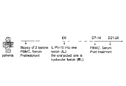
The study schema is shown in Figure 1A. Two study
lesions in each paticnt were sampled by biopsy pre-
treatment.
Table 1
Demographics of IL PV-10 treated Patients*
Patient Age Gender Stage Prior Therapy
PV001 48 IIIC Isolated Limb Infusion, IFN
PV002 81 F IIIB none
PV003 72 M IIIC Isolated Limb Infusion, ipi
PV004 77 F IIIB Isolated Limb Infusion, ipi
PV005 60 F IIIC none
PV006 77 B IV ipi, nivo
-40-
CA 03006302 2018-05-24
WO 2017/105993
PCT/US2016/065542
PV007 59 F IV Isolated Limb Infusion, vem
Isolated Limb Infusion, chemo,
PV008 75 M IIIC ipi, nivo
PV009 80 M IIIC none
PV010 86 F IIIC none
FV011 75 M IIIC none
PV012 80 IIIB Isolated Limb Infusion, PEG-IFN
PV013 86 F IIIB Isolated Limb Infusion, ipi
PV014 77 F IV Isolated Limb Infusion
PV015 69 F II1C none
* IFN - interferon; PEG-IFN = PEGylated interferon; ipi
ipilimumab; vem = vemurafenib; nivo = nivolumab.
Seven days later [trial day zero (D0)], one
of the two lesions was injected with IL PV-10. Seven
to fourteen days after PV-10 injection, both sites
were completely excised. Biopsy specimens were
fixed, stained with hematoxylin and eosin (H&E), and
evaluated by a pathologist. Comparisons of
pathologic complete response (pCR) in treated and
untreated specimens, before and after IL PV-10
injection were made, and the results were confirmed
with immunohistochemical staining for the melanoma
antigen Melan-A/MART-1 (melA).
As shown in Fig. 1B and Fig. 1C, tumors
completely regressed in both the PV-10-treated and
bystander lesions. That regression was noted in 4 of
12 patients. Additionally, 11 of 12 patients
exhibited at least partial regression of the injected
lesion, with a 4-fold decrease in frequency of me1A+
cells (mean value: 26.8 3.6 vs 6.4 2.8). Still
further, 10 of 12 patients demonstrated partial
-41-
CA 03006302 2018-05-24
WO 2017/105993
PCT/US2016/065542
regression of the bystander lesion with a 3-fold
decrease in the proportion of melAF cells (mean
value: 37.5 6.7 vs 12.2 4.1).
These results indicate that IL PV-10 can
induce a systemic response secondary to direct
ablation by IL PV-10 injection. To examine the role
of immune cells, the percentage of CD3+, CD4+ and CD8+
T-cells in PV-10 treated and bystander lesions were
compared before and after treatment with IL PV-10.
However, very few infiltrates were detected in the
lesions when tumor completely regressed, and no
significant changes were measured.
Next, immune cells were examined in the
peripheral blood before and after treatment with IL
PV-10. There was a significant increase in
circulating CD4+ T-cells, CD8+ T-cells, and NK T-cells
after PV-10 treatment (Figs. 1D-1F).
To determine whether the CD8+ T-cells can
recognize melanoma tumors, circulating CD8+ T-cells
were purified and co-cultured with autologous tumor
cells in vitro. There was a significant increase in
interferon-gamma (IFN-y) production after treatment
with IL PV-10 (Fig. 1G), indicating that PV-10
treatment enhances tumor-specific immune responses.
As autologous tumors were not available for
all patients, HLA-matched tumor cell lines were also
used. IFN-y levels in circulating CD8+ T-cells were
increased after IL PV-10 injection in 4 of the 6
patients tested. No change was measured when CD8+
T-cells were co-cultured with HLA-mismatched cell
lines. Together, these studies demonstrate that IL
PV-10 injection enhances tumor-specific immune
responses in melanoma patients.
-42-
CA 03006302 2018-05-24
WO 2017/105993
PCT/US2016/065542
IL PV-10 in M05-bearing mice elicits a tumor-specific
immune response
To investigate the underlying mechanism of
the tumor-specific immune response elicited by PV-10,
C57BL/6 mice bearing M05 tumor cells were used. M05
tumor cells are B16 melanoma cells that express the
ovalbumin (OVA) protein. Similar to the finding in
the B16 model [Toomey et al., PloS one 8:e68561
(2013)], IL injection of PV-10 directly inhibited
tumor growth (Fig. 2A). IL PV-10 therapy led to
increased OVA-specific CD8+ T-cells in the draining
lymph nodes (DLNs) of PV-10-treated mice, compared to
the PBS-treated group (Fig. 23).
To determine whether IL injection of PV-10
induced T cells with memory characteristics,
splenocytes from mice treated twice with IL PV-10
were cultured in vitro in the presence of OVA peptide
and media supplemented with the cytokines IL-15 and
IL-21, which are required for maintaining CD8+ memory
I-cells [Nguyen et al., J Leukocyte Biol 87:43-49
(2010)]. T-cells from PV-10-treated mice
demonstrated an about 2 fold increase in secretion of
IFN-y in response to M05 cells, compared to T-cells
isolated from PBS-treated mice. This indicates that
IL PV-10 can induce tumor-specific T-cells with
memory characteristics in M05 melanoma-bearing mice.
To monitor the CD8+ T-cell response after IL
PV-10, PV-10 or PBS were injected into M05 melanoma-
bearing mice on day 13 and adoptively-transferred
OT-1 T-cells that were labeled with Celltracker
violet dye. OT-1 T-cells are CD8+ T-cells that
specifically recognize the OVA 257-264 peptide
derived from the OVA protein [Rotzschke et al., Eur J
Immunol 21(11):2891-2894 (1991); Lipford et al., J
-43-
CA 03006302 2018-05-24
WO 2017/105993
PCT/US2016/065542
Immunol. 150(4):1212-1222 (1993)]. The combination
of IL injection of PV-10 and adoptive transfer of
OT-1 cells significantly impeded tumor progression
and increased survival (Figs. 3A and B).
Adoptive transfer of OT-1 cells alone or
treatment with IL PV-10 alone was not enough to
prevent tumor progression when treatment began at day
13 with an average tumor size of 52 mm2. The
proliferation of OT-1 T cells in the tumor, lymph
nodes (LNs) and spleen was examined after IL PV-10
injection by measurement of dye dilution of the
injected CD45.1+ OT-1 T-cells (Fig. 3C). OT-1 cells
robustly proliferated at both the tumor site and in
the proximal lymph nodes in PV-10- or PBS-treated
mice on day 4 post-transfer, with more than 70% of
cells having at least one additional division
(compared to the cells before transfer, hereafter
called "divided" T cells) (Fig. 3D).
An increased number of divided T cells were
measured in spleens, tumors, and distal LNs in the
mice treated with IL PV-10, compared to the mice
treated with PBS (Figs. 3 C-F). There was no change
found in the proximal LNs (data not shown),
indicating that the OT-1 T-cell response in the
draining LN may be too robust to measure a difference
between treated and non-treated mice. Together,
these data indicate that IL injection of PV-10 can
boost tumor-specific CD8+ T-cell responses leading to
prevention of tumor progression in M05-bearing mice.
IL PV-10 leads to Dendritic Cell (Dx7) activation
Because DCs are professional antigen-
presenting cells capable of priming T cells, the
presence of DCs in spleen and LNs was examined after
-44-
CA 03006302 2018-05-24
WO 2017/105993
PCT/US2016/065542
IL PV-10 treatment. There was no change in the
absolute number and percentage of splenic DCs on day
7 post-PV-10 injection. Consistent with previous
findings, other immune subsets including CD84-T cells
and myeloid-derived suppressor cells (MDSCs) were
also unchanged (data not shown), [Toomey et al., PIGS
one 8:e68561 (2013)].
However, the number of infiltrating DCs in
the draining LNs (DLNs), but not 1n non-draining LNs
(NDLNs), increased after 24 hours (i.e., about 1 day)
of PV-10 treatment (Fig. 4A). The overall total
number of cells in the DLNs was not significantly
changed after treatment. After 72 hours, the number
of infiltrating DCs in DLN of PV-10-treated mice
decreased to the base level as in the PBS-treated
mice, suggesting that the infiltration of DCs is
transient.
To examine whether DCs infiltrated from the
site of tumor, OVA protein labeled with FITC (FITC-
OVA) was injected IL 4 hours after IL injection of
PV-10 or PBS. The increased FITC+ DCs were measured
in the DLNs, but not in the NDLNs (Fig. 4B) in the
PV-10-treated mice, suggesting that IL PV-10 can
induce DCs to uptake antigens and infiltrate into the
DLNs from the tumor site.
Because enhanced OT-1 T-cell proliferation
and increased DC infiltration were measured in DLNs
after IL PV-10 treatment, it was hypothesized that IL
injection of PV-10 leads to DC activation that is
required for the tumor-specific response. To test
this hypothesis, BM-derived DCs were co-cultured with
supernatants from B16 tumors that were previously
injected with IL PV-10 or PBS.
-45-
CA 03006302 2018-05-24
WO 2017/105993
PCT/US2016/065542
After being pulsed with OVA protein, DCs
were co-cultured with OT-1 T-cells. DCs cultured
with supernatants derived from B16 tumors from PV-10-
treated mice induced increased proliferation of OT-I
T-cells, compared to DC cultured with supernatants
derived from B16 tumors from PBS-treated mice (Fig.
4C). In addition, DC pulsed with cell lysates of PV-
10-treated cells were able to better stimulate the
secretion of IFN-7 by OT-1 T-cells than DC pulsed with
cell lysatos of PBS-treated cells (Fig. 4D).
Together, these results support the role of IL PV-10
in the activation of DCs and the increased
infiltration of DCs from the tumor site into DLNs.
PV-10 treatment increases DC activation via INGB1.
To examine how tumor death induced by IL
PV-10 may be linked to the activation of DCs, PV-10-
mediated cell death was investigated, as were
potential factors released by tumor cells that can
contribute to DC activation.
First, the cytotoxicity of murine melanoma
B16 cells mediated by PV-10 was investigated in
vitro. As shown in Fig. 5A, PV-10 exhibited a dose-
dependent cytotoxicity in B16 cells, with an IC50
value of 60 M after 48 hours of treatment. There
was less cytotoxicity in mouse embryonic NIH3T3
fibroblasts, with an IC50 value of 110 M after 48
hours of treatment with PV-10 (Fig. 5A). The IC50
value of PV-10 on B16 and 3T3 cells was similar at 6,
12, and 24 hours.
There was a significant increase in
necrosis (4',6-diamidino-2-phenylindole dilactate
Stain; DAPI+) of B16 cells and human primary melanoma
-46-
CA 03006302 2018-05-24
WO 2017/105993
PCT/US2016/065542
(P) cells after 48 hours of treatment with 50 M of
PV-10, whereas there was little effect on 3T3 or
human embryonic kidney 293T cells (Fig. 5B).
However, a relatively small proportion of cells were
in early apoptosis, which is evidenced by Annexin V+
DAPI- (BioVision, Inc., Milpitas CA; Fig. 5C). This
also occurred 6, 12, and 24 hours after treatment.
This result indicates that treatment with 50 M of
PV-10 leads to cell death through necrosis rather
than apoptosis. Very few 3T3 fibroblast and human
embryonic kidney 293 T cells were necrotic or
apoptotic in the presence of the same dose of PV-10
(Figs. 5B and 5C). These studies illustrate that PV-
can kill tumor cells at a specific dose that is
not toxic to non-tumor cells.
It has previously been shown that necrosis
can be associated with the disruption of the
integrity of the cell membrane and uncontrolled
release of cytosolic contents into extracellular
space. Whether tumor cells treated with PV-10
released any damage-associated molecular pattern
molecules (DAMPs) was next examined.
DAMPs such as HMGB1, IL-la, and HSP
proteins are known byproducts of necrosis and stimuli
of DC activation. B16, 888 melanoma cells and 3T3
fibroblasts were treated with 0, 100 or 200 M PV-10
for 48 hours. Equal amounts of supernatant were
immunoblotted for HMGB1, HSP70, HSP90 or IL-la.
Treatment with PV-10 led to the release of HMGB1 into
the supernatant in a dose-dependent manner from B16
cells and human melanoma 888 cells but not from 3T3
cells (Figs. 6A and 6B). HSP 70 and IL-la were not
-47-
CA 03006302 2018-05-24
WO 2017/105993
PCT/US2016/065542
detected and HSP90 was unchanged after treatment with
PV-10 (data not shown).
To determine if secreted HMGB1 contributed
to DC activation, bone marrow- (BM-)derived DCs were
incubated with 10% supernatant (TS) from B16 cells
treated with PV-10 for 2 days in the presence of
HMGB1 neutralizing antibody or isotype control
antibody. The HMGB1 neutralizing antibody was
validated by the blockade of TNF-a secretion from
RAW264.7 macrophages.
Tumor supernatant from PV-10-treated cells
led to DC maturation, with up-regulation of surface
CD40. Neutralization of HMGB1 significantly
decreased CD40 expression (Fig. 6C). Treatment of
DCs with PV-10 directly did not change CD40
expression, suggesting that PV-10 itself does not
affect DC maturation. Other co-stimulatory markers
on DCs, including CD86 and CD80 were not up-
regulated.
To compare the antigen presentation
capacity of DCs, BM-derived DC were incubated with
the supernatant from M05 tumor treated with IL PV-10
in the presence of HMGB1 neutralizing antibody or
isotype control antibody for 2 days. The pre-treated
DCs were pulsed with OVA protein and then co-cultured
with OT-1 cells to examine OT-1 T-cell proliferation.
The blockade of HMGB1 reduced the ability of DCs to
stimulate OT-1 T-cell proliferation (Fig. 60), as
determined by [3-H]-thymidine incorporation. This
result indicates that treatment of tumor cells such
as melanoma cells with PV-10 leads to the release of
HMGB1, which is essential for DC activation.
To determine whether HMGB1 release was
relevant to patients treated with IL PV-10, the level
-48-
CA 03006302 2018-05-24
WO 2017/105993
PCT/US2016/065542
of HMGB1 in patients' serum before- and post-
treatment with IL PV-10 was compared by measurement
of IFN-y in patient serum. The concentration of
HMGB1 in patients' serum was significantly increased
in samples collected 7-14 days after treatment with
IL PV-10 (Fig. 7). Therefore, HMGB1 secretion
appears to contribute to the bystander effect in
patients with a tumor such as metastatic melanoma
treated IL with PV-10.
EXAMPLES
The following examples are intended to be
illustrative and non-limiting, and represent specific
embodiments of the present invention.
General Methods
Human subjects
Fourteen patients with dermal and/or
subcutaneous metastatic melanoma were enrolled in a
phase 1 clinical trial. (NCT01760499). Six of 14
patients had metastatic disease refractory to
previous ipilimumab, anti-PD-1 and/or vemurafenib
therapy, (Table 1). Two tumor lesions in each
patient were sampled by biopsy pre-treatment; one of
the two lesions was injected with IL PV-10, then both
residual sites were completely excised 7-14 days
later. Biopsy specimens were fixed in formalin and
embedded in paraffin. The specimens were sectioned
at 5-8 pm thickness, stained with hematoxylin and
eosin stains for determination of pathologic complete
response.
Immunohistochemistry was performed using
CD8, CD4, CD56 and melA. Peripheral blood and serum
were collected prior to, 7-14 days after IL PV-10
-49-
CA 03006302 2018-05-24
WO 2017/105993
PCT/US2016/065542
injection, and 21-28 days after IL PV-10 injection
for analysis. Peripheral blood mononuclear cells
(PBMC) were isolated by Ficoll-Paque0 Plus (GE
Healthcare, Pittsburgh, PA). Serum was prepared by
collecting the supernatant after incubation of blood
at room temperature for 1 hour and centrifugation at
1,000 X g.
Animals
Female C57BL/6 mice (6-8 weeks old) were
purchased from Harlan Laboratories (Indianapolis,
IN). Mice were housed at the Animal Research
Facility of the H. Lee Moffitt Cancer Center and
Research Institute. Mice were humanely euthanized by
CO2 inhalation according to the American Veterinary
Medical Association Guidelines. Mice were observed
daily and were humanely euthanized if a solitary
subcutaneous tumor exceeded 200 mm2 in area or mice
showed signs referable to metastatic cancer. All
animal experiments were approved by the Institutional
Animal Care and Use Committee and performed in
accordance with the U.S. Public Health Service policy
and National Research Council guidelines.
Cell lines and cell culture
NIH3T3, 293T cells and melanoma B16 cells
were obtained from ATCC (Manassas, VA). Human
melanoma cells 526, 624 and 888 were obtained from
the NIH (Bethesda, MD). Cells were cultured in RPMI
media (cRPMI) supplemented with 10% heat-inactivated
FBS, 0.1 mM nonessential amino acids, 1 mM sodium
pyruvate, 2 mM fresh L-glutamine, 100 mg/ml
streptomycin, 100 U/ml penicillin, 50 mg/ml
gentamicin, 0.5 mg/ml fungizonc (all from Life
-50-
CA 03006302 2018-05-24
WO 2017/105993
PCT/US2016/065542
Technologies, Rockville, MD), and 0.05 mM 2-ME
(Sigma-Aldrich, St. Louis, MO). M05 cells expressing
OVA protein [Palo et al., Nature Med 1:649-653 (1995)]
were maintained in cRPMI supplemented with 0.8 mg/m1
0418. The cell lines tested negative for mycoplasma
contamination. All cell lines were passaged fewer
than 10 times after initial revival from frozen
stocks.
For in vitro study, tumor cells were
incubated with a different dose of PV-10 for the
indicated time. Cell supernatants were collected for
functional assay or western blot. PV-10 was obtained
from Provectus Biopharmaceuticals, Inc., Knoxville,
TN.
For in vivo study, 3x10⁵ tumor cells were
injected into one flank of mice subcutaneously
(s.c.), and on days 7 or 13 PV-10 or PBS was injected
intralesionally (IL). Tumors were isolated from mice
after 18 hours and digested with a tumor dissociation
kit (Miltenyi Biotec, San Diego, CA) and GentleMACS'
(Miltenyi Biotec). Live cells were resuspended in
cRPMI at a concentration of 5x10⁵ cells/ml. Cell
supernatants were collected for functional assays
after 2 days.
EACS and tetramer staining
Single-cell suspensions from the indicated
tissues were prepared by pressing cells through a 70
pm cell strainer. After RBC lysis with ACK buffer,
cells were stained in FACS buffer with the following
antibodies for flow cytometric analysis: anti-human
CD3, CD4, CD8, and CD56; anti-mouse CD11c, I-Ab,
0045.1, CD45.2, CD86, CD80 and CD40 (all from BD
Biosciences, San Diego, CA). For the tetramer
-51-
CA 03006302 2018-05-24
WO 2017/105993
PCT/US2016/065542
staining, cells were stained with H-2 Eb/SIINFEKL
tetramer (MBL international, Woburn, MA) at room
temperature for 20 minutes, followed by an additional
20 minute incubation with additional antibodies on
ice according to the manufacturer's instructions.
Live/dead fixable near-IR or aqua fluorescent
reactive dyes (InvitrogenTM) were used to exclude dead
cells before analysis. Cells were acquired by LSR II
equipped with four lasers (BD Biosciences), and the
data were analyzed with FlowJo software (Tree Star,
Ashland, OR).
Assessment of IFN-y and HMGB1 by ELISA
For detection of IFN-y from human samples,
CD8+ T-cells were isolated from PBMCs with a human
CDe T-cell isolation kit (Miltenyi Biotec). lx105
cells were co-cultured with tumor cells in triplicate
at a ratio of 1:1, in a U-bottom 96-well plate.
After 48 hours, the IEN-y level in the cell
supernatant was measured with an IFN-y ELISA kit (R&D
Systems, Minneapolis, MN) according to the
manufacturer's instruction.
For detection of HMGB1 in patient serum, a
HMGB1 ELISA kit from IBL international (Toronto,
Canada) was used according to the manufacturer's
instructions.
For detection of IEN-y in mouse samples,
mice received s.c. 3x10⁵ M05 cells. On day 7 after
initial tumor cell injection, mice received a second
injection of 3x10⁵ M05 cells s.c. on the opposite
flank. PV-10 or PBS (50 L) was IL injected into the
initial M05 tumor lesion on days 7 and 17. On day
23, spienocytes were collected and cultured for 7
-52-
CA 03006302 2018-05-24
WO 2017/105993
PCT/US2016/065542
days, in cRPMI supplemented with 1 g/ml SIINFEKL
peptide, 20 ng/ml IL-15 and 20 ng/ml I1-21 (R&D
Systems) (Cheng et al., J Exp Med 205:2235-2249
(2008). The expanded cells were mixed with
irradiated M05 cells at a ratio of 10:1, and IFN-y
production in the supernatants was measured after 48
hours with a IFN-y ELISA kit (BD Biosciences)
according to the manufacturer's protocol.
Adoptive transfer of T cells
CD45.1 OT-1 T-cells were purified with a
T-cell enrichment column (R&D Systems) and were
incubated with CellTrackerTm Violet (Life
Technologies, now Thermo Fisher Scientific, Inc.
Waltham, MA) for 20 minutes at 37 C. After two
washes in PBS, 3x10⁵ labeled cells were resuspended
in 100 1 of PBS and injected i.v. into M05 tumor-
bearing mice. After 4 days, spleen, lymph nodes
(LNs) and tumors were harvested and stained with
antibodies to CD45.1 and to CD45.2. The
CD45.1+GD45.2- cells with at least one division were
considered "divided cells".
DC functional assay
Bone marrow obtained from C57BL/6 mice was
cultured with recombinant murine 20 ng/ml GM-CSF and
ng/ml IL-4 (R&D Systems, Minneapolis, MN) after
RBC lysis [Liu et al., J Immunol 191:1916-1926
(2013)]. On day 5, DCs were purified with Opti_prcpTM
gradient (Axis-Shield, Oslo, Norway) according to the
manufacturer's protocol and cultured in the presence
of GM-CSF and IL-4 at a cell density of 5x10⁵
-53-
CA 03006302 2018-05-24
WO 2017/105993
PCT/US2016/065542
cells/ml [Vohra et al., Cancer immunol Immun CII
59:729-736 2010)].
DCs were cultured with 10% supernatant from
B16 cells incubated with 100 M PV-10 for 2 days in
the presence of an antagonistic antibody against
HMGB1 (IBL international, Toronto, Canada) or the
relevant isotype. Next, DCs were pulsed for 2 hours
with 10 g/m1 of OVA protein (Sigma-Aldrich, St.
Louis, MO). After multiple washes, DCs were co-
cultured with 1e5 responder OT-1 T-cells in
triplicate, in U-bottom 96-well plates at different
stimulator-to-responder ratios for 3 days.
3H-thymidine (1 pCi) was added to each well 18 hours
prior to cell harvesting. T-cell proliferation was
measured by 3H-thymidine incorporation in a liquid
scintillation counter Microbeta Trilux (PerkinElmer,
Waltham, MA).
Determination of IC50
Cells were incubated with 12.5, 25, 50,
100, or 200 M PV-10 or PBS in a 12-well plate for 6,
12, 24 and 48 hours. All wells were collected, mixed
with counting beads and acquired by LSR II. DAPI was
used to exclude dead cells before analysis. The
absolute number of live cells was calculated by
comparing the ratio of bead events to cell events.
The half maximal inhibition of PV-10 on cell growth
was determined as 1050 using GraphPad Prism software
(GraphPad Software, Inc., La Jolla, CA).
Western blot
For removal of cellular debris, cell
supernatants were centrifuged at 14,000 X g. The
-54-
protein concentration of each sample was determined.
Equal amounts of protein were separated on a NuPAGEe
Novex' 4-12% Bis-Tris Gels (Life Technologies), then
transferred onto a polyvinylidene difluoride membrane
(Millipore). Membranes were blocked for 1 hour with 5%
BSA (w/v) in PBS and probed with HMGB1 (cat no. 3935),
HSP70 (D69), or HSP90 (045G5) antibodies overnight at
4 C (all from Cell Signaling Technology, Danvers,
MA). Immunoreactivity was visualized by incubation
with a horseradish peroxidase-linked secondary
antibody and treatment with enhanced chemiluminescence
reagents.
Statistical analysis
The data were analyzed with a two-tailed
Student's t test using GraphPad PrIen software or
Wilcoxon matched pairs test. A p value of < 0.05 was
considered to be statistically significant.
The foregoing description and the
examples are intended as illustrative and are not to
be taken as limiting. Still other variations within
the spirit and scope of this invention are possible
and will readily present themselves to those skilled
in the art.
-55-
CA 3006302 2019-05-07