Note : Les descriptions sont présentées dans la langue officielle dans laquelle elles ont été soumises.
CA 03008444 2018-06-14
WO 2017/100828
PCT/AU2016/051157
DENTAL APPARATUS
Field of invention
The present invention relates to an apparatus assisting in dental procedures.
Background of invention
Since the inception of modern dentistry during the late nineteenth century,
dental
diagnosis and treatment has relied largely on the same techniques. For
example, dentists have
been using a handpiece encasing a rotary engine with an attached cutting bur
to manually
prepare a cavity within a tooth, or reduce it circumferentially to accommodate
for a filling
material, crown or a mix of both.
The same tenet applies to the diagnosis of tooth decay (dental caries), which
is one of
the most ubiquitous diseases in human populations. The importance of reaching
a definite
diagnosis cannot be overemphasized, as it will govern the choice of a
consequent treatment
modality, be it prevention, monitoring, operation (drilling) or extraction.
Basically, diagnosis
depends on the practitioner's clinical acuity, as carious lesions can prove to
be elusive to assess
particularly when asymptomatic, located in the morphologically complex pits
and fissures, or
when caries has not yet progressed to induce visible cavitation. Hidden
caries, which advance
deep under an otherwise apparently sound surface, is yet another challenging
situation for the
diagnostician.
Therefore, when faced by such circumstances, a dentist may resort to
diagnostic aids
such as radiographs, optical magnifiers, staining dyes, thermal tests,
fluorescence or
transillumination. However, their efficiency and effectiveness in dental
settings are often
compromised by technique sensitivity (radiographic reproducibility for
example), increased
cost, fear of radiation exposure, proper chronological archiving and retrieval
of results, as well
as the time and effort of using different standalone devices and systems which
may also
inconvenience the patient and staff.
In addition, the accessibility to a particular tooth may be a determining
factor in
diagnosis, planning and performing- dental treatment. Plausibly, the more
posterior a tooth is in
the mouth, the less amenable it is for instrumentation and handling. Apart
from accessibility,
careful surgical precision must be maintained at all times throughout the
drilling procedure, as
the clinician is not only working within confined and miniscule structures,
but he also ought to
strictly avoid inadvertent damage to sound tissues around and within the tooth
such as the
delicate and centrally located pulp.
Substitute Sheet
(Rule 26) RO/AU
CA 03008444 2018-06-14
WO 2017/100828
PCT/AU2016/051157
2
Consequently, the outcome of dental care remains technique, equipment and time
sensitive, and thus quite vulnerable to human error, misjudgement and
indexterity. This serves
to limit the number of patients or teeth a practitioner can attend to, and
accounts for steadily
rising costs. This relative unimproved productivity of the healthcare
professions (as compared
to other fields that became more affordable and productive with industrial
automation) has been
described as the "Baumol Effect-.
It would be advantageous if there was a way to improve and streamline the
diagnosis
process, enhance visibility and access, minimise human error, reduce manual
effort and fatigue,
and/or perform rapid, precise and ideal tooth preparation.
Summary of invention
In accordance with a first aspect of the present invention there is provided
an
apparatus assisting in dental procedure, comprising: a body adapted to be
received with an
oral cavity, an anchoring means for securing the body within the oral cavity
such that it at
least partially surrounds one or more teeth, a dental device located within
the body and
adapted to move in a plurality of planes relative to the one or more teeth;
and a controller for
controlling the dental device to move into a suitable position relative to the
one or more teeth
and once located in position carry out work on or about the one or more teeth.
In an embodiment the body is adapted to be wholly received within the oral
cavity.
In an embodiment the work performed by the dental device includes at least one
pre-
programed procedure.
In an embodiment the anchoring means is a clamp that is adapted to secure to
the one
or more teeth and/or a tooth or teeth adjacent to the one or more teeth.
In an embodiment the clamp comprises one or more clamp peaks and one or more
clamp bows and wherein the body is coupled to the bow(s).
In an embodiment the body comprises one or more cameras operable to take an
image
of the work site and wherein the controller is configured to process the
resultant image data
for use in determining at least one of:
a. the position to locate the dental device for performing the work;
CA 03008444 2018-06-14
WO 2017/100828
PCT/A1J2016/051157
3
b. appropriate selection of the preprogramed procedure;
c. monitor in real time the operations performed by the apparatus; and
d. diagnose anomalies of the tooth structures by means of direct imagery,
transillumination and/or fluorescence.
In an embodiment the dental device comprises one or more lights for
illuminating the
oral cavity.
In an embodiment the one or more lights are transillumination lights adapted
to
transilluminate the one or more teeth.
In an embodiment the controller comprises a processing unit programmed to
instruct
the work device to perform work.
In an embodiment the apparatus further comprises a conduit for supplying or
expelling contents to the apparatus.
In an embodiment the contents are at least one of fluids, gases or solids.
In an embodiment the dental device is at least one of a mechanical bur,
forceps, root
elevators, scalpel, a mirror, a laser, a camera, an excavator, a dental
burnisher, a dental
plugger, a scaler, local anaesthetic vehicle, a curette or other dental
instrument.
In an embodiment movement of the dental device in the at least three planes is
caused
by the dental device moving along one or more sets of rails
In an embodiment at least one of the one or more sets of rails are adapted to
move
under the control of an actuator.
In an embodiment the apparatus further comprises a rotary motor within the
body that
can be either air, water, steam or electrically powered.
In accordance with a second aspect there is provided a method assisting in
dental
procedure, comprising utilising the dental apparatus according to any one of
the preceding
claims for performing the dental procedure.
In accordance with a third aspect there is provided a computer readable medium
storing computer program code which, when executed by a computer processor, is
operable
to control the controller of claim 1 to operate the dental work device.
CA 03008444 2018-06-14
WO 2017/100828
PCT/A1J2016/051157
4
Brief description of drawin2s
Embodiments of the present invention will now be described, by way of example
only, with reference to the accompanying drawings, in which:
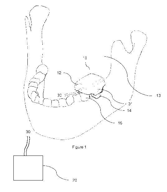
Figure. 1 is a schematic of an apparatus located within a patient's mouth, in
accordance with an embodiment of the present invention;
Figure 2a is a top view of the apparatus of Figure 1, anchored to a single
tooth;
Figure 2b is a sectional view through C-C shown in Figure 2a;
Figure 2c is an isometric view of the apparatus;
Figure 2d is a section view through A-A shown in Figure 2e;
Figure 2e is a side view of the apparatus;
Figure 2f is a further isometric view of the apparatus;
Figure 3 is an isometric view of a clamp used for anchoring the apparatus, in
accordance with an embodiment;
Figure 4a is an internal view of the body of the Figure 1 apparatus;
Figure 4b is a close up view of feature A shown in Figure 4a;
Figure 4c is a close up view of feature B shown in Figure 4a;
Figure 5a is a schematic of a dental work device of the apparatus, in
accordance with
an embodiment; and
Figure 5b is a schematic of a dental work device in accordance with an
alternative
embodiment; and
Figure 6 is a schematic of the apparatus attached to an extra oral base for
carving a
filling
Detailed description
Embodiments of the invention described herein relate to an apparatus for
assisting in
dental procedures. With reference to Figures I to 5, the apparatus 10
comprises a body 12
adapted to be received within an oral cavity 13 of a patient The body 12 is
secured within the
CA 03008444 2018-06-14
WO 2017/100828
PCT/AU2016/051157
oral cavity 13 via an anchoring means 14, such that the body 12 at least
partially surrounds
one or more teeth 16. A dental work device 18 (see particularly Figures 2 and
5) is movable
within the body 12 and is adapted to move in a plurality of planes relative to
the one or more
teeth 16. A controller 20 is operable to cause the dental work device 18 to
move into a
desired position and thereafter actuate the dental work device 18 for carrying
out work on or
about the one or more teeth 16. The work performed by the apparatus 10
includes, but is not
limited to, at least one of dental drilling, suction, imaging and
illumination.
Configuration of the Apparatus
In more detail, and with particular reference to the elevation and section
views
depicted in Figure 2, the body 12 of the apparatus 10 is generally rectangular
in shape with
chamfered edges for minimising patient discomfort. The body 12 may be formed
from any
suitable material, including plastic, metal, a suitable composite material or
the like. The
dental work device 18 is housed within the body 12 and includes an attachment
17 for use in
carrying out the work. As previously stated, the apparatus 10 (and more
particularly the
body 12) is anchored within the oral cavity 13 via an anchoring means 14.
According to the
illustrated embodiment, the anchoring means 14 takes the form of a clamp 14
that secures to
the crown of a single tooth 16. Figure 3 shows the clamp 14 in isolation. With
reference to
both Figures 3 and 4, the clamp 14 comprises a pair of bows 14a, 14b that
detachably couple
respectively to ends 12a, 12b of the body 12. The bows 14a, 14b extend to
opposing clamp
beaks 14c that firmly secure to the crown of a tooth adjacent to the gingiva
(i.e. in the same
manner as existing dental clamps used by dentists to attach the rubber dam).
According to the
illustrated embodiment, the bows 14a, 14b attach to the body via moveable arms
that firmly
hook under the bows 14a, 14b and thereafter can be locked in place to prevent
the body from
detaching from the clamp 14. It will be understood that any suitable detaching
means could
be used for securing the clamp to the body 12 (.e.g. frictional, magnetic or
latch fit),
depending on the desired implementation. For example, the bows 14a, 14b may
include
openings for receiving screws that screw into the body for securing the clamp
thereto. In yet
another alternative embodiment, the clamp 14 may be fixedly secured to the
body, e.g. via an
adhesive. In yet another alternative embodiment, the clamps may be integrally
formed with
the body 12 (e.g. set in place during forming of the body).
As previously mentioned, the apparatus 10 is adapted to perform work within a
work
site. According to the illustrated embodiment the work site comprises a tooth
16 (or teeth)
CA 03008444 2018-06-14
WO 2017/100828
PCT/AU2016/051157
6
that the body 12 at least partially surrounds. The dental work device 18 is
moveable in three
planes relative to the tooth 16, as depicted in Figure 2c, for moving into a
desired position.
Furthermore, the dental work device 18 includes a rotator and pivot mechanism
for allowing
the attachment 17 to both rotate and pivot (i.e. in an additional two planes)
once the dental
work device 18 is located in a desired position.
In more detail, and with additional reference to Figure 4a, there is shown a
sectional
view of the internal housing of the body 12 for illustrating movement of the
dental work
device 18 in the X and Y planes. As shown, a pair of rails 23a, 23b extend
between side
walls I5a, 15b of the body 12. The dental work device 18 couples to the rails
23a, 23b for
sliding movement there along. According to the illustrated embodiment, the
rails 23a, 23b
are closely received in openings 24a, 24b (i.e. having an internal profile
that corresponds to
the sectional profile of the rails ensuring smooth sliding operation) that are
disposed on
opposite sides of a body 12 of the work device 18. A further opening 26
extends through the
body 12 of the dental work device 18 for receiving a threaded lead screw 28
extending from
an actuator in the form of a stepper motor 30. The further opening 26 has a
threaded internal
profile corresponding to the thread of the lead screw 28. Actuation of the
stepper motor 30
(either in the forward or reverse direction) causes the dental work device 18
to move along
the rails 23a, 23b thereby facilitating precision open-loop positioning of the
device 18 in the
X plane. Positioning along the Y plane is achieved by way of rails 32a, 32b
which extend
between ends 12a, 12b of the body 12 and fixed thereto. The rails 32a, 32b
pass through
openings 33 disposed in the rails 23a, 23b, as shown in the close-up view of
Figure 4a. A
second actuator (also in the form of a stepper motor 36) controls a second
lead screw 38
which passes through openings 35 disposed in rails 23a, 23b. An internal
profile of the
openings 35 has a corresponding thread to the second lead screw 38, thereby
facilitating
movement of the dental work device 18 in the Y plane as the stepper motor is
actuated (again
either in forward or reverse). The close up view of Figure 4c shows this in
more detail. It
will be understood that other configurations could be utilised for achieving
the sliding
movement of the dental work device 18 in the X and Y planes, for example using
tracks
embedded in an internal wall of the body 18 that receive and retain rollers
disposed on the
work device 18 and which may be powered to move along the tracks for suitably
positioning
the work device. According to the illustrated embodiment, the dental work
device 18 is
adapted to move up to 2cm in in the X and Y planes.
CA 03008444 2018-06-14
WO 2017/100828
PCT/AU2016/051157
7
Movement in the Z plane (i.e. for controlling the height of the dental work
device
attachment 17 relative to the tooth 16) is achieved by way of a third
actuator. With reference
to Figure 5a, there is shown a schematic of the dental work device 18 whereby
the third
actuator takes the form of a pair of a stepper motors 40. The stepper motors
40 turn threaded
lead screws 44 which pass through corresponding vertically oriented openings
42 (which
have corresponding internal threaded profile to the screws 44) in a mid-
section 42 of the
dental work device 18. Although not shown in Figure 5a, the mid-section 42
comprises the
longitudinally oriented openings 24a, 24b and 26 (i.e. that facilitate
coupling to rails 23a, 23b
and lead screw 28). In this manner, the stepper motors 40 can be
simultaneously actuated
(either in forward or reverse) for facilitating upward and downward movement
in the Z plane.
It will be understood that in an alternative embodiment, only one stepper
motor and lead
screw may be required for facilitating the movement in the Z plane. An
alternative
embodiments for facilitating movement in the Z plane is shown in Figure 5b.
According to
this embodiment, an actuator in the form of one or more micro air pumps are
used to extend
and contract the length of the dental work device 18 so as to adjust the
height of the
attachment 17 relative to the tooth 16. As shown in Figure 5b, a plurality of
telescoping
cylinders 46, 48, 50 have internal chambers that are connected by way of a
series of valves.
The cylinders 46, 48, 50 are caused to extend or retract relative to one
another by applying
pressure or suction to the chambers by way of the air pump(s). Although not
shown in Figure
5b, the upper most cylinder 46 comprises the longitudinally oriented openings
24a, 24b and
26 that facilitate coupling to rails 23a, 23b and lead screw 28. It will also
be understood that
the rails 23a, 23b and 32a, 32b (and corresponding openings 33, 38) may be of
any desired
cross sectional profile, depending on the desired implementation. Further, the
rails 23a, 23b,
32a, 32b may be positioned at any desired height within the body 12, depending
on the
desired implementation. According to the illustrated embodiment, the dental
work device 18
is adapted to move up to 2cm in in Z plane.
Still with reference to Figures 5a and 5b, the attachment 17 is in the form of
a bur that
removably couples to the work device 18 by way of an actuator in the form of
an air powered
turbine 35 ( e.g. by way of frictional, magnetic or latch fit). The attachment
17 couples to the
work device 18 by way of a rotator 37 (in this embodiment taking the form of a
pneumatic
rotary actuator 37 which rotates the attachment 17 between 0 and 360 degrees
as indicated by
reference numeral D) and pivot mechanism 39 that in turn couples to the work
device 18 by
way of a pair of arms 41. A further actuator (in this illustrated embodiment
being in the form
CA 03008444 2018-06-14
WO 2017/100828
PCT/AU2016/051157
8
of an air powered pneumatic motor 39) causes a pivot portion of the mechanism
to pivot
relative to the arms 41 (i.e. for moving between 0 to 180 degrees as indicated
by reference
numeral E). The rotator 37 and pivot mechanism 39 advantageously provide two
additional
planes of movement for performing the necessary work. It will be understood
that some or
all of the above described actuators/motors/engines for controlling movement
in the X, Y and
Z planes (as well as for rotating and pivoting the work attachment 17) could
be electrically
powered, air powered or electromagnetically powered, depending on the desired
implementation.
Also shown in the figures is a conduit 30 that is connected to the body 12 and
which,
in use, facilitates the delivery or removal of fluid to/from the work site
and/or dental work
device 18. For example, the conduit 30 may deliver compressed air to the work
site as
required for the procedure. The compressed air may also be directed to any one
or more air
powered actuators utilised by the apparatus 10. The conduit 30 may
additionally or
alternatively deliver water (e.g. pressurised water) to the work site. In
addition, or as an
alternative, the conduit 30 may be used to suction air and/or water from the
work site during
the procedure. As yet another additional or alternative use, the conduit 30
may carry
electrical and/or data cable for providing power and/or commands to any
electrically powered
actuators and/or circuitry provided by the apparatus 10. The conduit 30 may
also carry all of
the necessary control signals to/from the circuitry (e.g. image data captured
from the
cameras).
In a particular embodiment, the conduit 30 may be connected to a housing
containing,
an extra-oral air compressor (for delivering the compressed air), a water
chamber for
supplying water, a suction pump and a receptacle used to store any substances
received from
the work site via the conduit. The conduit 30 may comprise separate tubes for
carrying the
various fluids and cables. In an alternate embodiment, the conduit 30 may be
connected to
pre-existing air, water and electricity outlets that may be within structures
such as such as
existing conventional dental chairs (dental units).
In an embodiment, one or more lights are disposed on the body 12 to illuminate
the
teeth 16 and/or surrounding oral cavity 13. In a particular embodiment an LED
light is
located at each internal corner of the body 12 (i.e. directed toward the work
site). In addition,
the apparatus 10 may be provided with a bore light or other such light for
transillumination
(e.g. disposed on an inner wall of the body) The apparatus 10 additionally
comprises a
CA 03008444 2018-06-14
WO 2017/100828
PCT/AU2016/051157
9
camera 31 (again which may be disposed on an inner wall of the body 12 facing
the work
site) for capturing image data for the work site and surrounding area.
As shown in Figure 1, the apparatus 10 is connected to a controller 20 for
electrically
controlling actuation of the various apparatus actuators and imaging devices
to perform the
desired dental procedure. For example, with regards to the air powered
actuators, the controller
is operable to control the air supply unit to deliver the necessary amount of
air/suction for
achieving the necessary movement. For electrical actuators, the controller 20
may directly
deliver the necessary power or signal thereto via an electrical cable disposed
in the conduit 30.
Alternatively, the controller 20 may be configured to wirelessly communicate
with the
actuators, e.g. via Bluetooth, WiFi or the like. In a particular form, the
controller 20 may
comprise a microcontroller or other suitable processing system including a
memory storing
program code for automatically controlling the actuators (i.e. based on
predefined instructions).
Additionally, or alternatively, the controller 20 may include a user interface
that allows an
operator to manually control the actuators. This may be the case for dental
procedures, such
as drilling, where the operator may wish to manually control the angle and
depth for drilling.
The controller 20 may be configured to cause a display device to display
relevant real time
procedural information, including image data captured from the camera(s), as
well as any other
relevant information (including warnings, actuator feedback data, etc.). Thus,
in one example,
the controller 20 may include a wireless receiver configured to receive a
remote control signal,
for controlling the apparatus 10. In an alternative embodiment to that shown
in the figures, the
controller 20 may be embedded in or disposed on the body of apparatus 10 and
can be
programmed to wirelessly communicate with a remote control device. In yet
another
embodiment, the controller may be configured to connect to a remote network,
such as the
Internet, allowing the apparatus to be controlled remotely, for example as a
means of urgent
dental intervention, where dentists are not readily accessible such as in
extremely remote areas
(e.g. outer space) or those suffering conflicts or natural disasters. The
apparatus 10 might be
attended in site by a dental assistant or nurse or a person trained for this
purpose, and operated
remotely by a dentist from abroad.
CA 03008444 2018-06-14
WO 2017/100828
PCT/AU2016/051157
Examples of Work Performed by Apparatus:
Imaging
In a particular embodiment, the camera 31 is able to capture image data
(corresponding to images of the work site or oral cavity) that can be
processed by the
controller 20, e.g. for creating a three-dimensional model of the work site.
The model can,
for example, be referenced by the programs stored in the controller memory for
use in
determining how to implement a particular dental procedure. The model can also
be stored
for future evaluation (e.g. to monitor a particular condition, treatment,
etc.). Alternatively, or
additionally, the controller 20 may be programmed to accept a scanned model of
the work
place generated by a standalone intra-oral scanner.
The controller 20 may further be configured to display the image data on a
display
device, thereby allowing a practitioner to examine the tooth or teeth within
the work site (i.e.
obviating the need for an additional standalone intra oral camera or
magnifier). For example,
the image data may allow a practitioner to clearly identify the location, type
and size of an
anomaly and as a result suitably prepare for treatment. Additionally, after
completion of
cavity preparation, a three-dimensional cavity filler can be produced based on
the information
obtained from the one or more cameras.
The camera(s) 31 may be fitted with a zoom functionality to allow a close and
detailed inspection of the work site, which can aid in diagnosing dental
ailments. In some
instances, a microscopic lens can be added to the camera. Further, the
information captured
by the one or more cameras can allow a spatial relationship between the tooth
and the
apparatus 10 to be established. This special relationship informs the
controller 20 of where
the tooth or teeth are to thereby ensure that the work is performed with a
high degree of
accuracy.
In a particular embodiment, a practitioner can compare image data from the
work site
prior to and after treatment. For instance, the before image data may be
overlayed with an
image of the treated site. A person skilled in the art will appreciate that
there are numerous
methods to compare the before and after work site that do not depart from the
invention.
A fluorescent light may be fitted to the apparatus which allows the camera(s)
to detect
fluorescence emission by microbes in dental plaque on the tooth. The resultant
images that
CA 03008444 2018-06-14
WO 2017/100828
PCT/AU2016/051157
11
are obtained can be processed to detect caries which may be displayed on a
screen in a
different colour.
As previously stated, the apparatus 10 comprises a light suitable for
transillumination
(e.g. a bore light). In a particular embodiment, the controller 20 may turn on
the light to
highlight the structure of the tooth 16, which may also be used to identify
caries, cavities or
cracks in the tooth whether mesodistally or occlusally, as well as a range of
other ailments.
The camera 31 may be controlled to capture image data resulting from the
transillumination
which may be evaluated (e.g. manually or by the stored programs) to plan a
treatment for any
discovered ailments.
In other instances, the one or more lights coupled with one or more cameras
allow for
any drilling procedures that may be required to be evaluated, recorded and
visualised. This is
achieved as the one or more cameras are able to obtain the real-time
dimensions and imagery
of the tooth allowing the accurate location of where work must be performed on
the tooth. A
person skilled in the art would appreciate that a light fitted into the
apparatus 10 is only one
of the many ways the transillumination technique can be exploited by the
apparatus.
Alternatives include using a separate standalone transillumination device,
such as an external
light device, while also using the camera feature and/or drilling feature of
the apparatus 10 to
provide enhanced visualisation of the results.
Drilling and lboth Preparation
In a particular embodiment, the attachment 17 comprises a bur drill. A
specific
example of a drill is shown in Figures 5a and 5b. As illustrated, the bur
drill 17 attaches to
the work device 18 by way of the rotator 37 and pivot mechanism 39. In one
embodiment,
the drill 17 is air powered by a rotary turbine engine 35 that can create a
drill speed of up to
approximately 200,000 revolutions per minute. It will be understood that the
drill speed may
be controlled automatically by the controller 20 or manually under the control
of an operator
(via a suitable controller interface). In an alternative embodiment, the
engine 35 may be an
electrically powered engine, or other suitably powered engine for carrying out
the work. It
will also be understood that various shaped and sized burs may be attached by
the operator to
the work device 18, depending on the desired implementation using bur drill
attachments that
are well understood in the art.
CA 03008444 2018-06-14
WO 2017/100828
PCT/AU2016/051157
12
Fillings and Crowns
In an embodiment of the present invention, the apparatus 10 is also able to
create
fillings and crowns. As an initial step, the apparatus 10 creates a three
dimensional model of
the subject tooth using techniques as hereinbefore described. This can be
achieved through
the camera and history of the drilling, or using a commercially available
intraoral standalone
scanning device. The controller can use the fluorescence result, the dentist's
demarcation
through the mouse/joystick and his choice of a certain cavity type and
dimensions to design a
digital cavity. Then, the body 12 of the apparatus 10 is detached from the
tooth 16 and re-
attached to a specialised extraoral base 50, as shown in Figure 6. According
to the illustrated
embodiment, the bows of the clamp are respectively inserted into slots
disposed in a pair of
arms 51a, 5 lb that extend from base 50, so as to secure the body thereto. It
will be
understood that the body 12 could be attached to the base using any suitable
securing
technique (e.g. the bows could clamp over the arms 51a, 51b in the same way
they clamp to
the tooth 16). In yet another embodiment, the body 12 could be detached from
the clamp 14
and re-attached to the base 50 using an alternative securing means. A
prefabricated block of
filling material 52 is secured by the operator to a cradle 54 coupled to an
internally housed
rotator 56 that can be controlled by the controller 20. The cradle 54 may also
be moveable in
other planes (e.g. using a similar mechanism to that used for moving the
dental work device),
depending on the desired implementation. The controller 20 then controls the
various
actuators to carve the material 52 to conform to the cavity and thus produces
the desired
crown or filling that fits the prepared tooth. The dentist then cements this
structure using
dental cements. It will be understood that the base 50 may include its own
water and power
supply, depending on the desired implementation.
Other Aspects
Different embodiments of the apparatus 10 may be created to perform one or
more of
the following dental procedures: root canal therapy, implant placement,
surgical sectioning
and extraction, periodontal therapy and soft tissue surgery, delivering local
anaesthesia and
the application and maintenance of orthodontic appliances. As previously
stated, the body 12
of the apparatus 10 is adapted to be received within a patient's oral cavity
13. The size of the
body may vary based on the type of oral cavity 13 that it is intended to
perform dental
procedures within. For instance, a lager sized body 12 may be used for an
adult's oral cavity,
while a smaller apparatus 10 may be used for a child's oral cavity. Persons
skilled in the art
CA 03008444 2018-06-14
WO 2017/100828
PCT/AU2016/051157
13
will appreciate that the apparatus 10 as described herein can be used with
animals, and as
such the size of the device may be larger or smaller depending on the type of
animal, and the
type of teeth that will be treated. By way of example, the body 12 of an
apparatus 10 for use
with a human adult may be 3cm x 3cm x 2.3cm (allowing it to be received wholly
within the
oral cavity) but should not be seen as being limited to this size, and may be
bigger or smaller.
The apparatus 12 may also be fitted with a safety sensor that causes the
controller 20
to cease work in response to detecting a hazard. By way of example, a pressure
sensor may
be fitted to an upper surface of the body 12. If a patient bites down on the
apparatus 10, the
pressure sensor will detect the bite and cause a signal to be sent to the
controller 20 which
causes the controller 20 to initiate an emergency procedure including ceasing
operation of the
actuators and raising the dental work device 18 off the tooth.
As hereinbefore described, the anchoring means 14 took the form of a clamp
having a
pair of bows. It will be understood that the body 12 may be configured to
attach to any form
of clamp depending on the desired implementation. For example, as persons
skilled in the art
will appreciate, that there are many forms of clamp having varied
configurations that are
suited to the working site (e.g. that conform to the different morphologies of
teeth, such as
anterior, posterior, upper and lower) and a dentists preferences. By way of
example, the
apparatus 10 could be removably attached to a wingless clamp, a distal clamp,
a cervical or
labial clamp, a retention clamp, a retraction clamp, or the like Further, it
will be understood
that the body 12 may only secure to a single bow of the clamp.
In another anchoring embodiment, the body 12 may need to be anchored to more
than
one tooth simultaneously using more than one clamp. For example, one form of
clamp may
attach to two teeth (anterior and posterior) in operation. This is useful when
the a particular
tooth is badly broken down so that it is unable to receive clamping, or when
there is a need to
relieve the tooth of clamping and hence use adjacent teeth for apparatus
retention.
The apparatus 10 is adapted to perform work within at least one work site. The
work
site is the portion of the oral cavity that requires diagnosis or treatment.
In some instances,
this may be the entire upper or lower arch, whereas in other instances this
may only be a
portion of the oral cavity, such as one or more teeth, a portion of the
gingiva, or a
combination of the two. There may be one or more work sites where the
apparatus 10 is able
to perform work. Work may be performed on these work sites at the same time,
or at different
times.
CA 03008444 2018-06-14
WO 2017/100828 PCT/AU2016/051157
14
Persons skilled in the art will appreciate that the body and internal
components may
be formed of any suitable material that can be sterilized, such as alloy
stainless steel. It will
also be appreciated that any moving parts (such as motor gears used in the
engines) are
formed of a corrosion resistant material. An appropriate sterilization
technique that may be
employed includes placing the apparatus 10 in a steriliser, immersion or
spaying with
disinfecting solutions or the like.
In this specification, the word -comprising" is to be understood in its "open"
sense,
that is, in the sense of -including", and thus not limited to its -closed"
sense, that is the sense
of -consisting only of" A corresponding meaning is to be attributed to the
corresponding
words "comprise", "comprised" and "comprises" where they appear.
The preceding description is provided in relation to several embodiments which
may
share common characteristics and features. It is to be understood that one or
more features of
any one embodiment may be combinable with one or more features of the other
embodiments. In addition, any single feature or combination of features in any
of the
embodiments may constitute additional embodiments.
In addition, the foregoing describes only some embodiments of the inventions,
and
alterations, modifications, additions and/or changes can be made thereto
without departing
from the scope and spirit of the disclosed embodiments, the embodiments being
illustrative
and not restrictive.
Furthermore, the inventions have described in connection with what are
presently
considered to be the most practical and preferred embodiments, it is to be
understood that the
invention is not to be limited to the disclosed embodiments, but on the
contrary, is intended to
cover various modifications and equivalent arrangements included within the
spirit and scope
of the inventions. Also, the various embodiments described above may be
implemented in
conjunction with other embodiments, e.g., aspects of one embodiment may be
combined with
aspects of another embodiment to realize yet other embodiments. Further, each
independent
feature or component of any given assembly may constitute an additional
embodiment.