Note : Les descriptions sont présentées dans la langue officielle dans laquelle elles ont été soumises.
CA 03016282 2018-08-30
WO 2017/191487
PCT/1B2016/052496
OVERLAY IMAGING FOR REGISTRATION OF A PATIENT EYE FOR LASER
SURGERY
BACKGROUND
Field of the Disclosure
100011 The present disclosure relates to ophthalmic surgery, and more
specifically, to overlay
imaging for registration of a patient eye for laser surgery.
Description of the Related Art
100021 In ophthalmology, eye surgery, or ophthalmic surgery, saves and
improves the vision
of tens of thousands of patients every year. However, given the sensitivity of
vision to even
small changes in the eye and the minute and delicate nature of many eye
structures,
ophthalmic surgery is difficult to perform and the reduction of even minor or
uncommon
surgical errors or modest improvements in accuracy of surgical techniques can
make an
enormous difference in the patient's vision after the surgery.
100031 Ophthalmic surgery is performed on the eye and accessory visual
structures. More
specifically, various types of laser ablation surgery (or simply 'laser
surgery') may be
performed on the cornea, lens, or other structures in the eye for clinical
treatment purposes.
Typically, prior to laser surgery a diagnostic image of the eye is acquired to
characterize the
eye and to identify specific treatment locations, among other uses.
Subsequently, when the
laser surgery is initiated on the eye using a surgical laser system, an
initial step in the surgery
is registration of the eye to confirm the identity of the patient and to
correlate the eye to the
diagnostic image. Typical surgical laser systems display the diagnostic image
next to a
surgical image of the eye being acquired for registration purposes. During
registration, the
surgeon or other medical personnel may find that precise rotational alignment
of the eye
using the two images displayed next to one another, the diagnostic image and
the surgical
image, is difficult or time-consuming.
SUMMARY
100041 In one aspect a disclosed method is for registration of patient eyes
for ophthalmic
surgery. The method may include acquiring a surgical image including the
limbus of a
-1-
CA 03016282 2018-08-30
WO 2017/191487
PCT/1B2016/052496
patient eye subject to ophthalmic surgery, and overlaying the surgical image
on a diagnostic
image including the limbus, the diagnostic image being previously acquired
from the patient
eye. The method may also include aligning a first limbus center of the
surgical image with a
second limbus center of the diagnostic image, and displaying the surgical
image being rotated
about the first limbus center over an angular range. In the method, the
surgical image may
have a transparency enabling simultaneous viewing of the surgical image and
the diagnostic
image during at least a portion of the angular range. In the method, the
angular range may
include an alignment angle for the surgical image at which the surgical image
and the
diagnostic image are cyclotorsonally aligned with respect to the first limbus
center and the
second limbus center, respectively.
[0005] In any of the disclosed embodiments of the method, the surgical image
and the
diagnostic image may include at least a portion of the iris of the patient's
eye.
[0006] In any of the disclosed embodiments of the method, both the surgical
image and the
diagnostic image may respectively include at least one identical marker at a
common location
with respect to the iris of the patient's eye. In the method, the marker may
be an indication of
an iris structure of the iris displayed over a uniform background in place of
the iris. In the
method, a first marker of the surgical image has a first color, and a second
marker of the
diagnostic image has a second color. In the method, the marker may have a
hollow shape. In
the method, the hollow shape may include at least one of a polygon and an
ellipse.
[0007] In any of the disclosed embodiments of the method, displaying the
surgical image
may further include displaying the surgical image being incrementally rotated
about the first
limbus center over a step angle of rotation, in response to user input
indicating the step angle.
100081 In any of the disclosed embodiments, the method may further include
receiving user
input specifying the alignment angle, displaying the surgical image rotated
about the first
limbus center at the alignment angle, and varying the transparency of the
surgical image from
low transparency to high transparency.
[0009] In any of the disclosed embodiments, the method may further include
receiving user
input to select at least one of the displayed transparency, the range of
transparency from high
transparency to low transparency, a step transparency within the transparency
range, a first
color in the surgical image, a second color in the diagnostic image, a number
of markers, a
-2-
CA 03016282 2018-08-30
WO 2017/191487
PCT/1B2016/052496
type of marker, a location of a marker, a contrast of the iris, a displayed
portion of the iris, the
angular range, and a step angle within the angular range.
[0010] Another disclosed aspect includes an image processing system for
overlay imaging
for registration of a patient eye for ophthalmic surgery, the image processing
system
including a processor enable to access memory media storing instructions
executable by the
processor to perform the method, or any portions thereof. A further disclosed
aspect includes
an article of manufacture comprising non-transitory memory media for
characterizing
membranes at vitreoretinal interfaces, the memory media storing instructions
executable by a
processor to perform the method, or any portions thereof. .
BRIEF DESCRIPTION OF THE DRAWINGS
[0011] For a more complete understanding of the present invention and its
features and
advantages, reference is now made to the following description, taken in
conjunction with the
accompanying drawings, in which:
[0012] FIGURES lA and 1B show an embodiment of overlay imaging for
registration of a
patient eye depicting the iris;
100131 FIGURES 2A, 2B, and 2C shows an embodiment of overlay imaging for
registration
of a patient eye using iris markers;
[0014] FIGURES 3A, 3B, and 3C shows an embodiment of overlay imaging for
registration
of a patient eye using iris markers and shape markers;
[0015] FIGURE 4 is a block diagram of selected elements of an embodiment of an
image
processing system for overlay imaging for registration of a patient eye; and
100161 FIGURE 5 is a flow chart of selected elements of a method for overlay
imaging for
registration of a patient eye.
-3-
CA 03016282 2018-08-30
WO 2017/191487
PCT/1B2016/052496
DESCRIPTION OF PARTICULAR EMBODIMENT(S)
[0017] In the following description, details are set forth by way of example
to facilitate
discussion of the disclosed subject matter. It should be apparent to a person
of ordinary skill
in the field, however, that the disclosed embodiments are exemplary and not
exhaustive of all
possible embodiments.
100181 As used herein, a hyphenated form of a reference numeral refers to a
specific instance
of an element and the un-hyphenated form of the reference numeral refers to
the collective
element. Thus, for example, device '12-1' refers to an instance of a device
class, which may
be referred to collectively as devices '12' and any one of which may be
referred to
generically as a device '12'.
[0019] As noted above, registration of a patient's eye is performed as an
initial step during
various types of ophthalmic surgery. In particular, for laser surgery of the
cornea or eye lens,
an image of the limbus of the eye may be acquired during diagnosis, referred
to as a
'diagnostic image'. The diagnostic image may be used, along with other
acquired data and
measurements, to determine a treatment plan for the eye, such as a surgical
plan for the laser
surgery. When the patient is prepared for the laser surgery and is positioned
within the laser
surgery system, a 'surgical image' of the patient's eye, including the limbus,
is acquired and
used for registration of the eye. The registration using the surgical image
serves to identify
the patient and to confirm that the correct eye is undergoing laser surgery.
[0020] Additionally, because cyclotorsion of the eye may occur to some degree,
registration
may further involve a precise rotational alignment of the eye with respect to
the diagnostic
image. Because the ability to rotate the patient may be limited, an alignment
angle may be
determined by the surgical laser system representing a slight rotational
(angular) offset
between the diagnostic image and the surgical image. The alignment angle may
then be
recorded by the surgical laser system and used to rotationally offset laser
activity on the eye
accordingly. The alignment angle is generally measured about a rotation point
that is the
geometric center of the limbus, which provides a more stable rotational
alignment point than
the center of the pupil, which can change as the pupil size changes. To the
extent that the
pupil diameter is somewhat comparable in each of the images, the limbus also
provides a
stable circular structure that can be used for precise scaling alignment of
the diagnostic image
-4-
CA 03016282 2018-08-30
WO 2017/191487
PCT/1B2016/052496
to the surgical image, even when the two images differ in size, resolution,
color, and
wavelength of light used, among other aspects.
[0021] Because the precise rotational alignment of the eye is crucial for
properly performing
the surgical plan for the laser surgery, the surgeon or other surgical
personnel will typically
verify that the alignment angle determined has a high degree of accuracy.
Thus, the checking
and confirmation of the alignment angle may represent an important operation
during
registration of the eye for the laser surgery.
[0022] Conventional surgical laser systems are typically equipped to provide
the diagnostic
image next to the surgical image on a display screen that is viewed by an
operator of the
surgical laser system, often the surgeon or other surgical personnel. The
surgical image may
be acquired using microscope optics and a camera that is included with the
surgical laser
system. However, determining the alignment angle between the diagnostic image
and the
surgical image when the two images are displayed next to one another may be
difficult and
time consuming, because such an arrangement may be challenging for human
visual
cognition to interpret. Thus, by virtue of the side-by-side arrangement found
in conventional
surgical laser systems, the overall accuracy of the alignment angle determined
in this manner
may be limited, which is undesirable because it represents a potential source
of error for
performing the surgical plan.
100231 As will be described in further detail herein, the inventors of the
present disclosure
have provided methods and systems of overlay imaging for registration of a
patient eye for
laser surgery. Instead of presenting the diagnostic image next to the surgical
image during
registration, the methods and systems of overlay imaging for registration of a
patient eye
disclosed herein may display the two images as a single overlay image
comprised of the
surgical image superimposed on the diagnostic image. The methods and systems
of overlay
imaging for registration of a patient eye disclosed herein may further provide
a variable
degree of transparency, from low transparency to high transparency, of the
surgical image.
The methods and systems of overlay imaging for registration of a patient eye
disclosed herein
may align the diagnostic image and the surgical image based on the limbus in
each respective
image, and may enable rotation of the surgical image relative to the
diagnostic image about
the limbus center. The methods and systems of overlay imaging for registration
of a patient
eye disclosed herein may also enable displaying the surgical image being
rotated relative to
the underlying diagnostic image about the limbus center. For example, the
surgical image
-5-
CA 03016282 2018-08-30
WO 2017/191487
PCT/1B2016/052496
may be rotated about an angular range that includes the alignment angle, such
as with a video
loop created with individual frames at individual step angles of the rotation,
to allow human
visual cognition to more accurately and precisely determine or verify the
alignment angle.
The methods and systems of overlay imaging for registration of a patient eye
disclosed herein
may still further provide markers that are indicative of certain structures in
the eye, such as
iris structures that are unique to the eye. Other geometric markers, such as
polygons or
ellipses, may also be used. The methods and systems of overlay imaging for
registration of a
patient eye disclosed herein may enable selection and configuration by users
of various
features for flexible and customizable displays for determining and verifying
the alignment
angle. The methods and systems of overlay imaging for registration of a
patient eye
disclosed herein may be implemented using an image processing system that is
included with
or that operates with the surgical laser system.
100241 Referring now to the drawings, FIGURES lA and 1B show an embodiment of
overlay
imaging for registration of a patient eye depicting the iris. In FIGURES IA
and 1B overlay
images comprising a diagnostic image (lower or back image) over which a
surgical image
(upper or front image) has been superimposed and aligned at the limbus center
of each
respective image. In FIGURES IA and 1B, substantially all of the iris
structure as well as the
pupil is shown in both the diagnostic image and the surgical image, where the
pupil is slightly
larger in the diagnostic image as compared to the surgical image. As shown,
FIGURE IA
shows a first overlay image that is slightly out of rotational alignment,
while FIGURE 1B
shows a second overlay image that is more precisely aligned, as evident by the
improved
sharpness of the iris structures visible. In FIGURE 1B, the more sharp
alignment is visible in
the clarity of the iris structures, particularly as compared with FIGURE 1A.
Accordingly, the
image shown in FIGURE 1B may represent an optimal alignment for cyclotorsion
about the
limbus center between the diagnostic image and the surgical image, and the
alignment angle
may be determined, or estimated, from the angular offset applied in FIGURE 1B,
which is
known.
100251 As shown, the images in FIGURE lA and 1B represent examples of overlay
imaging
for registration of a patient eye, as disclosed herein. Accordingly, the
overlay images shown
in FIGURE lA and FIGURE 1B may be displayed in a variety of different manners
and
techniques, as described herein. Because the overlay images shown in FIGURE 1
A and
FIGURE 1B may be generated and displayed using an image processing system (see
-6-
CA 03016282 2018-08-30
WO 2017/191487
PCT/1B2016/052496
FIGURE 4), both the diagnostic image and the surgical image may be processed
and
displayed as digital images, allowing for various types of manipulation. For
example, user
input may be received to determine the actual angular offset displayed,
allowing a user to
manually determine the optimal alignment angle for cyclotorsion by 'tuning'
the offset angle,
as desired. In some embodiments, user input may be received to determine a
step angle of
rotation for rapid bidirectional manual rotation. Although only static images
can be displayed
in the drawings, it will be appreciated that a dynamic, or animated, view of
the images shown
in FIGURES IA and 1B may be generated by varying an offset angle of the
surgical image
relative to the diagnostic image. In various embodiments, the offset angle may
be varied over
an angular range about the limbus center that includes the alignment angle, or
the expected
alignment angle. The display of the offset angle over the angular range may be
repeated in a
loop with bidirectional rotation. In this manner, a user may be able to
confirm with a high
degree of certainty, accuracy, and precision that the alignment angle selected
is actually the
optimal alignment angle. The animation loop about the angular range described
above may
be activated or repeated, for example, when the alignment angle has been
tentatively
determined in a first iteration, to enable determination of the alignment
angle with increased,
or optimal, accuracy in degrees of rotation, such as limited by the resolution
of the raw image
data or limited by the processing ability of the surgical laser system or the
image processing
system.
100261 It is further noted that color and contrast may be varied (not shown)
for the diagnostic
image or the surgical image or both. For example, the diagnostic image may be
a first color,
while the surgical image may be a second color. When both images are aligned,
the resulting
image may accordingly appear as a third color that is a combination of the
first color and the
second color.
100271 Other types of displays may also be generated, as described herein. For
example, a
transparency of the overlaid surgical image may be varied (not shown) between
low
transparency (mostly or all opaque) to high transparency (mostly or all
transparent). The
transparency of the surgical image may be varied manually by the user, such as
by user input
specifying the transparency. As an animation, the transparency of the surgical
image may be
automatically varied from low transparency to high transparency. The user may
be enabled
to determine the speed of the animation, as well as the transparency levels
corresponding to
low transparency and high transparency, respectively. In this manner, the
animation may
-7-
CA 03016282 2018-08-30
WO 2017/191487
PCT/1B2016/052496
transition from the diagnostic image to the surgical image without a change in
the offset
angle between the two images, to provide another view that enables highly
precise and
accurate determination of the alignment angle by more clearly presenting
misaligned features
in the display output to the user. As with the other techniques for overlay
imaging disclosed
herein, the transparency animation may enable determination of the optimal
alignment angle
that appears best aligned in various different overlay displays.
[00281 Still further, as will be shown in the subsequent figures, certain
portions of the overlay
image may be masked out or covered with a solid region, instead of the entire
image within
the limbus, for clarity and visual simplicity. Various types of markers may be
used in the
overlay image, such as, but not limited to, iris markers that are indicative
of particular iris
structures, and geometric markers, such as a polygon or an ellipse, which may
also be hollow
markers for improved recognition of alignment. The number, type, location,
size, among
other features of the markers may be user selectable. The markers may include
optical
markers, such as regions of varied illumination, brightness, contrast, or
other optical image
property.
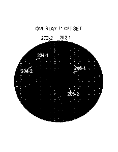
[00291 Referring now to FIGURES 2A, 2B, and 2C, an embodiment of overlay
imaging for
registration of a patient eye depicting the iris using iris markers 204 is
shown. Iris markers
204 may be indicative of a particular feature in the iris of the eye (see
FIGURES lA and 1B).
In FIGURES 2A, 2B, and 2C overlay images for the limbus are shown indicative
of a
diagnostic image (lower or back image) respectively superimposed over a
surgical image
(upper or front image) and aligned at the limbus center of each respective
image. In
FIGURES 2A, 2B, and 2C, the image of the iris has been replaced with a uniform
ring field
for greater optical clarity in viewing iris markers 204. In FIGURES 2A, 2B,
and 2C, a first
pupil 206-1 pupil is shown for the diagnostic image and a second pupil 206-2
is shown for
the surgical image, where first pupil 206-1 is slightly larger than second
pupil 206-2. It is
noted that in other embodiments, first pupil 206-1 and second pupil 206-2 may
be the same
size or different sizes, and the pupil size may be modulated by adjusting the
illumination on
the eye. In FIGURES 2A, 2B, and 2C, a first reticle 202-1 is also shown for
the diagnostic
image and a second reticle 202-2 is shown for the surgical image. Reticles 202
cross at a
common point that is the limbus center, which is not affected by a change in
size of pupils
206. As shown, FIGURE 2A shows a third overlay image that has an angular
offset of 70
about the limbus center and is accordingly out of rotational alignment, FIGURE
2B shows a
-8-
CA 03016282 2018-08-30
WO 2017/191487
PCT/1B2016/052496
fourth overlay image that has an angular offset of -70 about the limbus center
and is
accordingly out of rotational alignment. The diagnostic image having reticle
202-1 is shown
at alignment with vertical and horizontal. FIGURE 2C shows a fifth overlay
image that is
more precisely aligned, albeit at an offset of 3 from vertical or horizontal.
[0030] In FIGURE 2A, iris marker 204-2 is offset from iris marker 204-1, and
may be more
clearly visible that an actual picture of the iris that iris markers 204
represent. Iris markers
204 may be generated using the same image processing operation from a
respective iris
image. In FIGURE 2B, iris marker 204-2 is offset in an opposite direction from
iris marker
204-1 as compared to FIGURE 2A. In FIGURE 2C, iris markers 204 appear as
singular
markers due to the precise alignment in the overlay image, and reticle 202
also appears as a
single reticle.
[0031] As with FIGURES lA and 1B, it is noted that various different
modifications and
techniques may be used with the overlay images shown in FIGURES 2A, 2B, and
2C. In
some embodiments, certain portions of the iris may be blended in with a
varying degree of
transparency, as described above. Iris markers 204 may be shown along with the
actual iris
structure represented by iris markers 204, or any desired portion thereof
Color and
transparency of any of the image elements shown or described may be varied
manually or
automatically in an animation loop, as described above, with user
configuration of various
display parameters relating to color, time, transparency, among others. In
particular, when an
offset angle of the diagnostic image is not aligned with vertical or
horizontal, the alignment
angle may be determined relative to any offset angle of the diagnostic image,
as shown in
FIGURE 2C.
[0032] Referring now to FIGURES 3A, 3B, and 3C, an embodiment of overlay
imaging for
registration of a patient eye depicting the iris using iris markers 204 and
geometric markers
302. Iris markers 204 may be indicative of a particular feature in the iris of
the eye (see
FIGURES lA and 1B) and are the same as described above with respect to FIGURES
2A,
2B, and 2C. In FIGURES 3A, 3B, and 3C, overlay images for the limbus are shown
indicative of a diagnostic image (lower or back image) respectively
superimposed over a
surgical image (upper or front image) and aligned at the limbus center of each
respective
image. In FIGURES 3A, 3B, and 3C, the image of the iris has been replaced with
a uniform
ring field for greater optical clarity in viewing iris markers 204 and
geometric markers 302.
In FIGURES 3A, 3B, and 3C, a first pupil 206-1 pupil is shown for the
diagnostic image and
-9-
CA 03016282 2018-08-30
WO 2017/191487
PCT/1B2016/052496
a second pupil 206-2 is shown for the surgical image, where first pupil 206-1
is slightly larger
than second pupil 206-2. In FIGURES 3A, 3B, and 3C, a first reticle 202-1 is
also shown for
the diagnostic image and a second reticle 202-2 is shown for the surgical
image. Reticles 202
cross at a common point that is the limbus center, which is not affected by a
change in size of
pupils 206. As shown, FIGURE 3A shows a sixth overlay image that has an
angular offset of
2 about the limbus center and is accordingly out of rotational alignment,
FIGURE 3B shows
a seventh overlay image that has an angular offset of -2 about the limbus
center and is
accordingly out of rotational alignment. The diagnostic image having reticle
202-1 is shown
at alignment with vertical and horizontal. FIGURE 3C shows an eighth overlay
image that is
more precisely aligned, albeit at an offset of -3 from vertical or
horizontal.
100331 In FIGURES 3A, 3B, and 3C, geometric markers 302 are shown as circles,
which is a
special case of an ellipse more generally. Geometric markers 302 are further
shown as
hollow markers, which may enable a more precise or accurate alignment over the
entire
marker, as well as over the visible portion from the hollow space of the
underlying image
(here iris markers 204). In FIGURES 3A, 3B, and 3C, geometric marker 302-1 is
in the
diagnostic image, while geometric marker 302-2 is in the surgical image. As
with the
previous figures described in detail above, it is noted that various different
modifications and
techniques may be used with the overlay images shown in FIGURES 3A, 3B, and
3C.
100341 Referring now to FIGURE 4, a block diagram illustrating selected
elements of an
embodiment of an image processing system 400 is presented. In the embodiment
depicted in
FIGURE 4, image processing system 400 includes processor 401 coupled via
shared bus 402
to memory media collectively identified as memory 410.
[0035] Image processing system 400, as depicted in FIGURE 4, further includes
communication interface 420 that can interface image processing system 400 to
various
external entities, such as a network or instrumentation bus included with a
surgical laser
system. In embodiments suitable for overlay imaging for registration of a
patient eye for
laser surgery, image processing system 400, as depicted in FIGURE 4, includes
display
interface 404 that connects shared bus 402, or another bus, with an output
port for one or
more displays, such as a display of a surgical laser system, an ocular display
of a surgical
microscope, or another display. As shown, image processing system 400 may
further include
a camera interface 406 for acquisition of surgical images from a camera
included in the
-10-
CA 03016282 2018-08-30
WO 2017/191487
PCT/1B2016/052496
surgical laser system. In some embodiments, camera interface 406 includes a
camera and
suitable optics for overlay imaging for registration of a patient eye for
laser surgery.
[0036] In FIGURE 4, memory 410 encompasses persistent and volatile media,
fixed and
removable media, and magnetic and semiconductor media. Memory 410 is operable
to store
instructions, data, or both. Memory 410 as shown includes sets or sequences of
instructions,
namely, an operating system 412, and an overlay image processing application
414.
Operating system (OS) 412 may be a UNIX or UNIX-like operating system, a
Windows
family operating system, or another suitable operating system.
100371 In various embodiments, image processing system 400 may be integrated
with
different types of equipment. In one embodiment, image processing system 400
is integrated
with a surgical microscope. The surgical microscope may be integrated within
the surgical
laser system.
100381 Modifications, additions, or omissions may be made to image processing
system 400
without departing from the scope of the disclosure. The components and
elements of image
processing system 400, as described herein, may be integrated or separated
according to
particular applications. Image processing system 400 may be implemented using
more,
fewer, or different components in some embodiments.
[0039] Referring now to FIGURE 5, a flow chart of selected elements of an
embodiment of a
method 500 for overlay imaging for registration of a patient eye for laser
surgery, as
described herein, is depicted in flowchart form. It is noted that certain
operations described
in method 500 may be optional or may be rearranged in different embodiments.
Method 500
may be performed by overlay image processing application 414 in FIGURE 4.
[0040] Method 500 may begin, at step 502, by acquiring a surgical image
including the
limbus of a patient eye subject to ophthalmic surgery. The ophthalmic surgery
may be laser
surgery. At step 504, the surgical image is overlaid on a diagnostic image
including the
limbus, the diagnostic image being previously acquired from the patient eye.
At step 506, the
surgical image is displayed being rotated about the first limbus center over
an angular range,
the surgical image having a transparency enabling simultaneous viewing of the
surgical
image and the diagnostic image during at least a portion of the angular range,
and the angular
range including an alignment angle for the surgical image at which the
surgical image and the
diagnostic image are cyclotorsonally aligned with respect to the first limbus
center and the
-11-
second limbus center, respectively. At step 508, user input specifying the
alignment angle is
received. It is noted that step 508 may be repeated or performed iteratively
in conjunction
with other steps in method 500. At step 510, the surgical image is displayed
rotated about the
first limbus center at the alignment angle. At step 512, the transparency of
the surgical image
is varied from low transparency to high transparency.
[0041] As disclosed herein, methods and systems for overlay imaging for
registration of a
patient eye for laser surgery include aligning and overlaying a surgical image
of the limbus
over a previously acquired diagnostic image of the limbus. The surgical image
is displayed
with a degree of transparency and enabled to rotate about the limbus center.
Various types of
colors, markers, contrast, and backgrounds may be used to generate a display
for determining
the alignment angle between the diagnostic image and the surgical image.
-12-
Date Regue/Date Received 2022-07-06