Note : Les descriptions sont présentées dans la langue officielle dans laquelle elles ont été soumises.
ADAPTIVE PATIENT INTERFACE
BACKGROUND
[0001] This is a division of Canadian Patent Application 2,802,920 filed June
23, 2011
(PCT/US2011/041676).
Field of invention
[0002] This patent document relates to patient interfaces that attach an
ophthalmic
system to an eye for anterior segment eye procedures. More particularly, this
patent
document relates to adaptive patient interfaces that reduce a deformation of a
cornea of the
procedure eye.
Description of Related Art
[0003] This patent document describes examples and embodiments of techniques
and
devices for securing an ophthalmic system to an eye to perform an anterior
segment eye
procedure. These devices are often referred to as patient interfaces. Since
patient interfaces
serve to connect the ophthalmic system and the eye of the patient, their
performance is an
important contribution to the precision and success of the ophthalmic
procedures. Thus,
improvements in patient interfaces can lead to improvements in the precision
and reliability
of ophthalmic procedures.
SUMMARY
[0003a] Certain exemplary embodiments can provide a patient interface for an
ophthalmic system, comprising: an attachment module, attachable to the
ophthalmic
system; and a contact module, configured to accommodate a viscoelastic
substance between
the patient interface and a procedure eye.
[0003b] Certain exemplary embodiments can provide a method of utilizing a
patient
interface for an ophthalmic procedure, the method comprising: applying the
patient
interface to a procedure eye in preparation for the ophthalmic procedure; and
1
CA 3019055 2018-09-28
4
providing a viscoelastic substance to at least one of a cornea of the
procedure eye and a
contact portion of the patient interface, wherein the providing is performed
before, during or
after the applying.
10003c1 Certain exemplary embodiments can provide a patient
interface system for an
ophthalmic system, comprising: a multi-piece patient interface, comprising an
attachment
module, attachable to the ophthalmic system; a contact module, configured to
be docked to a
procedure eye, to accommodate a viscoelastic substance in an accommodation
space at least
partially bordered by the contact module and by the procedure eye, and to get
connected to
the attachment module; and a suction ring, coupled to the contact module,
connectable to a
vacuum suction system to create a partial vacuum between the suction ring and
the
procedure eye to reduce a mobility of the procedure eye for an ophthalmic
procedure; and a
degassing subsystem, coupled to the patient interface, comprising at least one
of a pressure-
reduction system, a heating system, a membrane degasification system, an inert
gas
substitution system, a surface tension manipulating system, and a reductant-
adding system.
[0004] Briefly and generally, a patient interface for an ophthalmic system can
include
an attachment module, attachable to the ophthalmic system; and a contact
module,
configured to accommodate a viscoelastic substance between the patient
interface and a
procedure eye.
[0005] In some implementations, the viscoelastic substance can include a
fluid, a
liquid, a gel, a cream, an artificial tear, a film, an elastic material, or a
viscous material.
[0006] In some implementations, a refractive index of the viscoelastic
substance is
closer to a refractive index of a cornea of the procedure eye than to a
refractive index of air
at an operating wavelength of the ophthalmic system.
[0007] In some implementations, a refractive index of the viscoelastic
substance is
within a range of approximately 1.24-1.52 at an operating wavelength of the
ophthalmic
system.
la
CA 3019055 2018-09-28
[0007] In some implementations, a refractive index of the
viscoelastic substance is
within a range of approximately 1.35-1.41 at an operating wavelength of the
ophthalmic
system.
[0008] In some implementations, the attachment module and the contact
module are
separate and connectable.
[0009] In some implementations, the attachment module and the contact
module are
integrated components of the patient interface.
[0010] In some implementations, a component of the patient interface
is at least one
of disposable, sterilizable, and reusable.
[0011] Some implementations can include one or more input ports to
introduce the
viscoelastic substance into an accommodation space at least partially defined
by the contact
module.
[0012] Some implementations can include one or more output openings
configured to
enable a discharge of air, gas, or the viscoelastic substance from the contact
module.
[0013] In some implementations, the one or more output openings can include
a vent
port, configured to keep a pressure in an accommodation space at least
partially defined by
the contact module at approximately ambient pressure.
[0014] Some implementations can include a suction subsystem
configured to at least
partially immobilize the procedure eye for an ophthalmic procedure.
[0015] In some implementations, the suction subsystem is arranged in
relation to the
contact module; and the suction subsystem is connectable to a vacuum suction
system to
create a partial vacuum between the suction subsystem and the procedure eye.
[0016] In some implementations, the patient interface is configured
to keep a change
of an apical curvature of a cornea of the procedure eye below 10% upon an
attachment of the
patient interface to the procedure eye.
[0017] In some implementations, the patient interface is configured
to keep a change
of the apical curvature of the cornea of the procedure eye below 3% upon the
attachment of
the patient interface to the procedure eye.
[0018] In some implementations, the ophthalmic system can include at
least one of
an imaging system, a diagnostic system, a laser system, and an ophthalmic
surgical system.
2
CA 3019055 2018-09-28
[0019] In some implementations, the contact module is
configured to accommodate
the viscoelastic substance before being attached to the procedure eye.
[0020] In some implementations, the contact module is
configured to accommodate
the viscoelastic substance after the viscoelastic substance has been applied
to the procedure
eye.
[0021] In some implementations, the contact module can
include a soft elastic film or
membrane, configured to contain the viscoelastic substance in an accommodation
space at
least partially defined by the contact module, and to form a soft and elastic
contact surface for
the procedure eye.
[0022] Some implementations can include a soft bag, containing the
viscoelastic
substance.
[0023] In some implementations, the patient interface can be
connectable to a
= degassing subsystem, configured to degas the viscoelastic substance.
[0024] In some implementations, a patient interface for an
ophthalmic system can
include a contact module, configured to be attachable to a first eye with an
apical corneal
radius of RI and separately to a second eye with an apical corneal radius of
R2; and to limit a
change of each apical corneal radius to less than 0.5* R1-R2 when the contact
module is
attached to the first eye and separately to the second eye, wherein the apical
corneal radii RI
and R2 are between 7.5 mm and 8.2 mm.
[0025] In some implementations, the contact module can be configured to
limit the
change of each apical corneal radius to less than 0.25*IR1-R21when the contact
module is
attached to the eyes.
[0026] Some implementations can include an attachment module
attachable to the
ophthalmic system, wherein the attachment module and the contact module can be
either
separate and connectable, or integrated components of the patient interface.
[0027] Some implementations can include one or more fluid
ports, configured to
introduce a fluid or gel into a containment space at least partially defined
by the procedure
eye and the patient interface.
[0028] Some implementations can include one or more output
ports, configured to
enable a discharge of at least one of air, fluid or gel from a containment
space at least
partially defined by the patient interface and the procedure eye.
3
CA 3019055 2018-09-28
[0029] Some implementations can include a suction port, configured to
enable a
creation of a partial vacuum between a portion of the patient interface and a
portion of the
procedure eye.
[0030] Some implementations can include a distal lens, wherein the
distal lens does
not contact a cornea of the procedure eye after an attachment of the patient
interface to the
procedure eye.
[0031] Some implementations can include a soft layer, configured to
contain a
viscoelastic substance, and to provide a soft contact surface for a procedure
eye.
[0032] In some implementations, a method of utilizing a patient
interface for an
ophthalmic procedure can include applying the patient interface to a procedure
eye in
preparation for the ophthalmic procedure; and providing a viscoelastic
substance to at least
one of a cornea of the procedure eye and a contact portion of the patient
interface, wherein
the providing is performed before, during or after the applying.
[0033] In some implementations, providing the viscoelastic substance
can include
providing a fluid, a liquid, a gel, a cream, an artificial tear, a film, an
elastic material, or a
viscous material.
[0034] In some implementations the providing can include introducing
the
viscoelastic substance through an input port of the patient interface into a
contact space, at
least partially bordered by the patient interface and the procedure eye after
the applying.
100351 In some implementations the providing can include introducing the
viscoelastic substance onto the cornea of the procedure eye before the
applying.
[0036] In some implementations the providing can include providing
the viscoclastic
substance at the contact portion of the patient interface before the applying.
[0037] In some implementations the providing can include providing
the viscoelastic
substance in a space at least partially defined by one or more soft films or
membranes.
[0038] In some implementations the providing can include using a
syringe to
introduce the viscoelastic substance.
[0039] In some implementations the ophthalmic procedure can include
at least one of
an imaging procedure, a diagnostic procedure, a laser-assisted procedure, and
an ophthalmic
surgical procedure.
[0040] Some implementations can include degassing the viscoelastic
substance.
4
CA 3019055 2018-09-28
[0041] In some implementations the degassing can include at least one
of reducing a
pressure, heating, performing membrane degasification, substituting an inert
gas, and adding
a reductant.
BRIEF DESCRIPTION OF DRAWINGS
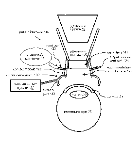
[0042] FIG. 1A illustrates an integrated patient interface 100.
[0043] FIG. 1B illustrates the patient interface 100 attached to the
ophthalmic system
and the procedure eye 20.
[0044] FIG. 2A-B illustrate a two-piece patient interface 100'.
[0045] FIG. 3 illustrates another embodiment of a two-piece patient
interface 100".
10 [0046] FIG. 4 illustrates a method of utilizing a patient interface.
[0047] FIGS. 5A-B illustrate different sequences of the method of
FIG. 4.
[0048] FIGS. 6A-H illustrate various implementations of providing a
viscoelastic
substance for the ophthalmic procedure.
DETAILED DESCRIPTION
[0049] Some laser eye surgical procedures, such as corneal refractive
corrections and
laser-assisted lens capsulotomies, may benefit from immobilizing the procedure
eye relative
to the surgical laser system during the procedure. Some systems include a so-
called patient
interface to carry out this task. One end of the patient interface can be
attached to the distal
end of the surgical laser system. The other end can include a contact lens
pressed against the
procedure eye. Such patient interfaces hold the eye steady relative to the
surgical laser,
enabling a high precision directing and focusing of the laser beam to a
predetermined target
location of the eye. Some patient interfaces can also be used to provide a
reference surface
for the targeting of the laser so that its focus depth can be defined relative
to the contact lens.
[0050] Some patient interfaces use flat contact "lenses", also called
applanation
plates. Others include curved contact lenses. In operation, either of these
contact lenses can
be pressed against the cornea of the eye, essentially immobilizing the eye and
forcing the
cornea to conform to the contact surface of the contact lens. To overcome the
slipperiness of
the tear film covering the eye, the contact lenses are typically held in place
by a vacuum
system, such as a suction ring.
[0051] While using rigid contact lenses has the benefit of providing a well-
defined
optical element for optimizing the beam properties of the laser, and possibly
of providing a
5
CA 3019055 2018-09-28
=
reference plane to direct the surgical laser with precision, there are also
disadvantages
associated with forcing rigid lenses against the procedure eye.
100521 One of the problems is that upon docking to the eye, the
contact lens typically
deforms the cornea, as their curvatures are generally different from each
other. This change
of the corneal curvature can cause internal deformations since the support
system of the lens
of the eye is very soft. Therefore, the docking of a rigid patient interface
typically shifts and
tilts the lens relative to the optical axis of the eye. This displacement and
tilt may make the
cuts of a typical cataract surgery, the circular capsulotomy cut on the
capsular bag and the
cataract surgical pattern cut in the lens itself, off-center and distorted,
leading to a
deterioration of the optical outcome of the cataract procedure.
[0053] Altering the cornea's natural curvature may also produce
wrinkles on the
surface of the cornea that could distort the laser beam. This distortion can
lead to increased
scattering and astigmatism of the beam, possibly requiring the use of a higher
energy laser
beam. The distortion can also lead to a loss of precision of the directing the
laser beam.
[0054] The corneal deformation can be reduced by designing the contact lens
to have
a curvature equaling that of a typical cornea. However, since corneal radii
vary considerably
from patient to patient, even these patient interfaces deform the corneas of
most patients.
[0055] To address these problems, some implementations of the
present invention
may use a patient interface whose lens does not make direct contact with the
cornea. Such an
embodiment can successfully minimize the corneal deformation, reducing the
aforementioned
problems, possibly even avoiding them altogether.
[0056] However, such non-contact designs may have their own
challenges as (I) the
light propagating through an air gap before entering the cornea may reduce the
beam quality
by increasing its astigmatism, for example; (2) the surface of the cornea can
dry out quickly,
increasing the light scattering at the corneal surface considerably; and (3)
the surgical eye
may have an excessive amount of movement because the patient interface does
not hold it
steady by direct contact.
[0057] Implementations of the present invention include patient
interfaces that reduce
the corneal deformation because their lenses do not make direct contact with
the cornea,
while at the same time offer solutions for the above three challenges.
[0058] FIG. lA illustrates an implementation of a patient
interface, or PI, 100. The
P1100 can include an attachment module 110 and a contact module 120. A
function of the
6
CA 3019055 2018-09-28
attachment module 100 can be to attach the P1100 to an ophthalmic system 10.
In some
embodiments the attachment module 110 can be connected to a distal end,
application tip, or
objective of the ophthalmic system 10. A function of the contact module 120
can be to form
a connection to an eye 20 on which an ophthalmic procedure is performed. This
eye will be
sometimes referred to as the procedure eye 20.
[0059] The ophthalmic system 10 can include an imaging system, a
diagnostic
system, a laser system or an ophthalmic surgical system.
[0060] The PI 100 can include a distal lens, or non-contact lens 111.
The distal lens
111 can be the last refractive element of the optical train of the ophthalmic
system 10. The
distal lens 111 can be a flat applanation plate or a lens with one or both
surfaces curved. Its
role can be similar to that of the contact lens of other patient interfaces,
with the difference
that in various embodiments the distal lens 111 does not contact a cornea 21
of the eye 20.
For this reason, the distal lens 111 does not deform the cornea 21, thus
avoiding the
displacement and tilt of lens 22, and the wrinkling of the cornea 21.
[0061] FIG. IB illustrates the P1100 after it has been connected or docked
to the eye
20. Visibly, in this implementation the distal lens or non-contact lens 111 is
indeed not in
direct contact with the cornea 21 of the eye 20. Because of this lack of
contact, the P1100
minimizes the deformation of the eye.
[0062] A measure of the deformation is the relative change of an
apical curvature of
the cornea of the procedure eye when the patient interface is attached to the
procedure eye.
Some embodiments of the P1100 keep the change of the apical curvature of the
cornea below
10% when the PI is attached to the eye. In other embodiments, the relative
change of the
apical corneal curvature can be kept below 3%.
[0063] Referring to FIG. IA, the contact module 120 can be formed to
accommodate
a viscoelastic substance 121 in a space between the PI 100 and the cornea 21.
This design
can address the above challenge (1), since when the viscoelastic substance 121
fills up the
space between the distal lens 111 and the cornea 21, the laser beam or light
of the ophthalmic
system 10 does not propagate through air.
[0064] When there is an air gap between the distal lens 111 and the
cornea of the
procedure eye 20, the surgical or diagnostic light beams arc refracted at the
posterior surface
of the distal lens 111 and at the anterior corneal surface. This latter
refraction is proportional
to (n(a)-n(c)), the difference between the refractive index n(a) of the air,
and n(c), that of the
cornea.
7
CA 3019055 2018-09-28
[0065] The deterioration of the beam quality can be reduced by
filling up the air gap
with the viscoelastic substance 121 between the patient interface 100 and the
cornea 21. in
this case, the beam refraction and astigmatism will be proportional to (n(v)-
n(c)), where n(v)
is an index of refraction of the viscoelastic substance 121.
[0066] Thus, in some embodiments the viscoelastic substance 121 can be
chosen to
have a refractive index n(v) closer to n(c), the refractive index of the
cornea, than to n(a), the
refractive index of air, at an operating wavelength of the ophthalmic system
10. Since an
index of refraction of the cornea is typically close to n(c)=1.38, in some
embodiments this
translates to the viscoelastic substance 121 having an index of refraction
n(v) in the
approximate range of 1.24-1.52. In other embodiments, n(v) can fall in the
approximate
range of 1.35-1.41.
[0067] Introducing the viscoelastic substance 121 to fill the space
between the distal
lens 111 of the patient interface and the cornea 21 also resolves challenge
(2) as the cornea is
not exposed to air in this design. Rather, the corneal surface can remain
wetted by the
viscoelastic substance 121, preventing the cornea 21 from drying out.
[0068] In various implementations, the viscoelastic substance 121 can
be one of a
wide variety of substances, including a fluid, a liquid, a gel, a cream, an
artificial tear, a film,
an elastic material, or a viscous material. In some cases, two or more of
these substances can
be present in the viscoelastic substance 121.
[0069] The viscoelastic substance 121 can be inserted through an input port
122 into
an accommodation space 123. The accommodation space 123 can have numerous
different
embodiments: it can be a concave space at least partially defined by the
contact module 120
and the distal lens 111, or it can be any recessed chamber of the patient
interface 100. It can
be also defined by a combination of the contact module 120, the distal lens
111 and the
accommodation module 110.
100701 FIG. IB illustrates that phase of the operation of the P1100
when the P1100
has been docked to the eye and the viscoelastic substance has been introduced
into the
accommodation space 123 through the input port 122, essentially filling up the
space or air
gap between the distal lens and the cornea.
100711 Implementations of the P1100 can include an output opening or vent
port 124.
The vent port 124 can have several functions, including discharging the air,
displaced by the
viscoelastic substance 121, from the accommodation space 123. Also, the
viscoelastic
substance 121 itself can be discharged from the accommodation space 123
through this vent
8
CA 3019055 2018-09-28
port 124, thus accelerating its introduction into the accommodation space 123.
Doing so also
increases the homogeneity of the spatial distribution of the viscoelastic
substance 121.
100721 Further, the vent port 124 can be configured to keep a
pressure in the
accommodation space 123 close to the ambient pressure. This functionality can
reduce or
prevent unintended gas seepage across the contact module 120. The vent port
124 can be
also used to degas the introduced viscoelastic substance 121, as described
below in more
detail.
[0073] In various embodiments, there can be more than one input ports
122 and more
than one output openings 124.
[0074] A vacuum suction system 130 can be attached to a suction subsystem
132
through a suction port 133 in some embodiments. The suction subsystem 132 can
be
configured to at least partially immobilize the procedure eye 20 for an
ophthalmic procedure.
An example of the suction subsystem 132 is a suction ring formed as part of
the contact
module 120. The suction ring 132 can include a skirt or vacuum seal formed to
make an
airtight contact with the eye. Applying suction through the suction port 133
can keep the eye
steady.
100751 Several different types of the suction subsystem 132 arc
known. The
aforementioned suction ring is one of them, where the partial vacuum acts on a
ring around
the cornea. In other implementations the partial vacuum can be applied to
larger portions of
the accommodation space 123. More than one suction chamber can also be formed.
[0076] In the implementation of FIG. 1A-B, the attachment module 110
and the
contact module 120 are components of a one-piece, integrated patient interface
100. An
aspect of this integrated P1100 is that sometimes the precise aligning and
docking of the PI
100 to the procedure eye 20 can be time consuming, as it can require the
adjustment of a
portion of the ophthalmic system 10. This movement can involve moving a gantry
or an
articulated arm of the ophthalmic system 10 that contains lenses and mirrors.
Therefore, this
movement can require more complex technical solutions.
[0077] FIG. 2A illustrates another embodiment of a patient interface
100' that
improves the efficiency of the docking of the P1100' and simplifies its
technology. The PI
100' achieves these features by having a separate attachment module 110' and a
separate
contact module 120'.
9
CA 3019055 2018-09-28
[0078] The attachment module 110' can be attached to the distal end
of the
ophthalmic system 10 or 10', such as to its objective, with ease, as this step
does not require
aligning the ophthalmic system with the eye. The separate contact module 120'
can include a
so-called gripper (not shown). A variety of the presently known grippers can
be combined
with the contact module 120' to provide improved control and ease of
manipulations for the
operator of the system. The contact module 120' is also relatively easy to
dock to the eye as
moving and adjusting it does not require moving an articulated arm of the
ophthalmic system
10'.
100791 FIG. 2B illustrates that once the attachment module 110' is
attached to the
ophthalmic system 10' and the contact module 120' is docked to the eye, an
operator of the
system, such as a surgeon, can gently move the gripper to align the contact
module 120' with
the attachment module 110'. When an alignment is achieved in the x-y
directions, the
attachment module 110' can be gently lowered onto the contact module 120' to
complete a
connection at a contact rim 126', completing the patient interface 100'. Once
a sufficient seal
has been established, a viscoelastic substance 121' can be provided through an
input port
122' into an accommodation space 123', at least partially defined by the
P1100' and the
cornea 21.
100801 Most of the elements of the P1100' are analogous to the
corresponding
elements of PI 100 and are labeled accordingly. Thus, their earlier
description is not repeated
here.
[0081] In the embodiment of FIGS. 2A-B of the P1100', a distal lens
111' can be
part of the attachment module 110'.
[0082] FIG. 3 illustrates another two-piece implementation P1100",
where a distal
lens 111" is either part of a contact module 120" or can be inserted into the
contact module
120". This P1100" can be completed by again docking the contact module 120" to
the eye,
attaching an attachment module 110" to an ophthalmic system 10", aligning the
two and
gently lowering the attachment module 110" onto the contact module 120" so
that a receiving
rim 113" of the attachment module 110" makes contact with a contact rim 126"
of the contact
module 120". After the contact was completed and a sufficient seal has been
established, a
viscoelastic substance 121" can be provided e.g. through an input port 122".
[0083] While the embodiments of FIG. 2 and FIG. 3 were referred to as
two-piece
PIs, the scope of the embodiments is broader and includes all multi-piece PIs
that have two or
CA 3019055 2018-09-28
more components or modules. These multiple components can be connectable to
form an
attachment module, a contact module, or additional modules with additional
functionalities.
100841 Of course, as in all medical processes, providing and securing
a sterile
environment for the patient is of paramount importance. This requirement can
be satisfied by
some embodiments of the patient interface 100, or one of its components, being
disposable.
In other embodiments, where the patient interface 100 or one of its components
is reusable,
this can be achieved e.g. by the P1100 being sterilizable.
[0085] One reason why implementations of the patient interface 100
can keep the
deformation of the cornea lower than previous systems is that they are
adaptive. The surface
that contacts the eye is not a rigid, or hard lens, but a deformable, soft
surface. Thus, after
docking the contact module 120 to the eye and introducing the viscoelastic
substance 121, the
radius of the contact surface, formed by the viscoelastic substance, can adapt
to the radius of
the cornea.
[0086] As pointed out above, even rigid contact lenses can limit the
deformation of a
particular cornea to a minimal degree, or even to zero. However, the corneal
radius of
curvature varies from patient to patient. Thus, rigid-lens systems cannot
minimize the
corneal deformation for a group of patients.
[0087] In contrast, the above-described patient interfaces with
adaptive, or
deformable, contact surfaces can minimize the corneal deformation of a group
of patients
with varying corneal radii. One way to capture this fact is that if the
contact module 120 of
the P1100 is attached to a first eye with an apical corneal radius of RI and
causes a 6R1
change of this corneal radius, and separately, it is attached to a second eye
with an apical
corneal radius of R2, and causes a 6122 change of that corneal radius, then
the contact module
120 is capable of limiting 6R1 and 6R2 to be less than 0.5*Al-R21, the lowest
value a rigid
contact lens could achieve as a joint limit for both radius changes.
Implementations of the
adaptive Pis can satisfy this condition in relation apical corneal radii RI
and R2 in the range
typical for human eyes, between 7.5 mm and 8.2 mm. In some other
implementations, the PI
100 can limit SRI and 6R2 to be less than 0.25*IR I -R21.
[0088] FIG. 4 illustrates a method 200 of utilizing the patient
interface 100 for an
ophthalmic procedure. The method 200 can include the following:
210 - applying the patient interface to a procedure eye in preparation for the
ophthalmic procedure; and
11
CA 3019055 2018-09-28
220 - providing a viscoelastic substance to at least one of a cornea of the
procedure
eye and an accommodation portion of the patient interface, wherein
the providing is performed before, during or after the applying.
[0089] The step 210 can include aligning a one-piece patient interface 100
with an
optical axis of the eye, followed by lowering and docking the patient
interface 100 to the eye.
After docking, the eye can be held steady by applying at least a partial
vacuum to a suction
subsystem of the patient interface 100. As mentioned before, two-piece patient
interfaces
100' and 100" can be applied to the eye by attaching the attachment module
110'/110" to the
distal end of the ophthalmic system 10'/10", docking the contact module
120'/120" to the
eye, aligning the attachment module 1107110" and the contact module 120'/120",
and finally
lowering the attachment module 110'/110" to dock it to the contact module
120'/120".
Again, the application of a partial vacuum can be used to hold the eye steady.
[0090] For either one-piece or two-piece interfaces, the providing step 220
can
include introducing the viscoelastic substance 121/121'/121" into the
accommodation space
123/123'/123". As before, the viscoelastic substance 121/121'/121" can include
a fluid, a
liquid, a gel, a cream, an artificial tear, a film, an elastic material, or a
viscous material.
[0091] in the method 200, the ophthalmic procedure can be an imaging
procedure, a
diagnostic procedure, a laser-assisted procedure, or an ophthalmic surgical
procedure.
[0092] FIG. 5A illustrates that a providing step 220' can be performed
after an
applying step 210'.
[0093] FIG. 58 illustrates that a providing step 220" can be performed
before an
applying step 210". In some cases, the providing step 220 and the applying
step 210 can be
performed in a partially overlapping manner.
[0094] FIGS. 6A-G illustrate that in various implementations the
viscoelastic
substance can be provided in several different manners. Elements analogous to
the elements
in earlier embodiments will not be expressly described and at some places will
even be
omitted for clarity. Nevertheless, combinations with the analogous elements
from FIGS. 1-5
are all within the scope of the invention.
[0095] FIGS. 6A-E illustrate various step-sequences for one-piece
integrated patient
interfaces.
[0096] FIG. 6A illustrates that the providing step 220 can include
providing the
viscoelastic substance 121 through the input port 122 of the patient interface
into the contact
12
CA 3019055 2018-09-28
space 123, where the contact space 123 is at least partially bordered by the
patient interface
and the procedure eye, after the applying step 210. Here and in subsequent
implementations,
the viscoelastic substance 121 can be provided e.g. by using a syringe, or any
other suitable
applicator.
[0097] FIG. 6B illustrates that in some implementations of the providing
step 220,
the viscoclastic substance 121 can be provided onto the cornea of the
procedure eye before
the patient interface is docked to the cornea. Again, a wide variety of
applicators can be
used, including syringes.
[0098] FIG. 6C illustrates that in some implementations of the
providing step 220,
the viscoelastic substance 121 can be provided at the contact module or
portion 120 of the
patient interface before the applying step 210. The viscoelastic substance 121
can be
introduced, for example, by a wide variety of applicators, including syringes.
In other cases,
the viscoelastic substance 121 can be disposed in the patient interface 100 by
its
manufacturer, affixed to the P1100 with e.g. a cover sheet or foil that can be
removed by the
surgeon to expose and provide the gel or cream of the viscoelastic substance
121.
[0099] The injection of certain viscoelastic substances 121, e.g.
with a syringe, may
lead to the formation of a large number of microscopic bubbles in the injected
gel or fluid.
Many of these microscopic bubbles can have diameters comparable to the
operational
wavelength of the laser or light beam, and thus can scatter the beam
intensely. For this
reason, the bubbles can lead to a pronounced deterioration of the optical
performance of the
system.
[00100] FIG. 6D illustrates that in some embodiments the formation of bubbles
can be
preempted by providing the viscoelastic substance 121 contained with or within
a soft elastic
film or membrane 150. In a preparatory step, the fluid or gel inside the soft
elastic film 150
can be carefully de-gassed and then the film 150 sealed airtight to prevent
the formation of
bubbles. When the patient interface is docked on the cornea, the soft elastic
film 150 is not
removed, thus preventing the formation of the microscopic bubbles. Since the
membrane 150
is soft and elastic, it still allows the extensive adaptation of the
viscoelastic substance 121 to
conform to the curvature of the cornea and thus minimize its deformation.
[00101] Additionally, bubbles may be generated at the contact surface where
the
viscoelastic substance 121 meets the cornea. Some embodiments manage these
bubbles by
providing the viscoelastic substance 121 with its maximum height, or apex,
close to the
optical axis of the ophthalmic system 10. With this design, when the P1100
makes first
13
CA 3019055 2018-09-28
contact with the viscoelastic substance 121, this contact happens at the
center or optical axis.
The continued lowering of the PI 100 extends the contact area moving radially
outward from
the center. Even if gas bubbles were trapped at the contact surface initially,
this design
presses and squeezes the bubbles radially outward, largely eliminating them
from the path of
the laser beam. This is to be contrasted with designs in which the
viscoelastic substance 121
does not have the maximal height at the center. In these designs gas bubbles
may remain
trapped at the contact surface, leading to enhanced light scattering.
[00102] FIG. 6E illustrates an embodiment where the viscoelastic substance is
provided at the contact portion 120 of an integrated one-piece patient
interface 100, contained
in a space defined by an anterior soft elastic film 150a on an anterior side
and a posterior soft
elastic film 150p on the posterior side of the viscoelastic substance 121.
[00103] This design can utilize two separate films or a single
membrane completely
surrounding the viscoelastic substance 121, in effect forming an elastic
containment bag.
Such implementations can provide additional control over the shape of the
viscoelastic
substance 121.
[00104] FIG. 6F illustrates a two-piece patient interface 100', where
the viscoelastic
substance 121' is contained between the two films 150a and 150p, or inside an
elastic bag
with two surfaces, at the contact module 120' before it is connected to the
attachment module
110'. In this embodiment, the distal lens is part of the attachment module
110'.
[00105] FIG. 6G illustrates a variant implementation of a two-piece patient
interface
100", where the viscoelastic substance 121" is again provided in an elastic
containment bag
or between two soft films 150a" and 150p". In this implementation a distal
lens 111" is part
of the contact module 120".
[00106] FIG. 6H illustrates yet another variant implementation, where
the viscoelastic
substance 121" can be provided and contained in a space at least partially
defined by a soft
elastic film 150" and the distal lens 111".
[00107] Some implementations can have additional modules to manage the gas or
bubbles, contained either in the viscoelastic substance 121 after its
injection into the
accommodation space 123, or trapped at the contact surface with the patient
interface 100.
These additional modules can include a degassing subsystem, connectable to the
patient
interface and configured to degas the viscoelastic substance 121 or the
contact surface.
Several such degassing systems and methods are known, among them: reducing a
pressure
experienced by the viscoelastic substance 121, heating the viscoelastic
substance 121,
14
CA 3019055 2018-09-28
performing a membrane-based degasification, substituting an inert gas for the
air atmosphere,
manipulating a surface tension of the viscoelastic substance 121, and adding a
reductant to it.
1001081 While this document contains many specifics, these should not
be construed as
limitations on the scope of the invention or of what may be claimed, but
rather as descriptions
of features specific to particular embodiments of the invention. Certain
features that are
described in this document in the context of separate embodiments can also be
implemented
in combination in a single embodiment. Conversely, various features that are
described in the
context of a single embodiment can also be implemented in multiple embodiments
separately
or in any suitable subcombination. Moreover, although features may be
described above as
acting in certain combinations and even initially claimed as such, one or more
features from a
claimed combination can in some cases be excised from the combination, and the
claimed
combination may be directed to a subcombination or a variation of a
subcombination. Also,
variations and enhancements of the described implementations, and other
implementations
can be made based on what is described.
15
CA 3019055 2018-09-28