Note : Les descriptions sont présentées dans la langue officielle dans laquelle elles ont été soumises.
CA 03022750 2018-10-30
WO 2017/200907 PCT/US2017/032613
EVANESCENT HEMOLYSIS DETECTION
Field of Technology
[00011 Aspects of the present disclosure are directed to the field of
clinical analyzers and
more particularly to a method and apparatus for measuring free hemoglobin in
plasma without
separating plasma from a whole blood sample.
Background
10002] In a variety of clinical settings, it is important to measure
certain chemical
characteristics of plasma from whole-blood samples. For example, it is
commonly important to
measure the analytes, extracellular hemoglobin, bilirubin, and lipid particles
in plasma. These
settings range from a routine visit of a patient to a physician's office, an
emergency room, or
monitoring of a hospitalized patient, for example. Numerous techniques and
apparatus are
commonly used for measuring chemical characteristics of body fluids in
clinical settings.
Measurement of an analyte in a body fluid sample may be accomplished by
numerous methods
one of which is by spectroscopic determination.
100031 Some techniques for analyzing body fluid are complex and may involve
numerous
steps such as centrifiagation to prepare a fluid sample for measurement. For
example, techniques
for measuring analyte content in the plasma portion of a blood sample may
involve preliminary
steps such as centrifugation of whole blood to separate blood cells from the
plasma portion.
These preliminary steps add time, complexity and cost to previously known
techniques for
measuring analyte content in a body fluid.
Summary
[0004] The disclosed apparatus and method may be implemented to measure
analytes or
components in the plasma fraction of a blood sample without any need for
separation of plasma
from the whole blood sample. Aspects of the present disclosure provide a
method and apparatus
for quantifying hemolysis in whole blood using frustrated total internal
evanescent wave
absorption at a prism/blood interface. According to an aspect of the present
disclosure, five
hemoglobin in a whole blood sample can be measured using evanescent wave
absorption without
red blood cell separation.
[0005] An apparatus for detecting analytes in whole blood without red
blood cell
separation from the whole blood, the apparatus according to an aspect of the
present disclosure
includes a channel for receiving a blood sample, and a prism adjacent to the
channel. A light
source directed through the prism at an angle of incidence greater than or
equal to a critical angle
relative to a normal of the interface, wherein the angle of incidence creates
total internal
reflection of light from the first light source and creates an evanescent
field extending into the
channel. The evanescent field decays to approximately zero within about 1
micron depth into the
channel. When whole blood is flowing in the channel, a substantially cell-free
plasma layer
occupies this thin boundary region of the channel. A light detector is aimed
to receive the light
from the light source that has been reflected through the prism from an
optical interface at the
boundary of the channel. Analyte content in a substantially cell-free plasma
layer of the blood
sample is determined by analysis of the reflected light. One aspect of the
present disclosure
describes an optical method for quantifying hemolysis in whole blood using
frustrated total
internal reflection caused by evanescent wave absorption at a prism/blood
interface.
[0005a] In accordance with an aspect is an apparatus for detecting analytes
in whole blood
without red blood cell separation from the whole blood, the apparatus
comprising:
a channel for receiving a blood sample;
a prism adjacent to the channel, wherein the prism includes a first surface
abutting the
channel, the first surface defining an optical interface between the prism and
the blood sample
when the blood sample is received in the channel;
a first light source directed through the prism to the optical interface at an
angle of
incidence greater than or equal to a critical angle relative to a normal of
the interface, wherein the
angle of incidence creates total internal reflection of light from the first
light source and creates an
evanescent field extending into the channel, wherein the evanescent field
extends into a plasma
layer of the blood sample adjacent to the interface and decays to
substantially zero before reaching
a portion of the channel containing blood cells; and
a first light detector aimed to receive the light from the first light source
that has been
reflected through the prism from the optical interface.
2
Date Recue/Date Received 2020-10-05
[0005b] In accordance with a further aspect is a method for detecting
analytes in whole
blood without red blood cell separation from the whole blood, the method
comprising:
receiving a whole blood sample in a channel, wherein a prism adjacent to the
channel
includes a first surface abutting the channel, the first surface defining an
optical interface between
the prism and the whole blood sample when the whole blood sample is received
in the channel;
directing a first light source through the prism to the optical interface at
an angle of
incidence greater than or equal to a critical angle relative to a normal of
the interface, wherein the
angle of incidence creates total internal reflection of light from the first
light source and creates an
evanescent field extending into the channel, wherein the evanescent field
extends into a plasma
layer of the whole blood sample adjacent to the interface and decays to
substantially zero before
reaching a portion of the channel containing blood cells;
aiming a first light detector to receive the light from the first light source
that has been
reflected through the prism from the optical interface; and
measuring absorption of the light from the first light source by an analytes
in only a plasma
layer of the whole blood sample within range of the evanescent field.
[0005c] In accordance with a further aspect is an apparatus for determining
an analyte
content in blood, the apparatus comprising:
an optical boundary between a flowing blood sample and an optically
transmissive media;
an evanescent optical field in the flowing blood sample adjacent to the
optical boundary
generated by a light source directed through the optically transmissive media
to the optical
boundary at an angle of incidence greater than or equal to a critical angle
relate to a normal of the
optical boundary, wherein the angle of incidence creates total internal
reflection of light from the
light source, wherein the evanescent optical field extends into a plasma layer
of the flowing blood
sample adjacent to an interface and decays to substantially zero before
reaching a portion of the
channel containing blood cells; and
a light detector configured to detect absorption of light in the evanescent
optical field at a
wavelength corresponding to an absorption wavelength of the analyte.
2a
Date Recue/Date Received 2020-10-05
Brief Description of the Drawings
[0006] The foregoing will be apparent from the following more particular
description of
example embodiments of the present disclosure, as illustrated in the
accompanying drawings in
which like reference characters refer to the same parts throughout the
different views. The
drawings, which are not necessarily to scale, emphasis illustrative
embodiments of the present
disclosure.
[0007] FIG. 1 is an illustration of an apparatus for detecting analytes in
whole blood
without red blood cell separation from the whole blood according to an aspect
of the present
disclosure.
[0008] FIG. 2 is an illustration of an apparatus for detecting analytes in
whole blood
without red blood cell separation from the whole blood according to another
aspect of the present
disclosure.
2b
Date Recue/Date Received 2020-10-05
CA 03022750 2018-10-30
WO 2017/200907 PCT/US2017/032613
[009] FIG. 3 is an illustration of a prism integrated with a flow cell
channel according to
another aspect of the present disclosure
PM FIG. 4 is a. graph showing light absorbance by a fluid sample having
free hemoglobin
versus wavelength of the light detected according to an aspect of the present
disclosure.
[00111 FIG. 5 is a graph showing light absorbance for three samples having
different
levels of hemolysis versus wavelength of light detected according to an aspect
of the present
disclosure.
[00121 FIG. 6-a graph &absorbance versus response time for six sample of
fluid having
different levels of hemolysis as measured according to an aspect of the
present disclosure.
[00131 FIG. 7 is a process flow diagram describing a method for detecting
analytes in whole
blood without red blood cell separation from the whole blood according to an
aspect of the
present disclosure.
Detailed Description
100141 When a whole blood sample flows through a channel having a small
cross sectional
diameter, such as a blood vessel in the body or a capillary on a chip, for
example, the sample
behaves as a flow stream in which a substantially cell-free plasma film is
present at the edges of
the channel. The substantially cell-free plasma film is a very thin layer
having a thickness in the
range of less than a micron to a few microns at the edge of the channel. It is
believed that the
substantially cell-free plasma film is present in blood vessels, for example,
to help prevent
clogging and reduce fluidic resistance of the small blood vessels in the body.
The small blood
vessels may have cross sectional diameter in a range of about 8 microns, for
example.
[0015] According to aspects of the present disclosure, absorption of light
is measured in the
narrow substantially cell free plasma layer at the boundary of the flow
channel and an optical
interface. To measure the absorption in this narrow region, light is incident
onto the boundary at
an angle greater than a critical angle. The incident light generates a field,
called an evanescent
wave, which penetrates into the flow cell. The optical field amplitude of the
evanescent wave
decays in less than l wavelength, approximately 500 tim, from the flow cell
surface. Because
3
CA 03022750 2018-10-30
WO 2017/200907 PCT/US2017/032613
this optical path-length is so much smaller than typical co-oximetry flow
cells (100 urn), optical
wavelengths corresponding to the maximum hemoglobin absorption, the Soret band
around 420
rim, are used instead of typical co-oximetry wavelengths in the range of 500-
650 rim.
[00161 An evanescent field is an optical field that is created at the
boundary of two materials
that have a different refractive index, e.g. between a glass prism, and a
fluid like blood. The
evanescent field exists only next to this interface and decays exponentially
as you move away
from the boundary. So, far away from the interface, the amplitude of the field
goes to zero.
Because the evanescent field exists only next to the boundary, the plasma
layer next to the
boundary can be measured without the field interacting with the cells.
100171 According to an aspect of the present disclosure, the boundary layer
is probed with an
evanescent field created by total internal reflection from a prism surface.
The presence of
various analytes in plasma can be measured next to the channel wall without
interference from
the cells because in the region very close to the wall the plasm.a is present
with no cells.
[0018] An evanescent field is generated by configuring the angle of
incident light with
respect to an axis normal to the boundary to be greater than a certain
critical angle by a margin of
approximately 1-5 degrees. The critical angle depends on the nature of the two
materials on
either side of the optical boundary. In an illustrative embodiment in which
the optical boundary
is formed between a prism made from BK7 glass and blood serum, for example,
the critical angle
is 62.4 degrees. When the angle of incidence is above the critical angle by a
large enough
margin, which depends on the light source being used, all of the incident
light is reflected. That
is called total internal reflection. Under conditions of total internal
reflection, the only light on
the other side of the boundary is called an evanescent field. On the other
hand, when the angle
of incidence is less than the critical angle, some of the incident light will
propagate into the blood
flow.
f00191 Because the evanescent light only penetrates a short distance into
the channel it
provides only a weak absorption signal. Therefore, it is important that the
light source emits
light in a part of the spectrum that provides good absorption by the analyte
being detected. An
illustrative embodiment of the disclosed apparatus configured for hemolysis
includes a light
source that emits light in the 410 rim - 420 run wavelength range because in
this range
4
CA 03022750 2018-10-30
WO 2017/200907 PCT/US2017/032613
hemoglobin exhibits a very strong absorption peak. In a particular embodiment,
a light source
that emits light at 405 urn is used for hemolysis. In another embodiment in
which the analyte
being detected. is bilirubin, a light source that emits light with a
wavelength of 535 nm may be
used. In still another embodiment in which the analyte being detected is
lipemia, a light source
that emits light with a wavelength of 671 nm may be used.
100201 According to an aspect of the present disclosure, two light sources
may be used for
hemolysis. Differential detection may be performed by comparing the absorption
at the
wavelength of a main signal with absorption at some off-resonant wavelength. A
first light
sources may provide a main signal in the 420 urn wavelength range, for
.hemolysis. The second
light source may be provided in another color to correct for scattering and/or
turbidity, or another
absorbing anal yte. The wavelength of the second light source is not as
critical as the wavelength
of the first light source. In an illustrative embodiment, the second light
source has a wavelength
of about 470 urn. Because one or two colors are used in certain embodiments of
the disclosed
apparatus, the light detectors in these embodiments can be implemented as just
one photodiode
for each color. It should be understood that the light detectors may
alternatively be implemented
as spectroscope in alternative embodiments. For example, an embodiment of the
disclosed
apparatus may be configured with light sources having numerous different
wavelengths. In these
embodiments absorption may be measured using a spectroscope, for example.
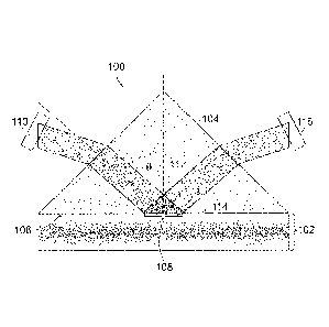
[0021] Referring to FIGURE 1, an apparatus 100 for detecting analytes in
whole blood
without red blood cell separation from the whole blood according to an aspect
of the present
disclosure includes a channel 102 for receiving a blood sample and a prism 104
adjacent to the
channel 102. The prism 104 includes a first surface 106 abutting the channel
102 and defining
an optical interface 108 between the prism 104 and the blood sample when the
blood sample is
received in the channel 102.
[0022] The apparatus 100 also includes a first light source 110 directed
through the prism
104 to the optical interface 108 at an angle of incidence 112 greater than or
equal to a critical
angle relative to a normal axis 114 of the interface. The angle of incidence
112 of optical
illumination in the prism 104 is greater than the critical angle of the
prism/plasma interface 108.
The angle of incidence 112 creates total internal reflection of light from the
first light source 110
CA 03022750 2018-10-30
WO 2017/200907 PCT/US2017/032613
and creates an evanescent field 114 extending into the channel 102. The
evanescent field 114
extends into a plasma layer of the blood sample adjacent to the interface 108
and decays to
substantially zero before reaching a portion of the channel 102 containing
blood cells.
100231 In an embodiment according to another aspect of the present
disclosure, the apparatus
100 may be configured for hemolysis detection in the whole blood. In this
embodiment the first
light source 110 has an emission wavelength in a range corresponding to a peak
in an absorption
spectra of hemoglobin. The emission wavelength of the first light source may
he between about
410 nanometers and 420 nanometers, for example.
100241 The apparatus 100 also includes a first light detector 116 aimed to
receive the
light from the first light source 110 that has been reflected through the
prism 104 from the
optical interface 108. The first light source 110 may include a first light
emitting diode and the
first light detector 116 may include a first photodiode. In another
illustrative embodiment, the
first light detector 116 may include a spectroscope, for example.
[00251 Comparison circuitry coupled to the first light detector 116 is
configured to identify a
presence of analytes in the evanescent field 114 by comparing intensity of the
light that has been
reflected through the prism 104 at a first wavelength, with a predetermined
intensity. The
predetermined intensity may be an intensity of light emitted from the first
tight source 110, for
example. The comparison circuitry may include one or more processors coupled
to computer
memory, data storage devices and/or communication circuitry and/or one or more
computer
networks. For example, the comparison circuitry may and may include
conventional general
purpose computer equipment or dedicated circuitry incorporated with an optical
analysis module
and configured for measuring and/or comparing signals received by the first
light detector. The
comparison circuitry may also be configured to output and/or store a measured
level of analyte
based on the measurements and/or comparisons of the signals received by the
first light detector,
for example.
[00261 Referring to FIGURE 2, an apparatus 200 for detecting analytes in
whole blood
without red blood cell separation from the whole blood according to another
aspect of the present
disclosure includes a first light source 210 second light source 220 having an
emission
wavelength different than the emission wavelength of the first light source
210 and directed
6
CA 03022750 2018-10-30
WO 2017/200907 PCT/US2017/032613
through the prism 204 to the optical interface 208 at a second angle of
incidence greater than or
equal to the critical angle relative to the normal of the interface. The
second angle of incidence
creates total internal reflection of light from the second light source 220
and creates a second
evanescent field extending into the channel 202. In this embodiment, the
apparatus 200 also
includes a second light detector 226 coupled to the comparison circuitry and
aimed to receive the
light from the second light source 220 that has been reflected through the
prism 204 from the
optical interface 208. In an illustrative embodiment, the comparison circuitry
may be configured
to compare the intensity of the light received by the first light detector 216
from the first light
source 210 'with an intensity of the light received by the second light
detector 226 from the
second light source 210.
100271 In an illustrative embodiment, the flow cell 230 may be a
conventional flow cell
bonded to a conventional prism 204, for example. The prism 204 may have a
rectangular face so
that the flow cell 230 can be much longer than. the optical path-length
through the prism 204.
According to aspects of the present disclosure, the prism 204 and/or the flow
cell 230 may be
made from injection molded plastic or other inexpensive materials, for
example. In alternative
embodiment according to an aspect of the present disclosure, the apparatus 200
may include a
prism 204 in which the channel 202 may be formed within the prism 204.
Referring to FIG. 3,
the prism 304 in this embodiment includes a flow cell channel 302 that has
been patterned into
one face of the prism 304 to produce a measurement region inside the prism
304.
100281 An embodiment of the disclosed apparatus may configured as a simple
device, having
only one or two LEDs or laser diodes as light sources, one or two photo-diodes
as light detectors,
and a prism. The prism may have an integrated flow cell channel as shown in
FIG. 3. In an
illustrative embodiment, the entire apparatus could be configured in a package
having millimeter
scale dimensions, for example.
[00291 FIG. 4 shows a graph 400 of light absorbance by a fluid sample
having 4.5 grams per
deci-liter of free hemoglobin in units of mini-optical density versus
wavelength of the detected
light. The graph 400 shows an absorption peak 402 of hemoglobin in the blue
410 nm - 420 ran
portion of the optical spectrum, which is about ten times higher than minor
peaks 404 at about
540 nm and 406 at about 570 nm in the green portion of the optical spectrum,
and 100 times to
7
CA 03022750 2018-10-30
WO 2017/200907
PCT/US2017/032613
1000 times higher than peaks in the red portion of the optical spectrum.
Configuring the light
source with a wavelength in the blue 410 ¨ 420 rim range for hemolysis allows
sufficient
absorption of the light by hemoglobin in the narrow cell-free boundary of the
channel and allows
a good signal to noise ratio in the light received by the light detector.
[0030] FIG. 5
shows a graph 500 of light absorbance for three samples having different
levels of hemolysis in units of milli-optical density versus wavelength of the
detected light. The
graph 500 shows a first signal 502 that shows no detectable peak for a sample
having no
hemolysis. A second signal 504 represents a sample having 2% hemolysis and has
a peak of
about 4 milli ¨ optical density units in the 410 nm ¨ 420 rim range. A third
signal 506 represents
a sample having 8% hemolysis and has a strong peak of about 18 milli-optical
density units in
the 410 nm ¨420 nm range. This shows that detection of absorbed light in the
410 urn ¨420 run
range by a fluid sample is a strong indicator of an amount of an amount of
hemolysis in the
sample.
[0031] FIG. 6
shows a graph 600 of absorbance in units of milli-optical density versus
response time in seconds for six sample of fluid having different levels of
hemolysis. The graph
600 that at a time of (trn) of measurement that is about 4 to 5 seconds after
a measurement start
time (t0) there is a significant separation of signal levels representing
absorption in the 410 ¨ 420
nm range for distinguishing different levels of hemolysis in a sample. For
example at the time of
measurement, a first signal 602 representing a first sample having no
hemolysis indicates no
absorption in the received light at wavelengths of 410 nrn ¨ 420 nm. At the
time of measurement
a second signal 604 representing a second sample having 50 mg/c1I, of
hemolysis indicates
absorption of about 1 milli-optical density units in the received light at
wavelengths of 410 nm
420 run. At the time of measurement a third signal 606 representing a third
sample having 100
mg/a, of hemolysis indicates absorption of about 1.5 milli-optical density
units in the received
light at wavelengths of 410 rim ¨420 nm. At the time of measurement a fourth
signal 608
representing a fourth sample having 200 mg/dL of hemolysis indicates
absorption of about 2.5
milli-optical density units in the received light at wavelengths of 410 nm 420
urn. At the time
of measurement a fifth signal 610 representing a fifth sample having 400
mg/di, of hemolysis
indicates absorption of about 5 milli-optical density units in the received
light at wavelengths of
410 rim ¨ 420 urn. At the time of measurement a sixth. signal 612 representing
a sixth sample
8
CA 03022750 2018-10-30
WO 2017/200907 PCT/US2017/032613
having 800 Ing/dI., of hemolysis indicates absorption of about 11.5 milli-
optical density units in
the received light at wavelengths of 410 nm ¨ 420 nm.
[00321 Referring to FIG. 7, another aspect of the present disclosure
includes a method 700
for detecting analytes in whole blood without red blood, cell separation from
the whole blood. At
block 702, the method 700 includes receiving a whole blood sample in a
channel. A prism
adjacent to the channel includes a first surface abutting the channel and
defining an optical
interface between the prism and the blood sample when the whole blood sample
is received in
the channel. At block 704, the method also includes directing a first light
source through a
prism to the optical interface at an angle of incidence greater than or equal
to a critical angle
relative to a normal of the interface. The angle of incidence creates total
internal reflection of
light from the first light source and creates an evanescent field extending
into the channel. At
block 706, the method also includes aiming a first light detector to receive
the light from the first
light source that has been reflected through the prism from the optical
interface. At block 708,
the method includes measuring absorption of the light from. the first light
source by an analytes
in only a plasma layer of the whole blood sample within range of the
evanescent field.
[00331 An apparatus for determining an analyte content in blood, according
to another aspect
of the present disclosure includes an optical boundary between a flowing blood
sample and an
optically transmissive media, such as a prism, for example. The apparatus
includes an
evanescent optical field in the flowing blood adjacent to the boundary, and a
light detector such
as a photo-diode or a spectroscope configured to detect absorption of light in
the evanescent field
at a wavelength corresponding to an absorption wavelength of the analyte.
According to another
aspect of the present disclosure, the apparatus also includes a light emitter,
such as an light
emitting diode or other light source, configured to direct light onto the
optical boundary at a
wavelength corresponding to the absorption wavelength of the analyte and at an
angle of
incidence selected to provide total internal reflection of the light within
the optically transmissive
media. According to another aspect of the present disclosure, the apparatus
also includes a
channel containing the flowing blood, wherein the channel is configured to
generate a cell free
layer of the flowing blood at a boundary of the channel. and wherein the
boundary of the channel
comprises the optical boundary.
9
CA 03022750 2018-10-30
WO 2017/200907 PCT/US2017/032613
(00341 In an illustrative embodiment the apparatus is configured for
determining free
hemoglobin content in the cell free layer of the flowing blood. In this
embodiment, according to
an aspect of the present disclosure, the light emitted by the light emitter
has a wavelength of
between 410 nanometers and 420 nanometers, and the light detector is
configured to detect light
absorption at wavelengths between 410 nanometers and 420 nanometers.
[0035] Although aspects of the present disclosure are described herein in
the context of
hemolysis, it should be understood by persons skilled in the art that aspects
of the present
disclosure can be implemented for detecting various analytes and other
constituents in a plasma
fraction of body fluid sample.
[00361 What is claimed is: