Note : Les descriptions sont présentées dans la langue officielle dans laquelle elles ont été soumises.
CA 03030238 2019-01-08
WO 2018/015778
PCT/IB2016/001164
A METHOD OF MODULATING EPILEPTOGENICITY IN A PATIENT'S
BRAIN
FIELD OF THE INVENTION
The invention relates to a method of modulating
epileptogenicity in a patient's brain.
BACKGROUND OF THE INVENTION
Personalized medicine proposes the customization of
healthcare with medical decisions, practices and products
being tailored to an individual patient. Individual
variability has clear effects upon the responsiveness to
treatment approaches. Thus, diagnostic testing is often
employed for selecting appropriate and optimal therapies
based on the context of a patient's genetic content or
other molecular and cellular analysis. Historically,
personalized medicine uses heavily genetic information,
but finds more and more viability on the systems level.
Structural and functional neuroimaging play a key role
and have already contributed concrete diagnostic tools
that are though mostly restricted to neurology, such as
presurgical evaluation of epilepsy or differential
diagnosis of coma. Other domains such as psychiatry
suffer from a void of diagnostic tools for routine
clinical practice.
One solution to this issue is to link the interpretation
of neuroimaging signals to computational brain models. So
far, modeling has focused on reproducing the set of
functionally active links between brain areas, but has
been hampered by the stationary nature of most
CONFIRMATION COPY
CA 03030238 2019-01-08
WO 2018/015778
PCT/IB2016/001164
2
connectivity based metrics applied to validate the
models. In fact, most meaningful situations and tasks in
neuroscience pose themselves as non-stationary processes
including the resting state, as well as cognitive and
motor behaviors. The same applies to pathological
behaviors also, of which seizure recruitment in epilepsy
is one example.
In partial epilepsy, seizures originate in a local
network, the so-called Epileptogenic Zone (EZ), before
recruiting other brain regions, the so-called Propagation
Zone (PZ). Correctly delineating the EZ is essential for
successful interventions as, for example, resective
surgery.
Accordingly, a need exists for identifying an EZ in the
brain of an epileptic patient, and for modulating
epileptogenicity in said patient's brain, which would
allow a successful intervention of said patient.
SUMMARY OF THE INVENTION
The invention relates to a method of modulating
epileptogenicity in a brain of an epileptic patient
comprising the steps of: providing a virtual brain;
providing a model of an epileptogenic and propagation
zones and loading said models in the virtual brain to
create a virtual epileptic brain; acquiring data of the
brain of the epileptic patient; identifying, in said
data, a location of at least one possible epileptogenic
zone; fitting the virtual epileptic brain against the
data acquired from the epileptic patient and
parametrizing at least one possible subset of said
epileptogenic zone in the virtual epileptic brain as an
CA 03030238 2019-01-08
WO 2018/015778
PCT/IB2016/001164
3
epileptogenic zone; and simulating, within the virtual
epileptic brain, the effect of a network modulation
mimicking a clinical intervention of the brain of the
patient.
Preferentially, - the virtual brain is a computerized
platform modelling various zones or nodes of a primate
brain and connectivity between said zones or nodes; - the
model of the epileptogenic zone is a mathematical model
describing the onset, the time-course and the offset of
epileptic discharges in said zone; - the mathematical
model of the epileptogenic zone is defined by state
variables describing fast discharges, defining spike and
wave events in the discharges, and a variable being a
slow permittivity variable, and differential equations; -
the structural data comprise magnetic resonance imaging,
diffusion-weighted magnetic resonance imaging, nuclear
magnetic resonance imaging, and/or magnetic resonance
tomography images data of the brain of the patient; - the
method further comprises the step of reconstructing the
patient brain in the virtual brain; - the method further
comprises the step of identifying, in the acquired
structural data of the patient brain, anomalies, and
incorporating said anomalies in the virtual brain; - the
method further comprises the step of identifying one or a
plurality of possible propagation zones and of one or a
plurality of possible other zones and parametrizing said
possible propagation and other zones as propagation and
other zones in the virtual brain; for
the
parametrization of the possible epileptogenic,
propagation and other zones, an excitability parameter
characterizing the degree of epileptogenicity is used; -
for the identification of the degree of epileptogenicity
of epileptogenic and propagation zone, the excitability
parameter is fit against functional patient data; a
plurality of simulations is carried out for a plurality
CA 03030238 2019-01-08
WO 2018/015778
PCT/IB2016/001164
4
of possible epileptogenic zones, distributions of
excitability parameters, and other network modulations
including resections and stimulations, mimicking the
effect of a clinical intervention.
BRIEF DESCRIPTION OF THE DRAWINGS
Other features and aspects of the present invention will
be apparent from the following description and the
accompanying drawings, in which:
Fig. 1 represents a spatial distribution map of
epileptogenicity, in a virtual brain network, for the
implementation of the method according to the invention;
Figs. 2A and 2B show, respectively, a simple and a
complex epileptic seizures that have been recorded for an
epileptic patient for the implementation of the method
according to the invention;
Figs. 3A, 3B and 3C are navigation charts that
relate to, respectively, the left thalamus, the left
hypothalamus and the left fusiform cortex providing from
simulations of epileptic discharges according to the
method of the invention;
Figs. SA, 5B and 5C are images showing,
respectively, the clinician's prediction of epileptogenic
and propagation zones in a patient's brain, a first
simulation of such zones in the virtual brain, and a
second simulation of such zones in said virtual brain
obtained using a prior data fitting, according to the
method of the invention;
Figs. 6A and 6B are graphs that demonstrate the
capacity of the method according to the invention to
identify minimally invasive approaches that may allow to
stop epileptic seizure propagation.
CA 03030238 2019-01-08
WO 2018/015778
PCT/IB2016/001164
DETAILLED DESCRIPTION OF THE INVENTION
5 The invention relates to a method of reducing
epileptogenicity in a patient's brain by identifying and
modulating the epileptogenic zone.
Epilepsy is a group of neurological diseases
characterized by epileptic seizures. Epileptic seizures
are episodes that can vary from brief and nearly
undetectable to long periods of vigorous shaking. In
epilepsy, seizures tend to recur, and have no immediate
underlying cause. The cause of most cases of epilepsy is
unknown, although some people develop epilepsy as the
result of brain injury, stroke, brain tumours, infections
of the brain, and birth defects. Known genetic mutations
are directly linked to a small proportion of cases.
Epileptic seizures are the result of excessive and
abnormal nerve cell activity in the cortex of the brain.
Epilepsy can often be confirmed with an
electroencephalogram (EEG). In partial epilepsy, seizures
arise from a localized area or network, called the
epileptogenic zone (EZ). They are called partial
seizures. Partial seizures can be asymptomatic, and their
spread to downstream brain regions is often linked to the
emergence of clinical symptoms including cognitive
impairment and loss of consciousness. How brain areas are
recruited during seizure propagation is not well
understood. Intracranial depth or stereotactic
electroencephalograms (SEEGs) are commonly used to
delineate the EZ in drug-resistant patient candidates for
neurosurgery. In clinical practice, direct stimulation of
brain regions with intracranial electrodes is used to
localize epileptogenic regions and assess their degree of
epileptogenicity. Time delays of seizure recruitment have
also been considered to be indicative for the strength of
CA 03030238 2019-01-08
WO 2018/015778
PCT/IB2016/001164
6
epileptogenicity, but remain controversial as there is a
large degree of spatial and temporal variation of
propagation, even within the same patient. Seizure
propagation from the epileptogenic zone toward
neighboring zones has been observed experimentally.
According to a first step of the invention, a virtual
brain is provided.
The virtual brain is a computerized platform modelling
various zones or nodes of a primate brain and
connectivity between said zones or nodes. An example of a
virtual brain is disclosed in the publication document
entitled "The Virtual Brain: a simulator of primate brain
network dynamics", Paula Sanz Leon et al., 11 June 2013,
which is incorporated herein, by citation of reference.
In this document, the virtual brain is disclosed as a
neuro-informatics platform for full brain network
simulations using biologically realistic connectivity.
This simulation environment enables the model-based
inference of neurophysiological mechanisms across
different brain scales that underlie the generation of
macroscopic neuroimaging signals including functional
Magnetic Resonance Imaging (fMRI), EEG and
Magnetoencephalography (MEG). It allows the reproduction
and evaluation of personalized configurations of the
brain by using individual subject data.
According to a further step of the invention, a model of
an epileptogenic zone (EZ) and a model of the propagation
of an epileptic discharge from an EZ to a propagation
zone (PZ) are provided, and loaded in the virtual brain.
The model of the epileptogenic zone is a mathematical
model describing the onset, the time-course and the
offset of epileptic discharges in said zone. Such a model
CA 03030238 2019-01-08
WO 2018/015778
PCT/IB2016/001164
7
is disclosed, for example, in the publication document
entitled "On the nature of seizure dynamics", Jirsa et
al., Brain 2014, 137, 2210-2230, which is incorporated
herein, by citation of reference. This model is named
Epileptor. It comprises five state variables acting on
three different time scales. On the fastest time scale,
state variables xi and yl account for the fast discharges
during the seizure. On the slowest time scale, the
permittivity state variable z accounts for slow processes
such as variation in extracellular ion concentrations,
energy consumption, and tissue oxygenation. The system
exhibits fast oscillations during the ictal state through
the variables xi and yi. Autonomous switching between
interictal and ictal states is realized via the
permittivity variable z through saddle-node and
homoclinic bifurcation mechanisms for the seizure onset
and offset, respectively. The switching is accompanied by
a direct current (DC) shift, which has been recorded in
vitro and in vivo. On the intermediate time scale, state
variables x2 and y2 describe the spike-and-wave
electrographic patterns observed during the seizure, as
well as the interictal and preictal spikes when excited
by the fastest system via the coupling g(xi) . The
equations of the model read as follows:
1-5T2 ¨y
2 = ¨ x0)¨ 2)
eco
3
-y2 ¨ x2 /2 +0.002g(x) ¨ 0.3(2 ¨33)=
Y2 ¨ -Y2 f2(xi,x2))
2
where
CA 03030238 2019-01-08
WO 2018/015778
PCT/IB2016/001164
8
1
,x3 ¨ax2
1 1 if xi < 0
(x2 ¨ 0.6 (4- ¨ 4)2 >, if xi ....> 0
(x ¨ 0 if x2 < ¨0.25 [6(x2 + 0.25)x1
2 if x .?.. ¨0.25
t
g(X1)= J67(1-7))C (c)dc
;
and xo = -1.6; To = 2857; T2 = 10; Ii = 3.1; 12 = 0.45; y =
0.01. The parameter xo controls the tissue excitability,
and is epileptogenic triggering seizures autonomously, if
xo is greater than a critical value, xoc = -2.05;
otherwise the tissue is healthy. Ii and 12 are passive
currents setting the operating point of the model.
The model of the propagation zone is identical to the one
of an EZ, however with an excitability parameter inferior
to the critical value xoc = -2.05. All other brain areas
may be modelled by Epileptors with excitability values
far from the threshold, or equivalently standard neural
population models as disclosed in Paula Sanz Leon et al.,
11 June 2013, which is incorporated herein, by citation
of reference. The coupling between brain areas follows a
mathematical model as disclosed in the publication
document entitled "Permittivity Coupling across Brain
Regions Determines Seizure Recruitment in Partial
Epilepsy", Timothee Proix et al., The Journal of
Neuroscience, November 5, 2014, 34(45):15009-15021, which
is incorporated herein, by citation of reference.
Permittivity coupling quantifies the influence of
CA 03030238 2019-01-08
WO 2018/015778
PCT/IB2016/001164
9
neuronal fast discharges xi j of a remote region j on the
local slow permittivity variable of a region i. Changes
in ion homeostasis are influenced by both local and
remote neuronal discharges via a linear difference
coupling function, which quantifies the deviation from
the interictal stable state as a perturbation
perpendicular to the synchronization manifold. The
linearity is justified as a first order approximation of
the Taylor expansion around the synchronized solution.
Permittivity coupling further includes the connectome
a scaling factor G, which both are absorbed in
Ky=GC,,
:,. The permittivity coupling from area j to area
N
T.
i reads Po , where xi
denotes the
signal transmission delay.
When loading the models of the epileptogenic zone (EZ)
and propagation zone (PZ) in the virtual brain, the
signal transmission time delays are here neglected,
because synchronization effects will not be considered,
but rather only the epileptic spread, which is determined
by the slow dynamics of the permittivity coupling.
Mathematically, the virtual brain then corresponds to the
following equations:
'XV '''. ¨ *i'
yll
..... .,.. / N2
1 . ( N
ii= - 4Cri ¨ xo),¨; ¨I; = (x14. ¨
j
3
1 7
YU =-----'17;V:2.1 + A (CWX;21,1 0
CA 03030238 2019-01-08
WO 2018/015778
PCT/IB2016/001164
According to a further step of the invention, structural
and functional data of the brain of the epileptic patient
are acquired. Structural data are for example images data
of the patient brain acquired using magnetic resonance
5 imaging (MRI), diffusion-weighted magnetic resonance
imaging (DW-MRI), nuclear magnetic resonance imaging
(NMRI), Or magnetic resonance tomography (MRT).
Functional data are for example electroencephalographic
signals of the patient brain acquired through EEG or
10 SEEG techniques.
According a further step of the invention, a structural
reconstruction of the patient brain is carried out in the
virtual brain, using the structural data acquired for
said patient brain.
Indeed, the non-invasive structural neuroimaging using
MRI and dMRI allows reconstruction of the patient's
individual brain network topography and connection
topology within a 3D physical space of the virtual brain.
Preferentially, the structural anomalies identified in
the patient brain structural data are incorporated into
the virtual brain.
Indeed, dramatic structural changes are induced by
anomalies changing the topology of the structural network
and thus altering the dynamical properties of the seizure
recruitment.
The structural anomalies are, for example, malignant or
non-malignant brain tumours including hamartoma, strokes,
pachygyria.
They generally appear as white or dark spots in the MRI
images.
CA 03030238 2019-01-08
WO 2018/015778
PCT/IB2016/001164
11
According to a further step of the invention, the
location of one or a plurality of possible epileptogenic
zones, one or a plurality of possible propagation zones
and of one or a plurality of possible other zones are
initially identified in the functional data of the
patient brain, and corresponding zones are parametrised
as epileptogenic, propagation or other zones in the
virtual brain. This initial parameter setting serves as a
prior for the subsequent data fitting procedures.
Indeed, non-invasive functional neuroimaging informs the
clinician expert on the evolution of the epileptic
seizure and allows the formulation of hypotheses on the
location of the EZ, i.e. the hypothetical area in the
brain responsible for the origin and early organisation
of the epileptic activity. The PZ comprises areas that
are recruited during the seizure evolution, but that are
by themselves not epileptogenic. Parameters are initially
set in the virtual brain network model following the
hypothesis on the EZ. Practically, a spatial map of
epileptogenicity is defined in the virtual brain, as
shown in Fig. 1. In this map, each node is characterized
by an excitability value xo, which quantifies the ability
of the model of a zone to trigger a seizure. For an
isolated zone, G = 0, the model can trigger seizures
autonomously if xo > xoc and is referred to as
epileptogenic. Inversely, if xo < xoc, the model does not
trigger seizures autonomously and is not epileptogenic.
The spatial map of epileptogenicity comprises the
excitability values of the EZ, the PZ and all other zone.
Of course, only the nodes in the EZ discharge
autonomously while embedded in the virtual brain.
The subsequent data fitting is thus carried out, the
target for said data fitting being the excitability
CA 03030238 2019-01-08
WO 2018/015778
PCT/IB2016/001164
12
parameter xo, which is estimated using automated
approaches. Obtaining such estimates of the parameters of
the network model, given the available functional data is
performed within a Bayesian framework, using a reduced
Epileptor model and reduced functional data set for the
fitting. The SEEG data are windowed and Fourier
transformed to obtain estimates of their spectral density
over time. Then SEEG power above 10 Hz is summed to
capture the temporal variation of the fast activity.
These time series are corrected to a preictal baseline,
log-transformed and linearly detrended over the time
window encompassing the seizure.
Hidden states in Bayesian modeling represent states of
the generative model that are not directly observable.
Uninformative priors are placed on the hidden states'
initial conditions, while their evolution follows a
Euler-Maruyama discretization of the corresponding
stochastic differential equations with linear additive
normally distributed noise. Uninformative priors are
placed on the excitability parameter per node xo,
observation baseline power, scale and noise. Finally, the
length of the seizure is also allowed to freely vary to
match that of a given recorded seizure. Structural
connectivity specifies a gamma prior on the connectivity
used in the generative method. This model is implemented
using a software for Bayesian inference, which implements
both Hamiltonian Monte-Carlo and automatic variational
inference algorithms for generic differential probability
models. This approach takes advantage of the efficiency
of the variational algorithm, which constructs an
approximate proxy distribution on the true posterior
optimized via stochastic gradient ascent.
According to further steps of the invention, a simulation
of the propagation of an epileptic discharge from said
CA 03030238 2019-01-08
WO 2018/015778
PCT/IB2016/001164
13
possible epileptogenic zone to other zones is carried out
in the virtual brain under systematic variation of model
parameters. These parameter variations correspond to
network modulations that may have inverse effects upon
the seizure numbers in different brain regions and are
thus non-trivial. Systematic simulations and
quantifications of these modulation effects provide
parameter spaces indicating the number of seizures in the
virtual brain and the extent of seizure propagation.
Changes in parameters are directly linked to therapeutic
network interventions, though the link is not always
evident, since the variation of a network parameter may
find different realizations in clinical practice. For
instance, the excitability of a brain region in the
network node model is a key parameter, which is
physiologically linked to variables such as balance of
excitation and inhibition, local synaptic efficacy,
extracellular ionic concentrations, or glial activity.
Alterations of these variables will result in
excitability changes in the tissue, and thus in the
desired network effects predicted by the virtual brain
model.
Practically, the patient's brain network model is
evaluated via simulation, data fitting and mathematical
analysis. It is used to "fingerprint" individual patient
brains by identifying a personalized parameter set
through data fitting. Systematic
computational
simulations further generate parameter maps outlining the
zones of seizures and seizure freedom. These maps will
give guidance of how to tune model parameters. The result
of this evaluation predicts the most likely propagation
patterns through the patient's brain and allows the
exploration of brain intervention strategies.
CA 03030238 2019-01-08
WO 2018/015778 PCT/IB2016/001164
14
The method according to the invention improves the
surgical outcome. First, following non-invasive EEG/MEG
and invasive SEEG exploration, the EZ hypotheses are fit
to the data and improved. Second, systematic network
modulations mimick clinical interventions strategies and
can be used to identify novel therapeutic strategies.
Modulations include stimulation paradigms, lesioning of
network links, resections of brain areas and changes of
local brain region parameters such as excitability. For
instance, surgical strategies are tested within the
virtual brain. So far, traditional approaches to surgery
apply one focal resection or ablation at the hypothesized
EZ, based on the dogmatic concept that medically
refractory epilepsy is ultimately a focal disease. A
large unknown remains the size, the number and the
specific anatomical location of possible resections or
thermal lesions designed to modulate large-scale
epileptic networks. The invention allows not only to
parametrically vary the size of the resection focus, but
also to employ multiple lesions at different locations
making thus full use of the network nature of the virtual
epileptic brain model. Technically, this is possible
nowadays: stereotactic-guided laser technology, for
instance, permits the modulation of large-scale networks
by allowing the placement of multiple lesions in key
components of previously mapped epileptic networks.
EXAMPLE: Identification of an epileptogenic zone in the
brain of a patient diagnosed with bitemporal epilepsy
A right-handed 41-year-old female patient initially
diagnosed with bitemporal epilepsy
underwent
comprehensive presurgical evaluation, including clinical
history, neurological examination, neuropsychological
testing, structural and diffusion MRI scanning, EEG and
SEEG recordings along with video monitoring. Nine SEEG
CA 03030238 2019-01-08
WO 2018/015778
PCT/IB2016/001164
electrodes were placed in critical regions based for the
presurgical evaluation. SEEG electrodes comprised 10 to
15 contacts. Each contact is 2 mm of length, 0.8 mm in
diameter and is 1.5 mm apart from other contacts. Brain
5 signals were recorded using a 128-channel DeltamedTM
system (sampling rate: 512 Hz, hardware band-pass
filtering: between 0.16 and 97 Hz). Structural and
diffusion MRI were acquired with a SiemensTM MagnetomTM
VerioTM 3T Scanner. Ti-weighted images were acquired with
10 a MPRAGE-sequence (TR = 1900 ms, TE = 2.19 ms, voxel size
= 1 x 1 x 1 mm3, 208 slices). The diffusion acquisition
used a DTI-MR sequence (angular gradient set of 64
directions, TR = 10.7 s, TE = 95 ms, 70 slices, voxel
size = 2 x 2 x 2 mm3, b-value = 1000s/mm2).
Structural reconstruction was then carried out. The
large-scale connectivity and the cortical surface of the
patient were reconstructed using SCRIPTSm, a processing
pipeline tailored for the virtual brain. The brain is
divided in several regions according to a parcellation
template, which is used for whole brain tractography to
develop the connectivity and delay matrices. Cortical and
subcortical surfaces are reconstructed and downsampled,
along with a mapping of vertices to corresponding region
labels. All processed data are formatted to facilitate
import into the virtual brain.
The MRI examination revealed a hypothalamic hamartoma.
Surface EEG recordings revealed interictal spikes and
indicated a bias towards the left hemisphere. Based on
the presurgical evaluation, seven SEEG electrodes were
implanted in the left hemisphere, and two in the right
hemisphere. One electrode was implanted in the
hypothalamic hamartoma. Figs. 2A and 2B show the
implantation scheme in the left column. During two weeks
of continuous SEEG recordings, 6 simple seizures
CA 03030238 2019-01-08
WO 2018/015778
PCT/IB2016/001164
16
localized in the right hippocampus and two complex
seizures starting in the right hippocampus and then
recruiting the left hippocampus, the left temporal lobe
and the hypothalamic hamartoma were recorded.
Representative seizure propagation patterns are shown in
Figs 2A and 2B.
This structural anomaly was integrated into the model.
Here, a hypothalamic hamartoma was integrated via a
modification of the local connectivity ' 'of the
hypothalamus. This hamartoma was delineated in the MRI
scan. It was used as a seed region of interest to
reconstruct the local connectivity. The local
connectivity strength was scaled up parametrically by the
scalar factor Ghyp to quantify the effect of the hamartoma
without changing its local connection topology.
Each node of the virtual brain network was loaded with
the Epileptor model. The nodes were connected via
permittivity coupling, which acts on a slow time scale
and allow the spread of the seizure through the network
by recruiting regions not in the HZ. The excitability
parameters for HZ, PZ and all other regions according to
clinical criteria comprising (i) regions involved in the
seizure; (ii) seizure length; (iii) length of time delays
before recruitment of other regions; (iv) seizure
frequency in each region, were set. The spatial
distribution of excitability was then heterogeneous
across the network, with high value of excitability for
regions in the HZ (xo > xoc + 0.4), smaller excitability
values for regions in the PZ (xoc + 0.4 > xo xoc), and
other nodes not epileptogenic (xo < xoc) . Once HZ and PZ
were defined, the systematic network modulation was
performed using a parameter space exploration by varying
the following parameters: (i) the global coupling
strength G, which is a scalar factor multiplying the
CA 03030238 2019-01-08
WO 2018/015778 PCT/IB2016/001164
17
whole connectivity matrix, (ii) the local coupling
strength of the hypothalamus Ghyp, which is a scalar
factor multiplying the contribution of the hypothalamus
to the connectivity matrix, (iii) the excitability values
xoright hippocampus of the right hippocampus, (iv) the
excitability values xoc)ther regions of the regions not
recruited in the propagation zone. The excitability
values of the other regions in the EZ and the PZ were
fixed as in Table 1 hereunder where xo=xoc+Axo.
Name of the region:o Zones
Right hippocampus 1.3 EZ
Left hippocampus 0.4 EZ
Left hypothalamus 0.4 EZ
Right hypothalamus 0.4 EZ
Brain Stem 0.31 PZ
Left parahippocampal 0.27 PZ
Left thalamus 0.24 PZ
Left temporal pole 0.16 PZ
Other regions -0.2 Other regions
To describe the virtual brain network behaviour in the
thus four-dimensional parameter space, the clinical
criteria i) through iv) for seizure quantification were
used. Figs. 3A to 30 show one of these quantifiers, the
frequency of recruitment for three different regions in a
seizure over a fixed simulation time as a function of the
four parameters G, Ghyp, xo right hippocampus and xo t her regions.
They illustrate the results of the systematic parameter
space explorations. These navigation charts offer the
clinician a tool for decision-making and hypothesis
CA 03030238 2019-01-08
WO 2018/015778
PCT/IB2016/001164
18
building. For instance, the figures demonstrate for this
particular patient that changes of excitability in the EZ
regions show fairly little influence on the number of
seizures in the VEP brain model, whereas reduction of
excitability outside of EZ/PZ regions is linked to
seizure reduction in the left thalamus and hypothalamus,
and to a lesser extent in the left parahippocampus (Figs
3A and 3B). A decrease of left hypothalamic connectivity
will always cause an increase of seizures in the left
hypothalamus, but not the left thalamus. The only means
of increasing the likelihood for seizures in the left
thalamus is the increase of the scaling of global
coupling G, while maintaining high values of hypothalamic
connectivity (Figure 3A). For all of the above scenarios,
the left parahippocampus shows fairly high seizure
numbers with one exception, that is high hypothalamic
connectivity and low overall strength of global coupling
G (Figure 30).
A representative set of parameters (G = 10, Ghyp= 10,
,6x0right hippocampus = 1 . 3, ,6x00ther regions = 0.2)
were selected
corresponding to the dot in Figs. 3A to 30 matching the
patient's seizure with regard to the clinical criteria i)
through iv). The virtual brain network model was
simulated over a period of 20 seizures and computed the
forward solution for the SEEG electrodes. Simple seizures
and complex seizures were generated with similar regions
recruited compared to the real SEEG recordings of Figs.
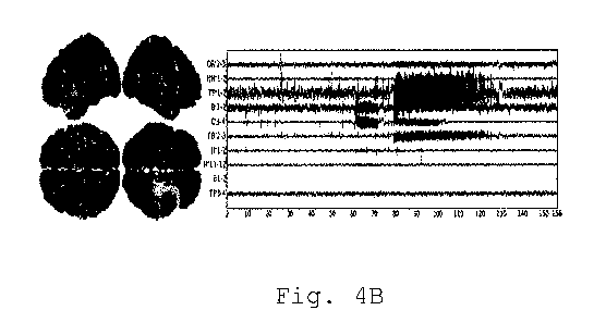
2A and 2B. These seizures are shown in Figs. 4A and 4B.
Fig. SA shows the spatial extent of the EZ and the PZ
such as estimated by clinician expertise. Fig. 53 shows
the spatial extent of the excitability zone expressed
= X
through the parameter distribution of
illustrated via its deviations .(3.) from the critical
value xoc = -2.05. Fig. 50 shows the comparison of the
CA 03030238 2019-01-08
WO 2018/015778
PCT/IB2016/001164
19
distribution of excitabilities found by fitting the model
to the SEEG data. In those figures, the EZ are
represented in light clear zones. It appears that data
fitting allows to identify a bilateral mesial temporal
EZ, a result well in agreement with the clinical
interpretation.
Figs. 6A and 6B demonstrate the capacity of the invention
to identify minimally invasive approaches to stop seizure
propagation as a function of the epileptogenic zone. In
Fig. 6A, the colour code (black/white) indicates seizure
propagation (white) or not (black). In Fig. 65, the size
of the propagation zone is plotted as a function of the
epileptogenic zone. For the virtual epileptic brain, a
small number of lesions is sufficient to stop seizure
propagation, up to 6 lesions as appearing in Fig. 6A. The
PZ reduces to 0, 1 or 2 areas after 5 to 6 lesions. If
the virtual brain's PZ is composed of 0 to 2 areas, the
network is not able to recruit any other regions.