Note : Les descriptions sont présentées dans la langue officielle dans laquelle elles ont été soumises.
CA 03030454 2019-01-09
WO 2018/017556 PCT/US2017/042560
IMPLANT AND METHOD FOR POSTERIOR SACROILIAC FUSION
CROSS-REFERENCE TO RELATED APPLICATIONS
This application claims priority to US Provisional Patent Application No.
62/363,752
(Attorney Docket No. 44057-712.101) filed on July 18, 2016, as well as US
Provisional
Patent Application No. 62/448,848 (Attorney Docket No. 44057-712.102) filed on
January
20, 2017, each of which is entirely incorporated herein by reference.
BACKGROUND OF THE INVENTION
[0001] The sacroiliac (SI) joint is the juncture between the sacrum at the
base of the spine
and the ilium of the pelvis. The SI joint is a synovial joint in which the
sacral surface has
hyaline cartilage that moves against fibrocartilage of the iliac surface. The
SI joint has
irregular elevations and depressions that produce interlocking of the two
bones.
[0002] Disorders of the SI joint can cause low back and radiating buttock
and leg pain.
Pain associated with the SI joint can be caused by traumatic fracture
dislocation of the pelvis,
degenerative arthritis, sacroili.itis, or other degenerative conditions.
Contributing factors
include post-traumatic injury, accelerated wear/instability after lumbar
fusion, post pregnancy
pain/instability, and longer life span combined with a more active lifestyle
in many patients.
[0003] The SI joint is increasingly being recognized as a pain generator as
SI joint
degenerative disease and instability are being diagnosed and treated more
commonly. It is
estimated that disorders of the sacroiliac joint are a source of pain for
millions of people
suffering from back and radicular symptoms.
[0004] Surgical treatment of these disorders includes stabilization and/or
arthrodesis.
Fusion of the SI joint can be accomplished by several different conventional
methods
encompassing an anterior approach, a posterior approach, and a lateral
approach. These
procedures typically involve fixation of the sacroiliac joint by placement of
one or more
trans-sacroiliac implants or by placement of implants into the Sl pedicle and
iliac bone.
[0005] While these methods have been utilized for fixation and fusion of
the Si joint over
the past several decades, in certain circumstances challenges with respect to
the fixation and
fusion of the Si joint may remain unresolved. Many of the Si joint fusion
procedures on the
market today fixate the SI joint from a challenging lateral approach.
Minimally invasive
procedures such as these are can be technically difficult requiring extensive
surgical training
1
CA 03030454 2019-01-09
WO 2018/017556 PCT/US2017/042560
and experience and may result in a substantial incidence of damage to the
lumbosacral
neurovascular elements. Furthermore, the lateral approach to the SI joint
often fixates the
joint with multiple implants that go across the joint, not offering a true
fusion approach into
the SI joint.
[0006] Additionally, current techniques and instruments typically allow for
either fixation
or fusion and thereby do not resolve both issues. Procedures are often
performed without
adequate removal of the articular joint surfaces or preparation of cortical
bone and thereby do
not always address the degenerative condition of the SI joint.
[0007] Failure to sufficiently stabilize and fuse the Si joint with the
implant structures
and methods may result in a failure to relieve the condition of the SI joint
being treated,
leading to continued or recurrent Si joint pain and instability requiring
additional surgery.
[0008] it would therefore be desirable to provide improved methods,
systems, and
devices that address at least some of these issues.
SUMMARY OF THE INVENTION
[0009] The present disclosure generally relates to medical devices and
methods, and more
particularly relates to implants and methods for fixation and fusion of the
sacroiliac joint.
[00010] It would be desirable to address the sacroiliac joint through a
posterior approach
while delivering both fusion and fixation of the joint. A posterior approach
allows for direct
visualization of the sacroiliac joint. In examples, the direct visualization
of the sacroiliac
joint when using a posterior approach is beneficial for effective
decortication to create a
proper fusion bed. In some embodiments, once the sacroiliac joint has been
decorticated, the
sacroiliac joint can be fixated with an implant. In some embodiments, the
sacroiliac may be
fixated with an implant prior to decortication. Optionally, in any embodiments
the sacroiliac
joint can be pre-packed with bone growth inducing material. Optionally, in any
embodiments
the sacroiliac joint can be post-packed with bone growth inducing material.
Optionally, in
any embodiments the traverse pin within the implant may allow for placement of
bone
growth inducing material extending from the ilium, through the sacroiliac
joint, and into the
sacrum.
[00011] An aspect of the invention provides a sacroiliac joint implant system
for fixating
and promoting fusion between an ilium, a sacrum, and a sacroiliac joint space.
The system
comprises a plate/pin component having a plate component and a pin component.
The plate
component has an iliac portion and a sacral portion, wherein the iliac portion
has a first
aperture, wherein the sacral portion has a second aperture, and wherein the
iliac portion and
2
CA 03030454 2019-01-09
WO 2018/017556 PCT/US2017/042560
said sacral portion have an angle between 95-175 degrees disposed
therebetween. The pin
component is connected with said iliac portion of said plate component, said
pin configured
for placement through said first aperture, through said ilium, across said
sacroiliac joint
space, and into said sacrum. The system further comprises a sacral screw
inserted into said
sacral portion of said plate component, said sacral screw configured for
placement through
said second aperture, through said sacrum.
[00012] Another aspect of the invention provides a sacroiliac joint implant
system for
fixating and promoting fusion between an ilium, a sacrum, and a sacroiliac
joint space. The
system comprises a plate having an iliac portion and a sacral portion, wherein
said iliac
portion has a first aperture, wherein said sacral portion has a second
aperture, and wherein
said iliac portion and said sacral portion have an angle between 95-175
degrees disposed
therebetween. The system also comprises a transverse pin inserted into said
iliac portion of
said plate, said transverse pin configured for placement through said first
aperture, through
said ilium, across said sacroiliac joint space, and into said sacrum, said
transverse pin
comprising a receiving component. Additionally, the system comprises a sacral
screw
inserted into said sacral portion of said plate, said sacral screw configured
for placement
through said second aperture, through said sacrum, and secured into said
receiving
component of said transverse pin.
[00013] A further aspect of the invention provides a method of fixation and
fusion of a
sacroiliac joint comprising an ilium, a sacrum, and a sacroiliac joint space.
The method
comprises decortating of said sacroiliac joint. The method also comprises
broaching a first
channel through said ilium, across said sacroiliac joint space, and into said
sacrum, wherein
said first channel is configured to receive a transverse pin of a sacroiliac
joint implant.
Additionally, the method comprises providing bone growth inducing material
within said
sacroiliac joint space. The method also comprises drilling a second channel
into said sacrum,
wherein said second channel is configured to receive a sacrum screw of said
sacroiliac joint
implant. The method further comprises placing a plate of said sacroiliac joint
implant across
said sacroiliac joint space, said place comprising a sacrum portion and an
ilium portion.
Additionally, the method comprises inserting said transverse pin through said
plate and
through said first channel. The method further comprises inserting said sacrum
screw
through said plate and through said second channel.
[00014] An additional aspect of the invention provides a method of fixation
and fusion of a
sacroiliac joint comprising an ilium, a sacrum, and a sacroiliac joint space.
The method
comprises decorticating the joint space with a rasp. The method also comprises
sliding the
3
CA 03030454 2019-01-09
WO 2018/017556 PCT/US2017/042560
plate over said rasp. Additionally, the method comprises inserting bone screws
on the sacral
side of the plate. The method also comprises removing the rasp through the
plate. The
method further comprises using a box cutter to create a track for the
transverse pin.
Additionally, the method comprises inserting the transverse pin. Further, the
method
comprises inserting bone screws on the iliac side.
[00015] Some embodiments of the apparatus can provide a plate component for
placement
over the posterior inferior end of SI joint. In embodiments, the plate
component may be
integrally connected with a pin component which transversely extends across
the SI joint.
Embodiments may additionally provide one or more bone screws which slides
through the
plate components. Optionally, in any embodiments at least one bone screw may
be inserted
into an opening on the sacral side of the plate, into the sacrum. Optionally,
in any
embodiments at least one bone screw may be inserted into an opening on the
iliac side of the
plate, into the sacrum. In some embodiments, the one or more bone screws may
be straddle
the pin. In some embodiments, one or more bone screws may be threaded into the
pin
component.
[00016] Some embodiments of the apparatus can provide a plate for placement
over the
posterior inferior end of SI joint. Embodiments may also provide a pin which
slides through
an opening on the iliac side of the plate and transversely extends across the
SI joint.
Embodiments may additionally provide a screw which slides through an opening
on the
sacral side of the plate, into the sacrum. In some embodiments, the screw may
thread into the
transverse pin.
[00017] The present disclosure also relates to a method including marking the
joint
location with fluoroscopy, creating an incision over the iliac wing,
dissecting down to the
sacroiliac joint, decorticating the joint space, placing the plate over the
posterior inferior end
of the sacroiliac joint, drilling a hole through the hole on the iliac side of
the plate, into the
ilium, through the sacroiliac joint, and into a sacrum, broaching the hole to
fit the shape of the
transverse pin, inserting the transverse pin into the plate and through the
hole, drilling a hole
through the hole in the sacral side of the plate and into the sacrum, and
inserting the sacral
screw into the plate and through the hole and threading the sacral screw into
the transverse
pin.
[00018] Optionally, in any embodiment the implant can provide a plurality of
bone
ingrowth aperture elements for receiving bone growth material and facilitating
fusion of the
sacroiliac joint.
4
CA 03030454 2019-01-09
WO 2018/017556
PCT/US2017/042560
[00019] Optionally, in any embodiment the methods can include packing the
implant with
bone growth inducing material before and/or after implantation.
[00020] Optionally, in any embodiment the implant can provide an additional
device or
method to guide the placement of holes in the sacrum and ilium. In
embodiments, the plate
may preferably be placed after the drilling/broaching of the holes/void.
Additionally, the
transverse pin may be inserted. Further, the sacral screw may be inserted and
threaded into
the transverse pin.
[00021] Additional optional embodiments will be apparent from the detailed
descriptions
and the claims herein.
BRIEF DESCRIPTION OF THE DRAWINGS
[00022] Figure 1 is a perspective view of an exemplary embodiment of an
implant for
sacroiliac joint fusion.
[00023] Figure 2 is a perspective view of an exemplary embodiment of the
transverse pin.
[00024] Figure 3 is a side cross-section view of an exemplary embodiment of
the
transverse pin in Figure 2.
[00025] Figure 4 is a side view of an exemplary embodiment of the sacral
screw.
[00026] Figure 5 is a top view of an exemplary embodiment of the sacral screw
of Figure
4.
[00027] Figure 6 is a cross-section through an exemplary embodiment of the
plate.
[00028] Figure 7 is a perspective view of the embodiment shown in Figure 1 in
the
sacroiliac joint space.
[00029] Figure 8 is a cross-section through the embodiment in Figure 1 in the
sacroiliac
joint space.
[00030] Figure 9 is a posterior view of a patient with an incision targeting
the posterior
inferior end of the sacroiliac joint.
[00031] Figure 10 is a lateral view of a patient with posterior dissection to
the sacroiliac
joint space.
[00032]
Figure 11 is a lateral cross-sectional view of decortication of the sacroiliac
joint
space.
[00033] Figure 12 is a lateral cross-sectional view of a broach creating a
void across the
sacroiliac joint space shaped to receive a transverse pin.
CA 03030454 2019-01-09
WO 2018/017556 PCT/US2017/042560
[00034] Figure 13 is a lateral cross-sectional view of a drill creating a void
through the
sacrum shaped to receive a sacral screw.
[00035] Figure 14 is a cross-sectional view of the implant being packed with
bone growth
inducing material after insertion in the sacroiliac joint space.
[00036] Figure 15 is a perspective view of the implant comprising a plate,
pin, and two
sacral screws.
[00037] Figure 16A is a perspective view of the implant comprising a plate,
pin, two sacral
screws, and one iliac screw.
[00038] Figure 16B is a perspective view of the implant comprising a plate,
pin, two sacral
screws, and two iliac screws.
[00039] Figure 17A is a perspective view of an exemplary embodiment of a bone
screw.
[00040] Figure 17B is a cross-section view of the embodiment in Figure 17A.
[00041] Figure 17C is a front view of the embodiment in Figure 17A.
[00042] Figure 17D is a top view of the embodiment in Figure 17B.
[00043] Figure 18A is a perspective view of an exemplary embodiment of the
plate/pin.
[00044] Figure 18B is a cross-sectional front view of the embodiment in Figure
18A.
[00045] Figure 18C is a cross-sectional side view of the embodiment in Figure
18A.
[00046] Figure 18D is a top view of the plate/pin shown in Figure 18A.
[00047] Figure 19 is a perspective view of an exemplary embodiment of a
plate/pin.
[00048] Figure 20 illustrates the plate/pin implantation position on a cross-
section of the
sacroiliac joint.
[00049] Figure 21 is a perspective view of another exemplary embodiment of a
transverse
pin.
[00050] Figure 22 is a perspective view of another exemplary embodiment of a
plate.
[00051] Figure 23A is a perspective view of an exemplary embodiment of an
implant
comprising a plate, curved pin, and four bone screws.
[00052] Figure 23B is a cross-sectional view of the sacrum and ilium to
illustrate
implantation position of the plate and curved pin shown in Figure 21.
[00053] Figure 23C is a cross-sectional front view of engagement between the
transverse
pin and plate of Figures 21 and 22, respectively.
[00054] Figure 23D is a cross-sectional side view of engagement between the
transverse
pin and plate of Figures 21 and 22, respectively.
6
CA 03030454 2019-01-09
WO 2018/017556 PCT/US2017/042560
[00055] Figure 24 is a cross-sectional view of the sacrum and ilium to
illustrate
implantation position of the plate and curved pin shown in Figure 21 with the
addition of
bone growth inducing material between the sacrum and the ilium.
[00056] Figure 25 is a perspective view of an exemplary embodiment of an
implant
comprising an integrated plate/pin component having a plate component, a
curved pin
component, and four bone screws.
[00057] Figure 26 is a cross-sectional view of the sacrum and ilium to
illustrate
implantation position of the plate and curved pin shown in Figure 25.
DETAILED DESCRIPTION OF THE INVENTION
[00058] Specific embodiments of the disclosed device and method of use will
now be
described with reference to the drawings. Nothing in this detailed description
is intended to
imply that any particular component, feature, or step is essential to the
invention.
[00059] Figure 1 is a perspective view of an exemplary embodiment of an
implant for
sacroiliac joint fusion. Figure 1 illustrates an embodiment with optional
features, any of
which may be optionally used or substituted with other features in other
embodiments
discussed herein. The embodiment of the implant as provided in Figure 1
illustrates a
transverse pin 1 and a sacral screw 2 that pass through a plate 3. Optionally,
in any
embodiments components of the implant may be made of stainless steel.
Optionally, in any
embodiments components of the implant may be made of titanium. Optionally, in
any
embodiments some components of the implant may be made of stainless steel and
some
components of the implant may be made of titanium. Optionally, in any
embodiments the
transverse pin may be made of porous material. Optionally, in any embodiments
the
transverse pin may be made of porous titanium. Optionally, in any embodiments
the
transverse pin may be made of porous titanium coated for bone ingrowth.
[00060] The implant provided in Figure 1 may be used to immobilize the
sacroiliac joint.
In examples, a plate may be rectangular. In additional embodiments, a plate
may be circular,
triangular, elliptical, or another shape. In some embodiments, a plate may be
curved to match
the curvature of the sacrum and ilium across the sacroiliac joint. As anatomy
may differ
between patients, the curvature of a particular plate may differ from patient
to patient.
Optionally, in any embodiments curvature may be between 95 degrees and 175
degrees
Optionally, in any embodiments curvature may be between 120 degrees and 160
degrees.
Optionally, in any embodiments a plate of the implant, such as plate 3 as seen
in Figure 1,
may contain a cylindrical recess extending through the plate with a
countersink on one end of
7
CA 03030454 2019-01-09
WO 2018/017556 PCT/US2017/042560
the plate for receiving a sacral screw, such as sacral screw 2, and a recess
extending through
the plate with a countersink on the opposite end of the plate shaped to
receive a transverse
pin, such as transverse pin 1. The plate of an implant may be placed across
the sacroiliac
joint. In particular, the plate of an implant may be placed across the
sacroiliac joint such that
a portion of the plate is placed against the sacrum, a portion of the plate is
placed against the
ilium, and a portion of the plate is placed across the sacroiliac joint gap.
Additionally, the
transverse pin may be placed through its corresponding hole in the plate.
Optionally, in any
embodiments the transverse pin may be placed with a sliding fit through its
corresponding
hole in the plate. Optionally, in any embodiments the tranverse pin may be
threaded through
its corresponding hole in the plate. Further, the sacral screw may be placed
through its
corresponding hole in the plate. Optionally, in any embodiments the sacral
screw may be
placed with a sliding fit through its corresponding hole in the plate.
Optionally, in any
embodiments the sacral screw may be threaded through its corresponding hole in
the plate.
In examples, the sacral screw may be placed with a fit through it
corresponding hold in the
plate, through the sacrum, through the aperture in the transverse pin, and
then threaded into
the shaft of the transverse pin.
[00061] Optionally, in any embodiments the sacral screw comprises a
cylindrical body and
cylindrical head Optionally, in any embodiments, the body may be a different
shape, such as
a generally square-ish shape or a generally triangular shape, or another
example of a different
shape. Optionally, in any embodiments the head may be a different shape, such
as a
generally square-ish shape or a generally triangular shape, or another example
of a different
shape. Optionally, in any embodiments the head may have a larger diameter than
the body.
Optionally, in any embodiments the sacral screw may comprise a socket to
receive a driver
that can rotate the screw. Optionally, in any embodiments the socket is a hex
socket. In
some embodiments, the socket may be a different shape. In examples, the
cylindrical body
may consist of a non-threaded portion and threaded distal tip. Optionally, in
any
embodiments transverse pin 1 may comprise a predominantly triangular body of
three
intersecting circular regions with one flat side and head 4. Optionally, in
any embodiments
the transverse pin may comprise a different shape, such as a cylindrical shape
or a rectangular
box-like shape, among other examples. Optionally, in any embodiments head 4
may be of
similar but larger shape than the corresponding body of the traverse pin. The
aperture through
the main body of transverse pin 1 may consist of a flat upper part and angled
lower surface
such that it has a smaller opening on one side and a larger opening on the
opposite side.
Optionally, in any embodiments the angled lower surface by be between 30
degrees and 60
8
CA 03030454 2019-01-09
WO 2018/017556 PCT/US2017/042560
degrees. Optionally, in any embodiments the angled lower surface may be
approximately 45
degrees. Optionally, in any embodiments the angled lower surface may be based
on an angle
of entry that a sacral screw enters when inserted through a plate that has a
sacral component
that is angled based on geometry of a patient's anatomy. Because of this, the
angled lower
surface may have an angle that is customized based upon a patient's anatomy.
In examples,
the angled lower surface may have a receiving component to receive a distal
portion of a
sacral screw. In some embodiments, transverse pin may comprise a threaded
blind hole to
receive the distal threaded tip of sacral screw 2. In examples, an implant as
described herein
can be effectively placed using a posterior surgical approach. In embodiments,
the implants
may be sized according to the local anatomy. In particular, the implants may
be sized
accordingly to the local anatomy of particular patients.
[00062] Figure 2 is a perspective view of an exemplary embodiment of the
transverse pin,
such as transverse pin 1, that may be used in an implant described herein.
Figure 2 illustrates
an embodiment with optional features, any of which may be optionally used or
substituted
with other features in other embodiments discussed herein. Figure 2 shows a
preferred
embodiment of transverse pin 1. Transverse pin 1 may comprise a predominantly
triangular
body of three intersecting circular regions with one flat side and head 4 of
similar but larger
shape as the corresponding body of traverse pin 1. The head of the traverse
pin may engage
with a plate of an implant. In examples, a head 4 of traverse pin 1 may engage
with plate 3,
as shown in Figure 1. As seen in Figure 2, a cylindrical hole 5 extends from
head 4 to
aperture 15. Cylindrical hole 5 may extend from head 4 to aperture 15 so as to
allow for
packing of the aperture with bone growth inducing material after the
transverse pin is
implanted and aperture 15 through transverse pin 1 with a flat upper part and
angled lower
surface such that it has a smaller opening on one side and a larger opening on
the opposite
side.
[00063] Aperture 15 may allow for the insertion of bone growth inducing
material.
Examples of bone inducing material may include biologics, agents, medical
adhesives,
bonding cements, and/or bone healing substances. Optionally, in any
embodiments the
transverse pin may include some features to help contain bone growth inducing
material.
Optionally, in any embodiments aperture 15 may include retaining walls to help
pre-packed
bone growth inducing material during transverse pin insertion. Additionally,
transverse pin 1
may comprise a threaded, blind hole 6 shaped to receive the threaded portion
of a sacral
screw. In particular, traverse pin 1 may comprise a threaded, blind hole 6
shaped to receive
the threaded portion of sacral screw 2, as shown in Figure 1. Hole 6 may
extend normal to the
9
CA 03030454 2019-01-09
WO 2018/017556 PCT/US2017/042560
angled surface of aperture 15, which may contain an internal thread 7 for
connection to sacral
screw 2, as shown in Figure 1.
[00064] Figure 3 is a side cross-section view of an exemplary embodiment of
the
transverse pin in Figure 2. Figure 3 illustrates an embodiment with optional
features, any of
which may be optionally used or substituted with other features in other
embodiments
discussed herein. In particular, Figure 3 shows a cross-sectional view of the
transverse pin 1
in Figure 2. Transverse pin comprises a predominantly triangular body of three
intersecting
circular regions with one flat side and head 4 of similar but larger shape
than the triangular
body of traverse pin 1. Additionally, Figure 3 also illustrates longitudinal
pin axis 17 and
traversing thread axis 18. Thread axis 18 may be offset from pin axis 17 such
that when
transverse pin 1 and sacral screw 2 are attached to the plate, as shown in
Figure 1, the screw
axis 8 of sacral screw 2 is coaxial with thread axis 18. Optionally, in any
embodiments thread
axis 18 may be in-line with a threaded, blind hole shaped to receive the
threaded portion of
sacral screw 2, as shown in Figure 1. Optionally, in any embodiments
transverse pin 1 may
also contain an aperture with a flat upper part and angled lower surface such
that traverse pin
1 has a smaller opening on one side and a larger opening on the opposite side.
Transverse pin
1 may also contain a chamfered, cylindrical hole extending from head 4 to
aperture 15.
[00065] Figure 4 is a side view of an exemplary embodiment of a sacral screw
within an
implant. Figure 4 illustrates an embodiment with optional features, any of
which may be
optionally used or substituted with other features in other embodiments
discussed herein. In
particular, Figure 4 is a preferred embodiment of the sacral screw 2. The
sacral screw may
comprise a screw axis 8 longitudinal, a cylindrical body 14, and a pan head 9
for engaging a
driver tool that can rotate the screw. Optionally, in any embodiments a driver
tool may be
engaged by another type of head. The sacral screw may further comprise an
external
machine screw thread 10 along a distal portion of the length of the tubular
body 14 to engage
with transverse pin 1, as shown in Figure 1.
[00066] Figure 5 illustrates a top view of an exemplary embodiment of the
sacral screw of
Figure 4. Figure 5 illustrates an embodiment with optional features, any of
which may be
optionally used or substituted with other features in other embodiments
discussed herein. In
particular, Figure 5 illustrates a top view of a preferred embodiment of
sacral screw 2 head 9
with hexagonal recess 11 for receiving a driving tool. The diameter of
cylindrical head 9 may
be larger than the diameter of cylindrical body 14 as shown in Figure 4. In
examples, the
diameter of cylindrical head 9 may be larger than the diameter of cylindrical
body 14 so as to
provide a stop to prevent driving the screw in too deep into the bone of the
patient.
CA 03030454 2019-01-09
WO 2018/017556 PCT/US2017/042560
[00067] Figure 6 is a cross-section through an exemplary embodiment of the
plate. Figure
6 illustrates an embodiment with optional features, any of which may be
optionally used or
substituted with other features in other embodiments discussed herein. Figure
6 illustrates a
sacral portion and an iliac portion. Additionally, Figure 6 is a preferred
embodiment of plate
3. On one end, plate 3 comprises a cylindrical recess 12 extending through the
plate with a
countersink 15 for receiving sacral screw 2 and head 9, respectively.
Optionally, in any
embodiments cylindrical recess 12 may comprise a slotted region that may snap
into place
once a screw is screwed in so as to prevent a head of a sacral screw from
backing out of the
cylindrical recess. Optionally, in any embodiments the sacral portion may have
a different
shaped recess, such as a triangular recess or a square-shaped recess, among
other shapes.
Optionally, in any embodiments the shape of the sacral screw may match the
shape of the
recess within the sacral portion. Optionally, in any embodiments recesses 12
and/or 13
within a sacral portion or iliac portion, respectively, may be threaded.
[00068] In examples, plate 3 may contain on the opposite end and in another
plane
transverse that of the screw hole a recess 13 extending through the plate with
a countersink
16 to receive transverse pin 1 and head 4, respectively. Head 9 of sacral
screw 2 and head 4
of transverse pin 1 may be flush with the surface of the plate at recesses 12
and 13
respectively. Optionally, in any embodiments head 4 of transverse pin 1 may
not be flush
with recess 13. Optionally, in any embodiments head 9 of sacral screw 2 may
not be flush
with the surface of the plate at recess 12. Countersinks 15 and 16 may prevent
driving sacral
screw 2 and transverse pin 1, respectively, in too deep into the bone of the
patient. The bone
contacting the inner surface of the plate may be concave and the outer surface
may be
convex. More specifically, the plate may be angled 95 to 175 degrees such that
surface 55 is
flush with the sacrum and surface 56 is flush with the ilium allowing thread
10 of sacral
screw 2 to thread into thread 7 of transverse pin 1.
[00069] Figure 7 illustrates a perspective view of the embodiment shown in
Figure 1 in the
sacroiliac joint space. Figure 7 illustrates an embodiment with optional
features, any of
which may be optionally used or substituted with other features in other
embodiments
discussed herein. In particular, Figure 7 shows the transverse pin 1, the
sacral screw 2, and
the plate 3 fixating the sacrum 53 and ilium 54. In examples, the implant
illustrated in Figure
7 may be that of any implant described herein. In particular, the implant
illustrated in Figure
7 as being attached to the ilium and sacrum may be that of any implant
described herein.
[00070] Figure 8 illustrates a cross-section through the embodiment in Figure
1 in the
sacroiliac joint space. Figure 8 illustrates an embodiment with optional
features, any of
11
CA 03030454 2019-01-09
WO 2018/017556 PCT/US2017/042560
which may be optionally used or substituted with other features in other
embodiments
discussed herein. Figure 8 shows a cross section of the transverse pin 1, the
sacral screw 2,
and the plate 3 in the sacroiliac joint space. Figure 8 may be viewed to
highlight the surface
area available for bone growth inducing material 31. Bone growth inducing
material 31 may
be used to enhance fusion. Plate 3 may span across sacrum 53 and ilium 54.
Transverse pin 1
extends through plate 3, through ilium 54, and into sacrum 53. Sacral screw 2
may extend
through plate 4, into the sacrum, and may be threaded into transverse pin 1.
[00071] Figures 9-11 illustrates embodiments for delivering an implant,
such as in Figures
1-8 above, into a sacroiliac joint space of a patient. Figure 9 illustrates a
posterior view of a
patient with an incision targeting the posterior inferior end of the
sacroiliac joint. Figure 9
illustrates an embodiment with optional features, any of which may be
optionally used or
substituted with other features in other embodiments discussed herein. Figure
9 shows the
location of an incision 40 at the posterior inferior end of the sacroiliac
joint. The sacroiliac
joint is illustrated as having a sacro component 53 and an iliac component 54.
[00072] Figure 10 is a lateral view of a patient with posterior dissection to
the sacroiliac
joint space. Figure 10 illustrates an embodiment with optional features, any
of which may be
optionally used or substituted with other features in other embodiments
discussed herein.
Figure 10 illustrates dissection 41 down to the posterior inferior end of the
sacroiliac joint
space using a standard cobb-like instrument with a sweeping motion in the same
plane as the
joint. Optionally, in any embodiments an alternative instrument for dissection
41 may be
conducted using
[00073] Figure 11 is a lateral cross-sectional view of decortication of the
sacroiliac joint
space. Figure 11 illustrates an embodiment with optional features, any of
which may be
optionally used or substituted with other features in other embodiments
discussed herein.
Figure 11 illustrates decortication 42 of the sacroiliac joint space using a
rectangular rasp-like
instrument to remove outer cortical bone from both sacrum and ilium with a
reciprocating
in/out motion.
[00074] Figure 12 illustrates the use of a broach 43 to create a void 44
through the ilium
54 and into the sacrum 53 that is shaped to receive transverse pin 1. Figure
12 illustrates an
embodiment with optional features, any of which may be optionally used or
substituted with
other features in other embodiments discussed herein. A mallet may be used to
hammer on
the back end of the broach to advance the broach for void creation.
Optionally, in any
embodiments the broach may include depth markings to indicate when the broach
is at the
proper depth and thereby when the void length matches the length of the
intended transverse
12
CA 03030454 2019-01-09
WO 2018/017556 PCT/US2017/042560
pin. Optionally, in any embodiments the void matches the shape and length of
the pin. In
these examples, the amount of material that is removed is also based on the
shape and length
of the pin. Optionally, in any embodiments a void is created that is greater
than the shape and
length of the pin. Optionally, in any embodiments excess void area is filled
in bone growth
inducing material. Optionally, in any embodiments void 44 is generated after
decortication.
As seen in Figure 12, the space between the sacrum and ilium is larger in the
decorticated
section that was shown in Figure 11.
[00075] Figure 13 illustrates the use of a drill 45 to create a void 46
through the sacrum 53
that is shaped and located to receive sacral screw 2. Figure 13 illustrates an
embodiment with
optional features, any of which may be optionally used or substituted with
other features in
other embodiments discussed herein. The diameter of drill 45 is preferably
sized to match the
minor diameter of the threaded shaft of screw and may include depth markings
to indicate
what length screw to choose.
[00076] Figure 14 illustrates a bone growth inducing material delivery device
52
comprising a conical funnel and long tube for introducing bone growth inducing
material 31.
Figure 14 illustrates an embodiment with optional features, any of which may
be optionally
used or substituted with other features in other embodiments discussed herein.
Additional
examples may include a syringe injection for the bone growth inducing
material, hand
delivery of the bone growth inducing material, or other examples of providing
the bone
growth inducing material to the joint space. Examples of bone growth inducing
material may
include autograft, allograft, medical adhesives, bonding cements, and/or bone
healing
substances. Optionally, in any embodiments bone growth inducing material may
be provided
into the joint space through the transverse pin 1 after implant insertion in
the sacroiliac joint
space. Bone growth inducing material may be inserted into the funnel.
Optionally, in any
embodiments a plunging tool such as a cylindrical rod may be used to push the
bone growth
inducing material through the long tube and into the hole in the pin that
extends from the pin
head to the slotted region. In this way, the slotted region that crosses the
sacroiliac joint may
be filled with bone growth inducing material.
[00077] Figure 15 shows a preferred embodiment of the implant where a one-
piece or
integral/monolithic plate/pin 70 and two bone screws 60 in the sacrum may
immobilize the
sacroiliac joint. Figure 15 illustrates an embodiment with optional features,
any of which
may be optionally used or substituted with other features in other embodiments
discussed
herein. The two bone screws 60 as provided in Figure 15 are positioned so as
to straddle the
pin component of plate/pin 70. Optionally, in any embodiments the bone screws
60 may not
13
CA 03030454 2019-01-09
WO 2018/017556 PCT/US2017/042560
touch the pin component of plate/pin 70. Optionally, in any embodiments the
bone screws 60
may be non-parallel. The plate component and pin component of plate/pin 70 may
form an
L-shaped or J-shaped bracket having an elongated linear section extending
along an axis, and
a shorter section coupled to one end of the elongated linear section and
extending transverse
to the axis. Optionally, in any embodiments the elongated linear section and
the shorter
section may be integrally formed. Optionally, in any embodiments the elongated
linear
section and the shorter section may be coupleable together. The angle between
the shorter
section and the elongated linear section may preferably ranges from 15 to 75
degrees. The
short section, which may also be known as a plate component, may include one
or more holes
sized to receive a bone screw for securing the implant to the sacrum. The
elongated linear
section, which may also be known as a pin component, may form a pin that
traverses through
the ilium and sacrum. Optionally, in any embodiments the pin component that is
formed may
be sized such that it does not contact the bone screw(s). Additionally, the
pin component
may have a rectangular slotted region extending through the elongated linear
section in which
bone graft material may be disposed for facilitating fusion
[00078] Figures 16A and 16B are similar to Figure 15 except that Figures 16A
and 16B
also comprise one or more bone screws to be inserted into the ilium. Similar
to Figure 15,
Figures 16A and 16B include a rectangular aperture within a pin portion of
plate/pin 80 that
may be used to deliver bone growth inducing material. Additionally, similar to
Figure 15,
Figures 16A and 16B include an irregular aperture within a plate portion of
plate/pin 80 that
may be used to deliver bone growth inducing material. In particular, Figure
16A and 16B
show a preferred embodiment of the implant where a one-piece integral or
monolithic
plate/pin 80, bone screws 60 in the sacrum, and one or more bone screws in the
ilium
immobilize the sacroiliac joint. Figure 16A illustrates a monolithic plate/pin
80 having two
bone screws to be inserted into the sacrum and one bone screw to be inserted
into the ilium.
Figure 16A illustrates an embodiment with optional features, any of which may
be optionally
used or substituted with other features in other embodiments discussed herein.
Figure 16B
illustrates a monolithic plate/pin 80 having two bone screws to be inserted in
to the sacrum
and two bone screws to be inserted into the ilium. Figure 16B illustrates an
embodiment with
optional features, any of which may be optionally used or substituted with
other features in
other embodiments discussed herein. In examples, an implant may have more than
two bone
screws to be inserted into the sacrum. In examples, an implant may have more
than two bone
screws inserted into the sacrum. The holes for receiving the screws may be
angled relative to
one another so that the screws are also angled relative to one another and so
that the screws
14
CA 03030454 2019-01-09
WO 2018/017556 PCT/US2017/042560
enter the bone at a desired angle. In embodiments, the angles may be any angle
that matches
the patient's anatomy. In preferred embodiments the relative angle between any
two adjacent
iliac or sacral screws may preferably diverge 5-30 degrees, more preferably 10-
20 degrees,
and more preferably diverge about 15 degrees. In examples, two or more bone
screws may
be non-parallel with respect to one another.
[00079] In examples, the plate component and pin component of plate/pin 80 may
form an
L-shaped or J-shaped bracket having an elongated rectangular or linear section
extending
along an axis, and a shorter section coupled to one end of the elongated
linear section and
extending transverse to the axis. The short section may include one or more
holes sized to
receive a bone screw for securing the implant to the sacrum. The elongated
linear section
may form a pin that traverses through the iliac and sacrum, and a distal
portion of the
elongated linear portion may be beveled or otherwise shaped to have a sharp
point to help
penetrate bone. It may have a rectangular slotted region extending through the
elongated
linear section in which bone graft material may be disposed for facilitating
fusion. As seen in
Figure 16A, the two sacral screws may be placed through two holes on the short
section of
the plate and into the sacrum and preferably disposed on either side of and
not in contact with
the elongated linear section. Also as seen in Figure. 16A, one iliac screw may
be placed
through a hole. The hole may be centrally located through the junction between
the short
section of the plate and the elongated linear section. The iliac screw may be
placed through
the hole into the ilium. As seen in Figure 16B, two sacral screws may be
placed through two
holes on the short section of the plate and into the sacrum. The two sacral
screws may be
preferably disposed on either side of, and not in contact with, the elongated
linear section. As
also seen in Figure 16B, two iliac screws may be placed through two holes, the
holes located
through the junction between the short section of the plate and the elongated
linear section,
and into the ilium. Optionally, in any embodiments the shorter sacral portion
may be flat and
planar. Optionally, in any embodiments the shorter sacral portion may have an
arcuate
surface that matches the contours of the sacrum. Optionally, in any
embodiments the screw
heads may preferably fit into the holes such that the screw heads are flush.
Optionally, in any
embodiments the screw heads may fit below the outer surface of the plate.
[00080] Figures 17A-17D show a preferred embodiment of the bone screw 60 that
may be
used with any of the implants described herein. Figure 17A illustrates a
perspective view of
an exemplary embodiment of a bone screw. Figure 17A illustrates an embodiment
with
optional features, any of which may be optionally used or substituted with
other features in
other embodiments discussed herein. Figure 17B illustrates a cross-section
view of the
CA 03030454 2019-01-09
WO 2018/017556 PCT/US2017/042560
embodiment in Figure 17A. Figure 17B illustrates an embodiment with optional
features,
any of which may be optionally used or substituted with other features in
other embodiments
discussed herein. Figure 17C illustrates a front view of the embodiment in
Figure 17A.
Figure 17C illustrates an embodiment with optional features, any of which may
be optionally
used or substituted with other features in other embodiments discussed herein.
Figure 17D
illustrates a top view of the embodiment in Figure 17B. Figure 17D illustrates
an embodiment
with optional features, any of which may be optionally used or substituted
with other features
in other embodiments discussed herein.
[00081] The bone screw may contain an externally threaded, cylindrical head 62
that
extends along the same axis as an elongated, externally threaded, cylindrical
shaft 63 of
smaller diameter. Threaded head 62 may engage with internally threaded holes
78 or 81 of
plate/pin 70 or plate/pin 80 as shown in Figures 15 and 16, respectively. In
examples, the
threaded head may engage with internally threaded holes of any plate/pin
embodiment
disclosed herein. Threaded shaft 63 may be disposed through the plate/pin
along the axis of
the threaded holes. In examples, threaded shaft 63 may be preferably disposed
along the
majority/entire length of the screw shaft, may engage with bone, and may
thereby secure the
implant to the sacrum or ilium. Threaded shaft 63 may also contain cutting
flute 64 adjacent
the distal end and a tapered tip 66 at the distal portion thereof, to aid in
insertion, both of
which extend along the screw's axis. Threaded head 62 has larger diameter than
screw shaft
and may also contain countersunk hex socket 61 and internal threaded hole 65
to engage with
an insertion tool, again extending along its axis. Bone screw 60 may vary in
length 10 to
100mm and may vary in diameter 4 to 12mm, although these dimensions are not
limiting and
may be adjusted based on the patient's anatomy being treated.
[00082] Figures 18A-18D show a preferred embodiment of plate/pin 70 which
contains an
oval, elongated linear section or "pin member" 72 extending along an axis and
a shorter
rectangular section or "plate member" 71 coupled to one end of the elongated
linear section
and extending transverse to the longitudinal axis of the elongated linear
section. Figure 18A
is a perspective view of an exemplary embodiment of the plate/pin. Figure 18A
illustrates an
embodiment with optional features, any of which may be optionally used or
substituted with
other features in other embodiments discussed herein. Figure 18B is a cross-
sectional front
view of the embodiment in Figure 18A. Figure 18B illustrates an embodiment
with optional
features, any of which may be optionally used or substituted with other
features in other
embodiments discussed herein. Figure 18C is a cross-sectional side view of the
embodiment
in Figure 18A. Figure 18C illustrates an embodiment with optional features,
any of which
16
CA 03030454 2019-01-09
WO 2018/017556 PCT/US2017/042560
may be optionally used or substituted with other features in other embodiments
discussed
herein. Figure 18D is a top view of the plate/pin shown in Figure 18A. Figure
18D illustrates
an embodiment with optional features, any of which may be optionally used or
substituted
with other features in other embodiments discussed herein.
[00083] The pin member and the plate member may optionally take the same form
as
described in other embodiments, such as in Figs. 15 or 16. Plate member 71 may
contain a
sacrum contacting surface 73 and ilium contacting surface 74, the angle
(indicated by a
dotted curve in Figure 18B) between which may vary preferably 95 to 175
degrees, although
other angles are possible depending on the patient's anatomy. Plate member 71
may also
contain a slotted region 76 which may be cruciform shaped. In additional
examples, the
slotted region may be another desired shape such as round, rectangular,
triangular, oval, as
well as other examples. The slotted region may allow the surgeon to see the
joint and pack
additional bone graft material through it if desired. Plate member 71 may also
contain a hex
socket or similar feature 79 and threaded hole 78 which extend along or
substantially parallel
to the axis of the pin member and engage with an impaction tool that helps
drive the pin into
bone. Plate 71 also contains diverging, preferably 5 to 30 degrees, internally
chamfered and
threaded sacral screws holes 77 with unthreaded, cylindrical section 82
adjacent to the bone
contacting surface of the plate member to prevent bone screws 60 from
translating through
the plate. Pin member 72 traverses the sacroiliac joint, first passing through
the ilium, then
crossing the joint, and finally entering the sacrum, as shown in the
implantation position in
Figure 20. Pin member 72 varies in length preferably 20 to 60mm and may
contain a
rectangular slotted region 75 extending through the pin member in which bone
graft material
may be disposed for facilitating fusion. Other aspects of the implant may
generally take the
same form as other embodiments described herein.
[00084] Figure 19 shows a preferred embodiment of plate/pin 80 which contains
an
elongated linear section or "pin member" 72 extending along an axis, and a
shorter section or
"plate member" 71 coupled to one end of the elongated linear section and
extending
transverse to the axis. Figure 19 illustrates an embodiment with optional
features, any of
which may be optionally used or substituted with other features in other
embodiments
discussed herein. Plate/pin 80 also contains internally threaded bone screw
hole(s) 77 and 81
to receive bone screw(s) 60 to engage with the sacrum and ilium, respectively.
Bone screw
hole(s) 81 also features an unthreaded cylindrical section 82 adjacent to the
bone contacting
surface of the plate/pin to prevent bone screw(s) 60 from translating through
the device.
17
CA 03030454 2019-01-09
WO 2018/017556 PCT/US2017/042560
[00085] Figure 20 illustrates the plate/pin 70 or 80 implantation position on
a cross-section
of the sacrum 53 and ilium 54. Figure 20 illustrates an embodiment with
optional features,
any of which may be optionally used or substituted with other features in
other embodiments
discussed herein. Here the elongate linear portion, or pin 72, passes through
the sacrum and
ilium and the shorter portion, or plate 71, has an arcuate surface that allows
the shorter
portion to conform to the sacrum and ilium where it may be screwed into
position as
described in other figures. The implant comprising plate/pin 70 or 80 also
includes bone
screw(s) (not shown) that pass into the sacrum and bone screw(s) (not shown)
that pass into
the ilium. Examples of bone screws inserted into a plate/pin, such as
plate/pin 70 or 80, are
provided throughout the disclosure. Embodiments of bone screws as described
herein may be
used in accordance with plate/pin 70 or 80, as well as other implant
configurations.
[00086] Figure 21 is a perspective view of another exemplary embodiment of a
transverse
pin 83 having a curved oval main body 85. Figure 21 illustrates an embodiment
with
optional features, any of which may be optionally used or substituted with
other features in
other embodiments discussed herein. Curved main body 85 has a tapered end 86
to allow for
placement of transverse pin 83 between and not in contact with two bone screws
in the
sacrum and extension 95 traverse to the main body. Extension 95 contains two
partially
threaded diverging bone screw holes 77 with unthreaded section 82 to receive
and act as a
stop for bone screws to be placed through transverse pin 83 and into the
ilium. Optionally, in
any embodiments different inserter features may be provided for inserting bone
screws.
Extension 95 also contains threaded blind hole 90. In examples, blind hole 98
may be used to
engage with an inserter tool. Additionally, extension 95 also comprises
overhang 89.
Overhang 89 may hook around a corresponding pocket in plate 84 in Figure 22 so
as to stop
from driving transverse pin 83 in too deep. In examples, overhang 89 may act
as a hard stop
during insertion. In examples, overhang 89 may help prevent rotation of the
pin relative to
the plate. Transverse pin 83 may also contain two rectangular rails 88
extending along the
narrow ends 86 of oval main body 85. Ends 86 of oval main body 85 may be
tapered.
Rectangular rails 88 may follow the curve partially along extension 95. Rails,
such as
rectangular rails 88, may act as a guide for transverse pin 83 implantation.
In examples, rails
may act as a guide for inserting transverse pin 83 during implantation. Rails
may contain flat
end 94 that, upon engagement with plate 84 in Figure 22, may prevent
transverse pin 83 from
backing out. Transverse pin 83 also contains aperture 87. Aperture 87 extends
through
transverse pin 83 and, as illustrated in Figure 21, has a generally
rectangular portion that
tapers towards an end. In examples, aperture 87 may approximate the shape of
main body 85
18
CA 03030454 2019-01-09
WO 2018/017556 PCT/US2017/042560
and taper 86. Optionally, in any embodiments aperture 87 may be packed with
bone growth
inducing material. Optionally, in any embodiments aperture 87 may be packed
with bone
growth inducing material prior to implantation. Optionally, in any embodiments
bone growth
inducing material that is provided to a sacroiliac joint gap, or an adjacent
area, may be
provided through aperture 87. Examples of bone growth inducing material may
include
biologics, agents, medical adhesives, bonding cements and/or bone healing
substances. In
examples, a curved transverse pin, such as pin 83 illustrated in Figure 21,
may be shaped for
placement through the ilium, through the sacroiliac joint space, and into the
sacrum.
[00087] Optionally, in any embodiments a transverse pin, such as transverse
pin 83, may
be between 20mm-60mm in length. Optionally, in any embodiments a transverse
pin may be
less than 20mm, 20mm, 25mm, 30mm, 35mm, 40mm, 45mm, 50mm, 55mm, 60mm, or more
than 60mm. In examples, pin lengths may vary. In examples, pins described
throughout this
disclosure may vary in length. Optionally, in any embodiments a pin may vary
in length
based on anatomy of a patient. In examples, bone screws may have a divergence
between 0
degrees-30 degrees. Optionally, in any embodiments bone screws may have a
divergence of
0 degrees, 5 degrees, 10 degrees, 15 degrees, 20 degrees, 25 degrees, 30
degrees, or more
than 30 degrees.
[00088] Figure 22 is a perspective view of another exemplary embodiment of a
plate 84
which is rectangular in shape with sacrum contacting surface 73 and ilium
contacting surface
74, the angle between which may vary preferably 95 to 175 degrees, although
other angles
are possible depending on the patient's anatomy. Figure 22 illustrates an
embodiment with
optional features, any of which may be optionally used or substituted with
other features in
other embodiments discussed herein. Plate 84 also contains a slotted region 76
which may be
cruciform shaped or any other desired shape such as round, rectangular,
triangular, oval, etc.,
and allows the surgeon to see the joint, aiding in proper plate placement, and
pack additional
bone graft material through it if desired. Plate 84 also contains diverging,
preferably 5 to 30
degrees, internally chamfered and threaded sacral screws holes 77 with
unthreaded,
cylindrical section 82 adjacent to the bone contacting surface of the plate
member to prevent
bone screws 60 from translating through the plate. Plate 84 also contains two
slots 92 traverse
to the iliac contact surface of the plate and pocket 93 partially extending
along the lateral
outer edge of plate 84. Slots 92 and pocket 93 may be shaped to receive rails
88 and overhang
89, respectively, of transverse pin 83 in Figure 21. Plate 84 also contains
two arms 96 on
either side of the iliac side of the plate, with the long edge parallel to
ilium contact surface 74
19
CA 03030454 2019-01-09
WO 2018/017556 PCT/US2017/042560
that may flare outward during transverse pin 83 insertion and return to
neutral position (e.g.,
that shown in Figure 22) once transverse pin 83 is fully seated.
[00089] Figure 23A is a perspective view of another exemplary embodiment of an
implant
comprising a transverse pin, such as transverse pin 83 of Figure 21; a plate,
such as plate 84
of Figure 22; and four bone screws, such as bone screws 60 of Figure 17.
Figure 23A
illustrates an embodiment with optional features, any of which may be
optionally used or
substituted with other features in other embodiments discussed herein. As seen
in Figure 23,
bone screws 60 include two sacral screws and two iliac screws. Figure 23B is a
cross-
sectional view of the sacrum 53 and ilium 54 to illustrate implantation
position of plate 84
and curved pin 83 shown in Figure 21. Figure 23B illustrates an embodiment
with optional
features, any of which may be optionally used or substituted with other
features in other
embodiments discussed herein. As seen in Figure 23B, plate 84 is placed with
sacrum
contacting surface 73 adjacent to the sacrum 53 and ilium 54 contacting
surface 74 adjacent
to ilium 54 and curved pin 83 shown in Figure 21. Transverse pin 83 is placed
through plate
84, extending through ilium 54, and partially extending into the sacrum 53.
Figure 23C is a
cross-sectional front view of engagement between the overhang 89 of transverse
pin 83 and
pocket 93 of plate 84 of Figures 21 and 22, respectively. Figure 23C
illustrates an
embodiment with optional features, any of which may be optionally used or
substituted with
other features in other embodiments discussed herein. Figure 23D is a cross-
sectional side
view of engagement between rail end 94 of transverse pin 83 and snap 91 of arm
96 of plate
84 of Figures 21 and 22, respectively. Figure 23D illustrates an embodiment
with optional
features, any of which may be optionally used or substituted with other
features in other
embodiments discussed herein. As seen in Figure 23D, snap 91 is a square
extension at the
lateral end of each arm 96 extending towards the center of plate 84 such that
when transverse
pin 83 is seated in plate 84, snap 91 blocks rail end 94 to prevent transverse
pin 83 from
backing out.
[00090] Figure 24 is a cross-sectional view of sacrum 53 and ilium 54. Figure
24
illustrates an example of an implantation position of plate 84 and curved pin
83 shown in
Figure 21 with the addition of bone growth inducing material 97. Figure 24
illustrates an
embodiment with optional features, any of which may be optionally used or
substituted with
other features in other embodiments discussed herein. As seen in Figure 24,
bone growth
inducing material 97 has been sized to fit through joint visualization window
76 seen in
Figure 22, between sacrum 53 and ilium 54, and through aperture 87 seen in
Figure 21.
Optionally, in any embodiments the bone growth inducing material may be
integrated within
CA 03030454 2019-01-09
WO 2018/017556 PCT/US2017/042560
the plate. Optionally, in any embodiments the bone growth inducing material
may be
integrated within the plate such that the bone growth inducing material may
slide into the
sacroiliac joint as the plate is being placed. Optionally, in any embodiments
the bone growth
inducing material may be connected to the plate. Optionally, in any
embodiments the bone
growth inducing material may be connected to the plate such that the bone
growth inducing
material may slide into the sacroiliac joint as the plate is being placed.
Embodiments having
bone growth inducing material that is connected to and/or integrated within
the plate may be
optionally used or substituted with other features in other embodiments
discussed herein.
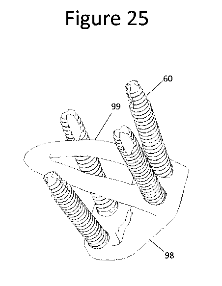
[00091] Figure 25 is a perspective view of an exemplary embodiment of an
implant
comprising an integrated plate/pin component having a plate component 98, a
curved pin
component 99, and four bone screws 60. Figure 25 illustrates an embodiment
with optional
features, any of which may be optionally used or substituted with other
features in other
embodiments discussed herein. In particular, the implant comprises two sacral
screws and
two iliac screws. As seen in Figure 25, plate component 98 and pin component
99 of the
illustrated integrated pin/plate component are formed of a single body.
[00092] Figure 26 is a cross-sectional view of the sacrum 53 and ilium 54 to
illustrate
implantation position of the plate component 98 and curved pin component 99
shown in
Figure 25. As seen in Figure 26, plate component 98 is placed with sacrum-
contacting
surface 73 adjacent to the sacrum 53 and ilium-contacting surface 74 adjacent
to ilium 54. Pin
component 99 extends through plate component 98, further extending through
ilium 54,
extending across a sacroiliac joint space between said sacrum 53 and ilium 54,
and partially
extending into the sacrum 53.
[00093] While preferred embodiments of the present disclosure have been shown
and
described herein, it will be obvious to those skilled in the art that such
embodiments are
provided by way of example only. Numerous variations, changes, and
substitutions will now
occur to those skilled in the art without departing from the invention. It
should be understood
that various alternatives to the embodiments described herein may be employed
in practicing
the invention. It is intended that the following claims define the scope of
the invention and
that methods and structures within the scope of these claims and their
equivalents be covered
thereby.
21