Note : Les descriptions sont présentées dans la langue officielle dans laquelle elles ont été soumises.
SCAR ASSESSMENT
CROSS-REFERENCE TO RELATED APPLICATION
This application claims the benefit of U.S. Provisional
Patent Application 62/623,022, filed 29 January 2018 and U.S.
Patent Application 16/231,995, filed 25 December 2018, which
are incorporated herein by reference.
FIELD OF THE INVENTION
This invention relates generally to investigating human
tissue, and specifically to assessing a degree of scarring of
cardiac tissue.
BACKGROUND OF THE INVENTION
In diagnosing problems associated with the heart, it is
well known that scarring of portions of the myocardium may
contribute to the problems, and this is considered to be the
case, for example, in atrial fibrillation. The scarring can be
identified non-invasively using magnetic resonance imaging
(MRI) protocols, and/or invasively using bipolar voltage
mapping.
SUMMARY OF THE INVENTION
An embodiment of the present invention provides apparatus
for assessing scarring of cardiac tissue, consisting of:
a probe including:
one or more electrodes, which are configured to contact
the cardiac tissue at a plurality of positions and to sense
respective voltages in the tissue at the positions; and
a processor which is configured to:
receive the respective voltages,
1
CA 3031281 2019-01-24
compute a triangular mesh that is representative of a
surface of the cardiac tissue and that has multiple triangles
having vertices corresponding to the positions contacted by the
one or more electrodes,
calculate respective scar areas within the triangles by
comparing the respective voltages sensed at the positions
corresponding to the vertices to a predefined range of the
voltages that is associated with scarring, and
compute a sum of the respective areas, and compare the sum
to a total area of the triangles so as to assess a degree of
the scarring of the tissue.
Typically the voltages are peak-peak bipolar voltages.
In a disclosed embodiment the range includes a minimum
voltage associated with the scarring of the tissue and a
maximum voltage associated with the scarring of the tissue.
Typically, the disclosed embodiment includes, for a given
triangle in the multiple triangles, associating the minimum and
maximum voltages with edges of the given triangle so as to
define points on the edges. The disclosed embodiment may also
include joining the points to form a polygon, wherein the
polygon includes, for the given triangle, the respective scar
area.
In a further disclosed embodiment the one or more
electrodes consist of two electrodes.
In a yet further disclosed embodiment the probe further
includes a sensor configured to provide respective signals to
the processor indicative of the positions.
In an alternative embodiment the one or more electrodes
are configured to provide respective signals to the processor
indicative of the positions.
2
CA 3031281 2019-01-24
In a further alternative embodiment the predefined range
is selected for the scarring of the tissue to consist of dense
scar. Alternatively or additionally, the predefined range is
selected for the scarring of the tissue to consist of
hibernating myocardium.
There is further provided, according to an embodiment of
the present invention, a method for assessing scarring of
cardiac tissue, including:
contacting the cardiac tissue with one or more electrodes
at a plurality of positions;
sensing respective voltages in the tissue at the
positions;
computing a triangular mesh that is representative of a
surface of the cardiac tissue and that consists of multiple
triangles having vertices corresponding to the positions
contacted by the one or more electrodes;
calculating respective scar areas within the triangles by
comparing the respective voltages sensed at the positions
corresponding to the vertices to a predefined range of the
voltages that is associated with scarring; and
computing a sum of the respective areas, and comparing the
sum to a total area of the triangles so as to assess a degree
of the scarring of the tissue.
The present disclosure will be more fully understood from
the following detailed description of the embodiments thereof,
taken together with the drawings, in which:
BRIEF DESCRIPTION OF THE DRAWINGS
3
CA 3031281 2019-01-24
Fig. 1 is a schematic illustration of an invasive medical
procedure using an apparatus, according to an embodiment of the
present invention;
Fig. 2 is a schematic illustration of a distal end of a
probe used in the apparatus, according to an embodiment of the
present invention;
Fig. 3 schematically illustrates an array of recorded
positions and line segments joining the positions to form a
mesh, according to an embodiment of the present invention;
Fig. 4A is a schematic diagram illustrating a typical
triangle of the mesh, and Fig. 4B is a schematic diagram
illustrating a figure topologically equivalent to the triangle
of Fig. 4A, according to an embodiment of the present
invention;
Fig. 5 is a table illustrating numerical examples of
different triangles, polygons generated for the triangles, and
the area of each of the polygons, according to an embodiment of
the present invention;
Fig. 6 is a flowchart of an algorithm implemented by a
processor and/or a professional in analyzing tissue of a
patient, according to an embodiment of the present invention;
and
Fig. 7 is a schematic of a triangle illustrating steps of
the algorithm, according to an embodiment of the present
invention.
DETAILED DESCRIPTION OF EMBODIMENTS
Overview
In analyzing cardiac tissue, the assessment of the degree
of scarring of the tissue, also termed the scar burden, has
been fraught with difficulty. For example, if a non-invasive
4
CA 3031281 2019-01-24
MRI (magnetic resonance imaging) protocol is used the
complexity and limited availability of the MRI protocol causes
problems. For an invasive assessment using bipolar voltages,
the lack of consistency of the bipolar sampling techniques and
measurement is also problematic. In many cases the assessment
of scar burden relies on a visual estimation, and this has been
shown to be inaccurate.
Embodiments of the present invention provide a
quantitative and objective assessment of the scar burden
associated with cardiac tissue being investigated. In a
procedure performed on a patient, a probe is introduced into
the patient, and the probe is used to acquire positions in the
tissue and corresponding bipolar voltages associated with the
positions. From the positions a triangular mesh is generated,
and a respective bipolar voltage is associated with each vertex
of every triangle in the mesh. Minimum and maximum bipolar
voltages, that may be used in identifying scarred tissue, are
applied to edges of the triangles, so as to define points on
the edges. For a given triangle the points are connected,
together with at least one or more sections of the edges of the
triangle, to form a polygon within the triangle, so that the
region within the polygon corresponds to an area of scarred
tissue.
A quantitative assessment of the scar burden may be made
by finding a ratio of the areas of all the polygons to the
areas of all the triangles of the mesh.
A disclosed embodiment of the present invention provides
apparatus for assessing scarring of cardiac tissue, the
apparatus comprising a probe and a processor coupled to the
probe.
5
CA 3031281 2019-01-24
The probe has one or more electrodes, typically a pair of
electrodes, which are configured to contact the cardiac tissue
at a plurality of positions and to sense respective voltages,
typically bipolar voltages, in the tissue at the positions.
The processor receives the voltages from the probe. The
processor also computes a triangular mesh that is
representative of a surface of the cardiac tissue and comprises
multiple triangles having vertices corresponding to the
positions contacted by the one or more electrodes.
The processor calculates respective scar areas within the
,
triangles by comparing the respective voltages sensed at the
positions corresponding to the vertices to a predefined range
of the voltages that is associated with scarring. A sum of the
respective areas is computed, and the sum is compared to a
total area of the triangles so as to assess a degree of the
scarring of the tissue.
Detailed Description
In the following description, like elements in the
drawings are identified by like numerals, and like elements are
differentiated as necessary by appending a letter to the
identifying numeral.
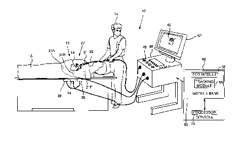
Fig. 1 is a schematic illustration of an invasive medical
procedure using apparatus 12, and Fig. 2 is a schematic
illustration of a distal end 22 of a probe 26 used in the
apparatus, according to an embodiment of the present invention.
The procedure is performed by a medical professional 14, and,
in the description hereinbelow the procedure is assumed to
comprise an investigation comprising electropotential mapping
6
CA 3031281 2019-01-24
of at least a portion of tissue 15 of a myocardium 16 of the
heart of a human patient 18.
The investigation also comprises using the mapping to
analyze tissue 15 to find out how scarred the tissue is. The
investigation may use results that have been previously
acquired, i.e., as a retrospective investigation. Alternatively
or additionally, the investigation may use results acquired in
real time, i.e., as a real time investigation.
In order to perform the investigation, professional 14
inserts probe 20 into a sheath 21 that has been pre-positioned
in a lumen of the patient. Sheath 21 is positioned so that
distal end 22 of the probe enters the heart of the patient.
Distal end 22 comprises a position sensor 24 that enables the
location and orientation of the distal end to be tracked.
As is illustrated in Fig. 2, distal end 22 comprises
generally similar pairs of cylindrical electrodes 34, 36, e. g.
electrodes 34A, 36A; 34B, 36B; 340, 360; _ . In the disclosure,
for simplicity a pair of electrodes 34, 36 may also be referred
to as electrodes 38, so that, for example, electrodes 34A, 36A
may also be referred to as electrodes 38A. Electrodes 38
acquire electropotentials of regions with which they are in
contact, and in the following description electrodes 38A, 38B,
380,
are assumed to be respectively in contact with locations
20A, 20B, 20C, of the myocardium.
Apparatus 12 is controlled by a system processor 46, which
is located in an operating console 48 of the apparatus. Console
48 comprises controls 49 which are used by professional 14 to
communicate with the processor. The software for processor 46,
which comprises software for an algorithm described
hereinbelow, may be downloaded to the processor in electronic
7
CA 3031281 2019-01-24
form, over a network, for example. Alternatively or
additionally, the software may be provided on non-transitory
tangible media, such as optical, magnetic, or electronic
storage media. The track of distal end 22 is typically
displayed on a three-dimensional representation 60 of the heart
of patient 18 that is displayed on a screen 62.
In order to operate apparatus 12, processor 46
communicates with a module bank 50, which has a number of
modules used by the processor to operate the apparatus. Thus,
bank 50 comprises an electrocardiograph (ECG) module 56 which
acquires and analyzes signals from electrodes 38, and a
tracking module 58 which receives and analyzes signals from
position sensor 24, and which uses the signal analysis together
with the processor to generate a location and an orientation of
distal end 22, as well as a location and orientation of
electrodes 38. In some embodiments sensor 24 comprises one or
more coils which provide the sensor signals in response to
alternating magnetic fields traversing the coils. In these
embodiments, in addition to receiving and analyzing signals
from sensor 24, tracking module 58 also controls radiators 32,
34, and 36 which radiate the alternating magnetic fields
traversing sensor 24. The radiators are positioned in proximity
to myocardium 16, and are configured to radiate the alternating
magnetic fields into a region in proximity to the myocardium.
The Carto system produced by Biosense Webster, of 33
Technology Drive, Irvine, CA 92618 USA, uses such a magnetic
tracking system.
ECG module 56 typically acquires, from electrodes 38,
bipolar voltages of the region in contact with the electrodes.
In some embodiments one or both electrodes 34, 36 (of a given
8
CA 3031281 2019-01-24
set of electrodes 38) may be used to acquire unipolar voltages
from regions in contact with the electrodes. In some
embodiments one or both electrodes 34, 36 may also be
configured to apply ablation. Additionally or alternatively,
one or both electrodes 34, 36 may be used as location sensing
electrodes, determining a location in contact with the
electrodes. The use as location sensing electrodes is described
in more detail below.
Alternatively or additionally, embodiments of the present
invention may incorporate other modes of tracking distal end 22
and electrodes 38. As one example, tracking module 58 may be
configured to inject current into a given one of electrodes 38,
and the module may record values of currents received by skin
patches 31A, 31B, ... 31F. There are typically six such patches,
three of which are shown in the figure, attached to patient 18.
The position of the given electrode may be estimated from the
values of the currents acquired by the patches, and/or by the
impedances of the,patches, as registered by the module.
As a second example, tracking module 58 may be used to
generate, again using skin patches attached to the patient,
electric fields (typically three approximately orthogonal
fields) in the patient, and the module may record the voltages
generated on a given one of electrodes 38 in response to the
fields. The position of the given electrode may be estimated
from the recorded voltages. All such modes of tracking are
assumed to be comprised within the scope of the present
invention.
Bank 50 typically also comprises other modules, such as a
force module, a power module, an irrigation module, and a
9
CA 3031281 2019-01-24
temperature module. For simplicity the functions of these
modules are not described herein.
For the electropotential mapping investigation described
herein, distal end 22 is moved so that electrodes 38 contact
different regions of tissue 15. While the electrodes are in
contact, processor 46 uses ECG module 58 to acquire and record
cardiac electropotentials generated during the period of
contact. Also while the electrodes are in contact, the
processor uses tracking module 58 to determine and record the
position of the contacting electrodes. It will be understood
that the position of the contacting electrodes is a three-
dimensional position.
In an embodiment of the present invention, the cardiac
electropotentials acquired by electrodes 38 and recorded by
module 56 are bipolar voltages. Processor 46 analyzes the
recorded bipolar voltages from a given set of electrodes 38 to
find a peak-peak voltage value for the set of electrodes.
Typically, the processor interpolates the peak-peak voltage
values from locations in proximity to a given recorded position
to find a peak-peak voltage for the given position. Thus for
each recorded position on tissue 15, processor 46 is able to
generate an ordered pair of values comprising a value of the
position, and a peak-peak voltage value of electropotentials
recorded by the electrodes for the recorded position.
Fig. 3 schematically illustrates an array of recorded
positions and line segments joining the positions to form a
mesh, according to an embodiment of the present invention. As
stated above, electrodes 38 and/or sensor 24 are used by
processor 46 to acquire and record positions of the electrodes,
herein referred to as recorded positions 74A, 743, 74C, ..., and
CA 3031281 2019-01-24
generically as recorded positions 74. Recorded positions 74
form an array 70 of positions. Using processes well-known in
the art processor 46 joins array 70 of recorded positions 74 by
line segments 78A, 78B, 78C, ..., generically referred to herein
as line segments 78, for example by using a ball-pivot
algorithm. In embodiments of the present invention, line
segments 78 are generated so as to join positions 74 in a
triangular mesh 82 comprising multiple triangles 90. While mesh
82 is typically in three dimensions, it will be understood that
each triangle 90 of the mesh is a planar, two-dimensional (2D),
triangle.
Each position 74 is at the vertex of at least one
triangle, and is typically at the vertex of many triangles.
There is a respective peak-peak voltage associated with each
vertex.
Fig. 4A is a schematic diagram illustrating a typical
triangle of mesh 82, and Fig. 4B is a schematic diagram
illustrating a figure topologically equivalent to the triangle
of Fig. 4A, according to an embodiment of the present
invention. As illustrated in Fig. 4A, a triangle 90A of mesh 82
has vertices A, B, C, corresponding to positions 74. The
vertices are joined by straight line segments AS, BC, CA, also
herein referred to as edges AB. BC, CA, corresponding to line
segments 78.
In the following description of the analysis of triangle
90A, vertices A, B, and C are assumed, as a general case, to
have respective peak-peak voltages al, a2, and a3, typically
measured in mV, where al < a2 < a3. (Examples of degenerate
cases, such as al - a2, a2 = a3, or a3 = al, are given below.)
11
CA 3031281 2019-01-24
A figure 92 is topologically equivalent to triangle 90A.
Figure 92 comprises a straight line segment A'B'C' and a curved
line A'C', where A', B', C' respectively correspond to A, B, C.
Segment A'B'C' may be considered to be a first number line,
where A' has the value al, C' has the value a3, and B' has the
value a2 (between al and a3). Curved line A'C' may be
considered to be a second number line.
In embodiments of the present invention, tissue 15 is
assumed to comprise scar tissue if the peak-peak voltages
formed at the tissue, and acquired by electrodes 38, lie in a
range between amin and amax, where amin is a minimum peak-
peak voltage value identifying scar tissue, and amax is a
maximum peak-peak voltage value identifying the scar tissue.
amin and amax are also typically measured in mV.
As is known in the art, scar tissue may be classified
according to assigned values of amin and amax. Table I gives
some of these classifications, together with values of amin and
amax.
Type of Scar Approximate value Approximate value
Tissue of amin (mV) of amax (mV)
Dense 0 0.5
Hibernating 0.5 1.5
Myocardium
Table I
Processor 46 uses amin and amax in analyzing triangles 90.
Typically al < amin, amax < a3, and this inequality is assumed
12
CA 3031281 2019-01-24
for triangle 90A. Examples of cases where the inequality,
herein referred to as the boundary inequality, does not hold
are provided below.
Prior to the analysis of the triangles, both values amin,
amax are typically pre-set by operator 15. In the analysis,
processor 46 may position amin, amax on curved number line
A'C', and thus on edge AC at points D,E, as is illustrated in
Figs. 4A and 4B. In addition both values amin, amax may be
positioned on straight number line A'B'C'. By way of example,
Figs. 4A and 4B illustrate amin as being on line segment A'B',
and thus on edge AB at a point F, and amax as being on line
segment B'C', and thus on edge BC at a point G. However, it
will be understood that both values amin, amax may be
positioned on line segment A'B', or both may be positioned on
line segment B'C'.
From the description above it will be understood that
edges AB, BC, and CA of triangle 90A are also considered herein
as number lines, each of the lines terminating in two of the
values al, a2, and a3. Points D, E, F, G are positioned on the
number lines according the numerical values of the points,
i.e., in accordance with amin and amax.
Once values amin, amax have been positioned on edges,
i.e., number lines, of triangle 90A processor 46 constructs a
first line DF joining the edge points amin, and a second line
EG joining the edge points amax. A shaded region 94, of an area
enclosed by polygon DFBGE comprises regions of the triangle
that are assumed to have voltage values between amin and amax,
and thus to identify scar tissue.
13
CA 3031281 2019-01-24
Fig. 5 is a table illustrating numerical examples of
different triangles 90, the polygons generated for the
triangles, and the area of each of the polygons, according to
an embodiment of the present invention. The table gives nominal
cartesian coordinates (x,y) of the vertex of each triangle, and
nominal peak-peak voltages (al, a2, a3) associated with each
vertex. For each triangle there is an example of a nominal
minimum and maximum peak-peak voltage (amin, amax). The diagram
on the left of each row illustrates the triangle, with the
generated polygon shown as a shaded region. The calculated area
of each shaded region is also given, as well as the area of its
triangle.
Triangles 1, 3, and 4 are examples of the degenerate cases
referred to above. Triangles 1 and 2 are examples where the
boundary inequality provided above does not hold.
Fig. 6 is a flowchart of an algorithm implemented by
processor 46 and/or professional 14 in analyzing tissue 15 of
patient 18, and Fig. 7 is a schematic of a triangle
illustrating steps of the algorithm, according to an embodiment
of the present invention. In an initial step 100 of the
algorithm, probe 26 is inserted into patient 18, electrodes 38
are used to acquire peak-peak voltages of locations of tissue
15, and the processor generates and records ordered pairs of
positions and respective peak-peak voltages of the positions on
the tissue, as described above.
In a mesh producing step 102, the processor produces a
triangular mesh, comprising line segments joining the recorded
positions found in step 100, and evaluates the lengths of the
line segments of each of the triangles of the mesh.
14
CA 3031281 2019-01-24
For each triangle vertex, in a vertex voltage step 104,
the processor assigns a peak-peak voltage to the vertex of each
identified triangle.
In a scar bounding step 106, professional 14 selects
values for minimum peak-peak voltage value amin and maximum
peak-peak voltage value amax and provides the values to the
processor.
For each given triangle of the mesh formed in step 102, in
an analysis step 108 the processor analyzes each edge of the
triangle and applies the values of amin and amax to the edges,
as described above with respect to Figs. 4A and 4B.
As is also described above with respect to Figs. 4A and
4B, in a polygon formulation step 110, once the values of amin
and amax have been applied to the edges of a given triangle,
the processor delineates the polygon defined by the triangle
edges and the applied values of amin and amax.
The processor then calculates the area for the delineated
polygon, and the area of the triangle containing the polygon,
in an area calculation step 112.
Fig. 7 is an example of a triangle A"B"C" formed in
step 102. By way of example, the lengths of the edges of
triangle A"B"C" are assumed to be: A"B"= 0.9mm,
B"C"=1.3mm, A"C"=1.0mm.
In step 104, processor 46 assigns the vertices of the
triangle, using the data acquired in step 100, the following
peak-peak voltages: A" 0.2 mV; B" 2.5 mV; C" 3.0 mV.
In step 106 it is assumed that professional 14 selects
values of amin and amax corresponding to hibernating
myocardium, i.e., amin = 0.5 mV and amax =1.5 mV.
CA 3031281 2019-01-24
In step 108 the processor applies the values of amin and
amax to the edges of triangle A"B"C". This generates points
D" with a value of 0.5 mV and E" with a value of 1.5 mV on
edge A"B". The application also generates points F" with a
value of 0.5 mV and G" with a value of 1.5 mV on edge A"C".
The processor may calculate the positions of points D",
E", F", G" according to the lengths of the respective edges
upon which they lie, and according to the values of the
voltages of the vertices of the edges, considering the edges as
number lines. Typical calculations are given in equations (1) -
(4).
0.5mV-0.2mV
A"D " = = 0.9mm = 0.117mm (1)
2.5mV-0.2mV
t5mV-0.2mV
A"E" = = 0.9mm = 0.509mm (2)
2.5mV-0.2mV
" 0.5mV-0.2mV
A"F" = = 1.0mm = 0.107mm (3)
10mV-0.2mV
1.5mV-0.2mV
A"G" = = 1.0mm = 0.464mm (4)
3.0mV-0.2mV
Using the positions of points D", E", F", G" the
processor in step 110 delineates the polygon D"E"G"F".
In step 112 the processor calculates the area of polygon
D"E"G"F" as 0.11145 mm2. The processor also calculates the
area of triangle A"B"C", for example from the lengths of the
triangle sides or by any other suitable method for calculating
the area of a triangle, giving the area as 0.449 mm2.
Returning to the flowchart of the algorithm, the processor
iterates steps 108, 110, and 112 for all triangles in the mesh
generated in step 102, and in a comparison step 114 the
16
CA 3031281 2019-01-24
processor checks if the iteration has been performed for all
triangles. If comparison 114 returns negative, control of the
flowchart returns to step 108. If comparison 114 returns
positive, indicating that all triangles of the mesh have been
analyzed, control continues to a final step 116 of the
flowchart.
In final step 116 of the algorithm the processor sums the
total area of all polygons identified for the mesh, and the
total area of all the triangles of the mesh. The processor then
calculates the ratio of the two areas, and provides the ratio
to professional 14, typically using screen 62.
It will be understood that the ratio determined by the
flowchart is a measure of the fraction of the total area of
tissue 15 which lies between the scar identifying peak-peak
minimum and maximum values amin and amax. The ratio is also
referred to herein as the scar burden associated with tissue
15. Professional 14 may use the ratio, i.e., the scar burden,
to judge how scarred tissue 15 is, and also to decide if a
procedure, such as ablation of a section of tissue 15, is to be
performed.
The above description has described, by way of example,
how a predefined range of bipolar voltages may be applied to
vertices of triangles in a triangular mesh to evaluate a
selected classification of scar burden, by calculating areas
within the triangles of the mesh. Those having ordinary skill
in the art will be able to adapt the description, mutatis
mutandis, to evaluate the scar burden for other scar
classifications. It will be appreciated that the embodiments
described above are cited by way of example, and that the
present invention is not limited to what has been particularly
17
CA 3031281 2019-01-24
shown and described hereinabove.
Rather, the scope of the
present invention includes both combinations
and
subcombinations of the various features described hereinabove,
as well as variations and modifications thereof which would
occur to persons skilled in the art upon reading the foregoing
description and which are not disclosed in the prior art.
18
CA 3031281 2019-01-24