Note : Les descriptions sont présentées dans la langue officielle dans laquelle elles ont été soumises.
CA 03031761 2019-01-23
WO 2018/005848 PCT/US2017/040074
TREATMENT OF SEPSIS AND RELATED INFLAMMATORY CONDITIONS BY
LOCAL NEUROMODULATION OF THE AUTONOMIC NERVOUS SYSTEM
INCORPORATION BY REFERENCE TO ANY PRIORITY APPLICATIONS
[0001] This application claims the benefit under 35 U.S.C. 119(e) as
a nonprovisional
application of U.S. Prov. App. No. 62/355,889 filed on June 29, 2016, which is
hereby incorporated
by reference in its entirety.
FIELD OF THE INVENTION
[0002] Some aspects of the invention relate to methods, drugs and
their formulations to
modulate the immune system, particularly for the treatment of life-threatening
inflammatory
disease, are described.
BACKGROUND
[0003] Sepsis is a major public health issue and one of the most
frequent causes of
death in hospitalized patients. It is a clinical syndrome of physiologic,
pathologic and biochemical
abnormalities induced by infection or injury. Recent publications in the
Journal of the American
Medical Association (JAMA) indicate that in-hospital mortality rates in
infected patients are
alarmingly high ranging between 18-54%. Patients that survive sepsis can
suffer physical and
cognitive impairment, which can more than double their 5-year mortality risk,
compared to
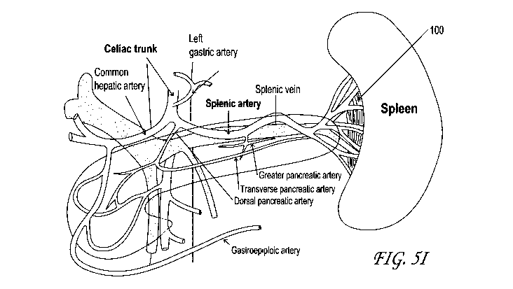
hospitalized controls.
[0004] Nearly a million people are affected by sepsis annually in the
United States and
over 200,000 people die, placing a significant burden on the healthcare
system. Estimates suggest
that over $20 billion were spent in 2011 on sepsis-related intensive care unit
(ICU) hospitalizations,
which represents 5.2% of the total US hospital costs.
[0005] The cellular and molecular mechanisms influencing pathogenesis
of sepsis are
not well understood. It affects all age groups irrespective of race, gender,
geography, or health
status. Sepsis develops in patients affected by an infection or tissue injury
from noninfectious
sources such as pancreatitis, ischemia reperfusion injury, cancer, and a host
of other disorders that
1
CA 03031761 2019-01-23
WO 2018/005848 PCT/US2017/040074
are inflammatory in nature. The host immunological response and reaction to
infection and injury
plays an equally important role in the restoration or deterioration of organ
function. There are no
reliable diagnostic blood markers or cellular markers in organs or tissue for
the detection of sepsis.
Common symptoms include fever, increased respiratory rate, increased heart
rate, lethargy, edema,
confusion and low blood pressure.
[0006] Antibiotics and intravenous fluids (fluid replacement therapy)
are used to treat
septic patients in the intensive care unit. Mechanical ventilation and
dialysis are used to assist
respiratory and kidney function. Medications like vasopressin may be used to
control blood
pressure. The use of corticosteroids is controversial and treatment using the
drug drotrecogin-alfa
has not been effective and the drug has been withdrawn from the market. No FDA-
approved drugs
are available for the treatment of sepsis. Current mortality rates from
sepsis, severe sepsis and
septic shock conditions are currently about 30%, 50% and 80%, respectively.
[0007] Newer device-based treatments using vagus nerve stimulation
(VNS),
noninvasive therapeutic ultrasound delivery, and membrane filters are in
development. Electrical
stimulation of the vagus nerve has been shown to activate the splenic release
of acetylcholine and
suppress pro-inflammatory cytokine release via the brain-immune cholinergic
anti-inflammatory
pathway (CAP) and treat sepsis in animal models. Non-invasive ultrasound
treatment, before renal
ischemic reperfusion injury (IRI), also has been found to stimulate CAP in
rats and protect the
kidney. Hollow-fiber dialysis and cytopheretic membrane filters, which bind
and sequester the
activated leukocytes from blood circulation, have been clinically tested in
septic patients.
[0008] All these methods have significant limitations. VNS requires
the surgical
implantation of an expensive electrical generator and placement of electrodes
in critically-ill
patients. Also, VNS may result in unwanted side effects when delivered at the
cervical level
because branches innervate many off-target organs. A randomized clinical study
in experimental
endotoxemia failed to show a similar reduction in cytokines as had been
observed in preclinical
studies. Non-invasive ultrasound energy treatment is not targeted and may
damage surrounding
tissue. Finally, the cytopheretic device therapy also did not show clinical
benefit beyond small
open-label clinical studies.
[0009] We describe in some embodiments therapeutic drugs, compositions
and methods
of administration that can overcome these limitations. The spleen is one of
the largest secondary
lymphoid tissues and plays a significant role in the neuro-immune axis of
inflammation and
maintaining immune homeostasis. Nerve signaling through the splenic nerve and
its branches may
2
CA 03031761 2019-01-23
WO 2018/005848 PCT/US2017/040074
modulate the production of cytokines and may activate other molecular pathways
that indirectly
lead to inflammation and symptoms consistent with sepsis. Methods described in
some
embodiments of the invention provide a treatment strategy for sepsis by the
administration of a
drug to alter the pro- and anti-inflammatory neuro-immune signaling pathways
between the spleen
and the brain. Other methods described herein disclose the treatment of a
patient having symptoms
consistent with sepsis by the administration of drug to an organ containing
primary or secondary
lymphoid tissues. In addition, some embodiments of the invention describe
methods to access
points of innervation between an organ containing lymphoid tissue and the
brain, verify the nerve
site and measure the splenic nerve signals before, during and after treatment.
Specific drugs,
compositions and formulations are also described.
SUMMARY
[0010] Methods of use, formulations, and devices for delivering
therapeutic drugs
locally to the region of the spleen are described herein. In one embodiment, a
method for treating
sepsis and other inflammatory disease conditions comprises inserting a drug
delivery system inside
the body, advancing the device to the spleen through the splenic artery,
splenic vein or other blood
vessel adjacent to the splenic nerves. In one embodiment, the drug delivery
system is advanced
next to the splenic nerve and the splenic nerve activity may be measured at
the target tissue site
before a small volume of therapeutic formulation is administered locally to
the splenic nerve and
nerve branches, related nerve plexi or ganglia to stimulate, modulate or alter
neuro-immune
activity. The change in splenic nerve activity may be measured to assess
treatment effect prior to
the delivery device removal from the body. In some embodiments, the nerve
activity may be
attenuated to achieve the desired immune response and maintain immune
homeostasis.
[0011] In yet another embodiment, the drug delivery therapy can be
advanced through
the vasculature beyond the hilum into one or more post-hilum segments of the
spleen prior to
injecting the drug delivery system more distally. The drug delivery system may
target the delivery
of drugs to the postganglionic catecholaminergic neurons innervating the
spleen or to target
immune cells in the spleen directly. Drug delivery systems may be delivered in
a formulation that
can be administered into the blood vessels such that the system(s) are
sequestered in the vasculature
until the drug is released and the carrier cleared from the site.
Alternatively, drug delivery systems
may be delivered transvascularly via a drug delivery microcatheter into the
spleen itself in order to
achieve this. In yet another embodiment, a drug coated balloon is deployed
within the splenic
3
CA 03031761 2019-01-23
WO 2018/005848 PCT/US2017/040074
vasculature to deliver drug transarterially or transvenously. In yet another
embodiment, a
bioerodible stent is placed in the vasculature to deliver drug both
transvascularly and into the blood
stream distal to the site of placement.
[0012] In comparison to conventional intravenous therapy, the drug may
be pre-loaded
and delivered through a catheter, needle-syringe system or pump and wherein
drug may be
administered in a manner that perfuses the organ directly to provide for a
more rapid intervention.
In one embodiment, the drug is delivered from a drug delivery catheter placed
in the splenic artery
directly through the arterial system to the spleen. By administering the drug
formulation prior to the
splenic artery branching into terminal branches, the entire organ, such as the
spleen may be bathed
in the drug in this manner. If the drug is coated on or encapsulated within
nanoparticle or
microparticles, these nano- and micro-particles will course through the
vasculature where they may
either get trapped in a progressively smaller arteriole or capillary or
alternatively extravasate into
the splenic tissue. In this manner, sustained release formulations of agents
can be delivered locally
into the spleen.
[0013] Drug may be administered near organ innervation nodes, for
example the splenic
nerves, directly or may be mixed with excipients and polymers to provide
sustained drug release
over time to stimulate nerves or permanently affect nerve function to have
durable treatment effects
lasting a few days to several weeks.
[0014] The neuromodulatory effects of drug compositions described
below may
stimulate or upregulate nerve activity to enhance or inhibit the release of
anti- or pro-inflammatory
cytokines, alter the host immune response to inflammation, and maintain immune
homeostasis.
Other effects of blocking nerves and attenuating or downregulating nerve
activity to enhance or
inhibit the release of anti- or pro-inflammatory cytokines, over short or long
periods of time, are
also described.
[0015] Methods and devices for accessing the splenic nerve and other
nerve targets
involved in the brain-immune pathway are also described. The application also
describes methods
for visualizing nerves and measuring local autonomic activity before locally
administering the drug
formulation near the splenic nerve; and monitoring nerve feedback during and
after treatment.
[0016] Methods described here may in some cases be used either as an
adjunctive
treatment to therapies currently in clinical practice or therapies under
investigation to treat sepsis
and other inflammatory disorders or medical conditions. Treatments described
here may be
performed before or after the primary procedure to allow sufficient time to
regulate the local and
4
CA 03031761 2019-01-23
WO 2018/005848 PCT/US2017/040074
systemic hormone, cytokine and catecholamine levels to achieve optimal
clinical efficacy and
restore immune homeostasis.
[0017] Other nerve targets innervating other target organs inside the
body and involved
in neuro-immune signaling and inflammatory disorders and medical conditions
are also described.
Drug formulations may be injected locally at one or more target nerve sites
inside the body to treat
sepsis. Drug formulations may be administered at different nerve sites to
achieve the desired
therapeutic benefit at specific locations over the desired time periods.
[0018] In some embodiments, a method of modulating inflammation in a
patient is
disclosed. The method can include, for example, providing a therapeutic agent
delivery system
comprising at least one therapeutic agent; accessing between the folds of one
or more ligaments
directly connected to a splenic hilum of the patient, wherein the one or more
ligaments comprise
the splenorenal ligament and the gastrosplenic ligament of the patient; and
delivering the
therapeutic agent delivery system between the folds of the one or more
ligaments.
[0019] In some embodiments, accessing between the folds of the one or
more ligaments
comprises: inserting a catheter into a first blood vessel; advancing the
catheter into a second blood
vessel (e.g., a splenic artery, splenic vein, splenic artery end branches,
etc.); and penetrating a wall
of the second blood vessel with a portion of the catheter to a position
between the folds of the one
or more ligaments. In some embodiments, accessing between the folds of the one
or more
ligaments includes inserting a catheter percutaneously (e.g., between ribs in
some cases); and
positioning the catheter between the folds of the one or more ligaments with a
portion of the
catheter. The one or more ligaments could include the splenorenal ligament,
gastrosplenic ligament,
or others. The therapeutic delivery system can be an implant delivered between
the folds of the one
or more ligaments, and can coil around a blood vessel in some cases. The
delivery system could
include, for example, microspheres, or a gel such as a hydrogel, that can be
in situ cross-linking in
some cases, an injectable hydrogel slurry, be biodegradable, or combinations
of the foregoing.
[0020] In some embodiments, the method, e.g., delivering the
therapeutic agent delivery
system treats or prevents systemic inflammatory response syndrome, sepsis,
septic shock, an
autoimmune disease, or acute respiratory distress syndrome. The therapeutic
agent could include,
for example, a sympathomimetic agent, such as an alpha-1, alpha-2, alpha-
nonselective, beta-1,
beta-2, or beta-nonselective agonists. In some embodiments, the therapeutic
agent includes a
nicotinic acetylcholine receptor agonist, such as nicotine or acetylcholine,
for example. Delivering
the therapeutic agent delivery system can neuromodulate sympathetic and/or
parasympathetic
CA 03031761 2019-01-23
WO 2018/005848 PCT/US2017/040074
nerves, and/or cells residing in the spleen, such as immune cells, including T-
cells, B-cells,
macrophages, polymorphonuclear cells, eosinophils, basophils, NK cells, or
other cells.
[0021] In some embodiments, a method of modulating inflammation of a
patient can
include accessing between the folds of one or more ligaments directly
connected to a splenic hilum
of the patient, wherein the one or more ligaments comprise the splenorenal
ligament and the
gastrosplenic ligament of the patient; and flowing a gel comprising a
therapeutic agent between the
folds of the one or more ligaments such that the folds of the one or more
ligaments serves as a
boundary and limits the spread of the gel to between the folds of the one or
more ligaments.
[0022] In some embodiments, a method of modulating inflammation in a
patient can
include providing a therapeutic agent delivery system comprising at least one
therapeutic agent;
accessing the splenic hilum of the patient; and delivering a therapeutic agent
delivery system
comprising a hydrogel to the splenic hilum.
[0023] In some embodiments, a system configured for modulating
inflammation in a
patient can include a catheter sized and configured for being positioned
percutaneously within a
blood vessel directly proximate and for delivering a therapeutic agent to the
splenic hilum; and a
first hydrogel comprising one or more of: a nicotinic acetylcholine receptor
agonist and a
sympathomimetic agent. A hydrogel for use modulating inflammation by delivery
to the splenic
hilum percutaneously or transvascularly, such as through the wall of the
splenic artery, end
branches thereof, or the splenic vein, directly within the folds of the
splenorenal ligament or the
gastrosplenic ligament of a patient can include a therapeutic agent comprising
one or more of: a
nicotinic acetylcholine receptor agonist and a sympathomimetic agent.
BRIEF DESCRIPTION OF THE DRAWINGS
[0024] FIGURE 1 shows examples of the pathways associated with injury,
infection,
inflammation, sepsis and resulting effects on restoring organ function or
organ failure and death.
[0025] FIGURES 2A-2B shows the various afferent (sensory) and efferent
(motor)
neuronal pathways that maintain organ homeostasis inside the human body
mediated by the vagus
nerve and the sympathetic chain.
[0026] FIGURE 3 shows the immune control through the cholinergic anti-
inflammatory
pathway (CAP) in the spleen and the underlying cellular mechanisms.
6
CA 03031761 2019-01-23
WO 2018/005848 PCT/US2017/040074
[0027] FIGURES 4A-4B shows the location of the spleen inside the body
(4A) and
structure of the spleen (4B) illustrating the blood vessels and nerve fibers
innervating the white
pulp, red pulp and the marginal zone.
[0028] FIGURE 4C illustrates the position of the spleen in relation to
the left 9th
through 11th ribs.
[0029] FIGURE 4D also schematically illustrates a histological section
of the spleen
and selected features.
[0030] FIGURE 5A shows the blood circulatory system supplying the
spleen and
nearby organs (stomach and pancreas). FIGURES 5B-5E illustrate various views
of spleen and
associated anatomy, including the splenorenal and gastrosplenic ligaments.
FIGURE 5F illustrates
an axial section at T12 illustrating the gastrosplenic ligament attachment
site to the spleen at the
splenic hilum. FIGURES 5G-5I schematically illustrate non-limiting examples of
potential drug
delivery sites.
[0031] FIGURES 6A-6B shows the sympathetic (6A) and parasympathetic
(6B)
nervous system innervating the spleen and other organs.
[0032] FIGURES 7A-7B show the sympathetic and parasympathetic neuronal
pathways
in inflammation and sepsis.
[0033] FIGURE 8 illustrates the effect of vagus nerve on inflammatory
pathways in the
spleen.
[0034] FIGURES 9A-9C shows (A) the anatomical location of the thymus,
(B) thymic
vessels providing blood supply, and (C) sympathetic and vagus
(parasympathetic) nervous systems
connected to the thymus, relative to adjacent organs.
[0035] FIGURES 10A-10B illustrates selected lung anatomy.
DETAILED DESCRIPTION
[0036] Local drug delivery systems to modulate and prevent or treat
infection, trauma,
injury, inflammation, sepsis, septicemia, septic shock, systemic inflammatory
response syndrome
(SIRS) and acute respiratory distress syndrome (ARDS) through abrogation of
neuro-immune axis-
specific signaling, by the administration of drug to an organ containing
lymphoid tissue near the
site of innervation, are described. Drug delivery systems may be injected
locally near autonomic
nerves innervating the spleen or other target organ to affect neuro-immune
signaling and effector
7
CA 03031761 2019-01-23
WO 2018/005848 PCT/US2017/040074
pathways for the treatment of inflammatory diseases. Alternatively, drug
delivery systems may be
injected in proximity to the effector or target cells that are modulated by
the autonomic nervous
system in order to directly modulate these cells. Other nerve target sites and
methods to affect and
improve the immune function are also described.
[0037] Methods, drugs, drug formulations and devices to treat
inflammation, sepsis,
septicemia, septic shock, systemic inflammatory response syndrome, acute
respiratory distress
syndrome and related inflammatory medical conditions through local chemical
neuromodulation of
the splenic nerve are described. Other nerve target sites of the autonomic
nervous system, ganglia
and nerve plexi inside the body that affect neuronal, neuro-immune and neuro-
humoral pathways of
inflammation, sepsis and related conditions to restore and preserve organ
function are also
described.
Sepsis and Other Inflammation-Mediated Medical Conditions
[0038] Sepsis can be considered a syndrome or a medical condition and
not a disease
per se. It is a life-threatening condition when the body's response to an
infection injures its own
tissues and organs. The pathophysiology is unknown and there are no standard
diagnostic tests or
blood markers for detecting sepsis. Sepsis can be identified by a set of
clinical symptoms in
patients with a suspected infection or injury/trauma to tissue from
noninfectious sources such as
pancreatitis, renal ischemia reperfusion injury (IRI), cancer, and a host of
other disorders. For
example, immune response after IRI contributes to renal tissue damage and
reduced glomerular
filtration rate (GFR) in patients that suffer acute kidney injury (AKI). The
infection, host body
response and organ dysfunction are the three clinical factors used to
identification and treatment of
sepsis. Common symptoms are fever, increased respiratory rate, increased heart
rate, confusion and
low blood pressure. Sepsis is the most common cause of multiple-organ failure.
[0039] Sepsis can be caused by pathogen factors and host factors.
Microbes and
pathogens from an infectious source invade the body and enter the bloodstream
leading to signs of
systemic illness. Immune response to antigens and foreign bodies involves
interactions between the
pro- and anti-inflammatory cytokines released through the inflammation
process. Pro-inflammatory
cytokines (PICs) include tumor necrosis factor (TNF-a), interleukin (IL)-1, IL-
la, IL-lb, IL-6, IL-
8, IL-12, IL-18, gamma-interferon (IFN-y), platelet-activating factor (PAF),
macrophage migration
inhibitory factor (MIF), granulocyte-macrophage colony stimulating factor, and
high mobility
group protein 1 (HMG-1). IL-4, IL-10, IL-13, alpha-interferon (IFN-a) and
transforming growth
8
CA 03031761 2019-01-23
WO 2018/005848 PCT/US2017/040074
factor-beta (TGF-b) are considered to be anti-inflammatory cytokines (AICs).
Cytokines are
produced by immune cells including, monocytes, macrophages and neutrophils,
and non-immune
cells such as fibroblasts, osteoblasts, smooth muscle cells, epithelial cells,
and neurons. Monocytes
and macrophages may be classified as pro-inflammatory (classically-activated,
or M1 cells that can
be differentiated by IFN-y) and anti-inflammatory (alternatively-activated, or
M2 cells that are
stimulated by IL-4). M1 cells secrete high levels of PICs (TNFa, IL-113, IL-6
and IL-12), while M2
cells secrete AICs (IL-10 and TGF-(3). Under normal conditions the balanced
inflammatory
response and feedback loop between AICs and PICs resolves the infection,
restores organ function
and maintains immune homeostasis.
[0040] Under abnormal conditions, imbalance in the feedback loop may
lead to
deleterious effects. The initial local tissue response, appropriate to
infection, becomes amplified
primarily by the innate immune system. Both pro- and anti-inflammatory
cytokines are activated
and a hyperinflammatory reaction, or a cytokine storm, of pro-inflammatory
cytokines and
activated leukocytes can exacerbate tissue damage and lead to non-resolving
inflammation and
patient death. Imbalance in the production and release of (excessive) pro-
inflammatory and
(reduced) anti-inflammatory cytokines can gradually escalate from inflammation
into sepsis, septic
shock, and organ failure. In addition, the host immune response may become
abnormal and damage
tissue and organs. With time, the persistent failure of the innate immunity
(natural immune system)
and adaptive immunity (defined as the acquired antigen-specific immune
response developed and
memorized over time) may further lead to multiple organ failure and ultimately
patient death. In
other words, patients could die from the body's dysfunctional immune response
to infection rather
than from the infection itself. Sepsis has been shown to involve early
activation of pro- and anti-
inflammatory responses along with major changes in non-immunological pathways
such as
cardiovascular, neuronal, autonomic, hormonal, bioenergetic, metabolic and
coagulation pathways.
Some infections may cause organ failure without the influence of a
dysfunctional host response.
[0041] New definitions, published recently in JAMA, define sepsis as a
medical
condition with evidence of infection and life-threatening organ dysfunction.
Septic shock is
considered a more severe form of sepsis in which the underlying circulatory
and cellular metabolic
abnormalities are greater or in a state of acute circulatory failure. Patients
in septic shock are
hypotensive, despite the use of adequate fluid therapy, hyperlactatemic (serum
lactate levels > 2
millimolar per liter or >18 milligrams per deciliter) and need vasopressor
therapy to maintain a
mean blood pressure of 65 mm of Hg or above. Changes in brain function (mental
status), lung
9
CA 03031761 2019-01-23
WO 2018/005848 PCT/US2017/040074
function (Pa02/Fi02 < 280, without other pulmonary or cardiovascular disease
as the cause) and
kidney function (oliguria or urinary output < 0.5 mL/kg for at least 2 hours)
are also indicators of
organ dysfunction and septic shock.
[0042] Systemic inflammatory response syndrome (SIRS) is another life-
threatening
inflammatory medical condition that is prevalent among hospitalized patients
with or without an
infection. Tachycardia (heart rate > 90 beats/minute), tachypnea (respiratory
rate > 20/minute or
PaCO2 < 32 mm Hg in a spontaneously breathing patient), hyperthermia
(temperature > 38 C),
hypothermia (temperature < 36 C) and abnormalities in white blood cell count
(> 12000/mm3 or <
4000/mm3) are common features of SIRS. Like sepsis, SIRS may follow a variety
of clinical
insults, including infection, pancreatitis, ischemia, multiple trauma, tissue
injury, hemorrhagic
shock, or immune-mediated organ injury. SIRS is considered a medical condition
with an adaptive
host response.
[0043] Acute respiratory distress syndrome (ARDS) or lung shock is
another life-
threatening medical condition that is characterized by widespread inflammation
in the lungs
triggered by pathologies like trauma and pneumonia. Symptoms may include
shortness of breath,
fast breathing, and a low oxygen level in the blood. ARDS often occurs with
the failure of other
organ systems such as the liver or kidneys.
[0044] Gastric and colorectal cancer, among other cancers may also be
targeted with
splenic neuromodulation system that blocks the pro-carcinogenic inflammation
in the spleen. By
blocking release of splenic TFF2, an anti-inflammatory peptide from T-cells,
the expansion of
myeloid-derived suppressor cells (MDSCs) can be suppressed.
[0045] Stroke, ischemic and hemorrhagic, may both be potentially
treated with a drug
delivery system targeted at the spleen. Preclinical testing suggests that in
stroke, the activation of
the spleen has a detrimental effect on stroke-induced neurodegeneration. A
drug delivery system
that can temporarily block the activation of the CAP through blocking
sympathetic nerve firing or
release of norepinephrine, would be desirable. Local drug delivery with
(alphal, beta, pan)
adrenergic receptor blockers such as carvedilol, prazosin, or propranolol, may
be desirable.
[0046] Several other medical conditions may be caused by uncontrolled
inflammation,
imbalance in cytokines released and resultant cell death. These conditions
include diseases related
to the gastrointestinal tract (appendicitis, peptic, gastric and duodenal
ulcers, peritonitis,
pancreatitis, ulcerative colitis, pseudomembranous, acute and ischemic
colitis, diverticulitis,
CA 03031761 2019-01-23
WO 2018/005848 PCT/US2017/040074
epiglottitis, achalasia, cholangitis, coeliac disease, cholecystitis,
hepatitis, Crohn's disease,
enteritis, and Whipple's disease); related to systemic or local inflammation
(asthma, allergy,
anaphylactic shock, immune complex disease, organ ischemia, reperfusion
injury, organ necrosis,
hay fever, sepsis, septicemia, endotoxic shock, cachexia, hyperpyrexia,
eosinophilic granuloma,
granulomatosis, and sarcoidosis); diseases related to the urogenital system
(septic abortion,
epididymitis, vaginitis, prostatitis and urethritis); related to the
respiratory system (bronchitis,
emphysema, rhinitis, cystic fibrosis, adult respiratory distress syndrome,
pneumonitis,
pneumoultramicroscopic silicovolcanoconiosis, alveolitis, bronchiolitis,
pharyngitis, pleurisy, and
sinusitis); hemorrhagic shock, infectious diseases from viruses (influenza,
respiratory syncytial
virus, HIV, hepatitis B virus, hepatitis C virus and herpes), bacteria
(disseminated bacteremia,
Dengue fever), fungi (candidiasis), and protozoal and multicellular parasites
(malaria, filariasis,
amebiasis, and hydatid cysts); dermatological and skin diseases (e.g.,
dermatitis, dermatomyositis,
sunburn, urticaria, warts, and wheals); cardiovascular diseases (like
vasculitis, angiitis,
endocarditis, arteritis, atherosclerosis, thrombophlebitis, pericarditis,
myocarditis, myocardial
ischemia, congestive heart failure, periarteritis nodosa, and rheumatic
fever); diseases related to the
nervous system (Alzheimer's disease, meningitis, encephalitis, multiple
sclerosis, cerebral
infarction, cerebral embolism, Guillain-Barre syndrome, neuritis, neuralgia,
spinal cord injury,
paralysis, and uveitis); diseases of the bones, joints, muscles and connective
tissues (various
arthritides and arthralgias, osteomyelitis, fasciitis, Paget's disease, gout,
periodontal disease,
rheumatoid arthritis, and synovitis); other autoimmune and inflammatory
disorders (such as
myasthenia gravis, thyroiditis, systemic lupus erythematosus (including in
patients with functional
asplenia), Goodpasture's syndrome, Behcets's syndrome, allograft rejection,
graft-versus-host
disease, Type I diabetes, ankylosing spondylitis, Berger's disease, Type II
diabetes, ankylosing
spondylitis, Reiter's syndrome); as well as various cancers, tumors and
proliferative disorders (e.g.,
Hodgkin's disease).
[0047] In other embodiments, the patients suffering from other
conditions mediated by
inflammatory cytokines may be treated using methods described above. These
include
inflammation of the gut and gastrointestinal tract, such as, appendicitis,
peptic, gastric or duodenal
ulcers, peritonitis, pancreatitis, ulcerative colitis, pseudomembranous
colitis, acute or ischemic
colitis, diverticulitis, epiglottitis, achalasia, cholangitis, cholecystitis,
hepatitis, Crohn's disease,
enteritis, Whipple's disease; systemic and local inflammatory diseases like
asthma, allergy,
anaphylactic shock, immune complex disease, organ ischemia, reperfusion
injury, organ necrosis,
11
CA 03031761 2019-01-23
WO 2018/005848 PCT/US2017/040074
hay fever, sepsis, septicemia, endotoxic shock, cachexia, hyperpyrexia,
eosinophilic granuloma,
granulomatosis, sarcoidosis, septic abortion, epididymitis, vaginitis,
prostatitis, urethritis,
bronchitis, emphysema, rhinitis, cystic fibrosis,
pneumonitis,
pneumoultramicroscopic silicovolc anoconio s is, alvealitis, bronchiolitis,
pharyngitis, pleurisy,
sinusitis, influenza, respiratory syncytial virus infection, herpes infection,
HIV infection, hepatitis
B virus infection, hepatitis C virus infection, disseminated bacteremia,
Dengue fever, candidiasis,
malaria, filariasis, amebiasis, hydatid cysts; dermatological diseases and
conditions of the skin such
as, for example, burns, dermatitis, dermatomyositis, sunburn, urticaria,
warts, wheals; conditions
involving the cardiovascular or cerebrovascular systems and related tissues
like, vasculitis, angiitis,
endocarditis, arteritis, atherosclerosis, cerebrovascular accident, sleep
apnea, hypertension,
thrombophlebitis, pericarditis, myocarditis, myocardial ischemia,
periarteritis nodosa, rheumatic
fever, coeliac disease, congestive heart failure, adult/acute respiratory
distress syndrome;
inflammatory conditions involving the central and peripheral nervous system
like Alzheimer's
disease, meningitis, encephalitis, multiple sclerosis, cerebral infarction,
cerebral embolism,
Guillain-Barre syndrome, neuritis, neuralgia, spinal cord injury, paralysis
and uveitis; diseases of
the bones, joints, and muscles and connective tissues such as various forms of
arthritis and
arthralgia, osteomyelitis, fasciitis, Paget's disease, gout, periodontal
disease, rheumatoid arthritis,
synovitis; other autoimmune and inflammatory disorders like, myasthenia
gravis, thyroiditis,
systemic lupus erythematosus, Goodpasture's syndrome, Behcets's syndrome,
allograft rejection,
graft-versus-host disease, Type I diabetes, ankylosing spondylitis, Berger's
disease, Type II
diabetes and Reiter's syndrome; as well as various cancers (of the breast,
esophagus, prostrate,
colon endometrial or kidney), tumors and proliferative disorders such as
Hodgkin's disease; and
other abnormal host responses to any of the primary diseases like polycystic
ovary syndrome,
metabolic syndrome, osteoarthritis, Pickwickian syndrome and obesity-related
insulin resistance.
Other conditions and diseases that may benefit from the therapy are described
in U.S. Pat. Pub.
Nos. 2005/0075702 Al to Shafer, 2006/0287678 Al to Shafer, 2009/0247934 Al to
Tracey et al.,
and U.S. Pat. Nos. 6,610,713 B2 to Tracey, 7,273,872 B2 to Tracey et al., and
7,769,442 B2 to
Shafer, each of which are hereby incorporated by reference in their
entireties.
Molecular Pathways and Mechanisms
[0048] Exact molecular pathways and mechanisms for sepsis, ARDS, SIRS
and other
inflammatory medical conditions are not well understood. Not to be limited by
theory, FIGURE 1
12
CA 03031761 2019-01-23
WO 2018/005848 PCT/US2017/040074
illustrates some of the complex pathways by which the inflammatory response to
injury or infection
occurs inside the body. Under normal conditions, the pathways are effective in
controlling
infection, injury, trauma etc., regulating immune response and restoring
immune homeostasis
without affecting organ function. Under abnormal conditions, they may be
ineffective in controlling
inflammation and lead to sepsis, inflammatory syndromes like SIRS and ARDS,
septic shock,
organ failure and death. At a cellular level, the insult from infection or
trauma triggers danger-
associated molecular patterns (DAMPs) and pathogen-associated molecular
patterns (PAMPs),
which activate innate immune cells to produce a wide range of pro- and anti-
inflammatory
cytokines. PAMPs and/or DAMPs sense pathogen activity by pattern recognition
mechanisms
(such as pattern recognition receptors or PRRs) on cell surfaces, within the
cytosol and in the
nucleus. Different types of cells, tissues, organs, proteins, and other
molecules can act as sensors
and effectors including complex protein systems (complement and coagulation
systems), vascular
and tissue cells (endothelial cells, epithelial cells and adipose tissue), and
blood and lymphatic cells
(granulocytes, macrophages, monocytes, T-cells and B-cells).
[0049] The effector cells mediate immune response by releasing
different pro- or anti-
inflammatory biomarkers like complement components 5a and 3a (C5a and C3a);
C5a receptor
protein (C5aR); terminal complement complex (C5b-9); activated partial
thromboplastin time
(aPTT); prothrombin time (PT); antithrombin (AT); high-mobility-group protein
B1 (HMGB1);
endothelial leukocyte adhesion molecule 1 (ECAM-1); intercellular adhesion
molecule 1 (ICAM-
1); C-reactive protein (CRP); liposaccharide-binding protein (LBP);
procalcitonin (PCT); IL-6, IL-
8, IL-10; macrophage migration inhibitory factor (MIF); soluble tumor necrosis
factor (sTNF);
soluble urokinase type plasminogen activator receptor (suPAR); soluble
triggering receptor
expressed on myeloid cells 1 (sTREM-1); monocytic human leukocyte antigen DR
(mHLA-DR);
CD64 and CD48 integral membrane glycoproteins; disseminated intravascular
coagulation (DIC),
to influence organ function and regulate the host immune response. These
mediators may be
effective in clearing the infection and restoring organ function under normal
conditions of immune
homeostasis. However, the uncontrolled production of PICs like TNF, IL-la, IL-
lb, IL-6, IL-8,
IFN-y, PAF, MIF and HMG-1 or HMGB1 can cause sepsis. Glucocorticoids and IL-10
anti-
inflammatory mediators can suppress inflammation. The ineffective regulation
between the
biomarker and cytokine release may lead to continued deterioration in organ
function, multiple
organ failure and patient death.
13
CA 03031761 2019-01-23
WO 2018/005848 PCT/US2017/040074
[0050] Cytokine functional response depends on a number of factors.
They can act as
pro- and anti-inflammatory depending on the amount of cytokine, the nature of
the target cell, the
nature of the activating signal, the nature of cytokine produced, the timing,
the sequence of
cytokine action and the experimental animal model used to study inflammation
and sepsis. For
example, a high concentration of TGF-b suppresses cell proliferation and
produces excessive
amounts of extracellular matrix (fibrosis); low concentrations of TGF-b may
cause excessive cell
proliferation and result in impaired wound healing. As noted above, there are
two types of
monocyte/macrophage cells and they can be activated by different signals. Pro-
inflammatory M1
monocytes can be differentially induced by IFN-y and anti-inflammatory M2
monocytes are
stimulated by IL-4. As a result, M1 cells secrete high levels of the TNFa, IL-
113, IL-6 and IL-12
PICs while M2 cells secrete IL-10 and TGF-(3 AICs. M1 cells are known to be
associated with
inflammatory or autoimmune disorders; M2 cells are known to restore immune
homeostasis and
organ function. Timing and sequence of cytokines released can affect
inflammatory response.
When IL-4 and IL-13 are administered simultaneously to activated monocytes,
they inhibited the
production of IL-6, IL-12, MCP-1 and TNF; IL-6 and TNF levels were found to be
enhanced, when
they were delivered before activating signals. Similarly, the simultaneous
delivery of TNF and
IFN-y at the same time was found to have no effect on production of nitric
oxide (NO) by
macrophages; but IFN-y can prime the cells and produce significant amount of
NO when exposed
to TNF later. The local administration of drug formulations described in this
invention, the timing
and their sequence of delivery can regulate the pro- and anti-inflammatory
cytokine levels to treat
inflammatory disorders like sepsis and restore organ function.
[0051] Innate immunity refers to nonspecific defense mechanisms that
come into play
immediately or within hours of an antigen's entry and detection in the body.
These mechanisms
include physical barriers such as skin, chemicals in the blood, and immune
system cells that attack
foreign cells in the body. The innate immune response is activated by chemical
properties of the
antigen. Adaptive immunity refers to antigen-specific immune response and is
more complex than
the innate immunity. The antigen first must be processed and recognized. Once
an antigen has been
recognized, the adaptive immune system creates an army of immune cells
specifically designed to
attack that antigen. Adaptive immunity also includes "memory" effects that
make the future host
response against a specific antigen more efficient. Under normal conditions,
the antigen-specific
immune response fights the infection and cytokines return to their homeostasis
levels.
14
CA 03031761 2019-01-23
WO 2018/005848 PCT/US2017/040074
[0052] Two mechanisms have been proposed to explain the host response
to injury,
inflammation and sepsis. One mechanism suggests that both PICs and AICs are
activated after
injury and infection and early deaths from sepsis are caused by the
hyperinflammatory reaction or a
cytokine storm. The second proposal suggests that activation of
cytokines/innate immunity and
suppression of the adaptive immunity occurs after the onset of sepsis, leading
to uncontrolled
inflammation, tissue injury and organ damage. Late deaths from sepsis are
believed to be from
failure of the adaptive immune system to regulate uncontrolled infection and
death.
[0053] Recent work has demonstrated that immunity and the impaired
host response are
coordinated by interactions between the nervous and immune systems. There is
direct evidence that
the immune system is functionally and anatomically connected to the nervous
system. Neural
circuits of the autonomic nervous system (ANS) and the central nervous system
(CNS) operate
reflexively by sensing injury and infection and activate immune pathways to
combat inflammation
through various biomarkers, cytokines, catecholamines and neurotransmitters.
The ANS is
composed of afferent (sensory) nerves and efferent (motor) nerves which
control body movement,
organ function, heart rate, etc. to maintain normal homeostasis. The ANS also
controls the
inflammatory response through the inflammatory reflex circuit in which
afferent signals sense
injury and infection in different parts inside the body and efferent signals
from the brain (CNS)
regulate cytokine release to reduce inflammation. Immune cells express
different neurotransmitter
receptors which are modulated based on their activation status. Failure of the
inflammatory reflex
or pathway disrupts immune homeostasis in afferent and efferent signaling in
both the immune and
nervous systems and contributes to non-resolving inflammation and sepsis. In
particular, preclinical
studies have shown that the immune cells and the immune response are
controlled by the
cholinergic anti-inflammatory pathway (CAP) or reflex, mainly acting through
autonomic
innervation of the spleen.
[0054] Inflammation inside the body may be mediated by humoral,
cellular and neural
mechanisms. FIGURE 1 describes some of the humoral and cellular mechanisms of
inflammation.
Corticosteroids, glucocorticoids, macrophage-derived tissue growth factor (TGF-
b), IL-10, soluble
cytokine receptors, eicosanoids and oxygenated and nitrated lipids are some
examples of anti-
inflammatory mediators that target the humoral component of inflammation. TGF-
b and IL-4 (that
stimulate macrophages to assume anti-inflammatory phenotypes), regulatory T-
cells and myeloid-
derived suppressor cells are examples of mediators of the cellular mechanisms
of inflammation.
The central nervous system receives information from the immune system from
sensory neurons in
CA 03031761 2019-01-23
WO 2018/005848 PCT/US2017/040074
response to changes in cytokine levels, pH, oxygen content and other
molecular/chemical changes.
Afferent neurons express receptors for TNF, IL-1, LPS and other products of
inflammation.
[0055] FIGURES 2 and 3 illustrate the neuronal and neuroendocrinal
pathways that
maintain immune homeostasis inside the body. The inflammatory reflex can
include afferent and
efferent signals transmitted through the vagus nerve in response to the
molecular products (or
biomarkers) of infection and injury, including cytokines, eicosanoids, DAMPs,
and PAMPs. Since
acetylcholine is the primary neurotransmitter of the vagus nerve, this
mechanism of
immunosuppression is also referred to as the cholinergic anti-inflammatory
pathway (CAP) to
mediate the neural control of systemic inflammation. The signals from
biomarkers of inflammation
(or cytokines) in the organs activate afferent signals in the vagus nerve to
the nucleus tractus
solitaries (NTS) of the brain stem, which are modulated in the dorsal root
ganglia and transmitted
to the brain via the spinal cord (to nuclei located in the hypothalamus and
brain stem). As shown in
FIGURE 2, afferent vagal signals can be activated from different organs
including the lung, liver,
spleen, pituitary gland and endothelial cells of other organs (intestine,
stomach and colon). Efferent
signals from the nucleus ambiguus (NA) and dorsal motor nucleus (DMV) return
through the vagus
nerve and the preganglionic efferent nerves through the rostral ventrolateral
medullary (RVLM)
which originate in the sympathetic trunk. Vagal afferent signals terminate in
the celiac ganglion
and interact with the adrenergic nerve cell bodies that project distally via
the splenic nerve.
Sympathetic pre-ganglionic nerves also connect at the celiac superior
mesenteric plexus ganglion
and innervate the spleen, liver, stomach, pancreas, adrenal glands and
intestines.
[0056] As shown in FIGURES 2 and 3, the splenic nerve endings release
norepinephrine (NE) in the spleen, which in turn stimulates T-cells
(expressing choline
acetyltransferase, ChAT) and enhances acetylcholine (ACh) production. ACh
interacts with
a7nACh receptors (a7nAChRs) on macrophages, prevents activation of the NF-kB
(nuclear factor,
kappa-light-chain-enhancer of activated B cells) pathway and suppresses the
release of pro-
inflammatory cytokines. ACh may also inhibit the activation of the JAK (janus-
kinase)-STAT3
(signal transducer and activator of transcription) signaling pathway for
transmitting extracellular
chemical signals and limit or reduce the release of pro-inflammatory cytokines
TNF-a, IL- lb, IL-6,
HMGB1, IFN-y and CXCL-2 (cytokine belonging to the CXC family, also called
macrophage
inflammatory protein 2-alpha, or MIP2-alpha). Neuromodulation or activation of
the sympathetic
chain can enhance the release of NE in target tissues. NE stimulation of alpha-
adrenergic receptors
enhances cytokine release. NE stimulation of beta-adrenergic receptors
suppresses cytokine release
16
CA 03031761 2019-01-23
WO 2018/005848 PCT/US2017/040074
and treats inflammation and sepsis. Local administration of drug formulations
described in this
invention near various target organ nerve sites can affect neuronal signaling
and regulate PIC and
AIC levels to treat sepsis and other inflammatory disorders. Preclinical
studies have shown that
stimulating the vagus nerve suppresses innate immune responses and
downregulates PIC release in
the spleen through the a7nAChR mechanism.
[0057] Activation of the inflammatory reflex by sensory input to the
brain or CNS can
also trigger efferent signals to other organs or affect the cytokine levels
through neuro-humoral
pathways. As shown in FIGURE 3, the signals are transmitted to the adrenal
gland through
hypothalamic-pituitary-adrenal (HPA) axis can increase the release of
glucocorticoid hormones and
provide another method for neuronal control of the humoral anti-inflammatory
pathway to regulate
the immune response and restore immune homeostasis.
[0058] Under normal conditions, the vagus nerve inhibits activity of
the innate immune
response to pathogen associated molecular products. The inhibitory activity of
the inflammatory
reflex can be enhanced by increasing adrenergic signals in the splenic nerve
by electrical
stimulation of the vagus or splenic nerves or by pharmacologically activating
adrenergic splenic
neurons using cholinergic agonists. The inflammatory reflex can also be
inhibited by increasing
splenic adrenergic activity by altering signals from the preganglionic neurons
arising on the
sympathetic chain, or by altering signals arriving from the vagus nerve that
terminate on
interneurons residing in the celiac ganglion that can modulate the signals
arising from the
sympathetic chain. In addition, adrenergic neurons in the spleen may be
modified by the onset of
inflammation leading to an impaired inflammatory reflex and resulting in
abnormal (increased)
inflammation and cytokine levels.
[0059] Experimental studies have demonstrated that stimulation of the
vagus nerve may
attenuate cytokine release in sepsis, renal ischemia reperfusion injury (IRI),
and other states of
inflammation. Electrical stimulation of the splenic tissue, both ex vivo and
in vivo, through the
(cholinergic) vagus nerve reduced cytokine production when challenged with
inflammatory stimuli.
Administration of cholinergic agonists and surgical methods to stimulate the
vagus nerve may also
be promising pathways to treat sepsis. Drug formulations and methods of
administration to alter
these signaling pathways and optimize the expression of cytokines for
resolving inflammation and
treat sepsis and related medical conditions are described.
[0060] Preclinical work in mice showed that ultrasound energy can
protect mice from
IRI and prevent acute tissue injury and resulting fibrosis through the splenic
CAP and preserve
17
CA 03031761 2019-01-23
WO 2018/005848 PCT/US2017/040074
kidney morphology and function. Splenectomy and other studies revealed that
CD4+ T cells in the
spleen may mediate the protective effects; blockade or genetic deficiency of
the a7nAChR nullified
the protective effect and an a7nAChR agonist promoted the therapeutic effect.
Although ultrasound
energy-based treatment has been proposed for the prevention of AKI, by
stimulating the splenic
CAP, its clinical benefit on sepsis-associated AKI has not been established.
[0061] Nicotinic acetylcholine receptors (nAChRs) are also involved in
mechanisms of
immune regulation. nAChR ligands such as nicotine may protect mice against
various
inflammatory diseases like rheumatoid arthritis and sepsis. In preclinical
models, nicotine acts on
monocytes (macrophages) and inhibit the release of PICs (TNFa, IL-113, IL-6
and IL-12) and the
concomitant upregulation and secretion of AICs (IL-10, TGF-(3). a7 and a9
subunits of nAChRs
may be involved in the production of bone marrow M1 monocytes.
[0062] Other neuro and/or immune pathways and organs may also affect
inflammation
and cytokine release. Vagal nerve signals may modulate the release of dopamine
from the adrenal
medulla. The stimulation of D1 receptors on monocytes and macrophages may
limit cytokine
expression and/or cytokine release. Inflammatory afferent signals to the brain
from endocrine
system may enable cytokine transfer across the attenuated blood-brain barrier
of the hypothalamic-
pituitary junction, and trigger cytokine production by cells in the central
nervous system (CNS).
[0063] Melanocyte-stimulating hormone (MSH), thyroid stimulating
hormone (TSH),
glucocorticoids, leptin, ghrelin, and adrenocorticotropin (ACTH) are some of
the factors that
modulate cytokine production in the CNS. In addition, the hypothalamic
response to cytokines may
alter the release of ACTH, TSH, prolactin (PRO), growth hormone (GH), and
follicle stimulating
hormone. Monocyte and macrophage activity and cytokine production may also be
altered by
thyroid hormones (T3, T4). Similarly, both T and B cells function may be
decreased by estradiols
(EST) and increased by androgens (AND); GH, prolactin, and insulin stimulate T
cell activity.
Such neuro-hormonal signaling pathways in the adrenal glands, liver, lungs
kidney, hypothalamus,
pituitary gland and the CNS may be affected using methods and devices
described in the following
sections to resolve uncontrolled inflammation and pro-inflammatory cytokine
release, treat sepsis
and restore organ function.
Current Treatments for Sepsis
[0064] There are no approved drugs to treat sepsis. Antibiotics,
oxygen and intravenous
fluids (fluid replacement therapy) are used to treat sepsis patients in the
intensive care unit.
18
CA 03031761 2019-01-23
WO 2018/005848 PCT/US2017/040074
Mechanical ventilation and dialysis are also used to assist lung and kidney
function. Medications to
control blood pressure (e.g., vasopressin, dopamine, neosynephrine,
norepinephrine) may be used.
The use of corticosteroids is controversial, and the use of activated
drotrecogin alfa (a drug
marketed for severe sepsis) has been discontinued and withdrawn from the
market due to bleeding
complications. Mortality rates from sepsis, severe sepsis and septic shock
conditions can be as high
as 30%, 50% and 80%, respectively.
[0065] Accordingly, in some embodiments, a method can involve a
minimally-invasive
therapy to treat sepsis using local chemo neuromodulation without the need for
a permanent
implant inside the body. A small volume of drug or a drug delivery system may
be administered
locally near the splenic nerve, which runs along the splenic artery and
splenic vein, with the clinical
goal of treating sepsis and providing mortality benefit. The drug may be
injected near the target
nerve site using percutaneous needle-based techniques under external
ultrasound or CT imaging
guidance, or using an endovascular catheter under x-ray fluoroscopy guidance.
In one embodiment,
the injectable drug may be administered one time to affect local nerve
signaling, causing changes in
neuronal and/or immune function through different neuronal and neuro-hormonal
pathways to
control and resolve inflammation and sepsis. In other embodiments, the drug
may be administered
over a period of time by administering a sustained/controlled release
formulation of the drug or by
drug infusion, over a period of a few hours, days or weeks to modulate the
immune and nervous
systems and treat sepsis. These methods are described in detail below.
Other mechanisms of sepsis and treatment:
[0066] Other mechanisms may also be involved in the development of
sepsis. It can be
caused by, e.g., bacterial pneumonia or peritonitis from leaking of intestinal
contents. Subsequent
events include apoptotic deletion of T and B cells, defective DCs, and onset
of immunosuppression,
together with defective innate immunity. These events may lead to loss of the
ability to clear
bacteria, resulting in development of multi-organ failure (MOF) and death.
Repetitive systemic
administration of cardiac glycosides has been shown to down-modulate pro-
inflammatory B and T
cells. Other studies have shown that regular administration of cardiac
glycoside can down-modulate
the expression of type I interferons. We describe in some embodiments a new
method to treat
diseases associated with inflammatory signaling by administering a site-
specific bolus of drug,
locally over a period of time, directly into an innervated organ with lymphoid
tissue to prevent
sepsis and restore organ function.
19
CA 03031761 2019-01-23
WO 2018/005848 PCT/US2017/040074
[0067] Sepsis may also be caused from inflammation induced by defects
or dysfunction
of the redox balance between reactive oxygen species (ROS) and anti-oxidant
enzymes inside the
body. ROS buildup may lead to high levels of sustained inflammation and other
immune activation
states in endothelial cells and leukocytes, ultimately causing organ failure
and death.
Neuromodulation, by local administration of drug formulations described below
near target organs
and target tissue (including neurons) may affect the redox balance and restore
immune function.
[0068] Examples include inducers of Nrf2, a basic leucine zipper
protein that regulates
expression of anti-oxidant proteins. Dietary products, such as sulforaphane,
may cause of induction
of Nrf2 and may be candidates for reversal of the redox imbalance in sepsis.
[0069] Cellular depletion of adenosine tri-phosphate (ATP) may cause
inflammation
and sepsis. Under normal conditions peroxisome proliferator activity receptors
(PPARs) respond to
oxidative stresses and preserve mitochondrial function to contain
inflammation. Sepsis may reduce
PPAR levels, lead to a reduction in the mitochondrial ATP levels and cause
uncontrolled
inflammation. Neuromodulation by local administration of drug formulations
described below, near
target organs and target tissue, may alter neuronal and/or immune signaling,
affect cellular ATP
levels and restore immune homeostasis. Sepsis may also be caused by defective
phagocytosis from
dysfunction in macrophages and dendritic cells (DCs), T-cell and B-cell death,
and expression of
inhibitory ligands and receptors that suppress immune response. Defective
phagocytes are unable to
defend pathogens like bacteria and fungi. IL-7 has anti-apoptotic effects and
promotes T and B cell
proliferation. Neuromodulation by local administration of drug formulations
described below, near
target organs and target tissue, may alter IL-7 production, control
inflammation and restore immune
homeostasis.
[0070] Recent studies have shown that infectious pathogens may also be
involved in
electrical signaling by affecting nerve conduction, inflammation and
circulating cytokine levels.
Specifically, bacteria are found to interact through ion channels in addition
to communication
through the transmission of chemical molecules. For example, bacterial
communication is believed
to be one of the reasons why biofilms (bacteria trapped in an extracellular
matrix) are resistant to
antibiotics and can act like a microorganism. Bacteria on the outer surface
sense the (harmful)
antibiotic and can trigger an immune response to prevent the antimicrobial
agent from entering the
core of the biofilm. This may be one of the reasons why sepsis patients may
not respond to
antibiotics and other drugs since the collective signaling from bacteria
(pathogens) may alter the
body's immune response. Neuromodulation, by local administration of drug
formulations described
CA 03031761 2019-01-23
WO 2018/005848 PCT/US2017/040074
below near target organs and target tissue may alter the tissue (endothelial
and/or epithelial)
response, nerve signaling, and cytokine levels, to control inflammation and
restore immune
homeostasis.
Immune function of the Spleen and local chemo neuromodulation
[0071] The spleen is an important organ for mediating inflammation
inside the body.
Tissue expression of proinflammatory cytokines like interleukin (IL)-1, IL-6,
IL-8, tumor necrosis
factor (TNF)-a, and IL-12] and elevated plasma levels are detected within
hours after macrophages
sense the bacteria. Large amounts of cytokines are produced in these tissues,
with peak TNF-a
mRNA expression occurring around 3 h after septic surgery or
lipopolysaccharide (LPS, an
endotoxin) injection in mice, resulting in the engulfment of bacteria by
macrophages. The spleen
produces nearly 10-fold more TNF-a than the liver and lung on a per-gram-of-
tissue basis.
Preclinical data show that a reduction in inflammatory cytokines can reduce
inflammation,
endotoxemia and improve survival from sepsis.
[0072] Studies have shown that TNF-producing macrophages are found in
the spleen
near the catecholaminergic nerve terminals suggesting the vagus nerve controls
immune function
and inflammation through the CAP mechanism involving two serially-connected
nerves. The first
is the pre-ganglionic parasympathetic (afferent) vagus nerve, which senses
pathogens, ischemia,
injury and cytokine levels and sends sensory signals to the brain via the NTS
(FIGURES 2 and 3).
Polysynaptic relays in the brain stem then connect to ANS outflow centers, the
rostral ventrolateral
medullary (RVLM) sympathoexcitatory neurons and the vagal motor neurons in the
nucleus
ambiguus (NA) and the dorsal vagal motor nucleus. The vagal efferent signals
from the brain arrive
at the celiac ganglion through the vagus nerve. The second nerve involved in
the CAP mechanism
is the post-ganglionic sympathetic (efferent) splenic nerve which originates
in the celiac-superior
mesenteric plexus and travels along the splenic artery. Signals from the brain
through the efferent
vagus and efferent splenic nerve trigger the splenic CAP mechanism, attenuate
PIC levels and treat
sepsis and other inflammatory disorders.
[0073] Electrical stimulation of the cervical vagus nerve has been
found to attenuate
systemic TNF levels in control rats subjected to sham surgery. In contrast,
vagus nerve stimulation
(VNS), after surgical ablation of the splenic nerve, was not effective in
reducing TNF levels
suggesting the role of the spleen in mediating inflammation. Studies by Tracey
et al [2008] also
show that the vagus nerve functionally communicates to the splenic nerve. VNS
increased the
pancreatic NE levels independent of muscarinic receptors. Electric stimulation
of the splenic nerve
21
CA 03031761 2019-01-23
WO 2018/005848 PCT/US2017/040074
enhanced NE release from the spleen and attenuated LPS-induced TNF through a
beta-adrenergic-
dependent mechanism, in ex¨vivo models. In vitro, acetylcholine and other
cholinergic agonists
were shown to reduce LPS-induced TNF in human and mouse macrophages and in
mouse
splenocytes through the a7-nicotinic acetylcholine receptor (a7- nAchR)
mechanism. The nicotinic
acetylcholine receptor subunit-7 is expressed in autonomic ganglia, where it
may mediate fast
synaptic transmission. Acetylcholine released by the vagus nerve may act on a7-
nAChR receptors
expressed in the ganglia of the celiac superior mesenteric plexus and modulate
splenic nerve
function. This mechanism is supported by evidence that VNS activity does not
suppress TNF
production in a7 knock-out mice.
[0074] As shown in FIGURE 4B, activation of the adrenergic splenic
nerve results in
the release of NE. NE binds to beta-adrenergic receptors in the vicinity of
CD4+ T cells in the
white pulp of the spleen. The binding stimulates T cells to express choline
acetyltransferase
(ChAT) and enhances the secretion of acetylcholine (ACh). ACh then crosses the
marginal zone
into the red pulp of the spleen, where it binds to a7nAChR receptors on
splenic myeloid cells (or
macrophages). A7nAChR signal transduction suppresses the synthesis and release
of
proinflammatory cytokines such as TNF-a, IL-lb, IL-18, HMGB1, and other
cytokines. The
suppression initially occurs in the spleen, which in turn lowers the systemic
cytokine levels and
limits inflammatory cytokine expression and release during sepsis and related
medical conditions.
This cholinergic anti-inflammatory pathway, mediated by the parasympathetic
nervous system, is
summarized in FIGURE 7A.
[0075] Similarly, the SNS may also influence inflammation and sepsis
(FIGURE 7B).
Activation of the sympathetic chain leads to release of NE in target organ
tissues of the spleen,
lung, adrenal glands, pancreas, stomach, gut, intestines. NE stimulation can
increase or suppress
inflammation depending on adrenergic receptor type involved. Alpha- adrenergic
receptors (a-ARs)
enhance cytokine release and 13-AR stimulation suppresses cytokine release.
Thus activation of
SNS pathway may suppress the inflammatory response in the presence of f32AR
agonists
(formoterol, albuterol, salmeterol) or may intensify the inflammatory response
in the presence of
a2AR agonists (epinephrine, norepinephrine). These mechanisms and pathways
provide new nerve
target sites to modulate SNS and PSNS activity through local chemo
neuromodulation and
influence the inflammatory response inside the body using local administration
of drug
formulations described below. The therapeutic agent could be a nonselective
beta agonist such as
22
CA 03031761 2019-01-23
WO 2018/005848 PCT/US2017/040074
isoprenaline, or a beta-1 or beta-2 selective agonist in some embodiments. The
therapeutic agent
could be an alpha-1 agonist or alpha-2 agonist (e.g., clonidine) in some
embodiments.
[0076] In one embodiment, a drug formulation may be administered
locally within the
splenic tissue using delivery methods described below. Local neuromodulation
of adrenergic
receptors on macrophages may enhance or decrease TNF production depending on
whether a or (3
receptors are activated. NE release may attenuate the production of TNF in the
spleen through 0
receptors are activated. NE release may attenuate the production of
catecholaminergic activation of
the a7nAChR signaling in CAP to release cytokines. Since there are no
cholinergic nerve fibers in
the spleen, the acetylcholine may be produced by non-neuronal endothelial
cells and lymphocytes
like splenic T-cells, and B-cells which are richly innervated by the
adrenergic axons of the splenic
nerve.
[0077] In another embodiment, the drug formulation may be delivered
locally near the
splenic nerve to stimulate and upregulate the production of NE. The splenic
nerve is an inherent
component of a pathway that originates in the brain and terminates in the
spleen to regulate the
immune response. Electrical stimulation of the hypothalamus and central
administration of
angiotensin, IL-113, or IFN-a have been shown to modulate spleen immune cell
function via the
splenic nerve, an effect that has been ascribed solely to the sympathetic
nervous system.
[0078] Additionally, NE release may activate the CAP pathway (through
a7nAChR
signaling and T-cell mediated macrophage activity), and suppress cytokine
release.
[0079] FIGURE 8 illustrates the vagus nerve and the sympathetic chain
network that
innervates the spleen and surrounding organs. In one embodiment, the drug
formulation may be
delivered locally to a portion of the vagus nerve to induce neuromodulation
and suppress the
release of pro-inflammatory cytokines in the spleen. Following activation of
the inflammatory
reflex by sensory input to the brainstem, the signals are relayed to the
nuclei controlling the
function of the hypothalamic-pituitary-adrenal (HPA) axis, which increases
glucocorticoid
hormone release by the adrenal gland. This provides another pathway and
potential nerve target site
for local neuromodulation, through a one-time administration of drug
formulations described
below, and affect the neural networks, the compensatory nerve and molecular
signals to adjust
immune responses, and the humoral anti-inflammatory mechanisms that may more
chronically
modulate innate and adaptive immune responses.
[0080] In another embodiment, the immune and cytokine activity may be
controlled by
modulating the sympathetic nerves originating from the sympathetic chain
through the local
23
CA 03031761 2019-01-23
WO 2018/005848 PCT/US2017/040074
administration of drug formulations described below. The drug acts to block
nerve conduction,
attenuate neurotransmitter levels and reduce cytokine levels. Specific
formulations and methods to
treat sepsis are described in the following sections.
Anatomy and Physiology of the Spleen
[0081] The spleen plays an important role in the body's immune system,
and filters
blood and mediates the immune system against bacterial infection and multi-
organ dysfunction or
failure. It is an organ of the lymphatic system and is located in the upper
left quadrant of the
abdomen, to the left of the stomach, as illustrated in FIGURE 4A. The spleen
has a diaphragmatic
surface, which extends between the 9th ribs to the 11th ribs on the lateral
aspect at the left side, as
shown in FIGURE 4C. The spleen also has a visceral surface. The two surfaces
meet at a sharp
superior margin, which carries the splenic notch. Below the notch is the angle
at the same superior
margin. The visceral surface includes the following four impression: the
gastric impression for the
stomach; the pancreatic impression for the pancreas; the colic impression for
the splenic flexure;
and the renal impression placed at its hilus for the left kidney.
[0082] Hilum. The hilum is located on the inferomedial part of the
gastric impression
and contains splenic arteries, nerves, and veins. The hilum is also the
location of attachment to the
gastrosplenic and splenorenal (lienorenal) ligaments. Double layered
peritoneal folds (e.g., with an
anterior layer and a posterior layer in some cases), variously named as
ligaments, omenta and
mesenteries, connect the intraperitoneal organs to the abdominal wall. Some of
these ligaments
contain blood vessels and lymph nodes while others are avascular. The
peritoneal folds can act as
conduits for the passage of blood vessels and lymphatics from the
retroperitoneum to reach
intraperitoneal organs, The gastrosplenic ligament is a fold of the peritoneum
that extends from the
hilum of the spleen to the greater curvature of the stomach and contains short
gastric vessels,
lymphatics, and sympathetic nerves, including the short gastric vessels and
left gastro-epiploic
vessels. The splenorenal/lienorenal ligament is a fold of peritoneum that
extends from the hilum to
the anterior surface of the left kidney and also contains the splenic vessels
and splenic nerves (e.g.,
where the splenic artery branches into several end arteries within the
splenorenal ligament). The
phrenicocolic ligament is a fold of peritoneum that extends from the splenic
fixture of the colon to
the diaphragm along the midaxilary line. Branches of the splenic artery enter
the hilum where the
gastrosplenic and splenorenal ligaments attach. Some of these anatomic
features are illustrated, for
example, in the different anatomic views of FIGURES 5B-5D. FIGURE 5E
schematically
24
CA 03031761 2019-01-23
WO 2018/005848 PCT/US2017/040074
illustrates a cross-section illustrating, from anterior to posterior, the
presplenic fold, gastrosplenic
ligament, and the splenorenal ligament. The presplenic fold can include veins
from the lower pole
of the spleen. The gastrosplenic ligament can include short gastric and
gastroepiploic arteries
between its folds. A lymph node and accessory spleen is also shown. The
splenorenal ligament can
include between its folds the pancreas, splenic artery, splenic vein, and an
accessory spleen.
FIGURE 5F illustrates an axial cross-section through the body of the T12
vertebra (and proximate
the 9th, 10th, and 11th ribs) showing where the gastrosplenic ligament
attaches to the spleen (Sp)
which defines the hilum of the spleen.
[0083] There are two main types of tissue in the spleen that are
specialized for their
functions. The spleen includes regions containing red pulp, white pulp and a
marginal zone, as
illustrated in FIGURE 4B. The white pulp includes ovoid masses of lymph tissue
called Malpighian
corpuscles, or lymph follicles, within which may be seen germinal centers.
Here, lymphoid
aggregations including (B- and T-) lymphocytes and macrophages are arranged
around arteries.
The red pulp forms the greater part of the splenic substance, including the
reticular meshwork and
venous sinuses between which are splenic cords of cells. FIGURE 4D also
schematically illustrates
a histological section of the spleen and selected features, including the
trabecular arteries (branches
of the splenic artery after it passes into the trabeculae of the spleen, where
it branches), central
arteries (when the trabecular arteries reach the white pulp and become covered
with periarteriolar
lymphoid sheaths), peripheral white pulp, marginal zone sinuses, trabecula,
germinative center,
penicillar arterioles (when branches of the central arteries are given to the
red pulp), sinusoids,
trabecular vein, and pulp vein.
[0084] Once bacteria or other infectious organisms enter the body, the
reticuloendothelial system that includes phagocytic myeloid cells
(macrophages) in the spleen,
liver, lung and the peritoneum filter and scavenge the organisms from blood.
Although the liver is
the largest organ, the red pulp of the spleen is more efficient in removing
debris through
phagocytosis. The red pulp mechanically filters the old red blood cells and
platelets, and maintains
a reserve of red blood cells, platelets and monocytes. White pulp removes
antibody coated bacteria
and blood cells, moving through the blood and lymph node circulation, by
active immune response
through different humoral and cell-mediated pathways described below. Blood
supply to and from
the spleen primarily occurs through the splenic artery and splenic vein,
respectively, as shown in
FIGURE 5A, which also illustrates arterial (top) and venous (bottom) anatomy
relevant to the
spleen, pancreas, and duodenum.
CA 03031761 2019-01-23
WO 2018/005848 PCT/US2017/040074
[0085] Artery. The splenic artery is the primary vessel supplying
blood to the spleen
and is the largest branch of the celiac trunk. The splenic artery may be
tortuous in adults (10%) and
the tortuosity is thought to increase with age. The artery typically ranges in
diameter between 7
and 8mm of diameter.
[0086] The splenic artery originates from the celiac trunk the
majority of the time
(90.6%) but also the abdominal aorta (8.1%) and other sites (1.3%). The
splenic artery typically
courses across the superior surface of the pancreas to reach the spleen. While
the artery assumes a
suprapancreatic course 74.1% of the time, an enteropancreatic (18.5%),
intrapancreatic (4.6%), and
retropancreatic (2.8%) course have also been observed. Occasionally, the
splenic artery divides into
two or more branches with supra- and entero- pancreatic courses. The artery
reaches the hilum by
passing through the splenorenal ligament. Prior to entering the hilum of the
spleen, the splenic
artery typically divides into terminal branches (97%). Two terminal branches
are common (63.1%)
followed by four (18.8%) six (9.7%) and more than six (5.6%). These terminal
branches are also
known as lobar arteries, since each branch supplies a corresponding lobe (a
lobe is also referred to
as a segment) and then may divide into subsequent two to four lobular
branches. The lobar arteries
do not anastomose with one another and therefore supply individual segments of
the spleen but the
lobular arteries do anastomose with one another. The majority of the time,
there are two primary
lobes/segments (92.8%) but three primary segments have been observed. In
association with these,
a superior polar segment (29.3%), inferior polar segment (44.8%), and both
superior and inferior
polar segments (10.5%) are present. The arteries follow the trabeculae and
pass into the red pulp.
Almost immediately, each artery is invested in the white pulp (lymph
follicle). Having given off
capillaries to the follicle it re-enters the red pulp and divides into several
parallel penicillate vessels.
[0087] The splenic artery also gives off branches to the pancreas and
stomach. Five to
seven short gastric branches arise from the terminal spleen or left
gastroepiploic artery to supply
the gastric cardia and fundus. For example, the posterior gastric artery
arises from the middle of the
splenic artery in approximately 40% of patients. The inferior polar artery
usually gives rise to the
left gastroepiploic artery. The branch called the dorsal pancreatic artery and
the greater pancreatic
artery arise from the proximal and middle segments of the splenic artery,
respectively. These
branches may also provide minor collateral supply to the spleen.
[0088] Vein. The splenic vein form the principal drainage of the
spleen and is part of
the hepatic portal system. The splenic vein has a straighter course than the
artery, but runs generally
in close relation to the artery. Five or six large veins draining the spleen
at the hilum unite to form
26
CA 03031761 2019-01-23
WO 2018/005848 PCT/US2017/040074
the splenic vein which grooves on the pancreas on the upper back side of the
pancreas, below the
splenic artery. The splenic vein connects with the inferior mesenteric vein
(returning blood from
the rectum, sigmoid, descending colon) before joining the superior mesenteric
vein (returning from
the small intestine and cecum, ascending, transverse colon) to form the portal
vein, which drains
the blood from these organs into the inferior vena cava. Other veins that
drain into it include the
short gastric, left gastroomental, and pancreatic veins.
Innervation of the Spleen and the Inflammatory Reflex
[0089] Sympathetic innervation: As shown in FIGURE 6A, the sympathetic
pre-
ganglionic fibers from the T6-T9 thoracic sympathetic chain ganglia travel
along the greater
splanchnic nerve and to celiac plexus, the celiac ganglion and the esophageal
plexus. Sympathetic
nerves from these celiac, mesenteric and esophageal plexi and ganglion
innervate the spleen and
other organs involved with inflammation, cytokine activation and release. They
include the liver,
stomach, pancreas the adrenal medulla. In addition, some nerve fibers from the
thoracic T9, T10,
lumbar (L1-L5) and sacral (S1-S4) sympathetic chain ganglia may also travel
along the lesser
splanchnic nerve, lumbar splanchnic nerve and sacral splanchnic nerves
synapsing near the near the
superior mesenteric ganglion, inferior mesenteric plexus and ganglion and the
hypogastric plexus.
[0090] In most cases, sympathetic and parasympathetic nervous systems
have opposing
actions of activating and inhibiting a physiological response and form
feedback loops to regulate
organ function and maintain homeostasis inside the body. There is increasing
evidence that the two
arms act together in certain diseases and, in particular, at certain stages of
disease. A method to
treat sepsis, by leveraging the sympathetic nervous systems actions in concert
with the
parasympathetic nervous system to resolve inflammation and develop immune
memory to combat
infections from the same pathogens in the future can be desirable.
[0091] The spleen is primarily innervated by the splenic nerve/nerves
which course
along the splenic artery (formerly known as the lienal artery). The cell
bodies of these nerves
primarily originate in the mesenteric/superior mesenteric and celiac ganglion,
although fibers may
also be found to originate from other ganglia. Although the majority of the
splenic innervation is
perivascular in distribution, noradrenergic nerves also accompany smaller
vessels without smooth
muscles cells or travel through the parenchyma, suggesting a direct
immunomodulatory role.
Noradrenaline or norepinephrine is the primary neurotransmitter thought to
mediate this given the
presence of adrenoreceptors on lymphoid cells. Noradrenaline may control the
equilibrium between
27
CA 03031761 2019-01-23
WO 2018/005848 PCT/US2017/040074
pro-inflammatory and anti-inflammatory neuro-immunomodulation. The nerve
bundle(s) primary
contain noradrenergic postganglionic nerve fibers (sympathetic efferent,
catecholaminergic, 98%)
which enter the spleen together with the splenic artery, run along the
trabeculae in plexuses and
extend into the white pulp along the central artery where they terminate in
the periarterial
lymphatic sheath and marginal zone/sinus and the parafollicular zone. The
greatest density of nerve
fibers are found in the periarterial lymphatic sheath. Nerve fibers may be co-
localized with T-cells,
macrophages (e.g. ED3+ cells), as well as B cells residing in the marginal
zone where lymphocytes
enter the spleen. The richest innervation is in T-cell zones and in areas of
mast cells and
macrophages, whereas follicular and nodular zones where B cells mature are
poorly innervated.
Scattered nerve fibers have been observed traveling into the red pulp.
[0092] Peptidergic innervation has been identified in the spleen
through neuropeptide
immunoreactivity illustrating NPY-like, Met-enkephalin-like, and
cholecystokinin-8 (CCK)-like,
neurotensin-like labelling of the central artery of the white pulp and its
smaller arteries. Also, VIP-
positive nerves accompany large arteries and central arterioles ending in the
white pulp. Several
groups have also found 'low-pressure baroreceptors' in the spleen that are
thought to reflex with
the sympathetic nerves, however significant afferent innervation in humans is
thought to be
unlikely, particularly outside of the hilum/hilus of the spleen. Other organs
and related neural
networks are involved with inflammation and cytokine release. The lesser
splanchnic nerve
originates from T10 and T11 thoracic sympathetic chain ganglia and connects to
the superior
mesenteric ganglion plexus (SMGP). Post-ganglionic nerve fibers from the SMGP
innervate the
small and large intestine. Pre-ganglionic sympathetic nerve fibers from the
T12 thoracic and Li and
L2 lumbar ganglia form the lumbar splanchnic nerves and connect near the
inferior mesenteric
ganglion plexus (IMGP); post-ganglionic renal nerves from the IMGP innervate
the kidney. Post-
ganglionic sympathetic nerves from the cervicothoracic (C3-T4 T12) ganglia
form the cardiac and
pulmonary plexus and innervate the lung, which is an important organ for the
entry of infectious
pathogens into the body and resolution of inflammation. Local chemical
neuromodulation of these
nerve pathways to regulate the activation and circulation of cytokines and
treat sepsis and related
inflammatory disorders are described in the present invention.
[0093] Parasympathetic innervation: The parasympathetic nervous system
modulates
the neural activity in of the spleen and thus is an indirect modulator through
the vagus nerve
(cranial nerve X), as shown in FIGURE 6B. There are few, if any, vagal nerve
fibers innervating
the spleen. The pre-ganglionic vagus nerve branches at several locations along
the thoracic trunk
28
CA 03031761 2019-01-23
WO 2018/005848 PCT/US2017/040074
forming various autonomic nerve plexi (or networks); the parasympathetic
fibers (cholinergic,
acetylcholine) synapse on the celiac, superior mesenteric, and other ganglia
in the vicinity. Some of
these fibers innervate the postganglionic sympathetic nerves that innervate
the spleen.
Clinical Procedure and Devices
[0094] Drug formulations described above may be injected locally, near
the splenic
nerve and other nerve target sites inside the body, under x-ray (fluoroscopy
or angiography),
electron beam computed tomography (EBCT or CT), magnetic resonance imaging
(MRI), optical
coherence tomography (OCT), external ultrasound (ultrasonography, color
Doppler sonography) or
intravascular ultrasound (IVUS) imaging using a device. Imaging may be used to
insert the device
into the body and advance it near the nerve target site, visualize the nerves
and surrounding tissue,
verify location and inject the drug locally near the splenic nerve or other
nerve tissue. Nerve
stimulation may be performed to confirm the target nerves innervating organs,
nerve sites for drug
administration and measure the nerve signaling activity before, during and
after treatment.
[0095] The splenic nerve and other nerve target tissue sites
associated with
inflammation and/or sepsis may be accessed using different routes. These
include open surgery,
laparoscopic methods, direct access using percutaneous needle or catheter-
based techniques or
endovascular methods using a catheter-based device. Among these, the
percutaneous and
endovascular methods are preferred to minimize the additional injury, trauma
and infection in
critically ill patients from surgery or laparoscopy.
[0096] Percutaneous Access Needle and catheter-based devices: The
device may be, in
some embodiments, a simple syringe connected to a long 18-33 gauge needle
(like a biopsy needle)
to reach the splenic nerve site under x-ray, CT, MRI, or ultrasound imaging.
Ultrasound and CT
can be preferred for needle-based treatment because of real-time guidance and
shorter time to reach
the target. The splenic artery, splenic hilum and surrounding tissue and
organs may be used as land
marks to administer the drug near the splenic nerves or to the spleen itself.
Coaxial needles may be
used in order to provide better structural support for low-caliber needles to
penetrate the tissue. In
addition, the coaxial method allows the introduction of microelectrodes and
electrophysiology (EP)
catheters to measure nerve activity and/or stimulate the nerve tissue before,
during and after
treatment through a single needle.
[0097] Different approaches may be used to access the splenic nerve.
The patient is
consciously sedated and placed in the lateral decubitus position. The needle
is inserted using a
subcostal approach under CT, along a direct and shortest trajectory, avoiding
the colon, kidney,
29
CA 03031761 2019-01-23
WO 2018/005848 PCT/US2017/040074
lung and pleura. Once the needle is in position, a negative aspiration test is
performed to verify that
the location of the needle is outside blood vessels (no puncture of the
splenic artery, splenic vein, or
other surrounding blood vessels is indicated by the lack of blood draw during
aspiration) or the
peritoneum. Next a small amount of dilute contrast material is injected under
fluoroscopic or CT
guidance to further ensure that the needle tip is in the correct position and
there no peritoneal spill,
no puncture of nearby blood vessels, bowel, or the kidney. Once the position
is verified about
0.01 0 10 mL of the drug delivery system is injected near the splenic nerve or
another nerve target
site using the syringe. The needle is removed and the puncture site is closed.
In one embodiment,
direct venous access can be obtained in the perihilar splenic vein with a 21
gauge Chiba needle
under ultrasound or fluoroscopic guidance. Optionally, access may be possible
through a
transsplenic route. The puncture site is typically between the 7th and 9th
intercostal space on the left
midaxillary line. For hilar delivery, the needle is advanced to the splenic
hilus and, after
confirmation that the needle is not in a vessel, the drug delivery system can
be administered.
Alternatively, if vascular access is desired, the core of the needle can be
removed and pulling back
slowly until blood can be aspirated followed by contrast medium to confirm
that the tip of the
needle is inside the vein or artery. A 0.018" guide wire can be introduced and
then a 5 F catheter
sheath is pushed into the artery/vein through the exchanged guide wire. Direct
percutaneous
endovascular access of the splenic vessels provides a less cumbersome approach
than femoral
artery puncture and improves the maneuverability of the catheter tip with
fewer exchanges.
Microcatheters advanced through the lumen of the needle can then access the
spleen at the terminal,
lobar, and lobular arteries and arterioles, allowing for the placement of the
drug delivery system in
closer approximation to the immune cells.
[0098] In another embodiment of the device, the needle may be
exchanged for a flexible
4-10F catheter tube. After aspiration of the catheter and injection of
contrast through the catheter to
confirm target location, 0.01-10mL of the drug formulation may be injected
near the nerve target
site.
[0099] In yet another embodiment an EP catheter or a microelectrode
may be inserted to
reach the nerve target site to confirm the location of the nerve target site.
The nerve tissue may be
electrically stimulated and the reactive signal response may be measured to
verify the disease
condition before treatment. After local administration of the drug formulation
to affect nerve or
ganglion function, the EP catheter or the electrode may be stimulated again to
measure the nerve
signaling, to verify that the treatment is effective and complete. The EP
stimulation catheters and
CA 03031761 2019-01-23
WO 2018/005848 PCT/US2017/040074
microelectrodes may be introduced through the inner lumen of the needle.
Alternatively, the
catheter and electrode may be left in place adjacent to the needle while
treatment is being delivered
to continuously monitor the change in nerve signaling during treatment. In
some embodiments, the
target nerves can be stimulated either before or after drug delivery as a
synergistic combination.
[0100] An ultrasound (e.g., 3.5 MHz curvilinear probe) probe may be
used to locate the
spleen and surrounding tissue/organs in real-time and select the best
trajectory to advance the
needle and the catheter. Color Doppler imaging may be used to avoid damage to
major blood
vessels along the needle tract. When ultrasound is not sufficient for image
guidance, CT may be
used for needle or catheter guidance with minimal disruption to the patient.
For CT-based
treatment, a spiral non-contrast CT of the spleen may be performed with a
radiopaque marker or
grid to identify the shortest and safest route for introducing the needle and
the catheter.
[0101] In one embodiment of the needle-based device, one or more
nanoelectrode
sensors are incorporated at the tip of the needle (on outer the surface), to
measure the electrical
signals transmitted from the target nerve tissue, ganglion or portion of the
nerve. Nerve activity
may be measured directly using a wired connection to a data logger or remotely
using a wireless
connection. For example, planar nanoelectrode arrays (PNAs) have been used to
measure SNA near
the stellate ganglion using a wireless transmitter.
[0102] In another embodiment, the needle itself may be used to measure
the SNA
activity of the nerve, ganglion or portion of the nerve. Typically, 18-33G
needles are made from
stainless steel, high-carbon steel and cobalt-chromium alloys. They may be
coated with high
conductivity elements like gold, tungsten, tantalum, niobium and chromium,
etc., to improve
conduction and the nerve signal measurements. Such measurements may be used to
study the
efficacy of treatment by monitoring the signal, before, during and after
treatment, e.g., local
administration of a drug formulation to modulate and/or interrupt nerve
signaling.
[0103] In another embodiment of the present invention the treatment
may be performed
under MRI. MRI may be used to locate the splenic nerve or other nerve targets
involved with
inflammation and sepsis, and introduce the needle to the target nerve
location. Most needles are
made of metals and alloys like stainless steel, high-carbon steel and cobalt-
chromium alloys.
Metallic needles may cause artifacts under CT and MRI imaging which makes it
difficult to
identify surrounding tissue during clinical treatment. MRI and CT compatible
needles may be made
from niobium, tantalum, platinum, zirconium and palladium-based alloys, which
have low
magnetic susceptibility and reduce artifact size and enhance needle
visibility. Other examples of
31
CA 03031761 2019-01-23
WO 2018/005848 PCT/US2017/040074
non-metallic materials include ceramics (zirconia, alumina), carbon fibers,
polymers and their
composites, etc. Needle tip designs may be coated with bismuth and other low
magnetic
susceptibility materials to clearly identify the target nerve tissue.
Locations of drug delivery.
[0104] In one embodiment, the drug delivery system can be delivered
after saline
hydrodissection to create a potential space between the peritoneum and the
capsule of the spleen or
the two folds of the peritoneum. In another embodiment, the drug delivery
system can be injected
or placed within the folds of the splenorenal ligament, which contains the
splenic artery, vein, and
sympathetic nerves. In this manner, the drug delivery system can be delivered
proximal to the
hilum or within the hilum itself. The ligament itself advantageously provides
a boundary for the
spread of an injectable drug delivery system such as an in situ forming gel,
which can be entirely or
substantially entirely contained within the folds of the ligament in some
embodiments without
spreading into unintended locations outside of the folds of the ligament. In
another embodiment,
the drug delivery system is delivered along, e.g., within the folds of the
gastrosplenic ligament to
communicate with the sympathetic nerves there. In some embodiments, the
therapeutic agent is
delivered at or proximate a location where the splenic artery enters the
splenorenal ligament, and
the therapeutic agent is allowed to flow in a controlled manner, e.g.,
unidirectionally between the
folds of the splenorenal ligament toward the splenic hilum.
[0105] In another embodiment, a gel depot can be placed between the
splenic capsule
and the diaphragm or peritoneum. A potential space can be created by, e.g.,
blunt or saline
hydrodissection or the formulation itself can hydrodissect and then a drug
delivery system can
spread across the surface of the spleen. Depending on the extent of the
spread, about 10% to about
100% of the spleen may be covered in the drug delivery system. An in situ
forming hydrogel, in
which the components are a low-viscosity liquid on injection and then react to
form a viscous
hydrogel, would be desirable in some embodiments as the low-viscosity liquid
can readily spread
across the spleen. In yet another embodiment, the lienorenal ligament and
gastrosplenic
(gastrolienal) ligament bound the spread of the depot system.
[0106] In yet another embodiment, the spleen can be accessed from the
stomach, lung,
kidney or descending colon at the lower ribs (R9-R11) or lower thoracic (T11-
T12) levels. In this
manner, the system can be delivered to the spleen hilum or the spleen itself.
In these embodiments,
32
CA 03031761 2019-01-23
WO 2018/005848 PCT/US2017/040074
a minimally invasive needleoscopic approach would provide access from the
lumen of one organ,
such as the stomach, to the hilum of the spleen.
[0107] In another embodiment, a solid depot system can be placed in
one or more
trabeculae passing into the spleen. In another embodiment, a drug delivery
implant can be delivered
into the vascular sinusoid through a needle with the lumen of a 25 XXTW
needle. In another
embodiment, the drug depot system may be delivered to or in proximity to the
pancreatosplenic
lymph nodes which lie along the splenic artery. In yet another embodiment, the
drug depot system
may be delivered to the nodes in the hilus of the spleen which receive lymph
vessels from the
splenic capsule. Of note, only the lymph from the capsule and trabeculae, pass
to the
pancreatosplenic lymph nodes. In another embodiment, the drug depot system is
placed around the
splenic vein as this is the route by which the pulp drains from the spleen. In
patients with accessory
spleens, as may be found in the splenorenal ligament, the therapy can be
delivered to these as well
as needed.
Endovascular Access-Catheter-based Devices:
[0108] Another embodiment of the device may be an endovascular
catheter, with
multiple lumens, ports and elements, to assist navigation through the human
blood circulatory
system to reach the splenic nerve or other nerve target sites, and locally
administer the drug
formulation to treat sepsis and related inflammatory diseases. The catheter
device may be
introduced and advanced from the arterial or venous vessels of the circulatory
system. Typically,
the treatments involve the use of introducer kits, flexible guidewires,
guiding catheters, sheaths and
other ancillary devices by those skilled in the art (interventional physician
specialists) to reach the
target tissue location, and are not described here.
[0109] Typical endovascular access or puncture sites for introducing
the catheter are the
femoral artery, femoral vein, brachial artery, brachial vein, radial artery,
radial vein, carotid artery,
jugular vein, subclavian artery and subclavian vein. After the vessel is
punctured, catheters are
advanced from the puncture sites to reach one of the blood vessels adjacent to
the splenic nerve or
other target nerve locations under x-ray fluoroscopy, CT, ultrasound, optical
coherence tomography
(OCT) or MRI guidance.
[0110] A typical endovascular catheter used to deliver the drug
formulation near the
splenic nerve or other nerve target sites comprises at least three design
elements. The first element
can be a long hollow cylindrical shaft that facilitates advancement through
blood vessels along a
33
CA 03031761 2019-01-23
WO 2018/005848 PCT/US2017/040074
thin guidewire used in endovascular interventional procedures. The second
element can be a
positioning anchor that locks the position of the catheter relative to the
nerve target site. In an
exemplary catheter, the positioning anchor could be a balloon, which upon
activation (or dilation
though a lumen that exits through an inflation port on the proximal end,
outside the body),
conforms to the luminal surface of the vessel and stabilized the position of
the device. The third
element can be an injection component that may be in fluid communication
through a separate port
on the proximal end of the catheter (outside the body). When the injection
element is activated, it
penetrates the vascular wall to reach the target nerve tissue and locally
administers the drug
formulation near the target nerve tissue with minimal injury to the vessel
wall.
[0111] In some procedures, the catheter may introduced into the
femoral artery and
advanced through the iliac artery, the abdominal aorta and the celiac trunk
into the splenic artery
under x-ray fluoroscopy guidance. Once the position of the catheter is
confirmed under
angiography, the balloon may be inflated to lock the position of the catheter.
The microneedle
(injection element) may then be activated to penetrate through the splenic
artery wall and reach the
perivascular space surrounding the splenic artery. A small amount of contrast
may be injected to
verify the location of the microneedle relative to the splenic nerve under
angiography. After
confirming the location, the drug formulation may be injected near the splenic
nerve. After
treatment, the injection element may be deactivated, the balloon is deflated
and the catheter is
removed from the body. The splenic artery may be accessed through other routes
(radial artery,
radial vein, femoral vein, brachial artery). Other nerves may similarly be
accessed through different
vessels of the circulatory system that are adjacent to the nerve target sites.
[0112] As shown in anatomical target sites illustrated, for example,
in FIGURES 2B,
4A and FIGURE 5A, the catheter may be advanced to other vessel locations and
deployed at those
locations to reach the splenic nerve. These vessels include the celiac artery,
celiac vein, bifurcation
of the celiac and splenic artery, bifurcation of the celiac and splenic vein,
the left gastro-epiploic (or
gastro-omental) artery, the left gastro-epiploic vein, the splenic hilum
(venous and arterial access),
the left gastric artery, the left gastric vein, the short gastric arteries,
the short gastric veins, and
related bifurcations and ostia of these vessels. FIGURE 5G schematically
illustrates drug delivery,
such as via a gel 100 for example which can be delivered proximate the celiac
trunk, where the
splenic artery arises. FIGURE 5H schematically illustrates drug delivery, such
as via a gel 100 for
example which can be delivered proximate where the splenic artery branches
into smaller splenic
branches, e.g., after the splenic artery enters the splenorenal ligament.
FIGURE 51 schematically
34
CA 03031761 2019-01-23
WO 2018/005848 PCT/US2017/040074
illustrates drug delivery, such as via a gel 100 for example into the hilum of
the spleen, which can
be accomplished via delivery of a therapeutic agent delivery system directly
into the hilum, or in
some embodiments flowing into the hilum between the folds of the splenorenal
ligament, for
example (e.g., in some embodiments as a result of delayed flow distally into
the hilum after the
initial delivery location of FIGURE 5H in some cases). Other potential
therapeutic agent delivery
sites can be utilized, including but not limited to any anatomical site(s) and
approach method
disclosed herein depending on the desired clinical result.
[0113] In one embodiment of the device, the anchoring element to
stabilize the position
of the catheter may be a compliant balloon made from a homogenous material to
accommodate
different sizes (diameters) of the splenic artery. In other embodiments, the
balloon may be made
from different materials so that portions of the balloon are compliant to
ensure that the injection
element is oriented towards the target nerve location. In another embodiment,
the balloon may
incorporate an electrical sensor (that accommodates balloon expansion) to
locate the splenic nerve
or other nerve sites, ganglia and nerve plexi, based on local nerve-signaling
activity. In yet another
embodiment, the anchoring element may be a spring or self-expanding mesh or
stent-like structure.
The anchoring element is constrained with a sheath of the catheter. Once the
catheter is advanced to
the target nerve site, the sheath is retracted so that the anchoring element
is released to expand and
conform to the vessel wall.
[0114] The self-expanding anchoring element may be pre-shaped and
constrained in
such a way as to orient the injection element towards the nerve target site
upon release.
[0115] In one embodiment of the device, the injection element is a
microneedle (or an
equivalent drug delivery element). The injection element may be activated by
the anchoring
element (balloon expansion or unconstraining the self-expanding mesh) or
activating it separately
using a handle to advance the microneedle across the vessel wall to reach the
target nerve location.
The microneedle is designed to of sufficient strength and caliber (between 10-
200 microns in
diameter) to penetrate the vessel wall, yet small in diameter to minimize
vessel trauma, vessel
perforation and bleeding complications.
[0116] In another embodiment, the injection element may be a needle-
less balloon with
a micro-aperture. The injection balloon element may administer a small volume
(10-500
microliters) of drug formulation to the target nerve tissue through a small
aperture from a reservoir,
by piercing the tissue under transient conditions (< 1 second) of high
pressure (between 100-10,000
psi). The method comprises of positioning a delivery device comprising the
aperture with the
CA 03031761 2019-01-23
WO 2018/005848 PCT/US2017/040074
artery and injecting the drug formulation at high velocity out of the
aperture, across the artery wall
and interacts with the nerve tissue to disrupt nerve signal transmission and
treat disease. The
micro-aperture can be of sufficient caliber (between, e.g., 10-200 microns in
diameter) to avoid
injury to the vessel wall (perforation, bleeding) and surrounding tissue. The
needleless component
is in communication with the drug reservoir and the high pressure injection
system on one end
(outside the body) and the microaperture in contact with the vessel wall
(inside the body). The
needleless component could be a balloon, a long injection tube or a series of
injection tubes, with a
microaperture, in fluid communication with the drug reservoir.
[0117] The microneedle injection elements may be used as
microelectrodes to monitor
nerve signal activity before, during and after treatment. In one embodiment,
one or more
nanoelectrode sensors are embedded on surfaces of the microneedle tips to
measure the electrical
signals near target nerves, plexi or ganglia. Both wired and wireless sensors
may be used to monitor
local nerve activity.
[0118] In another embodiment, the microneedles may be used to
stimulate the local
nerves and measure the extent of nerve overactivity or identify/verify the
regions of abnormal nerve
activity or abnormal cytokine activity or abnormal biomarker activity before
treatment near the
target nerve site. The microelectrodes may be connected to a generator to
stimulate the nerves and
monitor nerve signal activity before, during and after treatment. In one
embodiment, one or more
nanoelectrode sensors may be incorporated into the anchoring element to
amplify the local nerve
signal and assist measurement. Injection and anchoring elements may
incorporate additional
sensors for activating, amplifying and receiving local nerve signals.
[0119] In one embodiment, tip of a 5-Fr or 6-Fr catheter is advanced
distally through
the splenic artery until it is positioned beyond the dorsal pancreatic artery
to prevent off-target
delivery to the gastrointestinal tract and pancreas. Similarly, as needed
other vessels can be avoided
including the pancreatica magna and short gastric branches. A coaxial system
may be needed due
to the size and tortuosity of the vessels. A coaxial microcatheter may safely
allow for access to
splenic arteries distal to the hilum. Intraarterial injection of lidocaine (50
to 100 mg) may be
performed to decrease patient discomfort and abdominal pain during the
procedure. In another
embodiment, a 3 French (3-Fr) microcatheter can be advanced into the spleen
through the terminal
and lobar branches to deliver the formulation beyond the hilum, or posthilar
deployment, similar to
those employed for superselective distal embolization of the spleen after
trauma, may be used to
36
CA 03031761 2019-01-23
WO 2018/005848 PCT/US2017/040074
advance the catheter within the spleen. In this manner, specific segments of
the spleen can be
selectively targeted as needed.
[0120] In another embodiment, the catheter may be introduced through
the radial artery
or brachial artery to reach the splenic artery and the splenic nerves
surrounding the splenic arteries
and veins. The radial or brachial veins may also be punctured to introduce and
advance the catheter
through the splenic vein to access the splenic nerve fibers, other target
nerves, ganglia and nerve
plexi.
[0121] Delivery systems for injecting polymer and gel-based
formulations: The
composition mixtures of the dehydrated hydrogel precursors and the drug
molecules may be
delivered using several delivery systems. They may be delivered using, for
example, pre-filled
syringes or gas powered atomizers. Other delivery methods include aerosolizing
apparatus (Inhale
Therapeutics, Aradigm Corp.) and pneumatic, needleless injectors (Powderject
Ltd., U.K.; Bioject,
Portland, OR). Pneumatic injectors may be actuated by compressed gases (argon,
carbon dioxide,
nitrogen, or helium) or springs. The injectors may be partially or fully
disposable and often come
packaged with a fill needle or vial adaptor to draw the medication or an
implant-forming material
or solution from a vial into a syringe.
Other Target Organs and Sites: Liver, Lung and Thymus
[0122] Liver: The liver is a vital organ located below the diaphragm
in the upper right
side of the abdomen (FIGURE 2B). It plays a major role in metabolism by
regulating glycogen
storage, plasma protein synthesis, hormone production and detoxification of
metabolites.
[0123] Blood supply to the liver is provided by the hepatic artery and
the hepatic portal
vein. The hepatic artery branches from the celiac trunk, which originates from
the aorta, and
supplies oxygen-rich blood to the liver. Then, it subdivides into the proper
hepatic artery (supplying
the gall bladder through the cystic artery), gastroduodenal artery (which
further branches into the
right gastroepiploic artery and the superior pancreatic-duodenal artery) and
the right gastric artery
(supplying the stomach). These vessels further branch into small capillary
vessels called sinusoids,
which lead to lobules. The lobules are functional units of the liver and are
made up of millions of
hepatocytes. Blood flows through liver sinusoids and empties into the central
vein of each lobule.
The central veins from each lobule merge into the hepatic portal vein, which
carries nutrient-rich
37
CA 03031761 2019-01-23
WO 2018/005848 PCT/US2017/040074
blood, collected from the spleen, gastrointestinal tract, related organs,
pancreas and liver, and drains
it into the inferior vena cava.
[0124] The liver is innervated by sympathetic and parasympathetic
nerves containing
afferent and efferent fibers, as shown in FIGURES 6A and 6B. Sympathetic
splanchnic nerves
originate from the celiac and superior mesenteric ganglia, which are
innervated by pre-ganglionic
neurons located in the intermediolateral column of the spinal cord between the
T7-T12 thoracic
vertebrae. Parasympathetic innervation is provided by branches of the vagus
nerve (cranial nerve
X) which originates in the dorsal motor nucleus (DMV). The nerve fibers enter
the liver at the porta
hepatis and follow the course of branches of the hepatic artery and portal
vein. In addition, the
fibrous covering of the liver is innervated by branches of the lower
intercostal nerves.
[0125] Lymphatic vessels of the liver drain into hepatic lymph nodes,
which lie along
the hepatic vessels and ducts in the lesser omentum, and empty in the coelic
lymph nodes.
[0126] Thymus: The thymus is a lymphoid organ of the immune system
which produces
T cells or T-lymphocytes and maintains the T-cell levels in the circulation.
FIGURES 9A-9C
shows (A) the anatomical location of the thymus, (B) thymic vessels providing
blood supply, and
(C) sympathetic and vagus (parasympathetic) nervous systems connected to the
thymus, relative to
adjacent organs. As noted above, T cells can play a critical role on the
body's immune response,
specifically the adaptive immune response against external pathogens. It can
include two identical
lobes that are located in the anterior superior mediastinum, between the
sternum and the heart,
extending from part of the neck to the thorax. Each lobe can include several
lobules, made up of
small nodules, which enclosed in a capsule. The cortical portion is composed
of lymphocytes
supported by epithelial reticular cells. The medullary portion may have fewer
lymphocytes with a
network of coarser reticular cells, called Hassall' s corpuscles. The medulla
is the site where
thymocyte development (T-cell receptor gene arrangement) is completed.
[0127] Blood supply to the thymus is provided by the internal thoracic
artery, the
superior thyroid artery and inferior thyroid artery. They subdivided into
capillaries and coalesce to
form the thoracic, thyroid, and the left brachiocephalic vein (innominate
vein).
[0128] Nerve supply to the thymus is provided by the vagus
(parasympathetic) and
sympathetic nerves. Branches from the descendens hypoglossi and phrenic nerves
mostly innervate
the thymus capsule and the cortex. Postganglionic sympathetic nerve fibers
extend from nerve
bundles and plexi surrounding blood vessels (perivascular) and innervate the
thymic capsule, cortex
and corticomedullary junction. Most thymic sympathetic (adrenergic) nerves
fibers are located on
38
CA 03031761 2019-01-23
WO 2018/005848 PCT/US2017/040074
the outer cortex and do not penetrate the medulla. Thymic parasympathetic
(cholinergic) nerve
fibers originate from the recurrent laryngeal and phrenic branches of the
vagus nerve and innervate
the cortex and the medulla. In addition, peptidergic nerves (that release
peptide neurotransmitters
like tachykinin, substance P, neurokinin A and vasoactive intestinal peptide
(VIP), calcitonin gene-
related peptide (CGRP)) are also found in the thymic lymphoid
microenvironment.
[0129] Lung: The lung is the primary organ of respiration which
extracts oxygen from
the atmosphere and releases carbon dioxide from the blood stream. FIGURES 10A-
10B illustrates
selected lung anatomy. It is a critical organ in the development of
inflammation and progression to
sepsis and other chronic diseases like chronic obstructive pulmonary disease
(COPD), emphysema
etc., The lungs are enclosed within pleural sacs and divided into lobes. The
lobes are subdivided
into bronchopulmonary segments and lobules. The lungs begin at the trachea and
branch into
bronchi, bronchioles and alveoli, where the gas exchange takes place.
[0130] In addition to respiration, the lungs also protect humans
against infection. The
lung is lined by epithelial cells which secrete immunoglobulin A and carry
mucous (containing
antimicrobial compounds like defensins, antiproteases, and antioxidates) that
trap airborne
pathogens, dust particles and bacteria. The lining also contains macrophages
and immune cells that
kill microbes and dendritic cells which present antigens to activate
components of the adaptive
immune system such as T-cells and B-cells.
[0131] Blood supply to the lungs is provided by the bronchial
circulation of three
bronchial arteries, one bronchial artery to the right lung and two arteries to
the left lung. They
branch from the descending thoracic aorta and supply the bronchial tree. The
bronchial veins
collect the blood and empty into the azygos vein on the right lung and
accessory hemiazygos vein
on the left lung. The bronchial arteries and veins constitute the bronchial
circulation. In addition,
the lung is supplied by the pulmonary blood circulation system from the heart.
Two pulmonary
arteries (right and left) carry deoxygenated blood to the lungs and branch
into thin-walled
capillaries. After blood passes through and gas exchange takes place in the
alveoli, the capillaries
then coalesce to form the pulmonary veins and supply the heart with oxygenated
blood. Some
bronchial vein branches are connected to the pulmonary veins. Blood supply to
the visceral pleura
and the parietal pleura are provided by the bronchial circulation and the
intercostal arteries,
respectively.
[0132] The sympathetic and vagus (parasympathetic) nervous systems are
connected to
the lung and its constituents, related blood vessels and their anatomical
locations. Sympathetic and
39
CA 03031761 2019-01-23
WO 2018/005848 PCT/US2017/040074
parasympathetic nerve fibers from the pulmonary plexus follow the trachea and
bronchial tree
(primary, secondary and tertiary bronchi and alveoli) and innervate the smooth
muscle and glands
of the lungs. Parasympathetic nerve fibers originate from the vagus nerve;
release of acetylcholine
constricts the smooth muscle lining of the bronchial tree, vasodilated the
pulmonary vessels and
increases mucous secretion from bronchial glands. The lungs are also
innervated by post-
ganglionic sympathetic (adrenergic) nerves originating from the Ti -T4
sympathetic chain ganglia
from the pulmonary plexus. Release of NE acts on b2-adrenergic receptors and
causes
bronchodilation, vasoconstriction of pulmonary vessels and inhibits mucous
secretion.
[0133] The lymphatic system of the lungs drains into pulmonary and
bronchopulmonary
(hilar) nodes, followed by the tracheobronchial (carinal) and paratracheal
nodes before connecting
with the right lymphatic ducts (right lung) and the thoracic duct (left lung).
No lymph nodes are
present in the alveolar sacs.
[0134] In one embodiment, a drug formulation may be administered
locally within the
hepatic tissue (including nerves supplying the liver, and its constituents)
using delivery methods
described below. The drug may cause local neuromodulation or changes in nerve
signaling to
activate complex protein systems (complement and coagulation systems),
vascular and tissue cells
(endothelial cells, epithelial cells and adipose tissue) or blood and
lymphatic cells (granulocytes,
macrophages, monocytes, lymphocytes, T-cells and B-cells), as shown in FIGURE
1. The local
drug-induced neuromodulation may enhance or decrease the production of
inflammation mediators,
biomarkers and cytokines and affect the inflammatory response. Examples of
cytokines include
C5a, C3a, C5aR, C5b-9, ELAM-1, ICAM-1, aPTT, PT, AT, CRP, LBP, IL-6, IL-8, IL-
10, CD64,
HMGB1, CD48, etc., as listed in FIGURE 1.
[0135] In another embodiment, a drug formulation may be administered
locally within
the thymic tissue (including nerves supplying the thymus and its constituents)
using delivery
methods described below. The drug may cause local neuromodulation or changes
in nerve signaling
to activate complex protein systems (complement and coagulation systems),
vascular and tissue
cells (endothelial cells, epithelial cells and adipose tissue) or blood and
lymphatic cells
(granulocytes, macrophages, monocytes, lymphocytes, T-cells and B-cells), as
shown in FIGURE
1. The local drug-induced neuromodulation may enhance or decrease the
production of
inflammation mediators, biomarkers and cytokines and affect the inflammatory
response. Examples
of cytokines include C5a, C3a, C5aR, C5b-9, ELAM-1, ICAM-1, aPTT, PT, AT, CRP,
LBP, IL-6,
IL-8, IL-10, CD64, HMGB1, CD48, etc., as listed in FIGURE 1.
CA 03031761 2019-01-23
WO 2018/005848 PCT/US2017/040074
[0136] In another embodiment, a drug formulation may be administered
locally within
the lung tissue (including nerves supplying the lung and its constituents)
using delivery methods
described below. The drug may cause local neuromodulation or changes in nerve
signaling to
activate complex protein systems (complement and coagulation systems),
vascular and tissue cells
(endothelial cells, epithelial cells and adipose tissue) or blood and
lymphatic cells (granulocytes,
macrophages, monocytes, lymphocytes, T-cells and B-cells), as shown in FIGURE
1. The local
drug-induced neuromodulation may enhance or decrease the production of
inflammation mediators,
biomarkers and cytokines and affect the inflammatory response. Examples of
cytokines include
C5a, C3a, C5aR, C5b-9, ELAM-1, ICAM-1, aPTT, PT, AT, CRP, LBP, IL-6, IL-8, IL-
10, CD64,
HMGB1, CD48, etc., as listed in FIGURE 1.
[0137] Other organs and associated nerve tissue may also be treated
through local
administration of drug formulations described below to treat inflammation,
sepsis and restore organ
function (FIGURE 1). The target tissue and organs comprise, and not limited to
renal nerves,
adrenal gland, adrenal nerves, pulmonary nerves, splanchnic nerves, lymph
nodes, celiac ganglion,
sympathetic chain ganglia, pancreas, intestine, gut and associated nerves that
innervate these organs
and immune cells affecting circulating cytokine levels.
Drug Neuromodulatory effects: Mechanism of action
[0138] In order to leverage the sympathetic nervous systems modulation
of splenic
immune function, the 1) sympathetic nervous system can be stimulated directly
to stimulate
endogenous norepinephrine production and other co-transmitters within the
spleen, 2) the
sympathetic nerve release of norepinephrine can be augmented to provide higher
local levels of
neurotransmitters, cotransmitters, or others agents that act to mimic the
effect of norepinephrine, 3)
drug delivery directly to the target cells that bind norepinephrine, such as
the macrophages and
lymphocytes, can be modulated directly and independently of the sympathetic
nervous system.
[0139] Neurotransmitters. Neurotransmitters and other chemicals
released by the
nervous system to primary and secondary lymphoid organs. The ANS, and
particularly sympathetic
afferent and efferent nerves liberate catecholamines, acetylcholine, and
peptide transmitters at the
synapse to their effector cells in the spleen. These include neuropeptide Y
(NPY), substance P
(SP), calcitonin-gene related peptide (CGRP) and vasoactive intestinal peptide
(VIP), among
others. Noradrenergic and neuropeptidergic nerve fibers are found adjacent to
immune cells in the
41
CA 03031761 2019-01-23
WO 2018/005848 PCT/US2017/040074
spleen and regulate immune responsiveness and thus would be of interest to
deliver directly into the
lymphoid microenvironment directly through a drug delivery system.
[0140] As mentioned previously, the alpha-7 nicotinic acetylcholine
receptor (a7
nAChR) is also found on these immune cell and therefore the local delivery of
nicotine, for
example, may be desirable to stimulate. In one embodiment, 10 micrograms to 1
gram of nicotine
can be delivered in a drug delivery system to the target site. Also as
mentioned previously, the
immune cells are also modulated through their adrenergic receptors, such as
beta-adrenergic
receptors which bind to norepinephrine released from the catecholaminergic
nerves. In one
embodiment, beta-agonists are delivered locally to the spleen to modulate
immune cells.
[0141] The following describes drugs that are directed primarily
towards modulating the
splenic nerve directly. Neuronal noise is a general term that is defined
herein as the random
influence on the transmembrane voltage of single neurons, and by extension,
the firing frequency of
neural networks. This noise may influence the transmission and integration of
signals from other
neurons, as well as, alter the firing activity of neurons in isolation. The
noise may also affect
innervated tissue and generate disturbances in cell signaling and organ
function. Abnormalities in
nerve signaling may lead to, or may be associated with, different inflammatory
conditions listed in
the aforementioned embodiments.
[0142] Ion pump and ion channel antagonists: Ion channels are ion-
permeable pores in
the lipid membranes of all cells. The channels open and close in response to
stimuli, and thus gate
the flow of specific small ions. The ions flow downhill thermodynamically to
enter or egress cells.
[0143] Ion pumps are non-ion permeable pumps in the lipid membranes of
all cells that
use chemical energy (in the form of adenosine triphosphate (ATP) hydrolysis)
to power the
transport of ions against an electrochemical gradient (uphill,
thermodynamically).
[0144] Both ion channels and ion pumps are highly abundant on cells in
a ganglion, as
ion homeostasis (the regulation of ions that enable maintenance of normal
cellular responses) is a
hallmark of a neuron. Indeed, the average charge difference across a neuronal
membrane when at
rest (-70mV) differs significantly from the charge difference across the
membrane of an actively
firing neuron (-30mV). The neuron utilizes both ion channels and ion pumps for
membrane
depolarization (opening of sodium channels) and repolarization (opening of
potassium channels).
The Na+/K+ pump is responsible for maintaining the electrochemical gradient of
the resting
potential (-70mV).
42
CA 03031761 2019-01-23
WO 2018/005848 PCT/US2017/040074
[0145] Perturbations in neuronal activity may lead to prolonged
resting periods,
cessation in neuronal firing (block) and/or nerve death.
[0146] Conductance fluctuations in ion channels may be driven by
thermal fluctuations,
and in some sense, amplify these fluctuations. These protein channels are made
up of subunits and
complex domains that weave in and out of the cytoplasmic membrane, and undergo
spontaneous
changes in conformations between various open and closed states in a heat-
influenced manner. The
open state is characterized by a pore that allows specific types of ionic
species to migrate through
the membrane, under the influence of an electrochemical driving force. Such a
force arises due to
gradients in voltage and ionic concentration across the neural membrane. In a
neuron, where there
are a large number of channels, single channel fluctuations have minimal
impact on neuronal ion
homeostasis; multiple channel fluctuations may be required in a neuron to
cause action potentials.
[0147] The main component of noise experienced by a neuron originates
in the myriad
of synapses made by other cells onto it. Every spike arriving at this synapse
contributes a random
amount of charge to the cell due to the release noise. During the time a
channel is open, ions
migrate in complex ways and varying amounts across the membrane. The
associated fluctuations
are called channel shot noise. Continued perturbations may lead to downstream
dysfunction within
a neuron and downstream from said neuron. Discussed herein are drugs that may
be used to
regulate ion flow by agonistic or antagonistic interaction with ion channels
or ion pumps to reduce
shot noise, synaptic noise, or to regulate neuronal activity in the ANS.
[0148] In some embodiments, it is advantageous to contact a tissue
with a channel
blocker to affect ganglionic activity in the adjacent tissue. In other
embodiments, it is advantageous
to contact a tissue with an ion pump antagonist to affect ganglionic activity
in the adjacent tissue.
Examples of channel blockers and ion pump antagonists for use in modulating
ANS activity in
ganglionic cells, nerve fibers, ganglia and nerve plexi include the following.
[0149] Na/K, H/K and vacuolar ATPase blockers: Cardiac glycosides may
be used to
locally modulate the ANS. They inhibit Na(+)/K(+) ATPase, disrupt ion
homeostasis, control
aberrant ion homeostasis, induce cell block or induce cytotoxicity in neurons.
Cardiac glycosides
may also regulate gene expression of MDR (Pgp), MRP (MRP1), CFTR or cAMP-
activated Cl-
channels, and others. 3,4,5,6-Tetrahydroxyxanthone is another Na/K-ATPase
inhibitor that may
inhibit pump function without activating the kinase signaling function. It
inhibits Na/K ATPase
pump action with an affinity comparable to ouabain, but does not alter sodium
or ATP affinity, is
not blocked by potassium, and it does not activate the Src complex or
downstream kinases. Other
43
CA 03031761 2019-01-23
WO 2018/005848 PCT/US2017/040074
examples of cardiac glycosides that may be used to locally neuromodulate the
ANS, related nerves
to alter neuronal and/or immune function to treat inflammation and sepsis
include acetyldigoxin; G-
strophanthin; digoxin; digitoxin; ouabain; ouabagenin; lanatoside C;
proscillaridin; bufalin;
oleandrin; deslanoside; marinobufagenin and their variants.
[0150] SCH-28080 is a potent inhibitor of gastric H+ and K+-ATPase.
The novel
antiulcer agents, SCH-28080 and SCH-32651 were examined for their ability to
inhibit the H+/K+
ATPase enzyme activity in a preparation of microsomal membranes from rabbit
fundic mucosa.
SCH-28080 inhibited the isolated enzyme activity with a potency similar to
omeprazole, IC50s of
2.5 and 4.0 11M respectively. SCH 32651 was less potent exhibiting an IC50 of
200.0 p.M. Both
compounds may therefore exert their antisecretory activity via a direct
inhibition of the parietal cell
H+K+ ATPase.
[0151] Rabeprazole sodium is gastric proton pump inhibitor. It may
suppress the
production of acid in the stomach by inhibiting the gastric H+/K+ATPase
(hydrogen-potassium
adenosine triphosphatase) at the secretory surface of the gastric parietal
cell. Rabeprazole sodium
has been used clinically to treat acid-reflux disorders (GERD), peptic ulcer
disease, H. pylori
eradication, and prevent gastrointestinal bleeds associated with NSAID use.
[0152] KM91104 is a cell-permeable vacuolar ATPase (V-ATPase)
inhibitor that
specifically targets the V-ATPase a3-B2 subunits interaction. Bafilomycin Al
is another specific
inhibitor of V-ATPase. Both may be used in small volumes to locally
neuromodulate the ANS and
treat chronic medical conditions.
[0153] Na/K, Na/H and Na/Ca blockers: Apamin, a potent Na/K channel
blocker, and
amiloride and its variants are selective inhibitors of Na/H exchangers may be
good candidates for
local chemo neuromodulation of the ANS. The sodium-proton (Na/H) exchange is a
predominant
pathway for sodium to entry into an energy-deficient neuron, especially under
ischemia-induced
intracellular acidosis. The inhibition of the Na/H pump by amiloride or its
derivative ethyl-
isopropyl-amiloride may be used to treat ANS dysfunction and treat
inflammation.
[0154] Cariporide is a selective inhibitor of the Na+/H+ exchanger
subtype 1 (NHE-1),
also known as the Na+/H+ antiporter. Cariporide has shown to have
cardioprotective and
antiarrhythmic effects, and has recently been investigated for anticancer
activity. Cariporide may be
administered locally to treat ANS dysfunction, inflammation and
neuromodulation, to restore
immune homeostasis.
44
CA 03031761 2019-01-23
WO 2018/005848 PCT/US2017/040074
[0155] Zoniporide is a potent and selective inhibitor of Na+/H+
exchanger isoform 1
(NHE-1) with an IC50 = 59 nM at NHE- 1, vs. 12,000 nM for NHE-2. It has been
shown to inhibit
NHE-1-dependent Na+ uptake with an IC50 of 14 nM and have cardioprotective
effects against
myocardial injuries and ischemic insults. It inhibits the swelling human
platelets and attenuates
cardiac contractile dysfunction in rats. Zoniporide may have neurotoxic
effects as it causes
peripheral sensory axonopathy. Zoniporide may be administered locally to treat
ANS dysfunction,
inflammation and neuromodulation, to restore immune homeostasis.
[0156] KK4389KR is a Na+/H+ exchanger-1 (NHE-1) inhibitor (IC50 = 0.23
11M) that
may treat ANS dysfunction. It may inhibit NHE-1-mediated rabbit platelet
swelling. In anesthetized
rats, KK4389KR reduced infarct size from 67% (control) to 43% (at 0.1 mg/kg)
and 24% (at 1.0
mg/kg); reduced number of ventricular premature beats from 530 to 266 (at 0.1
mg/kg) and 115 (at
1.0 mg/kg); reduced VF incidence from 17 to 8 (0.1 mg/kg) and 0 (1.0 mg/kg);
with demonstrated
efficacy for research and treatment of myocardial ischemic diseases in animal
model. Herein, we
present its use to modulate NHE-1 activity on NHE-1 expressing neurons.
[0157] CGP-37157 is a specific inhibitor of mitochondrial Na+/Ca2+
exchanger NCLX,
as well as sarcoplasmic reticulum calcium-stimulated ATPase and possibly other
calcium channels
to neuromodulate the ANS. 3',4'-dichlorobenzamil may be used to modulate ANS
by inhibiting the
Na+/Ca2+ exchanger, Na+ transport and sarcoplasmic reticulum Ca2+ release
channels. KB-R7943
(mesylate) is a reverse Na/Ca exchanger inhibitor that can treat ANS
disorders.
[0158] Na, K, Ca channel blockers: Prilocaine, novocaine, articaine,
bupivacaine and
lidocaine block sodium channels and are currently used for local nerve block
and for spinal
anesthesia. These drugs may also be used in conjunction with the above drugs.
They may also be
mixed with polymers to construct drug formulations where the anesthetic is
released over a
sustained period of time (days to years) and its effects may last a few weeks
to a few years.
[0159] Specific methods and formulations are described in the
following sections.
[0160] Other candidate drugs for local administration and
neuromodulation of the ANS
to treat inflammation and related medical indications are QX-314 (chloride, a
selective sodium
channel blocker), glyburide (a potassium channel inhibitor, and has been shown
to stimulate insulin
secretion), and mibefradil hydrochloride (which is used as a general calcium
channel blocker).
[0161] Other TRPA, KCNQ and HCN channel blockers: TRPA is a family of
transient
receptor potential ion channels and TRPA1 is its sole member. It is expressed
in the dorsal root
CA 03031761 2019-01-23
WO 2018/005848 PCT/US2017/040074
ganglia and trigeminal ganglion. A-967079 is a potent inhibitor of TRPA1,
which can delivered
locally near nerves and ganglia to modulate the ANS.
[0162] Humans have over 70 potassium channel genes, but only some are
linked to
medical conditions. For example, mutations in the KCNQ family of voltage-gated
potassium
channels (KQT-like, subfamily Q) are associated with cardiac arrhythmias (long
QT syndrome 1),
deafness and epilepsy. XE 991 is an inhibitor of KCNQ channels, and may be
injected locally near
nerves or ganglia to treat ANS disorders.
[0163] Hyperpolarization-activated cyclic nucleotide-gated (HCN)
channels are proteins
that serve as non-selective ligand-gated cation channels in the plasma
membranes of heart and brain
cells. HCN channels are also called pacemaker channels because they help
generate within the
group of neurons and cardiomyocytes. Zatebradine is a HCN channel blocker that
is under
investigation for bradycardic activity. It may be delivered locally near
neurons and ganglia to
modulate autonomic dysfunction.
[0164] Voltage-gated channel blockers: Lamotrigine is a voltage-gated
sodium channel
inhibitor. Oxcarbazepine is an inhibitor of voltage gated sodium channels.
Phenytoin blocks voltage
gated calcium channels and may be used as an anticonvulsant. Tetrodotoxin,
saxitoxin, conotoxin,
dendrotoxin, iberiotoxin and heteropodatoxin are naturally occurring or
synthetic and block
sodium, voltage-gated sodium or potassium channels. These drugs may be used to
locally
neuromodulate nerves, ganglia, plexi or portion of a nerve to treat chronic
medical conditions.
[0165] Na/C1, K/C1, Na/HCO3 co-transport inhibitors: The Na-K-Cl
cotransporter
(NKCC) is a protein that aids in the active transport of sodium (Na),
potassium (K), and chloride
(Cl) ions across the cell membrane. Two isoforms or this membrane transport
protein, NKCC1 and
NKCC2, are encoded. Bumetanide is an inhibitor of Nat/ICE/Cr co-transporter
that may be used to
treat ANS 0 mediated diseases. CLP257 is a selective K-F 0 Cl¨ co- transporter
and KCC2
(Potassium chloride transporter, a neuron-specific membrane protein expressed
in the central
nervous system) activator that can be used to restore impaired Cl¨ transport
in neurons with
reduced KCC2 activity.
[0166] Activating the KCC2 transporter is a new mechanism for the
treatment of
neuropathic pain. Published evidence suggests that CLP257 can modulate
plasmalemmal KCC2
protein turnover post-translationally. KCC2 agonists may also be good
candidates for local
neuromodulation, using methods described above.
46
CA 03031761 2019-01-23
WO 2018/005848 PCT/US2017/040074
[0167] Torsemide is a loop diuretic of the pyridine-sulfonylurea class
with anti-
aldosteronergic properties and inhibitor of the Na+/K+/2C1- carrier system. It
functions in the thick
ascending limb of the loop of Henle and enhances the excretion of sodium,
chloride and water from
the luminal side of the cells. Furthermore, torsemide may treat edematous
conditions that are
associated with diseases such as liver cirrhosis, kidney disorders and chronic
congestive heart
failure. Here, this drug can chemically neuromodulate the ANS by locally
administering the drug
near target organs and tissue (described above) and treat inflammation and
sepsis.
[0168] VU0240551 is a potent, selective KCC2 inhibitor. KCC2 is a
potassium-chloride
exchanger expressed specifically in neurons. KCC2 functions to lower
intracellular chloride
concentrations below the electrochemical potential of the cells, thereby
increasing the
hyperexcitability of the neurons. KCC2 activity enhances GABA and other
inhibitory
neurotransmission and is implicated in pain processing. VU0240551 was
discovered in a high-
throughput screen, followed by directed medicinal chemistry. VU0240551 is
selective for KCC2
over NKCC1 (Na-K-Cl cotransporter). It binds competitively to the K+ site and
binds
noncompetitively to the Cl- site. It is the only small molecule with
specificity for a KCC family
member. VU0240551 can in some embodiments be used to chemically neuromodulate
the ANS
locally near select locations inside the body (target organs and tissue
described above) and treat
inflammation, sepsis and restore immune homeostasis.
[0169] Chlorthalidone is a thiazide-like diuretic, an inhibitor of the
Na+-C1- co-
transporter. It inhibits Na+ ion transport across the renal tubular epithelium
increasing the delivery
of Na to the distal renal tubule and indirectly increasing potassium excretion
via the Na-K
exchange mechanism. Chlorthalidone also promotes Ca++ reabsorption by an
unknown
mechanism. Recent studies show that chlorthalidone may be a better drug in
preventing
cardiovascular events than hydrochlorothiazide. It may also be used to
modulate GABA-mediated
neurotransmission, intracellular chloride concentration, and hypoexcitability
or hyperexcitability.
Chlorthalidone may also be used to cause neuronal edema and cytolysis by local
administration
near organs and neuronal tissue (described in previous sections) and treat
inflammation and sepsis.
S0859 is a selective high-affinity generic inhibitor of the Na/HCO3 - sodium
bicarbonate co-
transporter (NBC). S0859 does not inhibit Na+-H exchange (NHE). It may be a
strong mediator
of ANS when delivered locally near specific neurons and ganglia and a good
candidate to
47
CA 03031761 2019-01-23
WO 2018/005848 PCT/US2017/040074
chemically neuromodulate target organs and tissue (described above) and treat
inflammation and
sepsis.
[0170] Other drugs: Concanamycin A may be used to inhibit
acidification of organelles
and perforin-mediated cytotoxicity. Sanguinarine is a benzophenanthridine
alkaloid isolated from
plants belonging to the family Papaveracea. It exhibits anti-bacterial, anti-
fungal, anti-
inflammatory and anti-cancer properties. It induces cell cycle arrest and
sensitizes cancer cells to
apoptosis by activating TNF-related apoptosis inducing ligand. It inhibits
STAT3, MMP-2, MMP-
9, interacts with glutathione, induces generation of ROS, disrupts the
microtubule assembly and
causes DNA damage resulting the death of the cancer cells. It has potential to
affect nerve cells and
may be a modulator of ANS when delivered locally near specific neurons and
ganglia and a good
candidate to chemically neuromodulate target organs and tissue (described
above) and treat
inflammation and sepsis.
[0171] Stevioside is a noncaloric natural sweetener, 300 times more
potent than sucrose.
It inhibits transepithelial transport of p-aminohippurate (PAH) by interfering
with the organic anion
transport system. At 0.5-1 mM, it showed no interaction with any organic anion
transporters
(OAT). Stevioside reportedly has genotoxic effects in cultured mammalian
cells. It may be a strong
mediator of ANS when delivered locally near specific neurons and ganglia and a
good candidate to
chemically neuromodulate target organs and tissue (described above) and treat
inflammation and
sepsis.
[0172] TGN-020 is an inhibitor of Aquaporin 4 (AQP4), the most
abundant water
channel in brain. Aquaporins (AQPs) are water channels required for
maintaining fluid homeostasis
and enabling water movement across barrier membranes, but may enhance
pathological cellular
volume changes and cause edema in injury states.
[0173] Pretreatment with the AQP4 inhibitor TGN-020 significantly
reduced the
volume of brain edema associated with ischemic injury in a mouse model of
focal cerebral
ischemia. It may be an ANS modulator when delivered locally near specific
neurons and ganglia
and a good candidate to treat inflammation and sepsis.
[0174] Xipamide is a sulfonamide diuretic that blocks sodium
reabsorption in the distal
tubules of the kidney, resulting in increased urine output. Xiopamide also
blocks the cystic fibrosis
transmembrane conductance regulator (CFTR) chloride channel. It may delivered
locally near
neurons, ganglia and nerve plexi to treat autonomic imbalance.
48
CA 03031761 2019-01-23
WO 2018/005848 PCT/US2017/040074
[0175] GPCR agonists and antagonists: G-protein coupled receptors
(GPCR) comprise a
large superfamily of receptors typically sharing a common structural motif of
seven transmembrane
helical domains. Some GPCRs instead can be single-spanning transmembrane
receptors for
cytokines such as erythropoietin, epidermal growth factor (EGF), insulin,
insulin-like growth
factors I and II, transforming growth factor (TGF), or multi-polypeptide
receptors such as GPIb-V-
LX or the collagen receptor that exhibit outside-in-signaling via G proteins.
GPCRs play a vital role
in signaling processes that control cellular metabolism, cell growth and
filamentation,
inflammation, neuronal signaling, and blood coagulation. GPCRs also have an
important role as
targets for molecules such as hormones, neurotransmitters and physiologically
active substances,
and act in a manner that controls, regulates or adjusts the function of said
GPCRs in a particular
molecular and cellular context. For instance, GPCRs include receptors for
biogenic amines, e.g.,
dopamine, epinephrine, histamine, glutamate, acetylcholine, and serotonin; for
lipid mediators of
inflammation such as prostaglandins, platelet activating factor, and
leukotrienes; for peptide
hormones such as calcitonin, C5a anaphylatoxin, follicle stimulating hormone,
gonadotropin
releasing hormone, neurokinin, oxytocin, and for proteases such as thrombin,
trypsin, and factor
VIIa/Xa; and for sensory signal mediators, e.g., retinal photopigments and
olfactory stimulatory
molecules. In short, GPCRs are a major target for the modulation of ganglionic
cell activity and
ANS.
[0176] Unlike fast ligand-gated receptors, GPCRs are not ion channels.
GPCR actions
take 100 millisecond to minutes. Fast chemical synapses signal in a fraction
of a millisecond. They
can evoke complex pleiotropic responses typically involving G proteins, second
messengers, and
numerous intracellular targets. Fast chemical synaptic receptors only change
the membrane
potential and sometimes admit calcium ions into the cell. The GPCR coupled
monoamines and
peptides have longer extracellular lifetimes and thus cannot be targeted for
point-to-point wiring to
a single postsynaptic cell in a circuit. They work on larger groups of cells.
[0177] Common GPCR agonists that signal GPCRs located in ganglia are
monoamines
like, adrenaline, noradrenaline, serotonin, dopamine and histamine; small
neurotransmitters like
acetylcholine (mACh), gamma aminobutyric acid (GABAB), glutamate
(metabotropic, mGluR),
ATP (P2Y), adenosine and cannabinoids; peptide neurotransmitters and hormones
like opioids,
somatostatin, NPY, oxytocin, vasopressin, neurotensins, VIP, galanin, kinins,
releasing hormones,
and many more; and sensory modalities like light (rhodopsin), odorants, some
tastetants including
sweet, bitter, and umami.
49
CA 03031761 2019-01-23
WO 2018/005848 PCT/US2017/040074
[0178] For most of these GPCR agonists, there are multiple different
sensitive GPCRs.
In some examples, one agonist may give rise to different intracellular
responses depending on the
receptor subtypes and splice variants expressed on ganglionic cells. For
example, there are nine
genes encoding receptors for adrenaline and noradrenaline. Three of them
couple to the G-protein
Gq, often inducing intracellular calcium signaling (al adrenergic receptors),
three of them couple
to Gi, often inhibiting adenylyl cyclase activity, activating GIRK channels,
or inhibiting calcium
channel activity (a2 adrenergic receptors), and three of them couple to Gs,
often stimulating
adenylyl cyclase activity (0 adrenergic receptors).
[0179] GPCR agonists are typically released at nerve terminals and
varicosities, these
fast chemical synapses where presynaptic ACh, glutamate, GABA, or glycine
release may activate
post-synaptic receptors within nanometers of the release site, triggering the
opening of ion channels
in one post-synaptic neuron within a fraction of a millisecond. Such agonist
action stops in a few
milliseconds because agonist is quickly removed from the synaptic cleft. GPCR
signaling is
fundamentally different because GPCR agonists typically have a half-life of
200 milliseconds to
several minutes in tissue.
[0180] Importantly, agonist spread over such a time period can act on
many cells. Thus,
GPCR agonist spread beyond a single synapse (called spillover) can have a
distal effect. Agonists
may thus be used to affect the mode of operation of neural circuits in a
paracrine, hormone-like
manner rather than providing specific modulatory effects on a single neuronal
bundle.
[0181] Accordingly, in some embodiments, the GPCR agonist drugs may be
administered locally near neurons and ganglia connected to specific organs and
upregulate
ganglionic activity, control inflammation and restore homeostasis.
[0182] Agonist drugs that may be administered locally to target the
GPCR on nerve
tissue and modulate the ANS include: capsaicin; nicotine; glutamate;
medroxyprogesterone acetate;
genistein; acetylcholine; carbachol; cytosine; nifene; suxamethonium;
epibatidine; varenicline;
noradrenaline; amantadine; dextromethorphan; mecamylamine; memantine;
methylcaconitine;
phenylephrine; methoxamine; cirazoline; xylometazoline; midodrine;
metaraminol;
chloroethylchlonidine; agmatine; dexmedetomidine; medetomidine; romifidine;
clonidine;
chloroethylclonidine; brimonidine; detomidine; lofexidine; xylazine;
tizanidine; guanfacine;
amitraz ; dobutamine; isoprenaline; noradrenaline; salbutamol; albuterol;
bitolterol mesylate;
formoterol; is oprenaline ; levalbuterol; metaproterenol; salmeterol;
terbutaline; ritadrine; L796568;
amibegron; solabegron; mirabegron; and others.
CA 03031761 2019-01-23
WO 2018/005848 PCT/US2017/040074
[0183] In other embodiments, GPCR antagonist drugs may be administered
locally to
downregulate ganglionic cell activity and affect inflammation to treat sepsis.
Antagonist drugs that
can be administered near neural tissue to target the GPCR are: NPB112; MAb 1 ;
MAb23
monoclonal antibody; Nb6B 9 nanobody; acepromazine; alfuzo sin ; doxazo sin ;
phenoxybenzamine;
phentolamine; prazosin; tamsulosin; terazosin; trazodone; amitriptyline;
clomipramine; doxepin;
trimipramine; hydroxyzine; yohimbine; idazoxan; atipamezole; metoprolol;
atenolol; bisprolol;
propranolol; timolol; nebivolol; vortioxetine; butoxamine; SR59230A; fasudil;
guanfacine;
chlonidine; scopolamine; trimethaphan camsylate; guanethidine; galantamine;
pentolinium;
pancuronium; bupropion; dextromethorphan; diphenidol; ibogaine; hexamethonium;
mecamylamine; trimetaphan; conotoxin; bungarotoxin; MDMA; dihydro-beta-
erythroidine; and
others.
[0184] Other examples of drugs that can be administered in a local
fashion for the
modulation of ganglionic cells via GPCR to control inflammation and treat
sepsis are listed in Drug
Tables 1-2.
[0185] Table 1 ¨ Non-limiting Examples of Drug candidates for local
chemoneuromodulation
Agonist FFA1 GPR120
pEC50 pEC5o
Palmitic Acid 5.2-5.3 4.3
(C16:0)
Oleic acid 4.4-5.7 4.5
(C18:1)
DHA (C22:6) 5.4-6.0 5.4
INNAIOillliiatiMSIN111111111111111111111111111111111111111111111111111111111111
1111111111111111111111111111111111111111111111111111111111111111111111111111111
1111111111111111111111111111111111111111111111111111111111111111111111111111111
1111111111111111111111111111111111111111111111111111111111111111111111111111111
1111111111111111111111111111111111111111111111111111111111111111111111111111111
11111
Rosiglitazone 5.0-5.6* N.D.
FFARI
AGONISTS
MEDICA16 5.5-5.9* <5.0
GW9508 6.6-7.3 5.5
Cpd B 7.1 N.D.
Cpd C 6.8
TUG424 7.5b
N.D.
Cpd 37 7.1b
TAK-875 7.1' N.D.
CiPRINtiiiiiiiimiggEmEmmEgmEgmEgm
AGONISTS
51
CA 03031761 2019-01-23
WO 2018/005848 PCT/US2017/040074
Grifolic acid N.D. N.D.
NCG21 (Cpd 4.7 5.9
12)
Isoindo1in-1- N.D. 6.7
one series (Cpd
2)
Phenyl- N.D. 7.2
isoxazol-3-ol
series (Cpd 15)
Metabolex N.D >6.0
(Cpd 36)
Agonist pEC50 values quoted were obtained from fluorescent indicator
measurements of Ca2.-
mobilization, except ''Srnitli et al. (2009) compared TZD agonism for FFAI FRK
activation,
while Kotarsky et al. (2003) measured FFAI Ca . signaling using an aequorin
reporter gene,
*measurement of insulin secretion/DMR assay; *measurement of inositol
phosphate
accumulation. N,D, ¨ not determined; pEC50 values have not been published.
52
CA 03031761 2019-01-23
WO 2018/005848
PCT/US2017/0400 74
[0186] Table 2 ¨ Non-limiting Examples of Drug candidates for local
chemoneuromodulation
Receptor Antibody Company Disease Indication Status
Target
gg14
Kw40.6.11.AMG.76.11M=NOWNOWKWOMgEgO**(4401.tj'4Ø1tROIWOOMM
ApprovOiiilINKM4j.i.aPg
................
...............................................................................
...........................
MttaM4R~.MMMMMMMMN:N:N:N:N:N:N:N::::::::N:::::0g.T.PK::::::::::::N:N:N:N::N:N:N
:N:iFP.WVMMM
..........¨liCtiPliemiTa)41.41041:N:N:N:N:N:N:N4ti..42:
lynitilii*OniiiiiehMgENg:::F.1#0.0:1:=:=:=:=:=:=:=:=:=:=:=:=:=:=:=:=:=:=:'
AT008 Atom
rreE.:::::::::::::::::::::::::::::::::::::::::::::::::::::::::::::%ftata(Anigen
)
Afflteeh Allergy Pit-
clinical
Cancer
..... ............................... ........... .........................
... ......... ............... ....... ............................. ......
... ..... ..........................
C.:CRS
;iiig:::::::::::::::::::1111PRikt6ifiiiEkil::::::::::::::::::::
:::::::::...:::::¨::::.:::*:::::::
tfC:'4,..S )0.23 )11113)a:K*11:CoPe
1.:11cer4t*ee
4*to::::::::::::::::::::::::::::::::::::::::::Ommii1A1011003:4):::::::::
fl3wmfM0::::::::::::::::::::
Ticii#440g.Oom
IjkAi*OOMMEMMONRi1400attblsovoT1::::::::
..........,.................::::::::::::::.:::::::::::::
V¨alati- t. tivizoifk= Crystat fi..10K1Ogge
':::MgMgil:1111111111
it(fttrplf..3.04.
C$i.t1:2 NiN104Mac.-1(41:1)::::::::::::::::::::.=Ta.4:40N011ggp*WSqt.t.03-
..::::::::::::.:.:.:.:::::.:.:::::.:.:..
Wm
...............................................................................
...............................................................................
............................................................ .......
...............................................................................
...............................................................................
....................................................................
COKII-R ANIC034 Alum=phya
Ila iluslicsinienoNtio '''''''''':':':::::::::::Ufgial:
....õ.........,...:::::::::::::::::::::::::::::::
CXCR4 .M0X-133.3 Metlaroxillristol-Myers B cell cancers
(AML, OWL ]:::::yna:Se::1:::::::::
(1:01936564) .sii)*k 130...11,$)
DiVb4fint)ex)1P.ha$0 :1)
Al X,065 I 1.Aanskody) Ab Ipm StiiiiiiiilI mobilintion
Discontinucti 1Phase 2)
./...X-7.#jA,-3:07 .,,
Eli LAY .:::::::::::::::::::::::::::::::::::::::::::::::::::::::::::g11111111=
PxMittiCal
MitK Preelkicat
11111111::::::::::::::::11:1 3 IRV Pierru.Fabre
Cancer/HIV Pralinical
MEOW* CX-02 &C.N.-0 .1\k)rtweStillottletaPettlit
CAnCil DiVb4fint)ex)1P.ha$0 :1)
AMC1477 AnIvit Type 2 diabetts
NCtRit .(1)iwovery.)
iiiiiiig'.X014.A ,, Np$000 Undiulos0 ttaso
I
;SA14.114244
CCR9 Takeda-Millennium Inflammation (Crohn's disease) --
Discontinued (preclinical)
VPAC-1 Thrombogenics Thrombocytopenla Discontinued
(preclinical)
FPRL Yes Biotech (Anogen)
Alzheimer's Disease NORR (preclinical)
BK2 DM-204 DiaMedica Type 2 diabetes Preclinical
CCR6 G2Therapies Inflammation Preclinical
S I P3 7H9 Expression Drug Designs Cancer Preclinical
CXCR2 Crystal Bioscience Cancer Discovery
MorphoSys Cancer Discovery
82AR Crystal Bioscience Respiratory Discovery
PAR I Crystal Bioscience Cancer Discovery
A11)010 Affltech Inflammation Discovery
SIT)-R Pepscan Undisclosed NDRR
(Discovery)
CCR7 Pepscan Cancer, immunological disorders
Discovery
CXCR7 Pepscan Cancer Discovery
GLP-IR Abbott/HOS Type I or 2 diabetes NDRR
(early stage)
Neurological/metabolic
CCR8 ICOS/Ell Lilly Inflammation Early
stage (patent)
C3aR Human motile Sciences Asthma Early
stage (patent)
PARZ Boettri'nger Ingelheim Inflammation (1.805
Early stage (publication)
Amgen
Early stage (ixttent)
I.GKS -Kyowa thick(' Kirin Cancer Early stage
(publication)
CRTH2 Sasei/Abgenix Inflammation NDRR
53
CA 03031761 2019-01-23
WO 2018/005848 PCT/US2017/040074
[0187] Drug formulation dose, concentration and volume used for the
local chemo
neuromodulation of ganglionic cells, nerves, portions of nerves, plexi or
ganglionated plexi by the
antagonism of ion channels and ion pumps may vary based on drug half-life,
proximity of target
ganglia (and other neuronal sites of interest) from the site of
administration, pharmacodynamics
and pharmacokinetics. In general, the total dose of the antagonist drug
administered to a patient to
modulate the ganglia and other target neuronal sites is between 0.1 nanograms
and 15 milligrams.
In other embodiments, the more preferred total doses of the ion- and pump-
antagonist drugs are in
the range of 10 nanograms and 30 micrograms. If incorporated in a drug
delivery system that
permits sustained release, 10 micrograms to 1 gram of the agent may be loaded
into the drug
delivery system at a loading level of between 10 and 80%, more preferably 30
to 60%, allowing for
delivery for a period of hours to months, more preferably one to two weeks.
[0188] Different drug formulations and doses may be delivered near
different target
nerves based on their size, morphology, structure and function. In general,
higher drug doses may
be delivered locally to generate prolonged ganglionic cell-block or
neurotoxicity. Specifically,
higher doses may be needed to achieve the desired distribution of the drug to
affect cell soma and
modulate the ganglia. The total dose of ion-channel or ion-pump antagonist
drug delivered to a
local tissue for ganglionic cytotoxicity can be between 0.001 and 15 milligram
dose. A smaller
volume of drug and a different or diluted concentration may be desirable to
modulate individual
nerve fibers. Doses used for modulation of ganglionic cells to control
inflammation by agonism or
antagonism of GPCRs may vary based on drug half-life, proximity of target
ganglia from the site of
administration (ganglia, plexus, nerve, axon, ganglionated plexus or fat
pads), pharmacodynamics
and pharmacokinetics. In general, the total dose of GPCR agonist drugs may be
lower than the total
dose of GPCR antagonist drug. Additionally, the total dose of drug targeting
GPCR in a manner to
induce neuronal toxicity may be higher than the total dose of GPCR-targeted
drug to stimulate or
downregulate neuronal activity. The total dose of GPCR drug administered to a
patient to modulate
the autonomic ganglia may be between 0.1 nanograms and 30 milligrams. In other
embodiments,
the total dose of GPCR agonist or antagonist drug administered may be between
10 nanograms and
1 microgram.
[0189] In other embodiments, higher doses may be delivered locally, to
achieve
prolonged ganglionic cell block or cell death in order to control
inflammation. In these cases, the
total dose of GPCR-targeted drug administered locally may be between 0.01 and
30 mg. Yet in
other embodiments, lower doses may be delivered locally by mixing the drug
with a polymer and
54
CA 03031761 2019-01-23
WO 2018/005848 PCT/US2017/040074
releasing the drug over a sustained period of time ranging between a few weeks
to few months or a
few years, as described below.
[0190] In other embodiments, different formulations may be delivered
to different organ
tissue target sites inside the body. For example GPCR antagonist-based
formulations may be
delivered to the sympathetic ganglia regulating the SNS, and GPCR agonist-
based formulations
may be delivered near the vagus system. Or in other embodiments, channel
blockers may be
delivered to the sympathetic chain ganglia and GPCR based formulations may be
delivered to the
specific nerve fibers innervating the organs.
[0191] Other drug classes: Chemotherapeutic agents like doxorubicin,
anthracyclines,
paclitaxel, taxol and cisplatin may be injected locally near nerves and
ganglia to neuromodulate and
affect nerve function, organ function and treat inflammation, sepsis and
related medical conditions.
Injection of demyelinating agents (like lipocalin-2) and angiogenesis
inhibitors (that specifically
targets proliferating endothelial cells, like, vasostatin) may also be used
for local neuromodulation
to treat inflammation and sepsis.
Drug Combinations
[0192] The described formulations may contain one or more drugs and
other
constituents for specific functions beyond excipients and buffers used in
pharmaceuticals to achieve
the desired pH level, viscosity, and solubility. These include compounds to
improve the visibility of
the drug formulation during delivery to the target tissue under different
imaging conditions;
anesthetics to reduce local pain associated with nerve block and nerve damage
during the
procedure. Currently, in pain blockade, a combination of local anesthetic,
epinephrine, a steroid and
an opioid is often used to achieve temporary nerve block. Epinephrine
constricts blood vessels to
slow the diffusion rate of the anesthetic, the steroid is used to reduce
inflammation surrounding the
overactive ganglionic cells and the opioids block the pain. These embodiments
may be included
into the drug formulation as an injectable for local injection or into a
polymer. Specific compounds
and polymers are described in detail in the following sections.
[0193] In addition, two or more drugs may be used in combinatorial
form to develop a
therapeutically efficacious drug formulation for local neuromodulation using
individual drug
component dose levels that are safe and significantly below their individual
local dose or
concentration levels required for neuromodulation. This mitigates the risk for
toxicity associated
with potentially higher dose needed for local therapeutic neuromodulation. In
one embodiment,
CA 03031761 2019-01-23
WO 2018/005848 PCT/US2017/040074
patients may be pretreated with precursor agents, either systemically or
locally, to prepare the
nerve, ganglion or tissue for neuromodulation. Pretreatment of the nerve with
a precursor drug
formulation facilitates the local injection of a lower drug dose (volume or
concentration) locally, to
achieve prolonged nerve block, neuro-immune signaling, ganglionic cell block
or cell death. This
allows for the selective use of drugs and concentrations that are below their
systemic toxicity
levels, yet be efficacious to locally neuromodulate and treat inflammation,
sepsis and related
medical conditions. One example of such combinatorial treatment is to pretreat
patients with
parasympathomimetic and b-adrenolytic agents that diminish the toxicity of
cardiac glycosides.
[0194] Specifically, diazepam could be administered as a precursor
agent before local
neuromodulation of nerves and ganglia using cardiac glycosides and other ion
channel blockers.
Combination Therapies
[0195] Methods and formulations described here may be used to treat
patients in
combination with currently available treatments for inflammation and sepsis.
These include and are
not limited to administration of antibiotics, fluids, crystalloid solutions,
vasopressors (e.g.,
vasopressin, dopamine, norepinephrine, neosynephrine, etc.), corticosteroid
replacement therapy,
volume resuscitation, respiratory (oxygen) support, circulatory support,
metabolic and nutritional
support.
[0196] Methods and formulations described here may be used to treat
patients in
combination with new therapies currently in development to treat inflammation
and sepsis. These
include and are not limited to vagus nerve stimulation (VNS), splenic nerve
(or stimulation of other
nerves disclosed herein) stimulation (electrical or otherwise), ultrasound
energy treatment, and use
of membrane filters.
Drug formulations
[0197] One or more of the active pharmaceutical ingredient (API) or
bioactive
molecule(s described above is/are present in a therapeutically effective
amount, e.g., an amount
sufficient when administered locally to treat a disease or medical condition
mediated thereby. The
compositions may also include various other agents to enhance delivery,
safety, efficacy, and
stability of the active ingredients.
56
CA 03031761 2019-01-23
WO 2018/005848 PCT/US2017/040074
[0198] In some therapeutic strategies for sepsis, a drug having an
affinity for an ion
pump or ion channel or a G-protein coupled receptor (GPCR) may be administered
to an organ
comprising lymphoid tissue, near a point of innervation. Suitable drugs for
the treatment of sepsis
in a patient include: members of the cardiac glycosides, such as digoxin,
digitoxin, ouabain,
proscillaridin, bufalin, digitoxigenin, digoxigenin, marinobufagenin, and
their derivatives; ion
channel blockers, such as amlodipine, diltiazem, felodipine, isradipine,
nicardipine, nifedipine,
nisoldipine, verapamil, quinidine, ajmaline, procainamide, dispyramide,
phenytoin, mexiletine,
moricizine, propafenone, carvedilol, propranolol, esmolol, timolol,
metoprolol, atenolol, bisoprolol,
dronedarone, ibutilide, sotalol and their derivatives; members of the G-
protein coupled receptor
agonists and antagonists, such as 2-thiazoleethanamine, betahistine,
demethylbetahistine, betazole,
dimaprit, imetit, amthamine, impromidine, SKF91488, azelastine, cetirizine,
chlorpheniramine,
chemastine, cyclizine, desloratadine, dexchlorpheniramine, dimetindene,
diphenhydramine,
doxepin, doxylamine, ebastine, embramine, fexofenadine, levocetirizine,
loratadine, meclizine,
pheniramine, promethazine, quetiapine, burimamide, cimetidine, lafutidine,
nizatidine,
[0199] For example, the drug compositions may also include, depending
on the
formulation desired, pharmaceutically-acceptable, non-toxic carriers such as
polyethylene glycol
(PEG) or diluents, which are defined as vehicles commonly used to formulate
pharmaceutical
compositions for animal or human administration. The diluent is selected so as
not to affect the
biological activity of the combination. Examples of such diluents are
distilled water, buffered
water, physiological saline, phosphate-buffered saline (PBS), Ringer's
solution, dextrose solution,
and Hank's solution. In addition, the pharmaceutical composition or
formulation may include other
carriers, adjuvants, or non-toxic, nontherapeutic, non-immunogenic
stabilizers, excipients and the
like. The compositions may also include additional substances to approximate
physiological
conditions, such as pH adjusting and buffering agents, toxicity adjusting
agents, wetting agents and
detergents. The composition may also include any of a variety of stabilizing
agents, such as an
antioxidant.
[0200] The pharmaceutical compositions may be administered for
prophylactic and/or
therapeutic treatments. Toxicity and therapeutic efficacy of the active
ingredient can be determined
according to standard pharmaceutical procedures in cell cultures and/or
experimental animals,
including, for example, determining the LD50 (the dose lethal to 50% of the
population) and the
ED50 (the dose therapeutically effective in 50% of the population). The dose
ratio between toxic
57
CA 03031761 2019-01-23
WO 2018/005848 PCT/US2017/040074
and therapeutic effects is the therapeutic index and it can be expressed as
the ratio LD50/ED50.
Compounds that exhibit large therapeutic indices can be preferred.
[0201] The data obtained from cell culture and/or animal studies may
be used in
formulating a range of dosages for humans. The dosage of the active ingredient
typically lies within
a range of circulating concentrations that include the ED50 with little or no
toxicity. The dosage
may vary within this range depending upon the dosage form employed and the
route of
administration utilized.
[0202] To achieve local drug administration, a parenteral liquid
formulation may be
generated by reconstituting lyophilized drug with solubilizer. Reconstituted
drug and its
formulation can be packaged in a vial, ampule or prefilled syringe. Said
liquid can be a solution,
emulsion or suspension. To generate said formulation, an effective amount of
neuromodulatory
drug may be formulated in the presence of solubilizer, stabilizer, buffer,
tonicity modifier, bulking
agent, viscosity modifier, surfactant, chelating agent and adjuvant.
[0203] In a preferred embodiment, the drug may be formulated with a
hydrophobic
moiety. A hydrophobic moiety is either a lipid moiety or an amino acid.
Equally preferably, the
hydrophobic moiety may be selected from the group comprising: phospholipids,
steroids,
sphingosines, ceramides, octyl-glycine, 2-cyclohexylalanine,
benzolylphenylalanine, propionoyl
(C3); butanoyl; pentanoyl (C5); caproyl (C6); heptanoyl (C7); capryloyl (C8);
nonanoyl (Cg);
capryl (C10); undecanoyl (C11); lauroyl (C12); tridecanoyl (C13); myristoyl;
pentadecanoyl (C15);
A
palmitoyl (C16); phtanoyl ((CH3)4); heptadecanoyl (C17); stearoyl (C18);
nonadecanoyl (C );
arachidoyl (C20); heniecosanoyl (C21); behenoyl (C22); tracisanoyl (C23);
lignoceroyl (C2);
alcohols; glycerol; polyethylene glycol; dimethylsulfoxide; mineral oil, and
cholesterol; wherein
said hydrophobic moiety is formulated in the presence of drug.
[0204] In another preferred embodiment, the drug may be formulated
with a salt. In yet
another embodiment, the drug may be formulated in the presence of an ion. For
example, anions of
chloride; fluoride; or bromide may be used. Additionally, cations of calcium;
potassium; sodium; or
zinc may be used.
[0205] In yet another embodiment, the drug composition may include a
non-therapeutic
compound (contrast agent) to assist with the visualization of the drug
injection to the target nerve
tissue under different body imaging conditions. Specific contrast agents that
may be mixed into the
58
CA 03031761 2019-01-23
WO 2018/005848 PCT/US2017/040074
drug formulation for visibility under x-ray, electron-beam CT, external and
intravascular ultrasound
and MRI and described elsewhere herein.
[0206] The components used to formulate the pharmaceutical
compositions are
preferably of high purity and are substantially free of potentially harmful
contaminants (e.g., at
least National Food (NF) grade, generally at least analytical grade, and more
typically at least
pharmaceutical grade). Moreover, compositions intended for in vivo use are
preferably sterile. To
the extent that a given compound must be synthesized prior to use, the
resulting product is
preferably substantially free of any potentially toxic agents, such as any
endotoxins, which may be
present during the synthesis or purification process. Compositions for
parental administration are
also preferably sterile, substantially isotonic and made under GMP conditions.
Sustained-release formulations
[0207] The above drugs and drug formulations may also be incorporated
into a
polymeric or lipid matrix to release the drug over a period of time, ranging
between a few weeks to
a few months/years, and affect the nerves and immune function. The polymers
may be biostable or
biodegradable and constitute biocompatible matrices for sustained or
controlled drug delivery.
Using different delivery methods and devices, different composite hydrogel-
based drug
formulations, gels, plugs, microimplants, nanorods, nanoparticles and
microspheres containing the
therapeutic drug molecules may be administered near the specific nerve target
sites, ganglia, nerve
fibers, spleen, and immune cells to treat disease by local chemical
neuromodulation.
[0208] In one embodiment, the system is designed to provide on/off
pulsatile release of
drugs to mimic the vagal neurostimulation platforms that have shown promise in
preclinical and
clinical studies. In one embodiment, a solid microimplant comprised of layers
of multiple
alternating coatings with drug loaded and drug-free layers allow for release
of the drug, for
example for 10 minutes to 6 hours, more preferably one hour, followed by an
'off-period' during
which no drug is delivered to the site. In this manner, a bolus or sustained
release of drug can be
provided intermittently. For example, 1 microgram to 60 micrograms can be
delivered over 10
minutes to 6 hours, at a rate of 1 to approximately 10 micrograms per hour.
The microimplant is
designed as a surface eroding implant in which the advancing front of
hydrolysis of the
biodegradable polymer (e.g. polyanhydride-based polymers) permits diffusion of
the drug from the
implant. Alternatively, pulsatile release can be achieved through progressive
swelling of the
59
CA 03031761 2019-01-23
WO 2018/005848 PCT/US2017/040074
microimplant as a water front permits solubulization and release of the drug
or with a non-swelling
microimplant.
[0209] In some embodiments, the system provides sustained release of
drug for a period
of one day to 6 months, e.g., one week to two weeks. As sepsis is an emergent
and acute condition
that a patient typically either survives and recovers or rapidly deteriorates,
a drug delivery system
supporting the immune system could deliver drug until the patient recovers and
then be cleared
from the body. In this manner, continued modulation of the splenic nerve,
spleen, immune cells
could be avoided beyond the treatment and recovery from the condition itself.
Preferably,
biodegradable or bioerodible drug delivery systems should be employed to
provide sustained or
controlled drug delivery to the target tissue and then be cleared from the
system and allow the body
to return to normal or baseline physiologic neuro-immune modulation.
[0210] In some preferred embodiments, formulations than can be
injected in a low-
viscosity state through a higher gauge needle that then provide for sustained
release of drug are
desirable. Typically, this is achieved through a passive or active state
change in which the viscosity
of the formulation changes to permit sustained release of drugs. These drug
delivery systems are
known in the art and may take the form of shear-thinning polymers, such as
hyaluronic acid, in situ
crosslinking or polymerizing systems, such as those with polyethylene
backbones or other
configurations (e.g. star-shaped). To the inventors' knowledge, these systems
have not been
developed before to deliver immunomodulatory agents to the spleen or
neuromodulatory agents to
the splenic nerve(s). In some embodiments, the therapeutic agents could be as
described or
modified for use with those described in U.S. Pub. No. 2016/0317621 Al to
Bright, which is
hereby incorporated by reference in its entirety.
[0211] The bioactive agent or therapeutic drug molecule can be trapped
in a polymeric
network of hydrophobic regions which prevent the loss of the drug. In some
cases, the composite
material has two phases, where both phases are absorbable, but are not
miscible. The continuous
phase may be a hydrophilic network (such as a hydrogel, which may or may not
be crosslinked)
and the dispersed phase be hydrophobic (such as an oil, fat, fatty acid, wax
or fluorocarbon, or
other synthetic or water immiscible phase). In some cases, especially water
soluble drugs, a release
rate modifying agent may also be used to incorporate the drug and control its
release profile.
Examples of macromers, polymers, cross-linkable groups, hydrophilic components
and
hydrophobic components and rate-releasing modifying agents are described
below.
CA 03031761 2019-01-23
WO 2018/005848 PCT/US2017/040074
[0212] In a preferred embodiment, biodegradable macromers are provided
in an
acceptable carrier and crosslinking, covalently or non-covalently, to form
hydrogels which are
thermoresponsive. The drug formulations described above (biologically active
drugs) may be
incorporated in the macromer solution or in the resulting hydrogel after
crosslinking. The hydrogel
formulations can be optimized are optimized for volume and drug release rate,
which are
temperature dependent. The hydrogels may be formed in situ, for example, at a
tissue site, and may
be used for controlled release of drugs near nerve tissue. The macromers used
to form the
hydrogels may also be optimized for selective properties including
hydrophobicity, hydrophilicity,
thermosensitivity or biodegradability, and combinations thereof. The gels
permit controlled drug
delivery and release the drug or biologically active agent in a predictable
and controlled manner
locally at the targeted nerve site.
[0213] The macromers preferably include cross-linkable groups which
form covalent
bonds with other compounds, while in aqueous solution. This allows
crosslinking of the macromers
to form a gel, either after, or independently from thermally dependent
gellation of the macromer.
Chemically or ionically crosslinkable groups known in the art may be provided
in the macromers.
Polymerization chemistries may include, for example, reaction of amines or
alcohols with
isocyanate or isothiocyanate, or of amines or thiols with aldehydes, epoxides,
oxiranes, or cyclic
imines; where either the amine or thiol, or the other reactant, or both, may
be covalently attached to
a macromer. Mixtures of covalent polymerization system, sulfonic acid or
carboxylic acid groups
may be used.
[0214] The macromers may include hydrophobic domains and the
hydrophobicity of the
gel may be tailored to achieve the desired drug-release profile. The cell
membrane is composed of a
lipid bilayer with the inner region being hydrophobic. A hydrophobic tail may
be incorporated into
the macromer so that the biologically active drug molecule can diffuse into
the lipid bilayer.
Examples of tail groups are fatty acids, diacylglycerols; molecules from
membranes such as
phosphatidylserine, and polycyclic hydrocarbons and derivatives, such as
cholesterol, cholic acid,
steroids and the like. In addition, more than one hydrophobic group can be
incorporated into the
macromer to improve adherence of the hydrogel to the target tissue, the
neuron. Examples of
hydrophobic groups include oligomers of hydroxy acids such as lactic acid or
glycolic acid, or
oligomers of caprolactone, amino acids, anhydrides, orthoesters, phosphazenes,
phosphates,
polyhydroxy acids or copolymers of these subunits. Also, the hydrophobic
regions may be formed
of poly(propylene oxide), poly (butylene oxide), or a hydrophobic non-block
mixed poly (alkylene
61
CA 03031761 2019-01-23
WO 2018/005848 PCT/US2017/040074
oxide) or copolymers thereof. Poly L-lactide, or poly D,L-lactide or
polyester, which is a
copolymer of poly(lactic-co glycolic) acid (PLGA), may also be used.
[0215] The biodegradable macromers may also include hydrophilic
regions by
incorporating water-soluble hydrophilic oligomers available in the art. They
may include polymer
blocks of poly(ethylene glycol), poly(ethylene oxide), poly(vinyl alcohol),
poly(vinylpyrrolidone),
poly(ethyloxaZoline), or polysaccharides or carbohydrates such as hyaluronic
acid, dextran,
heparan sulfate, chondroitin sulfate, heparin, or alginate, or proteins such
as gelatin, collagen,
albumin, ovalbumin, or polyamino acids.
[0216] The biodegradable polymers incorporated into the formulation
are preferably
hydrolyzable under in vivo conditions. Hydrolyzable groups of interest include
polymers and
oligomers of glycolide, lactide, epsilon-caprolactone, and other hydroxy
acids. Preferred
poly(alpha-hydroxy acids) are poly(glycolic acid), poly(DL-lactic acid) and
poly(L-lactic acid).
Other materials include poly (amino acids), polycarbonates, poly(anhydrides),
poly (orthoesters),
poly(phosphazines) and poly(phosphoesters). Polylactones such as poly(epsilon-
caprolactone),
poly(delta caprolactone), poly(delta-valerolactone) and poly(gamma
butyrolactone). Monomeric,
dimeric, trimeric, oligomeric, and polymeric regions may be used to yield a
target polymer-drug
formulation that is substantially water soluble.
[0217] Release rate modifying agents may also be incorporated into the
drug-polymer
formulations to control drug release. Hydrophobic agents are able to form a
relatively stable
dispersed phase within the continuous hydrogel matrix and may be used as a
secondary container
for substantially water soluble therapeutic drugs. Degradation times and drug
release profiles may
be tailored by selecting appropriate polymers or monomers using linkages
susceptible to
biodegradation, such as ester, peptide, anhydride, orthoester, phosphazine,
and phosphoester bonds.
Crystallinity and molecular weight can also significantly alter degradation
rates.
[0218] Release rate. The release of the drug may be diffusion
controlled, chemically or
biodegradably controlled, solubility controlled (of the drug), solvent
controlled (swelling, osmosis,
rupture), or externally activated/modulated (e.g. magnetic system in which
micromovement of
magnetic beads within a hydrogel causes movement and thus drug release, low
frequency
ultrasound, electroporation), controlled by the extent of crosslinking and
crystallinity, the size,
thickness or volume of the drug delivery system, the porosity, and the
solubility of the system (e.g.
plasticizers or the additional of hydrophilic agents (e.g. glucose, mannitol))
that are rapidly
dissolved and create a network or pathway for dissolution of the drug out of
the system, or
62
CA 03031761 2019-01-23
WO 2018/005848 PCT/US2017/040074
controlled by the degradation of the hydrogel scaffold. The release rate of
the drug may also be
controlled by the pH, ionic strength, temperature, magnetic field, ultrasound,
or electrical
stimulation. Preferably, the release of the agent is not controlled by the
degradation of the polymer.
The release rate may be monomodal, bimodal or polymodal. The release rate may
include a burst
phase and then a linear continuous sustained release phase. The solubility of
the drug in the
aqueous phase drives the rate of drug release with poorly water soluble drugs
providing longer
release than the higher solubility drugs.
[0219] The hydrogel matrix preferably also includes a biologically
active agent, either
singly or in combination with different agents. Examples of therapeutic or
bioactive agents are
described in previous sections.
[0220] Expanding or Filling the potential space. In some embodiments,
it can be
desirable to deliver an agent, such as a hydrogel, to fill the entire space
within the folds or outer
layers of a ligament associated with the spleen, for example. Delivering the
solution in a suitably
viscous formulation, such as a hydrogel, slurry, an injectable foam, a glue or
an in situ forming
injectable scaffold, including a hydrogel, slurry or other gel that can fill
the majority of, or
substantially the entire location to be treated. Some examples of slurries
that can be used with
embodiments disclosed herein can be found, for example, in U.S. Pat. No.
7,057,019 to Pathak,
which is hereby incorporated by reference in its entirety. In one embodiment,
the therapy is a
viscous solution or gel that can be injected with a minimally invasive
technique to fill an
anatomical space and adheres to the edges of the tissue.
[0221] Porosity. Controlling the pore size of the gel provides another
mechanism to
control the release of drugs, particularly low molecular weight drugs, as well
as to prevent cellular
infiltration or axonal regeneration within or across the hydrogel. In some
embodiments, the gels
can have a pore size of less than 50 p.m, 20 p.m, 10 p.m, or even less. These
gels can be non-porous
or minimally porous for a period of time (e.g., 2-3 months) until the polymer
beings to degrade. In
some embodiments, the pores are too small for Schwann or immune cell ingrowth
(e.g., less than 8
p.m), and the density of pores is not such that a network is formed between
the pores. In one
embodiment, the use of low MW polymer chains between crosslinks reduces the
chain flexibility,
reduces mesh size/pore size, and convers an advantage to delay the release of
drugs out of the gel.
In one embodiment, small pores (<8 p.m) assist with the echogenicity of the
hydrogel but are
smaller than infiltrative cells such as Schwann cells, other supporting cells,
immune cells and
axons. In still another embodiment, the pores are microporous (e.g., from
about 100-500
63
CA 03031761 2019-01-23
WO 2018/005848 PCT/US2017/040074
Angstroms). Some examples of hydrogels with pores can be found, for example,
in U.S. Pat. No.
8,399,443 to Seward, which is hereby incorporated by reference in its
entirety.
[0222] In one embodiment, polymers with small pore or mesh sizes act
as the rate-
limiting factor in diffusion of drug out of the hydrogel. By controlling the
pore size to less than 5
microns, or more preferably less than 1 micron, for example, a small molecule
may diffuse out of
the scaffold but cells such as axons, glia and inflammatory cells cannot enter
the scaffold. Pore size
can be varied with the degree of crosslinking and the molecular weight of the
crosslinks of the gel.
[0223] In another embodiment, the pore size of the hydrogel can be
controlled to pores
less than about 50 microns, 20 microns, or 10 microns.
[0224] Bioadhesive. The hydrogel can be designed, in some cases, to
covalently or
noncovalently, ionically or nonionically, adhere to the adjacent tissue. In
one embodiment, it
adheres directly to the nerves that it is surrounding through crosslinking
with neural tissue. In one
embodiment, cationic interactions improve the adhesion of a hydrogel to the
tissue. Systems that
maintain a stable position and adhere to the site at which they were delivered
for several months
and do run the risk of migrating or compressing adjacent structures such as
the lung or spinal cord
can be desirable.
[0225] Echogenicity. In one embodiment, the hydrogel is naturally
echogenic, such
that its injection and spread is visible under ultrasound guidance. In another
embodiment, an agent
or microbubbles or some either echogenic component is added to the hydrogel to
improve its
echogenicity. In some embodiments, the combination of the neuromodulatory
agent and the
hydrogel improves the echogenicity and/or allows the hydrogel to be visualized
under color
Doppler.
[0226] Flexibility. In some embodiments, the gel can be flexible and
compliant given
its close approximation to the appropriate anatomy.
[0227] Swelling. In some embodiments, the drug delivery systems
undergo less than
about 10%, 5%, or substantially no swelling at all when placed in situ for
safety reasons.
[0228] A bioerodible drug delivery system that can control the spread
of a low-
molecular weight neuromodulatory drug over a period of days or months, that
has the appropriate
rheological and mechanical characteristics to permit the hydrogel spread
within the target location
and reduce the off-target spread, provide a non-permissive substrate and/or
functions as a tissue
sealant can be desirable in some embodiments.
64
CA 03031761 2019-01-23
WO 2018/005848 PCT/US2017/040074
[0229] In situ forming gels. Of interest in some cases are in situ
crosslinking synthetic
polymers. In situ forming materials can be advantageous because they can be
injected through a
fine gauge needle as a liquid to the target zone and then form a solid
scaffold in vivo that matches
the contours of the potential space. In situ forming gels may transition from
a solution to a gel as a
result of pH, temperature, salt, light, biomolecules, solvent-exchange, UV-
irradiation, ionic
crosslinking, covalent crosslinking, electromagnetic field. Different types of
crosslinking are
described in U.S. Pub. No. 2014/0363382 Al to Campbell et al., which is hereby
incorporated by
reference in its entirety.
[0230] Cross-linked. For cross-linked gels, in which two precursor
solutions are
typically mixed containing functional groups that react with each other to
form a crosslinked gel,
by varying the ratio of the precursor solutions, the concentration of an
accelerator or crosslinking
agent, the rate at which the two solutions form a solid hydrogel can be
varied. Upon mixing the
two precursors (low viscosity solutions approximating that of water), but
before the formation of
the solidified hydrogel, an 'intermediate' state of the gel in which the
viscosity is between that of
the precursor solution and the solidified hydrogel forms and can be injected
into the desired
anatomical location.
[0231] Crosslinked PEG. In one embodiment, a hydrogel such as one from
the group
of in situ polymerizing poly(ethylene glycol)-based hydrogels is selected for
the delivery of drugs.
Crosslinked PEG-based polymers are biocompatible, have controlled
crosslinking, degradation,
flexibility, and relatively high adhesion strength. In particular the use of
multi-arm PEGs, such as
4-armed PEG that are functionalized to cross-link with one another can be of
interest. Additional
spacers can be added between the 4-armed PEGs to vary the mechanical and drug
delivery
properties (if desired) of the polymer. The molecular weights of the PEG arms,
on average, may be
between about 200 Da to 20 kDa, preferably between about 1 kDa and 8 kDa, more
preferably
between about 2kDa and 5 KDa in some embodiments. The molecular weight of the
PEG precursor
can be, in some embodiments, between about 4 KDa and 100 kDa, more preferably
between about
8 kDa and 10 kDa or 20 kDa and 35 kDa. Generally, about 4 to 30% w/w
concentration of
precursors are used to prepare gels in some embodiments.
[0232] The precursors may be a combination of an ester group on one
PEG (precursor
A) and a trilysine amine (precursor B). In some embodiments, the precursor A
is a 20 kDa N-
hydroxysuccinimide end capped PEG which is resuspended at the time of delivery
in sodium
phosphate buffer, the accelerator. The precursor B can be, in some cases, a
trilysine acetate in a
CA 03031761 2019-01-23
WO 2018/005848 PCT/US2017/040074
0.075 M sodium borate decahydrate buffer (pH 10.2). A preservative may be
added, for example
butylated hydroxytoluene (BHT). In another embodiment, the PEG precursor is a
higher molecular
weight 31.5 kDa N-hydroxysuccinimide end capped PEG, with the same buffer and
trilysine
acetate buffer, which together form a gel in about 10 seconds. In this
embodiment, the PEG
precursor (lyophilized) is mixed with a diluent (e.g., the trilysine acetate
buffer) in a dedicated
syringe. The accelerator, the sodium phosphate buffer remains in a separate
syringe.
[0233] These hydrogels can remain at the desired anatomical location
for, e.g., between
2 to 3 months and then erode through hydrolysis, are resorbed, and fully
cleared through renal
filtration within, e.g., approximately 4 to 6 months. These in situ
polymerizing hydrogels have
been commercially developed as an absorbable perirectal spacer (Space0AR), and
as a dural
sealant (DuraSeal, Covidien). In addition to these technologies, other types
of major hemostats,
sealants and adhesives described by Mehdizadeh and Yang, Macromol. Biosci.
(March 2013) are
incorporated by reference in its entirety. By varying the ratio of the
precursors, the in situ gelation
time can be varied. Newer PEG hydrogel formulations have less swelling, which
can be an
advantageous characteristic in a formulation delivered to certain anatomical
locations.
[0234] In one embodiment, a 4 arm PEG amine (-NH2) and a 4 arm PEG NHS
ester are
mixed in the presence of HC1. The molecular weights and ratios of the two PEGs
can be varied to
control the properties of the polymer. In one embodiment, after the precursors
are mixed, the sol to
gel transition can be quick (2-13 seconds) or prolonged (1-2 minutes), to
allow the gel time to
migrate before removing the delivery system. In some embodiments, the liquid
forms a gel in about
2 seconds, 10 seconds, 20 seconds, 120 seconds, or 240 seconds.
[0235] In another embodiment, hyaluronic acid is added to the
precursor formulation to
increase the viscosity of the solution in order that it can travel within, for
example, a splenic
ligament, and then gelling after that. For example, the PEG/HA mixture can be
delivered at a first
location and the agent flows out of the needle/catheter both rostrally and
caudally. The ultrasound
probe is advanced rostrally with the flow of the agent and when it reaches the
lower border of the
desired location, the flow of material is halted. In another embodiment, when
the materials reach
the middle of the border of the desired location, the flow of material is
halted. In some cases, when
the material reaches the superior or most rostral border of the desired
location, the flow is halted
and the caudal spread of the agent is noted prior to removal of the needle. In
one embodiment, HA
is crosslinked with bifunctionalized maleimide-PEG-maleimide polymer using
enzymatic
66
CA 03031761 2019-01-23
WO 2018/005848 PCT/US2017/040074
crosslinking and then crosslinked with a DA click chemistry reaction to have
outstanding shape
memory and anti-fatigue properties.
[0236] In yet another embodiment, the crosslinked PEGs can be mixed
with low
molecular weight PEG, such as PEGs with a molecular weight less than 3.35kDa,
including 200
Da, 400 Da, 1 kDa, or 2 kDa linear PEGs. These PEGs can assist in modulating
the release of
drugs from the polymer.
[0237] These crosslinked PEGs can be delivered through needles, such
as for example
17G or 18 G needles or with needles as high as 33G, or about 27G, giving them
flexibility in terms
of routes of administration (catheter-based or needle-based).
[0238] Other technologies that may be adapted for use with systems and
methods as
disclosed herein include the Focal Seal product, which forms in situ through
photochemical/chemical polymerization of acrylate-capped PEG-PLL and
poly(trimethylene
carbonate), or CoSeal, is a covalently crosslinked PEG product comprised of
two 4-arm PEGs with
glutaryl-succinimidyl ester and thiol terminal groups.
[0239] PEG Generally. PEG-based hydrogels are biocompatible, have
controlled
degradation, flexibility, and relatively high adhesion strength, particularly
when crosslinked.
Through careful selection of the molecular weight, the number of arms, and the
reaction conditions,
other in situ forming PEG hydrogels can be synthesized. The drug delivery
systems may be
comprised of functionalized linear PEG or multi-arm PEG derivatives (with
reactive groups) such
as those available from JenKem Technology or Nanocs. These functionalized
systems may be
crosslinked with one another through a covalent interaction. PEG may be
functionalized with an
amine group (or other acid reactive chemical group) that binds to a carboxylic
group (or other
amine reactive group). These include 3 arm PEG amine (-NH2), 4 arm PEG amine (-
NH2), 4 arm
PEG carboxyl(-COOH), 4 arm PEG SCM (4 arm PEG NHS ester), 4 arm PEG
Succinimidyl
glutaramide (-SGA) with a longer half-life than the ¨SCM) 4 arm PEG
Nitrophenyl carbonate (-
NPC) with a carbonate linker between the PEG and NHS ester in which the
release of p-nitrophenol
can be traced by UV spectroscopy, 4 arm PEG succinimidyl carbonate (-SC) with
a carbonate
linker and a longer half-life than ¨SCM, 4 arm PEG Maleimide (-MAL) which is
selective for thiol
groups and reacts at pH 5-6.5, 4 arm PEG Acrylate (-ACLT) for use in vinyl
polymerization or co-
polymerization, 4 arm PEG Thiol (-SH), 4 arm PEG Vinylsulfone (-VS) which
binds to free thiol
groups in aqueous buffer between 6.5 and 8.5 pH at room temperature, 4 arm PEG
Succinimidyl
Succinate (-SS) with a cleavable ester linker to make it a biodegradable
hydrogel, 4 arm PEG
67
CA 03031761 2019-01-23
WO 2018/005848 PCT/US2017/040074
Succinimidyl Glutarate (-SG) with a ester linker, 4 arm PEG Isocianate, 4 arm
PEG Azide, 4 arm
PEG norbornene. Similar reactive groups described above can be used with other
multi-arm PEGs
such as 6-arm and 8-arm PEGS s. The molecular weight of these polymers may
vary from 1 KDa to
500 KDa. In a preferred embodiment, the polymer includes 4 arms although PEG-
arms may
increase to 16 arms. Similarly, any of the aforementioned polymers can be
combined to form co-
polymers, e.g. PEG-co-alginate, PEG-co-hyaluronic acid, etc. Alternatively,
heterobifunctional
PEGs, methoxy PEGs (-acrylate,-aldehyde,-amine,-biotin,-carbonate, -carboxyl, -
hydrazide, -
maleimide, -NHS, -oligopeptide, -phospholipid) can be used, and the like. In
addition to these,
Lipid-PEG derivates are also available.
[0240] Thermosensitive. In another embodiment, the gel may be an in
situ
thermosetting/thermosensitive gel, which requires a change in temperature to
form a physical gel,
typically at or below body temperature but it can be administered through a
single lumen or channel
without a need for mixing. The concentration of polymer can be such that it is
in a low viscosity
state at room temperature (for example, 23-25 C) and a higher viscosity state
at body temperature,
or just below body temperature at 35 C.
[0241] Biodegradable PEG-based copolymers have been fabricated to
degrade through
hydrolytic, enzyme-catalyzed or mixed mechanisms. The majority of these ABA
triblock, BAB
and AB diblock copolymers are thermosensitive polymers that gel below body
temperature,
although some transition from in the opposite direction (gel at and above body
temperature). These
are not covalent bonds but the gel is formed through ionic or nonionic
interactions, such as through
chain alignment between their hydrophobic-hydrophobic regions. By controlling
the molecular
weight of these blocks, the gel transition temperature can occur between,
e.g., 25-37 C, more
preferably 30-35 C, more preferably 30-33 C. The % w/v of these gels is
typically between 5 and
50% concentration, preferably between 5 and 40% concentration, more preferably
between 10 and
20% concentration. Examples of amphiphilic ABA/BAB triblock and AB diblock
copolymers
follow: The hydrophilic A segment in this case is the PEG or PEO and the
hydrophobic B segment
is most a PPs/polyester/POE/PHB or a PEO penetrating the inner cavity of
cyclodextrins. PEG di-
block and tri-block copolymers can be formed with polyesters including PEG-
PLA, PEG-PGA,
PEG-PCL, MPEG-PCL, PEG-PLGA, PEG-LA-PEG, PLGA-PEG-PLGA, PEG-PLGA-PEG, PEG-
PCL-PEG, PEG-PGA-PEG, PCL-PEG-PCL or with trimethylene carbonate (PEG-TMC),
PEG-
chitosan, PEG-dextrose, PEG-gelatin, and other suitable combinations of
polymers may be
selected. In another embodiment poly(ethylene oxide-co-glycidol)-CHO is formed
by mixing
68
CA 03031761 2019-01-23
WO 2018/005848 PCT/US2017/040074
aqueous glycol chitosan and poly(E0-co-Gly)-CHO to form a cross-linked
hydrogels in situ.
Alternatively, an a-cyclodextrin/PEG-b-PCL-dodecanedioic acid-PCL-PEG hydrogel
(MPEG-
PCL-MPEG) showed promise for cardiac applications delivering cells and may be
suitable for use
in locations as described herein. Alternatively, a four-arm PPO-PEO block
copolymer (Tetonic) can
be modified with acrylates for crosslinking and NHS-group added for reaction
with tissue amines.
Alternatively, the PEO-CMC hydrogel (Oxiplex, MediShield, Dynavisc, Aril,
FzioMed) has many
of the characteristics to make it an excellent polymer to deliver drugs to
desired locations. Still
other polymers include, PEO-PHB-PEO hydrogels. PEG-PCL-PEG or PCL-PEG-PCL
(PCEP)
which transition from a solution at room temperature to a gel at body
temperature are described.
For example, in one embodiment, a PEG-PCL-PEG hydrogel (2K-2K-2K) forms a
thermosensitive
hydrogel that can be injected as a solution and forms a gel in situ.
Neuroprotective drugs can be
safety mixed into the hydrogel solution prior to injection in situ. Also, pH-
block copolymer
hydrogels may be well suited for this application and may include diblock
copolymers such as
PEG-PCL, PEG-PLA or triblock copolymers such as PEG-PLGA-PEG.
[0242] Pre-formed PEG hydrogels. In another embodiment, PEG can be
crosslinked
ex vivo, dehydrated and then crushed. These particles can then be resuspended
in an aqueous
buffer with or without drug and stored in a preloaded syringe for injection.
The advantage for this
type of delivery system is the ability to provide clinician with the drug
delivery system ready for
use. One example of this technology is the TraceIT hydrogel (Augmentix), which
is an injectable
hydrogel that is visible under ultrasound, CT, and MR that can be injected
with a 25G needle and
remains in place for approximately three months and gradually degrades through
hydrolysis and is
bioresorbed over 7 months. The iodinated PEG confers the visibility under CT
and MR. In one
embodiment, a PEG (non-iodinated) slurry is injected with a wt% of between
2.5% and 20%. The
neuromodulatory agents described may be incorporated into the hydrogel. Drugs
with low
solubility may be incorporated as crystals, particulates, or in a suspension.
Higher water solubility
drugs, incorporated in a hydrogel, typically only release for hours to days.
If they are additionally
incorporated into microspheres, liposomes, or nanoparticles, their release
rate can be delayed and
they can provide more sustained release. Further examples can be found, for
example, in U.S. Pub.
No. 2014/0363382 to Campbell et al., which is hereby incorporated by reference
in its entirety.
[0243] Hyaluronic acid. The hyaluronic acid (HA) can be formulated
with a range of
viscosities and modulus of elasticities. Since it is shear-thinning or
thixotropic, it can easily be
injected through higher gauge needles and after it is injected the gel returns
to its intramolecular
69
CA 03031761 2019-01-23
WO 2018/005848 PCT/US2017/040074
and intramolecular ionic links are restored. As the shear force is increased,
such as during injection,
the hydrogel becomes thinner (shear-thinning) allowing the delivery of some
hydrogels through a
standard syringe needle or catheter such as a 27 G or 29 G thin walled needle
or a 30 G needle, as
necessary.
[0244] By varying the molecular weight of HA, the degree of
crosslinking and the
concentration of reactive HA precursors, hydrogels of varying pore size and
viscosity and
degradation rate can be produced. HA is negatively charged and so it can
absorb a lot of water and
expand forming a loose hydrated network. The HA may be in the form of randomly
crosslinked
HA chains and neuromodulatory agents can be encapsulated in the network
without any covalent
linkage. HA can be reacted with an excess of glycidyl methacrylate (GMA) to
form crosslinked
HAHA can be crosslinked with bisepoxide, divinyl sulfone derivatives under
alkaline conditions,
glutaraldehyde, biscarbodiimide and hydrazides under acidic conditions.
[0245] HA-based hydrogel particles (HGPs) also known as microgels or
nanogels can
be synthesized from water in oil emulsion crosslinking to form aqueous
droplets of HA. These
microscopic gels provide a convenient method to deliver drugs in the aqueous
phase inside these
gels.
[0246] Considerable work has gone into developing HA-based gels to
solve the various
needs of dermal fillers based on if tissue plumping or filling versus small
wrinkle filling are
needed. As a result, these gels have a wide variety of viscosity after
injection. The complex
viscosity (n*) relates to how the hydrogel flows from the needle and then
later how much it
spreads. Generally, Restylane SubQ > Perlane > Restylane, in that order, are
more viscous
hyaluronic acid fillers than Juvederm, Voluma > Juvederm Ultra Plus > Juvederm
Ultra which have
low viscosity. In these embodiments, it is preferably to have a hyaluronic
acid based delivery
system with a higher viscosity filler so that the agent will remain in place.
[0247] The following hyaluronic acid/hyaluronan based products
include, for example,
Perlane, Juvederm (Ultra, Ultra XC, Volume XC), Restylane and Hyalform, and
collagen-based
products such as Evolence. Perlane is more viscous than Restylane containing
particles between
750 and 1000 microns, similarly Juvederm's line contains hyaluronic acids with
different
viscosities/thicknesses.
[0248] Another advantage to hyaluronic acid based products beyond
their extensive
clinical evaluation is that it is possible to dissolve excess filler with
hyaluronidase. In one
embodiment, the glycosidic bonds of hyaluronic acid can be cleaved with
Vitrase (ovine hyaluronic
CA 03031761 2019-01-23
WO 2018/005848 PCT/US2017/040074
acid, 200 USP/ml) which can be injected by itself or with saline into the site
containing the
hyaluronic acid to assist in the diffusion of fluid and clearance of the
hyaluronic acid. For example,
in one embodiment 20 mg/ml of crosslinked hyaluronic acid (cross-linked with
BDDE) is
suspended in PBS at neutral pH. Lidocaine (0.3%) can also be incorporated the
gels to reduce the
pain associated with injection Hyaluronidase is also delivered locally to
increase nerve permeability
and is sometimes used in conjunction with 10% hypertonic saline as a
neurolytic agent and to break
up adhesions in the spine (1500 U/10m1). Conventional hyaluronic acid hydrogel
crosslinking can
be employed, as disclosed, for example, in U.S. Pat. No. 4,582,865 to Balazs
et al., which is hereby
incorporated by reference in its entirety.
[0249]
Ethanol based systems. With hydrophobic drugs and hydrogel monomers or
hydrogels are soluble in ethanol, a high drug-loaded hydrogel can be created.
Since ethanol can act
as either a solvent for the polymer as well as a neurolytic agent and the
alcohol is rapidly absorbed
once placed in the body, novel hydrogels using alcohol may be possible. In one
embodiment the
neurolytic agent is coadministered with the hydrogel in an aqueous/ethanol
solution. The ethanol,
between, for example, 10 and 50 wt%, more preferably 30%, can be incorporated
in a HA- or PEG-
based hydrogel. With regard to the in situ forming crosslinked hydrogels, the
ethanol can either be
incorporated in the precursor solution prior to mixing the agents and
formation of the gel. This
may be reflected in the kit in which the alcohol is an additional vial.
[0250]
In another embodiment, the active agent is added to the polymer solution where
it is either dissolved (soluble) or dispersed (insoluble -
suspension/dispersion) in the polymer
solution. After the solution is injected into the target site, the solvent
(ethanol) diffuses away from
the polymer-drug mixture while water diffuses in, causing the polymer to turn
into a solid drug
delivery implant. The drug is subsequently released by diffusion or
dissolution. In one embodiment
the drug is dissolved in ethanol and the monomers PEG methyl ether (MPEG) ¨
PLA, acrylol
chloride macromonomer, itraconic acid, and MPEG methacrylate to form poly(LA-
IA-MEG). In
one embodiment, ethanol is added to the aqueous phase of the polymer and
modifies the gelation
time. Addition of ethanol, for example 25% ethanol, improves the mechanical
properties of the gel.
[0251]
Poloxamers. The Pluronic class of polymers are nonionic triblock copolymers
of poly(ethylene oxide)-poly(propylene oxide)-poly(ethylene oxide) (PEO-PPO-
PEO) that are
thermoreversible polymers that are thought to form as micelles aggregate
together above the
critical micellular concentration (CMC) to form a gel.
Poloxamers form hydrogels as
homopolymers or as uncomplexed multi-block copolymers. Poloxamer properties
can further be
71
CA 03031761 2019-01-23
WO 2018/005848 PCT/US2017/040074
controlled through crosslinking to improve the release of drug and modify the
sol-gel transition
behavior and critical gelation temperature and concentration. Poloxamers, such
as P407, can be
injected into the potential space and used to protect tissues encapsulated in
the semi-solid gel from
thermal damage such as RF, ultrasound, and radiation. Poloxamers form at
between 10 and 60%
wt/volume, more preferably between 20 and 50%, more preferably 25-35% wt/vol.
The P407 is
thermoreversible (15.4% in water) and transitions to a semi-solid at body
temperature. Pluronic F-
127 is a nonionic surfactant polyol (MW 12.5KDa) with 7% PPO that at low
concentrations forms
micelles and at high concentrations packs to form high modulus gels. HPMC can
be added to
Poloxamers to prolong the gelation time. In another example, a polaxamer-
heparin hydrogel if
formed from poloxamer (PEG-propylene glycol-PEG). In another example, 20%
ethanol is added
to the Poloxamer solution without affecting the concentration for gelation. At
30% ethanol and 35
wt% F-127 can form at 20 degrees Celsius. As another example, two Pluronic
block copolymers
can be mixed to vary the properties of the gel. In one embodiment, Pluronic
F127 can be loaded
with the therapeutic agent and then F-127 can be mixed with F-68 to assist in
reducing the gelation
temperature.
[0252] .. Other polymers. The aforementioned not limiting, there is an unmet
need for an
injectable gel, that includes a glue, slurry, scaffold, or hydrogel- or a more
simple emulsion or
other viscous solution formulation that can deliver a neuromodulatory agent or
combination of
neuromodulatory agents. In some embodiments, the therapy can include
neuromodulatory agent(s)
delivered in a gel. In some embodiments the neuromodulatory agent is co-
delivered with an
anesthetic and/or contrast agent. In some embodiments, the anesthetic, if
delivered, is administered
immediately prior to the injection of the therapy.
[0253] Formulations include gels, and more particularly hydrogels that can
form either
through physical crosslinking (ionic interactions, hydrogen bonding,
hydrophobic-hydrophobic
interactions) or chemical crosslinking (Schiff base cros slinking, Diels-Alder
crosslinking, Michael
addition, CuAAC, SPAAC, Thiol-ene, Oxime, and Radical polymerization. The
polymerization of
hydrogels can be induced by physical mixing, temperature, pH, UV light
exposure, and/or ionic
concentration. Polymeric gels may be homopolymers, copolymers, or multi-
polymer
interpenetrating polymeric hydrogels. The gels may be nonionic (neutral),
anionic, cationic,
amphoteric electrolytes (ampholytic, acid and base groups), or zwitterionic
(anionic and cationic
groups in each structural repeating unit).
72
CA 03031761 2019-01-23
WO 2018/005848 PCT/US2017/040074
[0254] Echogenicity In some embodiments, the gel can be sufficiently
echogenic to
allow the clinician administering the therapy to confirm its appropriate
delivery within the desired
anatomical location. In some embodiments, the gel has low to no internal
pores, decreasing the rate
water permeation through the gel, decreasing the rate of drug release.
[0255] After the gel has formed at the site or has been delivered to
the site, the gel may
provide for sustained or controlled release of the agent. This can provide
more effective means to
deliver therapeutic concentrations locally to the target tissue.
[0256] Polymers. The drug delivery system may be comprised of a
nondegradable
polymer such as silicone, cellulose or ethylene vinyl acetate copolymer
(EVAc), polystyrene,
acrylamide, or cyanoacrylate glues. However, in some embodiments, the drug
delivery system is
comprised of biodegradable or bioerodible polymers. The drug delivery systems
may be comprised
of natural polymers including, but not limited to glycosaminoglycans and
polysaccharides
including but not limited to collagen, alginate, chitosan, pullulan,
hyaluronic acid, hyaluronan,
gelatin, carboxymethylcellulose (CMC) silk fibroin, dermatan sulfate, chitin,
and chondroitin
sulfate and derivatives thereof. Synthetic biodegradable polymers such as
polylactic acid (D-, L-,
D/L, PLA), polyglycolic acid (PGA), polylactic-co-glycolic acid (PLGA),
polyaminoacids,
polyorthoesters (POE), polycaprolactone (PCL), polyphosphoesters (PPE),
poly(urethanes),
polyanhydrides, polyimide, propylene glycol, poly(ethylene oxide), olyethylene
glycol (PEG),
poly(2-hydroxyethyl methacrylate) (PHEMA), and poly N-(2-hydroxypropy1)-
methacrylamide
(PHPMA), poly(methylmethacrylate) (PMMA) (Artecoll or Artefill ¨ microspheres
in a collagen
gel), polyacrylamide (Aquamid) poly(ester urethane), cyclodextrin, poly(alkene
oxide), poly
(hydroxyalkanoate), poly(R-3-hydroxybutyrate) (PHB) and co- hetero- polymers
thereof. Other
components include glycerol, poly(glycerol-co-sebacic acid), and poly(ethylene
oxide) (PEO)
These polymers can be further modified to create hydrogels with cholesterol
methacrylate or 2-
ethoxyethyl methacrylate (EOEMA). The polymers can include linear backbones or
star or
branched polymers with molecular weights ranging from 1 kDa to 500 kDa, more
preferably 2 kDa
to 300 kDa. Some examples include but are not limited to poly(epsilon-
caprolactone-co-ethyl
ethylene phosphate, a copolymer of caprolactone and ethyl ethylene phosphate
(PCLEEP),
polilactofate-PLA (PPE-PLA) copolymer (Paclimer Microspheres), polyanhydride-
co-imide,
poly(TMA-Tyr-:SA:CPP 20:50:30) polymer (Chiba et al), poly(vinyl alcohol)
based cryogels. For
these purposes, polyscaccharides, N-isopropylacrylamide (NIPAAm) copolymers
(thermosensi),
poloxamer and its copolymers, pEO-P(D,L)LGA copolymers and liposome based
systems. In one
73
CA 03031761 2019-01-23
WO 2018/005848 PCT/US2017/040074
embodiment, copolymerization of NIPAAm, acrylic acid and hydroxymethacrylate
and TMC
(HEMAPTMC) may be suitable for injection.
[0257] Additional biodegradable polymers, solvents, aqueous carriers,
are described in,
for example, U.S. Pat. No. 6,545,067 to Buchner et al. and U.S. Pub. No.
2014/0363498 to
Sawhney et al., both of which are incorporated by reference in their
entireties).
[0258] Natural gels based gels: Chit s an-P-glyc eropho
sphate/hydroxyl-ethyl cellulose
(chitosan/f3-GP/HEC) hydrogels, chitosan-polylysine hydrogels, alginate
hydrogels, and collagen
hydrogels can also be utilized in some embodiments, as can rapid gelling
hydrogels composed of
mixtures of chitosan-thiol modified and polylysine-maleimide give gelation
times of between, e.g.,
about 15 and 215 seconds. These hydrogels have excellent hemostatic
properties. In another
embodiment gelatin methacrylate can be utilized.
[0259] Fibrin-based gels. Chondroitin sulfate proteoglycan gel
(CSPGs), such as
Aggregan, Neurocan, Brevican, Versican, and NG2 exert inhibitor influences on
axon growth as
can urinary bladder matrix (UBM). Fibrin and fibrinogen, whether mammalian or
non-mammalian,
may be used as an injectable gel but may be less desirable because of its
ability to support neurite
extension. Matrigel and other fibrin gels in some cases do not stay around for
long enough to
prevent regeneration. However, fibrin may be conjugated with PEG to improve
its characteristics.
In one embodiment, the drug is delivered in a crosslinked fibrin matrix,
sealant glue or slurry, such
as the FDA approved Tisseel. By varying the concentration of thrombin used to
induce
polymerization, the solution to gel transition can be controlled.
[0260] Other commercial formulations that may be suitable include
collagen based gels
such as Evolence (with Glymatrix technology), calcium hydroxyapatite
microspheres (CaHA,
Radiesse), and pro-fibrotic PLLA microspheres (Sculptra), and/or the fibrin
matrix or glue (Tisseel)
made of fibrin and thrombin.
[0261] Biodegradable alginate or collagen, or agarose-chitosan
hydrogels. In one
example a chitosan hydrogel is prepared by mixing chitosan (2% w/v) with
dibasic sodium
phosphate (DSP) to for a gel that at body temperature. In one embodiment, the
BST-Gel platform
(Biosyntech, Canada) is utilized, that includes chitosan neutralized with beta-
glycerophosphate
(GP) which forms a gel at room temperature.
[0262] Mechanism of drug release. Sustained release gels may
additionally
incorporate complexes, microspheres, nano spheres , nanocrystals, micelles,
liposomes,
nanoliposomes, or nanocomplexes, as known in the art. Alternatively, a viscous
formulation such
74
CA 03031761 2019-01-23
WO 2018/005848 PCT/US2017/040074
as a suspension, emulsion or a slurry can be delivered to the tissue, such as
a slurry of hydrogel
particles, in which the release rate is primarily controlled by the
environment into which it is
injected. Drug diffusion through gels can also be controlled by the polymer
concentration, the
degree of swelling (hydration factor).
[0263] Microspheres. In order to provide more controlled release and
reduce the burst,
the drugs may be loaded into microspheres. These microspheres can be delivered
in a slurry or
incorporated into a hydrogel. In one embodiment, the microspheres are
incorporated into an in situ
forming hydrogel. In another embodiment they are incorporated into a
lyophilized phase of the in
situ polymerizing hydrogel in which they will only get resuspended when they
are ready for use.
The microspheres may release the neuromodulatory agent with or without
neuromodulatory agent
also loaded in the hydrogel phase. Alternatively, the microspheres may release
one agent and the
aqueous phase of the hydrogel may release a different agent. In this
embodiment, the release rates
of the drug from the microsphere and gel phase may differ. Typically the
release of drug from the
microspheres will be slower than that from the hydrogel. In some embodiments,
the microspheres
are biodegradable so that they are eventually cleared from the site of
injection.
[0264] Microspheres can be formed by single or double-emulsion. In one
embodiment,
a poly(ethylene glycol) based microsphere system if formed with a water-in-
water emulsion
process. A single (W/O) or double W/O/W emulsion process can be used to
prepare the drug. By
adjusting the number of sites of hydrolysis, emulsion conditions and varying
the PEG molecular
weight the degradation and erosion can be controlled. In one embodiment, PEG-
diacrylate
(PEGDA) chains are reacted with dithiol molecules to form hydrolytically
labile ester linkages
proximal to thioether bonds, PEG-dithiol (PEG-DTT). A water-in-water emulsion
process is then
used to synthesize the PEG microspheres. Alternatively, the PEG-DTT polymer
solution can be
dispersed in a 40 kDa dextran-rich aqueous phase and the acrylate groups in
the droplets can be
crosslinked with UV light to form microspheres. The microspheres are removed
from the emulsion
by dilution of the dextran-rich phase and centrifugation.
[0265] Nanoparticles If intracellular delivery of these agents is
desired, the
neuromodulatory agent can be encapsulated within nanoparticles which are more
readily
endocytosed into the cells. Alternatively, the gold nanoparticles can be
conjugated directly to the
neuromodulatory agents as these readily accumulate within neurons.
[0266] Nanocrystals. For example, a drug may be formulated in
nanocrystals and
dispersed in a drug delivery system. The crystals can be sieved to achieve a
particular range of
CA 03031761 2019-01-23
WO 2018/005848 PCT/US2017/040074
particle size in order to better control the release of drug. Alternatively,
the drug may be
micronized to reduce the size of the drug particles.
[0267] In some embodiments, the drug release occurs through diffusion
of the drug
from the drug delivery system. In one embodiment, the drug crystals are loaded
into the hydrogel,
and the release of the drug occurs as the hydrogel absorbs water after
implantation causing
solubilization of the hydrophobic drug crystal and subsequent sustained
diffusion into the
surrounding tissues, thus the polymer hydrogel itself is imparting
[0268] Coprecipitates. Instead of microspheres, the poorly water
soluble drugs may be
complexed with one or more pharmacological carriers. In one embodiment an
inert water-soluble
carbohydrate is selected to form a coprecipitate with a neuromodulatory agent
in order to better
control the release profile of the drug. For example, the drug can be
coprecipitated with fructose,
polydextrose or xylose at a ratio of drug: carrier of between 1:5 to 1:20.
[0269] Embedded drug delivery systems to facilitate controlled release
of drugs from
the hydrogels include The drug is loaded into microspheres in a hydrogel that
provide the rate-
limiting release of the drug. The polymers may degrade by bulk or surface
erosion over a period of
days to weeks to months, as needed for a given application. For example, in
one embodiment, a
thermoresponsive Poloxamer gel is combined with pH sensitive chitosan
nanocomplexes
containing the active agent.
[0270] Polymer conjugation. The polymer may be conjugated to the drug
with an
enzymatic or hydrolytic linkage. In one embodiment, the linkage is a
hydrolytic linkage off of the
backbone of is the polymer and upon delivery into an aqueous environment,
hydrolysis causes
release of the drug.
[0271] Lipophilic for depots. Highly lipophilic agents may be
particularly desirable
agents to deliver to nerves and are efficient in forming depots in the fascia
and adipose tissue
through which these nerves run.
[0272] Differential sensitivity. In another embodiment, a chemical
agent is delivered
that is preferentially more sensitive to one type of neural fiber than
another. For example,
sympathetic efferent fibers are recognized to be more sensitive to anesthetic
than sensory afferent
fibers. In another embodiment, the soma themselves are targeted such as the
sympathetic ganglia or
the dorsal root ganglia.
[0273] A further embodiment includes adding proteolytically degradable
sites in the
PEG system, enabling both proteolytic and hydrolytic or mixed-mode
degradation.
76
CA 03031761 2019-01-23
WO 2018/005848 PCT/US2017/040074
[0274] Free base. Alternatively, the drug can be converted to its free
base, where
applicable, and injected or delivered as a viscous paste directly or
incorporated within a drug
delivery system.
[0275] Drug loading levels. The drug loading level can be in some
embodiments about
1% to 80%, about 5 to 50%, or about 5 to 20% in some cases
[0276] Volumes of agent or formulation administered. Although the
physician will
have the discretion to deliver the appropriate volume of therapy, in some
embodiments, volumes
from about 1 ml to 30 ml are delivered in and around various neural targets.
In some embodiments,
volumes between about 1 ml and 20 ml are delivered to treat the target vessels
or organs, such as
between about 2.5 and 10 ml, or 1 and 5 ml.
[0277] The drug and/or drug-polymer formulations may also incorporate
contrast agents
to visualize the target site for delivery during the clinical procedure. Ionic
contrast agents for
visibility under x-ray fluoroscopy and CT include diatrizoate (Hypapaque) and
metrizoate
(Isopaque 370) monomers and ioxaglate (Hexbrix) dimer; non-ionic kind include
iopamidol
(Isovue 370), iohexol (Omnipaque 350), iopromide (Oxilan 350), iopromide
(Ultravist 370),
iodixanol (Visipaque 320) monomers and ioversol dimer. Contrast agents for
visibility under
ultrasound include microbubbles of suphur hexafluoride (Sonovue, Bracco) and
albumin shell with
octofluoropropane gas (Optison, GE Healthcare) or lipid microspheres
(Perflexane, Alliance
Pharmaceutical; Perflutren). Barium sulphate may also be mixed into the
formulation to improve
the visibility of the drug formulation during injection to the target nerve
site. For treatment
procedures under MRI, contrast agents based on gadolinium like gadoterate
(Dotarem),
gadodiamide (Omniscan), gadobenate (MultiHance), gadopentetate (Magnevist),
gadoteridol
(ProHance), gadoversetamide (OptiMARK), gadobutrol (Gadavist), and
gadopentetic acid
dimeglumine (Magnetol) may be incorporated into the polymer and/or drug
formulation. Many
other gadolinium, iron-oxide, iron-platinum, manganese and protein-based
contrast agents may also
be incorporated into the drug or drug-polymer formulations to improve the
visibility of drug
injection under MRI.
[0278] The drug and/or polymer formulations may also incorporate
anesthetic agents to
reduce pain during the clinical procedure. Examples of ester-based anesthetic
agents that may be
incorporated into the formulation include, procaine, amethocaine, cocaine,
benzocaine, tetracaine.
Examples of amide-based are lidocaine, prilocaine, bupivicaine,
levobupivacaine, ropivacaine,
77
CA 03031761 2019-01-23
WO 2018/005848 PCT/US2017/040074
mepivacaine, dibucaine and etidocaine. They may be included in the injectable
(non-polymer) or
polymer-based drug formulations.
[0279] The polymer-based drug formulations may be prepared outside the
body in solid
or gel form and delivered using different delivery systems to the target nerve
locations. Ingredients
or precursors of the formulations may be pre-packaged and sterilized, in dry
or liquid forms, at a
manufacturing facility. The dry or aqueous precursors may be premixed by
medical personnel in
the clinical setting and injected at the target nerve site. Water in the
aqueous environment
surrounding the target nerve or ganglion initiates transformation and the
formation of the drug-
releasing hydrogel implant. Alternatively, the finished product may be mixed,
sterilized and
packaged at a manufacturing facility or mixed by medical personnel.
[0280] Dry powder formulations can comprise a mixture of two or more
individual
dehydrated precursors and the drug formulation. The precursors activate upon
exposure to water in
bodily tissue, dissolve and simultaneously cross link to form the hydrogel
implant containing the
drug formulation. In one embodiment, the precursors may comprise a
lyophilized, or freeze-dried
forms that are compounded together with the drug. As an example, a two-part
dehydrated hydrogel
precursor mixture may comprise of an electrophilic, multifunctional
poly(ethylene glycol) ("PEG")
precursor and a multifunctional, nucleophilic PEG precursor. These two
components may be
compounded together with the drug, when dry. Upon exposure to an aqueous
environment, rapid
chemical crosslinking occurs and forms a drug-releasing hydrogel implant.
Another embodiment
comprises a fully-synthetic, solid PEG particulate hydrogel composition. A
degradable PEG
hydrogel is fabricated, then dried or lyophilized, pulverized and mixed with
the drug (biologically
active ingredient) powder to form the hydrogel implant near the target nerve
site using specific
delivery systems.
[0281] Other polymer-based drug formulations may also be prepared or
cross-linked
inside the body to form the drug formulation described using different
delivery systems. Two or
more ingredient formulations may be prepared, packaged and sterilized at a
manufacturing facility
(separate packages or a combined package with multiple chambers). They can be
mixed using
mixers, injecting guns and delivery systems to that the polymers cross-link at
the target nerves site
location and release the drug over time.
[0282] In another embodiment the hydrogel-based drug formulation
product may be
fabricated in the anhydrous form and delivered to the target site in solid
form. In situ swelling after
the plug comes into contact with water in the tissue initiates drug release to
the target tissue.
78
CA 03031761 2019-01-23
WO 2018/005848 PCT/US2017/040074
Other Diseases, Targets and Pathways
[0283] Other inflammatory diseases, disorders and medical conditions
may also be
treated methods and drug formulations described above by administering the
drug locally near
other organs and tissue, such as the liver, kidneys, thymus, gut, pancreas,
adrenal gland, and the
hypothalamus. For example, efferent vagus nerve signaling has been implicated
in facilitating
lymphocyte release from the thymus via a nicotinic acetylcholine receptor
response. Studies have
shown that nicotine may be effective in treating some cases of inflammatory
bowel disease (IBD).
FIGURES 2, 3, 7, 8, 9 and 10 illustrate some of the targets and pathways to
control inflammation.
[0284] Efferent vagal innervation from the DMV innervates the gut
myenteric plexus
and muscularis externa where macrophage-like cells are in present near vagal
nerve endings. The
proximal colon is densely innervated by the vagus compared to the distal
segments that are
innervated by sympathetic nerves. The vagus nerve controls anti-inflammatory
activities through
ChAT-expressing T cells or B cells in the spleen and requires an intact
splenic nerve. ChAT-
expressing T cells and B cells are also found in gut-associated lymphoid
tissue (GALT) such as
Peyers patches. They interact with macrophages via secretion of ACh and
downregulate the
production of proinflammatory cytokines and regulate mucosal immune cells.
Sympathetic
innervation also exists for lymphoid organs in the gut (GALT) and mesenteric
lymph node (MLN).
Vagus nerve fibers in the gut, proximal colon, distal colon and the mesenteric
plexus may be
upregulated through local chemo neuromodulation to attenuate the release of
inflammatory
cytokines. Alternatively, the sympathetic nerve fibers involved with these
organs and their tissue
structures may be also downregulated through the local administration of drug
formulations.
[0285] Potential nerve sites and target ganglia to attenuate
inflammation through local
chemo neuromodulation and treat the medical conditions associated with
inflammation and sepsis
include the cranial nerve III, cranial nerve VII, cranial nerve IX,
sphenopalantine ganglion, ciliary
ganglion, submandibular ganglion, otic ganglion, sympathetic ganglia,
sympathetic ganglia,
cervical sympathetic ganglia, coccygeal ganglia, celiac ganglion, inferior
mesenteric ganglion,
inferior mesenteric ganglion, cardiac and pulmonary plexus, hypogastric
plexus, celiac plexus,
spinal nerves, post-ganglionic fibers to the spinal nerves, sympathetic
nerves, pelvic nerves, greater
splanchnic nerve, lumbar splanchnic nerves and the lesser splanchnic nerves.
[0286] These medical conditions may be treated through local chemo
neuromodulation
by a single administration of a small volume of drug formulation, using
methods described above.
79
CA 03031761 2019-01-23
WO 2018/005848 PCT/US2017/040074
The formulations may be injected using a needle or a catheter. Specific nerve
target sites, ganglia,
plexi, nerve fibers and portions of a nerve may be accessed by introducing the
device through the
femoral vein or artery and advanced to reach the cranial and cerebral arteries
and veins,
submandibular arteries and veins, otic artery and veins, intercostal arteries
and veins, celiac arteries
and veins, inferior mesenteric arteries and veins, cardiac arteries and veins,
pulmonary arteries and
veins, hypogastric arteries and veins, pelvic arteries and veins, hepatic
arteries and portal veins,
renal arteries and veins, the adrenal arteries and veins, the adrenal medulla
and other arteries and
veins supplying the thymus, hypothalamus, pituitary glands, etc.
[0287] Various other modifications, adaptations, and alternative
designs are of course
possible in light of the above teachings. Therefore, it should be understood
at this time that within
the scope of the appended claims the invention may be practiced otherwise than
as specifically
described herein. It is contemplated that various combinations or
subcombinations of the specific
features and aspects of the embodiments disclosed above may be made and still
fall within one or
more of the inventions. Further, the disclosure herein of any particular
feature, aspect, method,
property, characteristic, quality, attribute, element, or the like in
connection with an embodiment
can be used in all other embodiments set forth herein. Accordingly, it should
be understood that
various features and aspects of the disclosed embodiments can be combined with
or substituted for
one another in order to form varying modes of the disclosed inventions. Thus,
it is intended that
the scope of the present inventions herein disclosed should not be limited by
the particular
disclosed embodiments described above. Moreover, while the invention is
susceptible to various
modifications, and alternative forms, specific examples thereof have been
shown in the drawings
and are herein described in detail. It should be understood, however, that the
invention is not to be
limited to the particular forms or methods disclosed, but to the contrary, the
invention is to cover all
modifications, equivalents, and alternatives falling within the spirit and
scope of the various
embodiments described and the appended claims. Any methods disclosed herein
need not be
performed in the order recited. The methods disclosed herein include certain
actions taken by a
practitioner; however, they can also include any third-party instruction of
those actions, either
expressly or by implication. For example, actions such as "accessing the
splenorenal ligament"
includes "instructing the accessing of the splenorenal ligament." The ranges
disclosed herein also
encompass any and all overlap, sub-ranges, and combinations thereof. Language
such as "up to,"
"at least," "greater than," "less than," "between," and the like includes the
number recited.
Numbers preceded by a term such as "approximately", "about", and
"substantially" as used herein
CA 03031761 2019-01-23
WO 2018/005848 PCT/US2017/040074
include the recited numbers (e.g., about 10% = 10%), and also represent an
amount close to the
stated amount that still performs a desired function or achieves a desired
result. For example, the
terms "approximately", "about", and "substantially" may refer to an amount
that is within less than
10% of, within less than 5% of, within less than 1% of, within less than 0.1%
of, and within less
than 0.01% of the stated amount. Furthermore, various theories and possible
mechanisms of actions
are discussed herein but are not intended to be limiting.
81