Note : Les descriptions sont présentées dans la langue officielle dans laquelle elles ont été soumises.
CA 03032861 2019-02-01
WO 2018/026898 PCT/US2017/045060
LASER ABLATION SYSTEM
TECHNICAL FIELD
This invention relates to apparatus and methods for laser ablation for
cellular analysis.
BACKGROUND OF THE INVENTION
Laser ablation combined with mass spectrometry can be used for imaging of
biological
samples, such as cells, tissues, etc. (imaging mass spectrometry; IMS). The
samples can be
labeled with elemental tags/labelling atoms, thereby enabling imaging mass
cytometry
(IMC). Each laser pulse generates a plume of ablated material from the sample
which can be
transferred from where ablation occurs to an ionization system and mass
analyzer. The
information acquired from the laser pulses at each location on the sample can
then be used for
imaging the sample based on its analyzed content. However, this technique has
limitations in
its ability to separately resolve each discrete plume of ablated material
produced from each
laser ablation pulse on the sample.
BRIEF SUMMARY OF THE INVENTION
In the present invention, the inventor has devised numerous developments of
existing laser
ablation-based imaging mass cytometers and imaging mass spectrometers. In
particular, these
developments relate to modifications that minimize the transfer time that it
takes plumes of
sample material ablated from a sample to be transferred to the components of
the imaging
mass spectrometer or mass cytometer that ionize and analyze the sample
material.
The apparatus of the invention, such as an imaging mass spectrometer or an
imaging mass
cytometer, typically comprises three components. The first is a laser ablation
system for the
generation of plumes of vaporous and particulate material from the sample for
analysis.
Before the atoms in the plumes of ablated sample material (including any
detectable labelling
atoms as discussed below) can be detected by a mass spectrometer component (MS
component; the third component), the sample must be atomized and ionized (some
ionization
of the sample material may occur upon ablation, but space charge effects
result in the
1
CA 03032861 2019-02-01
WO 2018/026898 PCT/US2017/045060
neutralization of the charges well before they can be detected, thus the
apparatus requires a
separate ionization component). Accordingly, the apparatus comprises a second
component
which is an ionization system that ionizes the atoms to form elemental ions to
enable their
detection by the MS component based on mass/charge ratio. Between the laser
ablation
system and the ionization system is a transfer conduit, adapted to couple the
laser ablation
system with the ionization system; the transfer conduit having an inlet
positioned within the
laser ablation system, the inlet being configured for capturing the ablated
plume as the
ablated plume is generated; and for transferring the captured ablated plume to
the ionization
system (in some instances, such as where the ionization system is an
inductively coupled
plasma (ICP) the transfer conduit is the same conduit which introduces the
sample directly
into the ICP torch through the central injector tube, and in this instance the
transfer conduit
can be termed an injector). Thus in operation, the sample is taken into the
apparatus, is
ablated to generate vaporous/particulate material, which is ionized by the
ionization system,
and the ions of the sample are passed into the MS component. Although the MS
component
can detect many ions, most of these will be ions of the atoms that naturally
make up the
sample. In some applications, for example analysis of minerals, such as in
geological or
archaeological applications, this may be sufficient.
In some cases, for example when analyzing biological samples, the native
elemental
composition of the sample may not be suitably informative. This is because,
typically, all
proteins and nucleic acids are comprised of the same main constituent atoms,
and so while it
is possible to tell regions which contain protein/nucleic acid from those that
do not contain
such proteinaceous or nucleic acid material, it is not universally possible to
differentiate a
particular protein from all other proteins. However, by labelling the sample
with atoms not
present in the material being analyzed under normal conditions, or at least
not present in
significant amounts, (for example certain transition metal atoms, such as rare
earth metals;
see section on labelling below for further detail), specific characteristics
of the particle
sample can be determined. In common with IHC and FISH, the detectable labels
can be
attached to specific targets on or in the sample (such as fixed cells or a
tissue sample on a
slide), inter alia through the use of affinity reagents such as antibodies or
nucleic acids
targeting molecules on or in the sample. In order to detect the ionized label,
the MS
component is used, as it would be to detect ions from atoms naturally present
in the sample.
By linking the detected signals to the known positions of the laser ablations
which gave rise
to those signals it is possible to build-up an image of the atoms present at
each position, both
2
CA 03032861 2019-02-01
WO 2018/026898 PCT/US2017/045060
the native elemental composition and any labelling atoms (see e.g. Hutchinson
et al. (2005)
Anal. Biochem. 346:225-33, Seuma et al. (2008) Proteomics 8:3775-84, Giesen et
al. (2011)
Anal. Chem. 83:8177-83 and Giesen et al. (2014) Nature Methods. 11:417-422).
The
technique allows the analysis of many labels in parallel, which is a great
advantage in the
analysis of biological samples.
A limitation on the process of laser ablation-based imaging is how quickly the
plume of
ablated material can be transferred from the laser ablation system to the
ionization system and
detector. This is because when the plume of ablated material is generated by
ablation, that
plume of material continues to expand in the gaseous phase over time simply
due to
diffusion. Thus a longer duration from the timepoint of ablation to the
timepoint at which the
material is ionized means the transience of each ablation plume in the
ionization system and
ultimately the detector is longer, as more diffusion of the plume will have
occurred. This
lengthened detection time has one of two consequences: either (i) the rate at
which the
plumes are generated (i.e. rate of laser firing in the laser ablation system)
must be lowered to
maintain the discrete analysis of the plumes or (ii) it must be accepted that
the plumes
generated from discrete ablating laser pulses will begin to overlap (which can
lower the
quality of the image if the overlap becomes large, as it will no longer be
possible to precisely
allot the ions detected by the mass spectrometer to a particular ablated
location on the
sample; the acceptable degree of overlap therefore varies with the imaging
application).
The inventor has now made advances in IMS and IMC apparatus engineering to
improve
their use for the analysis of samples.
The inventor's improvements relate to the modification of the transfer conduit
that couples
the laser ablation system with the ionization system (or the injector where
the ionization
system is an ICP). The improvements include modifications at the inlet of the
transfer conduit
(e.g. injector) in the laser ablation system, modifications to the transfer
conduit (e.g. injector)
itself, and modifications at the outlet of the transfer conduit at the
ionization system end.
Accordingly, the invention provides an apparatus comprising:
(i) a laser ablation system, adapted to generate plumes of sample
material from
a sample;
3
CA 03032861 2019-02-01
WO 2018/026898 PCT/US2017/045060
(ii) an ionization system, adapted to receive material removed from the
sample
by the laser ablation system and to ionize said material to form elemental
ions;
(iii) a mass spectrometer to receive elemental ions from said ionization
system
and to analyze said elemental ions,
wherein the laser ablation system and the ionization system are coupled
together by
a transfer conduit, adapted to carry a flow of gas containing plumes of
ablated
sample material from the laser ablation system to the ionization system, and
wherein
the inlet of the transfer conduit within the laser ablation system comprises
an
asymmetric sample cone, with an aperture at the narrow end of the cone.
The invention also provides an apparatus comprising:
(i) a laser ablation system, adapted to generate plumes of sample material
from
a sample;
(ii) an ionization system, adapted to receive material removed from the
sample
by the laser ablation system and to ionize said material to form elemental
ions;
(iii) a mass spectrometer to receive elemental ions from said ionization
system
and to analyze said elemental ions,
wherein the laser ablation system and the ionization system are coupled
together by
a transfer conduit, adapted to carry a flow of gas containing plumes of
ablated
sample material from the laser ablation system to the ionization system,
wherein the
internal surface of the transfer conduit comprises a taper along at least a
portion of
its length from the inlet (at the laser ablation system end) to the outlet (at
the
ionization system end).
The invention also provides an apparatus comprising:
(i) a laser ablation system, adapted to generate plumes of sample material
from
a sample;
(ii) an ionization system, adapted to receive material removed from the
sample
by the laser ablation system and to ionize said material to form elemental
ions;
4
CA 03032861 2019-02-01
WO 2018/026898 PCT/US2017/045060
(iii) a mass spectrometer to receive elemental ions from said
ionization system
and to analyze said elemental ions,
wherein the laser ablation system and the ionization system are coupled
together by a transfer
conduit and a flow sacrificing system,
wherein the transfer conduit is adapted to carry a flow of gas containing
plumes of ablated
sample material from an inlet in the laser ablation system to an outlet in the
flow sacrificing
system,
wherein the flow sacrificing system comprises a chamber comprising:
(a) the outlet of the transfer conduit;
(b) an ionization system inlet, positioned to receive sample material from the
transfer
conduit outlet and to introduce the sample material into the ionization
system; and
(c) a sacrificial flow outlet,
wherein the flow sacrificing system is adapted to reduce the flow of gas
entering the
ionization system through the ionization system inlet compared to the flow of
gas entering the
flow sacrificing system through the transfer conduit, by directing some of the
flow of gas
entering the flow sacrificing system out of the sacrificial flow outlet, and
wherein the outlet of the transfer conduit in the flow sacrificing system is
optionally flared.
BRIEF DESCRIPTION OF THE DRAWINGS
The skilled person in the art will understand that the drawings, described
below, are for
illustration purposes only. The drawings are not intended to limit the scope
of the applicant's
teachings in any way.
FIG. 1 is a schematic view of a laser ablation mass cytometer.
FIG. 2 is a diagrammatic view of an embodiment of the laser ablation system of
FIG. 1
showing the sampling of the laser ablated plume through an aperture configured
for
transferring the plume into an injector.
CA 03032861 2019-02-01
WO 2018/026898 PCT/US2017/045060
FIG. 3A is a view of an alternative configuration similar to FIG. 2 with the
plume sampled
directly into the injector. FIG. 3B is a view of the tapered conduit
embodiment of this
configuration.
FIG. 4 and FIG. 5 are diagrammatic views of further various embodiments of the
laser
ablation system of FIG. 1 showing the generation and the sampling of the laser
ablated plume
within the injector.
FIG. 6 is a view of an alternative configuration similar to FIG. 2 but showing
a 'power wash'
flow directed normal to the plume formation to direct the plume for transfer
into the injector.
FIG. 7A shows a configuration where the sample under study is illuminated by
the laser
radiation from the top side. FIG. 7B is a view of an embodiment of this
configuration in
which the sample cone is asymmetric. FIG. 7C is a view of the tapered transfer
conduit
embodiment of this configuration.
FIG. 8A shows an embodiment in which a part of the sheath flow is discarded as
a sacrificial
flow while the core of the sheath flow containing capture flow and plume
material enters the
tube to the ionization system (e.g. injector). FIG. 8B shows an embodiment
where the internal
diameter of the transfer conduit and the inlet to the ionization system are
similar and the
transfer conduit is flared out at its outlet in the flow sacrificing system.
FIG. 8C shows an
adaptation of the FIG. 8B embodiment, where the flow sacrificing system is
adapted to cause
an even greater reduction in the proportion of the flow from the transfer
conduit that passes
into the inlet to an ICP ionization system. To increase flow rate to the
optimum for
introduction of sample into an ICP plasma, a makeup flow is introduced (the
make-up flow
comprises a different composition of gases from the transfer flow exiting the
transfer conduit
outlet in the flow sacrificing system). FIG. 8D shows a diagram of an ICP
plasma torch
including an inlet for make-up flow gas.
FIG. 9 shows an arrangement in which the plume is sampled into an injector
that passes
through the objective lens.
FIG. 10 shows an arrangement in which the plume is sampled into an injector
that passes
through the objective lens and a mirror.
6
CA 03032861 2019-02-01
WO 2018/026898 PCT/US2017/045060
DETAILED DESCRIPTION OF VARIOUS EMBODIMENTS
It should be understood that the phrase "a" or "an" used in conjunction with
the present
teachings with reference to various elements encompasses "one or more" or "at
least one"
unless the context clearly indicates otherwise.
The present invention relates to laser ablation combined with inductively
coupled plasma
mass spectrometry (LA-ICP-MS). LA-ICP-MS has been described for measurement of
endogenous elements in biological materials and, more recently, for imaging by
detection of
elemental-tagged antibodies. See, e.g., Antonov, A. and Bandura, D., 2012,
U.S. Pat. Pub.
2012/0061561, incorporated by reference herein; Seuma et al., "Combination of
immunohistochemistry and laser ablation ICP mass spectrometry for imaging of
cancer
biomarkers" 2008, Proteomics 8:3775-3784; Hutchinson et al. "Imaging and
spatial
distribution of P-amyloid peptide and metal ions in Alzheimer's plaques by
laser ablation¨
inductively coupled plasma¨mass spectrometry" Analytical biochemistry 2005,
346.2:225-
233; Becker et al. "Laser ablation inductively coupled plasma mass
spectrometry (LA-ICP-
MS) in elemental imaging of biological tissues and in proteomics." 2007,
Journal of
Analytical Atomic Spectrometry 22.7:736-744; Binet, et al., "Detection and
characterization
of zinc- and cadmium-binding proteins in Escherichia coli by gel
electrophoresis and laser
ablation-inductively coupled plasma-mass spectrometry" Analytical Biochemistry
2003,318:30-38; Quinn, et al., "Simultaneous determination of proteins using
an element-
tagged immunoassay coupled with ICP-MS detection Journal of Analytical Atomic
Spectrometry" 2002, 17:892-96; Sharma, et al., "Sesbania drummondii cell
cultures: ICP-MS
determination of the accumulation of Pb and Cu Microchemical Journal" 2005,
81:163-69;
and Giesen et al. "Multiplexed immunohistochemical detection of tumor markers
in breast
cancer tissue using laser ablation inductively coupled plasma mass
spectrometry" 2011, Anal.
Chem. 83:8177-8183, each of which is incorporated by reference herein.
The present invention provides methods of laser ablation mass cytometry
analysis in which
pulses of a laser beam are directed to a sample for generating a plume of
sample for each of
the pulses; capturing each plume distinctively for each of the pulses;
transferring each of the
distinctively captured plume to an ionization system; and ionizing each of the
distinctively
captured and transferred plumes in the ionization system and generating ions
for mass
analysis and apparatus for carrying out the method. In various embodiments,
the apparatus
has a laser ablation system for generating an ablated plume from a sample and
a transfer
conduit adapted to couple the laser ablation system with the ionization system
of the
7
CA 03032861 2019-02-01
WO 2018/026898 PCT/US2017/045060
apparatus. In some embodiments the transfer conduit can have an inlet
positioned within the
laser ablation system such that the inlet can be configured for capturing the
ablated plume as
the ablated plume is generated. A gas inlet can be coupled to the inlet of the
transfer conduit
for passing a gas there between for transferring the captured ablated plume
into the ionization
system. Where the ionization system is an ICP, the transfer conduit may be
called an injector,
if the output of the conduit is directly within the plasma of the ICP. The
laser ablation system,
ionization system, and mass spectrometer components are discussed in more
detail
individually below. As noted above, the focus of the present invention is
modifications to the
transfer conduit which connects the laser ablation system to the ionization
system.
Transfer conduit
The transfer conduit forms a link between the laser ablation system and the
ionization system,
and allows the transportation of plumes of sample material, generated by the
laser ablation
system, from the laser ablation system to the ionization system. Part (or all)
of the transfer
conduit may be formed, for example, by drilling through a suitable material to
produce a
lumen (e.g., a lumen with a circular, rectangular or other cross-section) for
transit of the
plume. The transfer conduit sometimes has an inner diameter in the range 0.2
mm to 3 mm.
In some embodiments, the internal diameter of the transfer conduit varies
along its length.
For example, the transfer conduit may be tapered at an end. A transfer conduit
sometimes has
a length in the range of 1 centimeter to 100 centimeters. In some embodiments
the length is
no more than 10 centimeters (e.g., 1-10 centimeters), no more than 5
centimeters (e.g., 1-5
centimeters), or no more than 3 cm (e.g., 0.1-3 centimeters). In some
embodiments the
transfer conduit lumen is straight along the entire distance, or nearly the
entire distance, from
the ablation system to the ionization system. In some embodiments the transfer
conduit lumen
is not straight for the entire distance and changes orientation. For example,
the transfer
conduit may make a gradual 90 degree turn. This configuration allows for the
plume
generated by ablation of a sample in the laser ablation system to move in a
vertical plane
initially while the axis at the transfer conduit inlet will be pointing
straight up, and move
horizontally as it approaches the ionization system (e.g. an ICP torch which
is commonly
oriented horizontally to take advantage of convectional cooling). In some
embodiments the
transfer conduit is straight for a distance of least 0.1 centimeters, at least
0.5 centimeters or at
least 1 centimeter from the inlet aperture though which the plume enters or is
formed. In
8
CA 03032861 2019-02-01
WO 2018/026898 PCT/US2017/045060
some embodiments, the transfer conduit is adapted to minimize the time it
takes to transfer
material from the laser ablation system to the ionization system.
Sample cone inlets
The transfer conduit comprises an inlet in the laser ablation system, which
receives sample
material ablated from a sample in the laser ablation system, and transfers it
to the ionization
system. In some instances, the laser ablation system inlet is the source of
all gas flow along
the transfer conduit to the ionization system (see for example FIG. 3 and FIG.
10). In some
instances, the laser ablation system inlet that receives material from the
laser ablation system
is an aperture in the wall of a conduit along which a second "transfer" gas is
flowed (as
disclosed, for example in W02014146724 and W02014147260) from a separate
transfer
flow inlet. In this instance, the transfer gas forms a significant proportion,
and in many
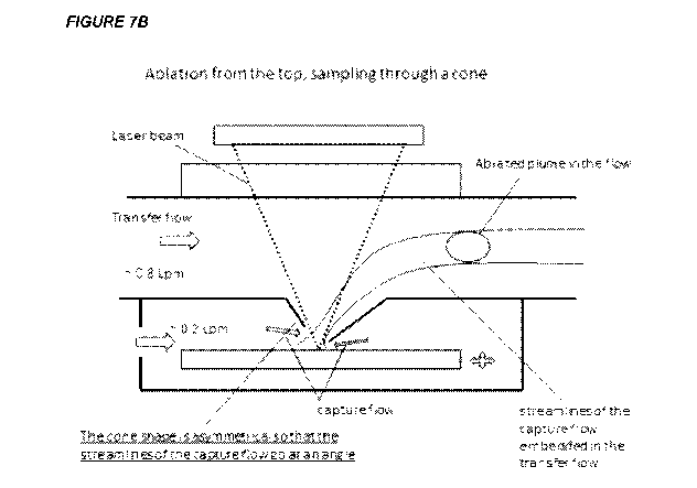
instances the majority of the gas flow to the ionization system. FIG. 7A shows
an
embodiment of this design. Here, the laser beam is focused through an
objective lens onto a
movable target through the ablation system inlet of the transfer conduit, to
generate plumes of
sample material for analysis. The ablation chamber of the laser ablation
system contains a gas
inlet (left hand side of chamber). Flowing gas into the chamber through this
inlet creates a
flow of gas out of the chamber at the cone through which the laser radiation
passes to ablate a
sample on the movable stage. This flow of gas captures plumes of ablated
material, and
entrains it as it flows up through the cone (in this embodiment, the cone is
the laser ablation
system inlet of the transfer conduit) and out of the ablation chamber into the
conduit passing
above the chamber. This conduit also has gas flowing into it from the separate
transfer flow
inlet (left hand side of the figure, indicated by the transfer flow arrow).
The component
comprising the transfer flow inlet, laser ablation system inlet and which
begins the transfer
conduit which carries the ablated sample material towards the ionization
system can also
termed a flow cell (as it is in W02014146724 and W02014147260).
The transfer flow fulfills at least three tasks: it flushes the plume entering
the transfer conduit
in the direction of the ionization system, and prevents the plume material
from contacting the
side walls of the transfer conduit; it forms a "protection region" above the
sample surface and
ensures that the ablation plume is carried out under a controlled atmosphere;
and it increases
the flow speed in the transfer conduit. In some embodiments the viscosity of
the capture gas
is lower than the viscosity of the primary transfer gas. This helps to confine
the plume of
sample material in the capture gas in the center of the transfer conduit and
to minimize the
9
CA 03032861 2019-02-01
WO 2018/026898 PCT/US2017/045060
diffusion of the plume of sample material downstream of the laser ablation
system (because
in the center of the flow, the transport rate is more constant and nearly
flat). The gas(es) may
be, for example, and without limitation, argon, xenon, helium, nitrogen, or
mixtures of these.
In some embodiments, the transfer gas is argon. Argon is particularly well-
suited for stopping
the diffusion of the plume before it reaches the walls of the transfer conduit
(and it also
assists improved instrumental sensitivity in apparatus where the ionization
system is an argon
gas-based ICP). The capture gas is preferably helium. However, the capture gas
may be
replaced by or contain other gases, e.g., hydrogen, nitrogen, or water vapor.
At 25 C, argon
has a viscosity of 22.6 Pas, whereas helium has a viscosity of 19.8 Pas. In
some
embodiments the capture gas is helium and the transfer gas is helium.
The use of a sample cone as in FIG. 7A minimizes the distance between the
target and the
conduit comprising the transfer flow of gas. Because of the reduced distance
through which
the capture gas flows at the point of the cone, this also leads to improved
capture of sample
material with less turbulence, and so reduced spreading of the plumes of
ablated sample
material. The inlet of the transfer conduit is therefore the aperture at the
tip of the sample
cone. The cone projects into the ablation chamber.
A modification of the sample cone is shown in FIG. 7B. Here, the sample cone
is
asymmetrical. When the cone is symmetrical, the gas flow from all directions
is symmetrical,
such that the overall flow of gas is zero (is neutralized) along the surface
of the sample at the
axis of the sample cone. By making the cone asymmetrical, a non-zero velocity
along the
sample surface is created, which assists in the washout of plume materials
from the ablation
chamber of the laser ablation system. FIG. 7B shows an asymmetry of the cone
that projects
the capture flow of gas entering the transfer conduit from the laser ablation
system in the
same direction as the transfer flow in the transfer conduit. This figure also
illustrates how the
asymmetry influences the projected streamlines of gas flow of the capture gas
flow within the
transport gas flow, together with a captured plume within the capture flow.
Accordingly, in
some embodiments, the sample cone of the transfer conduit is asymmetric. The
asymmetric
sample cone is adapted to cause a non-zero vector gas flow on the surface of a
sample at the
axis of the sample cone.
Thus, the invention provides an apparatus comprising:
(i) a laser ablation system, adapted to generate plumes of sample
material from
a sample;
CA 03032861 2019-02-01
WO 2018/026898 PCT/US2017/045060
(ii) an ionization system, adapted to receive material removed from the
sample
by the laser ablation system and to ionize said material to form elemental
ions;
(iii) a mass spectrometer to receive elemental ions from said ionization
system
and to analyze said elemental ions,
wherein the laser ablation system and the ionization system are coupled
together by a transfer
conduit, adapted to carry a flow of gas containing plumes of ablated sample
material from the
laser ablation system to the ionization system, and wherein the inlet of the
transfer conduit
within the laser ablation system is an asymmetric sample cone, with an
aperture at the narrow
end of the cone. Sometimes, the inlet within the laser ablation system is
asymmetric and
projects into the ablation chamber of the laser ablation system in a non-
horizontal (e.g.
vertical or perpendicular to the surface of the sample) direction (where an
asymmetric sample
cone is an example of such an inlet). The asymmetric inlet, such as the
asymmetric sample
cone, is adapted so that a higher capture flow enters the inlet on one side of
the inlet.
FIG. 7B shows a cone which is asymmetric because the one side of the cone
projects closer to
the target than the other side. In three dimensions, this represents a cone in
which the tip has
been truncated at an angle (i.e. non-parallel) to the base of the cone.
Accordingly in some
embodiments, the asymmetric sample cone is a truncated cone.
In practice, any modification of the sample cone that causes a non-zero vector
gas flow along
the surface of the sample at the axis of the cone may be employed. According,
in some
embodiments, the asymmetric cone comprises a notch or a series of notches,
adapted to
generate non-zero vector gas flow along the surface of the sample at the axis
of the cone. In
some embodiments, the asymmetric cone comprises an orifice in the side of the
cone, adapted
to generate non-zero vector gas flow along the surface of the sample at the
axis of the cone.
This orifice will imbalance gas flows around the cone, thereby again
generating a non-zero
vector gas flow along the surface of the sample at the axis of the cone at the
target. In some
instances, the side of the cone may comprise more than one orifice, such as
two, three, four,
five, six, seven, eight, nine, ten or more than 10 orifices. In some
embodiments, the sample
cone may include both one or more notches and one or more orifices. In some
embodiments,
the edges of the notch(es) and/or orifice(s) are smoothed, rounded or
chamfered in order to
prevent or minimize turbulence.
11
CA 03032861 2019-02-01
WO 2018/026898 PCT/US2017/045060
Different orientations of the asymmetry of the cone will be appropriate for
different
situations, dependent on the choice of capture and transfer gas and flow rates
thereof, and it is
within the abilities of the skilled person to appropriately identify the
combinations of gas and
flow rate for each orientation. In some embodiments, the asymmetry provides
increased
capture flow from the same source direction as the transfer flow (in other
words, the capture
flow direction is in line with the transport flow), as illustrated in FIG. 7B.
When the capture
flow is more in line with the transport flow, this can help to place the
streamlines of the
capture flow in the middle of the transfer flow without excessive turbulence.
According, in
some embodiments, the asymmetric inlet, such as an asymmetric sample cone, is
adapted so
that the streamlines of the capture flow are directed at an angle (i.e. not at
a right angle,
perpendicular to the surface of the sample).
A further kind of asymmetry is a cone formed from two elliptical halves, which
share a
common height (z) and one base diameter (the x diameter), but which differ in
the other base
(the y diameter) (or one elliptical and one circular half).
All of the above adaptations may be present in a single asymmetric sample cone
as use in the
invention. For example, the cone may be asymmetrically truncated and formed
from two
different elliptical cone halves, the cone may be asymmetrically truncated and
comprise one
of more orifices and so on.
The sample cone is therefore adapted to capture all or part of a plume of
material ablated
from a sample in the laser ablation system. The sample cone is positioned
operably proximate
to the sample, e.g. by maneuvering the sample within the laser ablation system
on a movable
sample carrier tray, as described in more detail below. As noted above, plumes
of ablated
sample material enter the transfer conduit through an aperture at the narrow
end of the sample
cone. In some embodiments, the diameter of the aperture a) is adjustable; b)
is sized to
prevent perturbation to the ablated plume as it passes into the transfer
conduit; and/or c) is
about the equal to the cross-sectional diameter of the ablated plume. In some
embodiments,
the diameter of the aperture is between about 100 p.m to lmm. For example, the
diameter of
the aperture is between about 200 p.m to 900 p.m, such as 300 p.m to 800 m.
In some
embodiments, the diameter of the aperture is between about 500 p.m to 700 p.m.
In some
embodiments, the diameter of the aperture is about 500 p.m. In some
embodiments, the
diameter of the aperture is about 700 p.m.
12
CA 03032861 2019-02-01
WO 2018/026898 PCT/US2017/045060
Tapered conduits
In tubes with a smaller internal diameter, the same flow rate of gas moves at
a higher speed.
Accordingly, by using a tube with a smaller internal diameter, a plume of
ablated sample
material carried in the gas flow can be transported across a defined distance
more rapidly at a
given flow rate (e.g. from the laser ablation system to the ionization system
in the transfer
conduit). One of the key factors in how quickly an individual plume can be
analyzed is how
much the plume has diffused during the time from its generation by ablation
through to the
time its component ions are detected as the mass spectrometer component of the
apparatus
(the transience time at the detector). Accordingly, by using a narrow transfer
conduit, the
time between ablation and detection is reduced, thereby meaning diffusion is
decreased
because there is less time in which it can occur, with the ultimate result
that the transience
time of each ablation plume at the detector is reduced. Lower transience times
mean that
more plumes can be generated and analyzed per unit time, thus producing images
of higher
quality and/or faster.
Accordingly, the invention also provides an apparatus comprising:
(i) a laser ablation system, adapted to generate plumes of sample material
from
a sample;
(ii) an ionization system, adapted to receive material removed from the
sample
by the laser ablation system and to ionize said material to form elemental
ions;
(iii) a mass spectrometer to receive elemental ions from said ionization
system
and to analyze said elemental ions,
wherein the laser ablation system and the ionization system are coupled
together by
a transfer conduit, adapted to carry a flow of gas containing plumes of
ablated
sample material from the laser ablation system to the ionization system,
wherein the
internal surface of the transfer conduit comprises a taper along at least a
portion of
its length from the inlet (at the laser ablation system end) to the outlet (at
the
ionization system end).
The taper may comprise a gradual change in the internal diameter of the
transfer conduit
along said portion of the length of the transfer conduit (i.e. the internal
diameter of the tube
were a cross section taken through it decreases along the portion from the end
of the portion
13
CA 03032861 2019-02-01
WO 2018/026898 PCT/US2017/045060
towards the inlet (at the laser ablation system end) to the outlet (at the
ionization system end).
As shown in FIG. 3B and 7C, the tapering modification to the transfer conduit
is applicable to
all embodiments of the apparatus described herein, whether they comprise a
direct injector
inlet, a sample cone, or any other structure at the ionization system inlet
end of the transfer
conduit. With reference to FIG. 3B, the region of the conduit near where
ablation occurs has
a relatively wide internal diameter. The larger volume of the conduit before
the taper
facilitates the confinement of the materials generated by ablation. When the
ablated particles
fly off from the ablated spot they travel at high velocities. The friction in
the gas slows these
particles down but the plume can still spread on a sub-millimeter to a
millimeter scale.
Allowing for sufficient distances to the walls helps with the containment of
the plume near
the center of the flow.
Because the wide internal diameter section is only short (of the order of 1-
2mm), it does not
contribute significantly to the overall transience time providing the plume
spends more time
in the longer portion of the transfer conduit with a narrower internal
diameter. Thus, a larger
internal diameter portion is used to capture the ablation product and a
smaller internal
diameter conduit is used to transport these particles rapidly to the
ionization system.
FIG. 7C shows the application of this development to apparatus comprising a
sample cone at
the ionization system inlet to the transfer conduit. As described above, the
conduit comprises
a wider internal diameter section and a taper down to a narrower internal
dimeter conduit,
which results in a shorter transfer time of ablated plumes to the ionization
system, and
ultimately shorter transience times for each plume at the mass spectrometer.
The portion of
the transfer conduit near the sample cone which receives plumes of material
following
ablation has a broad internal diameter, and as before is broad enough to
contain enough gas to
stop the plume material, generated by ablation of the sample, from hitting the
sides of the
conduit and to entrain the ablated sample material within the transfer flow
passing through
the flow cell from the transfer flow inlet. This broad portion will in many
instances be a
unitary component with the sample cone, and so the broadness of the internal
diameter (e.g.
approximately 2mm) also facilitates manufacture.
In some embodiments, the taper begins within 50mm of the ionization system
inlet to the
transfer conduit. In some embodiments, the taper begins within 40mm of the
ionization
system inlet, such as within 30mm, within 20mm, within 15mm, or within lOmm of
the
ionization system inlet. In some embodiments, the taper begins within 5mm,
within 4mm,
14
CA 03032861 2019-02-01
WO 2018/026898 PCT/US2017/045060
within 3mm, within 2mm or within lmm downstream of the ionization system
inlet. In some
embodiments, the taper begins 1-2mm downstream of the ionization system inlet.
The taper between the large internal diameter portion and the small internal
diameter region
can be made sufficiently gentle to avoid the onset of the turbulence. For
example, the taper
can be at an angle of at least 5 degrees. In some embodiments, the angle of
the taper can be at
least 10 degrees, such as at least 15 degrees, at least 20 degrees, at least
25 degrees, or 30
degrees or more, even such as 60 degrees. In some embodiments, the taper is at
an angle less
than 40 degrees, such as less than 30 degrees, less than 25 degrees, less than
20 degrees, less
than 15 degrees, or less than 10 degrees. In some embodiments, the taper is at
an angle less
than 8 degrees, such as less than 5 degrees, less than 4 degrees, less than 3
degrees, less than
2 degrees, or less than 1 degree. In some embodiments, the angle of the taper
is between 10
and 30 degrees. In some embodiments, the angle of the taper may increase or
decrease along
the length of the taper.
In some embodiments, the length of the taper is at least 5mm, for example at
least lOmm, at
least 20mm, at least 30mm, at least 40mm or at least 50mm or at least 100mm.
In some
embodiments, the length of the taper is less than lOmm, for example, less than
5mm, less
than 4mm, less than 3mm, less than 2mm or lmm or less.
The transfer conduit internal diameter can be x millimeters (mm) at the input
end of the
conduit but it can be tapered down 5-fold to x/5 mm near the output end (e.g.
4 mm at the
input end and 800 um at the output end). In some embodiments, the taper
reduces the internal
diameter of the transfer conduit by less than 5-fold, such as 4-fold or less,
3-fold or less, or 2-
fold or less. The internal diameter is the measure of the longest cross-
section through the
conduit. E.g. if the conduit is circular, the internal diameter is simply the
diameter of the
circle, but if the conduit is a rectangle, it is the diagonal. In some
embodiments, the internal
diameter of the conduit following the taper is narrower than 2mm, for example
narrower than
1.5mm, narrower than 1.25mm, narrower than lmm, narrower than 900um, narrower
than
800um, narrower than 700um, narrower than 600um, or 500um or narrower. In some
embodiments, the internal diameter of the conduit following the taper is 400um
or narrower,
300um or narrower, 200um or narrower or 100um or narrower.
The diameter of the narrow internal diameter section is limited by the
diameter corresponding
to the onset of turbulence. A Reynolds number can be calculated for a round
tube and a
known flow. In general a Reynolds number above 4000 will indicate a turbulent
flow, and
CA 03032861 2019-02-01
WO 2018/026898 PCT/US2017/045060
thus should be avoided. A Reynolds number above 2000 will indicate a
transitional flow
(between non-turbulent and turbulent flow), and thus may also be desired to be
avoided. For a
given mass flow of gas the Reynolds number is inversely proportional to the
diameter of the
conduit. Accordingly, in some embodiments, the internal diameter of the narrow
internal
diameter section of the transfer conduit is narrower than 2mm, for example
narrower than
1.5mm, narrower than 1.25mm, narrower than lmm, but greater than the diameter
at which a
flow of helium at 4 liters per minute in the conduit has a Reynolds number
greater than 4000.
Rough or even angular edges in the transitions between the constant diameter
portions of the
transfer conduit and the taper may cause turbulence in the gas flow.
Accordingly, in some
embodiments, the transitions into and from the taper should have smooth edges
adapted to
suppress the onset of turbulence. For instance, the edges may be rounded and
or chamfered.
Apparatus comprising a tapered conduit can also comprise a sample cone
(optionally
asymmetric). As would be understood by the skilled person, the tapered conduit
can be
employed in any of the apparatus described herein which use alternative
transfer conduit
arrangements, as illustrated e.g. in figures 2-10, and as discussed herein in
detail in the
following sections.
Sacrificial flow
At higher flows, the risk of turbulence occurring in the conduit increases.
This is particularly
the case where the transfer conduit has a small internal diameter (e.g. lmm).
The inventor has
discovered, however, that it is possible to achieve high speed transfer (up to
and in excess of
300m/s) in transfer conduits with a small internal diameter if a light gas,
such as helium or
hydrogen, is used instead of argon, which is traditionally used as the
transfer flow of gas. In
certain embodiments, a mixture of gas primarily comprising helium or hydrogen
is used.
High speed transfer presents problems insofar as it may cause the plumes of
ablated sample
material to be passed through the ionization system without an acceptable
level of ionization
occurring. The level of ionization can drop because the increased flow of cool
gas reduces the
temperature of the plasma at the end of the torch. If a plume of sample
material is not ionized
to a suitable level, information is lost from the ablated sample material ¨
because its
components (including any labelling atoms/elemental tags) cannot be detected
by the mass
spectrometer. For example, the sample may pass so quickly through the plasma
at the end of
the torch in an ICP ionization system that the plasma ions do not have
sufficient time to act
16
CA 03032861 2019-02-01
WO 2018/026898 PCT/US2017/045060
on the sample material to ionize it. The inventor has discovered that this
problem, caused by
high flow, high speed transfer in narrow internal diameter transfer conduits
can be solved by
the introduction of a flow sacrificing system at the outlet of the transfer
conduit. The flow
sacrificing system is adapted to receive the flow of gas from the transfer
conduit, and pass
only a portion of that flow (the central portion of the flow comprising any
plumes of ablated
sample material) onwards into the injector that leads to the ionization
system. To facilitate
dispersion of gas from the transfer conduit in the flow sacrificing system,
the transfer conduit
outlet can be flared out.
The flow sacrificing system is positioned close to the ionization system, so
that the length of
the tube (e.g. injector) that leads from the flow sacrificing system to the
ionization system is
short (e.g. ¨1cm long; compared to the length of the transfer conduit which is
usually of a
length of the order of tens of cm, such as ¨50cm). Thus the lower gas velocity
within the tube
leading from the flow sacrificing system to the ionization system does not
significantly affect
the total transfer time, as the relatively slower portion of the overall
transport system is much
shorter.
Accordingly, the invention provides an apparatus comprising:
(i) a laser ablation system, adapted to generate plumes of sample material
from
a sample;
(ii) an ionization system that is adapted to receive material removed from
the
sample by the laser ablation system and to ionize said material to form
elemental ions;
(iii) a mass spectrometer to receive elemental ions from said ionization
system
and to analyze said elemental ions,
wherein the laser ablation system and the ionization system are coupled
together by a transfer
conduit and a flow sacrificing system,
wherein the transfer conduit is adapted to carry a flow of gas containing
plumes of ablated
sample material from an inlet in the laser ablation system to an outlet in the
flow sacrificing
system,
wherein the flow sacrificing system comprises a chamber comprising:
(a) the outlet of the transfer conduit;
17
CA 03032861 2019-02-01
WO 2018/026898 PCT/US2017/045060
(b) an ionization system inlet, positioned to receive sample material from the
transfer
conduit outlet and to introduce the sample material into the ionization
system; and
(c) a sacrificial flow outlet,
wherein the flow sacrificing system is adapted to reduce the flow of gas
entering the
ionization system through the ionization system inlet compared to the flow of
gas entering the
flow sacrificing system through the transfer conduit, by directing some of the
flow of gas
entering the flow sacrificing system out of the sacrificial flow outlet, and
wherein the outlet of the transfer conduit in the flow sacrificing system is
optionally flared.
In some embodiments, the ionization system inlet is positioned co-axially to
the outlet of the
transfer conduit (because the plumes of sample material being transferred
along the conduit
will be entrained within the center of the transfer flow), to maximize
transmission of material
from the transfer conduit, through the flow sacrificing system, to the
ionization system inlet,
and so to the injector of the ionization system. In some embodiments, the
ratio of the internal
diameter of the transfer conduit to the internal diameter of the inlet of the
ionization system is
less than 2:1, for example 1.5:1 or 1:1. In some embodiments, the ratio of the
internal
diameter of the transfer conduit to the internal diameter of the injector of
the ionization
system is less than 2:1, for example 1.5:1 or 1:1. In some embodiments, the
internal diameter
of the injector of the ionization system (or the inlet to the ionization
system) has a greater
internal diameter than the transfer conduit. For example, in some embodiments,
the ratio of
the internal diameter of the transfer conduit to the internal diameter of the
inlet of the
ionization system is less than 1:1, for example 1:1.5 or 1:2. In some
embodiments, the ratio of
the internal diameter of the transfer conduit to the internal diameter of the
injector of the
ionization system is less than 1:1, for example 1:1.5 or 1:2.
In most arrangements, it is not desirable, or in some cases possible, to
significantly increase
the diameter of the tube (e.g. the injector) which passes from the flow
sacrificing system to
the ionization system as a way of reducing the speed of the gas at a
volumetric flow rate. For
example, where the ionization system is an ICP, the conduit from the flow
sacrificing system
forms the injector tube in the center of the ICP torch. When a wider internal
diameter injector
is used, there is a reduction in signal quality, because the plumes of ablated
sample material
cannot be injected so precisely into the center of the plasma (which is the
hottest and so the
most efficiently ionizing part of the plasma). The strong preference is for
injectors of lmm
internal diameter, or even narrower (e.g. an internal diameter of 800um or
less, such as
18
CA 03032861 2019-02-01
WO 2018/026898 PCT/US2017/045060
600 m or less, 500 m or less or 400 m or less). Other ionization techniques
rely on the
material to be ionized within a relatively small volume in three dimensional
space (because
the necessary energy density for ionization can only be achieved in a small
volume), and so a
conduit with a wider internal diameter means that much of the sample material
passing
through the conduit is outside of the zone in which energy density is
sufficient to ionize the
sample material. Thus narrow diameter tubes from the flow sacrificing system
into the
ionization system are also employed in apparatus with non-ICP ionization
systems. As noted
above, if a plume of sample material is not ionized to a suitable level,
information is lost from
the ablated sample material ¨ because its components (including any labelling
atoms/elemental tags) cannot be detected by the mass spectrometer.
Rough or even angular edges in the transition between the constant diameter
portion of the
transfer conduit and the flare at the outlet may cause turbulence in the gas
flow. Accordingly,
in some embodiments, the transition into the flare out should have smooth
edges adapted to
suppress the onset of turbulence. For example, the edges may be rounded.
Pumping can be used to help ensure a desired split ratio between the
sacrificial flow and the
flow passing into the inlet of the ionization system. Accordingly, in some
embodiments, the
flow sacrificing system comprises a pump attached to the sacrificial flow
outlet. A controlled
restrictor can be added to the pump to control the sacrificial flow.
Therefore, in some
embodiments, the pump of the flow sacrificing system further comprises a
restrictor adapted
to control the flow of gas through the sacrificial flow outlet. In some
embodiments, the flow
sacrificing system comprises a mass flow controller, adapted to control the
restrictor.
Where expensive gases are used, the gas pumped out of the sacrificial flow
outlet can be
cleaned up and recycled back into the same system using known methods of gas
purification.
Helium is particularly suited as a transport gas as noted above, but it is
expensive; thus, it is
advantageous to reduce the loss of helium in the system (i.e. when it is
passed into the
ionization system and ionized). The flow sacrificing system splits the helium
flow into a
near-axial flow and a sacrificial flow. The sacrificial flow can be cleaned up
and recycled in
the system while the near-axial flow (the central portion of the flow that
carries the entrained
particles from the ablated plume) will be passed into the ionization system
(e.g. the plasma of
an ICP torch). The helium from the near-axial flow will be lost for recovery.
Accordingly, in
some embodiments a gas purification system is connected to the sacrificial
flow outlet of the
flow sacrificing system. In some embodiments, the gas purification system
provides a
portion of the gas flowed into the apparatus, for example through an inlet
into the laser
19
CA 03032861 2019-02-01
WO 2018/026898 PCT/US2017/045060
ablation system's ablation chamber and/or through an inlet in the transfer
conduit (i.e. it is
used as either the capture flow and/or the gas that makes up most of the
transfer flow ¨
indicated by the arrows on the left hand side of FIG. 7).
A further refinement of the setup is provided in FIG. 8C, and is a particular
optimization of
the flow sacrificing system in apparatus in which the ionization system is an
ICP. As before,
a larger transfer flow rate is sent down the transfer conduit and only the
central portion of this
flow is allowed to become the part of the injector flow that will enter the
plasma of the ICP
torch. Typically, helium gas will be used as a transfer flow, because as noted
above its
properties are well suited for high velocity transport of the plume material
over a long
conduit (i.e. less chance to trigger the turbulence for the same flow velocity
(as compared to
argon). Even incorporating a gas purification system that recycles helium from
the sacrificial
flow, the near-axial flow of helium that continues through the flow
sacrificing system into the
ionization system is lost.
However, a further reduction of the near-axial flow in the setup of FIG. 8B
that is passed into
the ionization system inlet can have a negative consequence on the ionization
sampling
efficiencies in an inductively coupled plasma. The apparatus in FIG. 8C offers
a solution to
this problem. Here, another flow of a less valuable gas, such as Argon, is
added to make up
the flow in the injector of the ICP torch. The injector flow can be tuned to
optimize ionization
sampling efficiency. Argon gas is commonly used for the formation of a central
channel in
the inductively coupled plasma, and, accordingly, can be added to the injector
flow as shown
in FIG. 8C. Thus, the near-axial flow carried from the transfer conduit outlet
into the
ionization system inlet is chosen to be sufficiently small, but not so small
that plume
transients are significantly affected. A makeup flow of argon is chosen to
provide optimal
ionization conditions in the inductively coupled plasma. Accordingly, in some
embodiments,
the flow sacrificing system is adapted to reduce the flow of gas passing into
the ionization
system inlet (e.g. the injector of an ICP torch ionization system) to below
1Lpm, such as 0.5
Lpm or less, 0.4 Lpm or less, 0.3 Lpm or less, or 0.2 Lpm or less. In some
embodiments, the
ICP injector comprises a second inlet into which gas can be flowed to make up
the flow rate
in the injector. In some embodiments, the second inlet comprises a concentric
tube around the
injector attached to the ionization system inlet that introduces the make-up
gas as a sheath
flow around the sample-containing gas flow from the flow sacrificing system.
This make up
flow inlet is different from the flow of argon gas also provided in the middle
and outer
concentric tubes which support the plasma, as illustrated in FIG 8D. This
injector can also be
CA 03032861 2019-02-01
WO 2018/026898 PCT/US2017/045060
termed a dual concentric injector. Accordingly, in one aspect the invention
provides an
injector according to Figure 8D, which comprises a dual concentric portion.
Apparatus comprising a flow sacrificing system can also comprise a sample cone
(optionally
asymmetric) or a tapered conduit, as described above. In some embodiments, the
apparatus
comprise a flow sacrificing system, a sample cone (optionally asymmetric) and
a tapered
conduit, as described above. As would be understood by the skilled person, the
flow
sacrificing system can be employed in any of the apparatus described herein
which use
alternative transfer conduit arrangements, as illustrated e.g. in figures 2-
10, and as discussed
herein in detail in the following sections.
Laser ablation system
The laser ablation system, also referred to as the "ablation cell," houses the
sample during
ablation. Typically the ablation cell includes a laser transparent window to
allow laser energy
to strike the sample. Optionally the ablation cell includes a stage to hold
the sample to be
analyzed. In some embodiments the stage is movable in the x-y or x-y-z
dimensions. In
drawings and examples herein, the laser ablation system is sometimes shown as
an open
arrangement. However, such configurations are for illustration only, and it
will be recognized
that some form of suitable enclosure for preventing contamination or
infiltration from the
ambient environment is present. For example, a chamber configured with gas
inlets and/or
optical ports can be arranged around the laser ablation system to provide an
enclosed
environment suitable for capturing and transferring the ablated plume for mass
analysis (e.g.
FIG. 7). The gas inlets and optical port(s) are positioned so that the
orientation of the laser
beam, sample, plume expansion, and transfer conduit are suitable for the
methods and devices
disclosed herein. It will be appreciated that the ablation cell is generally
gas tight (except for
designed exits and ports).
Lasers used for laser ablation according to the invention generally fall into
three categories:
femtosecond pulsed lasers, deep UV pulsed lasers and pulsed lasers with a
wavelength
chosen for high absorption in the ablated material ("wavelength selective
lasers"). Deep UV
and wavelength specific lasers would likely operate with nanosecond or
picosecond pulses.
Each class of lasers has its drawbacks and benefits and can be chosen based on
a particular
application. In some embodiments, the laser is a femtosecond pulsed laser
configured to
operate with a pulse rate between 10 and 10000 Hz. Femtosecond laser are known
(see, e.g.,
21
CA 03032861 2019-02-01
WO 2018/026898 PCT/US2017/045060
Jhanis et al., "Rapid bulk analysis using femtosecond laser ablation
inductively coupled
plasma time-of-flight mass spectrometry" J. Anal. At. Spectrom., 2012, 27:1405-
1412.
Femtosecond lasers allow for laser ablation of virtually all materials with
the only
prerequisite for laser ablation being-sufficient power density. This can be
achieved even with
relatively low pulse energy when the beam is tightly focused, for instance to
1 micrometer
diameter and is short in duration (focused in time). Deep UV lasers also can
ablate a large
class of materials because most of the commonly used materials absorb deep UV
photons.
Wavelength selective laser ablation can utilize the lasers with the specific
laser wavelength
targeting absorption in the substrate material. A benefit of the wavelength
specific laser may
be the cost and simplicity of the laser and the optical system, albeit with a
more limited
spectrum of substrate materials. Suitable lasers can have different operating
principles such
as, for example, solid state (for instance a Nd:YAG laser), excimer lasers,
fiber lasers, and
OPO lasers.
A useful property of the femtosecond laser radiation is that it is absorbed
only where the
threshold power density is reached. Thus, a converging femtosecond laser
radiation can pass
through a thicker section of material without being absorbed or causing any
damage and yet
ablate the same material right at the surface where the focus is occurring.
The focus can then
be moved inside the material progressively as the sample layers are ablated.
Nanosecond
laser pulses might be partially absorbed by the substrate but can still work
for ablation since
the energy density at the focal point will be the highest (as long as it is
sufficient for
ablation).
The spatial resolution of signals generated in this way depends on two main
factors: (i) the
spot size of the laser, as signal is integrated over the total area which is
ablated; and (ii) the
speed at which a plume can be analyzed, relative to the speed at which plumes
are being
generated, to avoid overlap of signal from consecutive plumes, as discussed
above. The
distance referred to as spot size corresponds to the longest internal
dimension of the beam,
e.g. for a circular beam it is a beam of diameter 2[tm, and for a square beam
corresponds to
the length of the diagonal between opposed corners). The laser pulse may be
shaped using an
aperture, homogenized (if required) using a beam homogenizer, focused, e.g.,
using an
objective lens, to produce a desired spot size. Typically, the spot size is
100[tm or less, such
as 50[tm or less, 25[tm or less, 20[tm or less, 15[tm or less, or 10[tm or
less than 10 p.m.
Exemplary spot sizes include diameters (or equivalent sized ablation areas of
other shapes) in
22
CA 03032861 2019-02-01
WO 2018/026898 PCT/US2017/045060
the range of 0.10-3 p.m (e.g., about 0.3 p.m), 1-5 p.m (e.g., about 3 p.m), 1-
10 p.m (e.g., about
1, about 2, about 3, about 4 or about 5 p.m), less than 10 p.m, and less than
5 p.m. In particular
embodiments, a laser system is configured to operate with sufficiently focused
laser pulses to
ablate a sample area in the order of about 1 p.m, e.g., 100 nm to 1 p.m.
In order to analyze individual cells the laser in the laser ablation system
has a spot size which
is no larger than these cells. This size will depend on the particular cells
in a sample, but in
general the laser spot will therefore have a diameter of less than 4 p.m e.g.
within the range
0.1-4 p.m, 0.25-31.tm, or 0.4-2 p.m. Thus, a laser spot can have a diameter of
about 3 p.m or
less, about 2 p.m or less, about 1 p.m or less, about 0.5 p.m or less than 0.5
p.m, such as around
400nm or less, around 300nm or less, around 200nm or less, around 100nm or
less than
100nm. In order to analyze cells at a subcellular resolution the invention
uses a laser spot
size which is no larger than these cells, and more specifically uses a laser
spot size which can
ablate material with a subcellular resolution. Sometimes, single cell analysis
can be
performed using a spot size larger than the size of the cell, for example
where cells are spread
out on the slide, with space between the cells. Here, a larger spot size can
be used and single
cell characterization achieved, because the additional ablated area around the
cell of interest
does not comprise additional cells. The particular spot size used can
therefore be selected
appropriately dependent upon the size of the cells being analyzed. In
biological samples, the
cells will rarely all be of the same size, and so if subcellular resolution
imaging is desired, the
ablation spot size should be smaller than the smallest cell, if constant spot
size is maintained
throughout the ablation procedure. Small spot sizes can be achieved using
demagnification of
wider laser beams and near-field optics. A laser spot diameter of 1 p.m
corresponds to a laser
focus point (i.e. the diameter of the laser beam at the focal point of the
beam) of 1 p.m, but the
laser focus point can vary by +20% or more due to spatial distribution of
energy on the target
(for instance, Gaussian beam shape) and variation in total laser energy with
respect to the
ablation threshold energy. For example, using a 25 1.tm diameter laser beam,
and subjecting
this to 25-fold demagnification onto the tissue samples will give a spot size
with a 11.tm
diameter.
Ablation on this small scale produces very small amount of plume material that
in turn
ensures that the size of the plume is kept small. A smaller plume is more
likely to stay in the
middle of the capture flow without contacting the walls of the ablation cell
or of the transfer
conduit. Ablation on the 1 micrometer scale also means that the distance
between the ablated
surface and the area where plume expansion slows down and becomes dominated by
the
23
CA 03032861 2019-02-01
WO 2018/026898 PCT/US2017/045060
ambient gas is very short. This distance can range from a few micrometers to a
few hundred
micrometers. In some versions of the invention, the capture flow is present
where the plume
stops expanding. Therefore, for illustration and not limitation, several of
the appended figures
show the distance between the ablated surface and the region with capture flow
shown as
about 100 micrometers.
Although ablation on the 1 micrometer (or lower) scale is advantageous for
certain
applications (e.g., imaging), the methods and instruments of the invention are
also useful
when larger ablation spots are produced, such as ablation spots in the range
of about 5 to
about 35 microns diameter, for example in the range 5-15 microns, 10-20
microns, 15-25
microns, 20-30 microns and 25-35 microns. In some applications in which large
ablation
spots are produced, only a portion of the plume material is captured.
In some embodiments, the laser is situated outside the ablation chamber, and
the laser beam
(laser energy) enters the ablation chamber, e.g., though an optical window. As
used herein, a
laser beam may be described as being emitted from a surface (e.g., a laser
lens or mirror),
which surface may be oriented to direct the beam to a particular location or
pattern of
locations. For ease of description of the invention, the directed beam may be
considered to
have a particular orientation; the orientation of the beam can refer to an
imaginary line
aligned with the beam and extending beyond the actual beam (for example when
the beam
strikes a non-transparent surface). As will be apparent from context,
reference to the
orientation or position of a laser beam sometimes refers to the orientation or
position the
beam of an unpowered laser system would produce if the laser was in use.
For rapid analysis of a tissue sample a high frequency of ablation is needed,
for example
more than 20 Hz (i.e. more than 20 ablations per second, giving more than 20
plumes per
second). In some embodiments the frequency of ablation by the laser is at
least 40Hz, such as
at least 50Hz, or at least 100Hz. In some embodiments the frequency of
ablation by the laser
is within the range 40-2000 Hz, within the range 40-1500 Hz, within the range
40-500 Hz,
within the range 40-200 Hz, within the range 40-150 Hz, or within the range 75-
150 Hz. An
ablation frequency of more than 40 Hz allows imaging of typical tissue samples
to be
achieved in a reasonable time. The frequency with which laser pulses can be
directed at a
spot on the sample (assuming full ablation of the material at that spot) and
still be
individually resolved determines how quickly the pixels of the image can be
obtained.
Accordingly, if the duration of laser pulse required to ablate the material at
a point means that
24
CA 03032861 2019-02-01
WO 2018/026898 PCT/US2017/045060
only less than 5 pulses can be directed at a sample per second, the time taken
to study a lmm
x lmm area with ablation at a spot size of 11.tm would be over two days. With
a rate of 40Hz,
this would be around 6-7 hours, with further reductions in the analysis time
for further
increases in the frequency of pulses. At these frequencies the instrumentation
must be able to
analyze the ablated material rapidly enough to avoid substantial signal
overlap between
consecutive ablations, if it is desired to resolve each ablated plume
individually. It is
preferred that the overlap between signals originating from consecutive plumes
is <10% in
intensity, more preferably <5%, and ideally <2%. The time required for
analysis of a plume
will depend on the washout time of the ablation chamber (see ablation chamber
section
below), the transit time of the plume of sample material to and through the
ionization system
(optimizations of the transport to the ionization system are discussed above),
and the time
taken to analyze the ionized material. Each laser pulse can be correlated to a
pixel on the
image of the sample that is subsequently built up, as discussed in more detail
below.
Ablation chamber
An ablation chamber with a short washout time (e.g. 100 ms or less) is
advantageous for use
with the apparatus and methods of the invention. A cell with a long washout
time will either
limit the speed at which an image can be generated or will lead to overlap
between signals
originating from consecutive sample spots (e.g. Kindness et al. (2003) Clin
Chem 49:1916-
23, which had signal duration of over 10 seconds). Therefore the washout time
of a plume of
sample material from the laser ablation cell is a key limiting factor for
achieving high
resolution without increasing total scan time. Ablation chambers with washout
times of
<100 ms are known in the art. For example, Gurevich & Hergenroder (2007) J
Anal. At.
Spectrom., 22:1043-1050 discloses an ablation chamber with a washout time
below 100 ms.
An ablation chamber was disclosed in reference Wang et al. (2013) Anal. Chem.
85:10107-16
(see also reference WO 2014/146724) which has a washout time of 30 ms or less,
thereby
permitting a high ablation frequency (e.g. above 20 Hz) and thus rapid
analysis. Another such
ablation chamber is disclosed in reference WO 2014/127034. The ablation
chamber in this
document comprises a sample capture cell configured to be arranged operably
proximate to
the target (the sample capture cell described here is an example of a transfer
conduit inlet
modification which can be combined with the taper and flow sacrificing
modifications of the
transfer conduit as described above), the sample capture cell including: a
capture cavity
having an opening formed in a surface of the capture cell, wherein the capture
cavity is
CA 03032861 2019-02-01
WO 2018/026898 PCT/US2017/045060
configured to receive, through the opening, target material ejected or
generated from the laser
ablation site and a guide wall exposed within the capture cavity and
configured to direct a
flow of the carrier gas within the capture cavity from an inlet to an outlet
such that at least a
portion of the target material received within the capture cavity is
transferrable into the outlet
as a sample. The volume of the capture cavity in the ablation chamber of
reference WO
2014/127034 is less than 1cm3 and can be below 0.005cm3. Sometimes the
ablation chamber
has a washout time of 25ms or less, such as 20ms or 10ms or less. A sample
cone inlet of the
transfer conduit, for example an asymmetric sample cone, can also assist in
reducing the
washout time of the ablation chamber, and is an alternative to the capture
cell discussed here.
Ionization system
Sample material can be ionized by a variety of techniques. The use of an ICP
is suited for
IMS and IMC analyses. ICP is a plasma source in which the energy is supplied
by electric
currents produced by electromagnetic induction. Typically the plasma source is
based on
Argon gas. For example, the ionization system may comprise an ICP torch. IMC
using ICP in
the ionization system is reported on in, for example, Giesen et at. (2014)
Nature Methods.
11:417-422 and Wang et al. (2013) Anal. Chem. 85:10107-16.
The ionization system thus receives sample material from the laser sampling
system and
converts it into elemental ions for detection by the mass spectrometer. If the
sample material
is not atomized (e.g. the plume of sample material is still in the form of
molecules, or even an
aerosol of particulate material) then the ionization system acts to break down
the material into
elemental ions as part of the ionization process.
Mass spectrometer
As noted above, the third component of the apparatus is a mass spectrometer.
Mass analyzers
for use in the invention may be selected based on the needs of the operator or
specific
application. Exemplary types of mass analyzers include quadrupole, time of
flight (TOF),
magnetic sector, high resolution, single or multicollector based mass
spectrometers.
The time taken to analyze the ionized material will depend on the type of mass
analyzer/mass
spectrometer which is used for detection of ions. For example, instruments
which use
Faraday cups may be too slow for analyzing rapid signals, but not all analyses
will require the
rapid analysis of signals, and so the skilled person will be able to select
the mass
spectrometer or mass analyzer appropriately. Overall, the desired analysis
speed (and thus the
frequency with which ablation plumes can be interrogated) and degree of
multiplexing
26
CA 03032861 2019-02-01
WO 2018/026898 PCT/US2017/045060
(number of atoms to be monitored simultaneously/quasi-simultaneously) will
dictate the
type(s) of mass analyzer which should be used (or, conversely, the choice of
mass analyzer
will determine the speed and multiplexing which can be achieved).
Typically, time of flight mass spectrometers are used for the recording of
fast transient events
with the transit durations that are expected from a fast laser ablation setup.
TOF detectors can quasi-simultaneously register multiple masses in a single
sample. Whereas
TOF mass analyzers are normally unpopular for atomic analysis because of the
compromises
required to deal with the effects of space charge in the TOF accelerator and
flight tube, the
effectiveness of the technique can be improved by using it only to detect a
subset of ranges.
For example, in mass cytometry and imaging mass cytometry, a range may be
chosen only
such that ions from the labelling atoms used to mark target molecules in a
biological samples
are detected and so other atoms (e.g. those having an atomic mass below 80)
can be removed.
This results in a less dense ion beam, enriched in the masses in (for example)
the 80-210
dalton region, which can be manipulated and focused more efficiently, thereby
facilitating
TOF detection and taking advantage of the high spectral scan rate of TOF.
Thus, rapid
analyses can be achieved by combining TOF detection with choosing labelling
atoms that are
uncommon in the sample and ideally having masses above the masses seen in an
unlabeled
sample e.g. by using the higher mass transition elements. Further details on
mass cytometry
can be found in Tanner et at. Cancer Immunol Immunother (2013) 62:955-965 and
US patent
7479630, and on imaging mass cytometry in Giesen et al. (2014) Nature Methods.
11:417-
422.
Apparatus in use and additional variants of the invention to which the
transfer conduit
modifications described above can be applied
The apparatus of the invention may be used for analysis or imaging of a
biological sample,
which may be on transparent substrate. In imaging embodiments, generally the
laser may be
operated with continuous train of pulses or in bursts of pulses directed to
different positions
of the sample, referred to as "spots of interest," or "locations or zones of
ablation." The
pulses may be directed to spots in a set pattern, such as a raster for two-
dimensional imaging.
Alternatively, a plurality of individual spots at different locations (for
example,
corresponding to individual cells) may be ablated. In some embodiments, the
laser emits a
burst of pulses producing a plume coming from the same pixel (i.e. the same
location on the
target). Ablation plumes produced by individual pulses within the burst are
expected to fuse
27
CA 03032861 2019-02-01
WO 2018/026898 PCT/US2017/045060
into one plume and travel within the instrument in such a way that they will
be distinct from
the plume produced from another pixel. To distinguish individual pixels, the
time duration
between bursts (pixel interrogation that can be just one pulse or 100 pulses)
is maintained
above a certain limit determined by the time spreading of the ion signal (at
the detector) from
an individual pixel.
In accordance with the present teachings, each separate sample plume can be
distinctly
analyzed by the mass analyzer. In one aspect, the device is configured so that
spreading of the
plume in ablation cell (ablation system) and transfer conduit is smaller than
the spreading that
occurs in the ionization system and the mass analyzer. In one aspect, plumes
may be
distinctly analyzed by transferring each ablated plume to the ionization
system in a time
period that is within the cumulative transit time of the plume to the
ionization system and ion
detection by the mass analyzer. This can be accomplished by capturing each
sample plume
through a gas flow and under a transfer configuration such that the ratio
between the plume
broadening during transfer time period (i.e., transfer of the ablation plume
from the site of
ablation to the plasma) and the broadening during ion transit time period
(i.e., transfer of ions
from the plasma to the mass analyzer) is equal to or less than one.
Generally, the sample particle size limit for which an ionization system (e.g.
an ICP) can
effectively vaporize and ionize for the purpose of analytical detection is in
the order of about
p.m or less. Particles produced by the laser ablation at 1 micrometer scale
are below 1
micrometer and are well suited for an ICP ion source. For discrete particles
analysis (such as
may be carried out using CyTOF instrumentation, Fluidigm Canada Inc.), the
typical rate at
which these particles can be ionized and analytically detected can be a
function of the
cumulative broadening or spread of transit time of the sample in the plasma
while the
particles are being evaporated and ionized and of the ions' transit time
broadening or spread
between the ICP and its detection by the mass analyzer. Generally the
cumulative time
broadening or spread can be of the order of about 200 [is duration.
Consequently, for particles
of 10 p.m or less that are spatially separated, analyzing each distinct
particle can be achieved
by transferring each particle to the ionization system (e.g. ICP) in a time
period of the order
of 200 .is. In some embodiments the particles are transferred to the
ionization system (e.g.
ICP) in less than 200 [is, or less than 150 .is. Accordingly, in a sample
introduction system
where imaging of biological samples can be performed by laser ablation, a
laser system can
be configured to operate with sufficiently focused laser pulses to ablate a
sample area in the
order of about 1 p.m, such as the application of a femtosecond pulsed laser
for example. With
28
CA 03032861 2019-02-01
WO 2018/026898 PCT/US2017/045060
this configuration, the ablated plumes formed by each laser pulse can include
sample
particulates with dimensions typically about 1 p.m or less. Under certain
conditions as
described herein, these particulates can be captured and transferred to meet
the transfer time
period as required and, subsequently, each distinct plume can be effectively
vaporized and
ionized by the ionization system.
Additionally, while operating the laser with continuous series of pulses such
as in the case of
rasterizing across a sample surface for two dimensional imaging, the
distinctiveness of each
plume and the spatial separation between each subsequent plume can be
maintained between
the plume's zone of formation and the point of vaporization and ionization in
the ionization
system ion source. For example, as a plume is carried through a conduit, such
as the injector
tube shown in Fig. 1, the particles in the plume can spread and expand
outwardly in a radial
direction before it enters ionization system (e.g. the plasma of the ICP).
Spreading of the
particles produced in the plume can depend on its diffusion coefficient, the
velocity profile of
carrier flow and the distribution of particle density as it is formed and as
it evolves during
transit to the ionization system. For example, the femtosecond laser ablation
spot size of 1
p.m can produce a plume with an initial cross section diameter of about 100
p.m or less before
further spreading during its transit. The extent of spreading of the plume can
also be a
function of the dimension of the ablated particle; larger particles tend to
have lower diffusion
spreading but with higher momentum resulting in potential losses due to
contacting the inner
walls of the transfer conduit/injector tube. It is thus desirous to minimize
the plume spreading
and/or to transfer the plume to the ionization system within sufficient time
to vaporize and
ionize before the extent of spreading presents any challenging effects.
Accordingly, in various embodiments, the use of a laser for ablating 1 p.m
sample spots and
efficiently transporting the plume so that the spreading is maintained within
the internal
diameter of the transfer conduit/injector tube can be achieved by the
exemplary arrangements
described herein and in the accompanying drawings.
For a given laser ablation system and given sample, ablated plumes expand
after the laser
ablation until they reach a characteristic volume, referred to as the
"sampling volume." It is
desirable to configure the system to minimize the sampling volume, and to
increase the
velocity with which the gas flow carries the plume away from the sampling
volume. The
combination of a small sampling volume and fast gas flow reduces the time
spreading of the
plume transfer into the transfer conduit/injector. The sampling volume can be
described by
29
CA 03032861 2019-02-01
WO 2018/026898 PCT/US2017/045060
the envelope of the plume at the moment when the velocity of plume expansion
in any of the
dimensions falls substantially HO times) below the sonic velocity of the
surrounding gas
media. Without limitation, exemplary sampling volumes may be in the range 10-6
mm3 ¨ 10
mm3. Often the sampling volume is in the range 0.001 mm3 - 1 mm3. The capture
flow, where
present, flows into at least part of the sampling volume and carries at least
a portion of the
plume into the transfer conduit/injector whereupon it may be transported by
the transfer flow
to the ionization system (e.g. ICP). It is desirable that the velocity of
capture flow when it
enters the sampling volume be substantial (e.g., >1 m/s, >10 m/s, >100 m/s, or
> 500 m/s). In
some embodiments the velocity of capture flow when it enters the sampling
volume can be
estimated by measuring the velocity of the capture flow into the transfer
conduit/injector
(e.g., though the transfer conduit/injector aperture). In some embodiments
this measured
velocity is >1 m/s, >10 m/s, >100 m/s, or > 500 m/s. In contrast to the
present invention, if
the plume is not swept away rapidly, it will continue to expand and diffuse,
undesirably
filling the entire ablation cell.
In one aspect, the invention provides a laser ablation configuration in which
the laser beam is
directed to a target. In one embodiment, the target comprises a substrate and
a sample
disposed on the substrate. In one embodiment the substrate is transparent and
the target is a
transparent target.
In one aspect, the invention provides a laser ablation configuration
(discussed below in the
context of, but not limited to, Fig. 2), for "through-target" ablation. In
this configuration, the
pulse of a laser beam is directed through the transparent target and a sample
plume (the
"ablated plume" or the "plume") is formed downstream of the beam into a
transfer
conduit/injector. Also see Figs. 3-5. Through-target illumination is
advantageous for
optimizing transit time broadening due to the removal of optical elements
(windows,
objective lenses, etc.) from the straight path of the plume. In one aspect,
the invention
provides a laser ablation system comprising (a) a laser capable of producing
laser
illumination; (b) a laser ablation cell (or laser ablation system) into which
a transparent target
may be introduced and an transfer conduit/injector with an opening through
which an ablated
plume may enter, where the laser illumination originates from a surface on one
side of the
transparent target and the transfer conduit/injector opening is on the other
side. Other features
that may be included in the system are described throughout this disclosure
including the
examples.
CA 03032861 2019-02-01
WO 2018/026898 PCT/US2017/045060
In Fig. 1, a laser ablation mass cytometer comprises a laser ablation system
that can be
connected to an injector, such as a tube fabricated from quartz or other
generally suitable
material, and mounted for sample delivery into an inductively coupled plasma
(ICP) source,
also referred to as an ICP torch. The plasma of the ICP torch can vaporize and
ionize the
sample to form ions that can be received by a mass analyzer.
In various embodiments according to Fig. 2, the sample of interest can be
configured for laser
ablation by using a sample formatted to be compatible with a transparent
target. A sample can
be placed onto a transparent substrate, incorporated into a transparent
substrate or can be
formed as the transparent target. Suitable laser-transparent substrates may
comprise glass,
plastic, quartz and other materials. Generally the substrate is substantially
planar or flat. In
some embodiments the substrate is curved. Substrates are from 0.1 mm up to 3
mm thick, in
certain embodiments. In some embodiments, the substrate is encoded (see, e.g.,
Antonov, A.
and Bandura, D., 2012, U.S. Pat. Pub. 2012/0061561, incorporated by reference
herein). In
this configuration, the pulse of a laser beam is directed through the
transparent target and a
sample plume (the "ablated plume" or the "plume") is formed downstream of the
beam into a
transfer conduit/injector.
The transfer conduit can have an inlet configured to capture the ablated
plume; such as the
inlet formed as a sample cone having a small opening or aperture as
illustrated in Fig. 2. In
this configuration, the sample cone can be positioned near the area, or zone,
where the plume
is formed. For example, the opening of the sample cone may be positioned from
10 p.m to
1000 p.m from the transparent target, such as about 100 p.m away from the
transparent target.
Consequently, the ablated plume can be generated and formed at least partially
within the
expanding region of the cone. In some embodiments, the diameter of the
aperture and/or
dimensions of the spacing (including angles) are adjustable to permit
optimization under
various conditions. For example, with a plume having a cross sectional
diameter in the scale
of 100 p.m, the diameter of the aperture can be sized in the order of 100 p.m
with sufficient
clearance to prevent perturbation to the plume as it passes.
The transfer conduit can continue downstream of the sampling cone for
receiving the ablated
plume in such a configuration as to encourage the movement of the plume and
preserve the
spatial distinctiveness of each subsequent plume as a function of the laser
pulses.
Accordingly, a flow of gas can be introduced to aid in directing the plume
through the
aperture of the sampling cone in order to capture (capture flow) each plume
distinctively
31
CA 03032861 2019-02-01
WO 2018/026898 PCT/US2017/045060
while an additional flow of gas can be introduced to the transfer
conduit/injector for
transferring (transfer flow or sheath flow) each distinctly captured plume
towards the
ionization system. Another function of the transfer or sheath flow is to
prevent the particles
produced in the plume from contacting the walls of the transfer
conduit/injector. The gas(es)
may be, for example, and without limitation, argon, xenon, helium, nitrogen,
or mixtures of
these. In some embodiments the gas is argon. The capture flow gas and the
transfer flow gas
may be the same or different.
It is within the ability of one of ordinary skill in this field guided by this
disclosure to select
or determine gas flow rates suitable for the present invention. The total flow
through the
transfer conduit is typically dictated by the requirements of the ionization
source (e.g. an ICP
ionization source). The laser ablation setup needs to provide the flow that
would match these
requirements. For example, in FIG. 2, as well as other figures illustrating
various
configurations, the transfer conduit has been generally described with a 1 mm
inner diameter
in conjunction with the cumulative gas flow rate of about 1 liter per minute
(0.1 liter per
minute capture flow plus 0.9 liter per minute transfer flow). It would be
expected that smaller
or larger diameter transfer conduits, along with the correspondingly selected
gas flow rates,
can be applied to the various geometries presented with similar expected
results. Conditions
for maintaining non-turbulent gas dynamic within the transfer conduit in order
for preserving
the distinctiveness of each separate ablated plume are desirable.
As described herein, given a particular configuration of elements (e.g., a
particular
configuration of gas inlet positions, apertures, transfer conduit properties,
and other
elements), the capture and transfer flow rates are selected to result in
transfer of each ablated
plume to the ionization system (e.g. ICP) in a time period that is within the
cumulative transit
time of the plume between the ionization system and its detection by the mass
analyzer. This
can be accomplished by capturing each sample plume through a gas flow and
under a transfer
configuration such that the ratio between the plume broadening during transfer
time period
and the broadening during ion transit time period is equal to or less than
one. That is, the time
broadening (or time spreading) of the transit signal that is important. ICP-MS
devices (such
as the CyTOF ICP-TOF instrument, Fluidigm Canada Inc.) are characterized by
an inherent
broadening of the signal. In the case of laser ablation, the act of injecting
a single plume may
or may not be fast in comparison to the time spreading on the ICP-MS itself
The spreading
of the plume before ionization depends on the design of the laser ablation
system, and in
particular the ablation chamber and the transfer conduit. It is desirable that
the laser ablation
32
CA 03032861 2019-02-01
WO 2018/026898 PCT/US2017/045060
system and the transfer conduit do not spread the original ablation plume more
than the
inherent broadening of the remaining instrument. This condition ensures that
the spike in
detection signal produced by ablation plume is as sharp (in time) as it could
be for the chosen
instrument. If the spreading of the plume is much longer then the spreading in
an, for
example, ICP-MS system, an event of laser ablation from a single pulse will
come out much
broader at the detector. But, if the spreading in the laser ablation section
is smaller than the
instrument spreading the total spreading will be dominated by the instrument
spreading.
Thus, one can measure the instrument spreading using calibration beads and
then measure the
total spreading from a single laser pulse and compare these two numbers. If
the spreading
from the laser ablation is smaller than the spreading from the instrument, the
total spreading
will be less than 2-times of the instrument spreading.
The characteristic instrument time broadening can be measured experimentally,
for example
using labeled cells or calibration beads. Any time a single bead enters a mass
cytometer (e.g.,
CyTOF ICP-TOF instrument) the bead goes through evaporation and ionization in
plasma
and then goes through the mass analyzer until its signal reaches detector. The
transient event
is detected and used to record information about the particular bead, such as
the width of the
transient signal (which represents the time spread from a single event) and
the value of
spreading that occurs starting from the ICP source and ending at the detector.
In some embodiments, the device is configured to allow time spreading of
between 10 and
1000 microseconds for the path defined between the sample and the ion detector
of the mass
analyzer.
Typical capture flow rates are in the range of 0.1 to 1 Lpm. An optimal
capture flow rate can
be determined experimentally, but is usually at the lower end of the range
(e.g., about 0.1
Lpm). Typical transfer flow rates are in the range of 0.1 to 1 Lpm. An optimal
transfer flow
rate can be determined experimentally, but is usually at the higher end of the
range (e.g.,
about 0.9 Lpm). In some embodiments, the capture flow rate is lower than the
transfer flow
rate. The transfer flow rate can be 0 in some cases, for example if the
capture flow rate is
approximately 1 Lpm. Often the transfer flow rate is in the range of 0.4-1 Lpm
(e.g., 0.4, 0.6,
0.8 or 1 Lpm).
For illustration, in the configuration shown in Fig. 2, the flow rate of the
gas supplied for
capturing the plume through the sampling cone can be about 0.1 liters per
minute while the
transfer flow of about 0.9 liters per minute can pass through a 1 mm inner
diameter transfer
33
CA 03032861 2019-02-01
WO 2018/026898 PCT/US2017/045060
conduit/injector tube. The gas flows and their introduction orientation can be
optimized for
effective capture and transfer of each ablated plume so that each plume
maintains its
distinctiveness.
In various embodiments according to Fig. 3, the sampling cone of Fig. 2 can be
omitted so
that an open ended transfer conduit/injector can be positioned in place of the
aperture. In this
configuration the accumulative flow rate of about 1 liter per minute of the
supply gas can be
introduced in such a way as to be able to capture and to transfer each ablated
plume distinctly
and directly into the transfer conduit/injector. In some embodiments the
distance between the
surface of the transparent target and the transfer conduit/injector inlet is
500 p.m or less, such
as less than about 200 p.m, less than about 100 p.m or less than about 50 p.m.
In the
configuration of Fig. 3, there is no separate capture flow and transfer flow.
Instead, a single
gas flow directs the plume through the aperture and transfers the distinctly
captured plume
towards the ionization system (e.g. ICP). In this arrangement, the gas flow is
often in the
range of 0.2 liters per minute to 2 liters per minute.
In various embodiments, the ablated plume can be formed directly within the
transfer
conduit/injector tube with its direction of formation oriented in the
transverse direction as
indicated in Fig. 4 and Fig. 5. With the similar transparent target
configuration as described
according to Fig. 2, each ablated plume can be captured by the gas flow (about
1 liter per
minute) and drawn downstream to the ionization system (e.g. ICP). Since the
transparent
target illustrated in Fig. 4 is in a fixed position with respect to the
transfer conduit/injector
tube, the location of each ablation spot can be varied to provide scanning
capabilities. For
example, the incident laser beam ablation can be moved to various spots of
interest across the
stationary sample or moved in a raster pattern to provide greater imaging
capability.
Generally in raster operation, the pulsed laser operates continuously as the
location of
ablation changes according to a set pattern. Alternatively, in various
embodiments, the laser
beam can remain stationary while the target can be configured for movement to
provide
different spots for the ablation as illustrated in Fig. 5.
In various embodiments according to Fig. 6, the laser beam can be directed
incident onto the
target from the same side as the sample. In this instance, the sample can be
placed on a
substrate and each pulse of the laser beam can generate the ablated plume
expanding in the
direction of the incident laser. The laser radiation might be about orthogonal
to the substrate
or may be oriented at other angles, which will result in ablation spot that is
stretched (for
34
CA 03032861 2019-02-01
WO 2018/026898 PCT/US2017/045060
instance, elliptical instead of round). A constrain to the laser radiation
angle is that the
radiation itself converges in a cone. Focusing of the beam to 1 micrometer
scale requires the
cone angle to be quite wide (often expressed as operating at high numerical
aperture). This
means that significant tilting of the laser beam might affect the ability to
focus the laser to a
tight spot.
Fig. 6 illustrates the use of a "power wash." A 'power wash' flow of gas can
be directed near
(e.g., at about 100 p.m distance away) the zone from which the plume is
formed. The gas flow
from the 'power wash' can force the ablated plume, or redirect the plume,
towards the inlet
end of the transfer conduit/injector tube, effectively capturing each plume as
it is formed or
generated. With the similar configuration as described according to the above
examples, the
injector tube can be provided with a gas flow (about 0.9 liters per minute in
this illustration)
to capture and transfer the plume towards the ionization system (e.g. ICP). In
various
embodiments for example, the 'power wash' flow can be achieved with a flow of
gas (about
0.1 liter per minute) delivered through a narrow nozzle (about 100 p.m in
diameter for
example) for creating a gas jet suitable for redirecting each subsequent
ablated plume into the
transfer conduit/injector tube. The source of the power wash gas flow (e.g.,
nozzle) can be
referred to as a "gas inlet," because it is an inlet of the power wash gas
flow toward the
plume. Alternatively the source of the power wash gas flow can be referred to
as a "port." For
example, the 'power wash' flow of gas can emerge from a nozzle at a distance
of 50 p.m to
200 p.m from the laser ablation spot (the zone of formation of the plume). It
will be clear that,
as used in this context, "nozzle" does not refer to any particular structure,
but refers to the
outlet from which the power wash gas emerges. As illustrated in Fig. 6, the
diameter of the
power wash nozzle is smaller than the inner diameter (or equivalent cross-
sectional
dimension) of the transfer conduit/injector. For example, the diameter of the
nozzle may be
from 10% to 50% of the diameter of the transfer conduit/injector. In some
embodiments the
power wash directs the plume into a cone-shaped transfer conduit/injector
inlet.
FIG. 7 shows an embodiment where the sample under study is illuminated by the
laser
radiation from the top side. The laser radiation is focused by an objective
then passes through
an optical window and finally enters sealed ablation chamber through a conical
conduit. The
conical shape of the conduit allows for the laser radiation to pass to the
target while providing
a conduit for the capture gas to exit the chamber. The capture gas carries the
content of
ablation plume and then merges with the sheath flow. By choosing dimensions of
the gas
channels and flow rates one can ensure that the capture flow gets surrounded
by the sheath
CA 03032861 2019-02-01
WO 2018/026898 PCT/US2017/045060
flow and that the plug from an ablation plume stays near the axis of the
transfer
conduit/injector flow. This location of the plume facilitates the fastest
transfer of the plume
with reduced time spreading.
Fig. 8 shows a configuration similar to that of Fig. 7 and illustrates that a
stronger sheath flow
may be used to surround flow with plume material in the center of the flow.
Fig. 8 illustrates
that a part of the sheath flow is discarded as a sacrificial flow while the
core of the sheath
flow containing capture flow and plume material enters a short conduit that
supplies this flow
into the ICP.
The technique of utilizing sacrificial flow illustrated in Fig. 8 can be
applied to other
configurations described above. In such embodiments the transfer
conduit/injector can be
considered to have two portions with different inner diameters. A major
benefit of sacrificial
flow configuration is that the capture flow and the plume material stay near
the center of the
tubing where velocity profile of the gas flow is nearly flat, i.e. different
parts of the captured
plume advance with similar velocities.
Fig. 9 shows another embodiment with laser beam illumination on top of the
sample. Here
the plume is sampled into the sampling conduit arranged about normal to the
target. The
plume material is surrounded by the capture flow that also acts as a sheath
flow.
The gas dynamics of the capture of the plume in Fig. 9 resembles that of Fig.
3 where
through-target illumination is used. Since the laser radiation in Fig. 9 is
also positioned
normal to the target (as is the gas conduit) the objective lens and the
optical window have an
opening for the gas conduit. After passing through the objective lens the
conduit is bent to
take the sample away from the optical path and move it into the ionization
system.
Fig. 10 shows an arrangement in which laser ablation and plume sampling is
similar to the
embodiment shown in Fig. 9. However, to avoid bending the gas conduit further
downstream
the laser radiation is bent instead using a mirror. Here the optical window,
the objective
length and the mirror all have openings for the passing of gas conduit
carrying capture gas
and plume material.
While the present teachings are described in conjunction with various
embodiments, it is not
intended that the present teachings be limited to such embodiments. On the
contrary, the
present teachings encompass various alternatives, modifications, and
equivalents, as will be
appreciated by those of skill in the art. For example, in the various examples
illustrated in the
36
CA 03032861 2019-02-01
WO 2018/026898 PCT/US2017/045060
figures, the transfer conduit/injector tube has been generally described with
a 1 mm inner
diameter in conjunction with the cumulative gas flow rate of about 1 liter per
minute (0.1 plus
0.9 liter per minute). It would be expected that smaller or larger diameter
transfer
conduit/injectors, along with the correspondingly selected gas flow rates, can
be applied to
the various geometries presented with similar expected results. However,
conditions for
maintaining non-turbulent or nearly non-turbulent gas dynamic within the
injector tube in
order for preserving the distinctiveness of each separate ablated plume may be
desirable.
Furthermore, in some instances of elevated laser pulse rates, more than one
ablated plume can
be distinctly captured and transferred to the ionization system (e.g. ICP)
within the
cumulative transit time spread as discussed above. For example, at a
repetition rate of 10 kHz
a pulsed laser can generate two ablated plumes in 200 .is that can be
subsequently transferred
to the ICP for ionization. The ions generated from the two discrete plumes can
be analyzed as
a single discrete packet of ions by the mass analyzer. Consequently, while the
laser remains
at the same ablation spot or while the laser's rate of movement over a trace
of continuous
spots is less than the repetition rate, the ablated plumes, and the subsequent
ions, can provide
an accumulative mass analysis at the same ablation spot or provide an average
mass
distribution along the trace respectively. It should be noted that laser
repetition rate as high as
several MHz can be employed resulting in a signal that represents averaging of
many laser
pulses. The laser can also be fired in bursts to provide a gap in the data
flow between
individual sampling locations (or pixels).
It will be understood that the methods and devices of the invention may be
used with any of a
variety of types of samples, e.g., biological samples. In one approach the
sample is cellular
material, such as a tissue section, cell monolayer, cell preparation, or the
like. A sample may
be a thinly sectioned biological tissue up to 100 micrometers thickness, a
tissue sample in the
order of millimeters thickness, or an un-sectioned tissue sample. In one
example, thin tissue
sections (such as paraffin embedded sections) may be used. For illustration,
some tissue
sections have a thickness of 10 nanometers to -10 micrometers. In some cases,
the sample is a
group of cells, or one or more selected cells from a group of cells. See,
e.g., Antonov, A. and
Bandura, D., 2012, U.S. Pat. Pub. 2012/0061561, incorporated by reference
herein.
37
CA 03032861 2019-02-01
WO 2018/026898 PCT/US2017/045060
Constructing an image
IMS and IMC can provide signals for multiple labelling atoms/elemental tags in
plumes.
Detection of a label in a plume reveals the presence of its cognate target at
the position of
ablation (or, correspondingly, the position of desorption of the slug of
material). By
generating a series of plumes at known spatial locations on the sample's
surface the MS
signals reveal the location of the labels on the sample, and so the signals
can be used to
construct an image of the sample. By labelling multiple targets with
distinguishable labels it
is possible to associate the location of labelling atoms with the location of
cognate targets, so
the invention can build complex images, reaching levels of multiplexing which
far exceed
those achievable using existing techniques. For instance, the GRAPHIS package
from
Kylebank Software may be used, but other packages such as TERAPLOT, ImageJ and
CellProfiler can also be used. Imaging using MS data from techniques such as
MALDI-MSI
is known in the art e.g. Robichaud et at. (2013) J Am Soc Mass Spectrom
24(5):718-21
discloses the `MSiReader' interface to view and analyze MS imaging files on a
Matlab
platform, and there are also instruments for rapid data exploration and
visualization of both
2D and 3D MSI data sets in full spatial and spectral resolution e.g. the
`Datacube Explorer'
program.
Samples
The invention provides a method of imaging a sample. All kinds of samples can
be analysed
by the methods, including alloys, geological samples and archaeological
samples. Biological
samples can also be analyzed. Such samples comprise a plurality of cells, a
plurality of these
cells can be subjected to IMS and/or IMC in order to provide an image of these
cells in the
sample. In general, the invention can be used to analyze tissue samples which
are now
studied by IHC techniques, but with the use of labels which are suitable for
detection by
IMC.
Any suitable tissue sample can be analyzed. For example, the tissue can be
epithelium tissue,
muscle tissue, nerve tissue, etc., and combinations thereof. For diagnostic or
prognostic
purposes the tissue can be from a tumor. In some embodiments a sample may be
from a
known tissue, but it might be unknown whether the sample contains tumor cells.
Imaging can
reveal the presence of targets which indicate the presence of a tumor, thus
facilitating
38
CA 03032861 2019-02-01
WO 2018/026898 PCT/US2017/045060
diagnosis. The tissue sample may comprise breast cancer tissue, for example
human breast
cancer tissue or human mammary epithelial cells (HMLE). The tissue sample may
comprise
formalin-fixed, paraffin-embedded (FFPE) tissue, may be a frozen tissue, or
may be a tissue
embedded in a suitable resin. The tissues can be obtained from any living
multicellular
organism, but will usually be human.
The tissue sample will usually be a section e.g. having a thickness within the
range of 2-
p.m, such as between 4-6 p.m. Samples of less than 2[tm thickness can also be
analyzed,
such as less than l[tm, less than 500nm, less than 250nm or even 100nm or
less. A thinner
tissue sample would produce lower signal due to the reduction of the volume of
sample
ablated by a later pulse, but the thinner the section, the more sections can
be generated from a
tissue sample, which provides benefits in terms of 3-D imaging by imaging
multiple sections.
Techniques for preparing such sections are well known from the field of IHC
e.g. using
microtomes, including dehydration steps, including embedding, etc. Thus a
tissue may be
chemically fixed and then sections can be prepared in the desired plane.
Cryosectioning or
laser capture microdissection can also be used for preparing tissue samples.
Samples may be
permeabilized e.g. to permit of reagents for labelling of intracellular
targets (see above).
The size of a tissue sample to be analyzed will be similar to current IHC
methods, although
the maximum size will be dictated by the laser ablation apparatus, and in
particular by the
size of sample which can fit into its ablation chamber. A size of up to 5 mm x
5 mm is
typical, but smaller samples (e.g. 1 mm x 1 mm) are also useful (these
dimensions refer to the
size of the section, not its thickness).
Labelling of the tissue sample
In some embodiments, as described above, the apparatus and methods of the
invention detect
atoms that have been added to a sample (i.e. which are not normally present).
Such atoms are
called labelling atoms (the labelling atoms therefore represent an elemental
tag). The sample
is typically a biological sample comprising cells, and the labelling atoms are
used to label
target molecules in the cells/on the cell surface. In some embodiments,
simultaneous
detection of many more than one labelling atom, permitting multiplex label
detection e.g. at
least 3, 4, 5, 10, 20, 30, 32, 40, 50 or even 100 different labelling atoms is
enabled. By
labelling different targets with different labelling atoms it is possible to
determine the
presence of multiple targets on a single cell.
39
CA 03032861 2019-02-01
WO 2018/026898 PCT/US2017/045060
Labelling atoms that can be used with the invention include any species that
are detectable by
MS and that are substantially absent from the unlabelled sample. Thus, for
instance, "C
atoms would be unsuitable as labelling atoms because they are naturally
abundant, whereas
"C could in theory be used because it is an artificial isotope which does not
occur naturally.
In preferred embodiments, however, the labelling atoms are transition metals,
such as the rare
earth metals (the 15 lanthanides, plus scandium and yttrium). These 17
elements provide
many different isotopes which can be easily distinguished by MS. A wide
variety of these
elements are available in the form of enriched isotopes e.g. samarium has 6
stable isotopes,
and neodymium has 7 stable isotopes, all of which are available in enriched
form. The 15
lanthanide elements provide at least 37 isotopes that have non-redundantly
unique masses.
Examples of elements that are suitable for use as labelling atoms include
Lanthanum (La),
Cerium (Ce), Praseodymium (Pr), Neodymium (Nd), Promethium (Pm), Samarium
(Sm),
Europium (Eu), Gadolinium, (Gd), Terbium (Tb), Dysprosium (Dy), Holmium (Ho),
Erbium
(Er), Thulium (Tm), Ytterbium (Yb), Lutetium (Lu), Scandium (Sc), and Yttrium
(Y). In
addition to rare earth metals, other metal atoms are suitable for detection by
MS e.g. gold
(Au), platinum (Pt), iridium (Ir), rhodium (Rh), bismuth (Bi), etc. The use of
radioactive
isotopes is not preferred as they are less convenient to handle and are
unstable e.g. Pm is not
a preferred labelling atom among the lanthanides.
In order to facilitate TOF analysis (see above) it is helpful to use labelling
atoms with an
atomic mass within the range 80-250 e.g. within the range 80-210, or within
the range 100-
200. This range includes all of the lanthanides, but excludes Sc and Y. The
range of 100-200
permits a theoretical 101-plex analysis by using different labelling atoms,
while permitting
the invention to take advantage of the high spectral scan rate of TOF MS. As
mentioned
above, by choosing labelling atoms whose masses lie in a window above those
seen in an
unlabelled sample (e.g. within the range of 100-200), TOF detection can be
used to provide
rapid analyses at biologically significant levels.
Labelling the sample generally requires that the labelling atoms are attached
to one member
of a specific binding pair (sbp). This labelled sbp is contacted with a sample
such that it can
interact with the other member of the sbp (the target sbp member) if it is
present, thereby
localizing the labelling atom to a target molecule in the sample. The method
of the invention
then detects the presence of the labelling atom on a particle as it is
analyzed by the mass
cytometer. Rare earth metals and other labelling atoms can be conjugated to
sbp members by
known techniques e.g. Bruckner et al. (2013) Anal. Chem. 86:585-91 describes
the
CA 03032861 2019-02-01
WO 2018/026898 PCT/US2017/045060
attachment of lanthanide atoms to oligonucleotide probes for MS detection, Gao
& Yu (2007)
Biosensor Bioelectronics 22:933-40 describes the use of ruthenium to label
oligonucleotides,
and Fluidigm Canada sells the MaxParTM metal labelling kits which can be used
to conjugate
over 30 different labelling atoms to proteins (including antibodies).
Various numbers of labelling atoms can be attached to a single sbp member, and
greater
sensitivity can be achieved when more labelling atoms are attached to any sbp
member. For
example greater than 10, 20, 30, 40, 50, 60, 70, 80, 90 or 100 labelling atoms
can be attached
to a sbp member. For example, monodisperse polymers containing multiple
monomer units
may be used to form an elemental tag, each containing a chelator such as DTPA.
DTPA, for
example, binds 3+ lanthanide ions with a dissociation constant of about 10-6M
[Tanner et al.
Cancer Immunol Immunother (2013) 62:955-965]. These polymers can terminate in
a thiol-
reactive group (e.g. maleimide) which can be used for attaching to a sbp
member. For
example, the thiol-reactive group may bind to the Fc region of an antibody.
Other functional
groups can also be used for conjugation of these polymers e.g. amine-reactive
groups such as
N-hydroxy succinimide esters, or groups reactive against carboxyls or against
an antibody's
glycosylation. Any number of polymers may bind to each sbp member. Specific
examples of
polymers that may be used include straight-chain ("X8") polymers or third-
generation
dendritic ("DN3") polymers, both available as MaxParTM reagents. Use of metal
nanoparticles can also be used to increase the number of atoms in a label.
As mentioned above, labelling atoms are attached to a sbp member, and this
labelled sbp
member is contacted with the sample where it can find the target sbp member
(if present),
thereby forming a labelled sbp. The labelled sbp member can comprise any
chemical
structure that is suitable for attaching to a labelling atom and then for
detection according to
the invention.
In general terms, methods of the invention can be based on any sbp which is
already known
for use in determining the presence of target molecules in samples (e.g. as
used in IHC or
fluorescence in situ hybridisation, FISH) or fluorescence-based flow
cytometry, but the sbp
member which is contacted with the sample will carry a labelling atom which is
detectable by
MS. Thus, the invention can readily be implemented by using available flow
cytometry
reagents, merely by modifying the labels which have previously been used e.g.
to modify a
FISH probe to carry a label which can be detected by MS.
41
CA 03032861 2019-02-01
WO 2018/026898 PCT/US2017/045060
The sbp may comprise any of the following: a nucleic acid duplex; an
antibody/antigen
complex; a receptor/ligand pair; or an aptamer/target pair. Thus a labelling
atom can be
attached to a nucleic acid probe which is then contacted with a sample so that
the probe can
hybridize to complementary nucleic acid(s) therein e.g. to form a DNA/DNA
duplex, a
DNA/RNA duplex, or a RNA/RNA duplex. Similarly, a labelling atom can be
attached to an
antibody which is then contacted with a sample so that it can bind to its
antigen. A labelling
atom can be attached to a ligand which is then contacted with a sample so that
it can bind to
its receptor. A labelling atom can be attached to an aptamer ligand which is
then contacted
with a sample so that it can bind to its target. Thus labelled sbp members can
be used to
detect a variety of target molecules in a sample, including DNA sequences, RNA
sequences,
proteins, sugars, lipids, or metabolites.
In a typical embodiment the labelled sbp member is an antibody. Labelling of
the antibody
can be achieved through conjugation of one or more labelling atom binding
molecules to the
antibody, for example using the MaxParTM conjugation kit as described above.
The target
molecule of an antibody is called its antigen, and may be a protein,
carbohydrate, nucleic acid
etc. Antibodies which recognize cellular proteins that are useful for mass
cytometry are
already widely available for IHC usage, and by using labelling atoms instead
of current
labelling techniques (e.g. fluorescence) these known antibodies can be readily
adapted for use
in methods of the invention, but with the benefit of increasing multiplexing
capability.
Antibodies used with the invention can recognize targets on the cell surface
or targets within
a cell. Antibodies can recognize a variety of targets e.g. they can
specifically recognize
individual proteins, or can recognize multiple related proteins which share
common epitopes,
or can recognize specific post-translational modifications on proteins (e.g.
to distinguish
between tyrosine and phospho-tyrosine on a protein of interest, to distinguish
between lysine
and acetyl-lysine, to detect ubiquitination, etc.). After binding to its
target, labelling atom(s)
conjugated to an antibody can be detected to reveal the presence of that
target in a sample.
The labelled sbp member will usually interact directly with a target sbp
member in the
sample. In some embodiments, however, it is possible for the labelled sbp
member to interact
with a target sbp member indirectly e.g. a primary antibody may bind to the
target sbp
member, and a labelled secondary antibody can then bind to the primary
antibody, in the
manner of a sandwich assay. Usually, however, the invention relies on direct
interactions, as
this can be achieved more easily and permits higher multiplexing. In both
cases, however, a
42
CA 03032861 2019-02-01
WO 2018/026898 PCT/US2017/045060
sample is contacted with a sbp member which can bind to a target sbp member in
the sample,
and at a later stage label attached to the target sbp member is detected.
One feature of the invention is its ability to detect multiple (e.g. 10 or
more, and even up to
100 or more) different target sbp members in a sample e.g. to detect multiple
different
proteins and/or multiple different nucleic acid sequences in samples. To
permit differential
detection of these target sbp members their respective sbp members should
carry different
labelling atoms such that their signals can be distinguished by MS. For
instance, where ten
different proteins are being detected, ten different antibodies (each specific
for a different
target protein) can be used, each of which carries a unique label, such that
signals from the
different antibodies can be distinguished. In some embodiments, it is
desirable to use multiple
different antibodies against a single target e.g. which recognize different
epitopes on the same
protein.
If more than one labelled antibody is used, it is preferable that the
antibodies should have
similar affinities for their respective antigens, as this helps to ensure that
the relationship
between the quantity of labelling atoms detected by MS and the abundance of
the target
antigen will be more consistent across different sbps (particularly at high
scanning
frequencies).
If a target sbp member is located intracellularly, it will typically be
necessary to permeabilize
cell membranes before or during contacting of the sample with the labels. For
example when
the target is a DNA sequence but the labelled sbp member cannot penetrate the
membranes of
live cells, the cells of the sample can be fixed and permeabilized. The
labelled sbp member
can then enter the cell and form a sbp with the target sbp member.
Usually, a method of the invention will detect at least one intracellular
target and at least one
cell surface target. In some embodiments, however, the invention can be used
to detect a
plurality of cell surface targets while ignoring intracellular targets.
Overall, the choice of
targets will be determined by the information which is desired from the
method.
Labelling of the sample is not wholly reliant on sbp. In some instances
classical dyes can be
used to highlight desired features on the tissue. In a number of cases the
dyes used for
microscopy contain elements that are rare in the natural cell state. Thus, in
the process of
dyeing the tissue it gets enriched with particular elements that are readable
by apparatus and
methods described herein.
43
CA 03032861 2019-02-01
WO 2018/026898 PCT/US2017/045060
Accordingly, in some embodiments, the methods of analysis described above
comprise the
step of labelling a sample with at least one labelling atom. This atom can
then be detected
using the methods described above.
General
The term "comprising" encompasses "including" as well as "consisting" e.g. a
composition
"comprising" X may consist exclusively of X or may include something
additional e.g. X +
Y.
The term "about" in relation to a numerical value x is optional and means, for
example,
x+10%.
The word "substantially" does not exclude "completely" e.g. a composition
which is
"substantially free" from Y may be completely free from Y. Where necessary,
the word
"substantially" may be omitted from the definition of the invention.
While the foregoing invention has been described in some detail for purposes
of clarity and
understanding, it will be appreciated by those skilled in the relevant arts,
once they have been
made familiar with this disclosure, that various changes in form and detail
can be made
without departing from the true scope of the invention in the appended claims.
The invention
is therefore not to be limited to the exact components or details of
methodology or
construction set forth above. Except to the extent necessary or inherent in
the processes
themselves, no particular order to steps or stages of methods or processes
described in this
disclosure, including the Figures, is intended or implied. In many cases the
order of process
steps may be varied without changing the purpose, effect, or import of the
methods described.
All publications and patent documents cited herein are incorporated herein by
reference as if
each such publication or document was specifically and individually indicated
to be
incorporated herein by reference. Citation of publications and patent
documents (patents,
published patent applications, and unpublished patent applications) is not
intended as an
admission that any such document is pertinent prior art, nor does it
constitute any admission
as to the contents or date of the same.
44