Note : Les descriptions sont présentées dans la langue officielle dans laquelle elles ont été soumises.
CA 03036051 2019-03-06
WO 2018/048887 PCT/US2017/050257
- 1 ¨
NANODIAMOND PARTICLES AND RELATED DEVICES AND METHODS
RELATED APPLICATIONS
This application claims priority under 35 U.S.C. 119(e) to U.S. Provisional
Application No. 62/383,657, filed September 6, 2016, and entitled "Engineering
And
Utility Of Fluorescent Nanodiamond Particles (F-NDP) For Diagnostics And
Treatment
Of Blood Clots In Human And Veterinary Medicine," and to U.S. Provisional
Application No. 62/522,036, filed June 19, 2017, and entitled "Nanodiamond
Particles
And Related Devices And Methods," each of which is incorporated herein by
reference
in its entirety for all purposes.
TECHNICAL FIELD
Nanodiamond particles and related devices and methods, such as nanodiamond
particles for the detection and/or quantification of analytes, are generally
described. In
addition, the present invention relates to the field of medical and veterinary
diagnostics
and treatment. More specifically, the invention relates to a diagnostic
reagent, tool, and
system that are specific for detection of platelets and blood clots. In
addition, the
invention relates to detection of internal body bleeding sites in a variety of
diseases and
trauma conditions.
BACKGROUND
Cardiovascular diseases, such as stroke and heart attack, are the leading
cause of
mortality in developed countries. Deaths from strokes and heart attacks are
predominantly the consequence of blood clots (thrombi) formed in the cerebral
and
.. cardiac vessels or thrombo-embolic events (TEE) associated with blood clots
formed in
remote vessels (e.g., peripheral venous system, cardiac atria appendixes).
While several
factors are well known to contribute to a fatal TEE (e.g., atherosclerosis
vascular disease)
there is a clear "diagnostic and prognostic gap" in the assessment of the
specific and
"total clot burden" in individuals that carry known risk factors (e.g.,
atherosclerotic
vascular disease), let alone factors not yet fully vetted as predictive of
TEE. Invariably,
clinical presentation of consequences of blood flow occlusion by thrombi
leading to
stroke or heart attack command prompt medical investigations in search for the
TEE
culprits. Such investigations are mostly hospital-based imaging tests, such as
CA 03036051 2019-03-06
WO 2018/048887 PCT/US2017/050257
¨ 2 ¨
angiography, CAT scans, Mill, and ultrasound. The current technologies are
important
for timely and successful management of strokes and heart attacks, yet several
important
limiting factors must be addressed. First, the general population, especially
elderly
people who carry cardiovascular risks, often have limited access to hospital-
based
technologies to assess blood clots in vessels. Second, even in hospitals,
access to these
imaging technologies has a certain time requirement associated with the tests
and their
evaluation by specialists. In the case of a stroke, where the treatment window
is limited
to three to four and a half hours after the onset of the event, much of the
time is spent in
establishing patient eligibility for thrombolysis treatment, often to the
extent of missing
the critical window for treatment.
On the other extreme, once a diagnosis of cardiac arrhythmia, such as atrial
fibrillation (chronic, relapsing) is made, the risk of TEE mandates lifelong
treatment with
anti-coagulants even though the presence of clots in the cardiac chambers
(appendixes) is
unknown. These few examples point to a major "diagnostic gap" of TEE risks,
which
due to lack of early diagnosis and preventative measures often results in
fatal outcomes.
Early assessment of whole body clot burden or TEE risk, in ambulatory
settings, that
allows easy and broad access and affordable cost, is needed.
Furthermore, immunochromographic assays, such as lateral flow assays, are
generally used to detect the presence or absence of an analyte such as an
antigen in a
sample. However, such assays generally lack automated processing, accurate
quantification methods, and may, in some cases, require subjective
interpretation,
leading to false positives and/or false negatives.
Accordingly, improved devices and methods are needed.
SUMMARY
Nanodiamond particles and related devices and methods, such as nanodiamond
particles (e.g., fluorescent nanodiamond particles) for the detection and/or
quantification
of analytes, are generally described. The subject matter of the present
invention
involves, in some cases, interrelated products, alternative solutions to a
particular
problem, and/or a plurality of different uses of one or more systems and/or
articles.
In one aspect, diagnostic agents are provided. In some embodiments, the
diagnostic agents comprise a fluorescent nanodiamond particle chemically
bonded to a
polypeptide and/or polynucleotide.
CA 03036051 2019-03-06
WO 2018/048887
PCT/US2017/050257
¨ 3 ¨
In another aspect, fluidic devices are provided. In some embodiments, the
fluidic
device comprises a sample inlet, a reservoir in fluidic communication with the
sample
inlet, the reservoir comprising a plurality of fluorescent nanodiamond
particles, a
plurality of a first species bound to the plurality of fluorescent nanodiamond
particles, a
detection region in fluidic communication with the reservoir, the detection
region
comprising a plurality of a second species bound to the detection region, and
a control
region in fluidic communication with the detection region, the control region
comprising
a plurality of a third species bound to the control region.
In yet another aspect, systems are provided. In some embodiments, the system
comprises a sample inlet, a reservoir in fluidic communication with the sample
inlet, the
reservoir comprising a plurality of fluorescent nanodiamond particles, a
plurality of a
first species bound to the plurality of fluorescent nanodiamond particles, a
detection
region in fluidic communication with the reservoir, the detection region
comprising a
plurality of a second species bound to the detection region and a detector
configured to
quantify a fluorescent emission at the detection region.
In some embodiments, the system comprises a sample inlet, a reservoir in
fluidic
communication with the sample inlet, the reservoir comprising a plurality of
fluorescent
nanodiamond particles, a plurality of a first species bound to the plurality
of fluorescent
nanodiamond particles, a detection region in fluidic communication with the
reservoir,
the detection region comprising a plurality of a second species bound to the
detection
region and a detector configured to quantify an infrared signal at the
detection region.
In yet another aspect, methods are provided. In some embodiments, the method
comprises introducing, into a fluidic channel of a fluidic device, a sample
suspected of
containing an analyte, exposing the sample to a species bound to a plurality
of
fluorescent nanodiamond particles such that the analyte, if present, binds to
at least a
portion of the species bound to the plurality of fluorescent nanodiamond
particles,
removing any fluorescent nanodiamond particles and species not bound to the
analyte,
and quantifying a fluorescence emission of the plurality of fluorescent
nanodiamond
particles bound to the analyte.
In some embodiments, the method comprises administering, to a subject (e.g., a
human, a mammal) suspected of having a particular analyte, a plurality of
fluorescent
nanodiamond particles bound to a species such that the species may bind to the
analyte,
CA 03036051 2019-03-06
WO 2018/048887
PCT/US2017/050257
¨ 4 ¨
if present, and detecting a fluorescent emission of the plurality of
fluorescent
nanodiamond particles comprising the species bound to, if present, the
analyte.
Other advantages and novel features of the present invention will become
apparent from the following detailed description of various non-limiting
embodiments of
the invention when considered in conjunction with the accompanying figures. In
cases
where the present specification and a document incorporated by reference
include
conflicting and/or inconsistent disclosure, the present specification shall
control.
BRIEF DESCRIPTION OF THE DRAWINGS
Non-limiting embodiments of the present invention will be described by way of
example with reference to the accompanying figures, which are schematic and
are not
intended to be drawn to scale. In the figures, each identical or nearly
identical
component illustrated is typically represented by a single numeral. For
purposes of
clarity, not every component is labeled in every figure, nor is every
component of each
embodiment of the invention shown where illustration is not necessary to allow
those of
ordinary skill in the art to understand the invention. In the figures:
FIG. 1A is a schematic illustration of a system including fluorescent
nanodiamond particles, according to one set of embodiments;
FIG. 1B is a schematic illustration of a system including fluorescent
nanodiamond particles, according to one set of embodiments.
FIG. 2A is a schematic illustration of a system including fluorescent
nanodiamond particles for detection of an analyte, according to one set of
embodiments.
FIG. 2B is a schematic illustration of a system including fluorescent
nanodiamond particles for detection of an analyte upon introduction of a
sample
suspected of containing the analyte, according to one set of embodiments.
FIG. 2C is a schematic illustration of a system including fluorescent
nanodiamond particles for detection of an analyte after introduction of a
sample
suspected of containing the analyte, according to one set of embodiments.
FIG. 3 is a schematic illustration of an exemplary fluorescent nanodiamond
particle functionalized with a species such as human IgG, according to one set
of
embodiments.
FIG. 4 is a photograph of an exemplary system including a reservoir comprising
a
plurality of fluorescent nanodiamond particles, according to one set of
embodiments.
CA 03036051 2019-03-06
WO 2018/048887 PCT/US2017/050257
¨ 5 ¨
FIG. 5 is a plot of semi-ELISA for detection of human IgG bound to fluorescent
nanodiamond particles at various concentrations versus control (bovine serum
albumin
(BSA)), according to one set of embodiments.
FIG. 6 is a plot of the quantification of fluorescent nanodiamond particles
functionalized with bitistatin in a carotid artery clot versus control vessels
in rats,
according to one set of embodiments.
FIG. 7A shows scanning confocal microscopy images of carotid arteries from
rats
with generated clots with fluorescent nanodiamond particles introduced,
according to one
set of embodiments.
FIG. 7B shows a scanning confocal microscopy image of a carotid artery from a
rat with a generated clot with fluorescent nanodiamond particles introduced,
according to
one set of embodiments.
FIG. 8 shows images of tissue suspensions of clots treated or not with
fluorescent
nanodiamond particles, injected systematically to the femoral vein close to a
clot
generated in carotid artery, according to one set of embodiments;
FIG. 9 is a fluorescence scan result showing two dimensional screening of
excitation vs. emission wavelengths to optimize fluorescence of NDP-Fs used in
the
exemplary embodiment. The figure shows optimization of the maximal
fluorescence for
NDP-Fs. The heat map represents the results of fluorescence screening in the
entire
spectrum of excitation vs. emission wavelengths. The experiment was performed
using a
Tecan plate reader. NDP-Fs were applied on a 96-well plate (0.3 mg/0.1 ml) as
a
suspension in PBS. The blank, or negative control, was established using PBS
alone.
FIG. 10 presents a line graph indicating detection of Bt on NDP-Fs in a "semi-
ELISA" assay. NDPs were coupled to Bt or BSA, which were used in
concentrations as
indicated on the X-axis, per 1 mg of NDP-Fs. 0.2 mg of each NDP sample was
used for
semi-ELISA in three replicates. The experiment was performed on a 96-well
plate (U-
shape bottom) with gentle rotation during incubations. NDPs were blocked with
10%
goat serum before primary antibody against Bt was added. NDP samples were
incubated
with anti-Bt antibody for one hour at 37 C, and the plate was washed three
times with
PBST by centrifugation (1,000 x g) at room temperature. Goat anti-rabbit IgG
AP
conjugated (Sigma Inc.), diluted 1:2000, was added and incubated for one hour
as above.
Final washing was performed as above and substrate (pNPP) was added to AP.
Color
was developed for 30 minutes, and NDP samples were centrifuged. The
supernatant was
CA 03036051 2019-03-06
WO 2018/048887 PCT/US2017/050257
¨ 6 ¨
transferred to a 96-well plate and read using an ELISA plate reader under 405
nm
wavelength. Error bars represent standard deviation (SD) from three
independent
samples applied for the semi-ELISA procedure.
FIG. 11 is a line graph showing interaction of purified integrin with NDPs in
semi-ELISA. The experiment was performed using a U-shape bottom 96-well plate.
The plate was blocked overnight with 5% BSA, whereas NDPs were blocked with 3%
BSA by incubation for 1 hour at 37 C before application on the plate. Blocked
NDPs
were added to the wells (0.2 mg per well) and the plate was centrifuged
(10,000 x g) at
room temperature. Platelet Fibrinogen Receptor (PFR) at the indicated
concentrations
was added in 0.1 ml of Hanks' Balanced Salt Solution (HBSS) containing Ca2+
(as
CaCl2) and Mg2+ (as MgCl2) at physiological concentrations to each well and
incubated
for 1 hour at 37 C. NDPs were washed three times by centrifugation of the
plate (1,000
x g) at room temperature and primary polyclonal antibody against the
fibrinogen receptor
was added (2 pg/m1). Incubation was performed for one hour at 37 C, and
samples were
washed three times, as described above. Goat anti-rabbit IgG AP conjugated
(Sigma
Inc.), diluted 1:3000, was added and incubated for one hour as above. Final
washing was
performed as above and substrate (pNPP) was added to AP. Color was developed
for 30
minutes, and NDP samples on the plate were centrifuged at 10,000 x g at room
temperature to collect the NDP as a pellet. Supernatant was transferred to
flat bottom
96-well (0.1 ml) plates and read using an ELISA plate reader at 405 nm
wavelength.
FIG. 12 presents a line graph showing adhesion of NDP-F-Bt and NDP-BSA to
immobilized PFR. PFR was immobilized on a 96-well plate by overnight
incubation at
4 C in PBS. The plate and NDPs were blocked with 3% BSA. NDPs coupled to 1 mg
protein (Bt or BSA) were used in the experiment. NDPs (300 mg) were added to
the
wells. Incubation was performed for one hour at 37 C in HBSS buffer containing
calcium and magnesium at physiological concentrations. Unbound NDPs were
intensively washed out six times using the same buffer with vacuum aspiration.
Finally,
HBSS (100 pl) was added to the wells and fluorescence was read using a
fluorescence
plate reader with 485 nm (excitation) and 530 (emission) wavelengths.
FIG. 13 presents a line graph showing quantitation of adhesion of NDP-F-Bt and
NDP-BSA to immobilized fibrinogen receptor. Fibrinogen receptor was
immobilized on
8-well glass chamber slides by overnight incubation at 4 C. The wells were
blocked
with 3% BSA, and NDPs previously also blocked by 3% BSA were added (50 mg per
CA 03036051 2019-03-06
WO 2018/048887 PCT/US2017/050257
¨ 7 ¨
well per 200 ml) in HBSS containing calcium and magnesium at physiological
concentrations. The adhesion procedure was performed as per FIG. 12. In the
final step,
the slide was prepared with mounting buffer (Vector Lab). Images were analyzed
under
fluorescence microscope (400x) using an oil objective. The numbers of NDPs
were
calculated using ImageJ software. Error bars represent SD for three
independent pictures
taken for each concentration of fibrinogen receptor.
FIG. 14 depicts representative images of adhered NDP-F-Bt to immobilized PFR.
PFR (concentrations indicated) was immobilized on an 8-well chamber slide, and
the
experiment was performed as described in FIG. 13. In the legend, the bars
represent 20
pm.
FIG. 15 shows pictures of fluorescent images taken by IVIS and confocal
microscopy of carotid artery clots after treatment with FNDP via an external
carotid
artery infusion. FIG. 15A shows an in situ carotid bifurcation region image
indicating
fluorescence of a carotid arterial clot after treatment visible via IVIS
imaging after
exposure of the carotid bifurcation zone. FIGs. 15B and 15C are high
magnification
images of fluorescence emanating from the carotid bifurcation in vivo
suggesting
accumulation of FNDP in the clot. FIG. 15D shows ex vivo fluorescence of
carotid
artery bifurcation denoting one branch that shows fluorescence corresponding
to the clot
location within the carotid bifurcation. FIGs. 15E and 15F show confocal
images taken
on an Olympus IX83, showing that FNDP fluorescence is detected at an
excitation of
543 nm and an emission of 655-755 nm.
FIG. 16 shows fluorescent images taken by IVIS and confocal microscopy of
carotid artery clots after intravenous treatment with FNDP. FIG. 16A shows an
ex vivo
fluorescent image of a carotid artery from saline-treated control. FIG. 16B
shows an ex
vivo fluorescent image of a carotid artery from an IV FNDP-treated animal
showing
fluorescence localized to the branch with clot. FIGs. 16C-F show confocal
images taken
on an Olympus IX83. FIG. 16G is a graph showing the number of FNDPs present in
carotid clot lysates from animals treated locally via the external carotid
artery or
intravenously as compared with saline treated controls.
FIGs. 17A-17B show fluorescent microscopy of the specificity of the
interaction
of F-NDP-Bt for clot generation from rat blood plasma by thrombin (1 U/ml).
FIG. 17A
shows images of plasma clots obtained from fluorescence microscope Olympus
IX81
analysis, under 100x magnification. FIG. 17B shows images of plasma clots
obtained in
CA 03036051 2019-03-06
WO 2018/048887 PCT/US2017/050257
¨ 8 ¨
an IVIS 50 imaging system. Wavelengths used for measurement: excitation Cy5.5
BkG
(580-610 nm), emission Cy5.5 (695-770 nm). For background subtraction:
excitation
GFP (445-490 nm), emission Cy5.5 (695-770 nm). Exposure time: 1 minute. Arrows
point the localization of the clot.
FIGs. 18A-18B show images of vessels filled with F-NDP-Bt implanted
subcutaneously in a rat (post-mortem). FIG. 18A shows an image of the
implanted glass
capillaries filled with F-NDP-Bt (4 mg/ml) or PBS (control). Exposure time was
5
seconds. FIG. 18B shows an image of a rat aorta filled with F-NDP-Bt. The rat
aorta
was dissected from a euthanized female rat, washed with PBS to remove residues
of
coagulated blood, and filled with 300 Ill of F-NDP-Bt suspension (2 mg/ml) in
PBS.
FIG. 18C shows plots of average fluorescence intensity for F-NDP-Bt, F-NDP-Bt
+ lotrafiban (10, and F-NDP-BSA in thrombin-induced PRP clots.
FIG. 18D shows representative images of the clots in FIG. 18C from IVIS.
FIGs. 19A-19C show dose response curves with and without 700 nm diameter
fluorescent nanodiamond particles functionalized with bitistatin (i.e. probe)
for mean
maximum platelet aggregation +/- standard deviation linear regression lines
for
proteinase-activated receptor 4 (PAR4 AP, FIG. 19A), adenosine diphosphate
(ADP,
FIG. 19B), and arachidonic acid (AA, FIG. 19C).
FIGs. 20A-20C show box plots of 700 nm and 200 nm diameter probes binding
to platelet populations at various concentrations. FIG. 20A shows the
percentage of all
platelets, FIG. 20B shows the percentage of CD62 +ve platelets, and FIG. 20C
shows the
percentage of GPIIb/IIIa +ve platelets bound to the 700 nm and 200 nm probes
at various
concentrations. Data shown are the mean (+), the line within the box
represents the
median, upper and lower edges of the box represents 75th and 25th percentiles,
and upper
and lower whiskers represent the 95th and 5th percentiles.
FIG. 21 shows a plot of percentage of platelets bound to 700 nm diameter
probes
for stimulated versus unstimulated platelet populations. Data shown are mean
percentage
(+/- standard deviation) of all platelets bound to the probe in simulated
versus
unsimulated platelets at a concentration of 350 mcg/mL of probe.
FIG. 22 shows a plot of percentage of platelets bound to 200 nm diameter
probes
for stimulated versus unstimulated platelet populations. Data shown are mean
percentage
(+/- standard deviation) of all platelets bound to the probe in simulated
versus
unsimulated platelets at a concentration of 350 mcg/mL of probe.
CA 03036051 2019-03-06
WO 2018/048887
PCT/US2017/050257
¨ 9 ¨
FIGs. 23A-23D show flow cytometry dotplots for unsimulated and simulated
populations of cells and platelets.
FIGs. 24A-24D show flow cytometry dotplots for unsimulated and simulated
populations of cells and platelets.
FIGs. 25A-25B show plots of volume versus size (FIG. 25A) and fluorescence
intensity versus size (FIG. 25B) for F-NDPs before and after sterilization.
FIGs. 26A-26C show a comparison of NIR fluorescence intensity of F-NDP(NV)
and F-NDP(NVN) in suspensions. (FIG. 26A) F-NDP suspensions were scanned for
fluorescence in a fluorescence plate reader for a range of excitation and
emissions. The
fluorescence is normalized by subtracting a blank well and log10 processed.
(FIG. 26B)
Capillaries were filled with indicated density of F-NDP in PBS and analyzed by
IVIS
imaging using 5 seconds exposure. Insert indicates representative images of
capillaries.
Average fluorescence is presented in the plot. (FIG. 26C) Comparison of
fluorescence
intensity for different concentrations of F-NDP(NV) as function of exposure
time. Error
bars represent SD for three to five independent experiments. *Difference
between F-
NDP(NV) and F-NDP(NVN) (P<0.01)
FIGs. 27A-27D show a comparison of intensity of fluorescence of different
sizes
of F-NDP(NV) under different exposure times in IVIS. Left panes shows
representative
images of F-NDP(NV), presented in concentrations as pointed on the plot. Sizes
of
particles are (from the top): 100, 700, and 10,000 nm, respectively. Error
bars represent
SD for three independent experiments.
FIGs. 28A-28H show a comparison of the ability to detect F-NDP NIR
fluorescence through different biological barriers using IVIS. (FIG. 28A)
Capillaries
filled with F-NDP(NV) (4 mg/ml), F-NDP(NVN) (4 mg/ml), or PBS were positioned
under abdominal skin patch of euthanized rat. Positions of capillaries are
indicated by
arrows. (FIG. 28B) Capillary filled with F-NDP(NV) (4 mg/ml) covered by rat
quadriceps muscles ranged from 2 mm (flanking) to 5.9 mm (in the center).
(FIG. 28C)
Capillaries filled with F-NDP (4 mg/ml) or PBS were inserted into porcine
axillary vein.
(FIG. 28D) Capillaries filled with F-NDP (4mg/m1) or PBS were covered with
porcine
skin (2.5 mm) free of subdermis (FIG. 28E) Intensity of fluorescence for
different
concentrations of F-NDP(NV) through 2.5mm porcine skin free of subdermis.
(FIG.
28F) Porcine axillary veins filled with F-NDP(NV) (2 mg/ml) or PBS and covered
with
8mm porcine skin including dermis and subdermis. (FIG. 28G) Capillaries filled
with F-
CA 03036051 2019-03-06
WO 2018/048887 PCT/US2017/050257
- 10 ¨
NDP(NV) (20 mg/ml), F-NDP(NVN) (20 mg/ml) and PBS (same volume) covered with
porcine skin containing increased thickness of adipose tissue (presented on
the insert).
(FIG. 28H) Representative ultrasound of human carotid artery showing the
artery
11.89mm below the surface.
FIGs. 29A-29N show F-NDP(NV)-Bit infusion via external carotid artery.
Carotid arteries clots are imaged in situ and removed from the animal for
further direct
imaging and analysis. (FIGs. 29A-29F) Images of fluorescence recorded by an
IVIS
scanner designed for whole animal imaging using a 580-610nm excitation and a
695-
770nm emission passband with 2 second exposure. Auto-fluorescence was
subtracted
based on excitation at 445-490nm (FIG. 29A, FIG. 29B) In situ fluorescence
imaging of
carotid arterial clot after treatment in duplicate animals by IVIS (separation
of neck
particle by dissection) Scale bar = lcm. (FIGs. 29C-29F) Ex-vivo fluorescence
of
isolated carotid artery after F-NDP(NV)-Bit treatment (FIG. 29C, FIG. 29D) or
with
vehicle control of saline (FIG. 29E, FIG. 29F). Scale bar = lmm. (FIGs. 29G-
29J)
Confocal image stacks were taken on Olympus FV 1000. F-NDP(NV)-Bit
fluorescence
was detected at an excitation of 543nm and an emission of 655-755nm.
Background
fluorescence was collected from the same excitation, with emissions of 555-
625nm and
was subtracted from the foreground. Scale bar = lmm. Fluorescence of carotid
artery
after F-NDP(NV)-Bit treatment (FIG. 29G, FIG. 29H) or with saline treatment
(FIG. 291,
FIG. 29J). (FIG. 29K) In situ images of a FeCl3¨generated clot carotid artery
compared
with untreated contralateral artery. (FIGs. 29L-29N) Clots dissolved with RIPA
lysis
buffer and replicates are combined together to form a lysate. Aliquots of the
lysate was
then deposited onto a cover glass (10pL) and imaged with 20x objective. Scale
bar =
1000m. (FIG. 29K) Large numbers of F-NDP(NV)-Bit are visible. (FIG. 29L) After
IV
treatment at low dose (lmg), F-NDP(NV)-Bit are found in the lysate at the site
of clot
formation. (FIG. 29N) Almost no fluorescent particles are detected in saline
control.
FIGs. 30A-305 shows F-NDP(NV)-Bit intravenous infusion. (FIGs. 30A-30J)
images of fluorescence are performed on an IVIS scanner designed whole animal
imaging using a 580-610nm excitation and a 695-770nm emission passband with 2
second exposure. Autofluorsesence was subtracted based on excitation at 445-
490nm.
(FIGs. 30A-30C) Gross image indicating fluorescence of carotid arterial clot
after
treatment in triplicate animals is visible via IVIS imaging after exposure of
artery. Scale
bar = lcm. (FIGs. 30D-301) Ex-vivo fluorescence of carotid artery after F-
NDP(NV)-Bit
CA 03036051 2019-03-06
WO 2018/048887
PCT/US2017/050257
- 11 ¨
treatment (FIGs. 30D-30F) or with saline treatment (FIGs. 30G-301) Scale bar =
lmm.
(FIGs. 30J-300) Confocal image stacks taken on Olympus FV 1000. F-NDP(NV)-Bit
fluorescence is detected at an excitation of 543nm and an emission of 655-
755nm. Scale
bar = lmm. Fluorescence of carotid artery after F-NDP(NV)-Bit treatment (FIGs.
30J-
30L) or with saline treatment (FIGs. 30M-300). (FIG. 30P) Lysates from
solubilized
carotid arteries are imaged and F-NDP(NV)-Bit are counted by hemocytometer.
Contralateral untreated control is presented in insert. Scale bar = 1001.tm
(FIG. 30Q)
Total number of F-NDP(NV)-Bit detected per carotid bifurcation in the clotted
side
compared with the contralateral control as counted by hemocytometer. (FIG.
30R, FIG.
30S) Brightness after subtraction of background via IVIS (FIGs. 30D-30I) and
LSCM
(FIGs. 30J-300) imaging of treated and contralateral untreated carotid
bifurcations. Error
bars represent standard deviation. * = p<0.05, ** = p<0.01 vs control by t-
test.
DETAILED DESCRIPTION
Nanodiamond particles and related devices and methods, such as nanodiamond
particles for the detection and/or quantification of analytes, are generally
described. In
some embodiments, the present invention provides diagnostic agents and methods
for
risk assessment of subjects, including both humans and non-human animals at
risk of
thrombo-embolic events (TEE), which are the main cause of cardiovascular death
from
strokes and heart attacks. The diagnostic agents and methods of the invention
enable
assessment of the total body burden of intravascular clots and detection of
nascent
thrombi at common predilection sites for clot formation using non-radiation
(e.g., X-ray),
non-MRI, and non-ultrasound techniques. The technology generally comprises a
non-
invasive, telemetry-based fluorescent recording system suitable for use in a
fast and
affordable ambulatory setting. The invention includes multiple innovative
scientific and
engineering breakthroughs.
The invention is based, at least in part, on the recognition that fluorescent
nano-
diamond particles (NDP-F) functionalized with a species (e.g., a polypeptide
such as the
disintegrin Bitistatin (Bt), immunoglobulins such as IgG, and/or bovine serum
albumin
(BSA)) may have the innate ability to bind e.g., avidly to activated platelet
fibrinogen
receptors (PFR). For example, data presented herein demonstrate successful
coupling of
Bt to NDP-F and retention of Bt bioactivity. The methods, reagents, and de
novo
CA 03036051 2019-03-06
WO 2018/048887 PCT/US2017/050257
¨ 12 ¨
protocols used to accomplish the invention are not known to the inventors as
having been
reported by others before.
In some embodiments, the device comprises a plurality of nanodiamond particles
and a species (e.g., a polypeptide, a polynucleotide) bound to the nanodiamond
particles.
In certain embodiments, the plurality of nanodiamond particles may be exposed
to a
sample suspected of containing an analyte. In some cases, the analyte may bind
to the
species such that the presence of the analyte in the sample may be detected.
In some
embodiments, the devices, systems, and methods described herein are useful for
the
detection of an analyte in a sample obtained from a subject for, for example,
diagnostic
purposes. In some cases, the systems, devices, and methods described herein
may be
useful for diagnosing, prevent, treating, and/or managing a disease or bodily
condition.
In an exemplary embodiment, such systems, devices, and methods described
herein may
be useful for detecting and/or quantifying the presence of a virus (e.g.,
ebola) in a subject
and/or a sample obtained from the subject.
Advantageously, as compared to traditional systems for quantifying analytes
(e.g., viruses, bacteria, toxins, environmental pollutants, etc.) in a sample
from a subject,
the systems and methods described herein may comprise quantification of the
amount of
analyte present in the sample.
The present invention may also enable broad scale survey of TEE risks that may
be applicable in many medical emergency and life-threatening conditions beyond
strokes
and heart attacks. The technology not only can be used for individuals
suspected to be at
risk for TEE but also can be periodically deployed as part of primary health
care office
assessments, no different than annual mammography, lipids tests, or physical
examinations. For example, in some embodiments, the present invention monitors
fluorescent light emittance and is expected to be highly affordable, minimally
invasive
(requiring only a single injection of a safe dose of nanoparticles), and can
be conducted
and interpreted in an ambulatory setting by a trained emergency medical
technician or a
primary care physician, similar to ECG monitoring.
One general aspect of the invention is an imaging agent for detection of a
thrombus in a subject. The agent is composed, in an exemplary embodiment, of
three
elements, as follows: i) a fluorescent nano-diamond particle (NDP-F); ii) a
ligand that
functionalizes the NDP-F, such as a -COOH, -OH, -NH2, or -CO moiety; and iii)
a
protein attached to the ligand. The three can be bonded together in any order
and by any
CA 03036051 2019-03-06
WO 2018/048887 PCT/US2017/050257
¨ 13 ¨
type of chemical bonds, but are typically covalently bonded in the order
described. The
objective of the imaging agent is to specifically bind the agent to a discrete
biological
target in a human or non-human animal body.
Yet another aspect of the invention is a diagnostic method for detection of
activated platelets. In some cases, the diagnostic method is based in large
part on the
ability of a species such as a polypeptide (e.g., bitistatin, BSA, human IgG)
to bind to a
specific antigen (e.g., PFR) with high affinity and/or avidity. In some
embodiments, the
polypeptide (e.g., bitistatin) is configured to target the NDP-F to e.g.,
activated platelets,
and thus sites of thrombus formation. In some embodiments, the fluorescence of
the
NDP-F allows non-invasive and relatively harmless imaging of the location and
size of
the thrombus, or multiple thrombi. In certain embodiments, the diagnostic
method is
qualitative, and in other embodiments it is semi-quantitative or quantitative.
In some
embodiments, the method comprises: i) administering to a subject suspected of
having,
or potentially having, one or more sites of thrombus, a detectable amount of
the
diagnostic agent of the invention, ii) allowing sufficient time for the
diagnostic agent to
localize to the site(s) of thrombus, and iii) detecting the diagnostic agent
by detecting
fluorescence emission after excitation with a suitable electromagnetic
stimulus (e.g.,
excitation light, such as emission from a hand held device). It is to be
understood that, in
some embodiments, in step iii) the act of excitation can be omitted if the
fluorescent tag
is intrinsically fluorescent in the subject's body. The step of administering
can be any
action that results in introduction of the imaging agent into the systemic
blood stream of
the subject. It thus may be, for example and without limitation, via
intravenous injection
or infusion.
Yet further, and in accordance with the method described immediately above,
the
invention includes a method of detecting clots or clot formation in subjects.
As with the
method described above, this method can be considered a method of detecting or
imaging clots, clot formation, platelet activation, or pathological zones that
form a risk
for clot formation, such as an atherosclerotic vascular plaque or
inflammation. The
method steps are those described above. For example, in some embodiments, a
clot in a
subject may be detected by administering a plurality of nanodiamond particles
functionalized with a species (e.g., a polypeptide such as bitistatin) which
may bind to at
least a portion of a clot.
CA 03036051 2019-03-06
WO 2018/048887 PCT/US2017/050257
¨ 14 ¨
As those of skill in the art will immediately recognize, the diagnostic and
imaging methods of the present invention, by virtue of introduction of a non-
natural bio-
active substance into a subject's body, do not relate solely to collection of
data regarding
a biological event, but instead relate to physical and physiological changes
to the
subject's body. For example, introduction of the non-naturally occurring
imaging agent
into the blood stream of a subject physically alters the make-up of the blood
stream. In
addition, binding of the imaging agent to activated platelets alters the
body's natural
ability to interact with the activated platelets, and thus the clotting
cascade. Other
physical and physiological changes upon administration of the imaging agent of
the
invention will be apparent to the skilled artisan.
The present invention also encompasses kits for practicing the methods of the
invention. Broadly speaking, a kit according to the invention includes the
imaging agent
of the invention in packaged form suitable for distribution, delivery, and/or
storage for
use in a method of the invention. In customary fashion, the package is made of
a suitable
.. material, such as, but not limited to, a cardboard or plastic box and the
like, a metal
container and the like, or a foil pouch or the like. In some embodiments, the
kit includes
sufficient imaging agent in a container for a single administration, whereas
in other
embodiments, the kit includes sufficient imaging agent for two or more
administrations.
In embodiments where the kit includes sufficient imaging agent for two or more
administrations, the imaging agent can be supplied in a single container for
multiple uses
or in two or more containers, each containing sufficient imaging agent for a
single use.
In some cases, a combination of single-use and multiple-use containers can be
included
in a kit. In certain embodiments, the kit (regardless of how many containers
of imaging
agent are provided in the kit) can be provided in packaged combination with
one or more
reagents or devices for administration of the imaging agent to a subject. As
such, and
without limitation, a kit of the invention can include, in packaged
combination, the
imaging agent with an antiseptic (e.g., ethanol swabs or pads or iodine swabs
or pads),
one or more syringes, one or more needles adapted to connect with a syringe,
and/or one
or more pieces of gauze and/or adhesive to facilitate closure and healing of
the site of
administration.
In some embodiments, a plurality of nanodiamond particles (e.g., NDP-F) bound
to a species such as a polypeptide or polynucleotide may be introduced into a
sample
suspected of containing an analyte. In certain embodiments, the polypeptide
and/or
CA 03036051 2019-03-06
WO 2018/048887
PCT/US2017/050257
¨ 15 ¨
polynucleotide may be selected such that, if present, the polypeptide and/or
polynucleotide binds to the analyte.
In some embodiment, if an analyte is present in the sample, the analyte, the
species, and/or the nanodiamond particle may bind such that the presence of
the analyte
may be detected. In some cases, the detection of the analyte comprises
quantification of
an emission (e.g., a fluorescent emission, a near infrared emission) by the
nanodiamond
particle. In some embodiments, quantification of the emission comprises
quantification
of a relative intensity and/or quantification of a wavelength of the emission.
Without
wishing to be bound by theory, in some cases, the intensity of the emission
may be
proportional (e.g., directly proportional, exponentially proportional,
logarithmically
proportional) to the amount of analyte present in the sample.
In some embodiments, the plurality of (fluorescent) nanodiamond particles are
administered to a subject. In certain embodiments, the plurality of
nanodiamond
particles may be administered orally, rectally, vaginally, nasally, or
uretherally to the
subject. In some cases, the plurality of nanodiamond particles are
administered
surgically (e.g., implanted) and/or injected (e.g., into the systemic
circulation,
intraoccularly, into the spinal system, e.g., via syringe).
In another exemplary embodiment, the plurality of nanodiamond particles (e.g.,
the plurality of nanodiamond particles comprising a species bound to the
nanodiamond
particles) may be administered to a subject (e.g., for the detection of an
analyte suspected
of being present in the subject). For example, in some cases, the plurality of
nanodiamond particles comprising the species may be administered to the
subject and,
upon detection of an emission (e.g., fluorescent emission, near infrared
emission) of the
nanodiamond particles, demonstrate the presence of an analyte in the subject.
In some
cases, the analyte may be at least a portion of a (blood) clot capable of
binding to the
nanodiamond particles. In some embodiments, a detection device configured to
measure
and/or detect a fluorescent emission and/or a near infrared emission may be
applied to
the subject (e.g., on or near the skin, at a location internal of the subject)
such that, if the
analyte is present, the emission is detected and/or quantified.
As described above, the methods, devices, and systems described herein may be
useful for determining and/or quantifying the amount of analyte present in a
sample. In
some embodiments, the sample is a fluid. In certain embodiments, the sample is
whole
blood. In certain embodiments, the sample is obtained from a subject such as
whole
CA 03036051 2019-03-06
WO 2018/048887
PCT/US2017/050257
¨ 16 ¨
blood, plasma, urine, sputum, sweat, and/or other biological fluids. Methods
for
collecting such samples are known in the art. In some embodiments, the sample
is
introduced into a fluidic device (e.g., a fluidic device comprising a
reservoir comprising
a plurality of nanodiamond particles).
In certain embodiments, the sample may be diluted (e.g., prior to determining
and/or quantifying the amount of analyte present in the sample). For example,
in certain
embodiments, a buffer solution may be added to the fluidic device comprising
the
plurality of nanodiamond particles before, during, and/or after introducing
the sample to
the fluidic device such that the sample is diluted. In some embodiments, the
buffer
solution is contained within a reservoir in fluidic communication with one or
more
components of the fluidic device. The sample may be diluted in a buffer
solution prior
to, or during, the introduction of the plurality of nanodiamond particles to
the sample. In
some embodiments, an analyte in a sample is readily determinable without any
subsequent process steps. In some cases, the analyte is present in a subject
and the
nanodiamond particles may be administered to the subject, as described above.
The term `nanodiamond particle' generally refers to a diamond particle having
an
average cross-sectional dimension of less than 1 micrometer (e.g., less than
or equal to
900 nanometers, less than or equal to 800 nanometers, less than or equal to
700
nanometers, less than or equal to 600 nanometers, less than or equal to 500
nanometers,
less than or equal to 400 nanometers, less than or equal to 300 nanometers,
less than or
equal to 200 nanometers, less than or equal to 100 nanometers, less than or
equal to 90
nanometers, less than or equal to 80 nanometers, less than or equal to 70
nanometers,
less than or equal to 60 nanometers, less than or equal to 50 nanometers, less
than or
equal to 40 nanometers, less than or equal to 30 nanometers, less than or
equal to 20
.. nanometers, or less than or equal to 10 nanometers). In some cases, the
nanodiamond
particle may have an average cross-sectional dimension of greater than or
equal to 5
nanometers, greater than or equal to 10 nanometers, greater than or equal to
20
nanometers, greater than or equal to 30 nanometers, greater than or equal to
40
nanometers, greater than or equal to 50 nanometers, greater than or equal to
60
nanometers, greater than or equal to 70 nanometers, greater than or equal to
80
nanometers, greater than or equal to 90 nanometers, greater than or equal to
100
nanometers, greater than or equal to 200 nanometers, greater than or equal to
300
nanometers, greater than or equal to 400 nanometers, greater than or equal to
500
CA 03036051 2019-03-06
WO 2018/048887 PCT/US2017/050257
¨ 17 ¨
nanometers, greater than or equal to 600 nanometers, greater than or equal to
700
nanometers, greater than or equal to 800 nanometers, or greater than or equal
to 900
nanometers. Combinations of the above-referenced ranges are also possible
(e.g., less
than 1 micrometer and greater than or equal to 5 nanometers, less than or
equal to 700
nanometers and greater than or equal to 100 nanometers). Other ranges are also
possible.
Those of ordinary skill in the art would be capable of selecting suitable
methods for
determining the average cross-sectional dimension of a nanodiamond based upon
the
teachings of this specification.
Without wishing to be bound by theory, in some cases, the nanodiamond
particles
described herein may be auto-fluorescent (e.g., the nanodiamond particles emit
fluorescent light e.g., after absorption of electromagnetic radiation). In
some cases, the
nanodiamond particles may comprise one or more atomistic-type defects (e.g., a
point
defect such as a nitrogen-vacancy (NV) center, a point defect such as a
nitrogen-
vacancy-nitrogen (NVN) defect, combinations thereof) which result in near-
infrared
fluorescence and/or photoluminescence that may be detected and/or quantified.
Other
defects are also possible (e.g., Si-vacancy defects). In certain embodiments,
the
nanodiamond particles fluoresce in response to an applied electromagnetic
radiation.
For example, in some embodiments, the nanodiamond particle may be excited
(e.g., by applying electromagnetic radiation having a first wavelength) such
that the
nanodiamond particle emits a detectable emission (e.g., an electromagnetic
radiation
having a second wavelength, different than the first wavelength). In a
particular set of
embodiments, if an analyte is present in a sample, the analyte binds to the
nanodiamond
particle (e.g., binds to a species bound to the nanodiamond particle) such
that an
emission from the nanodiamond particle may be detected and/or quantified. In
some
cases, detection of an emission of nanodiamond particles in a subject may
indicate that
the nanodiamond particles are bound to the suspected analyte. In some such
cases, the
emission may be quantified (e.g., to determine the relative amount of analyte
present in
the subject).
In another set of embodiments, the sample suspected of containing the analyte
may be added to a fluidic device such that, if present, the analyte binds to
the
nanodiamond particles (e.g., to the species bound to the nanodiamond
particles) and to a
detection region in the fluidic device. In some such embodiments, the presence
of an
CA 03036051 2019-03-06
WO 2018/048887 PCT/US2017/050257
- 18 ¨
emission indicates the presence of the analyte in the sample. In some cases,
the intensity
and/or wavelength of the emission may be quantified.
As described herein, in some embodiments, the systems, devices, and methods
comprise a plurality of nanodiamond particles and a species bound to the
plurality of
nanodiamond particles. Advantageously, the devices and methods described
herein may,
in some embodiments, permit the analysis of analytes from whole blood without
additional filtering or separation steps and/or have relatively high
sensitivity as compared
to certain existing analyte quantification methods.
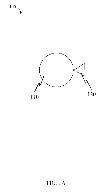
As illustrated in FIG. 1A, in some embodiments, device 100 comprises a
plurality
of nanodiamond particles 110 associated with a species 120 (e.g., a species
which may
bind to an analyte, if present). In some embodiments, the nanodiamond
particles are
associated with (e.g., bound to) the species via functionalization of the
nanodiamond
particle. For example, in some embodiments, a nanodiamond particle is
associated with
a species via formation of a bond, such as an ionic bond, a covalent bond, a
hydrogen
bond, Van der Waals interactions, and the like. The covalent bond may be, for
example,
a carbon-carbon, carbon-oxygen, oxygen-silicon, sulfur-sulfur, phosphorus-
nitrogen,
carbon-nitrogen, metal-oxygen, or other covalent bond. The hydrogen bond may
be, for
example, between hydroxyl, amine, carboxyl, thiol, and/or similar functional
groups.
For example, the species may include a functional group, such as a thiol,
aldehyde, ester,
carboxylic acid, hydroxyl, and the like, wherein the functional group forms a
bond with
the nanodiamond particle. In some cases, the species may be an electron-rich
or
electron-poor moiety wherein interaction between the nanodiamond particle and
the
species comprises an electrostatic interaction.
For example, the species may be associated with a functionalized nanodiamond
particle comprising a -COOH, -OH, -NH2, -SH, or -C=0 functional group by
reacting the
functionalized nanodiamond particle and the species in the presence of a cross-
linking
agent. Non-limiting examples of suitable cross-linking agents include
carbodiimides
such as 1-ethyl-343-dimethylaminopropyl]carbodiimide hydrochloride (EDC);
amine-
reactive compounds such as N-Hydroxysuccinimide ester, imidoester, and
hydromethylphosphine; sulfhydryl-reactive compounds such as maleimide, pyridyl
disulfides, and iodoacetyl; aldehyde-reactive compounds such as hydrazide and
alkoxyamine; and photoreactive cross-linking agents such as aryl azides and
diazirine.
Other cross-linking agents are also possible. Those of ordinary skill in the
art would be
CA 03036051 2019-03-06
WO 2018/048887 PCT/US2017/050257
¨ 19 ¨
capable of selecting suitable cross-linking agents based upon the type of
species selected
and the teachings of this specification.
In some embodiments, the species may bind with a target analyte. In some
cases,
the species may comprise a biological or a non-biological (chemical) group
capable of
binding another biological or chemical molecule in a sample (e.g., a
biological or
chemical molecule present on an analyte). For example, the species may include
a
functional group, such as a thiol, aldehyde, ester, carboxylic acid, hydroxyl,
amine,
polyethelene glycol and the like, wherein the functional group forms a bond
with the
analyte. In some cases, the species may be an electron-rich or electron-poor
moiety
wherein interaction between the analyte and the species comprises an
electrostatic
interaction.
In some embodiments, the species and analyte interact via a binding event
between pairs of biological molecules including proteins, nucleic acids,
glycoproteins,
carbohydrates, hormones, and the like. Specific examples include an
antibody/peptide
pair, an antibody/antigen pair, an antibody fragment/antigen pair, an
antibody/antigen
fragment pair, an antibody fragment/antigen fragment pair, an antibody/hapten
pair, an
enzyme/substrate pair, an enzyme/inhibitor pair, an enzyme/cofactor pair, a
protein/substrate pair, a nucleic acid/nucleic acid pair, a protein/nucleic
acid pair, a
peptide/peptide pair, a protein/protein pair, a protein/receptor pair, a small
molecule/protein pair, a glutathione/GST pair, an anti-GFP/GFP fusion protein
pair, a
Myc/Max pair, a maltose/maltose binding protein pair, a carbohydrate/protein
pair, a
carbohydrate derivative/protein pair, a metal binding tag/metal/chelate, a
peptide
tag/metal ion-metal chelate pair, a peptide/NTA pair, a lectin/carbohydrate
pair, a
receptor/hormone pair, a receptor/effector pair, a complementary nucleic
acid/nucleic
acid pair, a ligand/cell surface receptor pair, a virus/ligand pair, a Protein
A/antibody
pair, a Protein G/antibody pair, a Protein L/antibody pair, an Fc
receptor/antibody pair, a
biotin/avidin pair, a biotin/streptavidin pair, a drug/target pair, a zinc
finger/nucleic acid
pair, a small molecule/peptide pair, a small molecule/protein pair, a small
molecule/target pair, a carbohydrate/protein pair such as maltose/MBP (maltose
binding
protein), a small molecule/target pair, and a metal ion/chelating agent pair.
Specific non-
limiting examples of species include peptides, proteins, DNA, RNA, and PNA.
Other
species and binding pairs are also possible. In an exemplary embodiment, the
species is
an antibody (e.g., an antibody to a target analyte).
CA 03036051 2019-03-06
WO 2018/048887 PCT/US2017/050257
¨ 20 ¨
In an exemplary embodiment, the species and analyte interact via an
antibody/antigen pair binding event.
In another exemplary embodiments, the species and analyte interact via a
protein/receptor pair binding event. For example, the species may comprise a
protein
such as disintegrin (e.g., Bitistatin) and the analyte may comprise a receptor
molecule
such as a fibrinogen receptor.
In some embodiments, the species and the analyte interact via a binding event
between pairs of biological molecules including proteins, nucleic acids,
glycoproteins,
carbohydrates, hormones, or the like.
In some embodiments, the species is selected from the group consisting of
(poly)peptides, (poly)nucleotides, and ligands. In an exemplary embodiment,
the species
is a polypeptide, such as a protein, an antibody, and/or an antigen. For
example, in some
embodiments, the antibody is an immunoglobulin such as IgA, IgG, IgM, IgE, or
the
like, In another exemplary embodiment, the species is a (poly)nucleotide
(e.g., an
oligonucleotide), such as DNA or RNA. In yet another exemplary embodiment, the
species is a disintegrin such as albolabrin, applagin, barbourin,
batroxostatin, bitistatin,
obtustatin, schistatin, echistatin, elegantin, eristicophin, flavoridin,
halysin, kistrin,
mojastin, rubistatin, tergeminin, salmosin or triflavin. In some embodiments,
the species
may comprise a protein such as serum albumin (e.g., bovine serum albumin).
Examples of suitable nanodiamond particles functionalized with a species(e.g.,
Bitistatin) are discussed in more detail in U.S. Provisional Patent
Application No.
62/383,657, filed September 6, 2016, entitled "Engineering and Utility of
Fluorescent
Nanodiamond Particles (NDP-F) for Diagnostics and Treatment of Blood Clots in
Human and Veterinary Medicine," which is incorporated herein by reference in
its
entirety. Other functionalization methods are also possible.
Non-limiting examples of analytes that may be detected and/or quantified
include
a biological compound, a drug, a macromolecule, a salt, an electrolyte, an
enzyme, a
nucleic acid (e.g., a (poly)nucleotide), a carbohydrate, a (poly)peptide, a
protein, a lipid,
a phosphate, a sulfonate, a virus, a pathogen, a bacterium, a fungus, an
oxidant, a
reductant, a toxin, a surfactant, and combinations thereof.
In an exemplary set of embodiments, the analyte is a virus (e.g., ebola,
Marburg,
Bundibugyo, sudan, junin, lassa, MERS, small pox, Zika, pertussis, rubella,
rubeola). In
some embodiments, the analyte is a bacterial toxin (e.g., anthrax). In some
embodiments
CA 03036051 2019-03-06
WO 2018/048887
PCT/US2017/050257
¨ 21 ¨
the analyte is a biological entity associated with a particular parasite
and/or fungus.
Advantageously, the systems and methods described herein may be useful for the
detection and/or quantification of viruses such as ebola.
In yet another exemplary set of embodiments, the analyte is a receptor
molecule
(e.g., a fibrinogen receptor). Advantageously, the systems and methods
described herein
may be useful for the detection of blood clots.
In some cases, the analyte may comprise a marker/antigen for a particular
disease
or condition. For example, in some cases, the analyte may be a marker/antigen
associated with blood clots, traumatic brain injury, bone diseases (e.g.,
osteroporosis,
osteoarthrosis), inflammation and/or (auto)immune diseases (e.g., Crohn,
psoriasis)
ulcers, cardiac ischemia and stroke, atherosclerosis, muscle diseases,
Alzheimer's/Parkinson's, tumors and tumor metastasis, or others. For example,
the
systems and methods described herein may be useful for the detection and/or
diagnosis
in a subject of blood clots, traumatic brain injury, inflammation and/or
(auto)immune
diseases (e.g., Crohn's, psoriasis), ulcers, cardiac ischemia and stroke,
atherosclerosis,
muscle diseases, Alzheimer's/Parkinson's, tumors and tumor metastasis, or
others.
In some embodiments, the plurality of nanodiamond particles and the species
associated with the nanodiamond particles are present (e.g., in a reservoir)
and/or
introduced into a fluidic device. For example, as illustrated in FIG. 1B,
fluidic device
102 comprises a sample inlet 130 and a reservoir 140 in fluidic communication
with
sample inlet 130. In certain embodiments, reservoir 140 comprises a plurality
of
(fluorescent) nanodiamond particles 110 (and species 120 associated with
nanodiamond
particle 110).
In certain embodiments, a sample may be introduced to the sample inlet such
that
the sample flows into the reservoir and the sample interacts with the
plurality of
nanodiamond particles and species. In some embodiments, an analyte, if present
in the
sample, binds to the species.
In some cases, detection region 150 may be positioned downstream of, and/or in
fluidic communication with, reservoir 140.
In certain embodiments, a detector 160 may be positioned proximate detection
region 150. The detector, in some cases, may be configured to quantify an
emission
(e.g., an intensity of the emission, a wavelength of the emission) at the
detection region.
In some cases, the emission may be fluorescent and/or near-infrared. For
example, the
CA 03036051 2019-03-06
WO 2018/048887 PCT/US2017/050257
¨ 22 ¨
analyte, if present, may bind to the species. In some such cases, the analyte
may bind to
a second species (e.g., an antibody) associated with (e.g., bound to) the
detection region.
In some embodiments, the analyte bound to the second species and the first
species
(associated with the nanodiamond particles) may be detected and/or quantified
by
measuring (e.g., via the detector) the emission of the nanodiamond particles.
In some embodiments, a detector may be positioned proximate a region of a
subject suspected of containing an analyte and/or a clot. For example, the
plurality of
(fluorescent) nanodiamond particles functionalized with a species may be
administered
to a subject, and the detector may be positioned proximate the subject such
that any
nanodiamond particles bound to the analyte and/or clot may be detected (e.g.,
via an
emission of the nanodiamond particles).
Any suitable detector may be used with the devices and methods described
herein. For example, in some embodiments, the detector may be an optical
detector
(e.g., fluorescence detectors, visible light and/or UV detectors, near
infrared detectors,
microscopes).
In some embodiments, the emission is a fluorescent emission. In certain
embodiments, the wavelength of the emission is greater than or equal to 250
nm, greater
than or equal to 300 nm, greater than or equal to 350 nm, greater than or
equal to 400
nm, greater than or equal to 450 nm, greater than or equal to 500 nm, greater
than or
equal to 550 nm, greater than or equal to 600 nm, or greater than or equal to
650 nm. In
certain embodiments, the wavelength of the emission is less than or equal to
700 nm, less
than or equal to 650 nm, less than or equal to 600 nm, less than or equal to
550 nm, less
than or equal to 500 nm, less than or equal to 450 nm, less than or equal to
400 nm, less
than or equal to 350 nm, or less than or equal to 300 nm. Combinations of the
above-
referenced ranges are also possible (e.g., greater than or equal to 250 nm and
less than or
equal to 700 nm). Other ranges are also possible.
In certain embodiments, the emission is a near infrared emission. In some
embodiments, the wavelength of the emission is greater than 700 nm, greater
than or
equal to 750 nm, greater than or equal to 800 nm, greater than or equal to 850
nm,
greater than or equal to 900 nm, or greater than or equal to 950 nm. In
certain
embodiments, the wavelength of the emission is less than or equal to 1000 nm,
less than
or equal to 950 nm, less than or equal to 900 nm, less than or equal to 850
nm, less than
or equal to 800 nm, or less than or equal to 750 nm. Combinations of the above-
CA 03036051 2019-03-06
WO 2018/048887 PCT/US2017/050257
¨ 23 ¨
referenced ranges are also possible (e.g., greater than 700 nm and less than
or equal to
1000 nm). Other ranges are also possible.
In some embodiments, the nanodiamond particle may emit a fluorescent and/or
near infrared emission upon excitation by electromagnetic radiation having a
particular
wavelength. For example, in some embodiments, the nanodiamond particle may be
exposed to electromagnetic radiation having a wavelength of greater than or
equal to 250
nm, greater than or equal to 300 nm, greater than or equal to 350 nm, greater
than or
equal to 400 nm, greater than or equal to 450 nm, greater than or equal to 500
nm,
greater than or equal to 550 nm, greater than or equal to 600 nm, greater than
or equal to
650 nm, greater than or equal to 700 nm, greater than or equal to 750 nm,
greater than or
equal to 800 nm, greater than or equal to 850 nm, greater than or equal to 900
nm, or
greater than or equal to 950 nm (e.g., such that the nanodiamond particle
emits a
fluorescent emission and/or near infrared emission in one of the above-
referenced
ranges). In certain embodiments, the nanodiamond particle may be exposed to
.. electromagnetic radiation having a wavelength of less than or equal to 1000
nm, less than
or equal to 950 nm, less than or equal to 900 nm, less than or equal to 850
nm, less than
or equal to 800 nm, or less than or equal to 750 nm, less than or equal to 700
nm, less
than or equal to 650 nm, less than or equal to 600 nm, less than or equal to
550 nm, less
than or equal to 500 nm, less than or equal to 450 nm, less than or equal to
400 nm, less
than or equal to 350 nm, or less than or equal to 300 nm. Combinations of the
above-
referenced ranges are also possible (e.g., greater than or equal to 250 nm and
less than or
equal to 1000 nm, greater than or equal to 550 nm and less than or equal to
650 nm).
Other ranges are also possible.
While much of the description herein is in the context of (fluorescent)
.. nanodiamond particles, those of ordinary skill in the art would understand,
based upon
the teachings of this specification, that other particles are also possible.
For example, in
some embodiments, the device may comprise a particle such as a nanoparticle
(e.g., a
silica nanoparticle, a sapphire nanoparticle, a garnet nanoparticle, a ruby
nanoparticle)
having an emission in one of the above referenced ranges associated with a
species (e.g.,
.. a species capable of binding to one or more target analytes). In some
cases, the particle
may be autofluorescent. In other cases, the particle may be functionalized
with (e.g.,
associated with) a fluorescent molecule.
CA 03036051 2019-03-06
WO 2018/048887 PCT/US2017/050257
¨ 24 ¨
In some embodiments, the fluidic device (e.g., comprising a sample inlet and a
reservoir comprising a plurality of nanodiamond particles and a species
associated with
the nanodiamond particles) comprises a lateral flow assay configuration (e.g.,
in a lateral
flow device). Those of ordinary skill in the art would understand, based upon
the
teachings of this specification, how to incorporate a plurality of nanodiamond
particles
and a species associated (e.g., bound) to the nanodiamond particles into a
lateral flow
assay device. For example, as illustrated schematically in FIG. 2A, in an
exemplary
embodiment, system 200 comprises a lateral flow assay format. In certain
embodiments,
system 200 comprises reservoir 210 comprising a plurality of fluorescent
nanodiamond
particles 220 bound to a first species. Downstream of reservoir 210, in
certain
embodiments, is a second reservoir 230 comprising a second species (e.g., a
first
antibody to a target analyte) and a third reservoir 240 comprising a third
species (e.g., a
second antibody capable of binding to the antibody to the target analyte). In
certain
embodiments, the first species and the second species are the same.
Now referring to FIG. 2B, a sample suspected of containing an analyte 250 may
be introduced into system 200 (e.g., such that the sample flows and interacts
with
reservoir 210). In some embodiments, at least a portion of analyte 250 binds
to the
species on plurality of fluorescent nanodiamond particles 220. As illustrated
in FIG. 2C,
as the sample suspected of containing analyte 250 flows along the system, at
least a
portion of the analytes 250 (e.g., now bound to the species and/or plurality
of fluorescent
nanodiamond particles 220) bind to the second species in reservoir 230. In
some cases,
at least a portion of plurality of fluorescent nanodiamond particles 220 not
bound to the
analyte may be captured in reservoir 240 (e.g., where the third species is
capable of
binding to the first species).
In some cases, the devices and systems herein may be multiplexed. That is to
say, in some embodiments, the devices may comprise two or more, three or more,
four or
more, or five or more fluidic components and/or reservoirs comprising a
plurality of
nanodiamond particles. In some such embodiments, more than one analyte may be
detected, if present in the sample, in a single device. In certain
embodiments, one or
more analytes may be detected in a device comprising a plurality of fluidic
components
and/or reservoirs comprising a plurality of nanodiamond particles.
In an exemplary embodiment, a sample suspected of containing an analyte may
be introduced into a fluidic channel of a fluidic device, exposing the sample
to a species
CA 03036051 2019-03-06
WO 2018/048887 PCT/US2017/050257
¨ 25 ¨
bound to a plurality of fluorescent nanodiamond particles such that the
analyte, if
present, binds to at least a portion of the species bound to the plurality of
fluorescent
nanodiamond particles. In some embodiments, any fluorescent nanodiamond
particles
and species not bound to the analyte may be removed and a fluorescence
emission of the
plurality of fluorescent nanodiamond particles bound to the analyte may be
quantified.
As described herein, in some cases, the amount of analyte present in the
sample may be
correlated with the intensity of the fluorescence emission.
In another exemplary embodiment, a system comprises a sample inlet, a
reservoir
in fluidic communication with the sample inlet, the reservoir comprising a
plurality of
fluorescent nanodiamond particles, a plurality of a first species bound to the
plurality of
fluorescent nanodiamond particles, and a detection region in fluidic
communication with
the reservoir, the detection region comprising a plurality of a second species
bound to the
detection region. In some cases, the detector may be configured to quantify a
fluorescent
emission at the detection region and/or configured to quantify an infrared
signal at the
__ detection region.
In yet another exemplary embodiment, a fluidic device comprises a sample
inlet,
a reservoir in fluidic communication with the sample inlet, the reservoir
comprising a
plurality of fluorescent nanodiamond particles, a plurality of a first species
bound to the
plurality of fluorescent nanodiamond particles, and a detection region in
fluidic
communication with the reservoir, the detection region comprising a plurality
of a
second species bound to the detection region. In some embodiments, the fluidic
device
may further comprise a control region in fluidic communication with the
detection
region, the control region comprising a plurality of a third species bound to
the control
region. In some such embodiments, the third species may be the same or
different as the
first species and/or the second species. For example, the first species bound
to the
plurality of fluorescent nanodiamond particles may be a first antibody (e.g.,
capable of
binding selectively to the target analyte), the second species may be the
first antibody or
may be a second antibody different than the first antibody (e.g., capable of
binding
selectively to the target analyte), and the third species may be an anti-
antibody to the first
antibody. The control region may provide an indication, if an emission is
present, that
the system is working properly (e.g., the plurality of nanodiamond particles
were
properly featured and introduced into the sample).
CA 03036051 2019-03-06
WO 2018/048887 PCT/US2017/050257
¨ 26 ¨
In some embodiments, the fluidic device comprises an absorbent material. In
some embodiments, an absorbent region comprising the absorbent material is
positioned
downstream of, and in fluidic communication with, the detection region and/or
the
control region. In some cases, the absorbent material is associated with one
or more
components (e.g., the reservoir, the detection region) of the fluidic device.
In some
cases, the absorbent material may at least partially drive the flow of the
sample in the
fluidic device (e.g., wicking). In other embodiments, capillary action may at
least
partially drive the flow of the sample in the fluidic device.
Non-limiting examples of suitable absorbent materials include solid materials,
porous materials, particles, powders, and gels. In some embodiments, the
absorbent
material may comprise fabric, cellulose, cotton, and/or a polymer. Those of
ordinary
skill in the art would be capable of selecting suitable absorbent materials
based upon the
teachings of this specification.
In another exemplary embodiment, the methods described herein comprise
administering, to a subject suspected of having a particular analyte (e.g.,
present in the
bloodstream), a plurality of fluorescent nanodiamond particles bound to a
species such
that the species may bind to the analyte, if present, and detecting a
fluorescent and/or
near infrared emission of the plurality of fluorescent nanodiamond particles
comprising
the species bound to, if present, the analyte. In some cases, detecting a
fluorescent
emission indicates the presence of a blood clot in the subject. In certain
embodiments,
detecting a fluorescent emission indicates the presence of a virus (e.g.,
ebola, Marburg,
Bundibugyo, sudan, junin, lassa, MERS, etc.) in the subject.
As should be evident, in some embodiments, the present invention also provides
a diagnostic agent for detection or imaging of thrombotic events in a human or
non-
human animal, where the agent comprises a fluorescent nanodiamond particle
chemically
bonded to disintegrin Bitistatin (Bt). In certain embodiments, the fluorescent
nanodiamond particle and the Bt are covalently bonded. The diagnostic agent
may be
fluorescent as a result of an intrinsic property of the nanodiamond particle.
In some
embodiments, the diagnostic agent emits a detectable electromagnetic signal
when
excited by an electromagnetic source.
Additional exemplary embodiments relate to a method for diagnosis or prognosis
of a thrombo-embolic event. In these embodiments, the method comprises:
administering to a subject suspected of having suffered from or suspected of
being at risk
CA 03036051 2019-03-06
WO 2018/048887 PCT/US2017/050257
¨ 27 ¨
of, a thrombo-embolic event, a diagnostically effective amount of the
diagnostic agent of
the invention; allowing sufficient time for the diagnostic agent to localize
to the site(s) of
thrombus; and detecting the diagnostic agent by detecting fluorescence
emission of the
diagnostic agent. The method can be practiced as a method of detection of
activated
platelets and/or a method of detecting clots or clot formation in subjects.
Another exemplary embodiment of the invention relates to a kit. The kit may
include the diagnostic agent of the invention in packaged form suitable for
distribution,
delivery, and/or storage for use in a diagnostic method (e.g., a diagnostic
method for
detection of a thrombus). The packaged form may include a suitable material
for
distribution, delivery, and/or storage of the diagnostic agent. In certain
embodiments,
the kit further comprises, in packaged combination, one or more reagents or
devices for
administration of the diagnostic agent of the invention to a subject. The kit
can also
include a device that emits excitation energy for the diagnostic agent, and
preferably
further includes a detector for detection of emission response from the
diagnostic agent.
In a particular exemplary embodiment, the device (e.g., a device that emits
excitation
energy, a detector for detection of emission response) is a hand-held device.
It will be apparent to those skilled in the art that various modifications and
variations can be made in the practice of the present invention without
departing from
the scope or spirit of the invention. Other embodiments of the invention will
be apparent
to those skilled in the art from consideration of the specification and
practice of the
invention. It is intended that the specification and examples be considered as
exemplary
only, with a true scope and spirit of the invention being indicated by the
following
claims.
A "subject" or a "patient" refers to any mammal (e.g., a human), for example,
a
mammal that may be susceptible to a disease or bodily condition. Examples of
subjects
or patients include a human, a non-human primate, a cow, a horse, a pig, a
sheep, a goat,
a dog, a cat or a rodent such as a mouse, a rat, a hamster, or a guinea pig.
Generally, the
invention is directed toward use with humans. A patient may be a subject
diagnosed
with a certain disease or bodily condition or otherwise known to have a
disease or bodily
condition. In some embodiments, a patient may be diagnosed as, or known to be,
at risk
of developing a disease or bodily condition. In other embodiments, a patient
may be
suspected of having or developing a disease or bodily condition, e.g., based
on various
clinical factors and/or other data. In some cases, the patient may be
diagnosed with
CA 03036051 2019-03-06
WO 2018/048887 PCT/US2017/050257
¨ 28 ¨
having or developing a particular disease or bodily condition after the
detection and/or
quantification of an analyte (e.g., a virus, an antigen, an antibody) in a
sample obtained
from the patient. In some embodiments, a subject may demonstrate health
benefits, e.g.,
upon administration of the fluorescent nanodiamond particles.
As used herein, a "fluid" is given its ordinary meaning, i.e., a liquid or a
gas. A
fluid cannot maintain a defined shape and will flow during an observable time
frame to
fill the container in which it is put. Thus, the fluid may have any suitable
viscosity that
permits flow. If two or more fluids are present, each fluid may be
independently selected
among essentially any fluids (liquids, gases, and the like) by those of
ordinary skill in the
art.
The following examples are intended to illustrate certain embodiments of the
present invention, but do not exemplify the full scope of the invention.
EXAMPLES
Prophetic Example 1
The following example demonstrates an exemplary functionalized fluorescent
nanodiamond particle with a species (e.g., for binding to anti-human IgG-AP).
FIG. 3 illustrates the conjugation of a fluorescent nanodiamond particle (F-
NDP),
functionalized with a carboxylic acid group, and bound to a species (e.g.,
Bitistatin).
Such functionalized F-NDPs can be used, for example, to image and detect the
presence
of blood clots using external scanners (e.g., detectors for UV and/or NIR
emissions).
Example 2
The following example demonstrates the incorporation of fluorescent
nanodiamond particles into an exemplary system such as a lateral flow assay
format.
FIG. 4 shows the loading of three exemplary systems (system 400, system 410,
and system 420), each with a reservoir of fluorescent nanodiamond particles
(shown as
the dot in each system) placed on a nitrocellulose substrate. Upon
introduction of water
to system 400, water with 1% Tween 20 to system 410, and 10 mM sodium
phosphate
with 1% Tween 20 to system 420, flow of the fluorescent nanodiamond particles
along
the substrate of each device was observed (system 402, system 412, and system
422
respectively).
CA 03036051 2019-03-06
WO 2018/048887
PCT/US2017/050257
¨ 29 ¨
Example 3
The following example demonstrates the use of fluorescent nanodiamond
particles (F-NDP) conjugated with a species (e.g., human IgG) for the
detection of an
analyte (e.g., anti-human IgG).
FIG. 5 shows a plot of semi-ELISA for detection of human IgG coupled to the F-
NDP NV (700 nm). F-NDP were coupled to human IgG, or BSA (control), which were
used in concentrations as indicated, per 1 mg of F-NDP. 0.2 mg of each F-NDP-
IgG or
F-NDP-BSA sample were used for semi-ELISA, which was performed on a U-shape
bottom 96-well plate. The plate was blocked overnight with 3% BSA in PBST. F-
NDP-
IgG were blocked with 3% BSA in PBST by incubation for 1 hour at 37 C.
Blocking
agent was removed by centrifugation (1,000 g) at room temperature and washed
2x with
PBST. Goat anti-human IgG AP-conjugated (Sigma Inc.), diluted 1:5000, was
added
and incubated for 1 hr. as above. Final washing was performed as above, and p-
nitrophenyl phosphate substrate (pNPP) to alkaline phosphatase (AP) was added.
Color
was developed for 30 min, and NDP samples on plate were centrifuged.
Supernatant
was transferred to a flat bottom 96-well plate and read using an ELISA plate
reader
under 405 nm wave length. Error bars represent four repeats in semi-ELISA from
the
same samples.
Example 4
The following example demonstrates the use of F-NDPs functionalized with a
species (e.g., Bitistatin (Bt)) for the detection of blood clots. FIG. 6 shows
the
accumulation number of F-NDP-Bt in carotid arteries in rats with generated
clots.
Carotid arteries with generated clots or not (control) were dissected from
rats at the end
of the experiments. Vessels were solubilized with 12 N HC1 by overnight
incubation at
60 C in a ratio of 100 mg per ml. The resulting solution was diluted 10x with
water and
centrifuged using 14,000 rpm at room temperature and re-suspended in the same
volume
of water. F-NDP-Bt were counted using a hemocytometer with fluorescence
microscope
(40x objective, TRITC wavelengths). Presented numbers show total accumulated
particles. Error bars represent standard deviation from three rats (clot value
= 2.8 0.38
x 107; control value = 6.6 4.7 x 10⁵)
CA 03036051 2019-03-06
WO 2018/048887
PCT/US2017/050257
¨ 30 ¨
FIG. 7A is a scanning confocal microscope (SCM) image of carotid arteries from
rats with a generated clot. Generation of a clot was performed using ferric
chloride. F-
NDP-Bt suspension was injected locally, close to the clot. Rats were
euthanized, and
carotid arteries with clots were dissected. Imaging was performed using SCM.
Wavelengths used for measurement: excitation Cy5.5 BkG (580-610 nm), emission
Cy5.5 (695-770 nm). For background subtraction: excitation GFP (445-490 nm),
emission Cy5.5 (695-770 nm). NIR detection of F-NDP-Bit is indicated by the
white
arrows.
FIG. 7B shows a scanning confocal microscope image of carotid arteries from
rats with generated clot. Generation of a clot was performed using ferric
chloride. F-
NDP-Bt suspension was systemically injected to the animal tail. The rat was
euthanized,
and the carotid artery with clots was dissected. Imaging was performed using
SCM.
Wavelengths used for measurement: excitation Cy5.5 BkG (580-610 nm), emission
Cy5.5 (695-770 nm). For background subtraction: excitation GFP (445-490 nm),
emission Cy5.5 (695-770 nm). NIR detection is indicated by white arrows.
FIG. 8 shows images of tissue suspensions of clots treated or not with F-NDP-
Bt,
injected systematically to the femoral vein close (locally) to the clot
generated in carotid
artery. Tissues of the clot were manually homogenized in the presence of RIPA
buffer
(Triton X-100 based) in ratio one vein for 100 ml. Suspension of the lysate
was applied
on glass slide and immediately analyzed under the microscope. Magnification
100x.
Example 5
The following example demonstrates the functionalization of F-NDP (i.e., NDP-
F) with a polypeptide (e.g., Bitistatin (Bt)) and the detection of one or more
analytes
and/or blood clots.
The methodology of coupling proteins/peptides to carboxyl-functionalized NDP-
F is described herein. Preservation of the active domains responsible for the
biological
action of the coupled proteins/peptides remains challenging, and is generally
considered
to be a trial and error endeavor. A major innovation of the present invention
is the
demonstration of a concentration-dependent association of the engineered NDP-F-
Bt
agent to purified PFR. The selection of Bt was based on the high
selectivity/specificity
of Bt to the PFR, concomitant with negligible interactions with other RGD-
dependent
integrins, such as receptors for vitronectin (avb3) and fibronectin (a5b1).
The present
CA 03036051 2019-03-06
WO 2018/048887
PCT/US2017/050257
¨31 ¨
strategy to utilize Bt for clot imaging differs significantly from previous
studies aimed at
demonstrating the utility of Tc99-Bitistatin to map blood clots in vivo. The
present
imaging strategy is based on the innate near infrared (NIR) fluorescence
emitted upon
excitation of the NDP-F, thereby eliminating high radioactivity exposure
required by
other imaging techniques. It is also envisioned that coupling Bt (or other
polypeptides)
to a nanoparticle will be beneficial for the extension of the lifetime of the
Bt at the site of
its biological target.
Fluorescent nano-diamond particles (NDP-F or FNDP) functionalized by
carboxyl groups (-COOH) were purchased from Adamas Nanotechnologies, Inc.
(Raleigh, NC). Size distribution analysis revealed the peak of the diameter of
the NDP
was 734.5 ( 223.6, SD) nm. Optimization of fluorescence for the NDPs was
performed
by 2D screening of excitation vs. emission wavelengths (FIG. 9). Two areas of
optimal
fluorescence were established for NDPs, which should be useful for application
in
medical imaging (circled on FIG. 9). Detailed screening revealed two optimal
correlations of excitation vs. emission wavelengths: 480 nm vs. 520 nm, and
565 nm vs.
700 nm. The first correlation represents the typical green fluorescence,
whereas the
second is characterized by the long Stokes shift of fluorescence with near IR
emission.
The near IR emission is very useful for detection in vivo because it is in the
optical
therapeutic window of autofluorescence of factors present in human and non-
human
animal non-invasive imaging environments (e.g., water, hemoglobin,
oxyhemoglobin,
melanin).
The NDP-Fs were found to be resistant to photobleaching. Exposure of the
slides
containing NDPs to intense fluorescence light resulted in no changes in the
intensity of
their fluorescence in the time points up to 5 hours (data not shown).
Bitistatin is generally derived from snake venom and belongs to the
disintegrin
family of proteins. It has previously been investigated as a potential reagent
for
detection of deep venous thrombosis (DVT) using radioactive tags. This RGD-
disintegrin showed desirable parameters for detection of DVT when compared
with other
snake venom disintegrins, such as kistrin and barbourin. Therefore, this
fibrinogen
receptor-binding ligand was selected for coupling to NDP-Fs for the purpose of
detecting
activated platelets (or their aggregates) in clots present in the venous
circulation (vein
thrombus).
CA 03036051 2019-03-06
WO 2018/048887
PCT/US2017/050257
¨ 32 ¨
Bitistatin is generally an 83-amino acid polypeptide, which originally was
isolated from venom of Bitis arietants. This naturally occurring polypeptide
was
purified from the same snake venom using methodology developed in the
laboratory for
purification of other snake venom disintegrins (Marcinkiewicz, Cezary, et al.
"EC3, a
novel heterodimeric disintegrin from Echis carinatus venom, inhibits a4 and a5
integrins
in an RGD-independent manner." Journal of Biological Chemistry 274.18 (1999):
12468-12473.; incorporated herein by reference). Briefly, this method includes
two
steps of reverse phase HPLC with application of C18 column and a linear
gradient of
acetonitrile as a protein elution agent. Purity of the obtained Bt was tested
by SDS-
PAGE and quantified by digitization of bands on standard protein gels by
Coomassie
blue staining. The content of Bt was estimated at over 98% in the final
protein
preparation. This preparation was suspended in PBS for coupling to NDPs.
The active site of Bt was characterized and found to include an RGD motif,
which is important for ligand binding to certain integrins, such as 0E433,
avf33, and
a501. Bt is quite selective in that it has been shown to bind only the PFR,
0E433
integrin, which is exclusively expressed on circulating platelets. The
aspartic acid in the
RGD sequence was recognized by site-directed mutagenesis of recombinant
proteins and
short peptide synthesis as the most essential amino acid for disintegrin
binding to the
fibrinogen receptor. It was hypothesized by the inventors that coupling of Bt
to NDPs
that were functionalized by an attached amine group could affect the activity
of
disintegrin by engagement with carboxyl groups present in the side chain of
the aspartic
acid. Therefore, NDPs functionalized by a carboxyl group were selected to
attach the Bt
on NDPs surfaces.
Coupling of carboxyl (-COOH) groups present on NDPs to NH2 groups present
on Bt was performed using a standard protocol developed previously (Grabarek
and
Gergely, 1990). This method was based on the application of the cross-linker 1-
ethy1-3-
[3-dimethylaminopropyl]carbodiimide hydrochloride (EDC), which activates
carboxyl
groups to bind amine groups by forming an 0-acylisourea intermediate that
quickly
reacts with an amino group to form an amide bond. 0-acylisourea generally
undergoes
quick hydrolysis. Therefore, to increase the efficiency of coupling, sulfo-N-
hydroxysuccinimide (sulfo-NHS) was added to the reaction mixture to generate
an
amine-reactive sulfo-NHS ester.
BSA was coupled in parallel to the control NDPs.
CA 03036051 2019-03-06
WO 2018/048887 PCT/US2017/050257
¨33 ¨
A detailed description of an exemplary suitable coupling procedure follows:
Two samples of NDP, each containing 2 mg in 1.8 ml of 0.1M IVIES, 0.5M NaCl,
pH 6.0, were prepared. Sample A contained 1 mg Bt per 1 mg NDPs. Sample B
contained 1 mg BSA per 1 mg NDPs, and served as a control.
To each sample, 0.72 mg EDC was added and the mixture incubated at room
temperature (about 21 C to 25 C). with gentle rotating for 15 minutes. After
incubation,
2.5 1.1,1 of 2-mercaptoethanol (to give a final concentration of 20 mM) was
added to
quench the EDC and stop the carboxyl group activation.
The activated NDPs were transferred to 1.5 ml centrifuge tubes and centrifuged
at
10,000 x g for 5 minutes at room temperature. The NDP pellets were washed
twice with
1 ml PBS and suspended in coupling buffer (PBS). One milligram of either Bt or
BSA
in 1 ml of PBS was added to each sample tube. The tubes were incubated for two
hours
at room temperature with gentle rotating. Ten p,1 of 1M Hydroxylamine-HC1 (350
mg in
5 ml PBS) was added to each tube for quenching (final concentration of
Hydroxylamine-
HC1 should be about 10 mM).
The sample tubes were centrifuged at 10,000 x g for 5 minutes at room
temperature. The supernatants were saved for protein concentration
determination to
check coupling efficiency. The NDP-F-Bt particles were washed three times with
1 ml
PBS each time. The washed particles were suspended in a volume of PBS to
achieve a
.. concentration of 1 mg/ml.
Coupling efficiency was evaluated using immune detection of Bt on the surface
of NDPs (NDP-Bt). For ease of reference in this document, the detection method
is
called semi-ELISA. Broadly speaking, polyclonal antibody against Bt was
developed
commercially in rabbits (Chemicon/Millipore Inc.) using purified native
polypeptide as
the antigen. The saturated concentration of Bt was estimated at 1 mg per 1 mg
NDPs. A
representative plot from three independent experiments is presented in FIG.
10.
An exemplary suitable assay for determining coupling efficiency follows:
Using a 96-well plate having a U-shaped bottom and that has previously been
blocked by incubation overnight at 4 C with 5% BSA in PBS, 2001A1 of an NDP-F-
Bt or
NDP-F-BSA solution (1 mg/ml NDP in PBS) was each applied to a separate well.
The
plate was centrifuged for 10 minutes at 1,000 x g. The supernatant was removed
and the
plate gently shaken by vortexing to disperse and re-suspend the pellet of NDP-
F.
CA 03036051 2019-03-06
WO 2018/048887 PCT/US2017/050257
¨ 34 ¨
200 Ill of 10% goat serum in PBST (PBS + 0.05% Tween-20) was added and
mixed a few times by intensive aspiration and releasing from a pipet tip. The
mixture
was incubated for one hour at 37 C with slow agitation. The mixture was
centrifuged as
above and washed three times with 200 pi of PBST. After each centrifugation,
the pellet
was dispersed by vortexing. After the final wash, 100 pi of anti-bitistatin
(51.tg/m1 in
PBST) was added to the wells and incubated for one hour at room temperature.
The
plate was then washed three times with PBST as described above. To each
pellet, 100 pi
of a 1:2000 dilution of goat anti-rabbit IgG AP conjugate (from Sigma) in PBST
was
added to the wells and the mixture incubated for one hour at 37 C. The mixture
was
centrifuged at 1,000 x g for 10 minutes, and the pellet was washed three times
with
PBST as above.
150 pi of the AP substrate pNPP (from Sigma) was added and color was
developed for approximately 30 minutes (until the intensity of the yellow
color was
visible) at room temperature with gentle agitation. The reaction was blocked
by adding
100 pi of 3 M NaOH (this step is optional). The plate was then centrifuged and
100 pi of
the supernatant was transferred to a 96-well plate (flat bottom). The
absorbance of the
supernatant was measured at 405 nm using an ELISA plate reader.
The activity of NDP-Bt was tested using PFR purified from platelets, 0%03
integrin (Millipore Inc.). Two types of assays were applied to confirm the
presence of
disintegrin on NDP-Bt, including binding of soluble PFR to NDP-F-Bt and
adhesion of
NDP-F-Bt to fibrinogen receptor immobilized on the surface (plastic or glass).
Binding
of soluble PFR was determined on the basis of the functional "sandwich" ELISA,
which
is referred to herein as a functional semi-ELISA. In this case, immune-
detection of
0%03 integrin bound to the NDP-Bt was performed using a polyclonal antibody
against
the allf3 integrin subunit (purchased from Santa Cruz Inc.).
An exemplary suitable protocol for a functional semi-ELISA for detecting
binding of fibrinogen receptor to NDP-F-Bt follows:
Using a 96-well plate having a U-shaped bottom and that has previously been
blocked by incubation overnight at 4 C with 5% BSA in PBS, 200 Ill of an NDP-F-
Bt or
NDP-F-BSA suspension (1 mg/ml NDP in PBS) was each applied to a separate well.
The plate was centrifuged for 10 minutes at 1,000 x g. The supernatant was
removed
and the plate gently shaken by vortexing to disperse the pellet of NDP-F. 200
Ill of 3%
BSA PBST (PBS + 0.05% Tween-20) was added and the composition was mixed a few
CA 03036051 2019-03-06
WO 2018/048887 PCT/US2017/050257
¨35 ¨
times by intensive aspiration and releasing from a pipet tip. The mixture was
incubated
for one hour at 37 C with slow agitation. Alternatively, the blocking step may
be
performed in a tube before application on the 96-well plate. The mixture was
centrifuged as above and washed three times with 200 pi of PBST. After each
.. centrifugation, the pellet was dispersed by vortexing.
Fibrinogen receptor was then added at the desired concentrations (i.e., the
amounts required to generate a dose-response); see FIG. 11( in 200 Ill of HBSS
containing physiological concentrations of Ca2+ and Mg2+ and incubated for one
hour at
37 C. The plate was then washed three times with PBST as above. Next, 100 pi
of anti-
.. fibrinogen receptor (21.tg/m1 from Santa Cruz Inc.) polyclonal antibody in
PBST was
added to the wells and incubated for one hour at room temperature. The plate
was
washed three times with PBST as above. To the pellets, 100 Ill of a 1:3000
dilution of
goat anti-rabbit IgG AP conjugate (from Sigma) in PBST was added to the wells
and
incubated for one hour at 37 C. The mixture was centrifuged at 1,000 x g for
10
minutes, and the pellets washed three times with PBST as above.
150 pi of the AP substrate pNPP (from Sigma) was added and color was
developed for approximately 30 minutes at room temperature with gentle
agitation. The
reaction was blocked by adding 100 pi of 3 M NaOH (this step is optional). The
plate
was centrifuged and 100 pi of supernatant was transferred to the well of a 96-
well plate
(flat bottom). The absorbance of the supernatant was measured at 405 nm using
an
ELISA plate reader.
As can be seen from FIG. 11, the PFR bound to the NDP-Bt in a dose-dependent
manner, whereas the control NDP-BSA were not active in this assay.
Adhesion of NDP-Bt to immobilized fibrinogen receptor was performed in two
formats based on the estimation of the fluorescence of attached NDPs. First,
integrin
was immobilized on a 96-well plate and adhered NDPs were detected using a
fluorescence plate reader. The results obtained showed linear progression of
the
adhesion of NDP-Bt to increased concentration of immobilized purified receptor
(FIG.
12). Adhesion of NDP-Bt was also monitored under fluorescence microscopy. The
number of adhered NDPs was quantified using computer software (FIG. 13).
Representative images of adhered NDP-Bt are presented in FIG. 14.
To further characterize the effects of the FNDP, fluorescent images were taken
by
IVIS and confocal microscopy of carotid artery clots after treatment with FNDP
via
CA 03036051 2019-03-06
WO 2018/048887 PCT/US2017/050257
¨ 36 ¨
external carotid artery infusion. The results are shown in FIG. 15. In two
independent
experiments, infusion of FNDP via the external carotid artery commenced 3-5
minutes
after FeCl3 application and continued over 15 minutes (5 minutes beyond the
end of
FeCl3 infusion). The FNDP solution consisted of 1.5 ml of PBS where 5 mg/ml of
FNDP was suspended (after vortexing of the solution). This route of infusion
was
selected so as to avoid possible "first pass" elimination of the particles by
peripheral
organs. Following completion of FNDP infusion, the rat was euthanized and
subjected
to imaging by IVIS and/or fluorescent microscopy. FIGs. 15A-D show imaging of
fluorescence that was performed on an IVIS scanner designed for whole animal
imaging
using a 580-610 nm excitation and a 695-770 nm emission passband with a 2
second
exposure. Auto-fluorescence was subtracted based on excitation at 445-490 nm.
FIG.
15A shows an in situ carotid bifurcation region image, indicating fluorescence
of carotid
arterial clot after treatment visible via IVIS imaging after exposure of the
carotid
bifurcation zone. FIGs. 15B and 15C are high magnification images of
fluorescence
emanating from the carotid bifurcation in vivo, suggesting accumulation of
FNDP in the
clot. FIG. 15D is an ex vivo photograph of fluorescence of carotid artery
bifurcation
denoting one branch showing fluorescence corresponding to the clot location
within the
carotid bifurcation. FIGs. 15E and 15F are confocal images taken on an Olympus
IX83
of FNDP in which fluorescence is detected at an excitation of 543 nm and an
emission of
655-755 nm. Background fluorescence was collected from the same excitation,
with
emissions of 555-625 nm, and was subtracted from the foreground to reduce auto-
fluorescence. FIG. 15E shows ex vivo fluorescence of the carotid artery at 4x
magnification. FIG. 15F shows FNDP treated carotid arteries after they were
flushed
with RIPA lysis buffer and replicates were combined together to form a lysate.
Lysate
was then deposited onto a cover-glass and imaged at 20x magnification. Large
numbers
of FNDP at various aggregate sizes around platelets are visible.
FIG. 16 shows fluorescent images taken by IVIS and confocal microscopy of
carotid artery clots after intravenous treatment with FNDP. After clot
formation by
ferric chloride, treatment with saline control or FNDP by intravenous infusion
via tail
vein or femoral vein was performed in three rats. FNDP were infused (over 10
min) as a
suspension in PBS at 1 ml solution containing 1 mg/ml FNDP. Carotid arteries
were
removed from the animal for imaging and placed in 70% denatured ethanol for
preservation until imaging. FNDP were conspicuously identified at the site of
clot
CA 03036051 2019-03-06
WO 2018/048887 PCT/US2017/050257
¨ 37 ¨
formation. FNDP were identified in each of the three specimens obtained
following
intravenous infusion, yet the three specimens were treated together as one for
lysate
inspection. FIGs. 16A and 16B show imaging of fluorescence as performed on an
IVIS
scanner designed for whole animal imaging using a 580-610 nm excitation and a
695-
770 nm emission passband with a 2 second exposure. Autofluorescence was
subtracted
based on excitation at 445-490 nm. Figure 8A shows an ex vivo fluorescent
image of a
carotid artery from saline-treated control. Auto-fluorescence could not be
entirely
eliminated, but was evenly distributed across control specimen. FIG. 16B shows
an ex
vivo fluorescent image of a carotid artery from an IV FNDP-treated animal,
showing
fluorescence localized to the branch with a clot. FIGs. 16C-16F show confocal
images
taken on an Olympus IX83. The figures show that FNDP fluorescence was detected
at
an excitation of 543 nm and an emission of 655-755 nm. Background fluorescence
was
collected from the same excitation, with emissions of 555-625 nm and was
subtracted
from the foreground to reduce auto-fluorescence. FIGs. 16C and 16D show ex
vivo
fluorescence of carotid artery at 4x magnification of saline treated and FNDP
treated
animals, respectively. Auto-fluorescence could not be entirely eliminated, but
was
evenly distributed across control specimens, while fluorescence was localized
to the
branch with clot in the IV treated animal. (See panels 16E and 16F.) Treated
carotid
arteries were flushed with RIPA lysis buffer and replicates were combined
together to
form a lysate. Lysate was then deposited onto a cover-glass and imaged at 20x
magnification. In order to increase contrast for visual inspection, images
were processed
with an un-sharp mask in Imagek
FIG. 16E shows that the saline-treated control showed no detectable
fluorescence. FIG. 16F shows that FNDP appear as frequent fluorescent spots in
the
treated samples. FIG. 16G presents a graph showing the number of FNDPs present
in
carotid clot lysates from animals treated locally via the external carotid
artery or
intravenously as compared with saline treated controls. FNDPs were counted in
replicate
images after thresholding in Imagek
FIG. 17 depicts images of rat blood plasma clots following treatment with F-
NDP-Bt and F-NDP-BSA. Rat blood was collected by heart puncture and
centrifuged at
100 x g for 20 minutes at room temperature to obtain platelet rich plasma
(PRP). A
thrombus was generated by adding thrombin (1 U/ml) and incubating for 15
minutes at
37 C. The clot that was formed was washed 3x by decanting with HBSS containing
CA 03036051 2019-03-06
WO 2018/048887
PCT/US2017/050257
¨ 38 ¨
calcium and magnesium, and then sliced. Pieces of the clot were incubated with
a
suspension of F-NDP-Bt and F-NDP-BSA (50 pg/m1) in HBSS containing calcium and
magnesium for 60 minutes at 37 C, and washed 3x with the same buffer as above,
then
applied on the glass slide for imaging. Images of plasma clots obtained from
fluorescence microscope Olympus IX81analysis, under 100x magnification, are
shown in
FIG. 17A. Images of plasma clots obtained using an IVIS 50 imaging system are
shown
in FIG. 17B. Wavelengths used for measurement: excitation Cy5.5 BkG (580-610
nm),
emission Cy5.5 (695-770 nm). For background subtraction: excitation GFP (445-
490
nm), emission Cy5.5 (695-770 nm). Exposure time: 1 minute. Arrows in FIG. 17B
point the localization of the clot.
FIG. 17 shows the specificity of interaction of F-NDP-Bt with a clot generated
from rat blood plasma by thrombin (1 U/ml). Analysis of the clot under a
fluorescence
microscope (FIG. 17A) revealed that F-NDP-Bt accumulated on the surface of the
thrombus to a high extent, although this accumulation was not evenly
distributed.
Fluorescence microscopy imaging identified areas with high green fluorescence
intensity
(represented by bright spots in the black and white image), which may indicate
zones of
the condensation of activated platelets. Fluorescence live imaging system
(IVIS 50) also
exhibited binding of F-NDP-Bt to the plasma clot (FIG. 17B). However, in this
system
the near infrared (NIR) detection was set up based on the optimization
performed as
presented in FIG. 9. Control nanoparticles, containing coupled BSA to the
surface (F-
NDP-BSA), interacted with the clot to a negligible level in both imaging
assays.
Detection of F-NDP-Bt by NIR suggested a usefulness of functionally active F-
NDP-Bt
for imaging in living organisms because the emission wavelength was localized
within
an "optical therapeutic window" (600 - 1300 nm).
Therefore, detection of F-NDP-Bt was performed in a rat model for verification
of that hypothesis. The results are shown in FIG. 18. The skin areas of
observation
fields were prepared for implantation by hair shaving. The incision was made
by scalpel
and vessels were inserted under the skin of dead rats. The dead rats were
placed in IVIS
50 Imaging System, and measurement of fluorescence under NIR spectrum was
performed. Wavelengths used for measurement: excitation Cy5.5 BkG (580-610
nm),
emission Cy5.5 (695-770 nm). For background subtraction: excitation GFP (445-
490
nm), emission Cy5.5 (695-770 nm).
CA 03036051 2019-03-06
WO 2018/048887 PCT/US2017/050257
¨ 39 ¨
A representative image of the implanted glass capillaries filled with F-NDP-Bt
(4
mg/ml) or PBS (control) are shown in FIG. 18A. Exposure time was 5 seconds.
Figure
10B shows an image of a rat aorta filled with F-NDP-Bt. The rat aorta was
dissected
from a euthanized female rat, washed with PBS to remove residues of coagulated
blood,
and filled with 300 pi of F-NDP-Bt suspension (2 mg/ml) in PBS. The aorta was
secured
from both ends by knots of surgical sutures. Exposure time was 1 minute. FIG.
18A
demonstrates NIR imaging of F-NDP-Bt, experimentally implanted under rat skin.
Suspensions of F-NDP-Bt were infused into glass capillaries and into dissected
rat aorta
(FIG. 18B), before subcutaneous implantation. Clear images showed precise
localization
of both artificial (capillary) and natural (aorta) vessels in the rats.
Finally, the specificity of the interaction of F-NDP¨Bit with the fibrinogen
receptor present on activated platelets in a preformed PRP clot (FIGs. 18C-
18D) was
investigated. In two separate experiments, clots were incubated with F-NDP¨Bit
for 15
or 60 minutes, respectively. A clot was generated from rat PRP by thrombin (1
U/mL)
and incubated with F-NDP¨Bit (250 i.tg/mL) in the presence or absence of Lt
(4.67
i.tmol/mL). (FIG. 18C) IVIS imaging was performed using GFP filters
(excitation 445-
490 nm, emission 515-575 nm). The clot was incubated with F-NDP¨Bit for 15
minutes
(upper panel) or 60 minutes (lower panel). Intensity of fluorescence was
evaluated using
IVIS Living Image 4.3.1 software. Insets above the bars represent respective
images of
clots from IVIS. (FIG. 18D) Phase-contrast and fluorescence microscope images
of clots
(100x). Areas of accumulation of F-NDP¨Bit are framed in yellow. Irrespective
of the
duration of incubation, binding of F-NDP¨Bit was always 4-5 fold higher than
that of
the nonspecific control particles (F-NDP¨BSA). Furthermore, preincubation of
the clots
with lotrafiban (10 reduced F-NDP¨Bit binding to the level of the control F-
NDP¨BSA.
Bitistatin was purified from the venom of Bitis arientans (Latoxan
Serpentarium,
Valence, France) using two steps of reverse-phase HPLC. F-NDP, chemically
surface-
functionalized with carboxyl groups (-COOH), were purchased from Adamas
Nanotechnologies (Raleigh. NC, USA). Two strains of F-NDP were used: green
fluorescent F-NDP based on N-V-N color centers (F-NDP(NVN)) at 700nm (2x10⁸
particles/mg) and red fluorescent based on N-V (F-NDP(NV)) color centers at
100nm
(5x10¹¹ particles/mg), 700nm (2x10⁸ particles/mg), and 10,000nm (5x10⁵
particles/mg).
Isoflurane was purchased from Henry Schein (B34C16A Dublin, OH, USA). 70%
Denatured Ethyl Alcohol and PE-10 tubing were purchased from Fisher Scientific
CA 03036051 2019-03-06
WO 2018/048887
PCT/US2017/050257
¨ 40 ¨
(Pittsburgh, PA, USA). Silk Suture was purchased from Roboz SUT-15-1, Roboz
Surgical Instrument Co. (Gaithersburg, MD, USA). Parafilm and FeCl3 was
purchased
from Sigma-Aldrich, (St. Louis, MO, USA).
Bitistatin was coupled to the F-NDP of all types using EDC (1-ethyl-343
dimethylaminopropyl] carbodiimide hydrochloride) as a hetero-bifunctional
cross-linker.
Coupling efficiency and preservation of Bitistatin activity on the various
functionalized
nanodiamond particles (F-NDP-Bit) were verified using a semi-ELISA
methodology.
Example 6
The following example demonstrates the use of functionalized fluorescent
nanodiamond particles (e.g., (F-NDP-B0) to bind to platelets and
preferentially to
activated platelets from humans. The data also shows that the particles do not
substantially interfere with platelet aggregation.
For example, FIGs. 19A-19C show dose response curves for aggregation of
platelets with and without probe (i.e., F-NDP-Bt), determined as a function of
concentration of proteinase-activated receptor 4 (PAR4 AP), adenosine
diphosphate
(ADP), and arachidonic acid (AA).
FIGs. 20A-20C show the ability of a F-NDP-Bt probe having an average particle
diameter of 700 nm or 200 nm to bind to various platelet populations at
different
concentrations. FIGs. 21-222 shows the difference in binding of probes (700 nm
or 200
nm in diameter) in stimulated versus unstimulated platelet populations.
Flow cytometry data of stimulated and unstimulated platelets are shown in
FIGs.
23A-23D and FIGs. 24A-24D, using the F-NDP-Bt probes.
Example 7
The following example demonstrates an exemplary method for extracting,
isolating, and/or quantifying F-NDP-(NV) in blood and/or biological tissues.
1. F-NDP-BSA NV 700 nm were prepared using a coupling protocol as described
herein, in sterile conditions (see e.g., Example 8). Final concentrations of 5
mg/ml were
be prepared in PBS for animal administration.
2. 4 doses of 1 ml (5 mg/ml in PBS) of F-NDP-BSA were injected per one
subject (e.g., an animal). Blood (TBD) may be collected into 3.8% sodium
citrate (in
ratio 1:9) at different time intervals. Control group may be infused with
equal volume of
CA 03036051 2019-03-06
WO 2018/048887 PCT/US2017/050257
¨ 41 ¨
normal saline. Blood from PBS-injected animal may be collected in the same way
treated samples were processed. At the conclusion of the protocol
approximately 10 mL
of blood may be collected.
3. Blood containing F-NDP-BSA may be lyophilized using SpeedVac system
(SC110A Plus, Thermo Savant) with 4,680 xg. Control animal may be used for
preparation of standard curve. Standard curve may be prepared by mixing known
amount
of F-NDP-BSA with blood. 8 serial dilution will be prepared starting from the
highest, 2
mg/ml particles density.
4. Dry mass of particles containing blood collected in the course of the
experiment of may be dissolved in 12 N HC1 at same volume staring volume of
blood
(approx. 1 m1). Solubilization may be performed by overnight incubation at
60oC.
5. Supernatant may be removed by centrifugation (17,000 xg for 5 min at room
temperature) and pellet re-suspended in the same volume original volume (e.g.,
water 1
m1). Centrifugation will be repeated one time for washing. At this point,
particles may be
condensed using smaller amount of water, to improve sensitivity. For example,
final
volume of water may be twice lower than volume of blood used for
lyophilization, e.g.,
such that sensitivity for detection of F-NDP would be twice increased.
6. Suspension of F-NDP-BSA of investigated samples and standard curve may be
applied on 96-well plate (100 microliters per well) and plate may be read in
Tecan using
NIR wavelength (excitation 570 nm, emission 670 nm). In parallel, number of
particles
per ml may be established by disposable hemocytometer (Incyto Inc., Cheonan-
si,
Korea), counting under fluorescence microscope, e.g., using an Olympus IX81
with
TRICI wavelength and 400x magnification. Amount of F-NDP-BSA in investigated
samples may be deducted from the standard curve.
Example 8
The following example demonstrates an exemplary method for sterilization
and/or lyophilization of F-NDPs.
1. F-NDPs were suspended in 70% ethanol and incubate for 15 min at room
temperature (23+/-2 C), using gentle agitation on "Speci Mix Test Tube Rocker"
(ThermoScientific Inc., Waltham, MA USA). Density of F-NDPs was approximately
0.5
mg/ml.
CA 03036051 2019-03-06
WO 2018/048887
PCT/US2017/050257
¨ 42 ¨
2. The suspension of F-NDPs may be centrifuged (e.g., 17,000 xg for 5 min at
room temperature). Supernatant (ethanol) may be removed by vacuum aspiration
and
pellet suspended in water or buffer (e.g., PBS or MES) and centrifuged once
again under
the same speed conditions for washing purposes.
3. F-NDPs were suspended in water or working buffer (e.g., IVIES if coupling
to
protein will be performed) in desired density (e.g. 1 mg/ml).
4. Lyophilization of F-NDPs may be performed using a Speed Vac system
(SC110A Plus, Thermo Savant, and Holbrook, NY, USA). For example,
polypropylene
tubes 5 ml (Sarstedt Inc., Numbrecht, Germany) or 1.5 ml (Fisher Inc. Waltham,
MA
USA) were used for lyophilization. This system generally operates with
concentrator set
up for working in ambient temperature giving low drying rates. For example,
application
of high drying rates and work in a range of, for example, 43 C - 65 C
temperature may
be harmful for some polypeptides and/or polynucleotides attached to the F-NDP.
Concentrator may be used with 8,500 rpm maximal speed giving 4,680xg maximal
force.
Vacuum pump (model VLP120, Thermo Savant), may be set up for gas-ballast
control
"closed" position giving ultimate total pressure 1.5 x 10⁻³ Ton. Refrigerated
vapor trap
(model RVT400, Thermo Savant) may be used with 4 liter chamber capacity
giving,
approximate operating temperature -50 C.
Lyophilization may be performed from water or buffer suspension dependent on
required purposes (e.g. PBS may be used for in vivo application for diagnostic
approaches such as thromboembolic events in vasculature). Reconstitution of F-
NDP
may be performed using sterile deionized water, to the desired volume. All
procedures
may be performed in sterile conditions including all materials such as tubes
and tips.
4. Measuring the effect of ethanol sterilization and lyophilization on size
distribution may be performed using Zetasizer Nano ZS (Malvern Instruments
Ltd.,
Westborough, MA, USA). Working density of F-NDPs was estimated as 10 mg/ml.
FIGs. 25A-25B show a high overlap of same F-NDPs for Particles Size
Distribution
(Malvern, PSD) assayed before and after sterilization.
Example 9
The following example demonstrates the ability of fluorescent-NanoDiamond
particles (F-NDP) covalently conjugated with bitistatin (F-NDP-Bit) to detect
vascular
blood clots in vivo using extracorporeal near infrared (NIR) imaging.
Specifically, NIR
CA 03036051 2019-03-06
WO 2018/048887 PCT/US2017/050257
¨ 43 ¨
fluorescence properties of F-NDP were compared with color centers (NV vs. NVN)
and
sizes (100-10,000nm). Optimal NIR fluorescence and tissue penetration across
biological
tissues (rat skin, porcine axillary veins and skin) was obtained for F-NDP(NV)
with a
mean diameter of 700 nm. Interavital imaging ( IVIS ) in vitro revealed that F-
NDP(NV)
.. ¨ loaded glass capillaries could be detected across 6 mm of rat red-muscle
barrier and 12
mm porcine skin, which to average vertical distance of a human carotid artery
bifurcation
from the surface of the adjacent skin (14mm). In vivo, feasibility was
demonstrated a rat
model of FeCl3 -generated occlusive blood clots I carotid artery bifurcation.
Following
systemic infusions of F-NDP(NV)-Bit (3 or 15 mg/Kg) via the external carotid
artery
(ECA) or femoral vein (N=3), presence of the particles in the thrombi was
confirmed
both in situ via IVIS, and ex vivo, via confocal imaging. F-NDP(NV) presence
in the
vascular clots was further confirmed by direct counting of fluorescence
particles
extracted from clots following tissue solubilization. The data suggests that F-
NDP(NV)-
Bit associate with vascular blood clots, presumably by F-NDP(NV)-Bit binding
to
activated platelets within the blood clot. It is posited that F-NDP(NV)-Bit
could serve as
non-invasive platform technology for identification of vascular thrombi using
NIR
energy monitored by an extra-corporeal device.
Materials
Bitistatin was purified from the venom of Bitis arientans (Latoxan
Serpentarium,
Valence, France) using two steps of reverse-phase HPLC, as described above. F-
NDP,
chemically surface-functionalized with carboxyl groups (-COOH), were purchased
from
Adamas Nanotechnologies (Raleigh. NC, USA). Two strains of F-NDP were used:
green
fluorescent F-NDP based on N-V-N color centers (F-NDP(NVN)) at 700nm (2x10⁸
particles/mg) and red fluorescent based on N-V (F-NDP(NV)) color centers at
100nm
(5x10¹¹ particles/mg), 700nm (2x10⁸ particles/mg), and 10,000nm (5x10⁵
particles/mg). Isoflurane was purchased from Henry Schein (B34C16A Dublin, OH,
USA). 70% Denatured Ethyl Alcohol and PE-10 tubing were purchased from Fisher
Scientific (Pittsburgh, PA, USA). 5-0 Silk Suture was purchased from Roboz SUT-
15-1,
.. Roboz Surgical Instrument Co. (Gaithersburg, MD, USA). Parafilm and FeCl3
was
purchased from Sigma-Aldrich, (St. Louis, MO, USA).
Coupling of bitistatin to F-NDP
CA 03036051 2019-03-06
WO 2018/048887
PCT/US2017/050257
¨ 44 ¨
Bitistatin was coupled to the F-NDP of all types using EDC (1-ethyl-343
dimethylaminopropyl] carbodiimide hydrochloride) as a hetero-bifunctional
cross-linker.
Coupling efficiency and preservation of Bitistatin activity on the various
functionalized
nanodiamond particles (F-NDP-Bit) were verified using a semi-ELISA
methodology.
Characterization of NIR emission of F-NDP(NV) and F-NDP(NVN)
NIR fluorescence profiles of F-NDP were characterized using a Tecan Infinite
200 PRO (Tecan AG, Mannedorf, CH). 100111 of 3 mg/ml of 700nm F-NDP(NV) or F-
NDP(NVN) suspended in de-ionized (DI) water were loaded into 96-well
polystyrene.
Fluorescence was scanned for all wells with excitations from 230nm-850nm and
emissions from 290nm-850nm (Figure 1A) at 20 nm intervals. Data was processed
in
Matlab 2015b (Mathworks, Natick, MA, USA). Background fluorescence was
subtracted
from empty wells without F-NDP and the resulting net fluorescence value was
Log10
transformed for visualization.
Glass capillaries (40 mm length, 1 mm internal diameter, (Thermo Fisher
Scientific, Waltham, MA, USA) were filled with equal volumes (30 microliters)
of
suspensions of F-NDP at concentrations from 0.06 and up to 4 mg/ml (1.8-120m
total
particle mass) and sealed at each end by plasticine (Hasbro, Pawtucket, RI,
USA). The
NIR fluorescence intensity of the various suspensions in the capillaries were
analyzed
using an IVIS 50 Imaging System (PerkinElmer Inc., Akron OH) using an
excitation
filter set to `Cy5.5 BkG' (580-610 nm) and an emission filter set to `Cy5.5'
(695-770
nm) as these filters matched the desired NIR emission profile detected as
described
above. Imaging was completed with 'binning' set to 4, and a 10 cm field of
view, with
exposure times between 2 and 40 seconds. For imaging through biological
barriers (rat
and porcine skins and rat muscles) auto-fluorescence was imaged with the blue-
shifted
excitation `GFP' (445-490 nm) and the same emission filter `Cy5.5' (695-770
nm) with
similar imaging settings as above and subtracted from the foreground as
modified from
IVIS 50 protocol to compensate for the large stokes shift of the F-NDP(NV).12
This
correction was used for a simplified spectral un-mixing: A ratio of auto-
fluorescence
between the channel of interest and blue-shifted excitation channel is defined
in control
tissue. The same ratio was then used to subtract auto-fluorescence from the
channel of
interest based on the blue-shifted excitation channel in the test specimen.
This operates
under the assumption that the fluorophore being detected may have minimal
excitation at
CA 03036051 2019-03-06
WO 2018/048887 PCT/US2017/050257
¨ 45 ¨
the blue-shifted wavelength. To assess tissue penetration of NIR florescence
emission
from F-NDP, capillaries were placed under shaved abdominal rat skin (obtained
from
euthanized rats), covered with dissected rat quadriceps muscle (2 - 5.9 mm
thick), or
covered with porcine skin (isolated from shoulder of pig obtained from a local
butcher
shop)
Generation of carotid arterial blood clot and F-NDP infusion in rat
Technical procedures of the FeCl3-induced vascular thrombosis model are
generally known in the art. Specific modifications used in this particular
work are briefly
summarized below. All animal procedures were performed according to the
guidelines of
the US Animal Welfare Act and approved by the Institutional Animal Care & Use
Committee at SUNY Downstate Medical Center. In brief: adult male Sprague-
Dawley
rats (Charles River, 350 Gm +/-10% body weight), were anesthetized using 4%
isoflurane (IF, induction, in chamber) followed by 1-2% IF (maintenance)
adjusted
throughout the procedure. Rats were held in the supine position and subjected
to surgery
using clean instruments and aided by binoculars. The left carotid artery was
dissected
and exposed at the bifurcation region. A 5-0 surgical silk suture was wrapped
below the
common carotid (CCA), external carotid (ECA), and internal carotid (ICA)
arteries. A
PE-10 cannula was then inserted in the ECA for studies where F-NDP(NV)-Bit
were
injected locally. A PE-10 cannula was also inserted into the left femoral vein
for studies
where F-NDP(NV)-Bit was infused intravenously (IV). The ICA stem was wrapped
in
Parafilm soaked in 50% FeCl3 and kept in place for 10 minutes. Two to three
minutes
after placing the Parafilm onto the ICA, infusion of F-NDP(NV)-Bit suspension
in PBS
commenced either via the ECA (N=2, 15 mg/Kg in lmL), or via the femoral vein
(N=6)
at low dose (N=3, 3mg/Kg in 1 mL PBS), or at high dose (N=3, 45 mg/Kg in 3 mL
PBS). All infusions were completed over 10 minutes. Control rats were infused
with
vehicle at comparable volumes and duration.
Tissue fixation post F-NDP(NV)-Bit infusion
Following the completion of particles infusion, anesthesia was augmented to
produce deep hypnosis using 5% IF. The lower aorta was quickly isolated and
cut to
allow blood drainage. Tissue was fixed and residual blood removed by perfusion
with 10
mL of 70% denatured ethanol. Dissection of both bilateral carotid artery
bifurcation
CA 03036051 2019-03-06
WO 2018/048887
PCT/US2017/050257
¨ 46 ¨
regions was completed after whole body imaging (IVIS). Vessels were suspended
70%
denatured ethanol for further ex vivo NIR fluorescence evaluation.
In situ and ex vivo imaging of F-NDP(NV) fluorescence by IVIS
Briefly, NIR fluorescence was detected using a 580-610nm excitation and a 695-
770nm emission pass-band with 2 second exposure, 'binning' set to 4, and a 7
cm field
of view as described above. Auto-fluorescence was subtracted based on
excitation at
445-490nm under otherwise similar imaging conditions. Carotid arteries were
exposed
before imaging to enable clear visualization for in situ images. Following in
situ
imaging, carotid arterial bifurcations were removed from animals and placed on
a glass
plate for imaging ex vivo using identical imaging parameters to those used for
in situ
imaging. For each artery, the mean fluorescence intensity of the images
compensated for
auto-fluorescence was calculated using ImageJ (NIH, Bethesda, MD, USA).
Ex vivo imaging of F-NDP(NV) by fluorescence microscopy
Gross images of the entire carotid bifurcation region extracted from clot
bearing
or contralateral vessels were evaluated on a Fluoview FV1000 (Olympus, Tokyo,
Japan)
laser scanning confocal microscope (LSCM) using a 4x objective. NIR
fluorescence
emitted from F-NDP(NV) was detected at an excitation of 543nm and an emission
of
655-755nm. Confocal stacks were combined using a maximum intensity projection
so
that the entire vessel is brought into focus. The mean fluorescence intensity
of F-NDP in
each artery was calculated after subtraction of the local background using
Imagek
Isolation of F-NDP(NV) from vascular clot
F-NDP were isolated from extracted carotid arteries by homogenization in RIPA
lysis buffer (Teknova Inc. Hollister, CA, USA) at 100 mg/ml. Aliquots (10
microliters)
of the lysate suspension were applied on the microscope slides and analyzed
under
LSCM as above under 20x objective.
In animals treated with F-NDP(NV) via the femoral artery at high dose, F-
NDP(NV) were isolated from extracted carotid arteries by solubilizing the clot
bearing
vessel segment in 12 N hydrochloric acid (HC1) (Thermo Fisher Scientific,
Waltham,
MA, USA) overnight at 60 C at 100mg/ml. The solution was centrifuged (14,000xg
at
room temperature for 10 minutes) and the pellet was washed lx with distilled
water. The
CA 03036051 2019-03-06
WO 2018/048887 PCT/US2017/050257
¨ 47 ¨
pellet containing the insoluble F-NDP(NV)-Bit was re-suspended in DI-water
while
keeping the initial mass/volume ratio. An aliquot of the suspension was
applied to a
hemocytometer (Incyto Inc., Cheonan-si, Korea), which was standardized for
particles
counting in an inverted fluorescence microscope (Olympus IX81) with 40x
objective.
Images of F-NDP(NV)-Bit were taken from each observation field for counting
using
TRITC filter cube. Numbers of particles were calculated for the entire
solubilized tissue.
Statistical analysis
Unless stated otherwise, each experiment in this example was performed
independently three times in triplicate. No outlying data was excluded. Data
are
represented as mean SD. Statistical analyses were done by the Student's t
test using
(SigmaPlot 12 SPSS, Systat Software Inc., San Jose CA, USA). P <0.05 was
considered significant.
Results
Comparison of NIR fluorescence intensity of F-NDP(NVN) and F-NDP(NV)
fluorescence profile measurements revealed NIR fluorescence in both N-V and N-
V-N
particles. However, fluorescence in the NIR region was 20 times greater in the
N-V
particles (FIG. 26A). This experiment also revealed the peak excitation of F-
NDP(NV) at
.. 570nm and peak emission at 670nm. Using this peak excitation and emission
profile,
NIR emission of the F-NDPs was compared in the IVIS in a dose response manner
(FIG.
26B, FIG. 26C). Capillary studies revealed that under the same excitation
conditions,
NIR emission of the F-NDP(NV) was more effective than that of F-NDP(NVN) by
approximately an order of magnitude (FIG. 26B).
Fluorescent characteristics of F-NDP may vary with size of particle. The NIR
emission of identical mass per mL of F-NDP(NV) was compared for particles of
three
different sizes (FIGs. 27A-27D). The lowest fluorescence intensity was
observed for
smallest (100 nm) particles, while the highest fluorescence emission was
observed for
700 nm F-NDP. Fluorescence scaled linearly with exposure time revealing no
bleaching
at the higher exposure times tested. While fluorescence from the 700nm F-
NDP(NV)
particles saturated the detector in less than 20 seconds, longer exposure
times were
required to clearly display fluorescence from the F-NDP(NVN) particles. It is
noteworthy that for the same acquisition time the NIR emission of 700 nm and
10,000
CA 03036051 2019-03-06
WO 2018/048887 PCT/US2017/050257
¨ 48 ¨
nm was not augmented. In fact, at 0.5mg/m1 and lmg/ml, the emission of the
10,000 nm
particles, was significantly lower than that of the 700nm particles (p<0.01,
FIG. 27A).
We then tested the ability of NIR fluorescence emitted from equivalent sized
(700 nm)
F-NDP particles of the N-V and N-V-N strains of F-NDP to penetrate biological
barriers
as imaged in the IVIS (FIGs. 28A-28H). Capillaries filled with 4 mg/ml F-
NDP(NV)
particles could be imaged through rat skin (FIG. 28A) or quadriceps muscle
(FIG. 28B)
as well as porcine axillary vein (FIG. 28C) and 2.5mm of defatted porcine skin
(FIG.
28D), while capillaries filled with F-NDP(NVN) particles could not be
visualized with
similar imaging parameters in any of these circumstances. NIR fluorescence was
monitored through 2.5mm of defatted porcine skin at concentrations from 1-4
mg/ml
(30-120 micrograms total) (FIG. 28E) as the signal did not penetrate full-
thickness
tissue. Porcine axillary veins loaded with 1 mL of 2mg/mL of F-NDP(NV)
particles
could be visualized through 8mm of porcine skin (FIG. 28F). As a final test of
penetration capacity, an angled piece of full thickness porcine skin varying
from 9 to 14
mm in thickness was laid on top of capillaries containing 20mg/mL (600m total)
of F-
NDP(NV). A detectable signal from the F-NDP(NV) was recorded through the
porcine
skin up 12mm in thickness. In contrast, no light emission was detected at
equal
conditions of F-NDP(NVN) (FIG. 28G). Noteworthy is data obtained by ultrasound
imaging of human carotid arteries, where the carotid artery bifurcation
distance from the
skin surface was assessed at 14 mm below the skin surface. These findings
suggest a
translational prospect of F-NDP(NV) to detect blood clot in this area if
comparable
particle mass can be safely deposited on a blood clot in this region (FIG.
28H).
Detection of blood clot in rat model using F-NDP(NV)-Bit
The results depicted in FIGs. 26A-28H suggest that F-NDP(NV) (700 nm) are the
brightest of the particles tested and may be a useful F-NDP strain for imaging
in vivo.
Therefore, all subsequent in vivo studies were carried out with F-NDP(NV)
coupled with
Bit for detection of thrombi generated in the carotid artery bifurcation of
rats. After clot
formation and treatment with F-NDP(NV)-Bit via the ECA, carotid arteries were
imaged
in situ and removed from the animal for imaging and analysis. Injection of F-
NDP via
the ECA optimizes the exposure of the particles to the lesion site thus
avoiding
potentially confounding variables of distribution, uptake, and elimination.
Imaging of
fluorescence in the IVIS scanner demonstrated strong fluorescence in situ
(FIG. 29A,
CA 03036051 2019-03-06
WO 2018/048887 PCT/US2017/050257
¨ 49 ¨
FIG. 29B) in the vessel branches corresponding to the location of the clot in
the exposed
artery. After removing the carotid arterial bifurcations from treated and
untreated
animals, a strong fluorescent signal is detected (FIG. 29C, FIG. 29D vs FIG.
29E, FIG.
29F) in the FeCl3-treated arteries. This was further validated by confocal
imaging
confirming co-location of clot and deposited particles (FIG. 29G, FIG. 29H vs
FIG. 291,
FIG. 29J). As a final validation method, particles were imaged in pooled
lysates (2
lesions), showing large numbers of fluorescence particles in F-NDP(NV)¨treated
animals as compared to their absence in animals treated with vehicle (FIG.
29K).
Following in the initial proof of feasibility direct administration of the F-
NDP(NV)-Bit
via ECA, a low (3mg/Kg) and high (15mg/kg) dose of F-NDP(NV)-Bit were infused
systemically into animals via the femoral vein. The results from animals
treated with the
low dose were inconclusive, as the signal was not consistently above the
fluorescence
level detected from carotid bifurcation isolated from control animals treated
with vehicle
only. Despite this, pooled lysates (3 lesions) showed that F-NDP particles had
been
logged in the clots, while no particle associated fluorescence was detectable
from vehicle
treated animals (FIG. 29L).
After IV injection of the high dose of F-NDP(NV)-Bit, clot-associated
fluorescence was demonstrated in situ in all treated animals (FIGs. 30A-30C).
Arteries
with FeCl3 -generated lesions and the contralateral control were then
dissected and
imaged independently by IVIS and confocal microscopy. A strong fluorescence
signal in
the treated artery was observed by both IVIS (FIGs. 30D-30F vs FIGs. 30G-301)
and
confocal imaging (FIGs. 30J-30L vs FIGs. 30M-300). The difference in
brightness to
that of the control artery was statistically significant (FIG. 30R, FIG. 30S)
with p<0.05.
Lysates collected by dissolving the carotid arteries also demonstrated a large
number of
fluorescent particles residing in the lesioned artery and a low, but
consistent, number of
particles in the contralateral control arteries (FIG. 30P). This difference
was highly
statistically significant (FIG. 30Q) with p<0.01.
The two main objectives of the example described above were 1)
characterization
of the two strains of the F-NDP (F-NDP(NV) and F-NDP(NVN)) in terms of their
NIR
emission parameters and 2) identification of the F-NDP strain, which emits
sufficient
energy that most likely penetrates biological tissues over a distance required
for the
translational application in human NIR vascular pathology imaging. Given the
prospect
CA 03036051 2019-03-06
WO 2018/048887
PCT/US2017/050257
¨ 50 ¨
of longer (minutes) scale of imaging procedures in humans an important
consideration in
selection F-NDP includes the stability/durability of the NIR emission.
Nanoparticles in
general tend to have reduced toxicity with increased diameter. Specifically,
nanodiamonds display reduced toxicity (as compared to e.g. nanotubes) with
increasing
diameter (tested up to 100 nm), suggesting that the 700 nm F-NDP(NV) used in
this
study may be safe. Preliminary studies suggest no mortality or morbidity in
rats injected
via venous port, with 700nm F-NDP(NV) at 45mg/kg while evaluated for weight
and
neurobehavioral tests for up to 5 days.
The first objective was to systematically investigate the emission attributes
each
of F-NDP across four independent variables: a) the fluorescence spectrum and
brightness
resulting from the atomic manipulations (N-V, N-V-N); b) particles total mass
relationships to emission intensity; c) the relationship of particle diameter
to emission
intensity and d) speed and extent of NIR acquisition kinetics. These variables
have been
exercised 'head to head' between N-V and N-V-N strains using IVIS technology,
which
were considered all well suited for such comparison in due to its sensitivity
and its non-
invasive NIR light detection capability. The data presented in FIGs. 26A-27D
point to
the prospect of the F-NDP(NV) strain as useful for in vivo NIR fluorescent
penetration
through biological tissues. For example, in this example the 700 nm F-NDP(NV)
exhibited 10-60 higher NIR emission intensity than the F-NDP(NVN). Of note,
NIR
emission from F-NDP(NV) was not directly correlated with particle size, and
was
maximized for 700nm F-NDP(NV). At constant particle mass loading, the 700nm
particles were ¨4 times brighter in IVIS images, and consistently and across
all
acquisition periods tested; NIR fluorescence emission from 10,000 nm particles
was
weaker than from e 700 nm particles (Figure 2). While 10,000 nm F-NDP(NV) may
be
.. not likely to be useable for IV injection, the 10,000nm particles
demonstrated that under
equal mass conditions particle brightness was not correlated with diameter,
but may be
maximized for particles of a specific diameter range.
Commensurate with the physical properties delineated for the F-NDP(NV) (FIGs.
26A-27D) the data in Figs. 28A-28H further support the potential of the F-
NDP(NV) to
support in vivo vascular clot imaging. Figure 3 presents several conditions
where tissues
penetration of NIR fluorescence emitted from F-NDP(NV) was tested. The maximum
distance detectable by NIR through porcine skin, used as a human skin analog,
(FIG.
28G) was 12 mm. Fig. 28H presents an ultrasound recording of normal human
carotid
CA 03036051 2019-03-06
WO 2018/048887 PCT/US2017/050257
¨51 ¨
artery bifurcation annotated for the ICA, ECA and CCA. The linear dashed bar
in FIG.
28H indicates 11.89mm depth of the human carotid artery bifurcation from the
vertical
distance from the neck surface. Considering the dense epidermis of porcine
shoulder skin
as compared to human neck and the distance of the IVIS camera from the target
to be
monitored versus the use of a similar hand-held device placed directly on the
skin, it is
posited that NIR fluorescent imaging of a clot bin the carotid artery
bifurcation is likely
to be within an achievable diagnostic opportunity. Furthermore, NIR
fluorescence
recorded in FIG. 28H represents a source generated from ¨67Oug of 700 nm F-
NDP(NV)
particles. The same amount of particles tagged onto human clot in this region
could
enable clot detection by NIR fluorescence imaging. Results in FIG. 30Q
indicates a
minimum of 0.7% of injected dose was captured in the lesion of interest (2x10⁸
particles/mg in dose), which would imply a required dose of approximately
100mg (1.4
mg/kg) to reach a similar emission profile in human. In preliminary studies in
rat, doses
as high as 45mg/kg were well tolerated for 5 days without any adverse events
recorded
(data not shown).
In vivo studies were performed with an F-NDP(NV)-Bit covalently coupled with
bitistatin. The procedure used EDC-mediated covalent heterobifunctional
coupling,
yielding a stable amide bond, which is resilient in biological systems. These
F-
NDP(NV)-Bit, were administered systemically to anesthetized rats subjected to
an
established carotid artery clot procedure at the site of the bifurcation. In
the pilot
translational study F-NDP(NV)-Bit were first administered via the ECA (high
dose',
15mg/Kg) or via the femoral artery (low dose', 3 mg/Kg). This experimental
design was
selected since pharmacokinetics and particles distribution dynamics are as yet
unknown.
Therefore, to avoid possible loss of significant amount of particles via
'first path'
elimination or tissue distribution (potential 'false negative' outcome),
injection via the
ECA ascertains maximum exposure of the particles to the clot in the targeted
region. To
assess and confirm specific co-localization of F-NDP(NV)-Bit in the blood
three
independent methods were deployed: a) IVIS total body imaging (FIG. 29A, FIG.
29B);
b) LSCM of extracted vessels carrying clots (FIG. 29C, FIG. 29D) and c) direct
count of
particles extracted from clot-bearing vessels after solubilization of all
organic material
(FIG. 29L). Association of F-NDP(NV) by infusion of a high dose of F-NDP(NV)-
Bit
via the ECA is easily detected by all three methods. However, infusion of a
low dose (3
mg/Kg) of F-NDP(NV)-Bit via the femoral vein failed to detect emission by
either IVIS
CA 03036051 2019-03-06
WO 2018/048887 PCT/US2017/050257
¨ 52 ¨
or by LSCM (Figure 4E-F). A low amount of particles was counted in the clot
extract
(FIG. 29M), yet it is not clear whether this very small number is specifically
clot
associated, loaded into the vessel wall via "vasa-vasorum" or both. It is
clear however
that the low dose of F-NDP(NV)-Bit did not produce a credible emission signal
that
could be detected by IVIS or LSCM, even if particles were in fact target to
the clot.
Next, F-NDP(NV)-Bit was intravenously administered at a higher dose (15mg/Kg,
N=3).
As illustrated in FIGs. 30A-30C, all three animal tested displayed a strong
fluorescence
signal emanating from the carotid bifurcation zone (IVIS), as also clearly
visible in the
fluorescence of isolated vessels (FIGs. 30D-301) and under inspection by LSCM
(FIGs.
30J-300). Particles were also present in large numbers in solubilized clot-
bearing vessels
(FIG. 30P). FIGs. 30Q-30S show the quantitative analysis of the robust
deposition of
particles in clot-bearing vessels vs. the control contralateral.
In summary in this example evidence was demonstrated that the F-NDP(NV)
deployed in this study can associate with clot in vivo, such as a thrombus
formed in a rat
carotid artery bifurcation model. The proof-of-concept here is based on three
independent measures or the NIR fluorescence detected at the in situ clot
formation,
including direct counting of fluorescence particles isolated from the
extracted clot. The
data demonstrates the possibility to detect by NIR fluorescence imaging
emitted from F-
NDP(NV) over a distance corresponding to that present in vascular pathology
(e.g. clot
in the carotid artery bifurcation). If successfully translated to clinical
practice, this
minimally invasive procedure, conducted in ambulatory settings, could enhance
preventative measures, such as earlier initiation of anti-thromboembolic
medications.
While several embodiments of the present invention have been described and
illustrated herein, those of ordinary skill in the art will readily envision a
variety of other
means and/or structures for performing the functions and/or obtaining the
results and/or
one or more of the advantages described herein, and each of such variations
and/or
modifications is deemed to be within the scope of the present invention. More
generally,
those skilled in the art will readily appreciate that all parameters,
dimensions, materials,
and configurations described herein are meant to be exemplary and that the
actual
parameters, dimensions, materials, and/or configurations will depend upon the
specific
application or applications for which the teachings of the present invention
is/are used.
Those skilled in the art will recognize, or be able to ascertain using no more
than routine
CA 03036051 2019-03-06
WO 2018/048887
PCT/US2017/050257
¨ 53 ¨
experimentation, many equivalents to the specific embodiments of the invention
described herein. It is, therefore, to be understood that the foregoing
embodiments are
presented by way of example only and that, within the scope of the appended
claims and
equivalents thereto, the invention may be practiced otherwise than as
specifically
described and claimed. The present invention is directed to each individual
feature,
system, article, material, and/or method described herein. In addition, any
combination
of two or more such features, systems, articles, materials, and/or methods, if
such
features, systems, articles, materials, and/or methods are not mutually
inconsistent, is
included within the scope of the present invention.
The indefinite articles "a" and "an," as used herein in the specification and
in the
claims, unless clearly indicated to the contrary, should be understood to mean
"at least
one."
The phrase "and/or," as used herein in the specification and in the claims,
should
be understood to mean "either or both" of the elements so conjoined, i.e.,
elements that
are conjunctively present in some cases and disjunctively present in other
cases. Other
elements may optionally be present other than the elements specifically
identified by the
"and/or" clause, whether related or unrelated to those elements specifically
identified
unless clearly indicated to the contrary. Thus, as a non-limiting example, a
reference to
"A and/or B," when used in conjunction with open-ended language such as
"comprising"
can refer, in one embodiment, to A without B (optionally including elements
other than
B); in another embodiment, to B without A (optionally including elements other
than A);
in yet another embodiment, to both A and B (optionally including other
elements); etc.
As used herein in the specification and in the claims, "or" should be
understood
to have the same meaning as "and/or" as defined above. For example, when
separating
items in a list, "or" or "and/or" shall be interpreted as being inclusive,
i.e., the inclusion
of at least one, but also including more than one, of a number or list of
elements, and,
optionally, additional unlisted items. Only terms clearly indicated to the
contrary, such
as "only one of' or "exactly one of," or, when used in the claims, "consisting
of," will
refer to the inclusion of exactly one element of a number or list of elements.
In general,
the term "or" as used herein shall only be interpreted as indicating exclusive
alternatives
(i.e. "one or the other but not both") when preceded by terms of exclusivity,
such as
"either," "one of," "only one of," or "exactly one of" "Consisting essentially
of," when
used in the claims, shall have its ordinary meaning as used in the field of
patent law.
CA 03036051 2019-03-06
WO 2018/048887 PCT/US2017/050257
¨ 54 ¨
As used herein in the specification and in the claims, the phrase "at least
one," in
reference to a list of one or more elements, should be understood to mean at
least one
element selected from any one or more of the elements in the list of elements,
but not
necessarily including at least one of each and every element specifically
listed within the
.. list of elements and not excluding any combinations of elements in the list
of elements.
This definition also allows that elements may optionally be present other than
the
elements specifically identified within the list of elements to which the
phrase "at least
one" refers, whether related or unrelated to those elements specifically
identified. Thus,
as a non-limiting example, "at least one of A and B" (or, equivalently, "at
least one of A
or B," or, equivalently "at least one of A and/or B") can refer, in one
embodiment, to at
least one, optionally including more than one, A, with no B present (and
optionally
including elements other than B); in another embodiment, to at least one,
optionally
including more than one, B, with no A present (and optionally including
elements other
than A); in yet another embodiment, to at least one, optionally including more
than one,
.. A, and at least one, optionally including more than one, B (and optionally
including other
elements); etc.
In the claims, as well as in the specification above, all transitional phrases
such as
"comprising," "including," "carrying," "having," "containing," "involving,"
"holding,"
and the like are to be understood to be open-ended, i.e., to mean including
but not limited
to. Only the transitional phrases "consisting of' and "consisting essentially
of' shall be
closed or semi-closed transitional phrases, respectively, as set forth in the
United States
Patent Office Manual of Patent Examining Procedures, Section 2111.03.