Note : Les descriptions sont présentées dans la langue officielle dans laquelle elles ont été soumises.
CA 03037999 2019-03-21
WO 2018/075879 PCT/US2017/057569
FORCE MODULATING TISSUE BRIDGES,
ASSOCIATED TOOLS, KITS, AND METHODS
CROSS-REFERENCE TO RELATED APPLICATION
[0001] This application claims the benefit of U.S. Provisional Application
No. 62/411,023,
filed October 21, 2016, which is incorporated herein by reference, in its
entirety.
FIELD OF THE INVENTION
[0002] The present invention generally relates to medical articles for
covering wounds and/or
scars, and, more particularly, to wound closure and/or reducing wound tension.
BACKGROUND
[0003] Traditional methods of wound closure typically do not adequately
control wound
tension, which is well known to be a primary stimulus of excess scar
formation. In addition,
tension reduction is known to decrease the size, discoloration, and poor
appearance of scars
when applied during the wound healing period.
[0004] Therefore, a need exists for force modulating tissue bridges that
seek to allow wounds
to be closed accurately, and further seeks to provide simultaneous reduction
of tension on closed
wounds and scars in the healing phases.
SUMMARY
[0005] An aspect of this disclosure is the provision of a medical article
for at least partially
covering a wound and/or scar tissue. The medical article can include a body
comprising a
central section extending over an area, and flanges respectively extending
outwardly from
opposite lower sections of the central section. At least the central section
of the body can be
elastically configured to be deformed from an at rest configuration to an
extended configuration,
so that at least the central section can return toward the at rest
configuration in response to being
released from the extended configuration. The lower sections are typically
farther apart from
one another in the extended configuration than in the at rest configuration. A
first of the flanges
can have opposite upper and lower surfaces that are each larger than a
thickness defined
between the upper and lower surfaces of the first flange. The medical article
can further include
1
CA 03037999 2019-03-21
WO 2018/075879 PCT/US2017/057569
a foot pad connected to the first flange for at least partially moving with
the first flange. The
foot pad can extend inwardly into the area over which the central section
extends. The foot pad
can have opposite upper and lower surfaces that are each larger than a
thickness defined
between the upper and lower surfaces of the foot pad. The upper surface of the
foot pad and the
lower surface of the first flange can face toward one another.
[0006] The central section can comprise an arch extending over the area
over which the
central section extends. The foot pad can be a first foot pad. A second of the
flanges can have
opposite upper and lower surfaces that are each larger than a thickness
defined between the
upper and lower surfaces of the second flange. The medical article can further
comprise a
second foot pad connected to the second flange for at least partially moving
with the second
flange. The second foot pad can extend inwardly into the area over which the
central section
extends. The second foot pad can have opposite upper and lower surfaces that
are each larger
than a thickness defined between the upper and lower surfaces of the second
foot pad. The
upper surface of the second foot pad and the lower surface of the second
flange can face toward
one another.
[0007] The body can be stiffer than at least one of the foot pads. At least
one of the foot
pads can include an extension (e.g., strut) extending inwardly into the area
over which the
central section extends. At least one of the foot pads can comprise an outer
sheet configured to
be attached to a patient's tissue, and an inner sheet positioned between the
outer sheet and the
first flange, wherein the inner sheet can be stiffer than the outer sheet. The
outer sheet can be
larger than the inner sheet. An extension of the outer sheet can extend
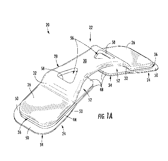
outwardly past an outer
edge of the inner sheet.
[0008] In accordance with another aspect of this disclosure, a medical
article comprises an
arch extending over an area, and a medial strut connected to the arch and
extending into the area
over which the arch extends. Optionally, the medial strut can comprise an
outer layer
configured to be attached to a patient's tissue, and an inner layer positioned
between the outer
layer and the arch. Optionally, the inner layer can be stiffer than the outer
layer, or vise versa.
The medial strut can be a first medial strut. A second medial strut can be
connected to the arch
and extend into the area over which the arch extends. The first and second
medial struts can be
positioned oppositely with respect to one another.
2
CA 03037999 2019-03-21
WO 2018/075879 PCT/US2017/057569
[0009] A medical article optionally can further comprise, or otherwise be
associated with, at
least one release liner adhered to the foot pad(s) and/or medial strut(s). A
medical article
optionally can include one or more features (e.g., a hole, receptacle, space
between the body and
at least a portion of a foot pad, and/or a catch part) configured for
interacting with an applicator
tool.
[0010] Another aspect of this disclosure is the provision of a tool
configured for being used
to manipulate a medical article. The tool can comprise first and second parts
that are spaced
apart from one another and each configured to releasably engage a medical
article, a
reconfigurable linkage connecting the first and second parts to one another,
and levers (e.g.,
handles) extending from proximate the linkage. The linkage and levers can be
cooperatively
configured so that at least portions the first and second parts are moved away
from one another
in response to at least portions of the levers being moved toward one another.
[0011] The first and second parts can be first and second catch parts
configured to releasably
attach to the medical article. Each of the catch parts can comprise a shank
and one or more
protrusion extending outwardly from the shank. The tool optionally can further
include a
bearing surface that is: positioned between the first and second catch parts,
connected to the first
and second catch parts by the linkage, and optionally configured to engage the
medical article
while the first and second catch parts are engaged to the medical article.
[0012] In another aspect of this disclosure, a tool configured for being
used to manipulate a
medical article can comprise a reconfigurable linkage connecting first and
second bodies to one
another. The first body can comprise a first lever connected to a first part.
The second body can
comprise a second lever connected to a second part. The first and second parts
can be
configured to respectively engage (e.g., at least partially receive) opposite
first and second ends
of a medical article. The reconfigurable linkage can be configured so that:
the first and second
bodies are pivotable relative to one another about first and second axes,
respectively, and the
first and second axes are movable toward and away from one another.
[0013] An aspect of this disclosure is the provision of a package having a
support comprising
a central section and outer sections respectively extending outwardly and
downwardly. A
medical article can be at least partially supported by the support. Foot pads
of the medical
article can respectively be proximate the outer sections of the support. One
or more gaps
3
CA 03037999 2019-03-21
WO 2018/075879 PCT/US2017/057569
between the support and the medial article can be configured to receive a
portion of a tool
configured for being used to manipulate a medical article.
[0014] A liner can be positioned between the support and the medical
article. The medical
article can be releasably mounted to the liner. At least a portion of the
liner can be fixedly
mounted to the support. The liner can comprise a line of disruption for at
least partially
facilitating relative movement between the medical article and the support.
The line of
disruption can at least partially define a flap in the liner. Such flaps can
be respectively
associated with foot pads of the medical article(s).
[0015] An aspect of this disclosure is the provision of a method for at
least deforming a
medical article from an at rest configuration to an extended configuration.
The deforming can
be comprised of reconfiguring a tool while the tool and the medical article
are engaged to one
another. The tool and the medical article being engaged to one another can be
comprised of a
first part of the tool and a first part of the medical article being in
engagement with one another,
and a second part of the tool and a second part of the medical article being
in engagement with
one another. The reconfiguring of the tool can be comprised of moving levers
of the tool
toward one another so that the first and second parts of the tool move away
from one another in
response to the moving of the levers of the tool toward one another, and the
first and second
parts of the medical article move away from one another in response to the
first and second parts
of the tool moving away from one another.
[0016] In accordance with an aspect of this disclosure, a method comprises
deforming a
medical article from an at rest configuration to an extended configuration so
that foot pads of
the medical article are farther apart from one another in the extended
configuration than in the at
rest configuration. Each of the foot pads can comprise an inner portion (e.g.,
medial strut)
extending inwardly from an outer portion of the foot pad, so that the inner
portions are
positioned between the outer portions of the foot pads. The inner portions can
be adhesively
mounted to a patient's tissue while the medical device is in its extended
configuration. Then the
medial article can reconfigure from the extended configuration to an
intermediate configuration
that is between the at rest configuration and the extended configuration. The
outer portions of
the pads can be adhesively mounted to the tissue while the medical device is
in its intermediate
configuration. Optionally, the adhesively mounting of the outer portions of
the pads to the
4
CA 03037999 2019-03-21
WO 2018/075879 PCT/US2017/057569
tissue can occur at least partially in response to the automatic / biased
reconfiguring of the
medial article.
[0017] The foregoing summary provides a few brief examples and is not
exhaustive, and the
present invention is not limited to the foregoing examples. The foregoing
examples, as well as
other examples, are further explained in the following detailed description
with reference to
accompanying drawings.
BRIEF DESCRIPTION OF THE DRAWINGS
[0018] Figs. 1A through 1D depict various views of a medical article that
may optionally be
referred to as a force modulating tissue bridge, or simply tissue bridge, and
which may be used
to at least partially cover a wound and/or scar, for example to help
facilitate wound closure
and/or reduce wound tension, in accordance with a first embodiment of this
disclosure.
[0019] Fig. 1E is a pictorial exploded view of the tissue bridge of Figs.
1A-1D, wherein Fig.
1E further depicts the tissue bridge exploded away from a schematically
depicted section of a
release liner and associated adhesive material, in accordance with the first
embodiment.
[0020] Figs. 2A through 2D depict various views of a tool configured for
being used to
manipulate the medical article or tissue bridge of Figs. 1A-1E, wherein the
tool may optionally
be referred to as an applicator tool, in accordance with the first embodiment.
[0021] Fig. 2E is a pictorial view of the applicator tool of Figs. 2A-2D
mounted to the tissue
bridge of Figs. 1A-1E, in accordance with the first embodiment.
[0022] Fig. 3A is a top pictorial view of at least a portion of a kit or
package comprising the
applicator tool of Figs. 2A-2D and several of the tissue bridges of Figs. 1A-
1E at least partially
contained in a tray, in accordance with the first embodiment.
[0023] Fig. 3B is a partially exploded view of some of the objects of the
package of Fig. 3A,
in accordance with the first embodiment.
[0024] Fig. 3C is an isolated, top plan view of the tray of Fig. 3A, in
accordance with the
first embodiment.
[0025] Fig. 3D is a top plan view of the tray of Fig. 3A containing the
applicator tool and
release liner, in accordance with the first embodiment.
[0026] Fig. 3E is a top plan view of the package of Fig. 3A, in accordance
with the first
embodiment.
CA 03037999 2019-03-21
WO 2018/075879 PCT/US2017/057569
[0027] Figs. 4A through 4F depict a sequence of steps of a method of using
the applicator
tool to remove a tissue bridge from the tray, in accordance with the first
embodiment.
[0028] Figs. 4G through 4K depict a sequence of steps of a method of using
the applicator
tool to apply the tissue bridge to a wound, in accordance with the first
embodiment.
[0029] Figs. 5A through 5D depict various views of a medical article or
tissue bridge having
a central release liner and release liner mounting straps affixed thereto, in
accordance with a
second embodiment of this disclosure.
[0030] Figs. 6A through 6F depict a sequence of steps of a method of
applying the tissue
bridge of Figs. 5A-5D to a wound, in accordance with the second embodiment.
[0031] Fig. 7 depicts a step of a method of applying the tissue bridge to a
wound, in
accordance with a third embodiment of this disclosure.
[0032] Fig. 8A is an isolated, pictorial view of an applicator tool, in
accordance with a fourth
embodiment of this disclosure.
[0033] Fig. 8B is a pictorial view of the applicator tool of Fig. 8A
mounted to a tissue bridge,
in accordance with the fourth embodiment.
[0034] Fig. 8C depicts a step of a method of applying the tissue bridge to
a wound, in
accordance with the fourth embodiment.
[0035] Fig. 9A is an isolated, pictorial view of an applicator tool, in
accordance with a fifth
embodiment of this disclosure.
[0036] Fig. 9B is a pictorial view of the applicator tool of Fig. 9A
mounted to a tissue bridge,
in accordance with the fifth embodiment.
[0037] Figs. 9C and 9D depict a step of a method of applying the tissue
bridge to a wound, in
accordance with the fifth embodiment.
[0038] Fig. 10A is a pictorial view of a tissue bridge, in accordance with
a sixth embodiment
of this disclosure.
[0039] Figs. 10B and 10C depict an applicator tool mounted to the tissue
bridge of Fig. 10A,
in accordance with the sixth embodiment.
[0040] Figs. 10D and 10E depict a step of a method of applying the tissue
bridge to a wound,
in accordance with the sixth embodiment.
[0041] Fig. 11A is a pictorial view of a tissue bridge, in accordance with
a seventh
embodiment of this disclosure.
6
CA 03037999 2019-03-21
WO 2018/075879 PCT/US2017/057569
[0042] Figs. 11B and 11C depict an applicator tool mounted to the tissue
bridge of Fig. 11A,
in accordance with the seventh embodiment.
[0043] Fig. 11D and 11E depict a step of a method of applying the tissue
bridge to a wound,
in accordance with the seventh embodiment.
[0044] Figs. 12A and 12B depict an applicator tool mounted to a tissue
bridge, in accordance
with an eighth embodiment.
[0045] Fig. 12C and 12D depict a step of a method of applying the tissue
bridge to a wound,
in accordance with the eighth embodiment.
[0046] Figs. 13A and 13B depict a tissue bridge, in accordance with a ninth
embodiment.
[0047] Fig. 13C is an exploded view of the tissue bridge, in accordance
with the ninth
embodiment.
[0048] Fig. 13D is a cross-sectional view of at least a portion of a kit or
package comprising
the tissue bridge at least partially contained in a tray, in accordance with
the ninth embodiment.
[0049] Figs. 13E and 13F depict a sequence of steps of a method of using an
applicator tool
to remove a tissue bridge from the tray, in accordance with the ninth
embodiment.
[0050] Fig. 14 is a bottom pictorial view of an applicator tool, in
accordance with a tenth
embodiment.
[0051] Fig. 15 is a top plan view of a tray at least partially containing
the applicator tool and
a series of tissue bridges, in accordance with the tenth embodiment.
[0052] Fig. 16A is a schematic top plan view of an elongate scar or wound
in tissue.
[0053] Fig. 16B is like Fig. 16A, except that the scar or wound is covered
by a strip.
[0054] Fig. 16C is like Fig. 16B, except that tissue bridges have been
mounted over the strip,
in accordance with an embodiment of this disclosure.
[0055] Fig. 16D is a side elevation view of a first version of the assembly
of 16C.
[0056] Fig. 16E is a side elevation view of a second version of the
assembly of Fig. 16D, and
Fig. 16E also depicts a tissue bridge in accordance with another embodiment of
this disclosure.
[0057] Fig. 16F is similar to Fig. 16E, in accordance with another
embodiment of this
disclosure.
[0058] Fig. 16G is a side elevation view of a tissue bridge in accordance
with an embodiment
of this disclosure.
7
CA 03037999 2019-03-21
WO 2018/075879 PCT/US2017/057569
[0059] Figs. 17A through 17C depict a tissue bridge in accordance with an
eleventh
embodiment.
[0060] Fig. 17D is a cross-sectional view taken along line 17D-17D of Fig.
17B.
[0061] Fig. 17E is an end elevation view of the tissue bridge of the
eleventh embodiment.
[0062] Fig. 17F is a top pictorial exploded view of the tissue bridge of
the eleventh
embodiment, wherein Fig. 17F further depicts the tissue bridge exploded away
from a
schematically depicted section of a release liner and associated adhesive
material, in accordance
with the eleventh embodiment.
[0063] Fig. 17G is a bottom pictorial exploded view of selected layers of
the tissue bridge of
the eleventh embodiment
[0064] Figs. 18A through 18C depict various views of an applicator tool in
accordance with
the eleventh embodiment.
[0065] Fig. 18D depicts the applicator tool mated to a tissue bridge of a
package including a
tray and series of tissue bridges, in accordance with the eleventh embodiment.
[0066] Figs. 19A through 19F depict a sequence of steps of a method of
using the applicator
tool to remove a tissue bridge from the tray, in accordance with the eleventh
embodiment.
[0067] Figs. 19G and 19H depict different confirmations of a release liner
associated with a
tissue bridge and tray, in accordance with the eleventh embodiment.
[0068] Figs. 191 through 19L depict a sequence of steps of a method of
using the applicator
tool to apply the tissue bridge to a wound, in accordance with the eleventh
embodiment.
[0069] Fig. 20A is top plan view of a tissue bridge of a variation of the
eleventh
embodiment.
[0070] Fig. 20B is an isolated, top plan view of a footpad of the tissue
bridge of the variation
of the eleventh embodiment.
[0071] Fig. 20C is an end elevation view of the tissue bridge of the
variation of the eleventh
embodiment.
[0072] Fig. 20D is a top pictorial exploded view of the tissue bridge of
the variation of the
eleventh embodiment, wherein Fig. 20D further depicts the tissue bridge
exploded away from a
section of a release liner, in accordance with the variation of the eleventh
embodiment.
[0073] Fig. 21 depicts a variation of the applicator tool of the eleventh
embodiment.
[0074] Fig. 22 depicts another variation of the applicator tool of the
eleventh embodiment.
8
CA 03037999 2019-03-21
WO 2018/075879 PCT/US2017/057569
[0075] Figs. 23A through 23C depict an applicator tool in accordance with a
twelfth
embodiment.
[0076] Figs. 24A through 24C depict an applicator tool in accordance with a
thirteenth
embodiment.
[0077] Figs. 25A through 25C depict an applicator tool in combination with
a tissue bridge,
in accordance with a fourteenth embodiment.
[0078] Figs. 26A through 26C depict an applicator tool in combination with
a tissue bridge,
in accordance with a variation of the fourteenth embodiment.
[0079] Figs. 27A and 27B depict an applicator tool in combination with a
tissue bridge, in
accordance with a variation of the fourteenth embodiment.
[0080] Figs. 28A and 28B depict an applicator tool in combination with a
tissue bridge, an in
accordance with a variation of the fourteenth embodiment.
[0081] Figs. 28D through 28E depict a sequence of steps of a method of
using the applicator
tool to apply the tissue bridge to a wound, in accordance with the fourteenth
embodiment.
[0082] Figs. 29A through 29C are top plan views of trays including mounting
features, in
accordance with other embodiments of this disclosure.
[0083] Fig. 30 depicts a tray that is carrying a series of tissue bridges,
and is equipped with
fastening straps, in accordance with an embodiment of this disclosure.
[0084] Fig. 31 depicts the tray with tissue bridges of Fig. 30 mounted on
the arm of a user, in
accordance with an embodiment of this disclosure.
[0085] Fig. 32 depicts the tray with tissue bridges of Fig. 30 with the
fastening straps
removed, wherein the tray is exploded away from a mounting base, in accordance
with an
embodiment of this disclosure.
[0086] Fig. 33 depicts a tray that is carrying a series of tissue bridges,
and is equipped with
fastening straps, in accordance with an embodiment of this disclosure.
[0087] Fig. 34 depicts a tray with tissue bridges exploded away from a
mounting base
equipped with clips and fastening straps, in accordance with an embodiment of
this disclosure.
[0088] Fig. 35 depicts a tray with tissue bridges exploded away from a
mounting base
equipped with adhesive material and fastening straps, in accordance with an
embodiment of this
disclosure.
9
CA 03037999 2019-03-21
WO 2018/075879 PCT/US2017/057569
DETAILED DESCRIPTION
[0089] Numerous embodiments are described below and illustrated in the
accompanying
figures, in which like numerals refer to like parts throughout the several
views. For convenience
of description and ease of understanding, and not for the purpose of limiting
the scope of this
disclosure or the associated inventions, some embodiments may be referred to
by number. The
embodiments described provide examples and should not be interpreted as
limiting the scope of
the invention. Other embodiments, and modifications and improvements of the
described
embodiments, will occur to those skilled in the art and all such other
embodiments, modifications
and improvements are within the scope of the invention.
[0090] Figs. 1A-1D depict an at least partially elastic (e.g., generally
elastic) medical article
20 in its undeformed or at rest configuration (e.g., relaxed state), in
accordance with a first
embodiment. The medical article 20 may optionally be referred to as a force
modulating tissue
bridge 20, or simply tissue bridge 20, and throughout this disclosure the
tissue bridge may be
more generally referred to as a medical article. In the following, first an
example of a method of
using the tissue bridge 20 is very briefly described, and thereafter the
tissue bridge and other
aspects of this disclosure are described in greater detail.
[0091] The tissue bridge 20 can be mounted to tissue such as, but not
limited to, a surface of
a patient's skin, for example the outer surface of the patient's epidermis.
The tissue bridge 20 is
typically mounted so that it extends across and at least partially covers a
wound and/or scar. In
the first embodiment, the tissue bridge 20 comprises generally elastic
material, and prior to the
tissue bridge being mounted on the patient, the tissue bridge can be generally
elastically
deformed from its undeformed or at rest configuration to a strained, deformed,
or extended
configuration. The tissue bridge 20 can at least begin to be mounted to the
tissue (e.g., skin
tissue), so that a central section of the tissue bridge extends across a wound
and/or scar, while the
tissue bridge is maintained in its extended configuration. After being at
least partially mounted
in its extended configuration, the tissue bridge 20 can be allowed to
generally elastically
reconfigure from its extended configuration at least partially toward its at
rest configuration,
which may, for example, reduce tension in the tissue, help close the wound,
help inhibit wound
reopening, and/or inhibit scar disfiguring (e.g., widening), as will be
discussed in greater detail
CA 03037999 2019-03-21
WO 2018/075879 PCT/US2017/057569
below. In the first embodiment, the tissue bridge 20 comprises material that
is at least generally
elastic, so that the tissue bridge is biased toward its at rest configuration
(e.g., relaxed state).
[0092] The tissue bridge 20 of the first embodiment comprises a generally
elastic body 22
and one or more multi-layer foot pads 24 mounted to the body, although in some
examples one
or more of the foot pads and/or portions thereof can be omitted (e.g., a foot-
pad may consist of,
or consist essentially of, a single layer). The body 22 can be generally
referred to as and/or
generally function as a backbone or other suitable structure configured to
movably connect two
or more of the foot pads 24 to one another. In the embodiment shown in Figs.
1A-1E, the body
22 includes at least two flanges 26 (e.g., feet) respectively extending
obliquely, for example
outwardly and downwardly, from opposite lower portions of a central section or
arch 28 of the
body. Each of the flanges 26 can be planar, or they can be substantially or
about planar since it
may not be critical that the flanges be exactly planar. The flanges 26 can
extend divergently
relative to one another, and obliquely relative to one another. The arch 28
can include a central
spanning section 30, and lower sections 32 respectively extending downwardly
from opposite
portions of the spanning section. The lower sections 32 of the arch 28 can
optionally be
configured as and/or referred to as shoulders 32. The flanges 26 can
respectively extend
obliquely, for example outwardly and downwardly, from lower portions of the
shoulders 32.
The shoulders 32 can provide a smoothly curved transition between the spanning
section 30 of
the arch 28 and the flanges 26. In the embodiment shown in Figs. 1A-1E, the
spanning section
30 of the arch 28 has a relatively low profile and is at least generally
arcuate, which can be
advantageous for an active person having the tissue bridge 20 mounted on their
skin.
Alternatively, it is believed that in some situations the arch 28 can be at
least more of a flat arch,
or the spanning section 30 of the arch can be flat, or the arch or features
thereof can be in any
other suitable configurations that will allow the tissue bridge 20 to function
generally or
substantially as described herein.
[0093] Each of the parts of the tissue bridge 20 will typically be
constructed of suitable
medical-grade materials. For example, the body 22 can be an injection-molded
or mechanically
thermoformed, unitary (e.g., single-piece) article such that the spanning
section 30, shoulders 32
and flanges 26 can be formed together as a single article from an injection-
moldable or formable,
generally elastic material such as, but not limited to, polycarbonate, or any
other suitable
injection-moldable or formable material. Referring to Fig. 1C, each of the
spanning section 30,
11
CA 03037999 2019-03-21
WO 2018/075879 PCT/US2017/057569
shoulders 32, and flanges 26 can be about the same thickness, or alternatively
the thickness of
the body 22 can vary along its length. Referring to Fig. 1B, the width of the
body 22 can, for
example, taper along its length, so that the spanning section 30 is relatively
narrow (e.g., has a
narrowed waist) as compared to the shoulder 32 and flanges 26, so that the
spanning section can
be more readily deformed as compared to the shoulders and flanges. For
example, the side edges
of the spanning section 30 can be inwardly curved or concave, as shown in
Figs. 1A, 1B and 1D,
or they may have a stepped or other suitable configurations. Alternatively,
the side edges of the
spanning section 30 can extend generally or substantially straight in a top
plan view of the tissue
bridge 20, or they can extend in any other suitable manner.
[0094] As shown in Figs. 1A-1D, the foot pads 24 can be spaced apart from
one another, and
the foot pads can be fixedly mounted to the flanges 26. Each foot pad 24 can
be or include be a
mat, laminate or other suitable structure comprising one or more layers of
material. For
example, in the first embodiment, each foot pad 24 includes an outer layer or
sheet 34
configured to be attached to tissue (e.g., skin tissue), and an inner layer or
sheet 36 positioned
between, and fixedly connected to each of, the outer sheet 34 and the
respective flange 26.
[0095] Referring to the exploded view of Fig. 1E, the tissue bridge 20 can
include inner,
intermediate and outer adhesive layers 38, 40, 42. The inner adhesive layers
38 can be between
and fixedly connect the inner sheets 36 to the flanges 26, the intermediate
adhesive layers 40
can be between and fixedly connect the outer sheets 34 to the inner sheets,
and the outer
adhesive layers 42 can be on the outer sides of the outer sheets for attaching
the tissue bridge 20
to tissue (e.g., a patient's skin), as will be discussed in greater detail
below.
[0096] The outer and inner sheets 34, 36 can be provided, for example, by
die cutting them
from appropriate webs or larger sheets of material, such as fabric or cast
microporous polymeric
sheet for the outer sheets 34, and an extruded polymer or plastic sheet for
the inner sheets 36.
The outer sheets 34 can be made of suitable fabric materials, cast materials,
films, or other
materials of the type from which skin-contact layers of bandages or other
wound dressings are
formed, or any other suitable material. The plastic inner sheets 36 can be
made of suitable
materials such as, for example, polyethylene, polyethylene terephthalate, or
any other suitable
materials. The inner and intermediate adhesive layers 38, 40 can respectively
comprise
adhesive materials that are compatible with the materials being connected
thereby. The outer
adhesive layer 42 (e.g., patient contact adhesive) can be, for example,
adhesive material of the
12
CA 03037999 2019-03-21
WO 2018/075879 PCT/US2017/057569
type that is typically used as an adhesive backing for bandages or other wound
dressings. In the
first embodiment, the outer adhesive layer 42 can have a lower adhesive
strength than the inner
and intermediate adhesive layers 38, 40, such as when the tissue bridge 20 is
to be removably
mounted to tissue (e.g., a patient's skin).
[0097] In the first embodiment, both the body 22 and the inner sheet 36
have a higher
modulus of elasticity (e.g., are formed from stiffer material) than the outer
sheet 34. More
generally, the body 22 and the inner sheet 36 can be stiffer than the outer
sheet 34 because of a
variety of factors, such as being larger, thicker, comprising material having
a higher modulus of
elasticity and/or being constructed to have an apparent modulus of elasticity.
Similarly, the
body 22 can have a higher modulus of elasticity than the foot pads 24.
[0098] As shown in Fig. 1C, the body 22, including its flanges 26, can be
thicker than each
of the outer and inner sheets 34, 36, and the arch 28 can extend over an area
into which portions
of the outer and inner sheets can optionally extend. The area over which the
arch 28 extends
may be referred to as a central area, a treatment area, an under-arch area,
and/or the like. The
thicknesses of the body 22 and sheets 34, 36 can be varied, for example,
independently as
necessary to produce tissue bridges 20 of different sizes and to function
optimally in different
anatomical areas and with different treatment tissue characteristics.
[0099] As shown in Figs. 1A and 1E, at each end of the tissue bridge 20,
the flange 26, outer
sheet 34, inner sheet 36, and adhesive layers 38, 40, 42 can be at least
partially superposed with
one another and can have different configurations from one another. For
example and as
schematically illustrated in Fig. 1E by dashed boundary lines on the outer and
inner sheets 34,
36, a congruent portion 44 of each inner sheet can be superposed with and
coextensive with the
respective flange 26, and a congruent portion 46 of the outer sheet 34 can be
superposed with
and coextensive with the respective inner sheet congruent portion 44. As other
examples that
are also at least partially schematically illustrated by the dashed boundary
lines in Fig. 1E, inner
extensions 48 of the outer and inner sheets 34, 36 can extend congruently with
one another into
the central area over which the arch 28 extends such that the inner extensions
48 are neither
superposed by nor coextensive with the flanges 26. More generally, each foot
pad 24 can
include at least one extension 48 that extends into the central area over
which the arch 28
extends such that the inner extension 48 can be neither superposed by nor
coextensive with the
flanges 26.
13
CA 03037999 2019-03-21
WO 2018/075879 PCT/US2017/057569
[00100] In the first embodiment, the inner extensions 48 may be referred to as
medial
extensions 48, for example since they extend toward the middle of the area
over which the arch
28 extends. As another example and as will be discussed in greater detail
below, the inner or
medial extensions 48 can be configured so that they at least partially resist
longitudinal
compression when the tissue bridge 20 in its extended configuration is mounted
to tissue (e.g.,
skin tissue) and then allowed to generally elastically reconfigure from its
extended configuration
at least partially toward its at rest configuration. Accordingly, the inner or
medial extensions 48
can be referred to as medial struts 48. In the first embodiment, each medial
strut 48 includes the
inner extensions 48 of both sheets 34, 36, but one or more layers or sheets of
the medial strut 48
can be omitted, such that each medial strut can be formed of one or more
layers of material.
[00101] Referring to Figs. 1A and 1E, one or more outer extensions 50 of the
foot pads 24, or
more specifically of the outer sheets 34, can extend outwardly such that they
are neither
superposed by nor coextensive with the flanges 26 or the medial struts 48. In
the example
shown in Fig. 1B, outer extensions 50 can extend outwardly past one or more of
the peripheral
edges (e.g., the outer end edges and side edges) of the flanges 26.
[00102] In the example shown in Fig. 1C, the medial struts 48 can be spaced
apart from (e.g.,
at least partially spaced apart from) the arch 28 and extend into the central
area over which the
arch extends, so that gaps or receptacles 52 are at least partially defined
between the medial
struts and the arch. The receptacles 52 can at least partially define, or be
at least part of, catch
parts configured for interacting with corresponding features of an applicator
tool that may be
used, for example, in the mounting of the tissue bridge 20 to tissue (e.g., a
patient's skin),
wherein the applicator tool is discussed in greater detail below. For example,
the tissue bridge
20 can include one or more catch parts, and the catch parts can respectively
comprise the
receptacles 52. A variety of differently configured catch parts are within the
scope of this
disclosure.
[00103] In the first embodiment, the body 22 includes at least two catch parts
that further
comprise holes 56 that extend through the body 22 and are open to the
receptacles 52. The holes
56 can be defined in the arch 28, or more specifically the holes can be
positioned in opposite end
portions of the spanning section 30. In the example shown in Fig. 1B, the
holes 56 can be open
to the central area over which the arch 28 extends, or more specifically the
holes can be open to
the receptacles 52; and the medial struts 48 can extend beneath the holes.
14
CA 03037999 2019-03-21
WO 2018/075879 PCT/US2017/057569
[00104] As shown in Figs. 1A and 1B, each of the holes 56 can be triangular,
with a side of
the triangle, or more specifically an edge 58 of the arch 28 that defines the
triangle, extending
crosswise to the length of the arch. The respective catch part can further
include the edge 58.
The edge 58 can extend parallel, or more generally substantially parallel or
about parallel, to the
boundary between the spanning section 30 and the respective shoulder 32. In
other words, the
edge can extend perpendicular to, or more generally substantially
perpendicular to or about
perpendicular to, the lengthwise or longitudinal axis of the body 22. In
addition, the holes 56,
when present, can reduce the area or volume of the outer portions of the
spanning section 30 in a
manner that enhances the deformability of the outer portions of the spanning
section.
[00105] Each catch part can further include a portion 60 (Fig. 1C) of the
lower surface of the
arch 28, wherein the surface portion 60 extends outwardly from the edge 58.
The catch parts,
receptacles 52, holes 56 and associated edges 58, and surface portions 60, or
the like, may be
optional, and other positions and configurations of catch parts, receptacles,
holes, or the like, are
within the scope of this disclosure.
[00106] In a version of the first embodiment, the foot pads 24 can be
described as including
the flanges 26, so that the flanges can be respective layers of the foot pads,
and the flanges can
be referred to as foot plates 26, or the like. As another example, the first
embodiment embraces
configurations of the tissue bridge 20 in which the foot plates 26 are not
integrally formed with
the arch 28. For example, the foot plates 26 can be formed separately from the
arch 28 and can
be fixedly or movably connected to the arch, such as by way of pivots, hinges,
or any other
suitable features. Other variations are also with the scope of this
disclosure. For example, the
foot plates 26 can have one or more holes formed therein or therethrough, as
discussed in greater
detail below. As another example alluded to above, one or more layers of each
foot pad 24 can
be omitted. For example and for each foot plate 26, it may be suitable in some
situations to omit
the layers between the foot plate and outer adhesive layer 42, so that the
outer adhesive layer,
which is for use in mounting the tissue bridge 20 to tissue (e.g., a patient's
skin), is mounted
directly to the underside of the foot plate. It is also within the scope of
the first embodiment for
the medial struts 48 to be integrally formed with the arch 28, flanges 26
and/or foot plates 26.
For example, the medial struts 48 can be extensions of the flanges 26 and/or
foot plates 26.
[00107] As alluded to above, Fig. 1E depicts the tissue bridge 20 in an
exploded
configuration. Additionally, Fig. 1E depicts the tissue bridge 20 exploded
away from a
CA 03037999 2019-03-21
WO 2018/075879 PCT/US2017/057569
schematically depicted section of a release liner 62 and associated adhesive
material 64. As an
example, after a tissue bridge 20 is manufactured or as part of the
manufacturing process for the
tissue bridge, the tissue bridge, or more specifically the outer sheets 34 by
way of the outer
adhesive layers 42, can be releasably mounted on the upper surface of the
release liner 62. In
addition, the lower surface of a portion of the release liner 62 can be
fixedly mounted to a
support by way of the adhesive material 64, as will be discussed in greater
detail below. The
release liner 62 can be, for example, a paper or plastic-based film sheet
coated with a release
agent that is engaged against the outer adhesive layers 42 so that the tissue
bridge 20 is
releasably mounted on the release liner.
[00108] In accordance with the first embodiment, the tissue bridge 20 can be
at least
somewhat translucent, and the tissue bridge can optionally include indicia,
visible design
elements and/or other visual features comprising one or more of color,
contrasting colors,
decorations, aligning marks, pictures, logos, images, characters, words, or
any other suitable
features that can be printed matter, or the like, wherein the printed matter,
or the like, can be
embedded or encapsulated in the tissue bridge and visible through one or more
exterior surfaces
of the tissue bridge. For example one or more of the components or layers of
the tissue bridge
20 can be at least generally transparent and/or at least generally
translucent, and the printed
matter, or the like, can be interior of exterior surfaces of the tissue bridge
20 and seen by a user
of the tissue bridge through at least one of the exterior surfaces of the
tissue bridge. For
example, the printed matter, or the like, can be on a layer or surface of the
tissue bridge 20 that
is internal to the tissue bridge (e.g., printed matter, or the like, can be
positioned or
"sandwiched" between the various layers of the tissue bridge). Referring to
Fig. 1E and as an
example, the body 22 and the inner sheets 36 can each be at least partially
transparent, and the
printed matter, or the like, can be on the top surface of at least one of the
outer sheets 34 so that
it is visible through the body 22 and the respective inner sheet 36. As
another example, the
body 22 can be at least partially transparent, and the printed matter, or the
like, can be on the top
surface of at least one inner sheet 36 so that it is visible through the body
22. Alternatively, it is
believed that the printed matter, or the like, can be contained in one of more
of the adhesive
layers 38, 40, 42 and/or in any other suitable location, with the
predetermined portion(s) of the
tissue bridge 20 being least partially transparent and/or translucent for
allowing the printed
matter, or the like, to be visible through one or more exterior surfaces of
the tissue bridge.
16
CA 03037999 2019-03-21
WO 2018/075879 PCT/US2017/057569
[00109] Figs. 2A-2D depict an applicator mechanism in the form of an
applicator tool 80 that
can be used, for example, to manipulate the tissue bridge 20 or another
suitable medical article,
for example as part of a method of mounting the tissue bridge to tissue (e.g.,
a patient's skin), in
accordance with the first embodiment. For example, the applicator tool 80 can
include one or
more parts or features that can be spaced apart from one another and can be
configured to
releasably engage the tissue bridge 20. In the first embodiment, the one or
more parts or features
of the applicator tool 80 that are configured to engage the tissue bridge 20
can comprise at least
one bearing or contact surface 82 and/or one or more catch parts 84. For
example, the contact
surface 82 can be positioned between the catch parts 84. The applicator tool
80 can further
include a reconfigurable frame connecting the contact surface 82 and catch
parts 84 to one
another. The applicator tool 80, and the like, can be more generally referred
to as a tool. For
example, the tool 80, or the like, may be used for more purposes than applying
a tissue bridge 20.
[00110] The frame can include a reconfigurable linkage (e.g., one or more
links 86A, 86B)
connecting the contact surface 82 and catch parts 84 to one another, and one
or more levers 88A,
88B extending upwardly from the links 86A, 86B. The applicator tool 80 can be
configured so
that when the bearing or contact surface 82 faces downwardly, the catch parts
84 extend
downwardly from the linkage (e.g., link(s) 86A, 86B), and the levers 88A, 88B
extend upwardly
from the linkage. The links 86A, 86B and the levers 88A, 88B can be
cooperatively configured
so that at least portions of the catch parts 84 move away from one another,
and the contact
surface 82 moves toward a line between the catch parts 84, in response to at
least portions of the
levers 88A, 88B being moved toward one another, as will be discussed in
greater detail below.
[00111] The links 86A, 86B can comprise several links, for example a central
link 86A and
outer links 86B. Similarly, the levers 88A, 88B can comprise several levers,
for example inner
levers 88A and outer levers 88B. In the example shown in Figs. 2A-2D, the
contact surface 82
can be a lower end face of the central link 86A, and the catch parts 84 can
include shanks 90
extending from lower ends of the outer links 86B and/or outer levers 88B. Each
catch part 84
can further include at least one protrusion 92 extending outwardly from the
lower end of the
shank 90 in a direction that is crosswise to the length of the shank. The
protrusions 92 can face
away from one another.
[00112] The outer links 86B can extend obliquely, outwardly and downwardly
from opposite
sides of an upper portion of the central link 86A respectively to upper
portions of the shanks 90.
17
CA 03037999 2019-03-21
WO 2018/075879 PCT/US2017/057569
The inner levers 88A can extend obliquely, outwardly and upwardly from
opposite sides of an
upper portion of the central link 86A. The outer levers 88B can extend
obliquely, outwardly and
upwardly respectively from upper portions of the shanks 90.
[00113] The levers 88A, 88B can be configured as and/or comprise handles 94.
For example,
in the embodiment depicted in Figs. 2A and 2B, a handle 94 can include
adjacent levers 88A,
88B that are connected to one another. These connections can comprise the
adjacent levers 88A,
88B being directly connected to one another proximate their upper ends, and
crossmembers 96
connected to and spanning between the adjacent levers.
[00114] The applicator tool 80 can be an injection-molded, unitary (e.g.,
single-piece) article
formed from an injection-moldable, generally elastic material such as, but not
limited to,
polycarbonate, polyethylene, or any other suitable injection-moldable
material. Alternatively,
the applicator tool 80 can be made of metal, metal alloys, steel, or any other
suitable materials
that can allow for re-sterilization. For example, hinges or other suitable
connections that allow
for relative movements between subparts can be included in the applicator
tools 80, such as when
the applicator tools are made of relatively rigid materials. As additional
examples, a variety of
different linkages, levers 88A, 88B, and handles 94 are within the scope of
this disclosure, as
will be discussed in greater detail below.
[00115] In accordance with an example of the first embodiment depicted in Fig.
2E, the tissue
bridge 20 and applicator tool 80 are cooperatively configured so that the
applicator tool can be
releasably engaged to the tissue bridge, and the applicator tool can be used
to manipulate the
tissue bridge as part of a method of mounting the tissue bridge to tissue. For
example, Fig. 2E
depicts the bearing or contact surface 82 in opposing-face-to-face relation
with an upper surface
of the arch 28, and the shanks 90 extending through the holes 56. In the
configuration of Fig.
2E, the protrusions 92 are hidden from view within the receptacles 52, as
partially schematically
illustrated by dashed lines. More generally, the above-discussed catch parts
of the applicator tool
80 and tissue bridge 20 are respectively engaged to one another in Fig. 2E.
However, a variety
of differently configured catch parts are within the scope of this disclosure.
[00116] The applicator tool 80 can optionally further include one or more
features for at least
partially facilitating predetermined cooperative interaction between the
applicator tool and the
tissue bridge 20. For example, the levers 88A, 88B and/or handles 94, or
features associated
therewith, can be configured to come into contact with one another when the
desired degree of
18
CA 03037999 2019-03-21
WO 2018/075879 PCT/US2017/057569
deformation is reached in the tissue bridge 20, thereby seeking to prevent
over distortion of the
tissue bridge. In addition or alternatively, the levers 88A, 88B and/or
handles 94, or features
associated therewith, can be configured to (e.g., can include one or more
catches, rows of
catches, or the like, configured to) cause the applicator tool 80 to hold the
tissue bridge 20 in one
or more predetermined states of deformation (e.g., one or more predetermined
strained,
deformed, or extended configurations) without requiring the user to
continually squeeze together
the handles 94, or the like. For example, the applicator tool 80 can include
mechanisms (e.g.
rows of catches) that can be sequentially activated, similarly to such
mechanisms of surgical
clamps, so one click (e.g., a first predetermined engagement between the
catches or the like) can
cause a relatively low state of deformation in the tissue bridge, two clicks
(e.g., a second
predetermined engagement between the catches or the like) can cause a
relatively medium state
of deformation in the tissue bridge, and three clicks (e.g., a third
predetermined engagement
between the catches or the like) can cause a relatively large state of
deformation in the tissue
bridge (e.g., the full deformation). In addition, the levers 88A, 88B, handles
94 and/or other
suitably associated features can have different shapes to assist in
ergonomically optimized use,
for example by comprising partial or complete rings, recesses shaped to accept
the user's digit(s)
and/or other suitable features. Cooperative interaction between the applicator
tool 80 and tissue
bridge 20, such as engagement between their catch parts, will be discussed in
greater detail
below, after a discussion of the option of the tissue bridges and applicator
tools being
conveniently provided as parts of kits.
[00117] In accordance with the first embodiment, and as at least partially
depicted in Fig. 3A,
one or more of the tissue bridges 20 and optionally at least one applicator
tool 80 can be
provided as part of a kit or package 120 that can further include the release
liner 62 and adhesive
material 64 (Fig. 1E). In the example shown in Figs. 3A-3C, the package 120
can include a
container in the form of an injection molded or vacuum-formed tray 122, or the
like. The tray
122 can have a base panel 124 and sidewalls 126 extending upwardly from the
periphery of the
base panel to define a cavity of the tray. Also, the package 120 and/or tray
122 can optionally
include at least one divider 128 that extends upwardly from the base panel 124
and is positioned
between and distant from opposite sidewalls 126 to at least partially divide
the tray cavity into at
least first and second compartments configured for respectively receiving the
applicator tool 80
and one or more tissue bridges 20. The first compartment of the tray 122, or
more specifically
19
CA 03037999 2019-03-21
WO 2018/075879 PCT/US2017/057569
the divider 128 and the sidewalls 126 that at least partially define the first
compartment of the
tray, can define a shape that is complementary to the peripheral shape of, and
about the same size
as, the applicator tool 80, so that a releasable interference fit can be
defined between the first
compartment and the applicator tool within the first compartment. As another
example, the first
compartment of the tray 122, divider 128 and applicator tool 80 can be omitted
from the tray
122. Any applicator tool 80 can be provided as part of a package that is
separate from the
package including the tray 122.
[00118] As an example, the tray 122 can be an inner tray that can be put in
either an outer
tray, a pouch and/or other suitable packaging. As other examples, the tray 122
can include other
features, for example slots or other surface features that can be used to
secure the tray to the
user's body (e.g., non-dominant forearm), to a fixture (e.g., a mayo tray), or
in other suitable
configurations.
[00119] Referring to the exploded view of Fig. 3B, and the isolated top plan
view of the tray
122 in Fig. 3C, the base panel 124 can be configured in the form of and/or to
define a generally
ridge-shaped support extending along at least a portion of the length of the
tray. The ridge-
shaped support can include an elevated central section 130 of the base panel
and downwardly
sloping outer sections 132 of the base panel. The central section 130 can be
flat, although exact
flatness may not be required such that the central section can be generally or
substantially flat, or
in any other suitable configuration. For example, the central section can be
concave, or the like.
The outer sections 132 can extend obliquely, or more specifically outwardly
and downwardly,
from opposite portions or edges of the raised central section 130. For
example, the outer sections
132 can extend obliquely, inwardly and upwardly from lower, marginal portions
134 of the base
panel 124. Two or more pairs of outer sections 132 can be included in each
tray 122.
Alternatively, the central section 130 can be recessed downwardly relative to
inner portions of
the outer sections 132.
[00120] Referring to Figs. 1E, 3A, 3B, 3D and 3E, the release liner 62 can
include a series of
lines of disruption 136. Each line of disruption 136 can comprise one or more
cuts, slits,
breachable lines of disruption, perforations and/or overlapping and/or
sequential combinations
thereof, for at least partially defining flaps 138 in the release liner 62, as
will be discussed in
greater detail below. The lines of disruption 136 can be configured in a
variety of patterns. In
some examples, the pattern may be symmetrical in relationship to each
associated (e.g.,
CA 03037999 2019-03-21
WO 2018/075879 PCT/US2017/057569
subsequently mounted) tissue bridge 20. In other examples, the lines of
disruption 136 can be
asymmetrical in relationship to each associated (e.g., subsequently mounted)
tissue bridge 20.
In the first embodiment, each line of disruption 136 extends partially around
the foot pad 24 that
is mounted to the flap 138 defined by the line of disruption, and opposite
ends of the line of
disruption extend beneath the food pad.
[00121] In accordance with the first embodiment, the tissue bridges 20 can be
manually
assembled and/or at least partially assembled by way of one or more automated
coating,
laminating and cutting processes. For example, the release liner 62 can be a
base ply or layer of
a laminate that is appropriately cut (e.g., die cut) and partially delaminated
to at least partially
form the foot pads 24 on the release liner, and thereafter the bodies 22 can
be respectively
mounted to the foot pads 24, or the like. The lines of disruption 136 can be
formed by an
appropriate one or more of the cutting (e.g., die cutting) steps, or the like,
such that the lines of
disruption (e.g., slits, perforations or other suitable cuts) may extend at
least partially into one or
more layers of the foot pad 24. As a more specific example, the lines of
disruption 136, or
extensions thereof, or the like, may extend into the outer adhesive layer 40
(Fig. 1E). At an
appropriate time, typically after the die cutting, or the like, the underside
of the release liner 62
can be fixedly secured to the surface of the outer sections 132 of the tray
122 by way of, for
example, adhesive material 64 (Fig. 1E) arranged in a pattern such that the
adhesive material is
omitted from between the flaps 138 in the release liner and the respective
portions (e.g., outer
sections 132) of the tray, so that the flaps can be moved relative to the
reminder of the release
liner, as will be discussed in greater detail below. More generally, any
adhesion or other
suitable connection between the flaps 138 and the respective portions (e.g.,
outer sections 132)
of the tray 122 is weaker than the adhesion or other suitable connection
between the remainder
of the release liner 62 and the tray, so that the flaps can be moved relative
to the reminder of the
release liner, as will be discussed in greater detail below.
[00122] Referring to Figs. 4A-4F, a method of using the applicator tool 80 to
remove a tissue
bridge 20 from the tray 122 is described in the following, in accordance with
the first
embodiment. In Figs. 4A-4F, the tray 122 and release liner 62 are cross-
sectioned along line 4-4
of Fig. 3E. Referring to Fig. 4A, initially, the applicator tool 80 (e.g., in
its undeformed or at
rest configuration) can be engaged against the tissue bridge 20 (e.g., in its
undeformed or at rest
configuration) by way of relative movement causing increased closeness between
the applicator
21
CA 03037999 2019-03-21
WO 2018/075879 PCT/US2017/057569
tool 80 and the tray 122 (e.g., movement of the applicator tool toward the
tissue bridge mounted
on the release liner 62 in the tray). Referring to Figs. 4A and 4B, in
response to the relative
movement, the protrusions 92 of the tool catch parts 84 can enter the
receptacles 52 by way of
the holes 56 (Figs. 1A and 1B). That is, the protrusions 92 can enter the
receptacles 52 by
traveling through the holes 56. For example, the applicator tool 80 can be
pushed downwardly
to engage the tissue bridge 20 in a manner so that the protrusions 92 of the
tool catch parts 84
enter the receptacles 52 by way of the holes 56, and optionally also the tool
contact surface 82
engages the central apex or any other suitable surface of the arch 28. The
applicator tool 80 can
be in its undeformed or at rest configuration throughout the step of the
protrusions 92 of the tool
catch parts 84 entering the receptacles 52 by way of the holes 56. As another
example, the
distance between the tips of the protrusions 92 can be greater than the
distance between the hole
edges 58 (Figs. 1A and 1B) so that the protrusions "snap" into the holes 56
and/or receptacles
52 and are optionally releasably contained in the receptacles by way of an
interference fit, or the
like.
[00123] The relative movement causing increased closeness between the
applicator tool 80
and the tray 122 may be facilitated by a user manually holding the handles 94
of the applicator
tool and moving the applicator tool toward the tissue bridge 20 in the tray,
or the tissue bridge
may be supported by any other suitable surface. Referring to Fig. 4B, the
protrusions 92 can
engage respective surfaces of the medial struts 48 in response to the relative
movement causing
increased closeness between the applicator tool 80 and the tray 122.
[00124] For serially achieving the configurations of Fig. 4C and Fig. 4D,
simultaneously
and/or in series, the relative movement causing increased closeness between
the applicator tool
80 and the tray 122 can continue, and the handles 94 can be manually squeezed
together (e.g.,
pushed toward one another) so that the applicator tool reconfigures toward its
actuated or
deformed configuration and applies deforming forces on the tissue bridge 20.
As the applicator
tool 80 is, for example, simultaneously pushed with greater force against the
tissue bridge 20
and caused to deform farther toward its deformed configuration, the applicator
tool applies
forces against the tissue bridge 20 so that the tissue bridge is responsively
deformed toward its
strained, deformed, or extended configuration. For example, the applicator
tool 80 can
simultaneously apply a downward force via the contact surface 82 and laterally
outward forces
via the catch parts 84.
22
CA 03037999 2019-03-21
WO 2018/075879 PCT/US2017/057569
[00125] In the transition from the configuration of Fig. 4C to the
configuration of Fig. 4D, the
outer portions of the foot pads 24 have moved, or more specifically pivoted,
away from the tray
outer sections 132. As discussed above, the flaps 138 can be attached to the
outer portions of
the foot pads 24 by way of the outer adhesive layer 42 (Fig. 1E). Therefore,
the flaps 138 can
be carried by, and pivot with, the outer portions of the foot pads 24.
Therefore, the flaps 138
pivot outwardly relative to a reminder of the release liner 62 that remains
fixedly mounted to the
tray base panel 124. That is, the flaps 138 can pivot outwardly relative to
(e.g., at least partially
delaminate from) a reminder of the release liner 62 and the tray 122 in
response to respective
movement, reconfiguring, and/or the like of the tissue bridge 20 and
applicator tool 80. In the
first embodiment, the release liner 62 is a support that supports the tissue
bridge, and each flap
138 can be referred to as a first section of the support, and the reminder of
the release liner 62
and/or the tray 122 can be referred to as a second section of the support, or
the like.
[00126] As another example, in the transition from the configuration of Fig.
4C to the
configuration of Fig. 4D, the protrusions 92 have pushed (e.g., deflected) the
medial struts 48
downwardly toward the recessed central section 130 of the tray 122 to close
one or more gaps
140 (Figs. 4A and 4B) positioned between the medial struts and the tray
central section.
However, such strut deflection and/or closure of the gaps 140 can be optional
and may not
occur. As other examples, the gaps 140 may be at least partially closed, only
partially closed
and/or it is believed that it may be possible in some situations to omit the
gaps 140. That is, in
an example, the medial struts 48 can pivot downwardly relative to a reminder
of the foot pads
24 in response to respective movement, reconfiguring, and/or the like of the
tissue bridge 20 and
applicator tool 80.
[00127] Referring to Fig. 4C-4F, the applicator tool 80 and tissue bridge 20
can be
cooperatively configured and engaged to one another in a predetermined manner
so that, in
response to the handles 94 being manually squeezed or pushed closer to one
another, at least
lower portions of the tool catch parts 84 are moved farther away from one
another and the
contact surface 82 moves toward a line between the catch parts 84, and this
movement of the
applicator tool 80 forces the tissue bridge 20 into its fully deformed or
extended configuration,
an example of which is shown in Fig. 4F. For example, the manual inward force
applied to the
opposite sides of the handles 94 to achieve this configuration can be in a
range of from more
than 0.2 pounds force (0.89 newtons) to less than 2 pounds force (8.9
newtons).
23
CA 03037999 2019-03-21
WO 2018/075879 PCT/US2017/057569
[00128] In the transition from the configuration of Fig. 4E to the
configuration of Fig. 4F, the
release liner 62 typically fully separates from the tissue bridge 20, and the
flaps 138 can pivot /
fall back into their original positions in response to relative movement
causing increased
distance between the applicator tool 80 and the tray 122. The release liner 62
typically fully
separates from the tissue bridge 20 in a manner that fully exposes the outer
adhesive layer 42
(Fig. 1E) (e.g., patient contact adhesive), so that there are no remnants of
the release liner stuck
to the tissue bridge and the outer adhesive layer is ready for being used to
secure the tissue
bridge to tissue, such as the skin of a patient.
[00129] For example, in Fig. 4F the tissue bridge 20 and applicator tool 80
are engaged to one
another, and both the tissue bridge and the applicator tool are in their
deformed configurations
so that the tissue bridge is securely grasped or otherwise held by the
applicator tool, so that as
the applicator tool is manually moved away from the tray 122 the applicator
tool carries the
tissue bridge away from the tray. While the tissue bridge 20 is securely held
by the applicator
tool 80, the applicator tool can be used to apply the tissue bridge to tissue,
such as the skin of a
patient.
[00130] Referring to Figs. 4G-4K, a method of using the applicator tool 80 to
apply a tissue
bridge 20 to tissue 152 on either side of a cut 150 in a patient's skin 152 is
described in the
following, in accordance with the first embodiment. Fig. 4G schematically
depicts with dashed
lines 154 the originally spaced apart edges of the cut 150, and a solid line
156 schematically
depicts that the edges of the cut may be manually pushed together prior to
applying the tissue
bridge 20 over the cut. The applicator tool 80 holding the tissue bridge 20
can be moved toward
the cut 150 so that the tissue bridge 20 extends crosswise to, or more
specifically substantially
perpendicular to, the length of the cut 150, and the first contact between the
tissue bridge and
the tissue or skin 152 occurs at the inner end sections or portions of the
medial struts 48 on
either side of the cut. Referring to Figs. 4H and 41, the applicator tool 80
can continue to be
pushed closer to the cut 150 so that the inner portions of medial struts 48
begin to become
adhered to the skin 152 by the outer adhesive layer 42 (Fig. 1E) (e.g.,
patient contact adhesive).
For example, the transmission of force from the applicator tool 80, by way of
the catch parts 84,
against the medial struts 48 can cause the pressure-sensitive adhesive layer
42 to be engaged
against the tissue 152 with sufficient force to cause the inner portions of
medial struts 48 to
become adhered to the tissue 152 at opposite sides of the cut 150. Then, the
manual force on the
24
CA 03037999 2019-03-21
WO 2018/075879 PCT/US2017/057569
handles 94 of the applicator tool 80 can be reduced, so that the tissue bridge
20 returns toward
its at rest configuration, and the medial struts 48 become closer together and
push the portions
of the tissue 152 to which they are adhered toward one another. Then, in
response to the tissue
bridge 20 returning farther toward its at rest configuration, the
reconfiguring of the tissue bridge
causes the outer portions of the foot pads 24 to move or pivot downwardly into
contact with the
tissue 152 at opposite sides of the cut 150. In one example, this contact
between the outer
portions of the foot pads 24 and the tissue 152 at opposite sides of the cut
150 may occur with
sufficient force to cause the pressure-sensitive adhesive layer 42 to securely
adhere the outer
portions of the foot pads 24 to the tissue 152 at opposite sides of the cut
150.
[00131] In accordance with the first embodiment, the inner portions of the
medial struts 48 are
adhesively mounting to the tissue 152 while the tissue bridge 20 is in its
deformed or extended
configuration; and thereafter as the tissue bridge 20 returns toward its at
rest configuration and
reaches an intermediate configuration that is between the extended and at rest
configurations,
the remainder or outer portions of the foot pads 24 are adhesively mounted to
the tissue. When
the tissue bridge 20 is first engaged against the tissue 152, the point of
first contact and adhesive
mounting to the tissue can be at the inner end sections or portions of the
medial struts 48, and
this mounting can occur while the medial struts are being pushed downwardly by
way of the
applicator tool 80. In the first embodiment, as the deforming force being
applied on the tissue
bridge 20 by the applicator tool 80 is reduced, the medial struts 48 move or
rotate inwards, thus
centrally pulling the tissues 152 to which they are adhesively mounted, and
this action by the
medial struts 48 occurs before the outer portions of the foot pads 24 are
adhesively attached to
the tissue. At this intermediate point, in which the medial struts 48 are at
least partially attached
to the tissue 152 and have moved inwards, and the outer portions of the foot
pads 24 are not yet
attached to the tissue, the shear stress and/or strain on predetermined tissue
(i.e., tissue that is
lateral to the lateral-most contact point between the medial strut and the
tissue) is distributed
laterally and in a gradual manner. Then, when the lateral or outer portions of
the foot pads 24
are pressed down and adhered to the tissue 152, the predetermined tissue
underneath and at the
lateral edges or outer edges of the foot pads 24 is secured (e.g., adhered to
the foot pads) in its
state in which the stress and/or strain in the predetermined tissue is
distributed laterally and in a
gradual manner, which seeks to prevent sudden, high sheer stress at the
lateral edges (e.g.,
opposite ends) of the tissue bridge 20.
CA 03037999 2019-03-21
WO 2018/075879 PCT/US2017/057569
[00132] Referring to Fig. 4K, the applicator tool 80 can then be removed from
the tissue
bridge 20 so that the tissue bridge remains mounted over the cut 150; then the
applicator tool
may be used to install another tissue bridge. As an example, the distance
between the tips of the
protrusions 92 can be greater than the distance between the hole edges 58
(Figs. 1A and 1B). In
such an example, upper surfaces or engagement shoulders 93 of the catch parts
84 can extend
obliquely downwardly from the shanks 90, so that there can be relative sliding
between the
shoulders 93 and the edges 83 when the applicator tool 80 is withdrawn from
the tissue bridge.
In response to such sliding, the catch parts 84 can pivot toward one another
and, thus, be freed
from the holes 56. Alternatively, the catch parts 84 can be moved toward one
another in any
other suitable manner as part of the catch parts 84 being freed from the holes
56. As another
example, after a tissue bridge 20 is mounted to a patient and the applicator
tool 80 is allowed to
return to its relaxed configuration, the tissue bridge 20 may reconfigure
toward its relaxed
configuration without fully reaching its relaxed configuration, so that the
protrusions 92 can
freely pass through the holes 56.
[00133] A user can push down manually with their fingers 157 on the foot pads
24, for
example with sufficient force to ensure that the pressure-sensitive adhesive
layer 42 securely
adheres the foot pads 24 to the tissue 152 at opposite sides of the cut 150.
In accordance with
the first embodiment, the tissue bridge 20 can be mounted to the tissue 152 in
a manner such
that the tissue bridge and tissue apply force against one another, and the
force applied by the
tissue typically restricts the tissue bridge from fully returning to its at
rest configuration. As a
result, the tissue bridge 20 applies compressive force to the tissue 152 by
way of the foot pads
24, as schematically depicted in Fig. 4K by arrows 158, in a manner that can,
for example,
reduce tension in the tissue, help close the wound 150, help inhibit wound
reopening and/or
inhibit scar disfiguring (e.g., widening). In the example shown in Fig. 4K,
the tissue 152
proximate the scar and/or wound 150 bulges into the central area over which
the arch 28
extends.
[00134] In association with the forces being applied against one another by
the tissue 152 and
the tissue bridge 20 when the tissue bridge is mounted, for example, as shown
in Fig. 4K, the
medial struts 48 are typically under longitudinal compression. In addition,
the medial struts 48
can be configured in a manner that seeks to allow the arch 28 and medial
struts to move in a
manner that provides a wide opening 160 (Fig. 4G) for at least partially
enveloping the subject
26
CA 03037999 2019-03-21
WO 2018/075879 PCT/US2017/057569
tissue 152, wound 150, scar, or the like, so that the tissue bridge 20 can
function to substantially
inhibit any tension in the tissue, wound reopening, and/or scar disfiguring
(e.g., widening). In
addition, the medial struts 48 can advantageously reach inwardly toward the
margins of the
wound (or scar). In addition, the medial struts 48 can flex upwards or
downwards
independently to compensate for changes in tissue tension (e.g. due to
swelling) or for off-
centered placement of the tissue bridge; therefore, the medial struts may help
align the margins
of the wound (or scar) to the same vertical height.
[00135] In alternative embodiments, the applicator tool 80 may be replaced
with one or more
other suitable tools or features configured for providing one or more forces
corresponding to the
downward force provided via the contact surface 82 and the laterally outward
forces provided
via the catch parts 84. Such alternative tools may comprise a plunger
mechanism, a side-
directed pliers mechanism, a cam-activated mechanism, and/or any other
suitable features, and
such alternative tools may be multi-piece tools. As another example, the
tissue bridges 20 may
be applied to tissue 152 using one or more straps, or the like, as will be
discussed in greater
detail below.
[00136] Numerous other embodiments are within the scope of this disclosure.
For example,
other embodiments (e.g., embodiments two through fourteen) of this disclosure
can be
configured and/or function at least generally like the first embodiment,
except for variations
noted and variations that will be reasonably apparent to those of ordinary
skill in the art.
Accordingly, some of the drawing reference numerals used in the foregoing are
used in the
following and/or reference numerals for similarly configured and/or similarly
functioning
features may be incremented by two hundred, or other amounts, in the
following.
[00137] Referring to Figs. 5A-5D regarding a tissue bridge 320 of a second
embodiment, the
outer extension(s) 350 of the outer sheets 334 extend farther outwardly as
compared to the
corresponding features of the first embodiment. In addition, the inner sheets
336 include outer
extensions 350' that extend outwardly past the outer edges of the respective
foot plate or flange
326, and are recessed inwardly relative to the outer edge of the respective
outer extension 350.
The holes 356 can be included in the bodies 322, or these holes can be
omitted, since the tissue
bridge 320 may be mounted using features other than the applicator tool 80 of
the first
embodiment.
27
CA 03037999 2019-03-21
WO 2018/075879 PCT/US2017/057569
[00138] As shown in Figs. 5A-5D, by way of the outer adhesive layer 42 (Fig.
1E) (e.g.,
patient contact adhesive), at least one central release or peel liner 462 and
one or more release or
peel liner mounting straps 464 are respectively mounted to the foot pads 324.
Inner ends of the
mounting straps 464 can include at least one folded over flap 465. The central
release liner 462
can define or include at least one tab 466, and the tab can extend from the
side of the central
release liner. In the second embodiment, the mounting straps 464 are longer
than the central
release liner 462 and configured so that they can be used as an applicator
tool for mounting the
tissue bridge 320 to tissue 452. Each of the release liners 462, 464 can be,
for example, a
release liner in the form of a paper or plastic-based film sheet coated with a
release agent that is
engaged against the outer adhesive layers 42 so that the tissue bridge 320 is
releasably mounted
on the release liners 462, 464.
[00139] Referring to Figs. 6A-6F, a method applying the tissue bridge 320 onto
tissue 452
including a wound 450 is described in the following, in accordance with the
second
embodiment. Referring to Fig. 6A and then Figs. 6B-6D, the tab 466 (Figs. 5A-
5C) can be
manually grasped and pulled to remove the central release liner 462 and expose
the outer
adhesive layer 42 on at least the medial struts 348. Then, the free ends of
the release liner
mounting straps 464 may be drawn together and pulled, such as by fingers 457,
while another
finger 457 pushes downwardly on the arch 328 so that deforming forces are
applied on the
tissue bridge 320, and the inner portions of the medial struts 348 begin to
become adhered to the
tissue 452 by the outer adhesive layer 42 (Fig. 1E) (e.g., patient contact
adhesive). In the
second embodiment, the mounting of the tissue bridge 320 onto the tissue 452
can be carried out
at least generally similarly to the first embodiment, so that after the medial
struts 348 are at least
partially attached to the tissue 452 and have moved inwards, and the outer
portions of the foot
pads 324 are not yet attached to the tissue, the shear stress and/or strain on
predetermined tissue
(i.e., tissue that is lateral to the lateral-most contact point between the
medial strut and the
tissue) is distributed laterally and in a gradual manner; and then, when the
lateral or outer
portions of the foot pads 324 are pressed down and adhered to the tissue 452,
the predetermined
tissue underneath and at the lateral edges or outer edges of the foot pads 324
is secured (e.g.,
adhered to the foot pads) in its state in which the stress and/or strain in
the predetermined tissue
is distributed laterally and in a gradual manner, which seeks to prevent
sudden, high sheer stress
at the lateral edges (e.g., opposite ends) of the tissue bridge 320.
28
CA 03037999 2019-03-21
WO 2018/075879 PCT/US2017/057569
[00140] Referring to Figs. 6E and 6F, the mounting straps 464 can be removed
from the tissue
bridge 320 by manually grasping and pulling the flaps 465. Then, a user can
push down
manually on the foot pads 324 with sufficient force to ensure that the
pressure-sensitive
adhesive layer 42 securely adheres the foot pads to the tissue 452 at opposite
sides of the scar or
wound 150.
[00141] Referring to Fig. 7, in a third embodiment, the outer ends of the
mounting straps 464
can be fixedly connected to one another by way of any suitable mechanical
fasteners 459,
adhesive material 459, or the like, so that a user may apply the tissue bridge
using the fingers
457 of a single hand. Referring to Figs. 6C and 7, joining the outer ends of
the mounting straps
464 together, for example manually and/or with one or more fasteners 459
and/or adhesive
material 459, can cause not only the arch 328 to flex, but also the shoulders
432 and foot plates
or flanges 426 to flex when the tissue bridge 320 is in its deformed or
extended configuration.
[00142] Fig. 8A depicts an applicator tool 580; Fig. 8B depicts the
applicator tool 580 in
receipt of a tissue bridge 520; and Fig. 8C depicts that the tissue bridge 520
and applicator tool
580 are engaged to one another, and both the tissue bridge and the applicator
tool are in their
deformed configurations so that the tissue bridge is securely grasped or
otherwise held by the
applicator tool, in accordance with a fourth embodiment. Referring to Fig. 8A,
the contact
surface 582 can be a lower end face of a central link 586A, and outer links
586B can extend
obliquely upward from the central link 586A to levers 588 or handles 594. The
catch parts 584
can include shanks 590 extending downwardly from lower ends of the levers 588
or handles 594.
Each catch part 584 can further include at least one protrusion 592 extending
outwardly from the
lower end of the shank 590 in a direction that can be crosswise to the length
of the shank. The
protrusions 592 can extend along, or more specifically parallel to, the length
of the contact
surface 582. Referring to Figs. 8B and 8C, the protrusions 592 can enter the
receptacles 552
(which are partially defined by the medial struts 548) through side openings
of the receptacles
552.
[00143] Fig. 9A depicts an applicator tool 780; Fig. 9B depicts the applicator
tool 780 in
receipt of a tissue bridge 720; and Figs. 9C and 9D depict that the tissue
bridge 720 and
applicator tool 780 are engaged to one another, and both the tissue bridge and
the applicator tool
are in their deformed configurations so that the tissue bridge is securely
grasped or otherwise
held by the applicator tool, in accordance with a fifth embodiment. Referring
to Fig. 9A, the
29
CA 03037999 2019-03-21
WO 2018/075879 PCT/US2017/057569
contact surface 782 can be a lower end face of a central link 786A, and outer
links 786B can
extend obliquely upward from the central link 786A to levers 788 or handles
794 joined to one
another at their upper ends. The catch parts 784 can include shanks 790
extending downwardly
from lower ends of the levers 788 or handles 794. Each catch part 784 can
further include at
least one protrusion 792 extending outwardly from the lower end of the shank
790 in a direction
that can be crosswise to the length of the shank. The protrusions 792 can
extend along, or more
specifically parallel to, the length of the contact surface 782. Referring to
Figs. 9B-9D, the
protrusions 792 can enter the receptacles 752 (which are partially defined by
the medial struts
748) through side openings of the receptacles 752.
[00144] Fig. 10A depicts a tissue bridge 920, in accordance with a sixth
embodiment. The
catch holes 956, which extend through the body 922 and are open to the
receptacles 952, can also
be open at the side edges of the body. Figs. 10B and 10C depict an applicator
tool 980 in receipt
of the tissue bridge 920; and Figs. 10D and 10E depict that the engaged
together tissue bridge
920 and applicator tool 980 are both in their deformed configurations so that
the tissue bridge is
securely grasped or otherwise held by the applicator tool, in accordance with
the sixth
embodiment. The catch part shanks 990 can enter respective catch holes 956
from the side, and
the catch part protrusions 992 can enter the receptacles 952 (which are
partially defined by the
medial struts 948) through side openings of the receptacles 952.
[00145] Fig. 11A depicts a tissue bridge 1120 of a seventh embodiment. In the
seventh
embodiment, the catch holes 1156 are in the form of a slot 1156 that extends
through the body
1122 and is open to the receptacles 1152. Figs. 11B and 11C depict an
applicator tool 1180 in
receipt of the tissue bridge 1120; and Figs. 11D and 11E depict that the
engaged together tissue
bridge 1120 and applicator tool 1180 are both in their deformed configurations
so that the tissue
bridge is securely grasped or otherwise held by the applicator tool, in
accordance with the
seventh embodiment. The catch parts 1184 extend downwardly through the slot
1156. An
enlarged section of the lower end of the applicator tool's central link 1186A
fits into the slot
1156, and protrusions 1187 extending from opposite sides of the lower end of
the central link
1186A engage against the upper surfaces of the arch 1128 at opposite sides of
the slot 1156. The
levers 1188 or handles 1194 can be joined to one another at their upper ends,
and tabs 1168 or
other suitable structures extending inwardly from the levers 1188 or handles
1194 can engage
one another to restrict any further inward movement of the levers 1188 or
handles 1194 when the
CA 03037999 2019-03-21
WO 2018/075879 PCT/US2017/057569
engaged together tissue bridge 1120 and applicator tool 1180 are both in their
deformed
configurations. For example, the tabs 1168 and or other suitable features can
be configured to
come into contact with one another when the desired degree of deformation is
reached in the
tissue bridge 1120, thereby seeking to prevent over distortion of the tissue
bridge.
[00146] As at least alluded to above, the tissue bridges and applicator
tools of the third
through seventh embodiments can be cooperatively configured with respect to
one another at
least generally like the tissue bridges and applicator tools of first
embodiment. For example and
referring back to Figs. 4C and 4D, the tissue bridges and applicator tools of
the third through
seventh embodiments can be cooperatively configured to function like tissue
bridges and
applicator tools of the first embodiment with regard to the catches or
protrusions 92 engaging
and pushing (e.g., deflecting) the medial struts 48 downwardly.
[00147] Figs. 12A and 12B depict an applicator tool 1380 in receipt of a
tissue bridge body
1320; and Figs. 12C and 12D depict that the engaged together applicator tool
1380 and tissue
bridge body 1320 are both in their deformed configurations so that the tissue
bridge body is
securely grasped or otherwise held by the applicator tool, in accordance with
an eighth
embodiment. The catch parts 1384 can comprise receptacles, or more
specifically slots, into
which opposite ends of the tissue bridge body 1320 respectively extend. The
levers 1388 or
handles 1394 can have loops, partial loops, handles and/or other suitable
features fixedly
connected at their upper ends or therealong for receiving fingers of an
operator of the tool 1380,
and another finger can be pressed downwardly on a platform and/or other
suitable features at the
upper end of the central link 1186A.
[00148] The applicator tool 1380 can have a first body comprising a first
lever 1388
connected to a first catch part 1384, and a second body comprising a second
lever 1388
connected to a second catch part 1384. Referring to Figs. 12B and 12C, the
reconfigurable
linkage (e.g., linkages 1186A, 1186B) can connect the first and second bodies
to one another,
and be configured so that (e.g., simultaneously): the first and second bodies
are pivotable
relative to one another about first and second axes 1371, 1372, respectively;
and the first and
second axes 1371, 1372 are movable toward and away from one another. The
levers 1388,
handles 1394, linkage (e.g., linkages 1186A, 1186B) and/or other suitably
associated features
can have a variety of different configurations to assist in ergonomically
optimized use, for
31
CA 03037999 2019-03-21
WO 2018/075879 PCT/US2017/057569
example by comprising partial or complete rings, recesses shaped to accept the
user's digit(s)
and/or other suitable features.
[00149] Figs. 13A and 13B depict a tissue bridge 1520 of a ninth embodiment.
As shown in
Fig. 13A-13C, the catch holes 1556 can extend through the foot plates or
flanges 1526.
Referring to the exploded view of Fig. 13C, the tissue bridge 1520 includes
inner and
intermediate adhesive layers 1538, 1540 respectively between and fixedly
connecting the inner
sheets 1536 to the flanges 1526, and the outer sheets 1534 to the inner
sheets. The tissue bridge
1520 can further include lower holes 1527 in the inner adhesive layers 1538
and inner sheets
1536. The lower holes 1527 can be open to the catch holes 1556, and the lower
holes 1527 can
also extend outwardly to be open at the outer edges of the inner adhesive
layers 1538 and inner
sheets 1536. As discussed in greater detail below, the catch holes 1556 can
mate with respective
portions of applicator tools. In addition and/or alternatively, the holes
1527, 1556 (e.g., open
widows) can provide pathways for ventilation / moisture transmission.
[00150] Fig. 13D depicts the tissue bridge 1520 mounted in a tray 1622, and
Figs. 13E and
13F depict some of the steps of a method of using the applicator tool 1580 to
remove the tissue
bridge from the tray 1622, in accordance with the ninth embodiment. For
example, in response
to relative movement causing increased closeness between the applicator tool
1580 and the tray
1622, or more specifically movement of the applicator tool toward the tissue
bridge 1520
mounted in the tray, the tool catch parts 1584 can enter the inner lower holes
1527 by way of the
outer holes 1556, so that respective portions of the outer sheets 1534 can be
engaged by, and
pushed (e.g., stretched) outwardly by, the protrusions 1592; and the catch
parts 84 become
releasably connected to the tissue bridge 1520. The inner lower holes 1527 can
further extend
through other layers of the foot pads 1524. In the ninth embodiment, when the
applicator tool
1580 is operated to cause the both the applicator tool and the tissue bridge
1520 to be in their
deformed configurations so that the tissue bridge is securely grasped or
otherwise held by the
applicator tool, not only the arch 1528 flexes, but also the shoulders 1532
and foot plates or
flanges 1526 flex, similarly to as shown in Figs. 6C and 7.
[00151] Fig. 14 is a bottom pictorial view of an applicator tool 1780, in
accordance with a
tenth embodiment. In the tenth embodiment, the bearing or contact surface
1782, which is
positioned between the catch parts 1784 for pressing against the apex of the
arch of a tissue
bridge, is defined at the juncture between links 1786 and/or at the lower
point of at least one link
32
CA 03037999 2019-03-21
WO 2018/075879 PCT/US2017/057569
1786. The one or more links 1786 can extend obliquely upward from the contact
surface 1782 to
the levers 1788 or handle 1794. Fig. 15 is a top plan view of a tray 1822 at
least partially
containing the applicator tool 1780 and a series of tissue bridges 1720.
[00152] Other embodiments are also within the scope of this disclosure. For
example, catch
parts of the tissue bridges can include hook-shaped members, shelves,
shoulders or other catch-
like features extending upwardly from the foot plates or flanges for engaging
with catches of the
applicator tools. In another example, the shoulders and foot plates or flanges
may be omitted
from the body of a tissue bridge, and curved foot pads, which can comprise
curved medial struts,
can be connected either directly or indirectly to the arch of such a tissue
bridge.
[00153] As another example, one or more therapeutic agents can optionally be
included in any
of the above-described kits, packages, and/or in or on the tissue bridges. For
example, a
medicinal, a biologic (e.g. amnion or chorion), growth factors, wound healing
factors, drugs
(wound modulators, steroids, antibiotics), and/or other suitable therapeutic
agents can be
included in the kits, packages, and/or in or on the tissue bridges in a manner
that seeks to
improve wound healing, reduce scars, and/or reduce complications. As one
specific example, an
element or medium that is in addition to or an additional part of a tissue
bridge may comprise the
therapeutic agent, and such a medium may be associated with any of the above-
descried tissue
bridges.
[00154] Figs. 16A-16C depict some of the steps of a method of associating a
medium with
one or more of the tissue bridges 20 of the first embodiment, although the
method may be carried
out with any other suitable tissue bridges, or the like. The medium can be in
the form of a strip
170 of material such as, but not limited to, silicone tape. Fig. 16A depicts
an elongate scar or
wound 150 in tissue 152, for example the outer surface of a patient's
epidermis. Referring also
to Fig. 16B, the scar or wound 150 can be covered with the strip 170, wherein
the covering strip
typically extends along the scar or wound. Referring to Figs. 16C and 16D, one
or more of the
tissue bridges 20 can be mounted to the tissue 152 so that the tissue bridges
extend across the
strip 170, and for each tissue bridge the strip extends between the inner
edges of the foot pads 24,
or more specifically the strip extends between the inner edges of the medial
struts 48.
[00155] In the example shown in Fig. 16D, the strip 170 is not fixedly
connected to the tissue
bridges 20, but can optionally be connected to the tissue 152 with releasable
adhesive material
172. Alternatively, in the example shown in Fig. 16E, the strip 170 can be
fixedly connected to
33
CA 03037999 2019-03-21
WO 2018/075879 PCT/US2017/057569
the arch 28 with adhesive material 174. Referring to Fig. 16E, the relative
sizes of features can
be such that strip 170 can be engaged to both the underside of the arch 28 and
the scar or wound
150, for example in a manner that seeks to holed the strip in place, whether
or not the adhesive
material 174 is present. For example, the strip or medium 170 can be a foam of
such a thickness
that the one or more tissue bridges 20 slightly push down on the foam to
maintain good contact
between the foam and scar or wound 150, wherein the foam is or can optionally
include one or
more therapeutic agents, such as silicone.
[00156] In addition or alternatively, the strip can be mounted to the arch 28.
For example,
regarding a method related to Fig. 16E, the underside of one or more of the
arches 28 can be
fixedly connected to the strip 170 by adhesive material 174 during original
manufacture of the
one or more tissue bridge, so that the strip 170 is originally engaged against
the scar or wound
150 in the tissue 152 at the same time that the one or more tissue bridges are
mounted to the
tissue. That is, multiple tissue bridges 20 may be fixedly mounted to a single
strip 170 and/or
each tissue bridge may be mounted to a separate strip. For the latter example,
for each tissue
bridge 20, the strip 170 can be wider than the width of the tissue bridge, so
that end sections of
adjacent the strips abut and/or overlap one another when a series of the
tissue bridges are applied
adjacent to one another.
[00157] Referring to Fig. 16F, in addition to the strip or medium 170 being
positioned
between the inner edges of the foot pads 24 or medial struts 48, the strip or
medium can also
extend at least partially over the medial struts, such as, for example, to
accommodate a relatively
larger area of the adhesive material 174. In one aspect of this disclosure,
the strip or medium
170 may or may not extend lateral to the level of the medial edges of the
medial struts 48 in the
at rest configuration of the tissue bridge 20. For example, Fig. 16G depicts a
side view of a
tissue bridge 20 in its at rest configuration, wherein the strip or medium 170
is fixedly connected
to the underside of the arch by the adhesive material 174, and the strip or
medium 170 is
configured to be positioned at least partially between the foot pads 24 and/or
medial struts 48
when the tissue bridge is mounted to tissue 152, for example as shown in one
or more of Figs.
16D-F.
[00158] Reiterating from above, in one example the strip 170 can comprise
silicone, or be a
silicone strip. As other examples, the strip 170 can be any suitable medium
comprising one or
more therapeutic agents, examples of which are mentioned above. As additional
examples, the
34
CA 03037999 2019-03-21
WO 2018/075879 PCT/US2017/057569
strips or mediums 170 can comprise or otherwise embrace other materials and
various
geometries/forms of materials such as, but limited to, foams, fabrics (wovens,
non-wovens,
and/or felts) laminations of same/differing materials, and the like, that can
carry and/or comprise
one or more therapeutic agents. The materials can have one or more properties
that work with or
enable the therapeutic agents. As another example, the strips or mediums 170
can comprise
biodegradable materials such as, but not limited to, polymers that can be
"doped" with
therapeutic agents so that as the polymers break down the therapeutic agents
are released.
Alternatively, the strip or medium 170 may not contain or otherwise carry any
therapeutic agent,
and can be, or can function as, a dressing, tape or other suitable medical
covering for engaging
the scar or wound 150. As another example, one or more therapeutic agents can
be applied to the
scar or wound 150 in a conventional manner, and thereafter the strip or medium
170 and tissue
bridge 20 may be mounted over the one or more therapeutic agents on the scar
or wound.
[00159] An eleventh embodiment of this disclosure is the same as the first and
ninth
embodiments discussed above, except for variations noted, and variations that
will be apparent to
those of ordinary skill in the art. Accordingly and for example, like drawing
reference numerals
are used for the first and eleventh embodiments, except that the reference
numerals for the
eleventh embodiment are typically incremented by two thousand as compared to
the first
embodiment.
[00160] Figs. 17A-17E depict the at least partially elastic (e.g.,
generally elastic) medical
article 2020 in its undeformed or at rest configuration, in accordance with
the eleventh
embodiment. At least partially reiterating from above, the medical article
2020 may optionally
be referred to as a force modulating tissue bridge 2020, or simply tissue
bridge 2020, and
throughout this disclosure the tissue bridge may be more generally referred to
as a medical
article.
[00161] Referring primarily to Fig. 17A, the tissue bridge 2020 of the
eleventh embodiment
comprises a generally elastic body 2022 and one or more multi-layer foot pads
2024 mounted to
the body, although in some examples one or more of the foot pads and/or
portions thereof can be
omitted (e.g., a foot-pad may consist of, or consist essentially of, a single
layer). The body 2022
can be generally referred to as and/or generally function as a backbone or
other suitable structure
configured to movably connect two or more of the foot pads 2024 to one
another. The body
2022 can include at least two flanges 2026 (e.g., feet) respectively extending
obliquely, for
CA 03037999 2019-03-21
WO 2018/075879 PCT/US2017/057569
example outwardly and downwardly, from opposite lower portions of a central
section or arch
2028 of the body. Each of the flanges 2026 can be planar, or they can be
substantially or about
planar since it may not be critical that the flanges be exactly planar. The
flanges 2026 can
extend divergently relative to one another, and obliquely relative to one
another. The arch 2028
can include a central spanning section 2030, and lower sections 2032
respectively extending
downwardly from opposite portions of the spanning section. The lower sections
2032 of the arch
2028 can optionally be configured as and/or referred to as shoulders 2032. The
flanges 2026 can
respectively extend obliquely, for example outwardly and downwardly, from
lower portions of
the shoulders 2032. The shoulders 2032 can provide a smoothly curved
transition between the
spanning section 2030 of the arch 2028 and the flanges 2026.
[00162] In one example, the arch's central spanning section 2030 can be
thicker than the
flanges 2026, and other variations are within the scope of this disclosure.
For example, whereas
the arch 2028 is typically depicted in the drawings as being at least
generally arcuate, it is
believed that in some situations the arch 2028 can be at least more of a flat
arch, or the spanning
section 2030 of the arch can be flat, or the arch or features thereof can be
in any other suitable
configurations that will allow the tissue bridge 2020 to function generally or
substantially as
described herein.
[00163] Each of the parts of the tissue bridge 2020 will typically be
constructed of suitable
medical-grade materials. For example, the body 2022 can be an injection-molded
or
mechanically thermoformed, unitary (e.g., single-piece) article such that the
spanning section
2030, shoulders 2032 and flanges 2026 can be formed together as a single
article from an
injection-moldable or formable, generally elastic material such as, but not
limited to,
polycarbonate, or any other suitable injection-moldable or formable material.
Each of the
spanning section 2030, shoulders 2032, and flanges 2026 can be about the same
thickness, or
alternatively the thickness of the body 2022 can vary along its length. The
width of the body
2022 can, for example, taper along its length, so that the spanning section
2030 is relatively
narrow (e.g., has a narrowed waist) as compared to the shoulder 2032 and
flanges 2026, so that
the spanning section can be more readily deformed as compared to the shoulders
and flanges.
For example, the side edges of the spanning section 2030 can be inwardly
curved or concave, or
they may have a stepped or other suitable configurations. Alternatively, the
side edges of the
36
CA 03037999 2019-03-21
WO 2018/075879 PCT/US2017/057569
spanning section 2030 can extend generally or substantially straight in a top
plan view of the
tissue bridge 2020, or they can extend in any other suitable manner.
[00164] The foot pads 2024 can be spaced apart from one another, and the foot
pads can be
fixedly mounted to the flanges 2026. Each foot pad 2024 can be or include be a
mat, laminate
or other suitable structure comprising one or more layers of material. For
example, each foot
pad 2024 can include an outer layer or sheet 2034 configured to be attached to
tissue (e.g., skin
tissue), and an inner layer or sheet 2036 positioned between, and fixedly
connected to each of,
the outer sheet 2034 and the respective flange 2026.
[00165] Referring to the exploded view of Fig. 17F, the tissue bridge 2020 can
include inner,
intermediate and outer adhesive layers 2038, 2040, 2042. The inner adhesive
layers 2038 can
be between and fixedly connect the inner sheets 2036 to the flanges 2026, the
intermediate
adhesive layers 2040 can be between and fixedly connect the outer sheets 2034
to the inner
sheets, and the outer adhesive layers 2042 can be on the outer sides of the
outer sheets for
attaching the tissue bridge 2020 to tissue (e.g., a patient's skin).
[00166] The outer and inner sheets 2034, 2036 can be provided by die cutting
them from
appropriate webs or larger sheets of material, such as fabric or cast
microporous polymeric sheet
for the outer sheets 2034, and an extruded polymer or plastic sheet for the
inner sheets 2036.
The outer sheets 2034 can be made of suitable fabric materials, cast
materials, films, or other
materials of the type from which skin-contact layers of bandages or other
wound dressings are
formed, or any other suitable material. The plastic inner sheets 2036 can be
made of suitable
materials such as, for example, polyethylene, polyethylene terephthalate, or
any other suitable
materials. The outer adhesive layer 2042 can have a lower adhesive strength
than the inner and
intermediate adhesive layers 2038, 2040, such as when the tissue bridge 2020
is to be removably
mounted to tissue (e.g., a patient's skin).
[00167] Both the body 2022 and the inner sheet 2036 can have a higher modulus
of elasticity
(e.g., are formed from stiffer material) than the outer sheet 2034. More
generally, the body
2022 and the inner sheet 2036 can be stiffer than the outer sheet 2034 because
of a variety of
factors, such as being larger, thicker and/or comprising material having a
higher modulus of
elasticity. The body 2022, including its flanges 2026, can be thicker than
each of the outer and
inner sheets 2034, 2036, although the thicknesses can be varied. The arch 2028
can extend over
an area into which portions of the outer and inner sheets can optionally
extend. The area over
37
CA 03037999 2019-03-21
WO 2018/075879 PCT/US2017/057569
which the arch 2028 extends may be referred to as a central area, a treatment
area, an under-arch
area, and/or the like.
[00168] Referring to Fig. 17C, inner extensions 2048 of the outer and inner
sheets 2034, 2036
can extend congruently with one another into the central area over which the
arch 2028 extends
such that the inner extensions 2048 are neither superposed by nor coextensive
with the flanges
2026. More generally, each foot pad 2024 can include at least one extension
2048 that extends
into the central area over which the arch 2028 extends such that the inner
extension 2048 can be
neither superposed by nor coextensive with the flanges 2026. The inner
extensions 2048 may
be referred to as medial extensions 2048, for example since they extend toward
the middle of
the area over which the arch 2028 extends. The inner or medial extensions 2048
can be
configured so that they at least partially resist longitudinal compression
when the tissue bridge
2020 in its extended configuration is mounted to tissue (e.g., skin tissue)
and then allowed to
generally elastically reconfigure from its extended configuration at least
partially toward its at
rest configuration. Accordingly, the inner or medial extensions 2048 can be
referred to as
medial struts 2048. A gap can be defined between adjacent ends of the medial
extensions 2048,
and the gap can be configured, for example, so that the adjacent ends of the
medial extensions
2048 are spaced apart from one another and do not contact one another.
[00169] Referring to Fig. 17B, for each foot pad 2024, one or more (e.g.,
four) margins or
outer extensions of the outer sheets 2034 can extend outwardly beyond the
inner sheet 2036
such that they are neither superposed by nor coextensive with the inner sheet
2036. In the
eleventh embodiment, each medial strut 2048 includes the inner extensions 2048
of both sheets
2034, 2036, but one or more layers or sheets of the medial strut 2048 can be
omitted, such that
each medial strut can be formed of one or more layers of material.
[00170] Referring to Fig. 17C, the medial struts 2048 can be spaced apart from
(e.g., at least
partially spaced apart from) the arch 2028 and extend into the central area
over which the arch
extends, so that gaps or receptacles 2052 are at least partially defined
between the medial struts
and the arch. The receptacles 2052 can at least partially define, or be at
least part of, catch parts
configured for interacting with corresponding features of an applicator tool
that may be used, for
example, in the mounting of the tissue bridge 2020 to tissue (e.g., a
patient's skin). For example,
the tissue bridge 2020 can include one or more catch parts that respectively
comprise the
38
CA 03037999 2019-03-21
WO 2018/075879 PCT/US2017/057569
receptacles 2052. A variety of differently configured catch parts are within
the scope of this
disclosure.
[00171] The body 2022 can include at least two catch parts that further
comprise inner holes
2056 that extend through the body 2022 and are open to the receptacles 2052.
The inner holes
2056 can be defined in the arch 2028, or more specifically the inner holes
2056 can be positioned
in opposite end portions of the spanning section 2030. The inner holes 2056
can be open to the
central area over which the arch 28 extends, or more specifically the inner
holes can be open to
the receptacles 2052; and the medial struts 2048 can extend beneath the inner
holes. The inner
holes 2056 can have any suitable shape. For example, they can be generally
polygonal, or
generally rectangular, with an edge 2058 of the arch 2028 that defines the
inner hole 2056
extending crosswise to the length of the arch.
[00172] The respective catch part can further include the edge 2058 and a
protrusion or rib
2057 extending downwardly from (e.g., downwardly from proximate) the edge
2058. The edge
2058 and rib 2057 can extend parallel, or more generally substantially
parallel or about parallel,
to the boundary between the spanning section 2030 and the respective shoulder
2032. In other
words, the edge 2058 and rib 2057 can extend perpendicular to, or more
generally substantially
perpendicular to or about perpendicular to, the lengthwise or longitudinal
axis of the body 2022.
In addition, the inner holes 2056, when present, can reduce the area or volume
of the outer
portions of the spanning section 2030 in a manner that enhances the
deformability of the outer
portions of the spanning section. At least partially reiterating from above,
each catch part can
include a protruding portion 2057 of the lower surface of the arch 2028,
wherein the protruding
portion or rib 2060 extends outwardly from, or adjacent to, the edge 2058.
[00173] The inner holes 2056 can be omitted and/or the body 2022 can have one
or more
other holes formed therein or therethrough. For example, outer holes 2059 can
extend through
the foot plates or flanges 2026. Referring to the exploded view of Fig. 17F,
the adhesive layers
2038, 2040 and inner sheets 2036 can further include lower holes 2027. The
lower holes 2027
can be open to the outer holes 2059, and the lower holes 2027 can also extend
outwardly to be
open at the outer edges of the adhesive layers 2038, 2040 and inner sheets
1536. The outer holes
2059 can have any suitable shape. For example, the outer holes 2059 can be
generally
polygonal, or generally rectangular. The lower holes 2027 can be shaped
complementary to the
outer holes 2059.
39
CA 03037999 2019-03-21
WO 2018/075879 PCT/US2017/057569
[00174] Fig. 17F, in addition to depicting the tissue bridge 2020 in an
exploded configuration,
further depicts the tissue bridge 2020 exploded away from a schematically
depicted section of a
release liner 2062 and associated adhesive material 2064. As an example, after
a tissue bridge
2020 is manufactured or as part of the manufacturing process for the tissue
bridge, the tissue
bridge, or more specifically the outer sheets 2034 by way of the outer
adhesive layers 2042, can
be releasably mounted on the upper surface of the release liner 2062. In
addition, the lower
surface of a portion of the release liner 2062 can be fixedly mounted to a
support (e.g., tray) by
way of the adhesive material 2064. The release liner 2062 can be, for example,
a paper or
plastic-based film sheet coated with a release agent that is engaged against
the outer adhesive
layers 2042 so that the tissue bridge 2020 is releasably mounted on the
release liner. The
release liner 2062 can include a series of lines of disruption 2136. Each line
of disruption 2136
can comprise one or more cuts, slits, breachable lines of disruption,
perforations and/or
overlapping and/or sequential combinations thereof, for at least partially
defining flaps 2138 in
the release liner 2062. The lines of disruption 2136 can be configured in a
variety of patterns.
In the eleventh embodiment, each line of disruption 2136 extends partially
around the foot pad
2024 that is mounted to the flap 2138 defined by the line of disruption 2136,
and opposite ends
of the line of disruption 2136 extend beneath the food pad. The lines of
disruption 2136 can be
formed as part of a cutting (e.g., die cutting) step, or the like, such that
the lines of disruption
2136 (e.g., slits, perforations or other suitable cuts) may extend at least
partially into one or
more layers of the foot pad 2024. As a more specific example, the lines of
disruption 2136, or
extensions thereof, or the like, may extend into the outer adhesive layer 2040
and outer sheet
2034. Similarly, perforations or additional lines of disruption 2137 (e.g.,
perforations arranged
in series) can be defined in the outer sheet 2034 and/or release liner 2062.
The adhesive
material 2064 is typically arranged in a pattern such that the adhesive
material is omitted from
between the flaps 2138 in the release liner 2062 and the respective portions
of the tray, so that
the flaps can be moved relative to the reminder of the release liner. In this
regard, the adhesive
material 2064 can be applied in any suitable pattern. For example, for a
representative flap
2138, the adhesive material 2064 can extend along at least one of the edges of
the flap without
adhering the flap to the associated tray, or the like.
[00175] For example and like the first embodiment and at least some of the
other
embodiments, the tissue bridge 2020 of the eleventh embodiment can be
configured so that each
CA 03037999 2019-03-21
WO 2018/075879 PCT/US2017/057569
flange 2026 has opposite upper and lower surfaces that are each larger than a
thickness defined
between the upper and lower surfaces of the flange; each foot pad 2024 has
opposite upper and
lower surfaces that are each larger than a thickness defined between the upper
and lower
surfaces of the foot pad; and for each flange 2026 and the respective foot pad
2024 connected
thereto, the upper surface of the foot pad and the lower surface of the
flange: can face toward
one another, can be superposed with one another, can be parallel (e.g.,
substantially parallel) to
one another, and/or can be directly connected to one another by the inner
adhesive layers 2038
positioned therebetween.
[00176] It is believed that the configuration (e.g., size) of the tissue
bridge 2020 can be
adjusted so that the tissue bridge can be well suited for a variety of
different situations.
Notwithstanding, for promoting ease of understanding, and not for the purpose
of limiting the
scope of the present invention, a few examples of dimensions and ratios are
provided in this
Detailed Description section of this disclosure. For example, in Fig. 17B, the
lengths "Li" and
widths "Wl" of the upper and lower surfaces of the flanges 2026 are
designated. In Fig. 17C
the thicknesses "T" defined between the upper and lower surfaces of the
flanges 2026 is
designated. The flange thicknesses T are smaller than the flange lengths Li
and widths Wl.
Also in Fig. 17B, a width "W2" of the central spanning section 2030 of the
arch 2028 is
designated, and it is narrower than the widths W1 of the flanges 2026. As one
example, the
overall length of the tissue bridge 2020 in its relaxed state (i.e., in its at
rest configuration) can
be in a range of from about 1 inch (about 25 mm) to about 1.6 inches (about 41
mm), or more
specifically about 1.3 inches (about 33 mm); the lengths Li of the upper and
lower surfaces of
the flanges 2026 can be in a range of from about 0.22 inches (about 5.6 mm) to
about 0.35
inches (about 8.9 mm), or more specifically about 0.28 inches (about 7.1 mm);
the widths W1 of
the upper and lower surfaces of the flanges 2026 can be in a range of from
about 0.3 inches
(about 7.6 mm) to about 0.5 inches (about 12.7 mm), or more specifically about
0.4 inches
(about 10.2 mm); the flange thicknesses T defined between the upper and lower
surfaces of the
flanges 2026 can be in a range of from about 0.016 inches (about 0.41 mm) to
about 0.024
inches (about 0.61 mm), or more specifically about 0.02 inches (about 0.51
mm); and the width
W2 of the arch central spanning section 2030 can be in a range of from about
0.2 inches (about
5.1 mm) to about 0.3 inches (about 7.6 mm), or more specifically about 0.25
inches (about 6.3
mm). Accordingly, the upper and lower surfaces of the flanges 2026 (e.g., the
flange lengths Li
41
CA 03037999 2019-03-21
WO 2018/075879 PCT/US2017/057569
and/or widths W1) can be at least two, several or many times larger than the
thickness T defined
between the upper and lower surfaces of the flange. Other dimensions and/or
ratios are within
the scope of this disclosure.
[00177] In other examples, the overall length of the tissue bridge 2020 in
its relaxed state (i.e.,
in its at rest configuration) can be in a range of from about 0.65 inch (about
16 mm) to about 3.5
inches (about 88 mm), or more specifically about 1.3 inches (about 33 mm); the
lengths Li of
the upper and lower surfaces of the flanges 2026 can be in a range of from
about 0.14 inches
(about 3.6 mm) to about 0.66 inches (about 16.7 mm), or more specifically
about 0.28 inches
(about 7.1 mm); the widths W1 of the upper and lower surfaces of the flanges
2026 can be in a
range of from about 0.2 inches (about 4.9 mm) to about 0.9 inches (about 23.8
mm), or more
specifically about 0.4 inches (about 10.2 mm); the flange thicknesses T
defined between the
upper and lower surfaces of the flanges 2026 can be in a range of from about
0.010 inches
(about 0.27 mm) to about 0.045 inches (about 1.14 mm), or more specifically
about 0.02 inches
(about 0.51 mm); and the width W2 of the arch central spanning section 2030
can be in a range
of from about 0.13 inches (about 3.3 mm) to about 0.56 inches (about 14.3 mm),
or more
specifically about 0.25 inches (about 6.3 mm). Other dimensions and/or ratios
are within the
scope of this disclosure.
[00178] With continued reference to Fig. 17B, the arch 2028 can have a length
"L2" of about
0.7 inches (about 17.8 mm), while the tissue bridge 2020 is in its relaxed
state. In the relaxed
state of the tissue bridge 2020, the ratio of the arch length L2 to the tissue
bridge's overall
length (L1 plus L2 plus L1) (i.e. L2 divided by (L1 plus L2 plus L1)) can
range of from greater
than 0.35 to less than 0.75, or more specifically can be about 0.3. In the
relaxed state of the
tissue bridge 2020, the ratio of the arch length L2 to the flange length Li
(i.e. L2 divided by L1)
can range of from greater than 1 to less than 5, or more specifically can be
about 0.23. The ratio
of the flange length Li to the flange width W1 (i.e. Li divided by W1) can
range of from
greater than 0.5 to less than 3, or more specifically can be about 0.77. Other
dimensions and/or
ratios are within the scope of this disclosure.
[00179] Similarly (e.g., measuring in the same directions as indicated by
lengths Li, L2,
widths Wl, W2 and thicknesses T designated in Figs. 17B and 17C), the tissue
bridge 2020 of
the eleventh embodiment can be configured so that each foot pad 2024 has
opposite upper and
42
CA 03037999 2019-03-21
WO 2018/075879 PCT/US2017/057569
lower surfaces that are each larger than a thickness defined between the upper
and lower
surfaces of the foot pad. As one example, the thicknesses defined between the
upper and lower
surfaces of each foot pad 2024 can be in a range of from about 0.01 inches
(about 0.25 mm) to
about 0.014 inches (about 0.36 mm), or more specifically can be about 0.012
inches (about 0.3
mm). The upper and lower surfaces of each foot pad 2024 can be at least two,
several or many
times larger than the thickness defined between the upper and lower surfaces
of the foot pad. In
one example, each inner sheet 2036 can be biaxially oriented polyethylene
terephthalate film
having a thickness in a range of from about 0.0025 inches (about 0.063 mm) to
about 0.015
inches (about 0.38 mm), or more specifically can be about 0.005 inches (about
0.13 mm). With
regard to the lengthwise and widthwise directions shown in Fig. 17B, in one
example, for each
inner sheet 2036, its length is at least twice its width, although there can
be other ratios. A
variety of other configurations (e.g., dimensions and/or ratios) are within
the scope of this
disclosure.
[00180] Referring to Fig. 17B, in a top plan view of the tissue bridge 2020,
the ratio of the
area of an outer hole 2059 (if present) to the area of the flange 2026 not
counting the hole (i.e.
the area of the outer hole 2059 divided by the area of the flange 2026 not
counting the hole) can
range of from greater than 1 to less than 3. Other dimensions and/or ratios
are within the scope
of this disclosure.
[00181] Referring to Fig. 17C, for a tissue bridge 2020 to be used on an area
of skin where
skin curvature is minimal, angles "Al" between a plane tangent to the top of
the arch 2028 and
the flanges 2026 can be within a range of from about 15 degrees to about 19
degrees, or more
specifically can be about 17 degrees, while the tissue bridge is in its
relaxed state. In another
example, angles Al can be within a range of from about 7 degrees to about 27
degrees, or more
specifically can be about 17 degrees, while the tissue bridge is in its
relaxed state. For areas of
skin with greater or lesser curvature, the angles Al can be adjusted to obtain
a desired level of
skin eversion. Other angles are within the scope of this disclosure.
[00182] Referring to Fig. 17D, for each medial strut 2048, the length "L3" of
the medial strut
can be less than the length "L4" of half of the arch 2028 in its relaxed
state. More specifically,
the ratio of the length L3 of the medial strut 2048 to the length L4 of half
of the arch 2028 (i.e.
L3 divided by L4) can be in a range of from greater than 0.3 to less than one,
while the tissue
43
CA 03037999 2019-03-21
WO 2018/075879 PCT/US2017/057569
bridge 2020 is in its relaxed state. Other dimensions and/or ratios are within
the scope of this
disclosure.
[00183] Referring to Fig. 17B, in a top plan view of the tissue bridge 2020,
each of the side
edges of the arch 2028 can have a radius of curvature of about 0.375 inches
(about 9.5 mm).
The ratio of this radius of curvature of the arch 2028 to the length L3 (Fig.
17D) of the medial
strut 2048 (i.e. the radius of curvature of the arch 2028 divided by the
length L3) can range of
from greater than 0.18 to less than 0.6, or more specifically can be about
0.3. Other dimensions
and/or ratios are within the scope of this disclosure.
[00184] Referring to Fig. 17D, the distance "Dl" between adjacent ends of the
medial struts
2048 can be in a range of from about 0.04 inches (about 1 mm) to about 0.7
inches (about 18
mm), or more specifically can be about 0.16 inches (about 4 mm). Other
dimensions and/or
ratios are within the scope of this disclosure.
[00185] Figs. 18A-18D depict an applicator mechanism in the form of an
applicator tool 2080
that can be used, for example, to manipulate a tissue bridge 2020 or another
suitable medical
article, for example as part of a method of mounting the tissue bridge to
tissue (e.g., a patient's
skin), in accordance with the eleventh embodiment. For example, the applicator
tool 2080 can
include one or more parts or features that can be spaced apart from one
another and can be
configured to releasably engage the tissue bridge 2020. In the eleventh
embodiment, the one or
more parts or features of the applicator tool 2080 that are configured to
engage the tissue bridge
2020 can comprise at least one bearing or contact surface 2082 and/or one or
more catch parts
2084. For example, the contact surface 2082 can be positioned between the
catch parts 2084.
The applicator tool 2080 can further include a reconfigurable frame connecting
the contact
surface 2082 and catch parts 2084 to one another.
[00186] The frame of the applicator tool 2080 can include a reconfigurable
linkage (e.g., one
or more links 2086) connecting the contact surface 2082 and catch parts 2084
to one another.
The frame of the applicator tool 2080 can further include one or more levers
2088 comprising
and/or extending upwardly from the links 2086. The applicator tool 2080 can be
configured so
that when the bearing or contact surface 2082 faces downwardly, the catch
parts 2084 extend
downwardly from the linkage (e.g., link(s) 2086), and the levers 2088 extend
upwardly from the
linkage. The links 2086 and the levers 2088 can be cooperatively configured so
that at least
44
CA 03037999 2019-03-21
WO 2018/075879 PCT/US2017/057569
portions of the catch parts 2084 move away from one another, and the contact
surface 2082
moves toward a line extending from one to the other of the catch parts 2084,
in response to at
least portions of the levers 2088 being moved toward one another, as will be
discussed in greater
detail below.
[00187] The contact surface 2082 can be a lower end face of a pivotable
junction (e.g.,
flexible joint, living hinge (e.g., area of reduced thickness), or the like)
between the links 2086.
The catch parts 2084 can include shanks 2090 extending from upper ends of the
links 2086
and/or lower ends of the levers 2088. The links 2086 can be referred to as
lower sections of the
levers 2088. Each catch part 2084 can further include at least one outer
protrusion 2092
extending outwardly from the lower end of the shank 2090 in a direction that
is crosswise to the
length of the shank. The outer protrusions 2092 can face away from one
another. Each outer
protrusion 2092 can be generally rectangular, and include an outer tip 2091
and an engagement
shoulder 2093. An acute angle can be defined between the engagement surfaces
2093 and the
shanks 2090. The links 2086 and the levers 2088 can be cooperatively
configured so that the
outer protrusion 2092 move away from one another in response to at least
portions of the levers
2088 being moved toward one another.
[00188] The applicator tool 2080 can optionally include additional
protrusions 2095 (e.g.,
stabilizing and/or alignment protrusions) extending outwardly from opposite
sides of the shanks
2090. A distance between the engagement shoulder 2093 and the most adjacent
shoulders of the
stabilizing and/or alignment protrusions 2095 can be about equal to, or
slightly larger than, a
thickness of the spanning section 2030 of the tissue bridge 2020. As another
example, the
distance between the engagement shoulder 2093 and the most adjacent shoulders
/ the lower
surfaces of the stabilizing and/or alignment protrusions 2095 can be in a
range of from about 1
times to about 3.5 times the thickness of the spanning section 2030 of the
tissue bridge 2020.
[00189] The links 2086 can extend obliquely, outwardly and upwardly from the
contact
surface 2082 respectively to upper portions of the shanks 2090. The levers
2088 can extend
obliquely, outwardly and upwardly respectively from upper portions of the
shanks 2090.
[00190] The levers 2088 can define or comprise handles 2094. For example, in
the
embodiment depicted in Figs. 18A-18D, the handles 2094 can be an inwardly
recessed, arcuate
and/or concave sections of the levers 2088, although differently configured
handles are within
the scope of this disclosure. As an example, Fig. 18B depicts two differently
configured handles
CA 03037999 2019-03-21
WO 2018/075879 PCT/US2017/057569
2094, each having an outer stop surface 2097 extending crosswise to the length
of the applicator
tool 2080 in a manner that seeks to restrict a user's fingers from
inadvertently sliding
downwardly out of the handle. The stop surface 2097 can be an upper surface of
a post or other
suitable protrusion. As another example, the stop surface 2097 can be a lower
portion of a
concave, arcuate or otherwise suitable curved surface. The handles 2094 can
also include, for
example, knurling configured in a manner that seeks to restrict a user's
fingers from
inadvertently sliding downwardly out of the handle. As depicted in Figs. 18A-
18D, the knurling
can be in the form of protrusions or hemispherical bumps, although other
features can be
included for restricting a user's fingers from inadvertently sliding
downwardly out of the handle.
[00191] Optionally, the levers 2088 can be lower levers 2088 that extend
obliquely upward
from (e.g., from proximate) the contact surface 2082 and/or catch parts 2084
to upper levers
2089. The upper levers 2089 can be joined to one another at their upper ends.
The upper
connection between the upper levers 2089 can be a pivotable junction (e.g.,
flexible joint, living
hinge (e.g., area of reduced thickness), or the like) between the upper levers
2089. Alternatively,
the upper levers 2089 may be omitted.
[00192] The applicator tool 2080 can be an injection-molded, unitary (e.g.,
single-piece)
article formed from an injection-moldable, generally elastic material such as,
but not limited to,
polycarbonate, polyethylene, or any other suitable injection-moldable
material. Alternatively,
the applicator tool 2080 can be made of metal, metal alloys, steel, or any
other suitable materials
that can allow for re-sterilization. For example, hinges (e.g., at the
junctions between respective
portions of the levers 2088, 2089 and/or links 2086) or other suitable
connections that allow for
relative movements between subparts can be included in the applicator tools
2080, such as when
the applicator tools are made of relatively rigid materials. As additional
examples, a variety of
different linkages, levers, and handles of the applicator tool 2088 are within
the scope of this
disclosure.
[00193] In accordance with examples of the eleventh embodiment depicted in
Figs. 18D-20L,
the tissue bridge 2020 and applicator tool 2080 are cooperatively configured
so that the
applicator tool can be releasably engaged to the tissue bridge, and the
applicator tool can be used
to manipulate the tissue bridge as part of a method of mounting the tissue
bridge to tissue. For
example, Fig. 18D depicts the bearing or contact surface 2082 in opposing-face-
to-face relation
with an upper surface of the arch 2028, and the shanks 2090 extending through
the inner holes
46
CA 03037999 2019-03-21
WO 2018/075879 PCT/US2017/057569
2056. In the configuration of Fig. 18D, the outer protrusions 2092 (Figs. 18A-
18C) are hidden
from view within the receptacles 2052. More generally, the catch parts of the
applicator tool
2080 and tissue bridge 2020 are respectively engaged to one another in Fig.
18D. However, a
variety of differently configured catch parts are within the scope of this
disclosure.
[00194] Referring to Figs. 19A-19F, a method of using the applicator tool 2080
to remove a
tissue bridge 2020 from the tray 122 is described in the following, in
accordance with the
eleventh embodiment. Referring to Fig. 19A, initially, the applicator tool
2080 (e.g., in its
undeformed or at rest configuration) can be engaged against the tissue bridge
2020 (e.g., in its
undeformed or at rest configuration) by way of relative movement causing
increased closeness
between the applicator tool 2080 and the tray 122 (e.g., movement of the
applicator tool toward
the tissue bridge mounted on the release liner 2062 in the tray). In the
eleventh embodiment, the
applicator tool 2080 is elastic and, thus, biased toward its undeformed or at
rest configuration.
[00195] Referring to Figs. 19A and 19B, in response to the relative movement,
the outer
protrusions 2092 of the tool catch parts 2084 can enter the receptacles 2052
by way of the inner
holes 2056 (Fig. 17A). That is, the outer protrusions 2092 can enter the
receptacles 2052 by
traveling through the inner holes 2056. For example, the applicator tool 2080
can be pushed
downwardly to engage the tissue bridge 2020 in a manner so that the outer
protrusions 2092 of
the tool catch parts 2084 enter the receptacles 2052 by way of the inner holes
2056, and
optionally also the tool contact surface 2082 engages, or at least becomes
more proximate to, the
central apex or any other suitable surface of the arch 2028. In some
embodiments, the tool
contact surface 2082 may not (e.g., may never) engage the tissue bridge 2020.
[00196] The applicator tool 2080 can be in its undeformed or at rest
configuration throughout
the step of the outer protrusions 2092 of the tool catch parts 2084 entering
the receptacles 2052
by way of the inner holes 2056. As another example, the distance between the
tips of the outer
protrusions 2092 can be greater than the distance between the hole edges 2058
(Fig. 17B) so that
the protrusions outer protrusions 2092 "snap" into the inner holes 2056 and/or
receptacles 2052
and are optionally releasably contained in the receptacles by way of an
interference fit, or the
like. The outward movement of the outer protrusions 2092 relative to one
another associated
with the "snap" is schematically represented by a pair of arrows in Fig. 19C.
[00197] In the eleventh embodiment, the inner holes 2056 are larger than the
outer protrusion
2092 so that the outer protrusions can pass through the inner holes. Measuring
in the same
47
CA 03037999 2019-03-21
WO 2018/075879 PCT/US2017/057569
directions as indicated by widths Wl, W2 in Figs. 17B, the widths of the outer
protrusions 2092
(e.g., the widths of the engagement shoulders 2093) can be within a range of
from less than
about 1 times to about 0.4 times the width (or length) of the edges 2058 of
the arch 2028 that
partially define the inner holes 2056.
[00198] The relative movement causing increased closeness between the
applicator tool 2080
and the tray 122 may be facilitated by a user manually holding the levers 2088
and/or handles
2094 (Figs. 18A-18C) of the applicator tool and moving the applicator tool
toward the tissue
bridge 2020 in the tray, or the tissue bridge may be supported by any other
suitable surface.
Referring to Fig. 19B, the outer protrusions 2092 can engage respective
surfaces of the medial
struts 2048 in response to the relative movement causing increased closeness
between the
applicator tool 2080 and the tray 122. As a more specific example depicted in
Fig. 19C, the
engagement shoulders or surfaces 2093 of the tool 2080 can engage the
engagement surfaces or
ribs 2057 of the tissue bridge 2020.
[00199] Then, for serially achieving the configurations of Fig. 19C and
Fig. 19D,
simultaneously and/or in series, the relative movement causing increased
closeness between the
applicator tool 2080 and the tray 122 can continue, and the handles 2094 can
be manually
squeezed together (e.g., pushed toward one another) so that the applicator
tool reconfigures
toward its actuated or deformed configuration and applies deforming forces on
the tissue bridge
2020. As the applicator tool 2080 is, for example, simultaneously pushed with
greater force
against the tissue bridge 2020 and caused to deform farther toward its
deformed configuration,
the applicator tool applies forces against the tissue bridge 2020 so that the
tissue bridge is
responsively deformed toward its strained, deformed, or extended
configuration. For example,
the applicator tool 2080 can simultaneously apply a downward force via the
contact surface
2082 and laterally outward forces via the catch parts 2084.
[00200] Referring to Fig. 19C, the distance between the medial struts 2048 and
the inner holes
2056 (Fig. 17A) can be about the same as, or smaller than, the distance
between the engagement
shoulders 2093 and the outer tips 2091 of the outer protrusions 2092. As a
result, the outer
protrusions 2092 (e.g., the outer tips 2091) can engage the medial struts 2048
and cause them to
deflect outwardly/downwardly, for example as shown in Fig. 19C. As another
example, the
distance between the engagement shoulders 2093 and the outer tips 2091 of the
outer
protrusions 2092 can be in a range of from about one to two times the distance
between the
48
CA 03037999 2019-03-21
WO 2018/075879 PCT/US2017/057569
medial struts 2048 and the inner holes 2056 in the relaxed configuration of
the tissue bridge
2020.
[00201] Continuing to refer to Fig. 19C, as the applicator tool 2080 of the
eleventh
embodiment is transitioned from its undeformed or at rest configuration (e.g.,
Fig. 19C) to its
actuated or deformed configuration (e.g., Fig. 19E), each side of the
applicator tool and the
associated tissue bridge 2020 can deform substantially symmetrically. For
example, in Fig.
19C, the distance "D2" schematically depicts the span of travel of the
junction between the
adjacent link 2086 and shank 2090 that occurs during in the transition of the
applicator tool
2080 from its undeformed or at rest configuration to its actuated or deformed
configuration, and
vice versa, and the span of travel D2 can be about 0.45 inches (11.5 mm). As
another example,
the distance "D3" schematically depicts how far the contact point or area
between the
engagement rib 2057 and engagement shoulder 2093 travels during in the
transition of the
applicator tool 2080 from its undeformed or at rest configuration to its
actuated or deformed
configuration, and vice versa, and the distance D3 can be about 2.1 inches
(5.4 mm).
[00202] As schematically depicted in Fig. 19C, a side of a triangle or
distance "Si" is defined
between the central point of the top surface of the tissue bridge arch 2028
and the junction of the
link 2086 and shank 2090. A side of the triangle or distance "S2" is defined
between the
junction of the link 2086 and shank 2090 and the contact point or area between
the engagement
rib 2057 and engagement shoulder 2093. An angle "A2" defined between a
triangle side "S3"
and triangle side S2 can be less than 120 degrees, or more specifically about
93 degrees. The
ratio of the distance Si to the distance S2 (i.e. Si divided by S2) can range
of from about 1.0 to
about 1.4, or can range of from about 1.1 to about 1.2, or more specifically
can be about 1.14.
[00203] In the transition from the configuration of Fig. 19C to the
configuration of Fig. 19D,
the outer portions of the foot pads 2024 have moved, or more specifically
pivoted, away from
the tray outer sections 132. As discussed above, the release liner flaps 2138
can be attached to
the outer portions of the foot pads 2024 by way of the outer adhesive layer
2042. Therefore, the
flaps 2138 can be carried by, and pivot with, the outer portions of the foot
pads 2024.
Therefore, the flaps 2138 pivot outwardly relative to a reminder of the
release liner 2062 that
remains fixedly mounted to the tray base panel 2124. That is, the flaps 2138
can pivot
outwardly relative to (e.g., at least partially delaminate from) a reminder of
the release liner
2062 and the tray 122 in response to respective movement, reconfiguring,
and/or the like of the
49
CA 03037999 2019-03-21
WO 2018/075879 PCT/US2017/057569
tissue bridge 2020 and applicator tool 2080. In the eleventh embodiment, the
release liner 2062
is a support that supports the tissue bridge 2020, and each flap 2138 can be
referred to as a first
section of the support, and the reminder of the release liner 2062 and/or the
tray 122 can be
referred to as a second section of the support, or the like.
[00204] As another example, in the transition from the configuration of Fig.
19C to the
configuration of Fig. 19D, the outer protrusions 2092 have pushed (e.g.,
deflected) the medial
struts 2048 downwardly toward the recessed central section 2130 of the tray
122. That is, in an
example, the medial struts 2048 can pivot downwardly relative to a reminder of
the foot pads
2024 in response to respective movement, reconfiguring, and/or the like of the
tissue bridge
2020 and applicator tool 2080.
[00205] Referring to Fig. 19C-19F, the applicator tool 2080 and tissue bridge
2020 can be
cooperatively configured and engaged to one another in a predetermined manner
so that, in
response to the handles 2094 being manually squeezed or pushed closer to one
another, at least
lower portions of the tool catch parts 2084 are moved farther away from one
another and the
contact surface 2082 moves toward a line between the catch parts 2084, and
this movement of
the applicator tool 2080 forces the tissue bridge 2020 into its fully deformed
or extended
configuration, an example of which is shown in Fig. 19F. For example, the
manual inward
force applied to the opposite sides of the handles 2094 to achieve this
configuration can be in a
range of from more than 0.2 pounds force (0.89 newtons) to less than 2 pounds
force (8.9
newtons).
[00206] In the transition from the configuration of Fig. 19D to the
configuration of Fig. 19E,
the release liner 2062 typically fully separates from the tissue bridge 2020,
and the flaps 2138
can pivot / fall back into their original positions in response to relative
movement causing
increased distance between the applicator tool 2080 and the tray 122. The
release liner 2062
typically fully separates from the tissue bridge 2020 in a manner that fully
exposes the outer
adhesive layer 2042 (e.g., patient contact adhesive), so that there are no
remnants of the release
liner 2062 stuck to the tissue bridge and the outer adhesive layer is ready
for being used to
secure the tissue bridge to tissue, such as the skin of a patient.
[00207] For example, in Fig. 19F the tissue bridge 2020 and applicator tool
2080 are engaged
to one another, and both the tissue bridge and the applicator tool are in
their deformed
configurations so that the tissue bridge is securely grasped or otherwise held
by the applicator
CA 03037999 2019-03-21
WO 2018/075879 PCT/US2017/057569
tool, so that as the applicator tool is manually moved away from the tray 122
the applicator tool
carries the tissue bridge away from the tray. While the tissue bridge 2020 is
securely held by
the applicator tool 2080, the applicator tool can be used to apply the tissue
bridge to tissue, such
as the skin of a patient.
[00208] At least Figs. 19C-19E may be characterized as schematically depicting
stretching of
the release liner 2062. However, any of such stretching can be avoided, for
example as shown
in Fig. 19G, by including a line of disruption 2063 (e.g., cut, slit or tear
line) between respective
sections of the release liner 2062. As another example depicted in Fig. 19H, a
length of the
portion of the release liner 2062 positioned between the inner ends of the
foot pads 2024 can be
greater than the distance between the inner ends of the foot pads 2024. The
excess length of the
portion of the release liner 2062 between the inner ends of the foot pads 2024
can be in the form
of a bulge or overlapping section of the release liner 2062, or the like.
[00209] Referring to Figs. 191-19L, a method of using the applicator tool 2080
to apply a
tissue bridge 2020 to tissue 2152 on either side of a cut 2150 in a patient's
skin 2152 is
described in the following, in accordance with the eleventh embodiment. Fig.
191 schematically
depicts with dashed lines 2154 the originally spaced apart edges of the cut
2150, and a solid line
2156 schematically depicts that the edges of the cut may be manually pushed
together prior to
applying the tissue bridge 220 over the cut. The applicator tool 2080 holding
the tissue bridge
2020 can be moved toward the cut 2150 so that the tissue bridge 2020 extends
crosswise to, or
more specifically substantially perpendicular to, the length of the cut 2150,
and the first contact
between the tissue bridge and the tissue or skin 2152 occurs at the inner end
sections or portions
of the medial struts 2048 on either side of the cut. Referring to Fig. 191,
the applicator tool
2080 can continue to be pushed closer to the cut 2150 so that the inner
portions of medial struts
2048 begin to become adhered to the skin 152 by the outer adhesive layer 2042
(e.g., patient
contact adhesive). For example, the transmission of force from the applicator
tool 2080, by way
of the catch parts 2084, against the medial struts 2048 can cause the pressure-
sensitive adhesive
layer 2042 to be engaged against the tissue 2152 with sufficient force to
cause the inner portions
of medial struts 2048 to become adhered to the tissue 2152 at opposite sides
of the cut 2150.
Then, for example as at least partially depicted in Fig. 19K, the manual force
on the handles
2094 of the applicator tool 2080 can be reduced, so that the tissue bridge
2020 returns toward its
at rest configuration, and the medial struts 2048 become closer together and
push the portions of
51
CA 03037999 2019-03-21
WO 2018/075879 PCT/US2017/057569
the tissue 2152 to which they are adhered toward one another. Then, in
response to the tissue
bridge 2020 returning farther toward its at rest configuration, the
reconfiguring of the tissue
bridge causes the outer portions of the foot pads 2024 to move or pivot
downwardly into contact
with the tissue 2152 at opposite sides of the cut 2150. In one example, this
contact between the
outer portions of the foot pads 2024 and the tissue 2152 at opposite sides of
the cut 2150 may
occur with sufficient force to cause the pressure-sensitive adhesive layer
2042 to securely
adhere the outer portions of the foot pads 2024 to the tissue 2152 at opposite
sides of the cut
2150.
[00210] In accordance with the eleventh embodiment, the inner portions of the
medial struts
2048 are adhesively mounting to the tissue 2152 while the tissue bridge 2020
is in its deformed
or extended configuration; and thereafter as the tissue bridge 2020 returns
toward its at rest
configuration and reaches an intermediate configuration that is between the
extended and at rest
configurations, the remainder or outer portions of the foot pads 2024 are
adhesively mounted to
the tissue. When the tissue bridge 2020 is first engaged against the tissue
2152, the point of first
contact and adhesive mounting to the tissue can be at the inner end sections
or portions of the
medial struts 2048, and this mounting can occur while the medial struts are
being pushed
downwardly by way of the applicator tool 2080. In the eleventh embodiment, as
the deforming
force being applied on the tissue bridge 2020 by the applicator tool 2080 is
reduced, the medial
struts 2048 move or rotate inwards, thus centrally pulling the tissues 2152 to
which they are
adhesively mounted, and this action by the medial struts 2048 occurs before
the outer portions
of the foot pads 2024 are adhesively attached to the tissue. At this
intermediate point, in which
the medial struts 2048 are at least partially attached to the tissue 2152 and
have moved inwards,
and the outer portions of the foot pads 2024 are not yet attached to the
tissue, the shear stress
and/or strain on predetermined tissue (i.e., tissue that is lateral to the
lateral-most contact point
between the medial strut and the tissue) is distributed laterally and in a
gradual manner. Then,
when the lateral or outer portions of the foot pads 2024 are pressed down and
adhered to the
tissue 2152, the predetermined tissue underneath and at the lateral edges or
outer edges of the
foot pads 2024 is secured (e.g., adhered to the foot pads) in its state in
which the stress and/or
strain in the predetermined tissue is distributed laterally and in a gradual
manner, which seeks to
prevent sudden, high sheer stress at the lateral edges (e.g., opposite ends)
of the tissue bridge
2020.
52
CA 03037999 2019-03-21
WO 2018/075879 PCT/US2017/057569
[00211] Referring to Fig. 19L, the applicator tool 2080 can then be removed
from the tissue
bridge 2020 so that the tissue bridge remains mounted over the cut 2150; then
the applicator tool
may be used to install another tissue bridge. In addition, a user can push
down manually with
their fingers 2157 on the foot pads 2024, for example with sufficient force to
ensure that the
pressure-sensitive adhesive layer 2042 securely adheres the foot pads 2024 to
the tissue 2152 at
opposite sides of the cut 2150. In accordance with the eleventh embodiment,
the tissue bridge
2020 can be mounted to the tissue 2152 in a manner such that the tissue bridge
and tissue apply
force against one another, and the force applied by the tissue typically
restricts the tissue bridge
from fully returning to its at rest configuration. As a result, the tissue
bridge 2020 applies
compressive force to the tissue 2152 by way of the foot pads 2024 in a manner
that can, for
example, reduce tension in the tissue, help close the wound 2150, help inhibit
wound reopening
and/or inhibit scar disfiguring (e.g., widening). In the example shown in Fig.
191, the tissue
2152 proximate the scar and/or wound 2150 bulges into the central area over
which the arch
2028 extends.
[00212] The tissue bridge 2020, release liner 2062 and applicator tool 2080
can be configured
differently than discussed above. For example, one or more of the layers of
the tissue bridge
2020 can be configured differently than discussed above. As a more specific
example, in the
variation of the tissue bridge 2020 depicted in Figs. 20A-20D, each of the
inner sheets 2036 and
the intermediate adhesive layers 2040 can include opposite, outwardly
extending lateral
extensions 2037, 2041. Accordingly, the inner sheets 2036, the intermediate
adhesive layers
2040 and foot pads 2024 become wider in the inward direction, and taper in the
outward
direction, such the medial strut 2048 sections of the foot pads are wider than
the outer sections
of the foot pads. Each of the outer sheets 2034 and outer adhesive layers 2042
can be generally
in the form of a quadrilateral, trapezoid, isosceles trapezoid, or any other
suitable shapes.
[00213] Referring to Fig. 20D and in an example of some versions of this
disclosure, the
release liner 2062 may not be associated with (e.g., mounted to) any tray 122
(Figs. 18A-18D).
For example, for the version depicted in Fig. 20D, when the tissue bridge 2020
is fully
assembled on the release liner 2062, thereafter at least a portion of the
release liner may be
removed, and then the tissue bridge 2020 may be applied by hand (e.g., not
using an applicator).
In this example, a user can manually grasp and pull a tab 2466 defined in the
release liner 2062
between a pair of breachable lines of disruption (e.g., tear lines). The tab
2466 can be part of a
53
CA 03037999 2019-03-21
WO 2018/075879 PCT/US2017/057569
central a central section 2462 of the release liner 2062 that is at least
partially defined between
the lines of disruption. Outer release liner portions 2464 can be manually
grasped and used for
mounting the tissue bridge 2020 to tissue.
[00214] As additional examples, Figs. 21 and 22 depict variations of the
applicator tool 2080,
wherein the outer protrusions 2092 are configured differently and the
stabilizing and/or
alignment protrusions 2095 can optionally be omitted. For example, in Fig. 21,
the engagement
shoulder 2093 can be at least partially defined by a bend in the shank 2090.
As another
example, in Fig. 22 the outer tip 2091 and engagement shoulder 2093 are
relatively rounded.
[00215] Figs. 23A - 23C depict an applicator tool 2080 in accordance with a
twelfth
embodiment, wherein the tissue bridge 2020 is schematically depicted in dashed
lines in Figs.
23B and 23C. The twelfth embodiment applicator tool 2080 includes parts or
features that are
spaced apart from one another and configured to releasably engage the tissue
bridge 2020. In the
twelfth embodiment, the parts or features of the applicator tool 2080 that are
configured to
engage the tissue bridge 2020 include bearing or contact surfaces 2082 and
catch parts 2084. For
example, the contact surfaces 2082 can be positioned between the catch parts
2084. The twelfth
embodiment applicator tool 2080 further includes a reconfigurable frame
connecting the contact
surfaces 2082 and catch parts 2084 to one another.
[00216] Referring to Fig. 23A, the frame of the twelfth embodiment applicator
tool 2080
include a reconfigurable linkage (e.g., links 2086A, 2086B) connecting the
contact surfaces 2082
and catch parts 2084 to one another. The catch parts 2084 extend downwardly
from the levers
2088 and/or handles 2094 and/or linkage (e.g., links 2086A), and the levers
2088 and/or handles
2094 extend upwardly from at least some of the linkage. The links 2086A are
connected to one
another by way of one or more additional links, for example the links 2086B,
which form a hinge
or hinge-like member (e.g., a pivotable junction, flexible joint, living hinge
(e.g., area of reduced
thickness), or the like). The contact surfaces 2082 can be lower surfaces of
protrusions
extending outwardly from lower end sections of the links 2086A. The links
2086A, 2086B and
levers 2088 and/or handles 2094 can be cooperatively configured so that at
least lower portions
of the catch parts 2084 move away from one another and the contact surfaces
2082 move toward
a line extending from one to the other of the catch parts 2084 in response to
at least portions of
the levers 2088 and/or handles 2094 being moved toward one another.
54
CA 03037999 2019-03-21
WO 2018/075879 PCT/US2017/057569
[00217] In the version of the twelfth embodiment depicted in Figs. 23B and
23C, the catch
parts 2084 of the applicator tool 2080 are configured to respectively mate
with the tissue bridge
outer holes 2059 (Fig. 17A), for example as discussed above with reference to
the ninth
embodiment. Similarly, the lower ends of the links 2086A are configured to
respectively mate
with (e.g., extend into and/or through) the tissue bridge inner holes 2056
(Fig. 17A). The
bearing or contact surface 2082 are configured to be in opposing-face-to-face
relation with (e.g.,
opposing-face-to-face contact with) the upper surface of the tissue bridge
arch 2028.
Additionally or alternatively, lower surfaces of the links 2086A and/or 2086B
can be configured
to be in opposing-face-to-face relation with (e.g., opposing-face-to-face
contact with) the upper
surface of the tissue bridge arch 2028. For example, Figs. 24A-24C depict a
thirteenth
embodiment applicator tool 2080 that is like the twelfth embodiment applicator
tool, except, for
example, that the bearing or contact surfaces 2082 are lower surfaces of the
links 2086B that are
configured to be in opposing-face-to-face relation with (e.g., opposing-face-
to-face contact with)
the upper surface of the tissue bridge arch 2028.
[00218] Figs. 25A-25C depict an applicator tool 2380 in receipt of a tissue
bridge 2020; and
Fig. 25C depicts that the engaged together applicator tool 2380 and tissue
bridge 2020 are both
in their deformed configurations so that the tissue bridge is securely grasped
or otherwise held
by the applicator tool, in accordance with a fourteenth embodiment. The catch
parts 2384
comprise receptacles, or more specifically inwardly open holes or slots that
can be defined
between parts, plates, slabs or other suitable features. In the fourteenth
embodiment, the
opposite ends of the tissue bridge 2020 respectively extend into the inwardly
open holes or slots
of the catch parts 2384. The thickness of the slots of the catch parts 2384
can be slightly larger
than the thickness of the opposite end sections of the tissue bridge 2020. The
handles 2394 can
be loops, partial loops, and/or other suitable features for receiving fingers
of an operator of the
tool 2380, and optionally another finger can be pressed downwardly on a
platform and/or other
suitable features positioned along the linkage (e.g., linkages 2186A, 1186B).
[00219] The applicator tool 2380 can have a first body comprising a first
lever 2388
connected to a first catch part 2384, and a second body comprising a second
lever 2388
connected to a second catch part 2384. Referring to Figs. 25B and 25C, the
reconfigurable
linkage (e.g., linkages 2186A, 2186B) can connect the first and second bodies
to one another,
and be configured so that (e.g., simultaneously): the first and second bodies
are pivotable
CA 03037999 2019-03-21
WO 2018/075879 PCT/US2017/057569
relative to one another about first and second axes 2371, 2372, respectively;
and the first and
second axes 2371, 2372 are movable toward and away from one another.
[00220] As depicted in Figs. 25A-25C, release or peel liner strips 2464 are
respectively
mounted to the foot pads 1024 and partially positioned in the receptacles or
slots of the catch
parts 2384 so that the patient contact adhesive is restricted from adhering to
the catch parts
2384. Inner ends of the release strips 2464 can include at least one folded
over flap 2465.
[00221] As depicted in Figs. 26A-C, the central link 2186A of the applicator
tool 2380 can
extend upwardly from the other links 2186B. A platform and/or other suitable
features at the
upper end of the central link 2186A can be pressed upon by a user as part of a
process of
deforming the tool 2380 and tissue bridge 2020. As depicted in Figs. 27A and
27B, at least a
portion of the linkage (e.g., linkages 1186A, 1186B of Figs. 25A-26C) can be
omitted. Referring
to Figs. 28A and 28B, the inner ends of the release strips 2464 can be pulled
away from the inner
portions of the foot pads 2024 to form the folded over flaps 2465. Outer end
sections of the
folded over flaps 2465 can be (e.g., releasably) secured to an outer surface
of the applicator tool
2380 with adhesive material 2381 and/or any other suitable fastening features
2381.
[00222] Referring to Figs. 28C-29E, a method using an applicator tool 2380 to
apply a tissue
bridge 2020 onto tissue 2052 including a wound 2150 is described in the
following, in
accordance with the fourteenth embodiment. Referring to Fig. 28C, the
applicator tool 2380 can
be deformed or otherwise reconfigured by manually pivoting the catch parts
2384 by way of the
levers 2088 and/or handles 2094 and/or any outer suitable features (e.g.,
linkage(s)), so that
deforming forces are applied on the tissue bridge 2020. Then, referring to
Fig. 28D, the inner
portions of the medial struts 2048 can begin to become adhered to the tissue
2052. After the
medial struts 2048 are at least partially attached to the tissue 2052, and the
outer portions of the
foot pads 2024 are not yet attached to the tissue, the lateral or outer
portions of the foot pads
2024 can be released from the catch parts 2384 so that the release liners 2464
are pulled off of
the foot pads 2024. The lateral or outer portions of the foot pads 2024 can be
pressed down and
adhered to the tissue 2052.
[00223] Referring to Figs. 29A-29C, the trays 122, which can be configured to
support one or
more of the above discussed tissue bridges (e.g., tissue bridges 2020) can
include or otherwise
be associated with one or more attachment features (e.g., fasteners, or the
like) for facilitating
attachment of the trays to a wide variety of suitable supports. For example,
the trays 122
56
CA 03037999 2019-03-21
WO 2018/075879 PCT/US2017/057569
depicted in Figs. Figs. 29A-29C include one or more attachment features in the
form of holes
4001 that can extend through the tray and be configured for receiving
fasteners, positions and/or
any other suitable features. The holes can extend through outer peripheral
flanges or any other
suitable portions of the trays 122, or the holes or other suitable attachment
features can be
associated with the trays in any other suitable manner.
[00224] Referring to Figs. 29A, 30 and 31, attachment features in the form of
straps 4002 with
fasteners 4003 (e.g., hook and loop releasable fasteners (e.g., VELCRO) or
other suitable
fasteners) can extend through the holes 4001 and be used to mount the tray 122
to structure. For
example, the structure can be the arm 4004 (e.g., forearm and/or wrist) of a
user, so that the
user's hands can remain free for other purposes, such as for using one of the
above-discussed
applicator tools, or another suitable tool, to retrieve the tissue bridges
2020 from the tray 122
and apply the tissue bridges accordingly. Other structures to which the tray
122 can be mounted
include, for example, a body part of a user or a patient, surgical drapes,
trays and/or any other
suitable structures. For example, the structure can be a finger or thumb of
the user, the back of a
user's hand, the user's clothing or any other suitable areas or structures.
[00225] Referring to Fig. 32, the holes 4001 or other attachment features of
the trays 122 can
be used to mount the trays to other suitable supports. For example, Fig. 32
depicts the tray 122
with tissue bridges 2020 of Fig. 30 with the fastening straps removed, wherein
the tray is
exploded away from a mounting base 4005, in accordance with an embodiment of
this
disclosure. The mounting base 4005 can include one or more attachment features
in the form of
protrusions 4006 or other suitable devices for extending into the holes 4001
to at least partially
secure the tray 122 to the mounting base 4005. The tray 122, or the tray 122
mounted to the
base 4004, can be sat on / supported by a tray, such as the tray of a mayo
stand, or the like.
[00226] Fig. 33 depicts a tray 122 that is carrying a series of tissue
bridges 2020 and is
equipped with other types of fastening straps 4002 (e.g., elastic straps),
wherein a variety of
differently configured attachment features (e.g., fastening straps) and
mounting bases are within
the scope of this disclosure.
[00227] Fig. 34 depicts a tray 122 with tissue bridges 2020 exploded away from
a mounting
base 4005 equipped with attachment features in the form of clips 4007 and
fastening straps
4002, in accordance with another embodiment of this disclosure. The straps
4002 can be
configured to removably mount the mounting base 4005 on the arm 4004 of a
user, and the clips
57
CA 03037999 2019-03-21
WO 2018/075879 PCT/US2017/057569
4007 can be configured to releasably engage edges of the tray 122, or other
suitable features
associated with the tray, to releasably secure the tray to the mounting base,
so that the mounting
base can be serially replenished with trays containing tissue bridges 2020
without having to
remove the mounting base from the user's arm.
[00228] As another example, Fig. 35 depicts a tray 122 with tissue bridges
2020 exploded
away from a mounting base 4004 equipped with adhesive material 4008 and
fastening straps
4002, in accordance with another embodiment of this disclosure. The straps
4002 can be
configured to removably mount the mounting base 4005 on the arm of a user, and
the adhesive
material 4008 can be configured to releasably engage the tray 122 to
releasably secure the tray
to the mounting base, so that the mounting base can be serially replenished
with trays containing
tissue bridges 2020 without having to remove the mounting base from the user's
arm. As
depicted in Figs. 34 and 35, the lower surface of the mounting base 4005 can
be shaped to
generally conform to the shape of the user's forearm, such as by having a
recessed, arcuate or
concave surface extending along the length of the lower surface of the
mounting base.
[00229] As additional examples, variations in the tissue bridge geometry can
be altered for
different relative effects and for use on or in different parts of the body
(e.g., to compensate for
different anatomical variations, such as different skin thicknesses, different
curvatures, different
orientations, different tissue qualities such as fat layer thickness or
changes due to aging). For
example, shoulders of the tissue bridges can be larger when it is desirable
for more medial strut
push down. As another example, the arch can be made narrower or wider, or
thicker or thinner.
For example, a relatively narrow arch may be used for thinner skin, and more
vertical forces
(eversion). A relatively wider arch can be used for thicker tissues, and more
horizontal forces.
A relatively thicker arch can be used for more tension reduction. A relatively
thinner arch can
be used over bony surfaces where there is more horizontal than vertical
movement. As another
example, the medial strut can be thicker or thinner, or can flare wider past
the width of the body.
A thicker medial strut may provide more medializing, and a thinner medial
strut may provide
more eversion. There can also be different distances defined by the gap
between adjacent ends
of the medial struts. Additionally, there can be different overall sizes of
the tissue bridges.
[00230] At least the tissue bridges would typically be sterilized prior to
being used, and any
suitable sterilization may be used. As one example, packages containing one or
more tissue
bridges and/or applicators may be exposed to radiation (e.g., (ionizing
radiation) in a manner that
58
CA 03037999 2019-03-21
WO 2018/075879 PCT/US2017/057569
sterilizes at least the tissue bridge(s) and/or applicator(s) therein.
Depending upon the types of
materials from which the tissue bridge(s) and/or applicator(s) are
constructed, the sterilization
(e.g., by exposure to radiation) may affect the properties of the materials.
In this regard,
materials from which the tissue bridge(s) and/or applicator(s) are constructed
may be selected
based upon the sterilization process to which the tissue bridge(s) and/or
applicator(s) may be
exposed.
[00231] As further examples, the above-discussed bodies of the tissue bridges
can be
constructed of formed polymers. The formed polymers can be polymers that are
shaped from a
constitutive polymer material via processes such as injection molding,
compression molding,
stamping, thermoforming, casting, and 3D printing such as fused deposition
modeling,
stereolithography, selective laser printing, polyjet processing, digital light
processing, and other
processes that are known to those of ordinary skill in the art. The formed
polymers may
comprise any materials suitable for the processing and application. Such
materials may include
light, non-allergic polymers such as acrylonitrile butadiene styrene,
polyoxymethylene,
polypropylene, polyethylene, polyethylene terephthalate, polycarbonate,
polyamide, polylactic
acid, polyvinyl chloride, polytetrafluoroethylene, polyaryletherketone,
polysulfone, and others
including their blends and copolymers with modifying additives such as
colorant, reinforcers,
impact modifiers, heat stabilizers, and others. Such polymer can have a
modulus between 0.1
and 10 GPa and elongation at between 1% and 100%. As a more specific example,
the bodies
of the tissue bridges can be constructed of polycarbonate having an elastic
modulus of about 2.4
GPa and an elongation at yield of about 7%.
[00232] As other examples, the above-discussed inner layers or sheets of the
foot pads can be
film layers. These film layers can be made from extruded polymers comprising
polymers such
as those discussed for formed polymers. The polymer for the film layer can
have a modulus
between 0.1 and 10 GPa and elongation at between 1% and 100%. As a more
specific example,
inner layers or sheets of the foot pads can be extruded polyethylene
terephthalate having an
elastic modulus of about 2.6 GPa and an elongation at yield about of 5%. The
inner layer can be
about 0.005 inches thick, or about 0.007 inches thick, and its opposite sides
can be treated with
Acrylic. The adhesive layer between the body and the inner layer can be a UV
curable
adhesive, or any other suitable adhesive.
59
CA 03037999 2019-03-21
WO 2018/075879
PCT/US2017/057569
[00233] As additional examples, the above-discussed outer layers or sheets of
the foot pads
can be fabric layers or other suitable substrates. These layers can produced
from non-woven or
woven polymers, microporous cast polymers, or other suitable substrates. The
fibers for the non-
woven or cast substrates can be made from polymers such as those discussed for
formed
polymers. The polymer for the outer layers or sheets of the foot pads can have
a modulus
between 0.1 and 10 GPa and elongation at between 1% and 100%. The apparent
elastic
properties of the outer layers or sheets of the foot pads can result from the
diameter, density, and
orientation of the fibers constituting the non-woven or non-woven or cast
substrates. Generally,
the resultant non-woven or cast substrate layer can have a modulus between
0.01 and 1 GPa and
elongation at between 10% and 1000%. As a more specific example, the outer
layers or sheets
of the foot pads can be a breathable polyurethane substrate have an apparent
elastic modulus of
about 0.2 GPa and an elongation at yield of 100%. The polyurethane substrate
can allow for
moisture transmission. The outer layer can be about 0.003 inches thick. In a
tissue bridge, both
the body and the inner sheet can have a higher modulus of elasticity than the
outer sheet. For
example, in a tissue bridge, both the body and the inner sheet can have an
elastic modulus in a
range of from about two times to about forty times the elastic modulus of the
outer sheet, from
about five times to about thirty times the elastic modulus of the outer sheet,
from about ten times
to about twenty five times the elastic modulus of the outer sheet and/or any
other subranges or
values therebetween. The adhesive between the inner layer and the outer layer
can be can be a
silicone pressure sensitive adhesive, or any other suitable adhesive.
[00234] As
an example, the release liners can be silicone release liners that are about
0.003
inches thick. The adhesive between the outer layer and the release liner can
be an acrylic
pressure sensitive adhesive, or any other suitable adhesive. The adhesive
between the outer
layer and the release liner is typically for adhering the tissue bridge to the
patient (e.g., the
patient-contact adhesive). The patient-contact adhesive can include one or
more additives, for
example one or more medicinal substances. The one or more medicinal substances
in the
patient-contact adhesive can include hydroquinone, sunblock, antihistamines,
steroids and/or
any other suitable additives.
[00235] A variety of examples are within the scope of this disclosure,
including the following.
[00236] Example 1. A medical article for at least partially covering a wound
and/or scar
tissue, the medical article comprising: a body comprising a central section
extending over an
CA 03037999 2019-03-21
WO 2018/075879 PCT/US2017/057569
area, and flanges respectively extending outwardly from opposite lower
sections of the central
section, at least the central section of the body being elastically configured
to be deformed from
an at rest configuration to an extended configuration, and to return toward
the at rest
configuration in response to being released from the extended configuration,
wherein the lower
sections are farther apart from one another in the extended configuration than
in the at rest
configuration, and a first of the flanges having opposite upper and lower
surfaces that are each
larger than a thickness defined between the upper and lower surfaces of the
first flange; and a
foot pad connected to the first flange for at least partially moving with the
first flange, the foot
pad extending inwardly into the area over which the central section extends,
the foot pad having
opposite upper and lower surfaces that are each larger than a thickness
defined between the
upper and lower surfaces of the foot pad, and the upper surface of the foot
pad and the lower
surface of the first flange facing toward one another.
[00237] Example 2. The medical article according to Example 1, wherein: the
central section
comprises an arch extending over the area over which the central section
extends; the foot pad is
a first foot pad; a second of the flanges has opposite upper and lower
surfaces that are each
larger than a thickness defined between the upper and lower surfaces of the
second flange; the
medical article comprises a second foot pad connected to the second flange for
at least partially
moving with the second flange; the second foot pad extends inwardly into the
area over which
the central section extends the second foot pad has opposite upper and lower
surfaces that are
each larger than a thickness defined between the upper and lower surfaces of
the second foot
pad, and the upper surface of the second foot pad and the lower surface of the
second flange
face toward one another.
[00238] Example 3. The medical article according to Example 1, wherein the
body is stiffer
than the foot pad.
[00239] Example 4. The medical article according to Example 1, wherein: the
central section
of the body extends at least partially around the area over which the central
section extends, and
the foot pad comprises a extension extending inwardly into the area over which
the central
section extends.
[00240] Example 5. The medical article according to Example 1, the upper
surface of the foot
pad is adhered to the lower surface of the first flange.
61
CA 03037999 2019-03-21
WO 2018/075879 PCT/US2017/057569
[00241] Example 6. The medical article according to Example 1, wherein the
foot pad
comprises: an outer sheet configured to be attached to tissue, and an inner
sheet positioned
between the outer sheet and the first flange, wherein the inner sheet is
stiffer than the outer
sheet.
[00242] Example 7. The medical article according to Example 6, wherein the
outer sheet is
larger than the inner sheet, and an extension of the outer sheet extends
outwardly past an outer
edge of the inner sheet.
[00243] Example 8. The medical article according to Example 1 in combination
with a tool
configured for being used to manipulate the medical article, wherein a portion
of the tool
extends into a space between the body and at least a portion of the foot pad.
[00244] Example 9. The medical article according to Example 1, wherein the
body further
comprises a catch part configured for interacting with a tool.
[00245] Example 10. The medical article according to Example 9, wherein: the
catch part
comprises a hole extending through the body, and at least a portion of the
foot pad extends
beneath the hole.
[00246] Example 11. The medical article according to Example 9, wherein a
section of the
foot pad extends: inwardly from proximate the first flange, and beneath the
catch part.
[00247] Example 12. The medical article according to Example 9, wherein the
catch part
comprises a receptacle configured to receive at least a portion of a tool.
[00248] Example 13. The medical article according to Example 12, wherein
the body and the
foot pad are cooperatively configured to together at least partially define
the receptacle.
[00249] Example 14. The medical article according to Example 12, wherein:
the body
comprises an arch, the arch extends partially around the area over which the
central section
extends, and the foot pad extends inwardly from the first flange into the area
over which the
central section extends to at least partially define the receptacle.
[00250] Example 15. The medical article according to Example 14, wherein the
catch part
comprises a hole that extends through the arch and is configured to receive an
end section of a
tool therethrough, such that the hole is an opening of the receptacle.
62
CA 03037999 2019-03-21
WO 2018/075879 PCT/US2017/057569
[00251] Example 16. A medical article for at least partially covering a wound
and/or scar
tissue, the medical article comprising: an arch extending over an area; a
medial strut connected
to the arch and extending into the area over which the central section
extends, the medial strut
comprising: an outer layer configured to be attached to tissue, and an inner
layer positioned
between the outer layer and the arch, wherein the inner layer is stiffer than
the outer layer.
[00252] Example 17. The medical article according to Example 16, wherein
the inner layer
has a higher modulus of elasticity than the outer layer.
[00253] Example 18. The medical article according to Example 16, wherein
the medical
article further comprises a release liner adhered to the medial strut.
[00254] Example 19. The medical article according to Example 16, further
comprising a foot
plate, wherein the medial strut is connected to the arch by way of at least
the foot plate.
[00255] Example 20. The medical article according to Example 19, wherein:
the medial strut
is a first medial strut, the foot plate is a first foot plate, the arch
comprises first and second lower
sections that are opposite from one another, the first medial strut is
connected to the first lower
section by way of at least the first foot plate, and the medical article
further comprises a second
medial strut connected to the second lower section by way of at least a second
foot plate.
[00256] Example 21. The medical article according to Example 20, further
comprising a
medium configured to be positioned at least partially between the first and
second medial struts.
[00257] Example 22. The medical article according to Example 21, wherein
the medium
comprises a therapeutic agent.
[00258] Example 23. The medical article according to Example 21, wherein
the medium
comprises a silicone strip.
[00259] Example 24. The medical article according to Example 21, wherein
the medium is
mounted to the arch.
[00260] Example 25. A tool configured for being used to manipulate a
medical article, the
tool comprising: first and second parts that are spaced apart from one another
and each
configured to releasably engage a medical article; a reconfigurable linkage
connecting the first
and second parts to one another; and levers extending from proximate the
linkage; the linkage
and the levers being cooperatively configured so that at least portions the
first and second parts
63
CA 03037999 2019-03-21
WO 2018/075879 PCT/US2017/057569
are moved away from one another in response to at least portions of the levers
being moved
toward one another.
[00261] Example 26. The tool according to Example 25, wherein the levers
comprise handles.
[00262] Example 27. The tool according to Example 25, wherein: the first part
comprises a
first catch part configured to releasably attach to a medical article, and the
second part
comprises a second catch part configured to releasably attach to the medical
article.
[00263] Example 28. The tool according to Example 27, wherein: each of the
first and second
catch parts comprises a shank and a protrusion extending outwardly from the
shank; and the
protrusions face away from one another.
[00264] Example 29. The tool according to Example 27, comprising a bearing
surface that is:
positioned between the first and second catch parts, connected to the first
and second catch parts
by the linkage, and configured to engage the medical article while the first
and second catch
parts are engaged to the medical article.
[00265] Example 30. The tool according to Example 29, wherein when the bearing
surface
faces downwardly:
[00266] the first and second catch parts extend downwardly from proximate the
linkage, and
the levers extend upwardly from proximate the linkage.
[00267] Example 31. A tool configured for being used to manipulate a medical
article, the tool
comprising: a first body comprising a first lever connected to a first part,
the first part being
configured to engage a first end of a medical article; a second body
comprising a second lever
connected to a second part, the second part being configured to engage a
second end of the
medical article; a reconfigurable linkage connecting the first and second
bodies to one another,
the linkage being configured so that: the first and second bodies are
pivotable relative to one
another about first and second axes, respectively, and the first and second
axes are movable
toward and away from one another.
[00268] Example 32. The tool according to Example 31, wherein the linkage
is configured so
that simultaneously: the first and second bodies are pivotable relative to one
another about first
and second axes, respectively, and the first and second axes are movable
toward and away from
one another.
64
CA 03037999 2019-03-21
WO 2018/075879 PCT/US2017/057569
[00269] Example 33. The tool according to Example 31, wherein the linkage
is constructed of
elastic material.
[00270] Example 34. The tool according to Example 31, wherein the levers
comprise handles.
[00271] Example 35. The tool according to Example 31, wherein: the first part
comprises a
receptacle configured to releasably receive a first end of a medical article,
and the second part
comprises a receptacle configured to receive a second end of the medical
article.
[00272] Example 36. The tool according to Example 35, comprising a bearing
surface that is
carried by the linkage and positioned between the first and second parts, and
configured to
engage the medical article while the first and second parts are engaged to the
medical article.
[00273] Example 37. The tool according to Example 36, wherein: the first lever
extends
outwardly from the first part; and the second lever extends outwardly from the
second part.
[00274] Example 38. A package, comprising: a support comprising a central
section and
outer sections respectively extending outwardly and downwardly; and a medical
article at least
partially contained in the package and supported by the support, the medical
article comprising a
central section and foot pads respectively extending outwardly and downwardly
from opposite
lower portions of the central section of the medical article, wherein the foot
pads are
respectively proximate the outer sections of the support, and a gap is defined
between at least a
portion of the central section of the medical article and the central section
of the support.
[00275] Example 39. The package according to Example 38, wherein the gap is
configured to
receive a portion of a tool.
[00276] Example 40. The package according to Example 38, further comprising a
liner
positioned between the support and the medical article, wherein: the medical
article is releasably
mounted to the liner, at least a portion of the liner is fixedly mounted to
the support, and the
liner comprises a line of disruption for at least partially facilitating
relative movement between
the medical article and the support.
[00277] Example 41. The package according to Example 40, wherein the line of
disruption at
least partially defines a flap in the liner.
[00278] Example 42. The package according to Example 40, wherein at least a
portion of the
line of disruption is positioned beneath a foot pad of the foot pads.
CA 03037999 2019-03-21
WO 2018/075879 PCT/US2017/057569
[00279] Example 43. The package according to Example 38, further comprising at
least one
strap configured to at least partially mount the support to a user's arm.
[00280] Example 44. The package according to Example 43 in combination with a
base,
wherein: the support comprises a tray; the tray is releasably connected to the
base; and at least
one strap is configured to at least partially mount the base to the user's
arm, for connecting the
tray to the user's arm by way of the base.
[00281] Example 45. The package according to Example 43, wherein at least a
portion of the
line of disruption is positioned beneath a foot pad of the foot pads.
[00282] Example 46. A method, comprising: deforming a medical article from
an at rest
configuration to an extended configuration, comprising reconfiguring a tool
while the tool and
the medical article are engaged to one another, wherein: the tool and the
medical article being
engaged to one another is comprised of: a first part of the tool and a first
part of the medical
article being in engagement with one another, and a second part of the tool
and a second part of
the medical article being in engagement with one another; the reconfiguring of
the tool is
comprised of moving levers of the tool toward one another so that: the first
and second parts of
the tool move away from one another in response to the moving of the levers of
the tool toward
one another, and the first and second parts of the medical article move away
from one another in
response to the first and second parts of the tool moving away from one
another.
[00283] Example 47. The method according to Example 46, wherein a support, to
which the
medical article is mounted, at least partially delaminates in response to at
least some of the
deforming of the medical article.
[00284] Example 48. The method according to Example 46, further comprising
there being
relative movement between first and second sections of a support for the
medical article in
response to at least some of the deforming of the medical article.
[00285] Example 49. The method according to Example 48, wherein the relative
movement
comprises pivoting the first section relative to the second section.
[00286] Example 50. The method according to Example 46, further comprising
unmounting
the medical article from a support while the tool and the medical article are
engaged to one
66
CA 03037999 2019-03-21
WO 2018/075879 PCT/US2017/057569
another, wherein at least some of the unmounting occurs after at least some of
the deforming of
the medical article.
[00287] Example 51. The method according to Example 46, further comprising
disengaging
the tool from the medical article after the deforming of the medical article,
wherein the
deforming of the medical article is comprised of deforming the medical article
so that the
medical article is biased toward the at rest configuration and reconfigures
from the extended
configuration in response to the disengaging of the tool from the medical
article.
[00288] Example 52. The method according to Example 51, further comprising,
before the
disengaging of the tool from the medical article, at least partially mounting
the medical article to
tissue while simultaneously: the medical article is in the extended
configuration, and the tool
and the medical article are engaged to one another.
[00289] Example 53. A method, comprising: deforming a medical article from an
at rest
configuration to an extended configuration so that foot pads of the medical
article are farther
apart from one another in the extended configuration than in the at rest
configuration, wherein
each of the foot pads comprises an inner portion extending inwardly from an
outer portion of the
foot pad, so that the inner portions are positioned between the outer portions
of the foot pads;
adhesively mounting the inner portions to tissue while the medical device is
in its extended
configuration; then the medial article reconfiguring from the extended
configuration to an
intermediate configuration that is between the at rest configuration and the
extended
configuration; and adhesively mounting the outer portions of the pads to the
tissue while the
medical device is in its intermediate configuration.
[00290] Example 54. The method according to Example 53, wherein the
adhesively mounting
of the outer portions of the pads to the tissue occurs at least partially in
response to the
reconfiguring of the medial article.
[00291] Example 55. The method according to Example 53, wherein: the inner
portions
comprise medial struts, and the reconfiguring from the extended configuration
to the
intermediate configuration comprises the medial struts becoming closer
together and pushing
portions of the tissue toward one another.
67
CA 03037999 2019-03-21
WO 2018/075879 PCT/US2017/057569
[00292] Example 56. A medical article for at least partially covering a wound
and/or scar
tissue, the medical article comprising: an arch comprising a spanning section
positioned
between opposite lower sections of the arch, wherein: the arch is configured
to be deformed
from an at rest configuration to an extended configuration, and in the at rest
configuration, any
space between the lower sections of the arch is less than a space between the
lower sections of
the arch in the extended configuration; and foot pads respectively connected
to the lower
sections of the arch for moving with the lower sections of the arch when the
arch is transitioned
between the at rest and extended configurations, wherein a first foot pad of
the foot pads
comprises: an outer layer configured to be attached to tissue, and an inner
layer positioned
between the outer layer and the arch, wherein the inner layer is stiffer than
the outer layer.
[00293] Example 57. The medical article according to Example 56, wherein the
inner layer
has a higher modulus of elasticity than the outer layer.
[00294] Example 58. The medical article according to Example 56, wherein the
inner layer is
a flange extending outwardly from the first lower section of the arch.
[00295] Example 59. The medical article according to Example 56, further
comprising a
flange, wherein the inner layer is positioned between the flange and the outer
layer.
[00296] Example 60. The medical article according to Example 56, further
comprising a foot
plate, wherein the inner layer is positioned between the foot plate and the
outer layer.
[00297] Example 61. The medical article according to Example 56, wherein: the
spanning
section extends over an area, a medial strut extends into the area, and the
medial strut comprises
at least a portion of each of the outer layer and the inner layer.
[00298] Example 62. The medical article according to Example 56, wherein: the
inner layer is
an intermediate layer of the first foot pad, and the first foot pad further
comprises an inner layer
positioned between the intermediate layer and the first lower section of the
arch.
[00299] Example 63. The medical article according to Example 62, wherein the
inner layer is
a flange extending outwardly from the first lower section of the arch.
[00300] Example 64. The medical article according to Example 62, wherein: the
spanning
section extends over an area, a medial strut extends into the area, and the
medial strut comprises
at least a portion of each of the intermediate layer and the inner layer.
68
CA 03037999 2019-03-21
WO 2018/075879 PCT/US2017/057569
[00301] Example 65. A medical article for at least partially covering a wound
and/or scar
tissue, the medical article comprising: a body comprising a central section
and flanges
respectively extending outwardly from opposite lower sections of the central
section, so that the
central section is positioned between the flanges, wherein the body is
configured to be deformed
from an at rest configuration to an extended configuration; a foot pad mounted
to at least a first
of the flanges for at least partially moving with the first flange, wherein
the foot pad comprises
an outer sheet configured to be attached to tissue, and an inner sheet
positioned between the
outer sheet and the first flange, wherein the inner sheet is stiffer than the
outer sheet.
[00302] Example 66. The medical article according to Example 65, wherein the
inner sheet
has a higher modulus of elasticity than the outer sheet.
[00303] Example 67. The medical article according to Example 65, wherein: the
central
section extends over an area, a medial strut extends into the area, and the
medial strut comprises
at least a portion of each of the outer sheet and the inner sheet.
[00304] Example 68. The medical article according to Example 65, wherein: the
extended
configuration comprises an extended configuration in which the first and
second flanges are
spaced apart by a distance, and in the at rest configuration, any space
between the first and
second flanges is less than the space between the first and second flanges in
the extended
configuration.
[00305] Example 69. The medical article according to Example 65, wherein: at
least a portion
of the body extends over and at least partially around an area, and the foot
pad comprises a
extension extending inwardly into the area.
[00306] Example 70. The medical article according to Example 65, wherein an
extension of
the outer sheet extends outwardly past an outer edge of the inner sheet.
[00307] Example 71. The medical article according to Example 19, wherein the
outer sheet is
larger than the inner sheet.
[00308] Example 72. The medical article according to Example 65, wherein the
foot pad is a
first foot pad, and the medical article further comprises a second foot pad
mounted to the second
flange for moving with the second flange, wherein the second foot pad
comprises an outer sheet
configured to be attached to tissue, and an inner sheet positioned between the
second flange and
69
CA 03037999 2019-03-21
WO 2018/075879 PCT/US2017/057569
the outer sheet of the second foot pad, wherein the inner sheet of the second
foot pad has a
higher modulus of elasticity than the outer sheet of the second foot pad.
[00309] Example 73. The medical article according to Example 65 in combination
with a tool
configured for being used to manipulate the medical article, wherein a portion
of the tool
extends into a space between the body and at least a portion of the foot pad.
[00310] Example 74. A medical article for at least partially covering a wound
and/or scar
tissue, the medical article comprising: a body comprising a central section,
flanges respectively
extending outwardly from lower portions of the central section, and a catch
part configured for
interacting with a tool; and a foot pad mounted to at least a first of the
flanges for at least
partially moving with the first flange, wherein a section of the foot pad
extends below the catch
part and is configured for interacting with a tool.
[00311] Example 75. The medical article according to Example 74, wherein: the
catch part
comprises a hole extending through the body, and at least a portion of the
foot pad extends
beneath the hole.
[00312] Example 76. The medical article according to Example 74, wherein a
section of the
foot pad extends inwardly from the first flange and beneath the catch part.
[00313] Example 77. The medical article according to Example 74, wherein a
section of the
foot pad is a medial strut.
[00314] Example 78. The medical article according to Example 74, wherein: the
catch part
comprises a hole extending through the spanning section of the body, and the
section of the foot
pad is a medial strut that extends beneath the hole.
[00315] Example 79. The medical article according to Example 78, wherein there
is a gap
between the hole and the medial strut.
[00316] Example 80. The medical article according to Example 74, wherein the
catch part
comprises a receptacle configured to receive at least a portion of a tool.
[00317] Example 81. The medical article according to Example 80, wherein the
body and the
foot pad are cooperatively configured to together at least partially define
the receptacle.
CA 03037999 2019-03-21
WO 2018/075879 PCT/US2017/057569
[00318] Example 82. The medical article according to Example 80, wherein: the
body
comprises an arch, the arch extends partially around an area, the foot pad
extends inwardly from
the first flange into the area to at least partially define the receptacle.
[00319] Example 83. The medical article according to Example 82, wherein the
catch part
comprises a hole that extends through the arch and is configured to receive an
end section of a
tool therethrough, such that the hole is an opening of the receptacle.
[00320] Example 84. A medical article for at least partially covering a wound
and/or scar
tissue, the medical article comprising: an arch extending over an area; medial
struts connected to
the arch and extending into the area; adhesive material connected to the
medial struts; and
[00321] a release liner connected to both of the medial struts by way of the
adhesive material.
[00322] Example 85. The medical article according to Example 84, further
comprising a first
foot plate and a second foot plate, wherein: a first medial strut of the
medial struts is connected
to the arch by way of at least the first foot plate; and a second medial strut
of the medial struts is
connected to the arch by way of at least the second foot plate.
[00323] Example 86. The medical article according to Example 84, wherein: the
release liner
is a first release liner, and the medical article further comprises a second
release liner connected
to the arch and extending outwardly relative to the first release liner.
[00324] Example 87. The medical article according to Example 86, further
comprising a third
release liner connected to a first lower section of the arch and extending
outwardly relative to
the first release liner, wherein: the first release liner is positioned
between the second and third
release liners, and the second release liner is connected to a second lower
section of the arch that
is opposite from the first lower section of the arch.
[00325] Example 88. The medical article according to Example 87, further
comprising a first
foot plate and a second foot plate, wherein: both the third release liner and
a first medial strut of
the medial struts are connected to the first lower section of the arch by way
of at least the first
foot plate; and both the second release liner and a second medial strut of the
medial struts are
connected to the second lower section of the arch by way of at least the
second foot plate.
[00326] Example 89. A medical article for at least partially covering a wound
and/or scar
tissue, the medical article comprising: an arch comprising opposite lower
sections, foot pads
71
CA 03037999 2019-03-21
WO 2018/075879 PCT/US2017/057569
respectively connected to the lower sections, and a medium configured to be
positioned at least
partially between the foot pads and optionally comprising a therapeutic agent.
[00327] Example 90. The medical article according to Example 89, wherein the
medium
comprises a silicone strip.
[00328] Example 91. The medical article according to Example 89, wherein the
medium is
mounted to the arch.
[00329] Example 92. The medical article according to Example 89, wherein: each
of the foot
pads comprises a medial strut extending into an area over which the arch
extends, and the
medium is positioned between the medial struts.
[00330] Example 93. The medical article according to Example 89, wherein the
medium
comprises a therapeutic agent.
[00331] Example 94. The medical article according to Example 92, wherein the
therapeutic
agent comprises silicone.
[00332] Example 95. A tool configured for being used to manipulate a medical
article, the tool
comprising: first and second parts that are spaced apart from one another and
each configured to
releasably engage a medical article; a reconfigurable linkage connecting the
first and second
parts to one another; and levers extending from the linkage; the linkage and
the levers being
cooperatively configured so that the first and second parts are moved away
from one another in
response to at least portions of the levers being moved toward one another.
[00333] Example 96. The tool according to Example 95, wherein the levers
comprise handles.
[00334] Example 97. The tool according to Example 95, wherein: the first part
comprises a
first catch part configured to releasably attach to a medical article, and the
second part
comprises a second catch part configured to releasably attach to the medical
article.
[00335] Example 98. The tool according to Example 97, wherein: each of the
first and second
catch parts comprises a receptacle for receiving a respective portion of the
medical article.
[00336] Example 99. The tool according to Example 97, wherein: each of the
first and second
catch parts comprises a shank and a protrusion extending outwardly from the
shank; and the
protrusions face away from one another.
72
CA 03037999 2019-03-21
WO 2018/075879 PCT/US2017/057569
[00337] Example 100. The tool according to Example 97, comprising a bearing
surface that is
positioned between the first and second catch parts, connected to the first
and second catch parts
by the linkage, and configured to engage the medical article while the first
and second catch
parts are engaged to the medical article.
[00338] Example 101. The tool according to Example 100, wherein when the
bearing surface
faces downwardly: the first and second catch parts extend downwardly from the
linkage, and the
levers extend upwardly from the linkage.
[00339] Example 102. A package, comprising: a support comprising a central
section and
outer sections respectively extending outwardly and downwardly from opposite
portions of the
central section; and a medical article at least partially contained in the
package and supported by
the support, the medical article comprising a central section and foot pads
respectively
extending outwardly and downwardly from opposite lower portions of the central
section of the
medical article, wherein the foot pads are respectively proximate the outer
sections of the
support, and a gap is defined between at least a portion of the central
section of the medical
article and the central section of the support.
[00340] Example 103. The package according to Example 102, wherein the gap is
configured
to receive a portion of a tool.
[00341] Example 104. The package according to Example 103, wherein: the
package at least
partially contains the tool, and the tool is configured for being used to
apply the medical article
to tissue.
[00342] Example 105. The package according to Example 102, further comprising
a liner
positioned between the support and the medical article, wherein: the medical
article is releasably
mounted to the liner, the liner is fixedly mounted to the support, and the
liner comprises a line
of disruption for at least partially facilitating relative movement between
the medical article and
the support.
[00343] Example 106. The package according to Example 105, wherein the line of
disruption
at least partially defines a flap in the liner.
[00344] Example 107. The package according to Example 105, wherein at least a
portion of
the line of disruption is positioned beneath a foot pad of the foot pads
73
CA 03037999 2019-03-21
WO 2018/075879 PCT/US2017/057569
[00345] Example 108. A method, comprising: deforming a medical article from an
at rest
configuration to an extended configuration, comprising reconfiguring a tool
while the tool and
the medical article are engaged to one another, wherein: the tool and the
medical article being
engaged to one another is comprised of: a first part of the tool and a first
part of the medical
article being in engagement with one another, and a second part of the tool
and a second part of
the medical article being in engagement with one another; the reconfiguring of
the tool is
comprised of moving levers of the tool toward one another so that: the first
and second parts of
the tool move away from one another in response to the moving of the levers of
the tool toward
one another, and the first and second parts of the medical article move away
from one another in
response to the first and second parts of the tool moving away from one
another.
[00346] Example 109. The method according to Example 108, wherein a support,
to which the
medical article is mounted, at least partially delaminates in response to at
least some of the
deforming of the medical article.
[00347] Example 110. The method according to Example 108, further comprising
there being
relative movement between first and second sections of a support for the
medical article in
response to at least some of the deforming of the medical article.
[00348] Example 111. The method according to Example 110, wherein the relative
movement
comprises pivoting the first section relative to the second section.
[00349] Example 112. The method according to Example 108, further comprising
unmounting
the medical article from a support while the tool and the medical article are
engaged to one
another, wherein at least some of the unmounting occurs after at least some of
the deforming of
the medical article.
[00350] Example 113. The method according to Example 108, further comprising
causing the
engagement between the tool and the medical article.
[00351] Example 114. The method according to Example 108, further comprising
disengaging
the tool from the medical article after the deforming of the medical article,
wherein the
deforming of the medical article is comprised of deforming the medical article
so that the
medical article is biased toward the at rest configuration and reconfigures
from the extended
configuration in response to the disengaging of the tool from the medical
article.
74
CA 03037999 2019-03-21
WO 2018/075879 PCT/US2017/057569
[00352] Example 115. The method according to Example 114, further comprising,
before the
disengaging of the tool from the medical article, at least partially mounting
the medical article to
tissue while simultaneously: the medical article is in the extended
configuration, and the tool
and the medical article are engaged to one another.
[00353] Example 116. Any one or more of Example 1 through Example 115 in
combination
with a therapeutic agent.
[00354] Example 117. Any one or more of Example 1 through Example 116 in
combination
with any one or more other of Example 1 through Example 116.
[00355] To supplement the present disclosure, this application incorporates
entirely by
reference the following patent application publications: United States Patent
Application
Publication No. 2014/0128819, and United States Patent Application Publication
No.
2014/0227483.
[00356] In the specification and/or figures, typical embodiments of the
invention have been
disclosed. The present invention is not limited to such exemplary embodiments.
For example,
the present invention is not limited to the specific details (e.g., dimensions
and ratios) that have
been disclosed. The use of the term "and/or" includes any and all combinations
of one or more
of the associated listed items. The figures may be schematic representations
and so are not
necessarily drawn to scale. Unless otherwise noted, specific terms have been
used in a generic
and descriptive sense and not for purposes of limitation.