Note : Les descriptions sont présentées dans la langue officielle dans laquelle elles ont été soumises.
CA 03042229 2019-04-29
WO 2018/081711 PCT/US2017/059044
HIGH SPEED DEEP TISSUE IMAGING SYSTEM USING
MULTIPLEXED SCANNED TEMPORAL FOCUSING
CROSS-REFERENCE TO RELATED APPLICTIONS
[0001] This application claims priority from U.S. provisional application
Serial No.
62/414,788, filed on October 30, 2016, the specification of which is
incorporated herein in its
entirety for all purposes.
BACKGROUND
[0002] A major goal of modern neuroscience is to understand how neural
networks perform
cognitively relevant functions. In order to achieve this goal, it is useful to
simultaneously and
independently record activities of large neurons that are building blocks of
even the simplest
neural networks. This task has been hampered, however, by shortcomings in
available tools and
technologies.
[0003] A type of optical microscope addresses this issue by enabling near
simultaneous
recording of activities from tens of thousands of neurons in rodent brains,
thereby enabling major
leaps in understanding fundamental principles of information processing in
mammalian brains,
including during various health and pathological states of human brain.
[0004] The microscope uses a Multiplexed Scanned Temporal Focusing (MuST)
strategy in
combination with laser systems with optimized pulse characteristics. MuST is a
transformative
technology, bridging local microcircuits to the level of complete cortical
networks. However, it
has remained a challenge to perform unbiased functional imaging of large
cortical volumes, for
example, larger than 500 by 500 by 500 micrometer (pm), with single-cell
resolution and
physiological time scales (for example, faster than 5 Hz).
[0005] Neuronal network activity in the mammalian cortex supports complex
brain
functions, such as sensory perception, generation of motor behaviors, or
memory formation. To
understand the process, it is necessary to record, with high spatio-temporal
resolution, from
ideally all neurons within large cortical volumes comprising a functional
cortical network. Over
CA 03042229 2019-04-29
WO 2018/081711 PCT/US2017/059044
the last decade, the combination of two-photon scanning microscopy (2PM) and
genetically encoded calcium indicators (GECIs) has emerged as an indispensable
tool for optical
readout of neuronal activity. Variants of GECIs, such as GCaMPs are widely
used for efficient
and cell type-specific labeling of neurons and sensitive optical recording of
changes in
intracellular calcium levels, a proxy for neuronal activity. However,
mechanical and optical
constrains in conventional two-photon scanning microscopy have severely
limited the effective
volumetric field-of-view (V-FOV) and temporal resolution at which neuronal
network dynamics
can be captured.
[0006] Two-photon scanning microscopy features almost diffraction limited
optical
resolution, excellent signal to noise ratio and importantly and, in contrast
to other high-speed
volume imaging approaches based on one-photon excitation, improved depth
penetration. These
advantages, however, come at the cost that a diffraction limited excitation
spot has to be scanned
in the lateral plane and along the axial direction in order to capture a
volumetric image, leading
to low temporal resolution. The known diffraction limited two-photon scanning
approaches have
varying performance. For concreteness, assuming a typical 350 by 350 p.m, or
512 by 512 pixel
plane, standard galvanometric point-scanning with a scanning frequency of 1
kHz (kilohertz)
yields a frame rate (which may also be referred to as a "temporal resolution"
of the imaging
system) of approximately 4 Hz (Hertz) for bidirectional scanning.
[0007] Strategies to overcome this speed limit include random access
scanning using
acousto-optical deflectors (A0Ds), a method designed for imaging with rates of
up to
approximately 50 kHz/N, where N is the number of points. As a second
possibility, fast plane
scanning using AODs or resonant scanners can significantly increase the frame
rate. For
example, a typical 8 kHz resonant scanner would be effective for achieving
"video rate" (for
example, 30 Hz) in a bidirectional scanning mode for the abovementioned 350 by
350 p.m (512
by 512 pixel) image, but could only cover approximately 5 to 10 individual z-
planes or about 50
p.m axially per second at a 30 Hz frame rate. Even with future improvements in
mechanical
scanning speed, fluorescence saturation is likely to, eventually, impose
limits on the overall scan
speed, since such an increase in scan speed has to be accompanied by an
increase in illumination
intensity to maintain a useful signal-to-noise level. Although other three
dimensional (3D)
imaging approaches exist that do not rely on point-like two-photon laser
scanning, most still fall
far short of the abovementioned performance goals either by only providing a
small V-FOV, not
2
CA 03042229 2019-04-29
WO 2018/081711 PCT/US2017/059044
achieving cellular-level imaging resolution, or due to their susceptibility to
scatter. Random
access scanning approaches, which may increase frame rate by limiting scanning
to targeted
locations, need prior knowledge on the location of neurons and are thus
difficult for application
with awake animals, as neurons move frame-by-frame.
SUMMARY
[0008] A tissue imaging system according to an aspect of the present
disclosure includes a
laser module for outputting a laser pulse, an optical delay module configured
to split a laser pulse
received from the laser module into a plurality of time-delayed sub-pulses, a
telescope for
delivering the sub-pulses from the optical delay module to a target volume and
a photodetector
configured to collect photons generated within the target volume in response
to excitation of the
target volume by the first and second sub-pulses. The sub-pulses include a
first sub-pulse and a
second sub-pulse, wherein the second sub-pulse is delayed with respect to the
first sub-pulse by
more than 3. The first sub-pulse may be focused at a first depth within the
target volume and the
second sub-pulse may be focused at a second depth within the target volume,
wherein the second
depth is different than the first depth. Alternatively, the first and second
sub-pulses can be
focused at the same depth within the target volume, but directed at adjacent
planes.
[0009] The optical delay module preferably includes a beam-splitter
configured to split the
laser pulse into the plurality of sub-pulses, at least two optical paths for
introducing the time
delay between the first sub-pulse and the second sub-pulse and an optical
combiner for
combining the first and second sub-pulses to form a temporally multiplexed
laser pulse
comprising the first and second sub-pulses, wherein the telescope delivers the
temporally
multiplexed laser pulse to the target volume. The optical paths may introduce
the time delay via
free-space propagation or via an optical fiber. The optical delay module
further preferably
includes a first focusing lens with a first divergence for focusing the first
sub-pulse received
from the beam splitter to the first depth within the target volume, and a
second focusing lens with
a second divergence for focusing the second sub-pulse received from the beam
splitter to the
second depth within the target volume.
[0010] The tissue imaging system further preferably includes a spatial
multiplexing module
configured to receive the temporally multiplexed laser pulse from the optical
delay module. The
3
CA 03042229 2019-04-29
WO 2018/081711 PCT/US2017/059044
spatial multiplexing module includes a beam splitter for splitting the
temporally multiplexed
laser pulse into a plurality of sub-beams including a first sub-beam and a
second sub-beam. The
first sub-beam and the second sub-beam are spatially separated with respect to
a first image
plane formed at the first depth within the target volume and with respect to a
second image plane
formed at the second depth within the target volume.
[0011] The spatial multiplexing module further preferably includes a
scanner for angularly
deflecting the first and second sub-beams, whereby the first and second sub-
beams are
respectively scanned on first and second focusing regions of the first and
second image planes.
In this aspect, the laser module is configured to output a laser beam
comprising light pulses
emitted at a first repetition rate, wherein the first repetition rate is at
least 1 MHz and each pulse
is less than 10 ps in duration. The spatial multiplexing module further
preferably includes a
controller configured to move the scanner at a rate based on the first
repetition rate, such that a
focus spot within the target volume for light output by the laser module is
deflected in a first
direction by approximately a width of the focus spot in the first direction
between emission of
successive pulses by the laser module.
[0012] The system further preferably includes a temporal focusing grating
for receiving the
angularly deflected first and second sub-beams from the scanner and for
dispersing light pulses
in the angularly deflected first and second sub-beams into their respective
spectral components.
In the embodiment where first and second temporal sub-pulses are to be
directed at adjacent
planes at the same depth of the target volume, multiple temporal focusing
gratings are used.
[0013] In addition, the photodetector preferably includes a photomultiplier
tube and a
microlens array for focusing the photons generated within the target volume.
It is also
conceivable that the present tissue imaging system is provided only with the
spatial multiplexing
module.
[0014] In another aspect of the present disclosure, a method for high-speed
imaging of
fluorophores within a target volume is provided. The method includes providing
a pulsed laser
beam comprising pulses each less than 10 ps in duration, splitting a pulse of
the pulsed laser
beam into a plurality of sub-pulses including a first sub-pulse and a second
sub-pulse,
introducing a time delay between the first sub-pulse entering the target
volume and the second
sub-pulse entering the target volume, the time delay being at least 3 ns, and
collecting photons
4
CA 03042229 2019-04-29
WO 2018/081711 PCT/US2017/059044
generated within the target volume in response to excitation of the target
volume by the first and
second sub-pulses. This method may further include focusing the first sub-
pulse at a first depth
within the target volume and focusing the second sub-pulse at a second depth
within the target
volume, wherein the second depth is different than the first depth.
[0015] The excitation of the target volume by the first and second sub-
pulses according to
the method preferably includes excitation of fluorophores within the target
volume with a two-
photon excitation scheme, a three-photon excitation scheme, or a combination
thereof.
[0016] Also, the method further preferably includes combining the first and
second pulses to
form a temporally multiplexed laser pulse comprising the first and second sub-
pulses, spatially
separating the temporally multiplexed laser pulse into a plurality of sub-
beams including a first
sub-beam and a second sub-beam, and delivering the spatially separated and
temporally
multiplexed laser pulse to the target volume, wherein the first sub-beam and
the second sub-
beam are spatially separated with respect to a first image plane formed at the
first depth within
the target volume and with respect to a second image plane formed at the
second depth within
the target volume.
[0017] The method further preferably includes angularly deflecting the
first and second sub-
beams with a scanner, whereby the first and second sub-beams are respectively
scanned on first
and second focusing regions of the first and second image planes. In addition,
the scanner is
preferably moved at a rate based on a repetition rate of the pulsed laser
beam, such that a focus
spot within the target volume for light output by the laser module is
deflected in a first direction
by approximately a width of the focus spot in the first direction between
emission of successive
pulses of the pulsed laser beam.
[0018] Here too, it is conceivable that the method only includes the steps
of spatially
multiplexing the beam.
BRIEF DESCRIPTION OF THE DRAWINGS
[0019] The drawing figures depict one or more implementations in accord
with the present
teachings, by way of example only, not by way of limitation. In the figures,
like reference
numerals refer to the same or similar elements.
CA 03042229 2019-04-29
WO 2018/081711 PCT/US2017/059044
[0020] FIG. 1 is a schematic view of an example imaging system according to
an aspect of
the present invention.
[0021] FIG. 2 is an enlarged view of the spatial multiplexing of temporally
focused laser
beamlets shown in FIG. 1.
[0022] FIG. 3 illustrates scanning of a spatially multiplexed sample,
according to an
implementation.
[0023] FIG. 4A-4D illustrate stages of scanning of the sample by the
imaging system,
according to an implementation.
[0024] FIG. 5 is a schematic view of a temporal multiplexing module,
according to an
implementation.
[0025] FIG. 6A illustrates temporally multiplexed sub-pulses penetrating in
a sample,
according to an implementation.
[0026] FIG. 6B illustrates temporally multiplexed sub-pulses penetrating in
a sample,
according to another implementation.
[0027] FIG. 7 is a schematic representation of a temporally and spatially
multiplexed beam
penetrating in a sample, according to a first sequence of an implementation of
a method
according to the present invention.
[0028] FIG. 8 is a schematic representation of a temporally and spatially
multiplexed beam
penetrating in a sample, according to a second sequence of an implementation
of a method
according to the present invention.
[0029] FIG. 9 is a schematic representation of a temporally and spatially
multiplexed beam
penetrating in a sample, according to a third sequence of an implementation of
a method
according to the present invention.
[0030] FIGS. 10A-10B illustrate scanning a focused spot for an optical
pulse (whether a
temporally multiplexed sub-pulse or not), according to an implementation.
[0031] FIG. 11 illustrates in vivo volume stack acquisition in auditory
cortex of mouse
expressing nuclear-confined red fluorescent protein, according to an
implementation.
6
CA 03042229 2019-04-29
WO 2018/081711 PCT/US2017/059044
[0032] FIG. 12 is a block diagram that illustrates a computer system upon
which aspects of
this disclosure may be implemented.
[0033] FIG. 13 shows a grating assembly for supporting multiple temporal
focusing gratings.
DETAILED DESCRIPTION
[0034] Two techniques for Multiplexed Scanned Temporal Focusing (MuST) are
herein
disclosed. The two techniques include two-photon scanning microscopy and three-
photon
scanning microscopy. Both disclosed MuST techniques (two and three photon)
provide superior
performance for volumetric calcium imaging at high frame rates compared to
known
technologies.
[0035] FIG. 1 illustrates a schematic view of an example imaging system
100. Imaging
system 100 includes a pulsed output laser module 139, which outputs (or emits)
a pulsed main
laser beam 109 comprising repeated ultrashort pulses of light (which may be
referred to as "laser
pulses"). For example, laser module 139 may be implemented using, for example,
a
commercially available or custom-built fiber-based chirped pulse amplifier
(FCPA). Laser
module 139 may output pulses of light at a repetition rate of, for example, 1
to 5 MHz. The
temporal duration of each pulse of light may each be, for example, less than
100 ps
(picoseconds), less than 50 ps, less than 20 ps, less than 10 ps, less than 5
ps, less than 1 ps, less
than 100 fs (femtoseconds), less than 50 fs, or less than 20 fs. Some
implementations of laser
module 139 may be referred to as "femtosecond lasers." Each pulse of light
delivers a laser
power effective for exciting an excitation volume of a sample 119 in a single
pulse of light. For
example, laser module may output the pulses of light at a 1040 nm wavelength
with a 1 MHz
repetition rate with each pulse delivering a power of about 100 nJ
(nanojoules) at a target
position within sample 119.
[0036] In the example illustrated in FIG. 1, the main beam output by laser
module 139 is
provided to a spatial multiplexing module 110 including a beam splitter 107
(which may also be
referred to as a "spatial separator"), which divides the received main beam to
multiple sub-beams
(which may be referred to as "beamlets"). The multiple sub-beams output by
beam splitter 107
may be collectively referred to as a "beam 109." In some implementations, beam
splitter 107
may include a multi-spot diffractive optical element (DOE) that splits a
received incident beam
7
CA 03042229 2019-04-29
WO 2018/081711 PCT/US2017/059044
into multiple sub-beams, which may be characterized by an equal intensity and
equal angle to
one another. There are both one-dimensional and two-dimensional (1D/2D) multi-
spot
diffractive optical elements. A 1D element splits a beam along a straight line
whereas a 2D
element produces beams arranged in a matrix of, for example, 2 by 2 or 3 by 3
spots. In some
examples, beam splitter 107 splits the main beam received from laser module
139 into four sub-
beams resulting in a 2 by 2 matrix of focus spots on a target (such as sample
119, as illustrated
by focus spots 201, 203, 205, and 207 in FIG. 2). The division of the main
beam output by laser
module 139 into multiple sub-beams and focusing of the sub-beams at different
positions in an
imaging plane (see, for example, the positioning of focus spots 201, 203, 205,
and 207 in FIG. 2)
may be referred to as "spatial multiplexing."
[0037] The multiple sub-beams included in beam 109 can be expanded and
directed towards
scanner 111, which performs a varying, selective, and controlled (for example,
by computing
device 131 or another element of imaging system 100) angular deflection of
beam 109, resulting
in controlled positioning of focus spots in sample 119. In some examples,
scanner 111 may be a
1D scanner that performs deflection of beam 109 along a straight line. In some
examples, as
illustrated by the examples in FIGS. 3 and 10A, scanner 111 may be a 2D
scanner that performs
angular deflection with respect to two axes. Scanner 111 may include, for
example, one or more
galvanometric mirrors, scanning refractive optics, one or more acousto-optic
deflectors, and/or
one or more electro-optic deflectors to controllably change the amount of
angular deflection of
beam 109.
[0038] After the deflection imparted by scanner 111, the beam 109 can be
translated by a
spherical scan lens 113 to focus the sub-beams included in beam 109 to form
foci on a spectral
dispersion element 115, such as a temporal focusing grating. The spectral
dispersion element
115 disperses the light pulses included in beam 109 into their spectral
components, which are
refocused in time and space by a telescope 117 including a temporal focusing
lens (TF-lens) 137
and an objective lens 133 and imaged at an image plane in the sample 119. For
example, a 4 by
4 or 4 by 2 spatial and temporal multiplexing can be provided by telescope 117
in connection
with other elements of imaging system 100. Examples of spatial multiplexing
are illustrated in,
and discussed in connection with, FIGS. 2 and 3; examples of temporal
multiplexing are
illustrated in, and discussed in connection with, FIGS. 5, 6A and 6B; and
examples of 4 by 3
spatial and temporal multiplexing are illustrated in, and discussed in
connection with, FIGS. 7-9.
8
CA 03042229 2019-04-29
WO 2018/081711 PCT/US2017/059044
[0039] Fluorophores (which may also be referred to as "fluorochromes"),
such as, but not
limited to, genetically encoded calcium indicators, re-emit light upon
excitation by beam 109
with an intensity (or amount of photons) corresponding to the amount of
fluorophores present in
excited voxels. A microlens array (ML) 125 in the delay path can be included
to focus the
fluorescence resulting from each sub-beam included in beam 109 onto respective
photodetectors,
such as respective anodes of a multi-anode photon multiplier tube (MA-PMT)
127. A dichroic
mirror 141 or similar optical element may be included to reduce an amount of
non-fluorescent
light reaching the photodetectors. A multi-channel counting card (dmCC) 129
can perform de-
multiplexing in the time domain to determine a fluorescent intensity for each
voxel. The de-
multiplexed intensities can be collected by a computing device 131 (which may
be further
configured to control various aspects of imaging system 100) and further
processed, displayed,
or stored in a local or global memory for further access and analysis.
[0040] The imaging system 100 can dramatically increase acquisition volume
and speed. In
some implementations, as a result of a 4 by 4 spatial and temporal
multiplexing of the light
pulses in the main laser beam output by laser module 139, FOVs of up to 1 by 1
mm can be
achieved using a 16 times objective. The parameters that determine the
expected fluorescent
signal in two-photon microscopy can be evaluated using equation (1). The
number of absorbed
photons per fluorophore, Na, and therefore the fluorescence signal in two-
photon excitation via a
pulsed laser source is proportional to:
p 2 2
N a ¨ ( 2
¨) A t (1)
[0041] f 2 A
[0042] In equation (1), Po is the average laser power at the sample plane
(e.g., a top surface
of sample 119), i f s the laser's pulse repetition rate, r is the pulse
length, A is the central
wavelength, A is the excitation area (e.g. diffraction limited area of the
laser focus in case of
standard two-photon scanning microscopy) at the sample, and A t is the dwell
(or exposure)
time. A key aspect of equation (1) is the quadratic dependence of the number
of absorbed
photons Na, on A and the linear dependence on A t. As an example, if the area
element A is
reduced by a factor of 10, the dwell time A t on a given location needs to be
reduced by the same
a
factor to maintain the same imaging frame rate. However, since N ¨ A t /A2,
the fluorescence
9
CA 03042229 2019-04-29
WO 2018/081711 PCT/US2017/059044
yield will be increased 10 fold in this example, thereby allowing further
decrease in dwell time
and hence an increase in frame rate.
[0043] Therefore, for a given V-FOV and resolution, setting the excited
area, for example the
size of the laser focus spot in sample 119, to approximately the desired
resolution may result in
optimizations to imaging speeds at a given average laser power. Furthermore,
the V-FOV,
resolution, and the desired temporal resolution determine a voxel imaging
rate, e.g., number of
voxels to be imaged per second. Equation (1) supports the notion that a
fluorescence yield may
be optimized when the repetition rate f is set equal to the voxel imaging
rate, as it allows pulse
energies to be maximized (excitation with a single pulse) for a given average
laser power.
[0044] The temporal focusing (TeFo) of the beam 109 circumvents limitations
due to the
coupling between the lateral and axial beam parameters, as shown in equation
(1). In TeFo, the
spectrum of a femtosecond pulsed laser is spatially dispersed by spectral
dispersion element 115
and imaged onto the sample by telescope 117 including TF-lens 137 and
objective lens 133.
Thereby, the frequency components of a pulse in beam 109 are geometrically
dispersed
everywhere but at the focus of the objective lens 137. This leads to an
effective reduction of the
peak pulse intensity and thus lowers two-photon excitation probability outside
the focal region.
The axial localization of excitation can be achieved by controlling the
dispersion of the pulse in
the sample, while the lateral excitation pattern can independently be set by
the choice of lenses
and objectives.
[0045] In one implementation, calcium imaging can be improved by realizing
an imaging
system capable of recording, with single-cell resolution, from the majority of
neurons in a 3D
volume of approximately 500 by 500 by 500 p.m or 1000 by 1000 by 700 p.m
(comparable to the
size of a cortical column in the mouse neocortex), with respective temporal
resolutions of greater
than 20 Hz and greater than 3 Hz.
[0046] In some implementations, deep-tissue imaging performance of system
100 can be
optimized by incorporating three-photon excitation. In general, conventional
two-photon
microscopy hippocampal imaging experiments involve invasive surgery, during
which the cortex
is removed and an approximately 1.5 mm deep imaging cannula-window is
implanted. Even
after such surgery, however, only the superficial hippocampal CA1 region
becomes accessible.
In contrast, the disclosed three-photon MuST imaging approach can be
significantly less
CA 03042229 2019-04-29
WO 2018/081711 PCT/US2017/059044
invasive, as it enables imaging the CA1 cell body layer at approximately 1.1
mm below the brain
surface without the need of cortical aspiration. The disclosed approach also
enables imaging
deep hippocampal regions (e.g., CA3, DG) after cortical surgery, that is,
leaving the
hippocampal CA1 and whole hippocampal circuitry intact (e.g., about 1 mm below
the
hippocampal dorsal surface).
[0047] Additionally, two-photon microscopy may not be well suited to
imaging below 1 mm
depth, due to scattering of the incoming laser pulses, which exponentially
reduces the excitation
probability with image depth. In contrast, three-photon microscopy with
excitation at
approximately 1700 nm is viable for imaging the fluorescence of red, non-
functional
fluorescence proteins such as red fluorescent protein (RFP). Additionally,
another spectral
window may also exist at around 1300 to 1400 nm where the combined attenuation
length by
scattering and water absorption can be advantageous for deep tissue imaging.
This wavelength
region would correspond to the three-photon excitation of green calcium
indicators such as
GCaMP. As discussed above, the laser module 139 outputs ultrashort pulses of
light, which are
particularly advantageous for three-photon microscopy, as an intensity of a
fluorescence signal
resulting from three-photon microscopy (S3P) scales inversely with the pulse
length (T) squared,
e.g., Sp - T-2.
[0048] Based on the theoretical calculation associated with the disclosed
approach, in typical
three-photon microscopy, the absorption cross-sections can be calculated as
approximately 10-82
CM2 (s/photon)2. In addition, the concentration expression of GCaMP can be
calculated as
approximately 20 p.m. The calculation results determine that the combination
of the disclosed
MuST approach with the three-photon excitation can provide sufficient signals
to facilitate high
frame rate (e.g., 10 frames per second and more) for in vivo imaging at depths
beyond 1 mm
over a FOV of 500 by 500 p.m.
[0049] The disclosed imaging system 100 can provide unbiased calcium
imaging of
unprecedentedly large V-F0Vs (e.g., 500 by 500 by 500 p.m at 20 Hz, or 1 by 1
by 0.7 mm at
3Hz) with faithful single-cell resolution at multi-hertz time resolution. Such
an imaging system
provides the ability to monitor the dynamics of network activity of tens of
thousands of neurons
near-simultaneously. In the mammalian cortex, this capability provides the
opportunity to gain
11
CA 03042229 2019-04-29
WO 2018/081711 PCT/US2017/059044
insights into the computational principles for information processing as it
will allow capturing
and correlating the dynamics of the network activity across cortical layers.
[0050] Further, the disclosed approach allows whole-brain imaging in
smaller model
organisms, such as Drosophila or zebrafish and with its extension to three-
photon imaging allows
for non-invasive cellular-resolution imaging at tissue depths that were
previously unavailable
(e.g., deep cortical areas greater than 1 mm).
[0051] Considering the average size of neurons in the mammalian cortex
(approximately 10
p.m diameter), spatial resolution can easily be reduced isotropically to
approximately 5 p.m while
still allowing faithful single-cell resolution. This is particularly the case,
even in dense cortical
regions, when nuclear localized calcium indicators are used. Fundamental
constraints from
optics, however, do not allow arbitrary shaping of the laser spot size to such
focal sizes, since
lateral localization of excitation (w) and axial localization of excitation
(z) are intrinsically
coupled through z ¨ 2w2. Thus, generating a laterally 5 p.m wide laser focus,
the same focus
would extend axially over approximately 40 p.m, thus not providing sufficient
optical sectioning
for single-neuron resolution. As noted above, Temporal Focusing (TeFo)
circumvents the above
limitation of the coupling between the lateral and axial beam parameters
[0052] In various instances, laser pulse energy of at most approximately
100 nJ at a
repetition rate of 4 MHz may be required to achieve high-speed single cell
resolution calcium
imaging by the imaging system 100 over the envisioned volume of 500 by 500 by
500 p.m at 3
Hz volume rate. The calculated laser power may further assume bi-directional
resonant laser
scanning with 12 kHz, with the excitation spot covering a volume of 5 by 5 by
5 p.m.
[0053] In an exemplary implementation, the imaging system 100 can be used
for functional
calcium imaging in scattering brain tissue, based on scanned temporal
focusing. To have a
reliable comparison between different optical imaging and scanning parameters
during the
characterization and test phase, standard non-living samples with stable
fluorescence properties,
which nevertheless resemble the scattering properties of live mouse brains (so-
called 'phantom-
tissues') can be employed. Furthermore, custom test samples with uniformly
distributed sub-
diffraction fluorescence beads can be used to measure and optimize the effect
of aberrations and
distortions due to the large scan angles and lengthy tube lens employed in the
disclosed setup.
The imaging system 100 can improve imaging speed and FOV. Improvements can be
made to
12
CA 03042229 2019-04-29
WO 2018/081711 PCT/US2017/059044
the imaging volume, FOV and volume speed by incorporating spatial as well as
temporal
multiplexing strategies into the scanned temporal focusing microscope.
[0054] In spatial multiplexing, the volumetric imaging speed can be mostly
limited by the
frequency of resonant scan mirrors (about 12 kHz), if used for scanner 111.
Therefore, a viable
route to increased overall speed is spatial multiplexing, in which the
excitation laser beam can,
for example, be divided into 4 sub-beams, which can then be directed to sub-
areas within the
FOV and scanned in parallel. Custom diffractive optical elements can be used
to divide the main
beam in order to increase efficiency and homogeneity compared to previous
approaches bases on
microlens arrays. The optical design for spatial multiplexing has to be
carefully chosen, such that
fluorescence excited by each beamlet can be imaged onto a separate
photodetector. To this end, a
multi-anode PMT (MA-PMT) 127 can be used. In one implementation, a MA-PMT 127
can be
used in combination with a custom microlens array 125 in the detection path
that focuses the
fluorescence from the sub-areas onto the detector units of the MA-PMT 127.
[0055] The excitation and detection pathways can be modeled using an
optical design
software such as, for example, ZEMAX using the computing device 131 to
minimize crosstalk
of the fluorescence on the individual detector elements for the given imaging
depth and tissue
scatter, while at the same time optimizing collection efficiency. This
crosstalk can be minimal
for foci separations of approximately 500 p.m and a depth less than 700 p.m.
Signals from
different MA-PMT 127 pixels can be collected synchronously in photon counting
mode by using
a multi-channel photon counter card 129. In addition, de-multiplexing
approaches such as
deconvolution in post-processing can further suppress scattering-induced
crosstalk.
[0056] Temporal multiplexing is a viable alternative to, and powerful in
combination with,
spatial multiplexing, in which the excitation laser can be split up and a
relative time delay can be
introduced to the individual beamlets. Each of the beamlets can then be either
directed to another
sub-area of the image FOV or focused to a different image plane. This is an
especially promising
approach, as the exemplary repetition rate of 4 MHz corresponds to 250 ns in
between pulses,
thus allowing ample time to divide the beamlets. In imaging system 100, the
individual beams
can be delayed by 10 ns, relative to each other, which is sufficiently long
compared to the typical
lifetime of the GCaMP fluorophores (e.g., about 3 ns). The beamlets can then
be focused to
different z-planes (shown in FIGS. 7 to 9) to allow simultaneous image
acquisition. The de-
13
CA 03042229 2019-04-29
WO 2018/081711 PCT/US2017/059044
multiplexing can then be performed in electronic post-processing using a fast
multi-channel
counter (dmCC) 129, by assigning the photons/signal to the area/plane based on
the arrival time
at the MA-PMT 127.
[0057] The required average laser power in milliwatts (mW) at the sample
119 can be plotted
as a function of volume size (FOV) and a diameter of temporal focused spot on
the sample 119,
which equals optical resolution in imaging system 100. For example, a
desirable trade-off can be
achieved where the V-FOV is 500 by 500 by 500 p.m, the spot size is 5 p.m
wide, and
approximately 150 mW power is needed. The 150 mW power can be equivalent of 50
nanojoules
(nJ) per pulse at a 3 MHz repetition rate.
[0058] As another example, if the scan speed and digitization are set such
that there is
effectively only one laser pulse per pixel, sufficient GCaMP signal can be
generated by
approximate1y100 nJ pulse energy, even at 500 p.m depth and with a laser
wavelength of 1040
nm, which is suboptimal for GCaMP.
[0059] In one implementation, the temporal focusing technique (TeFo) can be
used in a
wide-field configuration together with a camera-based detection scheme. A
scanned temporal
focusing scheme can be used, which is not prone to scattering. In addition,
the generated
fluorescence, including scattered components, can be efficiently coupled by
custom-designed
wide-angle collection optics onto a photomultiplier tube (PMT) 127 and
assigned to an image
pixel. Although the increased excitation volume of 5 by 5 by 5 p.m may
naturally lead to a
decreased optical resolution by the same magnitude, it can still be sufficient
to resolve individual
neuronal somata in the mouse cortex, a sensitive and most commonly used
surrogate readout for
neuronal output spiking in "in vivo" functional imaging.
[0060] In some exemplary implementations, high-speed 3D calcium imaging in
awake and
behaving mice can be enabled. For example, the 3D imaging can be performed at
volumes
extending to 500 by 500 by 500 p.m and at a temporal resolution higher than 3
Hz. The two-
photon microscopy design based on TeFo, which achieves the high-speed 3D
calcium imaging,
brings together several features. For example, the excitation volume can be
'shaped' according to
the size of the structure of interest. This results in an increased signal per
voxel while facilitating
operation in the non-saturated fluorescence regime. In addition, the fiber-
based amplified laser
source (FCPA) 130, as disclosed, can be designed to deliver maximum pulse
energy at a
14
CA 03042229 2019-04-29
WO 2018/081711 PCT/US2017/059044
repetition rate matching the voxel imaging rate thus resulting in optimized
signal-to-noise ratio.
Furthermore, the FCPA's repetition rate can be readily adjustable whereby the
optimized signal-
to-noise configuration can be maintained for different volumetric imaging
needs.
[0061] The modular design of the disclosed techniques allows a
straightforward
incorporation of a, for example, 4 to 8 times spatial and/or temporal
multiplexing into the design
directly translating into an increase of V-FOV and/or temporal resolution by
the same factor. In
addition, integration of three-photon scanning microscopy into the imaging
system 100 enables
non-invasive imaging of deep brain structures by massively reducing out of
focus fluorescence at
depth and scattering of the excitation beam. This can be achieved via an
optical parametric
amplifier (OPA) that can shift the FCPA's emission wavelength to about 1400
nm.
[0062] In various implementations, by employing a light-sculpting approach,
the excitation
area can be shaped in volume to the same order of magnitude as the structure
of interest (e.g.,
neuronal somata). Therefore, significantly more GCaMP fluorescence can be
collected per
imaging voxel, while retaining non-saturating excitation levels of the
fluorophores. The reduced
spatial resolution can in turn be traded for faster volume imaging rates, as
fewer points per line,
fewer lines per frame and fewer image planes per volume have to be scanned.
[0063] FIG. 2 illustrates an enlarged view of spatial multiplexing of
temporally focused laser
beamlets, according to an implementation. Depending on the availability of
laser power at
sample 119, a 4 times spatial multiplexing (e.g., 2 by 2) in the spatial
domain can be selected,
with focus spots for the beamlets 109a, 109b, 109c, and 109d included in beam
109 being
separated by, for example, 500 p.m. As shown in FIG. 2, the 2 by 2 spatial
multiplexing can be
applied by dividing the sample 119 into 4 quarters Ql, Q2, Q3 and Q4 and
focusing the
dispersed beamlets into the 4 quarters. Temporally focused spots 201, 203,
205, and 207 can be
scanned over the sample 119 such that each temporally focused spot is scanned
within one FOV
Ql, Q2, Q3 or Q4.
[0064] As previously discussed with regards to equation (1), spatial and
temporal
multiplexing depend on the available laser power, since each beamlet has to
maintain the same
pulse energy. A custom anti-reflection coated microscope objective 133, as
well as careful
selection of other temporal focusing optics ensures that losses due to optic
surface reflection can
be kept to a minimum.
CA 03042229 2019-04-29
WO 2018/081711 PCT/US2017/059044
[0065] FIG. 3 illustrates scanning of a spatially multiplexed sample,
according to an
implementation. Diagrams 301, 303, 305, 307, 309, and 311 of FIG. 3 illustrate
spatial
multiplexing of a sample 119 where the sample 119 is divided into 4 quarters
Ql, Q2, Q3 and
Q4. The temporally focused laser spots 201, 203, 205, and 207 scan the area of
sample 119
within the quarters Ql, Q2, Q3 and Q4, respectively. The scanning of the
sample can be
performed by the scanner 111 based on a sinuous pattern 313, although other
patterns may be
used. During the sinuous scan, each of the temporally focused spots 201-207
scan the respective
area Q1 to Q4 by moving within the scan area based on the sinuous pattern 313.
[0066] FIGS. 4A-4D illustrate stages of scanning of the sample 119 by the
imaging system,
according to an implementation. FIGS. 4A-4D are simplified representation of
movement of
beamlet 109a over the sample 119 by the imaging system 100 to scan the sample
119. The
beamlet 109 is focused on sample 119 by the telescope 117 (not shown in FIGS.
4A-4D) and the
beamlet 109a is scanned through the sample 119 by changes to an amount of
angular deflection
applied to beamlet 109a by a scanner element 407 included in scanner 111. The
scanner element
407 continuously changes the angle of deflection of the beamlet 109a, as shown
as arrow 403,
which results in the change in location of focus spot 201 between FIGS. 4A to
4D. As a result of
the moving beamlet 109a, the focused spot 201 can move over the sample 119 and
scan the
sample. For example, at time ti shown in FIG. 4A the focused spot 201 is in
the location shown.
At time t2 (FIG. 4B) the focused spot 201 has moved from location 405 (the
location of focused
spot 201) and moves in the direction shown as 409. At time t2, laser module
139 is not
outputting a light pulse, although FIG. 4B illustrates a path a hypothetical
beamlet 109a would
take if a pulse was being output at time t2. FIG. 4C illustrates the focused
spot 201 at time t3 (At
t3 - ti) where the displacement distance of focused spot 201 between initial
location 405 and
the location illustrated in FIG. 4C is shown as Ad. Similarly, after another
time lapse At from t3
to t4 in FIG. 4D (At ,,--,' t4 ¨ t3) the focused spot 201 moves from location
411 (the location of
focused spot 201 in FIG. 4C at time t3) to the location shown in FIG. 4D. As a
result, the scanner
111 scans the focused spot 201 through sample 119 by moving the beamlet 109a
continuously
such that the focused spot 201 of the beamlet 109a on the sample 119 moves a
distance Ad for
each time lapse At. Distance Ad is approximately equal to the width of focus
spot 201, as
illustrated by the difference in positions among initial location 405,
location 411, and the
location of focus spot 201 in FIG. 4D.
16
CA 03042229 2019-04-29
WO 2018/081711 PCT/US2017/059044
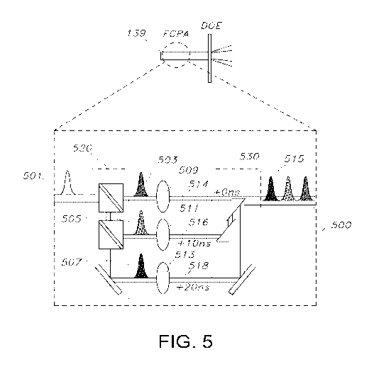
[0067] FIG. 5 illustrates a visualization of temporal multiplexing,
according to an
implementation. The laser module 139 may include an optical delay module 500
configured to
divide an optical pulse into multiple sub-pulses and introduce a relative time
delay between the
individual sub-pulses or an alternative way of introducing time delays between
sub-pulses. In
the example illustrated in FIG. 5, optical delay module 500 includes a three-
way beam splitter
520 that divides a main optical pulse 501 generated by laser module 139 into
three sub-pulses
503, 505, and 507. The three sub-pulses 503, 505 and 507 travel from the three-
way beam
splitter 520 to an optical combiner 530 via respective optical paths 514, 516
and 520. The
optical paths 514, 516 and 518 may simply be conduits for free space
propagation or they may
take the form of optical fibers. In each case, however, there is a difference
in the path lengths of
the optical paths so that the time required for each of the sub-pulses 503,
505, 507 to travel their
respective path is different.
[0068] Thus, the length of the second optical path 516 for the second sub-
pulse 505 can be
chosen to differ with respect to the length of the first optical path 514 for
the first sub-pulse 503
so that a first time delay of approximately 10 ns (nanoseconds) relative to
sub-pulse 503 is
introduced to sub-pulse 505. Similarly, a second time delay of approximately
20 ns relative to
sub-pulse 503 (and 10 ns relative to sub-pulse 505) is introduced to the third
sub-pulse 507 by
providing the third optical path 518 with an appropriate length with respect
to the first and
second optical paths.
[0069] The multiple sub-pulses 503, 505, and 507 are recombined by an
optical combiner
into a main beam output, or multiple outputs in close proximity by laser
module 139. The
optical combiner may be a combination of mirrors and/or transmissive elements
or other
elements arranged in a manner to redirect the sub-pulses in the same
direction. In particular, the
optical combiner can take the form of a polarizing or non-polarizing optical
beam splitter. For
convenience of discussion, the combined sub-pulses may be collectively
referred to as "optical
pulse 515" or a "set of sub-pulses."
[0070] The optical delay module 500 also preferably includes an arrangement
of focusing
lenses 509, 511, 513 disposed between the three-way beam splitter 520 and the
optical combiner
530. Sub-pulse 503 can be focused by lens 509 with a first divergence causing
it to be focused to
a first z-plane in sample 119. Sub-pulse 505 is focused by lens 511 with a
second divergence
17
CA 03042229 2019-04-29
WO 2018/081711 PCT/US2017/059044
causing it to be focused to a second z-plane in sample 119 that is different
than the first z-plane.
Sub-pulse 507 is focused by lens 513 with a third divergence causing it to be
focused to a third z-
plane in sample 119 that is different than the first and second z-planes. In
particular, these
focusing lenses and corresponding angle and position of the other optical
elements can be
arranged and chosen such that all pulses are focused at the same depth within
the target volume,
but directed at adjacent planes, as will be discussed further below with
respect to FIG. 6B.
[0071] In some examples, the optical pulse 515 may be sent to a beam
splitter, such as beam
splitter 107 illustrated in FIG. 1, to perform both spatial and temporal
multiplexing using
multiple sub-beams each comprising optical pulses, such as optical pulse 515,
that include
multiple sub-pulses. In some examples, an imaging device may use temporal
multiplexing with
spatial multiplexing, and not perform that beam splitting subsequent to
dividing main optical
pulse 501 into the series of time-delayed sub-pulses. Although the examples
illustrated in this
disclosure divide a main optical pulse into three sub-pulses for temporal
multiplexing, in some
examples a main optical pulse may instead be divided into two sub-pulses, or
may be divided
into 4 or more sub-pulses with a relative time delay between the sub-pulses.
[0072] Although the examples illustrated in this disclosure use a relative
time delay of
approximately 10 ns between the sub-pulses 503, 505, and 507 included in
optical pulse 515,
other amounts of time delay may be used. For example, where there is a
repetition rate of 4
MHz for main optical pulses 510, which corresponds to 250 ns between
successive pulses, for
three sub-pulses a relative time delay of up to 250 ns / 3, or approximately
80 ns may be used
between sub-pulses. The relative time delay should be longer than an expected
fluorescence
decay time, which is approximately 3 ns for the GCaMP fluorophores; thus, a
relative time delay
that is longer than approximately 3 ns is preferable for such applications. In
some examples, the
relative time delay may 100 ns or longer, 50 ns or longer, 20 ns or longer, 10
ns or longer, 5 ns or
longer, 2 ns or longer, or 1 ns or longer.
[0073] Depending on the desired amount of laser power at the sample 119, in
some examples
a 4 times multiplexing (e.g., 2 by 2) in the spatial domain can be selected,
with foci 109
separated by 500 p.m, in combination with up to 4 times multiplexing in the
temporal domain, to
simultaneously image several z-planes (similar to the 4 by 3 spatial and
temporal multiplexing
illustrated in FIGS. 6-9). With these improvements, a V-FOV 1 by 1 by 0.7 mm
can be achieved
18
CA 03042229 2019-04-29
WO 2018/081711 PCT/US2017/059044
with a frame rate of at least 3 Hz. Alternatively, temporal multiplexing can
be employed without
spatial multiplexing to image a V-FOV of 500 by 500 by 500 p.m with a frame
rate of at least 12
Hz. It is noted that even at higher pulse energies due to using a single
optical pulse for imaging
each voxel, bio-damage is not foreseen to be a limiting factor. This is
because the power is
distributed over a volume of approximately 5 by 5 by 5 p.m, which is about
1000 times larger
than the diffraction limited volume conventionally used in standard two-photon
microscopy.
[0074] FIG. 6A illustrates temporally multiplexed sub-pulses penetrating in
a sample,
according to an implementation. The temporally multiplexed sub-pulses 503,
505, and 507 of
FIG. 5 recombined into optical pulse 515 are sent to sample 119 much as
illustrated by the sub-
beam paths illustrated in FIG. 1. As a result of the different divergences
applied to each sub-
pulse 503, 505, and 507, each sub-pulse 503, 505, and 507 is focused to a
different respective z-
plane in sample 119. In the example illustrated in FIG. 6A, sub-pulse 503 is
focused to, and
produces a respective focus spot for exciting sample 119 on, a first z-plane
at depth Z1; sub-
pulse 505 (with a time delay of 10 ns relative to sub-pulse 503) is focused
to, and produces a
respective focus spot for exciting sample 119 on, a second z-plane at a depth
Z2 different than,
and greater than, depth Z1; and sub-pulse 507 (with a time delay of 20 ns
relative to sub-pulse
503 and a time delay of 10 ns relative to sub-pulse 505) is focused to, and
produces a respective
focus spot for exciting sample 119 on, a third z-plane at a depth Z3 different
than, and greater
than, depths Z1 and Z2. For convenience of discussion, this may also be
described as sub-pulse
503 penetrating to depth Z1, sub-pulse 505 penetrating to depth Z2, and sub-
pulse 507
penetrating to depth Z3.
[0075] To achieve focusing at different depths, as shown in FIG. 6A,
multiple temporal
focusing gratings 115' arranged at slightly different axial positions along
the beam propagation
are needed. A particular way of achieving this is to use a multi element
grating assembly 1300
that supports such multiple temporal focusing gratings 115' is shown in FIG.
13. The assembly
1300 generally includes a base 1302 formed with several legs 1304 extending
upwardly from the
base and a temporal focusing grating 115' supported at the end of each leg.
The legs 1304 have
different respective heights with respect to the base 1302 such that the
temporal focusing
gratings 115' will be positioned at different axial positions along the beam
path 1306, (which
will be perpendicular to the base 1302 when the assembly 1300 is installed in
the system).
19
CA 03042229 2019-04-29
WO 2018/081711 PCT/US2017/059044
[0076] FIG. 6B illustrates temporally multiplexed sub-pulses penetrating in
a sample,
according to an implementation using a single grating 115, as shown in FIG. 1.
The temporally
multiplexed sub-pulses 503, 505, and 507 of FIG. 5 are sent to sample 119 such
that the sub-
pulses are focused at the same depth of the sample, but are directed at
adjacent planes. In the
example illustrated in FIG. 6B, sub-pulse 503 is focused to, and produces a
respective focus spot
for exciting sample 119 on, a first planar region at depth Z1; sub-pulse 505
(with a time delay of
ns relative to sub-pulse 503) is focused to, and produces a respective focus
spot for exciting
sample 119 on, a second planar region at the same depth Z1; and sub-pulse 507
(with a time
delay of 20 ns relative to sub-pulse 503 and a time delay of 10 ns relative to
sub-pulse 505) is
focused to, and produces a respective focus spot for exciting sample 119 on, a
third planar region
at the same depth Z1 as the first and second planar regions.
[0077] Although only three temporally multiplexed sub-pulses are shown in
FIGS. 6A and
6B, the number of temporally multiplexed sub-pulses is not limited to three;
for example, four or
more sub-pulses with respective delays and focusing depths/planar regions can
be used.
Although FIG. 6A illustrates an example in which sub-pulses are focused to
increasing depth in
correlation with increasing time delay, focus depth and time delay may not be
related in this
manner in some examples.
[0078] FIGS. 7, 8, and 9 illustrate visualizations of a temporally and
spatially multiplexed
beam, comprising multiple sub-beams each delivering optical pulses that each
comprise multiple
sub-pulses, penetrating in a sample, according to an implementation.
Specifically, these
visualizations relate to an imaging system using 4 by 3 spatial and temporal
multiplexing, with 4
sub-beams 515a, 515b, 515c, and 515d each directed to a respect part (in these
examples,
respective quadrants Q1-Q4) and each repeatedly delivering sets of 3 sub-
pulses that are
relatively time delayed and focused at respective and different depths (in
respective z-planes).
The spatial multiplexing resulting in the four sub-beams 515a, 515b, 515c, and
515d may be
performed as described above, such as with respect to FIGS. 1-3, and the
temporal multiplexing
resulting in optical pulses each comprising sets of three sub-pulses may be
performed as
described above, such as with respect to FIGS. 5 and 6. Deflection of the sub-
beams 515a, 515b,
515c, and 515d to scan voxels within their respective parts of sample 119 may
be performed as
described above, such as with respect to FIGS. 1, 3, and 4. Although only four
spatially
multiplexed sub-beams 515a, 515b, 515c, and 515d are shown, the number of
spatially
CA 03042229 2019-04-29
WO 2018/081711 PCT/US2017/059044
multiplexed sub-beams is not limited to four. The sample 119 can be divided
into more than four
parts along with a corresponding increase in the number of sub-beams.
Likewise, more than
three sub-pulses may be included in each set of sub-pulses, with a
corresponding increase in the
number of z-planes
[0079] Each sub-beam 515a-515d comprises and repeatedly delivers temporally
multiplexed
optical pulses each comprising multiple sub-pulses, such as the temporally
multiplexed sub-
pulses 503, 505, and 507 included in optical pulse 515 illustrated in FIGS. 5
and 6, and the sub-
pulses penetrate into sample 119 at respective depths Z1 (corresponding to z-
plane #1), Z2
(corresponding to z-plane #2), and Z3 (corresponding to z-plane #3). In
addition, each sub-beam
515a-515d is used to scan one respective part or division (e.g., quarter Ql,
Q2, Q3, and Q4) of
the sample 119. As a result, a three dimensional scanning of the sample can be
provided in which
full 2D scanning sequence performed by an optical scanner (such as scanner 111
discussed in
connection with FIG. 1 performing one pass of the sinuous pattern 313
illustrated in FIG. 3)
results in the sample 119 being scanned by scanning 12 focus points through
the imaging planes
at three different depths shown as Z-plane#1, Z-plane#2, and Z-plane#3.
[0080] In FIG. 8, after imaging the first set of three planes z-plane #1, z-
plane #2, and z-
plane #3, the focus spots for the sub-beams 515a-515d continue penetrating
into the sample 119,
such that the Z-planes move in the z direction and, for example, Z-plane #1
moves from its
previous location to a new location Z-plane #4. Similarly, Z-plane #2 and Z-
plane #3 move to
new locations, Z-plane #5 and Z-plane #6, respectively. Such changes in
location may be
continued such that the voxels between z-plane #1 and z-plane #2 are all
scanned.
[0081] In FIG. 9, another of moving the planes is illustrated. Rather than
moving z-plane #1
to a new position between z-plane #1 and z-plane #2, z-plane #1 is moved to z-
plane #7 at a
depth greater than z-plane #3.
[0082] FIGS. 10A-10B illustrate scanning a focused spot for an optical
pulse (whether a
temporally multiplexed sub-pulse or not), according to an implementation. A
small sized
temporally focused spot 1021 can be scanned over the imaging field-of-view
(FOV) 1023. The
FOV 1023 can be a slice of sample 119, the image of which is captured by the
imaging system
100. The penetration of optical pulses (whether a temporally multiplexed sub-
pulses or not) into
the depth of sample 119 can scan a stack 1031 of slices of sample 119. Each
slice 1023 is similar
21
CA 03042229 2019-04-29
WO 2018/081711 PCT/US2017/059044
to a Z-plane in FIGS. 7 to 9. The focused spot 1021 may be similar to spots
201, 203, 205, and
207 shown in FIG. 2 and FIG. 7. For example, the size of the temporally
focused spot 1021 can
be approximately 5 by 5 by 5 p.m. Due to light sculpting, the excitation of
sample 119 can be
isotropically confined, hence providing single neuron optical sectioning
capability in the axial
direction x, y, or z as shown by coordinate systems 1025, 1027, and 1029.
Volume acquisition
can be performed by axial scanning of the sample 119. The axial scanning can
cause the excited
fluorescence to be detected by a photomultiplier tube (PMT) 127 (shown in FIG.
1). As
previously shown in FIG. 3, the scanning of slice 1023 of sample 119 by the
temporally focused
spot 1021 can be performed with a sinuous pattern 1033.
[0083] In some implementations, various modalities of wide-field temporal
focusing based
microscopy can be established. A scanning variant of temporal focusing, aptly
named scanned
temporal focusing is described herein. The scanned temporal focusing can be
combined with
latest state-of-the-art fiber-based laser amplifiers as well as spatial and
temporal multiplexing, to
circumvent and optimize design in two-photon laser scanning microscopy. For
example, by light-
sculpting an excitation volume of 5 by 5 by 5 p.m and rapidly scanning the
excitation volume
over the image FOV, plane acquisition speeds can dramatically be improved,
without sacrificing
single-neuron resolution. Matching the repetition rate (e.g., laser pulses per
second) to the
number of acquired voxels per second further provides optimal signal-to-noise
ratios, as only a
single laser pulse can be used to excite the sample during the image pixel
acquisition, as shown
in equation (1), whereby shot-noise is further minimized.
[0084] FIG. 11 illustrates in vivo volume stack acquisition in auditory
cortex of mouse
expressing nuclear-confined red fluorescent protein, according to an
implementation. The stack
1101 can be acquired with 5 p.m spot scanned temporal focusing configuration
and scanner 111
(e.g., galvanometric mirrors) as shown in FIG. 1. Average power of the laser
module 139 can be
between 25 and 50 mW, depending on the depth. Scale 1103 displays the depth
from 100 p.m to
600 p.m. Images 1105a, 1105b, 1105c and 1105d are magnified images of stack
1101. Neuronal
nuclei are clearly distinguishable, even at depth beyond 500um, as shown in
image 1105d.
[0085] FIG. 12 is a block diagram that illustrates a computer system 1200
upon which
aspects of this disclosure may be implemented, such as, but not limited to,
multi-channel
counting card (dmCC) 129 and computing device 131. Computer system 1200
includes a bus
22
CA 03042229 2019-04-29
WO 2018/081711 PCT/US2017/059044
1202 or other communication mechanism for communicating information, and a
processor 1204
coupled with bus 1202 for processing information. Computer system 1200 also
includes a main
memory 1206, such as a random access memory (RAM) or other dynamic storage
device,
coupled to bus 1202 for storing information and instructions to be executed by
processor 1204.
Main memory 1206 also may be used for storing temporary variables or other
intermediate
information during execution of instructions to be executed by processor 1204.
Computer
system 1200 further includes a read only memory (ROM) 1208 or other static
storage device
coupled to bus 1202 for storing static information and instructions for
processor 1204. A storage
device 1210, such as a magnetic disk or optical disk, is provided and coupled
to bus 1202 for
storing information and instructions.
[0086] Various other actions may be performed in response to identifying
WLAN issues. In
some situations, a replacement wireless router may be automatically dispatched
to a customer in
response to detecting a bad wireless router. In some situations, a customer
may be automatically
notified (through, for example, emails and pop-up windows) about potential
WLAN issues or
potentially problematic devices that may be impacting service quality at a
customer location. In
some situations, a system may automatically recommend WLAN changes, such as
upgrading a
wireless router, upgrading a client device, suggesting moving or placement of
a wireless router
or client device, and suggesting use of wireless network repeaters. In some
situations,
information about WLAN conditions may be automatically included in a monthly
bill or online
account webpage. In some situations, network conditions of customers who
contact customer
support may be gathered in a database, and used to dynamically and/or
automatically identify
reasons for customer dissatisfaction; for example, wireless router models may
be identified and
assessed for compatibility with other network hardware and client devices, and
information
about CPE 132 (which includes an integrated wireless router) may be collected
to improve
quality over time.
[0087] Computer system 1200 may be coupled via bus 1202 to a display 1212,
such as a
cathode ray tube (CRT) or liquid crystal display (LCD), for displaying
information to a computer
user. An input device 1214, including alphanumeric and other keys, is coupled
to bus 1202 for
communicating information and command selections to processor 1204. Another
type of user
input device is cursor control 1216, such as a mouse, a trackball, or cursor
direction keys for
communicating direction information and command selections to processor 1204
and for
23
CA 03042229 2019-04-29
WO 2018/081711 PCT/US2017/059044
controlling cursor movement on display 1212. This input device typically has
two degrees of
freedom in two axes, a first axis (e.g., x) and a second axis (e.g., y), that
allows the device to
specify positions in a plane. Another type of user input device is a
touchscreen, which generally
combines display 1212 with hardware that registers touches upon display 1212.
[0088] This disclosure is related to the use of computer systems such as
computer system
1200 for implementing the techniques described herein. In some examples, those
techniques are
performed by computer system 1200 in response to processor 1204 executing one
or more
sequences of one or more instructions contained in main memory 1206. Such
instructions may
be read into main memory 1206 from another machine-readable medium, such as
storage device
1210. Execution of the sequences of instructions contained in main memory 1206
causes
processor 1204 to perform the process steps described herein. In some
examples, hard-wired
circuitry may be used in place of or in combination with software instructions
to implement the
various aspects of this disclosure. Thus, implementations are not limited to
any specific
combination of hardware circuitry and software.
[0089] The term "machine-readable medium" as used herein refers to any
medium that
participates in providing data that causes a machine to operation in a
specific fashion. In some
examples implemented using computer system 1200, various machine-readable
media are
involved, for example, in providing instructions to processor 1204 for
execution. Such a
medium may take many forms, including but not limited to, non-volatile media,
volatile media,
and transmission media. Non-volatile media includes, for example, optical or
magnetic disks,
such as storage device 1210. Volatile media includes dynamic memory, such as
main memory
1206. Transmission media includes coaxial cables, copper wire and fiber
optics, including the
wires that comprise bus 1202. Transmission media can also take the form of
acoustic or light
waves, such as those generated during radio-wave and infra-red data
communications. All such
media must be tangible to enable the instructions carried by the media to be
detected by a
physical mechanism that reads the instructions into a machine.
[0090] Common forms of machine-readable media include, for example, a
floppy disk, a
flexible disk, hard disk, magnetic tape, or any other magnetic medium, a CD-
ROM, any other
optical medium, punchcards, papertape, any other physical medium with patterns
of holes, a
24
CA 03042229 2019-04-29
WO 2018/081711 PCT/US2017/059044
RAM, a PROM, and EPROM, a FLASH-EPROM, any other memory chip or cartridge, a
carrier
wave as described hereinafter, or any other medium from which a computer can
read.
[0091] Various forms of machine-readable media may be involved in carrying
one or more
sequences of one or more instructions to processor 1204 for execution. For
example, the
instructions may initially be carried on a magnetic disk of a remote computer.
The remote
computer can load the instructions into its dynamic memory and send the
instructions over a
telephone line using a modem. A modem local to computer system 1200 can
receive the data on
the telephone line and use an infra-red transmitter to convert the data to an
infra-red signal. An
infra-red detector can receive the data carried in the infra-red signal and
appropriate circuitry can
place the data on bus 1202. Bus 1202 carries the data to main memory 1206,
from which
processor 1204 retrieves and executes the instructions. The instructions
received by main
memory 1206 may optionally be stored on storage device 1210 either before or
after execution
by processor 1204.
[0092] Computer system 1200 also includes a communication interface 1218
coupled to bus
1202. Communication interface 1218 provides a two-way data communication
coupling to a
network link 1220 that is connected to a local network 1222. For example,
communication
interface 1218 may be an integrated services digital network (ISDN) card or a
modem to provide
a data communication connection to a corresponding type of telephone line. As
another
example, communication interface 1218 may be a local area network (LAN) card
to provide a
data communication connection to a compatible LAN. Wireless links may also be
implemented.
In any such implementation, communication interface 1218 sends and receives
electrical,
electromagnetic or optical signals that carry digital data streams
representing various types of
information.
[0093] Network link 1220 typically provides data communication through one
or more
networks to other data devices. For example, network link 1220 may provide a
connection
through local network 1222 to a host computer 1224 or to data equipment
operated by an Internet
Service Provider (ISP) 1226. ISP 1226 in turn provides data communication
services through
the world wide packet data communication network now commonly referred to as
the "Internet"
1228. Local network 1222 and Internet 1228 both use electrical,
electromagnetic or optical
signals that carry digital data streams. The signals through the various
networks and the signals
CA 03042229 2019-04-29
WO 2018/081711 PCT/US2017/059044
on network link 1220 and through communication interface 1218, which carry the
digital data to
and from computer system 1200, are exemplary forms of carrier waves
transporting the
information.
[0094] Computer system 1200 can send messages and receive data, including
program code,
through the network(s), network link 1220 and communication interface 1218. In
the Internet
example, a server 1230 might transmit a requested code for an application
program through
Internet 1228, ISP 1226, local network 1222 and communication interface 1218.
[0095] The received code may be executed by processor 1204 as it is
received, and/or stored
in storage device 1210, or other non-volatile storage for later execution. In
this manner,
computer system 1200 may obtain application code in the form of a carrier
wave.
[0096] The separation of various components in the examples described above
should not be
understood as requiring such separation in all examples, and it should be
understood that the
described components and systems can generally be integrated together in a
single package, or
into multiple systems.
[0097] While the foregoing has described what are considered to be the best
mode and/or
other examples, it is understood that various modifications may be made
therein and that the
subject matter disclosed herein may be implemented in various forms and
examples, and that the
teachings may be applied in numerous applications, only some of which have
been described
herein. It is intended by the following claims to claim any and all
applications, modifications and
variations that fall within the true scope of the present teachings.
[0098] Unless otherwise stated, all measurements, values, ratings,
positions, magnitudes,
sizes, and other specifications that are set forth in this specification,
including in the claims that
follow, are approximate, not exact. They are intended to have a reasonable
range that is
consistent with the functions to which they relate and with what is customary
in the technology
to which they pertain.
[0099] Except as stated immediately above, nothing that has been stated or
illustrated is
intended or should be interpreted to cause a dedication of any component,
step, feature, object,
benefit, advantage, or equivalent to the public, regardless of whether it is
or is not recited in the
claims.
26
CA 03042229 2019-04-29
WO 2018/081711 PCT/US2017/059044
[00100] It will be understood that the terms and expressions used herein have
the ordinary
meaning as is accorded to such terms and expressions with respect to their
corresponding
respective areas of inquiry and study except where specific meanings have
otherwise been set
forth herein. Relational terms such as first and second and the like may be
used solely to
distinguish one entity or action from another without necessarily requiring or
implying any
actual such relationship or order between such entities or actions. The terms
"comprises,"
"comprising," or any other variation thereof, are intended to cover a non-
exclusive inclusion,
such that a process, method, article, or apparatus that comprises a list of
elements does not
include only those elements but may include other elements not expressly
listed or inherent to
such process, method, article, or apparatus. An element proceeded by "a" or
"an" does not,
without further constraints, preclude the existence of additional identical
elements in the process,
method, article, or apparatus that comprises the element.
[00101] To the extent the aforementioned embodiments collect, store, or employ
personal
information provided by individuals, it should be understood that such
information shall be used
in accordance with all applicable laws concerning protection of personal
information. Additionally, the collection, storage, and use of such
information may be subject to
consent of the individual to such activity, for example, through well known
"opt-in" or "opt-out"
processes as may be appropriate for the situation and type of information.
Storage and use of
personal information may be in an appropriately secure manner reflective of
the type of
information, for example, through various encryption and anonymization
techniques for
particularly sensitive information.
[00102] In the foregoing Detailed Description, it can be seen that various
features are grouped
together in various examples for the purpose of streamlining the disclosure.
This method of
disclosure is not to be interpreted as reflecting an intention that the claims
require more features
than are expressly recited in each claim. Rather, as the following claims
reflect, inventive
subject matter lies in less than all features of a single disclosed example.
Thus the following
claims are hereby incorporated into the Detailed Description, with each claim
standing on its
own as a separately claimed subject matter.
27