Note : Les descriptions sont présentées dans la langue officielle dans laquelle elles ont été soumises.
CA 03042355 2019-04-30
WO 2018/116128 PCT/IB2017/058061
SYSTEMS AND METHODS FOR WIDE FIELD-OF-VIEW
OPTICAL COHERENCE TOMOGRAPHY
FIELD
[0001] The present disclosure relates to medical device imaging systems,
including
optical coherence tomography (OCT) systems.
BACKGROUND
[0002] Optical Coherence Tomography (OCT) is an imaging technique widely
adopted in
the biomedical fields, including ophthalmology. OCT systems perfoiin high-
resolution, cross
sectional imaging in semitransparent samples (such as biological tissues) by
measuring the
echo time delay of reflected light. OCT is often used by ophthalmic surgeons
to assist with
precision cutting and/or removal of tissues such as the vitreous. Providing
wide-field-of-
view OCT imaging across a curved surface such as a retina can be challenging
because the
images become curved and distorted at wide scan angles, particularly in highly
myopic
patients. Accordingly, there exists a need for improved wide-field-of-view OCT
imaging in
the ophthalmic context.
SUMMARY
[0003] In certain embodiments, an optical coherence tomography (OCT) system
includes
a light source configured to generate an OCT beam and a beam splitter,
configured to split
the OCT beam into a reference beam and an imaging beam, direct the reference
beam toward
a reference reflector, and direct the imaging beam toward a scanner. The
system also
includes a linear actuator, such as a piezoelectric actuator or voice coil
actuator, configured to
move the reference reflector to adjust the length of the reference beam and
the scanner,
configured to scan the imaging beam onto a target surface at a plurality of
scan angles,
wherein the scanner and target surface are separated by a sample distance that
varies at each
of the scan angles. The system further includes an OCT controller comprising a
processor
and instructions stored on a memory, the instructions executable by the
processor to cause the
OCT controller to generate signals to cause the scanner to scan the imaging
beam at each of
the scan angles at a first scan rate, and cause the actuator to adjust the
length of the reference
beam during the scan synchronously with the scan rate to match the variation
in sample
distance at each of the scan angles.
CA 03042355 2019-04-30
WO 2018/116128 PCT/IB2017/058061
[0004] In certain embodiments, the scan rate is between 200 Hz and 400 Hz,
or is at least
300 Hz. The scanner may be configured to scan the imaging beam at each of the
scan angles
according to a raster pattern, and the raster pattern may generate a B-scan at
least 12 mm in
length or at least 16 mm in length.
[0005] In certain embodiments, the linear actuator is configured to
translate the reference
reflector at least 2mm in a direction parallel to the reference beam. The
linear actuator may
further be configured to translate the reference reflector at least 4mm in a
direction parallel to
the reference beam.
[0006] The OCT system may comprise a spectral-domain OCT (SD-OCT) system or
a
swept-source OCT (SS-OCT) system.
[0007] In certain embodiments, an optical coherence tomography (OCT)
system,
comprises a light source configured to generate an OCT beam, and a beam
splitter,
configured to split the OCT beam into a reference beam and an imaging beam,
direct the
reference beam toward a reference reflector, and direct the imaging beam
toward a scanner.
The system also includes a linear actuator, such as a piezoelectric actuator
or voice coil
actuator, configured to move the reference reflector to change the length of
the reference
beam, and the scanner, configured to scan the imaging beam onto a target
surface over a
plurality of scan angles, wherein the scanner and target surface are separated
by a first sample
distance at a first scan angle and a second sample distance at a second scan
angle. The
system includes an OCT controller comprising a processor and instructions
stored on a
memory, the instructions executable by the processor to cause the OCT
controller to generate
signals to cause the scanner to scan the imaging beam onto the target surface
at the first scan
angle and the second scan angle according to a scan rate, and cause the
actuator to move the
reference reflector synchronously with the scan rate while the scanner scans
the imaging
beam onto the target surface, thereby adjusting the length of the reference
beam to account
for a difference between the first sample distance and the second sample
distance. The
system further includes a detector configured to receive the reference beam
reflected by the
reference reflector and the imaging beam reflected by the target surface, and
output an
interference signal based on the received reference beam and the imaging beam.
[0008] In certain embodiments, the linear actuator comprises a
piezoelectric stack or
voice coil configured to translate the reference reflector at least 2mm in a
direction parallel to
the reference beam. In certain embodiments, the first scan angle and the
second scan angle
are separated by at least 20 degrees. In certain embodiments, the scan
generates a B-scan at
2
CA 03042355 2019-04-30
WO 2018/116128 PCT/IB2017/058061
least 12 mm in length. The OCT system may comprise a spectral-domain OCT (SD-
OCT)
system or a swept-source OCT (SS-OCT) system.
[0009] According to certain embodiments, an optical coherence tomography
(OCT)
system comprises a light source, configured to generate an OCT beam, and a
beam splitter,
configured to split the OCT beam into a reference beam and an imaging beam,
direct the
reference beam toward a reference reflector, and direct the imaging beam
toward a scanner.
The system further includes a linear actuator, configured to translate the
reference reflector at
least 2mm in a direction parallel to the reference beam and the scanner,
configured to scan
the imaging beam onto a target surface at a plurality of scan angles. The
system includes an
OCT controller comprising a processor and instructions stored on a memory, the
instructions
executable by the processor to cause the OCT controller to generate signals to
cause the
scanner to scan the imaging beam at each of the scan angles at a first scan
rate, and cause the
actuator to translate the reference reflector synchronously with the scan
rate, such that a path
length of the reference beam is maintained within a tolerance range of a path
length of the
imaging beam throughout the scan.
[0010] In certain embodiments, the tolerance range is less than 0.5mm or 1
mm. The scan
rate may be between 200 Hz and 400 Hz. Further, the scanner may be configured
to scan the
imaging beam at each of the scan angles according to a raster pattern. The
linear actuator may
be a piezoelectric stack or voice coil configured to translate the reference
reflector at least
2mm in a direction parallel to the reference beam.
[0011] Certain embodiments may provide one or more technical advantages.
For
example, improved OCT imaging systems according to the disclosure may provide
ultra-wide
field-of-view OCT imaging with reduced distortion. Certain embodiments
generate OCT
images in which a target surface is centered throughout an OCT image window,
despite
relative variations in target depth. Thus, certain embodiments provide
improved live OCT
imaging of curved surfaces, such as high-myopia retinal surfaces. These and
other
advantages will be apparent to those skilled in the art in view of the present
drawings and
specification.
3
CA 03042355 2019-04-30
WO 2018/116128 PCT/IB2017/058061
BRIEF DESCRIPTION OF THE DRAWINGS
[0012] For a more complete understanding of the present disclosure and the
advantages
thereof, reference is now made to the following description taken in
conjunction with the
accompanying drawings in which like reference numerals indicate like features
and wherein:
[0013] FIG. 1 illustrates a block diagram of a conventional OCT system;
[0014] FIG. 2 illustrates a retinal image generated by a conventional OCT
imaging
system;
[0015] FIG. 3 illustrates a retinal image generated by an improved OCT
imaging system
according to certain embodiments;
[0016] FIG. 4 illustrates a block diagram of an improved OCT imaging system
according
to certain embodiments; and
[0017] FIG. 5 illustrates a method performed by an improved OCT imaging
system
according to certain embodiments.
[0018] One skilled in the art will understand that the drawings, described
below, are for
illustration purposes only, and are not intended to limit the scope of
applicant's disclosure.
4
CA 03042355 2019-04-30
WO 2018/116128 PCT/IB2017/058061
DETAILED DESCRIPTION
[0019] For the purposes of promoting an understanding of the principles of
the present
disclosure, reference will now be made to the embodiments illustrated in the
drawings, and
specific language will be used to describe the same. It will nevertheless be
understood that
no limitation of the scope of the disclosure is intended. Alterations and
further modifications
to the described systems, devices, and methods, and any further application of
the principles
of the present disclosure are contemplated as would normally occur to one
skilled in the art to
which the disclosure relates. In particular, it is contemplated that the
systems, devices, and/or
methods described with respect to one embodiment may be combined with the
features,
components, and/or steps described with respect to other embodiments of the
present
disclosure. For the sake of brevity, however, the numerous iterations of these
combinations
will not be described separately. For simplicity, in some instances the same
reference
numbers are used throughout the drawings to refer to the same or like parts.
[0020] Optical coherence tomographic (OCT) imaging systems are useful in an
array of
biological applications including ophthalmology, dentistry, cardiology,
gastroenterology, and
others. The general design and principles of OCT systems are known and
described in, for
example: (a) "Signal Processing Overview of Optical Coherence Tomography
Systems for
Medical Imaging," Texas Instruments White Paper SPRABB9 (June 2010) and (b)
"Biomedical Optical Imaging," Progress Report of the Research Laboratory of
Electronics at
MIT, No. 152 (2009-2010), each of which is incorporated by reference herein in
its entirety.
[0021] FIG. 1 is a simple schematic illustration of components in a
conventional OCT
system 100. System 100 may comprise a spectral-domain OCT (SD-OCT) system or
swept-
source (SS-OCT) system. In general, the components of such systems 100 are
well-known to
the skilled artisan. Among other things, system 100 includes a light source
102, beam
splitter/combiner 104, reference reflector 108, scanner 120, and a detector
124. Light source
102 may comprise any suitable low-coherence light source such as a super-
luminescent
diode, ultrashort (e.g., femtosecond) pulsed laser, or supercontinuum laser,
and may comprise
a frequency-swept or tunable laser in certain examples, such as SS-OCT
systems. Beam
splitter 104 may comprise a non-polarized beam splitter for splitting the OCT
beam into an
imaging beam and a reference beam and combining or directing reflected imaging
and
reference light toward detector 124. Reference reflector 108 is typically a
mirror, but may
comprise any suitable component which reflects the reference beam 106 toward
the detector
124. Scanner 120 may comprise one or more galvanometer-controlled mirrors to
scan the
CA 03042355 2019-04-30
WO 2018/116128 PCT/IB2017/058061
imaging beam in the x-y plane toward a target or sample, such as retina 114
(when discussing
the object being imaged, the terms "target" and "sample" are used
interchangeably herein).
In certain embodiments, scanner 120 may additionally include focusing optics
to scan the
imaging beam in a z-direction. Scanner 120 may comprise any suitable scanning
mirror
arrangement. Alternatively, scanner 120 may comprise any suitable scanner
components,
such as microelectromechanical systems (MEMS) or a resonant scanner. The
imaging beam
scanned by scanner 120 is directed through optical elements 122 which may
comprise
focusing and/or collimating lenses. Detector 124 comprises an interferometer
which receives
the imaging beam reflected from the target and the reference beam reflected
from the
reflector 108 and outputs an interference signal from which an OCT image can
be generated.
Particular components included in detector 124 depend on the type of OCT
system and may
include any suitable combination of spectrometers, photodetectors, array
detectors, analog-to-
digital converters (ADCs), diffraction grating(s), or other components known
to those skilled
in the art. For example, detector 124 in an SD-OCT system may include a
diffraction grating,
lenses, and an array detector such as a charge-coupled device (CCD). As
another example,
detector 124 in an SS-OCT system may include a photodetector and a analog-to-
digital
converter.
[0022] System 100 may include an OCT controller (not shown in FIG. 1)
comprising
hardware, firmware, and software configured to control components of system
100 to acquire
and display OCT images of a target. System 100 may additionally include one or
more
displays (not shown) to present OCT images generated by the OCT controller. In
various
examples, the display may include any one or more monitors, projectors,
oculars, heads-up
displays, screens, glasses, goggles, etc. The OCT images may be displayed as
2D or 3D
images.
[0023] In operation, light source 102 emits a low-coherence light beam
directed to beam
splitter 104, which splits the light into a reference beam 106 directed
through a reference arm
(which may comprise any suitable transmission and focusing optics including
optical fibers)
toward reflector 108 and an imaging beam 110 directed through an imaging arm
(which
likewise may include any suitable transmission and focusing optics including
optical fibers)
toward a scanner 120. Scanner 120 (under the control of the OCT controller)
may scan the
imaging beam toward optics 122 and the lens 112 of eye 101 according to a scan
pattern (e.g.,
raster scan, radial scan, cube scan, circle group scan, line group scan, etc.)
to generate the
desired scan (e.g., A-scan, B-scan, or C-scan). A depth-resolved axial scan (A-
scan)
comprises a measurement of the light signal interference at a point. Cross-
sectional images
6
CA 03042355 2019-04-30
WO 2018/116128 PCT/IB2017/058061
(B-scans) may be generated by scanning the OCT beam across the tissue surface
and
acquiring multiple axial measurements over a line, curve, circle, etc. A 3D
image may be
constructed from a series of B-scans generated over an area of the tissue
surface. Scanning
may be repeated at a scan rate or frequency to generate live or real-time OCT
images which
may useful for pre-operative diagnostics as well as intra-operative guidance.
[0024] Imaging beam light reflected by the retina 114 and reference beam
light reflected
by the reflector 108 may be received at detector 124, which interferes the
back-reflected or
backscattered imaging beam with the reference beam to generate OCT images.
Interference
occurs when the path length of the reference beam (i.e., the distance imaging
light travels
between source 102 and reflector 108) and the path length of the imaging beam
(i.e., the
stance imaging light travels between source 102 and a target such as retina
114) are matched
within the coherence length of the light emitted by light source 102. This
interference signal
conveys information about the target at a depth which corresponds to the
reference beam path
length.
[0025] Accordingly, OCT systems are calibrated prior to use by setting the
reference
beam path length according to the target depth, so that the path length of the
reference beam
is approximately equal to the path length of the imaging beam at the target
depth. The
difference between the path length of the reference beam and the path length
of the imaging
beam at the target depth in an OCT system is referred to as the optical path
difference (OPD).
Ideally, OPD is zero, though absolute precision necessary in practice. Thus,
in the example
of FIG. 1, if the primary target depth is the center surface of retina 114,
the reference beam
path length (illustrated as reference beam distance Rd) is set to match the
path length of the
imaging beam measured to the center of retina 114 (illustrated as center
sample distance
Sdc). In conventional spectral-domain OCT (SD-OCT) systems or swept-source OCT
(SS-
OCT) systems such as system 100, this reference beam path length is fixed at
the outset of the
imaging procedure and remains fixed throughout the OCT scan.
[0026] It is noted that OCT imaging systems may be broadly classified into
time-domain
OCT (TD-OCT) systems, SD-OCT systems, and SS-OCT systems. TD-OCT systems
obtain
an interference pattern by moving a reference mirror to vary the reference
path length at each
point in a scan pattern. That is, at a given point in a TD-OCT scan pattern,
the reference
mirror in the reference arm must be moved to change the reference path length.
The
movement of this mirror in the reference aim of TD-OCT systems is a speed
gating factor,
because the mirror must be moved through a distance (z-range) at each (x,y)
point of an OCT
scan pattern in order to generate the required interference signal.
7
CA 03042355 2019-04-30
WO 2018/116128 PCT/IB2017/058061
[0027] Conventional SD-OCT and SS-OCT systems operate according to
different
principles and avoid this speed gate by employing a fixed-position reference
reflector which
requires no mechanical scanning of the reference path at any point in a scan
pattern. SD-
OCT systems use a broadband light source and obtain depth infoiniation
measuring the
spectral density in the sample arm using a spectrometer. SS-OCT systems
utilize a
frequency-swept laser or tunable laser and a single-point detector. In both SD-
OCT and SS-
OCT systems, OCT images are generated from the received interference signal
using fast
Fourier transforms. Accordingly, the reference reflector position is fixed at
each (x,y) point
of an OCT scan pattern executed by conventional SD-OCT and SS-OCT systems.
[0028] Typical SD-OCT and SS-OCT systems for posterior-segment imaging may
scan
between 200 and 40 (e.g., 10 or 20 from a center position) across a
retinal target. Over
such scan angles, the targeted portion of the retina may be imaged without
significant
distortion because variations in the depth of the retina attributable to
retinal curvature are not
significant. Stated differently, the variations in OPD resulting from retinal
curvature are
typically not very significant across smaller scan angle ranges (e.g., between
20 and 40 ).
However, over wider fields-of-view (e.g., 40 or more), the curvature of the
retina across the
imaged area results in significant variation in OPD, particularly in high-
myopia patients.
This variation in OPD can cause distortion in the OCT image.
[0029] FIG. 2 illustrates an example wide field-of-view B-scan
(approximately 40 ) of a
retina generated by a conventional SD-OCT or SS-OCT system. As illustrated in
this
example, the image of the retina is curved in a wide "U" shape, such that the
edges appear to
"fall off' the image range on each side. This distortion results from
variations in the OPD
attributable to retinal curvature and the fixed reference beam path length.
That is, the
reference beam path length is calibrated to image at a particular depth, e.g.,
so that the OPD
is approximately zero at the center of the retina. However, the natural
curvature of the retina
results in the fundus surface outside that depth because the OPD changes as
the imaging
beam is scanned across tissues which are closer to scanner 120.
[0030] This characteristic "U"-shaped distortion is undesirable and
problematic. For
example, during a procedure, a surgeon may "zoom in" to a particular area of
the retina, such
one of windows A-C. Each of windows A-C represents an image area for
enlargement,
though it is noted that any portion of the image may be enlarged. Although the
retinal image
is generally horizontal in window B, windows A and C each display a portion of
the retinal
surface with a steep angular orientation in the image window. This angular
orientation
8
CA 03042355 2019-04-30
WO 2018/116128 PCT/IB2017/058061
results in distortion and truncation of the retinal image and, among other
things, it makes the
image more difficult to read and use, particularly in an intra-operative
context.
[0031] Embodiments of the present disclosure address this problem by
modulating the
position of a reference reflector, thereby adjusting the reference beam path
length to account
for or match variations of the target depth within a scan and "flatten" out
the OCT image as
shown in FIG. 3. In other words, the position of the reference reflector is
modulated so that
the system OPD is maintained at or near zero throughout a scan pattern.
Compared against
FIG. 2, image windows A and C of FIG. 3 display larger portions of the retina
with increased
clarity and reduced distortion. Accordingly, improved OCT systems according to
the present
disclosure facilitate high-speed (e.g., 200-400+ Hz), wide-angle scans (e.g.,
200- 900
sweeps) across large retinal cross-sections and provide improved images that
are substantially
free of distortion and easy to use during a surgical procedure.
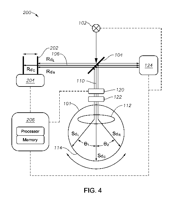
[0032] FIG. 4 illustrates an example of an improved OCT imaging system 200
to
generate images as shown in FIG. 3. System 200 may be a probe-based system, a
stand-alone
imaging system, or an imaging system integrated with other components, such as
a surgical
microscope. It is noted that FIG. 4 does not attempt to exhaustively
illustrate all components
of an OCT system, nor is it drawn to scale. Rather, it is provided to
qualitatively illustrate
how the optical path of the imaging beam 110 varies according to scan angle.
[0033] System 200 comprises an SD-OCT or SS-OCT imaging system which
includes
many of the same components as system 100 (like numerals indicate like
components). In
particular, system 200 includes a light source 102, beam splitter/combiner
104, scanner 120,
and a detector 124. Light source 102 may comprise any suitable low-coherence
light source
such as a super-luminescent diode, ultrashort (e.g., femtosecond) pulsed
laser, or
supercontinuum laser, and may comprise a frequency-swept or tunable laser in
certain
examples, such as SS-OCT systems. Beam splitter 104 may comprise a non-
polarized beam
splitter for splitting the OCT beam into an imaging beam transmitted through
the sample arm
and a reference beam transmitted through the reference aim (sometimes referred
to as a delay
line) of the OCT system. Beam splitter 104 also receives and combines
reflected imaging
light (reflected by the sample, such as eye 114) and reference light
(reflected by reference
reflector 202) toward detector 124. Scanner 120 may comprise one or more
galvanometer-
controlled mirrors to scan the imaging beam in the x-y plane through a sample
aim of the
OCT system toward the sample, such as retina 114. Scanner 120 may additionally
include
focusing optics to scan the imaging beam in a z-direction. Scanner 120 may
comprise any
suitable scanner, such as a galvanometer-controlled mirror scanner. The
imaging beam
9
CA 03042355 2019-04-30
WO 2018/116128 PCT/IB2017/058061
scanned by scanner 120 is directed through optical elements 122 which may
comprise
focusing and/or collimating lenses of the sample aiiii. Detector 124 comprises
an
interferometer which receives the imaging beam reflected from the target and
the reference
beam reflected from the reflector 202 and outputs an interference signal from
which an OCT
image can be generated. Particular components included in detector 124 depend
on the type
of OCT system and may include any suitable combination of spectrometers,
photodetectors,
array detectors, analog-to-digital converters (ADCs), diffraction grating(s),
or other
components known to those skilled in the art. Detector 124 in an SD-OCT system
may
include a diffraction grating, lenses, and an array detector such as a charge-
coupled device
(CCD). Detector 124 in an SS-OCT system may include a photodetector an analog-
to-digital
converter.
[0034] In contrast to system 100, system 200 includes a movable reflector
202 coupled to
an actuator 204, as well as an OCT controller 206 communicatively coupled to
actuator 204
and scanner 120. In certain embodiments, OCT controller 206 may also be
communicatively
coupled to detector 124 and light source 102. Reflector 202 typically
comprises a mirror, but
may comprise any reflector suitable for reflecting the reference beam of
system 200 towards
detector 124. In certain embodiments, actuator 204 comprises a linear
actuator, such as a
stacked piezoelectrionic array or linear voice coil actuator(s), configured to
translate reflector
202 laterally between positions Rdc and RdL/RdR, as indicated by the arrow
above reflector
202. In other embodiments, actuator 204 may comprise any suitable linear,
rotary, or
oscillatory actuator arranged to move reflector 202 and thereby adjust the
reference beam
path length. A stacked piezo array or voice coil actuator may provide
increased simplicity
compared with the galvanometer mirrors used for delay line modulation in time-
domain OCT
systems.
[0035] OCT controller 206 comprises hardware and software configured to
perform the
enhanced OCT imaging processes described herein. In certain embodiments, the
OCT
controller 206 includes one or more processors coupled to a memory. The
processor may
include one or more CPUs, microprocessors, field-programmable gate arrays
(FPGAs),
application-specific integrated circuits (ASICs), digital-signal processors
(DSPs), system-on-
chip (SoC) processors, or analogous components. The memory may include
volatile or non-
volatile memory including, magnetic media, optical media, random access memory
(RAM),
read-only memory (ROM), removable media, or analogous components. The memory
may
store instructions for software programs and algorithms that, when executed by
the processor,
allow the OCT controller 206 to direct the operation of (e.g., by generating
control signals
CA 03042355 2019-04-30
WO 2018/116128 PCT/IB2017/058061
sent to) scanner 120, actuator 204, light source 102, detector 124, and/or
other components of
system 200 to provide improved wide-field of view OCT imaging. As used in the
claims, the
terms "processor," "memory," and "instructions" each refers to a classes of
structures known
in the field of OCT imaging and familiar to those of ordinary skill in the
art. Accordingly,
these terms are to be understood as denoting structural rather than functional
elements of the
disclosed system.
[0036] In
operation, light source 102 generates an OCT beam which is split by beam
splitter 104 into a reference beam 106 and an imaging beam 110. Imaging beam
110 is
directed through an imaging or sample aim n
comprising transmission optics toward scanner
120 which, in response to signals generated by the OCT controller 206, scans
the imaging
beam 110 onto the target eye 101 according to a scan pattern to image a
portion of the retina
114. The scan pattern executed by system 200 may be any suitable pattern, such
as a raster
scan, radial scan, cube scan, circle group scan, line group scan, etc.
[0037]
While imaging beam 110 is scanned onto retina 114, reference beam 106 is
directed toward reflector 202 through a reference aim comprising transmission
optics.
Actuator 204 configured to move reflector 202 in response to signals generated
by the OCT
controller 206 modulate the position of reflector 202 while scanner 120 scans
imaging beam
110 onto retina 114 across a plurality scan angles in a scan pattern, so that
the system OPD is
maintained at or near zero. Detector 124 receives imaging light reflected from
retina 114 and
reference light reflected from the reflector 202 and outputs an interference
signal from which
an OCT image can be generated.
[0038] As
noted above, scanner 120 may scan the target surface according to a variety of
scan patterns. In certain embodiments, scanner 120 comprises two or more
galvanometer
scanners configured to scan imaging beam 110 according to a high-speed raster
pattern.
Raster patterns are typically generated using one fast galvanometer and one
slow
galvanometer. The fast galvanometer may sweep across a scan angle range at the
raster scan
frequency. In various embodiments of system 200, scanner 120 may implement a
raster scan
having a frequency in the range of 100-400 Hz, 150-350 Hz, 200-325 Hz, or 200-
300 Hz. In
certain examples, the raster scan frequency may be at least 200 Hz, 250 Hz,
275 Hz, 300 Hz,
325 Hz, 350 Hz, or 375 Hz, or 400 Hz. Further, the raster pattern may be
scanned across
scan angles of at least 20 degrees (40 sweep), 25 degrees (500 sweep), 30
degrees (60
sweep), 40 degrees (80 sweep), 50 degrees (100 sweep), 60 degrees (120
sweep), or
more. The pattern may generate a B-scan at least 12mm, 14mm, 16mm, 18mm, or
20mm,
22mm, or 24mm in length.
11
CA 03042355 2019-04-30
WO 2018/116128 PCT/IB2017/058061
[0039] It is noted that the trajectories of imaging beam 110 and reference
beam 106
depicted in FIG. 4 are simplified schematic illustrations provided to convey
the principles of
system 200, without concern for optical details of system 200. One skilled in
the art will
appreciate that, in practice, reference beam 110 and/or imaging beam 114 may
be refracted
and/or reflected by various elements in the beam path, including but not
limited to scanner
120, optics 122, and crystalline lens 112. For example, the path of imaging
beam 114 may be
reflected and/or refracted between scanner 120 and lens 112, though straight
paths are
depicted for simplicity. Moreover, imaging beam 114
[0040] As FIG. 4 illustrates, the surface of retina 114 is curved. Thus, as
imaging beam
110 is scanned across the curved surface of retina 114, the relative distance
between scanner
120 (an example fixed reference point along the image beam path) and the
retina 114 varies.
In this example, an initial scan angle 0, = 00 corresponds to a center-
position sample
distance, Sdc. Although scan angles ei in the example of FIG. 4 are based on a
point of
reference within lens 112 (where the path of imaging beam 110 at each scan
angle intersects),
one skilled in the art will appreciate that the location of the applicable
reference point by
which to measure a scan angle may vary in different embodiments.
[0041] During an imaging procedure, scanner 110 scans the imaging beam 110
so that it
sweeps across retina 114, as indicated by the curved arrow below retina 114 in
FIG. 4. As
the scanner directs the imaging beam to the left side of retina 114, the scan
angle increases
from 00 to 01, and the distance between scanner 120 and the scanned surface of
retina 114
decreases moving from Sdc to the left-position sample distance SdL (though it
is noted that
the actual change in beam path length may be impacted by other features in the
imaging ami
of system 200). Likewise, as scanner 120 causes the beam to sweep to the right
side of
retina 114, the scan angle returns to 00 at Sdc and then increases to 02, and
the distance
between scanner 120 and retina 114 returns to Sdc and then increases moving to
the right-
position sample distance SdR (again, the actual change in beam path length may
be also
impacted by other features in the imaging arm). Hence, the imaging beam path
length in
system 200 varies according to the scan angle of the imaging beam. Given a
fixed reference
beam path length, this variation can cause the OCT image to "fall off' at the
edges in a "U"
shape, as depicted in FIG. 2.
[0042] System 200 reduces or eliminates such distortion by adjusting the
position of
reflector 202 according to the scan angle to offset variations in the imaging
beam path length.
In particular, OCT controller 206 controls actuator 204 to modulate the
position of reflector
202 synchronously with the scan angle and maintain OPD at or near zero, or
within a
12
CA 03042355 2019-04-30
WO 2018/116128 PCT/IB2017/058061
tolerance range. For example, when scanner 120 scans imaging beam 110 to the
center of
retina 114, the sample beam 110 traverses a center-position path distance
represented by Sdc,
and reflector 202 is positioned at a corresponding center-position reference
beam distance
Rdc which is equal or approximately equal to Sdc, such that OPD is at or near
zero. When
scanner 120 scans imaging beam 110 at scan angle 81, imaging beam 110
traverses a path
represented by the left sample beam distance SdL, and reflector 202 is
positioned at a left
reference beam distance RdL such that the reflector 202 is translated a
distance commensurate
with the change in imaging beam path length (such that OPD is kept at or near
zero). This
may be performed at any number of points in the scan pattern. In this manner,
the path length
of reference beam 106 is actively adjusted during the scan to match the
variation in the path
length of imaging beam 110 at different scan angles in a scan pattern.
[0043] For example, if difference in the optical path length between Sdc
and SdL is 2mm,
then an actuator 204 may translate reflector 202 by a distance Rdc ¨ RdL to
reduce the
reference beam path length by an amount such that the OPD between reference
and sample
arms is kept at or near zero. It is noted that, in practice, it may be
necessary to translate
reflector 202 more or less than 2mm to maintain overall OPD at or near zero.
This may be at
least partially caused by differences between the optical paths of the imaging
beam 110 and
reference beam 106. For example, the sample arm of system 200 includes scanner
120,
optics 122, and eye 101. Within eye 101, the refractive index is approximately
n=1.3. On the
other hand, the reference beam 202 traversing the reference arm may be in air,
where n=1Ø
In such a system, to maintain overall OPD near zero given a 2mm change in
imaging beam
path length, it may be necessary to move reference reflector 202 more than
2mm.
Accordingly, in various embodiments, specific translation distances for
reference reflector
202 may be calibrated to account for system- and implementation-specific
factors to maintain
OPD at or near zero or within a tolerance range.
[0044] In some examples, system 200 may maintain equal imaging beam and
reference
beam path lengths (OPD = 0) for all scan angles Or, in a scan pattern.
However, in other
examples, it may not be necessary or feasible to maintain OPD at exactly zero
for all scan
angles. Accordingly, in certain embodiments OPD may be maintained within a
tolerance
value Tdx, such that any difference between the imaging beam path length and
reference
beam path length is less than or equal to Tdx (e.g., 10PDI _Tdx for all scan
angles On in a
scan pattern). In some examples, Tdx may be 0.1mm, 0.25mm, 0.5mm, lmm, or any
other
suitable value. In certain examples, Tdx may be variable. For example, Tdx may
increase or
decrease depending on the scan angle. Tdx may be set or configured by a system
operator.
13
CA 03042355 2019-04-30
WO 2018/116128 PCT/IB2017/058061
[0045] In
the context of a retinal imaging procedure, a raster pattern executed across
wide
angles at high rates presents particular challenges because the imaging beam
path length
changes most rapidly as retina 114 is scanned in a straight line. Hence, a
high-frequency
raster pattern requires that the reference beam path length must be modulated
at a very high
speed. To modulate the reference beam path length synchronously with the fast
galvanometer executing a high-speed, wide-angle raster scan, actuator 204 may
include one
or more linear actuators 204 configured to move reflector 202 (under the
control of OCT
controller 206) synchronously with the movement of scanner 120. For example,
linear
actuators 204 comprise stacked array of piezoelectric actuators having at
least 2mm of stroke,
operated in a double-path delay line to yield over 4mm of effective reference
beam path
length modulation (e.g., by moving reflector 202 across a 4+mm range between
Rdc and
RdORdR). In other examples, actuators 204 may comprise linear voice coil
actuator(s)
configured to modulate the position of reference reflector 202 across a a 4+mm
range
between Rdc and RddRdR.
[0046]
Values defining the correct position of reference reflector 202 at particular
scan
points and/or scan angles in a scan pattern may comprise pre-loaded default
values.
Alternatively, such values may be input by a system operator or generated from
patient-
specific data. Such patient-specific data may comprise eye modeling data,
biometric data,
OCT image data, and/or any other suitable infottnation, including data
obtained during a
preoperative procedure or during a calibration or initialization phase of an
imaging
procedure.
[0047] For
example, in certain embodiments, OCT controller 206 may cause scanner 120
to generate a calibration OCT image by scanning the imaging beam 110 according
to a scan
pattern while reflector 202 remains stationary in an initial position. OCT
controller 206 may
receive and analyze the generated calibration OCT image to determine a
plurality of sample
distance values (e.g., Sdi, Sd2,
Sdn) associated with particular scan angle values (e.g., 01,
02,..Ø). Based on the sample distance values, OCT controller 206 may
calculate a plurality
of reflector position values (e.g., Rp 1, Rp2,
Rpm) which will change the reference beam
path length to maintain the OPD within the specified tolerance. OCT controller
206 may then
associate the calculated reflector position values with corresponding scan
angle values and
store the association in memory. During an imaging procedure, OCT controller
206 may
generate signals which cause scanner 120 to scan imaging beam 110 across scan
angles in the
scan pattern and simultaneously control actuator 204 to position of reflector
202 according to
the stored reflector position values associated with each scan angle. As a
result, reflector 202
14
CA 03042355 2019-04-30
WO 2018/116128 PCT/IB2017/058061
may sweep across a plurality of positions synchronously with the scan rate,
thereby adjusting
the length of the reference beam to maintain OPD within a desired tolerance
Tdx.
[0048] Accordingly, embodiments of system 200 are capable of providing an
ultra-wide
field-of-view OCT image of a target, such as a retina, at high scan rates
without image
distortion characteristic of conventional OCT systems. Although a curved
target surface is
discussed in the example of FIG. 4, the systems and advantages described in
the present
disclosure may not be confined to imaging curved target surfaces but also
include enhanced
imaging of flat target surfaces based on the same principles.
[0049] FIG. 5 depicts a process performed by components of system 200 in
certain
embodiments. At step 502, an OCT controller 206 of system 200 associates one
or more scan
angles of a scan pattern with a plurality of reference reflector positions.
The associations
may be pre-loaded or calculated based on input by a system operator. In
certain
embodiments, the associations are determined by an OCT controller 206 based on
patient
data, eye modeling data, OCT image data, and/or other information. In certain
embodiments,
an OCT controller 206 calculates and stores a reference reflector position
value for each of a
plurality of scan angles in a scan pattern based on an analysis of a
calibration OCT image.
The calculated reflector position values for each scan angle may, in certain
embodiments,
also account for characteristics or features in the imaging beam path, such as
the refractive
index of eye 101. In some embodiments, the pattern may be scanned across scan
angles of at
least 20 degrees (40 sweep), 25 degrees (50 sweep), 30 degrees (60
sweep), 40
degrees (80 sweep), 50 degrees (100 sweep), 60 degrees (120 sweep), or
more. The
pattern may be a raster pattern generating a B-scan at least 12mm, 14mm, 16mm,
18mm,
20mm, 22mm, or 24mm in length. The scan pattern may be selected by a user or
automatically selected by system 200.
[0050] At step 504, an OCT controller 206 generates signals to cause
scanner 120 to scan
imaging beam 110 onto retina 114 at each scan angle within the scan pattern.
In certain
examples, the scan frequency may be at least 200 Hz, 250 Hz, 300 Hz, 325 Hz,
350 Hz, or
375 Hz, or 400 Hz.
[0051] At step 506, based on the association at step 502, the OCT
controller 206
generates signals causing the actuator 204 (e.g., a stacked piezo array or
voice coil
actuator(s)) to move reference reflector 202 while imaging beam 110 is scanned
at step 504
such that the reference beam path length is modulated according to the imaging
beam path
length throughout the scan pattern, so that the lOPD .Tdx for all or a subset
of scan angles
On in the scan pattern. In other embodiments, the OCT controller may generate
an instruction
CA 03042355 2019-04-30
WO 2018/116128 PCT/IB2017/058061
set which combines a reflector position sequence with the scan pattern. The
instruction set
may be executed by a processor of the OCT controller 206 without interruptions
or delays
attributable to on-the-fly calculations or lookup operations.
[0052] In this manner, an improved OCT image may be generated that
"flattens out" the
characteristic "U" shape, as shown in FIG. 3. This allows for imaging and
analysis of a
greater portion of the retinal surface may be imaged and, in contrast to FIG.
2, a surgeon may
easily "zoom in" to any of windows A, B, or C of FIG. 3 to view a particular
area of the
retina in greater detail. Compared with FIG. 2, the OCT image shown in FIG. 3
is more
easily readable and more useful to surgeons, particularly for intraoperative
real-time imaging.
[0053] Accordingly, embodiments of the disclosure provide methods and
systems for
wide field-of-view OCT imaging which overcomes limitations of conventional
systems and
methods. It will be appreciated that above-disclosed and other features and
functions, or
alternatives thereof, may be desirably combined into many other different
systems or
applications in accordance with the disclosure. It will also be appreciated
that various
presently unforeseen or unanticipated alternatives, modifications, variations
or improvements
therein may be subsequently made by those skilled in the art which
alternatives, variations
and improvements are also intended to be encompassed by the following claims.
16