Note : Les descriptions sont présentées dans la langue officielle dans laquelle elles ont été soumises.
CA 03042611 2019-05-02
WO 2018/083487 PCT/GB2017/053316
- 1 -
Sensing a property of a bladder wall
This invention relates to the sensing of a property of a bladder wall, for
example the
sensing of bladder-wall oxygen.
Tissue hypo-perfusion occurs when insufficient blood is reaching the body
tissue of a
human or animal. It is a major pathophysiological determinant of mortality and
morbidity in acutely ill patients and in high-risk patients undergoing
surgery. Medical
and surgical outcome has been linked to the degree of tissue-oxygen debt.
Insufficient
oxygen can lead to failure of organs (e.g. lung, kidney, gut) necessitating
admission to
an intensive care unit for organ support. Complications related to tissue hypo-
perfusion range from poor wound healing, secondary infection, inability to
tolerate
enteral feed, and gastric stress ulceration through to multiple organ failure
and death.
Uncomplicated survival is associated with prevention of, or rapid restoration
from, the
tissue oxygen debt. Mortality rates are high and long-term disability is
common in
survivors. Studies have shown how early resuscitation of the circulation in
these
patients can considerably improve outcomes.
It has been proposed to monitor oxygen partial pressure in the epithelial
layer of the
bladder in order to provide a convenient and early way of detecting if a
patient is
suffering from tissue hypo-perfusion.
Indwelling urinary catheters (also called Foley catheters) are well known
instruments
for draining urine from a bladder continuously. Human or animal patients who
are
unwell or undergoing major surgery routinely have a bladder (Foley type)
catheter
inserted via the urethra, in order to allow urine to be continuously drained
from the
bladder. A Foley catheter is inserted through the urethra so that the distal
end of the
catheter sits in the patient's bladder. A small balloon is inflated near the
tip of the
catheter, inside the bladder, to hold the catheter in place. The catheter has
a drainage
lumen, along its length, for draining urine, and a second lumen for inflating
and
deflating the balloon as required. An additional lumen is sometimes provided
for
irrigating the bladder.
CA 03042611 2019-05-02
WO 2018/083487 PCT/GB2017/053316
- 2 -
US 5,389,217 describes a modified Foley catheter in which an elongate oxygen
sensor, having an oxygen-sensing element enveloped within an oxygen-permeable
membrane, is completely accommodated within a channel of the catheter. When
the
apparatus is placed in the patient's bladder, the distal end of the sensor
passes
through an open port, at or near the distal termination of the catheter, and
extends
beyond the tip of the catheter. In a preferred embodiment, the terminal part
of the
channel carrying the oxygen sensor is defined by a bend where it meets the
port in the
tip of the catheter. This is said to allow the sensor, which is flexible, to
extend beyond
the catheter at an angle, when it emerges from the port, allegedly
facilitating
placement of the sensor tip in the epithelial wall of the bladder.
The present applicant believes, however, that such an arrangement does not
significantly facilitate placement of the sensor tip in the epithelial wall,
and that it is
problematic since it is incompatible with certain desirable sensor types.
The present invention seeks to provide a more versatile solution to sensing a
property
of a bladder wall, including (but not limited to) sending bladder-wall oxygen.
From a first aspect, the invention provides an apparatus for sensing a
property of a
bladder wall, comprising:
an elongate catheter; and
an elongate sensor,
wherein the catheter defines a path from a proximal end of the catheter to a
distal end
of the catheter, and wherein the catheter comprises a sensor channel for
guiding the
elongate sensor along at least a part of said path, the sensor channel opening
at a
sensor port towards the proximal end of the catheter, wherein the sensor
channel
comprises (i) an enclosed lumen portion, arranged to surround the sensor, and
(ii) an
open furrow portion, wherein the furrow is located nearer to the distal end of
the
catheter than is the enclosed lumen, and wherein the furrow is arranged to
allow the
sensor to exit the enclosed lumen in a direction substantially parallel to, or
tangential
to, the path of the catheter at a proximal end of the furrow.
The invention also encompasses methods of using such apparatus.
CA 03042611 2019-05-02
WO 2018/083487 PCT/GB2017/053316
- 3 -
Thus, from a second aspect, the invention provides a method of operating a
catheter
apparatus, the apparatus comprising:
an elongate catheter; and
an elongate sensor,
.. wherein the catheter defines a path from a proximal end of the catheter to
a distal end
of the catheter, and wherein the catheter comprises a sensor channel for
guiding the
elongate sensor along at least a part of said path, the sensor channel opening
at a
sensor port towards the proximal end of the catheter, wherein the sensor
channel
comprises (i) an enclosed lumen portion, configured to surround the sensor,
and (ii) an
open furrow portion, wherein the furrow is located nearer to the distal end of
the
catheter than is the enclosed lumen,
the method comprising moving the sensor within the lumen portion so as to
cause the
sensor to exit the enclosed lumen into the open furrow portion in a direction
substantially parallel to, or tangential to, the path of the catheter at a
proximal end of
the furrow.
Thus it will be seen by those skilled in the art that, in accordance with the
invention, a
furrow (or trough, or elongate depression) in the surface of the catheter
allows the
sensor to emerge from the enclosed sensor lumen of the catheter in a
substantially
straight line, without forcing the sensor to bend sharply when it emerges.
This
arrangement nevertheless still allows the sensor to emerge proximal of the
distal end
of the catheter, rather than emerging from the tip of the catheter. This
allows the tip of
the catheter to be completely enclosed, with no openings, which minimises
discomfort
to the patient during insertion of the catheter, and avoids an opening from
being
.. blocked by the wall of the bladder when the distal end of the catheter
abuts the
bladder wall. By enabling the sensor to pass along a relatively smooth path,
without
any abrupt bends, the catheter can be used with sensors that have relatively
inflexible
tips, such as a fibre-optic sensor having a metal cage at its tip.
The property may be a property that is indicative of a haemodynamic status of
the
bladder or of the bladder wall. The property may be bladder-wall oxygen. The
sensor
may be a sensor that is suitable for sensing the haemodynamic status of the
bladder
or bladder wall. The sensor may be a pH sensor, a NADH sensor, a p002 sensor,
a
laser Doppler flowmetry (LDF) sensor, an oxygen haemoglobin sensor, or any
other
CA 03042611 2019-05-02
WO 2018/083487 PCT/GB2017/053316
- 4 -
relevant sensor. In one preferred set of embodiments, the sensor is an oxygen
sensor.
The sensor may comprise a tip and a cable. The tip may be elongate¨e.g.,
between
5 mm and 20 mm long. The cable may contain one or more electrical conductors
and/or one or more optical fibres. The cable preferably comprises an outer
sleeve¨
e.g., of a plastics material. The tip may be less flexible than the cable. The
tip is
preferably oxygen-sensing and preferably comprises a material which is
reactive to
oxygen¨preferably a luminescent sensor material. For example, the tip may
comprise
a platinum-complex-based oxygen-sensitive indicator dye, such as platinum
octaethylporphyrin. In some embodiments, the tip comprises an outer metal cage
or
shield; this can help to protect the oxygen-sensitive material from damage.
The
sensor may be partially or substantially as described in WO 2006/095191, by
the
present applicant, the entire contents of which are hereby incorporated by
reference.
The open furrow preferably comprises a concave depression in an outer wall of
the
catheter. The furrow is preferably elongate along the path of the catheter.
The furrow
may have any depth, but preferably has a depth¨which may be a maximum depth,
or
which may be a mean depth measured along a deepest part of the furrow in the
direction of the path of the catheter¨that is less than half the thickness of
the catheter
adjacent the furrow. This depth may be measured relative to a lateral line
spanning
left and right side walls or surfaces of the furrow, or may be measured
relative to the
catheter¨e.g., adjacent to the furrow in the proximal or distal direction. The
depth of
the furrow is preferably more than the mean or maximum thickness of the tip of
the
sensor, and/or of the cable of the sensor adjacent the tip, and/or of a mean
or
maximum internal diameter of the enclosed sensor lumen. However, the depth of
the
furrow is preferably no more than twice or three times the mean or maximum
thickness
of the tip of the sensor, and/or of the cable of the sensor adjacent the tip,
and/or of a
mean or maximum internal diameter of the enclosed sensor lumen. In this way,
any
bending of the sensor as it leaves the furrow can be minimised. The enclosed
lumen
portion preferably opens into the furrow at a proximal end of the furrow,
preferably
wholly within the depth of the furrow¨e.g., through an orifice in a proximal
end surface
of the furrow.
CA 03042611 2019-05-02
WO 2018/083487 PCT/GB2017/053316
- 5 -
A base of the furrow is preferably at least as long as a tip, or other
relatively-inflexible
portion, of the sensor¨e.g., at least 10 mm, 15 mm or 20 mm long. In this way,
the tip
can be accommodated fully within the furrow, before being guided gently away
from
the catheter by a distal end surface of the furrow. The base may be a surface
(e.g., a
plane or semicylinder), or it may be a line defined in part by the interface
of two angled
side walls. The furrow could possibly have a laterally-oriented, planar end
wall (i.e.,
perpendicular to the path of the catheter); however, preferably the furrow
comprises an
inclined distal end surface, which is preferably angled at more than 90 or 120
degrees
from a base of the furrow (or from the path of the catheter), but preferably
less than
180 degrees¨for example, more than 135 or 150 degrees, and preferably less
than
175 or 170 degrees. In a preferred embodiment, the end surface is inclined at
160
degrees. In this way, the distal end face of the furrow will act to direct the
tip of the
sensor gently out of the furrow, as the sensor is pushed along the sensor
channel,
even when the tip of the sensor is relatively inflexible or rigid. The distal
end surface
may be planar or curved. It may be continuous with one or more side walls, or
may
join a side wall along an angled edge. If the distal end surface is curved,
then a plane
tangential to the surface is preferably angled as described above.
Methods of operating the apparatus may comprise pushing the sensor along the
sensor channel, such that a distal end face of the furrow directs the tip of
the sensor
out of the furrow.
The inner face of the enclosed sensor lumen is preferably lined or coated with
a layer
that has a lower coefficient of friction than that of the material that
defines the sensor
lumen (i.e., than the material that is being lined or coated, which may be
silicone in
some embodiments). The layer may comprise fluorinated ethylene propylene (FEP)
or
polytetrafluoroethylene (PTFE) or a perfluoroalkoxy alkane (PFA). This has
been
found to greatly facilitate placement of the sensor, by allowing it to move
easily within
the catheter. The layer may be bonded to the walls of the sensor lumen, or may
simple sit within it¨e.g., as a co-extruded tube within the sensor lumen.
The catheter preferably comprises a urinary drainage lumen connecting a drain
hole,
located towards the distal end of the catheter, and a drainage port, located
towards the
proximal end of the catheter. Being located towards the proximal end of the
catheter
may here mean anywhere between a mid-point of the catheter, or a point on the
CA 03042611 2019-05-02
WO 2018/083487 PCT/GB2017/053316
- 6 -
catheter that typically lies outside the patient's body, and the proximal end
of the
catheter. In particular, if the catheter comprises a transparent tube portion,
as
described below, the drainage port may be situated adjacent the distal end of
the
transparent tube¨i.e., near where the transparent tube joins a main body of
the
catheter¨rather than being adjacent the sensor port at the proximal end of the
transparent tube.
The catheter is preferably an indwelling urinary catheter, or Foley catheter.
The
catheter may be made primarily (e.g., at least half by mass) of silicone.
The drainage lumen and the sensor channel may be separate, or they may
comprise a
common channel or lumen for at least a part of the respective lengths.
The sensor port is preferably located on the path, or axis, of the catheter.
By contrast,
.. other ports, such as a drainage port and/or inflation port and/or
irrigation port, may be
displaced sideways from, and/or be at an angle to, the path of the catheter.
This
allows the sensor to enter the enclosed lumen, through the sensor port, on a
"straight-
through" path, with minimal or no bending. This reduces friction between the
sensor
and the catheter.
The sensor port preferably comprises a fastener or securing means for
resisting or
preventing movement of the sensor relative to the sensor channel. The fastener
or
securing means is preferably releasable. It may comprise a clamp or seal. It
may
additionally act to prevent fluid escaping around the outside of the sensor.
The sensor
port preferably comprises means for loosening the fastener or securing means,
to
allow the sensor to be moved freely within the catheter when required. The
sensor
port may, for instance, comprise a compressible annular bung¨e.g., made of
silicone¨and a compression mechanism¨e.g., a threaded plug. This may be used
to
compress the bung against the sensor, thereby increasing friction on the
sensor, to
.. resist movement, and effecting a seal around the sensor.
The catheter preferably comprises an inflatable balloon located towards the
distal end
of the catheter (e.g., within 1 to 10 cm of the distal end). The catheter
preferably also
comprises an inflation lumen, connecting to the inflatable balloon. An
inflation port
may be located towards the proximal end of the catheter. The drainage port and
CA 03042611 2019-05-02
WO 2018/083487 PCT/GB2017/053316
- 7 -
inflation port may be located adjacent each other in a connecting portion of
the
catheter. The balloon may have any appropriate size or shape, for retaining
the
catheter in a bladder of a patient, as is known in the art.
The catheter may additionally comprise an irrigation lumen.
One or both of the sensor and the catheter preferably comprises a displacement
indicator or scale, for measuring or indicating displacement of the sensor
relative to the
catheter. The displacement indicator may be marked on a transparent window of
the
catheter, or it may be marked on an outer face of the sensor. It may comprise
a
distance scale having a plurality of regularly-spaced marks, or it may
comprise one or
more marks that indicate one or more significant relative positions (e.g.,
when the
sensor tip is aligned with a distal mouth of the enclosed sensor lumen). In a
preferred
set of embodiments, the catheter comprises a transparent tube, through which
the
.. sensor may pass. The transparent tube may define the path of the catheter,
over the
length of the tube. There is preferably a plurality of regularly-spaced
markings on the
tube¨e.g., ten marks at one-centimetre intervals. The sensor may comprise one
or
more marks on an outer face which cooperate with the displacement indicator or
distance scale to allow the position of the sensor to be determined relative
to the
catheter. The displacement indicator or scale may be such that it can be
determined
when the distal tip of the sensor is located in, or near, the furrow.
Preferably, the
displacement indicator comprises one or more marks that indicate when the
sensor,
or more particularly the distal tip of the sensor, is wholly within the
enclosed sensor
lumen¨this can help to prevent injuring the patient during insertion and
withdrawal of
the catheter. Preferably, the displacement indicator comprises one or more
marks that
indicate when a tip of the sensor is located wholly beyond the distal end or
tip of the
catheter¨this can help prevent taking erroneous readings when the sensor tip
is still
touching the catheter. More generally, the displacement indicator or scale can
aid
accurate deployment of the sensor, by saving the operator from having to rely
on feel
.. alone, which may be unreliable if there is resistance to the passage of the
sensor
through the enclosed sensor lumen. The distal end of the transparent tube is
preferably joined to the rest of the catheter (referred to herein as the main
body of the
catheter) by a Luer lock. This can prevent any urine from leaking out of the
catheter.
The sensor port may be at the proximal end of the transparent tube.
CA 03042611 2019-05-02
WO 2018/083487 PCT/GB2017/053316
- 8 -
The apparatus is preferably suitable for measuring or monitoring the property
of the
bladder wall, e.g. bladder-wall oxygen, qualitatively and/or quantitatively.
An external
monitoring system may be connected to the sensor port. Once the sensor is
positioned against the bladder wall, the external monitoring system may take
.. measurements of the property continuously or at intervals. In one set of
embodiments,
the external monitoring system may take measurements of dissolved oxygen
levels
from bladder wall tissue¨e.g., in units of kPa or mmHg¨continuously or at
intervals.
The apparatus may thus comprise a monitoring system or control unit, connected
to
the sensor, for measuring or monitoring the bladder-wall property (e.g.,
bladder-wall
.. oxygen) in a human or animal patient.
Methods of operating the apparatus may comprise inserting the elongate
catheter into
a patient, preferably via the urethra, and preferably so as to locate a
proximal end of
the catheter in a bladder of the patient. Embodiments may comprise moving the
.. sensor along the enclosed lumen to position the tip of the sensor against a
bladder
wall of a patient. They may comprise using the sensor to measure or monitor
the
bladder-wall property for a patient. They may comprise the sensor being an
oxygen
sensor and using the oxygen sensor to measure or monitor bladder-wall oxygen
for a
patient.
Certain preferred embodiments of the invention will now be described, by way
of
example only, with reference to the accompanying drawings, in which:
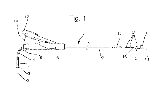
Figure 1 is a side view of a catheter and oxygen sensor embodying the
invention;
Figure 2 is a close-up side view, with hidden lines, of the distal end of the
catheter and oxygen sensor;
Figure 3 is a close-up perspective view of the distal end of the catheter; and
Figure 4 is a median-plane cross-sectional diagram of the male pelvis, showing
the catheter and oxygen sensor in place,
Figure 1 shows a Foley catheter 1 carrying an elongate oxygen sensor 2. In
other
embodiments, different types of sensor may be used. Figures 2 and 3 show close-
ups
of the distal end region of the catheter 1.
CA 03042611 2019-05-02
WO 2018/083487 PCT/GB2017/053316
- 9 -
The main body of the catheter 1 is approximately 40 cm long and is formed from
silicone. It comprises a relatively-rigid, proximal connecting portion 6, or
port hub, and
a relatively-flexible, cylindrical, elongate portion 7. A smooth, domed tip 8
is bonded to
the distal end of the elongate portion 7.
The elongate portion 7 of the catheter 1 defines three lumens, running side-by-
side
from the connecting portion 6 to near the tip 8 of the catheter 1: an
inflation lumen, a
drain lumen 11, and a sensor lumen 10. The inflation lumen runs from an
inflation port
12 in the connecting portion 6 to an inflatable balloon 13 which surrounds the
elongate
portion 7 and which is located around 5 cm proximal of the tip 8. The drain
lumen 11
runs from a drainage port 14 in the connecting portion 6 and terminates near
the tip 8.
The drain lumen 11 opens through a set of six drain holes 15 located along the
elongate portion 7 of the catheter 1, just proximal of the tip 8¨three holes
on each
side of the catheter 1. The sensor lumen 10 runs from a sensor port 3 towards
the tip
of the catheter 1. The inside of the sensor lumen 10 is lined with fluorinated
ethylene
propylene (FEP).
The following steps may be taken during manufacture of the catheter 1:
the main body of the catheter 1 is extruded in long lengths (typically several
hundred meters) and comprises a relatively-flexible, cylindrical, elongate,
silicone
structure, over-molded over an FEP tube, to provide a sensor lumen 10, as well
as a
cavity drain lumen 11 and another cavity inflation lumen;
the main body extrusion is cut into lengths of approximately 34 cm;
approximately 2 cm of silicone is removed from a proximal end, resulting in
approximately 2cm of FEP tube projecting from the main body extrusion;
the proximal end of the main body extrusion, containing the protruding 2 cm of
FEP tube, is loaded into a mold tool, and the connecting portion 6 is produced
by over-
molding silicone onto the main body of the catheter 1;
the distal end of main body extrusion 1 is loaded into a punching tool, and an
aperture is produced which intersects the inflation lumen, approximately 1 cm
from the
distal end;
the distal end of main body extrusion 1, containing the inflation aperture, is
loaded into a mold tool which over-molds a silicone portion comprising
drainage holes
15 connected to the drainage lumen 11, as well as an open furrow 16 connected
to the
sensor lumen 10; and
CA 03042611 2019-05-02
WO 2018/083487 PCT/GB2017/053316
- 10 -
a smooth, domed tip 8 (molded separately in silicone) is bonded to the distal
end of the silicone portion, beyond the drainage holes 15 and open furrow 16.
Figure 1 shows the oxygen sensor 2 entering the catheter 1 through a sensor
port 3.
The sensor port 3 contains a locking silicone bung, to provide a seal around
the
outside of the sensor 2. The oxygen sensor 2 then passes through a flexible
transparent tube 4, along which is marked a distance scale 5. The transparent
tube 4
is sealed to the connecting portion 6 by means of a Luer lock 9. The oxygen
sensor 2
passes from the transparent tube 4 into the sensor lumen 10 within the body of
the
catheter 1.
The inflation port 12 and drainage port 14 are displaced a little to the side
of the path
of the elongate portion 7 of the catheter 1. However, the sensor port 3 lies
on the path
of the catheter 1. This reduces resistance to movement of the oxygen sensor 2.
The distal end of the enclosed sensor lumen 10 opens into an open furrow 16,
at a
circular or oval mouth 23, approximately 3 cm before the tip 8. The furrow 16
runs for
approximately 15 mm towards the tip 8, and is defined by an elongate, concave
depression in the outer walls of the catheter 1. This can be seen particularly
clearly in
the close-up views of Figures 2 and 3. The furrow 16 is approximately 3 mm
wide and
approximately 2 mm deep when measured against the cylindrical shape of the
elongate portion 7. The sensor lumen 10 opens into the furrow 16 at one end,
within
the depth of the furrow 16. The furrow 16 is sized to accommodate the oxygen
sensor
2 within its depth. The side walls of the furrow 16 slope approximately
radially along
the cylindrical shape of the elongate portion 7 of the catheter 1. The furrow
16 has a
planar or curved base 22, approximately 13 mm long and approximately 1 mm
wide.
The distal end wall 17 of the furrow 16 slopes at an angle of approximately
160
degrees to the base 22.
The elongate oxygen sensor 2 comprises a flexible, plastic-coated outer sleeve
which
runs between a plug 19, at a proximal end of the sensor 2, to a sensor tip 18
at a distal
end of the sensor 2. The sleeve contains an optical fibre. The plug 19
contains optical
and electrical connections, and associated electronic circuitry, for plugging
the sensor
2 into a control unit (not shown). The sensor tip 18 is approximately 10 mm
long, and
contains an oxygen-sensitive luminescent material, such as platinum
CA 03042611 2019-05-02
WO 2018/083487 PCT/GB2017/053316
- 11 -
octaethylporphyrin, contained inside a rigid, elongate, perforated metal
cage¨for
example, in an arrangement substantially as described in WO 2006/095191.
Figure 4 shows the catheter 1 inside a human male. In use, a urine-collection
vessel
(not shown) is coupled to the drainage port 14, and the catheter 1 is inserted
along the
urethra 20 until its tip 8 is located inside the bladder 21. The inflation
port 12 is then
used to inflate the balloon 13, in order to prevent the catheter 1 from being
prematurely pulled out of the bladder 21.
The oxygen sensor 2 may initially be separate from the catheter 1, or may be
located
partially within the sensor lumen 10. The sensor port 3 is loosened, and the
oxygen
sensor 2 is pushed along the sensor lumen 10. The position of a reference mark
(not
shown) on the outside of the oxygen sensor 2 may be tracked against the
distance
scale 5 to determine the location of the oxygen sensor 2 relative to the
catheter tip 8.
It will be seen from Figure 4 that the path of the urethra 20 is far from
straight, typically
containing at least two significant bends. Nevertheless, the FEP-lining of the
sensor
lumen 10 ensures that the oxygen sensor 2 can easily be moved backwards and
forwards, by hand, within the sensor lumen 10.
After the sensor tip 18 reaches the distal end of the enclosed sensor lumen
10, it
emerges from the mouth 23 into the furrow 16. The furrow 16 is large enough to
fully
accommodate the rigid sensor tip 18. Unless a lateral force is causing the end
region
of the catheter 1 to bend significantly, with further pushing of the oxygen
sensor 2, the
sensor tip 18 will be deflected gently away from the catheter 1 as it meets
and is
guided outwardly by the sloping distal end wall 17 of the furrow 16. Further
insertion
of the oxygen sensor 2 will cause the sensor tip 18 to advance past the tip 8
of the
catheter 1, in substantially the same direction as the end region of the
catheter 1. The
flexible nature of the cable portion of the oxygen sensor 2 allows an oxygen-
sensing
side wall of the sensor tip 18 to come to rest against the wall of the bladder
21, in front
of the catheter 1.
Once the catheter 1 and oxygen sensor 2 have been correctly placed, the sensor
port
3 may be screwed tight in order to clamp the silicone bung around the sensor
sleeve
2, so as to prevent any movement of the oxygen sensor 2 within the sensor
lumen 10.
CA 03042611 2019-05-02
WO 2018/083487 PCT/GB2017/053316
- 12 -
The bung also prevents urine travelling along the sensor lumen 10 and leaking
out of
the sensor port 3.
The plug 19 of the oxygen sensor 2 can then be connected to a control unit
(not
shown), which performs measurements of the oxygen partial pressure in the wall
of the
bladder 21. The control unit may control the sensor tip 18 substantially as
described in
W02012/010884, by the present applicant, the entire contents of which are
hereby
incorporated by reference.
It will be appreciated by those skilled in the art that the invention has been
illustrated
by describing one or more specific embodiments thereof, but is not limited to
these
embodiments; many variations and modifications are possible, within the scope
of the
accompanying claims. For example, other types of sensor may be used, including
other types of oxygen sensor; the catheter 1 may contain additional lumens,
such as
an irrigation lumen; one of the lumens may be contained inside another of the
lumens;
the catheter 1 may be made of a material other than silicone; etc.