Note : Les descriptions sont présentées dans la langue officielle dans laquelle elles ont été soumises.
SINGLE AXIS SENSOR (SAS) WITH HALL SENSOR USING EXTERNAL
MAGNET
FIELD OF THE INVENTION
The present invention relates generally to tracking
of intrabody probes, and particularly to magnetic position
and orientation sensors in catheter-based tracking systems.
BACKGROUND OF THE INVENTION
Various techniques for magnetically tracking a
position and/or an orientation of intra-body probes were
previously proposed. For example, U.S. Patent Application
Publication 2010/0210939 describes a surgical navigation
system for tracking an instrument relative to a patient.
The system can track a portion of the patient, an
instrument, and/or both relative to image data, a
coordinate system, an atlas, a morphed atlas, or
combinations thereof. The system can include a tracking
device on the instrument to provide six degree of freedom
information regarding the location of the instrument. In
an embodiment, a location sensor in the device includes two
coils are which placed in an angle relative to one another,
such as an orthogonal angle. The sensor may use Hall sensors
alternatively to coils.
As another example, U.S. Patent Application
Publication 2016/0278746 describes a system and method that
pertains to an MR-guided breast biopsy procedure,
specifically as to real-time tracking and navigation of a
biopsy device. More particularly, the system utilizes a
diagnostic imaging modality such as magnetic resonance
imaging (MRI) to locate lesions in a human breast while
utilizing an inertial measurement unit to track advancement
of a biopsy device in real-time. One tracking approach is
based on a set of 3-axis Hall-effect-gyroscope-
1
CA 3053801 2019-09-03
accelerometer sensors: respectively, sensors/transducers
yield varying output voltages in response to the different
magnetic fields sensed, as well as for different
accelerations and angular velocities.
U.S. Patent Application Publication 2012/0143127
describes a variable magnet system for manipulating a
magnetic catheter. In one embodiment, a cluster of
electromagnets is configured to generate a desired magnetic
field. In one embodiment, one or more poles of the cluster
are moveable with respect to other poles in the cluster to
allow shaping of the magnetic field. In one embodiment, one
or more magnetic poles can be extended or retracted to
shape the magnetic field. In one embodiment, the
electromagnets can be positioned to generate magnetic
fields that exert a desired torque and/or movement force
on the catheter. In one embodiment, a magnetic field source
is used to create a magnetic field of sufficient strength
and orientation to move a magnetically-responsive catheter
tip in a desired direction by a desired amount.
U.S. Patent Application Publication 2003/0006759
describes a position sensor for a medical device that
comprises a core made of a high permeable material such as
Wiegand effect material comprising a mixture of cobalt,
vanadium, and iron. The position sensor has an outer
diameter of approximately 0.4 mm and is used in a medical
device having an outer diameter of approximately 0.67 mm.
SUMMARY OF THE INVENTION
An embodiment of the present invention provides a
catheter-based tracking system including one or more field
generators and a processor. The one or more field
generators, which are located adjacent to a Magnetic
Resonance Imagining (MRI) system, are configured to apply
one or more Alternating Current (AC) magnetic fields. The
2
CA 3053801 2019-09-03
processor is configured to receive signals, which are
produced responsively to the AC magnetic fields in a single
axis sensor (SAS) that is fitted at a distal end of a
catheter inserted into an organ of a patient, and, based
on the signals received from the SAS, calculate a direction
of the distal end. The processor is further configured to
receive, from a Hall effect sensor that is fitted at the
distal end of the catheter, a sensed component of a Direct
Current (DC) magnetic field of the magnetic resonance
imagining (MRI) system, and, based on the calculated
direction and the sensed component of the DC magnetic
field, calculate a roll-angle of the distal end inside the
organ.
In some embodiments, the processor is further
configured to calculate a position of the distal end based
on the signals received from the SAS.
In some embodiments, the processor is further
configured to present the roll angle of the distal end in
a coordinate system of the catheter-based tracking system.
There is additionally provided, in accordance with an
embodiment of the present invention, a method, including
applying one or more Alternating Current (AC) magnetic
fields using one or more field generators, which are
located adjacent to a Magnetic Resonance Imagining (MRI)
system. Signals are received, which are produced
responsively to the AC magnetic fields in a single axis
sensor (SAS) that is fitted at a distal end of a catheter
inserted into an organ of a patient. A direction of the
distal end is calculated based on the signals received from
the SAS. A sensed component of a Direct Current (DC)
magnetic field of the magnetic resonance imagining (MRI)
system is received from a Hall effect sensor that is fitted
at the distal end of the catheter. Based on the calculated
3
CA 3053801 2019-09-03
direction and the sensed component of the DC magnetic
field, a roll-angle of the distal end inside the organ is
calculated.
There is additionally provided, in accordance with an
embodiment of the present invention, a magnetic sensor
including a single axis sensor (SAS) and a Hall effect
sensor. The single axis sensor (SAS) is configured to, in
response to one or more Alternating Current (AC) magnetic
fields, generate signals indicative of a direction of a
single axis of the SAS in a given coordinate system. The
Hall effect sensor, which is configured to, in response to
a Direct Current (DC) magnetic field, generate a signal
indicative, in the given coordinate system, of a roll-angle
of the SAS about the single axis.
The present invention will be more fully understood
from the following detailed description of the embodiments
thereof, taken together with the drawings in which:
BRIEF DESCRIPTION OF THE DRAWINGS
Fig. 1 is a schematic, pictorial illustration of a
catheter-based magnetic position-tracking system, in
accordance with an embodiment of the present invention;
Figs. 2A and 2B are side views of a distal end of a
catheter comprising a Hall effect sensor oriented in a
parallel roll-angle and an orthogonal roll-angle,
respectively, in accordance with an embodiment of the
present invention;
Fig. 3 is a graph of normalized Hall voltage as a
function of roll-angle a, in accordance with an embodiment
of the present invention; and
Fig. 4 is a flow chart that schematically illustrates
a method for estimating the roll-angle of a catheter using
4
CA 3053801 2019-09-03
a Hall effect sensor, in accordance with an embodiment of
the present invention.
DETAILED DESCRIPTION OF EMBODIMENTS
OVERVIEW
Embodiments of the present invention that are
described hereinafter provide a magnetic catheter-based
tracking system that is configured to utilize a presence
of a large constant magnetic field, such as of a magnet a
magnetic resonance imaging (MRI) system, to measure a roll
angle of a distal end of the catheter. The MRI system is
available in many cases since it is being used for imaging
an organ of a patient to which the catheter is inserted.
In some embodiments, a processor of the magnetic
tracking system registers the axis (direction) of the
constant (Direct Current - DC) magnetic field of the MRI
system, with a coordinate system of the magnetic catheter-
based tracking system. For example, a component of the MRI
magnetic field that affects the roll-angle indicative
signals is projected by the processor onto the axes of the
coordinate system of the catheter-based tracking system.
With this additional projection, a full description of the
catheter distal end position, direction, and roll-angle,
is available in the coordinate system of the catheter-based
tracking system.
In some embodiments, a miniature magnetic sensor is
provided, which is fitted at the distal end of the catheter.
The magnetic sensor is capable of generating signals
indicative of the position, direction, and roll-angle of
the distal end of the catheter inside an organ of a patient
in a given coordinate system, such as the coordinate system
of the catheter-based tracking system. The disclosed
miniature magnetic sensor comprises a single-axis-sensor
5
CA 3053801 2019-09-03
(SAS - typically a coil) and a Hall effect sensor that are
both fitted at the catheter distal end, as described below.
In some embodiments, the SAS generates the position
and direction signals in response to one or more
alternating (Alternating Current - AC) magnetic fields of
the magnetic catheter-based tracking system. The Hall
effect sensor generates a roll-angle indicative signal in
response to a constant magnetic field that is induced by
the additional external magnet of the MRI system.
In the description hereinafter, a roll-angle of the
catheter distal end is defined as an amount of rotation of
the catheter distal end about a longitudinal axis of
symmetry of the distal end. The roll-angle can be used, for
example, to improve the accuracy of electroanatomical maps
produced by electrophysiological (EP) mapping catheters,
and/or of ablation procedures, by enabling, for example,
controlling the roll of a catheter that has asymmetric
electrodes for EP sensing or ablation. The roll-angle
indicative signal comprises a Hall voltage that varies as
the catheter rolls over its longitudinal axis of symmetry,
as described below.
In some embodiments, the SAS comprises a miniaturized
coil sensor, and the disclosed miniature magnetic sensor
structure combines the Hall effect sensor with the
miniaturized coil sensor. The magnetic sensor is also named
hereinafter "position, direction, and roll-angle (PDR)
sensor." In some embodiments, the PDR sensor is made of a
single coil that is wound to enclose the Hall effect sensor.
A direct (DC) electric current is applied to the Hall effect
sensor, and when the distal end is placed in the DC magnetic
field of the MRI, a Hall voltage is generated at the
terminals of the Hall effect sensor.
6
CA 3053801 2019-09-03
The maximal amplitude of the Hall voltage depends on
the direction of the distal end in space, via the projection
of the external DC magnetic field on that direction. Taking
into account the known (e.g., tracked) direction of the
distal end, and based on the registration of the coordinate
systems, a processor calculates a Hall voltage that is
uniquely indicative of the roll-angle. For example, in an
embodiment, the processor calculates an instantaneous
direction of the catheter from direction signals received
from the SAS, and based on the calculated direction, the
measured Hall voltage, and the aforementioned registration
of coordinate systems, derives the catheter distal end
roll-angle in a coordinate system of the catheter position
tracking system.
Typically, miniature Hall effect sensors are made of
a specific semiconductor, such as GaAs. Generally, however,
under a strong DC magnetic field, such as of an MRI system,
a measurable Hall voltage can be readily generated by many
types of conductors and semiconductors. In an embodiment,
one or more metallic electrodes that are disposed at the
distal end of the catheter are utilized as Hall sensors,
and terminals are connected to the electrodes to output
generated Hall voltage.
Note that bulky sensors, such as multi-coil sensors,
may alternatively be used for the simultaneous measurement
of position, direction, and roll-angle. Such sensors,
however, require an increased diameter of the catheter
distal end and may thus limit maneuverability in the body.
The disclosed miniature Hall sensor, on the other hand,
which has sub-millimeter dimensions, can be fitted in a
narrow diameter distal end of a catheter. The disclosed PDR
sensor, thus, adds signals indicative of its roll-angle on
top of the position and direction signals provided by a
7
CA 3053801 2019-09-03
legacy miniaturized SAS sensor, while presenting the same,
or a similar, physical form factor of the SAS.
The disclosed roll-angle tracking technique, which
utilizes a large DC magnetic field of an MRI system that
is available anyhow, to determine roll-angle using the
disclosed miniature PDR sensor, can improve catheter
accessibility and tracking accuracy, and therefore also
improve the overall quality of diagnostic and therapeutic
catheterization procedures.
SYSTEM DESCRIPTION
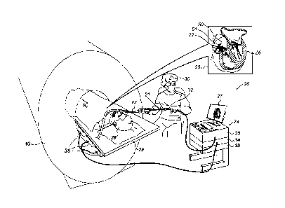
Fig. 1 is a schematic, pictorial illustration of a
magnetic catheter-based tracking system 20, in accordance
with an embodiment of the present invention. System 20
comprises a catheter 21, having a distal end 22 that is
navigated by a physician 30 into a heart 26 of a patient
28 via the vascular system. The catheter can be used for
ablation, EP sensing, or any other medical procedure. In
the pictured example, physician 30 inserts distal end 22
through a sheath 23, while manipulating distal end 22 using
a manipulator 32 near the proximal end of the catheter. As
shown in an inset 25, distal end 22 comprises a PDR sensor
51 which is contained within distal end 22, and an ablation
catheter 50.
In the embodiments described herein, catheter 21 is
used for ablation of tissue in heart 26. Although the
pictured embodiment relates specifically to the use of an
ablation catheter 50 for ablation of heart tissue, the
elements of system 20 and the methods described herein may
alternatively be applied to diagnostic applications, such
as electrophysiological mapping, using, for example, multi-
electrode catheters such as the Pentarayqp or the Lasso
8
CA 3053801 2019-09-03
catheters (both made by Biosense-Webster, Irvine,
California).
The proximal end of catheter 21 is connected to a
control console 24. Console 24 comprises a processor 39,
typically a general-purpose computer, with suitable front
end and interface circuits 38 for receiving signals from
catheter 21, as well as for applying energy via catheter
21 to ablate tissue in heart 26 and for controlling the
other components of system 20. Console 24 also comprises a
driver circuit 34, configured to drive magnetic field
generators 36.
During the navigation of distal end 22 in heart 26,
console 24 receives position and direction signals from PDR
sensor 51 in response to magnetic fields from external
field generators 36. Magnetic field generators 36 are
placed at known positions external to patient 28, e.g.,
below a table 29 on which the patient is lying. These
position and direction signals are indicative of the
position and direction of ablation catheter 50 in a
coordinate system of the position tracking system.
PDR sensor 51 further transmits a roll-angle
indicative signal to console 24, in response to a large
constant magnetic field Bo of an MRI system 40 (e.g., Bo
equals 1.5T). Bo defines a longitudinal axis for a
coordinate system of the MRI system.
Using the received signals, processor 39 calculates
the position, direction, and roll-angle of ablation
catheter 50 in the heart and, optionally, presents the
tracked position, direction, and roll-angle on a display
27.
The method of position and direction sensing using
external magnetic fields is implemented in various medical
applications, for example, in the CARTOTm system, produced
9
CA 3053801 2019-09-03
by Biosense Webster, and is described in detail in U.S.
Patents 5,391,199, 6,690,963,
6,484,118, 6,239,724,
6,618,612 and 6,332,089, in PCT Patent Publication WO
96/05768, and in U.S. Patent Application Publications
2002/0065455 Al, 2003/0120150 Al and 2004/0068178 Al, whose
disclosures are all incorporated herein by reference.
Processor 39 typically comprises a general-purpose
computer, which is programmed in software to carry out the
functions described herein. The software may be downloaded
to the computer in electronic form, over a network, for
example, or it may, alternatively or additionally, be
provided and/or stored on non-transitory tangible media,
such as magnetic, optical, or electronic memory.
SINGLE AXIS SENSOR (SAS) WITH HALL SENSOR USING EXTERNAL
MAGNET
Figs. 2A and 2B are side views of a distal end of a
catheter comprising a Hall effect sensor, oriented in a
parallel roll-angle and an orthogonal roll-angle,
respectively, in accordance with an embodiment of the
present invention. In Fig. 2A, a=00, meaning distal end 22,
is rolled parallel to an external magnetic field, as
described below. In Fig. 2B, distal end 22 is rolled at
a=90 relative to the external magnetic field.
As further seen, the MRI magnetic field vector Elo is
directed with an angle 55 (i.e., 0) relative to distal end
22, which in a coordinate system 53 is aligned with its
longitudinal axis parallel to the z-axis of coordinate
system 53. Roll-angle a of distal end 22 is therefore
defined relative to a projected component of magnetic field
vector Bo, Be=80=Sin(0).
CA 3053801 2019-09-03
As can be seen, whatever the roll-angle a of distal
end 22, the lines of the magnetic field face the same
effective area encompassed by coil 51a of sensor 51 (i.e.,
SAS 51a). Thus, using only coil 51a, tracking system 20 has
no roll-angle indicative signal.
As noted above, adding the disclosed Hall effect
sensor 60 provides the required roll-angle indicative
signal. Fig. 2A shows an electrical current 62 that flows
in the longitudinal direction through Hall effect sensor
60. Electrodes 60a and 60b of a Hall sensor 60 are aligned
to sense a resulting Hall voltage perpendicular to a
magnetic field component Be of vector Bo. At roll-angle
a=0 , exemplified by Fig. 2A, the resulting Hall voltage
falling between electrodes 60a and 60b is maximal. In Fig.
2B, distal end 22 is rolled by 900 relative to Fig. 2A, and
electrodes 60a and 60b are aligned parallel to magnetic
field component Be. In this case the resulting Hall voltage
between electrodes 60a and 60b is zero.
As further described below, the Hall voltage is
utilized to indicate a continuous roll-angle of distal end
22.
The example illustrations shown in Figs. 2A and 2B are
chosen purely for the sake of conceptual clarity. For
example, coil 51a, which is shown separated from Hall
sensor 60, is typically wound over sensor 60. Other system
elements, such as additional sensors and electrodes, are
omitted for simplicity.
Fig. 3 is a graph of normalized Hall voltage as a
function of roll-angle a, in accordance with an embodiment
of the present invention. The Hall voltage, VII, is
11
CA 3053801 2019-09-03
proportional to the cross product of current 62, I, and
magnetic field component Be, i.e.,
VH (a ,= 9) = K I =B0 =Sin (9) =Cos (a)
where a is the roll-angle, K is a known constant, and
B0 =Sin(9) Cos (a) is the Hall sensed component of the DC
magnetic field Bo.
As shown in Fig. 3, a normalized Hall voltage varies
with roll-angle a as a Cos(a) function. Thus, the measured
Hall voltage encodes the roll-angle, e.g., in the form of
a cosine function. The Hall voltage provides information
regarding the rotation of the SAS around its axis. For
example, as noted above, for a certain roll-angle the Hall
voltage is maximal. Further rotation of the catheter by 90
causes the Hall voltage to drop to zero. Another rotation
by 90 causes the Hall voltage to be maximal but with an
opposite polarity. In an embodiment, a processor uses a
lookup table comprising roll-angle as a function of Hall
voltage, so as to indicate a roll-angle of the distal end
in an organ of a patient.
In an embodiment, the roll-angle is indicated in the
coordinate system of the catheter tracking system (with
which the coordinate system of the MRI system is
registered, as described above).
In an optional embodiment, the processor uses slight
dVH
variations in roll-angle to calculate the slope, 7,T, of
the Hall voltage at a given roll-angle, so as to
differentiate between a roll-angle having a same VH, for
example, between a perpendicular (90 ) to anti
perpendicular (2700) roll-angles of the distal end 22, in
which for both VH=0.
12
CA 3053801 2019-09-03
Fig. 4 is a flow chart that schematically illustrates
a method for estimating the roll-angle of a catheter using
a Hall effect sensor, in accordance with an embodiment of
the present invention. The process begins with an MRI
system 40 imaging patient 28, at an MRI imaging step 70.
In parallel, typically in synchronization with the imaging
sequences to avoid electronic noises, catheter-based
tracking system 20 tracks a position and a direction of
distal end 22 of catheter 21, using modulated magnetic
fields that generators 36 produce, at a position and
direction tracking step 72. For that, system 20 uses a SAS
sensor 51a that is part of PDR sensor 51 that is fitted at
distal end 22 of catheter 21. Next, tracking system 20
senses a component of the DC magnetic field of MRI system
40 that includes the roll angle information, cos(a), using
Hall-effect sensor 60 that is also part of PDR sensor 51,
at a DC magnetic field component sensing step 74. In a
coordinate registration step 75, processor 39 registers a
direction defined by the DC magnetic field of MRI system
44, Bo, with the coordinate system of magnetic catheter-
based tracking system 20.
Then, based on the tracked direction, the Hall sensed
component of the DC magnetic field, and the aforementioned
registration of coordinate systems, processor 39 calculates
a roll-angle of distal end 22, at a roll-angle calculation
step 76. In an optional direction and roll-angle presenting
step 78, the indications provided by steps 72 and 76 are
presented on display 27, on a map of heart 26. Next,
physician 30 uses the indications provided by steps 72 and
76 to further spatially align the distal end inside heart
26 of patient 28, at a catheter aligning step 80. For
example, physician 30 may roll the distal end so as to be
13
CA 3053801 2019-09-03
able to ablate target tissue, and perform a procedure, such
as an ablation, at an optional perform procedure step 82.
The example flow chart shown in Fig. 4 is chosen purely
for the sake of conceptual clarity. In alternative
embodiments, additional steps, such as
electrophysiological sensing of aberrant cardiac activity
may be included.
Although the embodiments described herein mainly
address cardiac applications, the methods and systems
described herein can also be used in other applications,
such as in neurology and otolaryngology and nephrology.
It will thus be appreciated that the embodiments
described above are cited by way of example, and that the
present invention is not limited to what has been
particularly shown and described hereinabove. Rather, the
scope of the present invention includes both combinations
and sub-combinations of the various features described
hereinabove, as well as variations and modifications
thereof which would occur to persons skilled in the art
upon reading the foregoing description and which are not
disclosed in the prior art. Documents incorporated by
reference in the present patent application are to be
considered an integral part of the application except that
to the extent any terms are defined in these incorporated
documents in a manner that conflicts with the definitions
made explicitly or implicitly in the present specification,
only the definitions in the present specification should
be considered.
14
CA 3053801 2019-09-03