Note : Les descriptions sont présentées dans la langue officielle dans laquelle elles ont été soumises.
CA 03054470 2019-08-23
WO 2018/171869 PCT/EP2017/056705
METHODS AND DEVICES FOR MEASURING CHANGES IN THE POLARIZATION RESPONSE OF A
SAMPLE BY
TIME-DOMAIN INFRARED SPECTROSCOPY (FIELD-RESOLVED VIBRATIONAL SPECTROSCOPY)
Field of the invention
The invention relates to a method of measuring the polarization response of a
sample to optical
field excitation, in particular the vibrational response of molecules of a
biological sample, and
changes of the polarization response. The polarization response of the sample
is measured by
field-resolved spectroscopy, via directly sampling the rapidly oscillating
electric field radiated by
the induced sample polarization in the time domain. Furthermore, the invention
relates to a
spectroscopic apparatus for measuring the polarization response of a sample,
in particular a
biological sample. Applications of the invention include detection of changes
in the physical and
chemical properties/conditions of a sample, in particular changes in the
molecular composition of
biological samples. Possible biological samples include gaseous-, liquid- or
solid-phase samples
from a human or animal organism, in particular body fluids, tissues as well as
individual cells from
living organisms.
Technical background
Molecules are the smallest functional building blocks of living organisms.
Living systems require
the presence of an enormous variety of molecules. Their abundance is allowed
to vary within a
narrow range for an organism to function properly. Cells or blood, as
prominent examples, are
composed of tens of thousands of different molecules, the concentration of
which depends on
the physiological state of the body. Substantial changes in the abundance of
individual molecular
constituents of blood can thus serve as indicators of abnormal physiology.
Such changes are used
as a basis for molecular pathology for detection and subsequent monitoring of
progression of
disease, its response and resistance to treatment, and for assessing the
susceptibility of
individuals to particular disorders. Moreover, differences in molecular
composition of different
types of cells may be helpful in identifying cell types (such as e.g. stem
cells) and sorting cells from
one and the same organism.
The molecules with largest relative changes in concentration (incl. newly
appeared ones) lend
themselves as markers of a disease or to distinguishing different types of
cells from each other. A
1
CA 03054470 2019-08-23
WO 2018/171869 PCT/EP2017/056705
tiny fraction of them can be identified individually by antibody-based assays.
The conventional
techniques for sensing a large number of molecules simultaneously are e. g.
RNA sequencing and
mass spectrometry (detecting individual constituents) and vibrational
spectroscopy (measuring
global effects from a multitude of specimen). These techniques are mainly
sensitive to high-
abundance constituents, which dominate their observed signals, and they are
"blind" for a large
number of low-abundance molecules. However, changes in the concentration of
low-abundance
molecules can also be of high importance, a prominent example being e.g.
cytokines, even
miniscule concentration change of which is known to lead to extensive
physiological effects. Low-
abundance molecules may well incorporate several of possibly many different
ones with large
relative changes in concentration caused by abnormal physiology. Hence, they
might
¨ particularly in correlation ¨ be ideally suited for either disease marking
or cell identification/
sorting. All these potential molecular markers have been inaccessible to
molecular pathology and
cell biology to date. In conclusion, a persisting major challenge in molecular
pathology and cell
biology to date is the identification of smallest concentration changes of
high- as well as low-
abundance molecules in complex mixtures.
Vibrational spectroscopy acquires information related to the polarization
response of molecular
specimens induced by periodic oscillations of the atomic nuclei around their
equilibrium positions.
For decades, infrared spectroscopy and Raman spectroscopy (described below)
have been used to
acquire the amplitude response of molecular vibrations over an ever broader
spectral range. The
corresponding specimen-characteristic information is customarily referred to
as vibrational
molecular fingerprint, briefly: molecular fingerprint. Note that in literature
this designation has
also been used in the context of other physical observables, albeit always
with the aim of
associating a unique fingerprint (also called: spectral polarization response)
to a specific sample.
Despite of a plurality of measuring techniques, conventional fingerprinting
methods suffer from a
moderate sensitivity, preventing the reliable detection of small changes in
molecular composition
of samples and that of low-abundance constituents altogether.
Traditionally, molecular fingerprints are measured in the frequency domain,
either by
autocorrelation (Fourier-transform spectroscopy, FTS) or using
monochromator/spectrometer
arrangements, acquiring (indirectly or directly) spectral intensities. The
specific signature of a
sample manifests itself in changes of these intensities when placing the
sample in the beam path.
This brings about two severe limitations: first, intensity noise of the source
compromises the
ability of the approach/device to detect intensity changes that are induced by
the sample.
Second, the high intensity on top of which small changes are to be resolved
calls for a high
2
CA 03054470 2019-08-23
WO 2018/171869 PCT/EP2017/056705
dynamic range, the necessarily finite value of which sets a limit to power
scaling. Both effects
contribute to restricting the smallest detectable changes in sample
properties/conditions.
Most recently, a major progress in the detection limit of infrared absorption
spectroscopy has
been achieved. It is based on a sudden (preferably femtosecond-duration)
excitation of molecular
vibrations (or more generally: structural dynamics) and direct time-domain
sampling of the
rapidly oscillating electric field emitted by the induced polarization
response in the wake of the
sudden excitation. This field sampling acquires both the strength of the
excited vibrations
(amplitude response) as well as their retardation with which they react to an
external trigger
(phase response) and has been referred to as field-resolved spectroscopy
(FRS). This scheme,
described in WO 2016/102056 Al, substantially improves the sensitivity in the
detection of small
changes in the properties/conditions of a sample, in particular in specimen
concentration in
biological samples, however still suffers from the shortcoming of delivering a
signal that is
dominated by contributions from high-abundance constituents and, in addition,
its sensitivity is
still compromised by an ultraintense excitation pulse entering the detector
before the molecular
signal. In what follows, the physical principles underlying FRS are reviewed,
highlighting its
advantages beneficial for, and its limitations overcome by the present
invention.
Physical principles underlying FRS
Measuring the polarization response with FRS according to WO 2016/102056 Al is
based upon
the synchronism (or: coherence) with which molecules of the sample 1 under
investigation
(Figure 11, prior art) emit light waves when excited by coherent light
excitation waves 2, the field
oscillations of which are perfectly synchronized in space and time. As a
consequence, the
emission from individual molecules of the same type i add constructively,
resulting in a wave with
an electric field E1(t) the strength of which increases with the number of
emitters, Ni. The entire
wave, radiated by all the molecules of the sample 1 is the superposition of
all of these partial
waves, carrying what is referred to as the global molecular fingerprint (GMF)
of the sample, in the
form of the temporal variation of its electric field, EGmF (t) . The attribute
"global" stresses the
fact that the GMF of the sample 1 carries, in principle, information from all
of the molecules in
contrast to, e.g., a targeted search for biomarkers (see, e.g., P. E. Geyer et
al. in "Cell Syst." 2, 185
(2016)), restricted to a small subset of the constituents of the sample.
Exciting the molecules impulsively with the sudden, ultrashort excitation wave
2 (Figure 11, see
also A. Sommer et al. in "Nature" 534, 86 (2016)) that is much briefer than
the lifetime of
3
CA 03054470 2019-08-23
WO 2018/171869 PCT/EP2017/056705
molecular excitations will result in an electric field emanating from the
sample (sample wave 3)
consisting of two parts: the excitation laser pulse, modified by the sample's
instantaneous
polarization response (henceforth referred to as main pulse 2') and a (much
weaker) trailing part
arising from the sample's non-instantaneous polarization response, often
referred to as the free-
induction decay (FID), cf. Lanin et at. in "Nature Scientific Reports" 4, 6670
(2014) and Lauberau
and Kaiser in "Rev. Mod. Phys." 50, 607 (1978), also illustrated in Figure 11.
In the case of a biological sample the FID signal carries the GMF of the
sample, which we
henceforth refer to as the GMF wave (or: GMF signal). If the duration of the
main pulse 2' is
substantially shorter than that of the GMF signal, a direct time-domain
measurement of the latter
exhibits a fundamental advantage over (continuous-wave) frequency-domain
spectroscopy
techniques: the GMF signal can be accessed in a background-free manner owing
to the main pulse
2' decaying exponentially in time after its peak on a much shorter scale than
the duration of the
GMF signal.
This allows measurements of very weak signals generated by low-concentration
specimens, e.g.
improved sensitivity. In sharp contrast to frequency-domain implementation of
vibrational
spectroscopies, the intensity noise of the radiation source doesn't constitute
a limitation to the
minimum detectable GMF signal owing to its temporal separation from the
excitation. However,
.. the intensity noise of the source translates to relative amplitude noise of
the GMF signal, setting a
limit to the minimum detectable change in concentration of the molecular
constituents
contributing to the GMF signal.
Technical implementation ¨ prior art
Measuring the sample wave 3 is conducted with the spectroscopic apparatus 100
of Figure 12 as
disclosed in WO 2016/102056 Al. Driving pulses from a laser pulse source 10,
e. g. a femtosecond
laser as described by 0. Pronin et al. in "Nature Commun." 6, 6988, 2015, are
used for creating
the excitation pulses 2 as described by I. Pupeza et al. in "Nature Photon."
9, 721 (2015),
irradiating the sample 1 under investigation, and for providing sampling
pulses 5 for electro-optic
sampling of the sample wave 3 with an electro-optic detector device 20.
Electra-optic sampling
can directly measure EGmF(t) in excess of 200 THz (see S. Keiber et at. in
"Nature Photonics" 10,
p. 159, 2016). The excitation pulses 2 are created e. g. in a nonlinear
crystal (like a LiGaS2 crystal)
based on intra-pulse difference-frequency generation. The temporal amplitude
function of the
4
CA 03054470 2019-08-23
WO 2018/171869 PCT/EP2017/056705
sample wave 3 is subjected to a Fourier transformation directly yielding the
spectral response of
the sample 1.
As a further advantage, the technique of WO 2016/102056 Al measures the
electric field,
inherently accessing the full phase information in contrast to standard
frequency-domain
spectroscopy or time-domain measurements of the FID intensity as performed,
e.g., by Lanin et
al. in "Nature Scientific Reports" 4, 6670 (2014). As another advantage over
time-domain
measurements of the FID intensity, in FRS the FID signal rolls off linearly
with the decay of the
field amplitude rather than its squared value.
Notably, if the instrument according to Figure 12 is characterized by a linear
response, then the
measured sample wave 3 corresponds to the full electromagnetic response of the
sample to the
excitation field (measured by the same instrument with the sample removed).
This way one gains
access to the full information of the macroscopic polarization of the sample
1, with few-
femtosecond (to potentially sub-femtosecond) temporal resolution. Importantly,
increasing the
power of the driving pulse proportionally enhances the useful FID (henceforth:
GMF) signal above
detection noise floor, without any increase of disturbing background. Thus,
the scheme of WO
2016/102056 Al is truly power scalable with respect to the source: the
molecular signal
temporally separated from the (much more intense) excitation can be increased
by boosting the
source power without a dynamic range "exhausted" by the excitation power, in
contrast to the
above limitations of frequency-domain spectroscopy. Moreover, electro-optic
sampling ([OS) of
the excitation pulse 2 and the sample wave 3 obviates the need for poor-
sensitivity infrared
photon detectors. Nevertheless, the implementation of FRS with these sampling
techniques also
implies that the strong excitation pulse preceding the sample wave compromises
the sensitivity of
these sampling techniques for measuring smallest GMF signals (i.e. weakest
sample waves).
Very recent bench marking experiments were carried out with the prototype
embodiment of the
FRS technology disclosed in WO 2016/102056 Al. In a benchnnarking experiment,
a dilution series
of trehalose in water was investigated with both FRS and FTS. For the latter a
state-of-the-art
Fourier-transform spectrometer (MIRA-Analyzer, Micro Biolytics) was used. The
experiments
revealed concentration detection limits of lower than 0.001 mg/mL and
approximately 0.01
mg/mL for measurement times of 50 s for FRS and 45 s FTS, respectively. These
results confirm
the far superior performance of FRS regarding the detection limit of weak GMF
signals.
5
CA 03054470 2019-08-23
WO 2018/171869 PCT/EP2017/056705
Physical principles of stimulated Raman scattering
Another implementation of vibrational spectroscopy is based on stimulated
Raman scattering
(SRS), wherein the stimulated Raman process has been used to study the
temporal and spectral
vibrational structure of numerous molecular systems. In SRS, two excitation
fields at a pump
frequency, cop, and a Stokes frequency, Ws, are sent simultaneously into the
sample under study.
Molecular transitions are enhanced, if the difference frequency of the
excitation beams, Aw =
- coos, matches a vibrational frequency, Q, of a molecule of the sample,
resulting in loss and gain of
the transmitted pump and Stokes intensity, respectively. The induced changes
in these intensities
are generally small compared to the linear scattering or linear absorption of
the sample. This
shortcoming has been addressed by scaling the energy of the excitation fields
(McCamant et al. in
"Rev. Sci. lnstrum.", 75(11), 4971 (2004), or high-frequency modulation of the
excitation fields
(Freudiger et al. in "Science", 322(5909), 1857 (2008). However, the first
approach is of limited
utility for biological applications and the second one suffers from complexity
and long acquisition
time.
Using a broadband (near-octave-spanning) Stokes or pump pulse provides access
to the entire
spectrum of vibrational frequencies. For combining this advantage with high
spectral resolution,
one of the two pulses must be narrowband (with its spectral bandwidth
dictating the spectral
resolution of the measurement) and the other one is broadband. The GMF signal
here then
appears again as a wake of the broadband and ultrashort pump or Stokes
excitation pulse,
analogously to the implementation with a resonant infrared excitation pulse
described in WO
2016/102056 Al, and ¨ in its own spectral band ¨ in a background-free fashion.
However, SRS
measurements have not been addressed in WO 2016/102056 Al.
Limitations of FRS
(i) While FRS as disclosed in WO 2016/102056 Al has been demonstrated to be
superior to
frequency-domain vibrational spectroscopies in terms of sensitivity for the
molecular GMF signal
of interest, it still offers room for substantial improvement in several
respects. First, the detection
sensitivity of the electro-optic sampler measuring EGmF(t) is orders of
magnitude smaller than it
could be in the absence of the excitation pulse. This is because the sample
wave 3 beam carrying
both the main pulse 2' and the GMF wave can only be gently focused into the
sampler to avoid its
damage by the strong main pulse preceding the GMF wave. Removing the
excitation pulse would
6
CA 03054470 2019-08-23
WO 2018/171869 PCT/EP2017/056705
allow a much stronger focusing of the GMF/sample wave into the [OS detector,
resulting thereby
a correspondingly increased sensitivity in the detection of the weak
GMF/sample wave of interest.
(ii) Moreover, in molecular pathology and cell biology, where, as explained
above, the major
challenge consists in the identification of smallest changes in concentration
of both low- and high-
abundance molecules in complex mixtures. Actually, in the FRS scheme discussed
so far even high
relative changes in the concentration of low-abundance constituents may be
completely masked
by contributions from high-abundance specimens, leaving these potential
biomarkers unnoticed
(just as they are left unobserved by the limited dynamic range in all other
techniques capable of
detecting multiple constituents).
(iii) Last but not least, in the case of complex molecular mixtures, such as
biological samples, the
GMF signal consists of the superposition of the electric fields emitted by
molecules of numerous
different types, occurring with both low and high abundances. As the amplitude
of the GMF signal
.. increases with the number of emitters (which is very large in a complex
sample), so does the
relative intensity noise of EGmF(t), transferred to the GMF signal from the
excitation.
Consequently, amplitude variations due to radiation source noise mask the
temporal fingerprint
induced by small changes in the molecular composition of the sample. Moreover,
these changes
also need to overcome a possible background resulting from imperfections of
the measurement
system.
Objective of the invention
The objective of the invention is to provide an improved method of measuring a
temporal
polarization (or: spectral) response of a sample, in particular a biological
sample, and an improved
spectroscopic apparatus for measuring a temporal polarization (or: spectral)
response of a
sample, in particular a biological sample, being capable of circumventing
limitations of
conventional techniques, in particular the above-mentioned limitations of FRS.
The polarization
response is to be measured with improved sensitivity and/or reproducibility.
Summary of the invention
These objectives are correspondingly solved by a method and a spectroscopic
apparatus
comprising the features of the independent claims, respectively. Preferred
embodiments and
applications of the invention arise from the dependent claims.
7
CA 03054470 2019-08-23
WO 2018/171869 PCT/EP2017/056705
According to a first general aspect of the invention, the above objective is
solved by a method of
measuring a polarization response of a sample, in particular a biological
sample, comprising the
following steps.
A sequence of excitation waves is generated. The excitation waves (called
probe light in
conventional FRS) are generated as a train of laser pulses with a laser source
device, wherein each
excitation wave has a primary temporal shape and spectral content, preferably
with a center
wavelength in the infrared spectral range. Preferably, the full-width-at-half-
intensity-maximum
pulse duration of the excitation waves is equal to or below 1 ps, in
particular equal to or below
300 fs. If the sample to be investigated is in the gas phase, having sharp
vibrational bands and an
FID in a range of tens of ps, a narrowband excitation wave with a pulse
duration equal to or below
1 ps and above 500 fs can be provided. Otherwise, with sample in the liquid
phase having broad
vibrational bands and an FID in a range of 1 ps or shorter, a broadband
excitation wave with a
pulse duration equal to or below 300 Is can be provided.
The sample to be investigated is irradiated with the excitation waves,
including an interaction of
the excitation waves with the sample, so that a sequence of sample waves
(called modified probe
light in conventional FRS) is generated each including a superposition of an
instantaneous
polarization response of the sample, referred to as sample main pulse and a
(usually much
weaker) trailing part arising from the sample's non-instantaneous polarization
response to the
excitation wave, referred to as the free-induction decay, briefly FID signal
or, in particular in the
case of biological samples, a sample global molecular fingerprint (GMF) wave
(E GmF(sample) (t)))
briefly GMF wave or GMF signal. The modified temporal shape and spectrum of
the sample wave
deviate from the primary temporal shape and spectrum of the excitation wave
(respectively) by
features, which are determined by the polarization response of the sample. The
sample under
investigation is a solid, liquid or gas phase sample, in particular of
biological origin.
Furthermore, a reference sample (or: control sample) is provided, which is
another sample (in
solid, liquid or gas phase), in particular of biological and / or of synthetic
nature, to which the
sample to be investigated is to be compared in terms of its GMF. The reference
sample may
comprise e. g. a sample which does not include certain molecules of interest
or another which
includes the molecules of interest with another concentration (e. g. an elder
sample from the
same source like the sample under investigation). A synthetic reference sample
is a reference
sample with a well-known and highly reproducible molecular composition, in
particular
8
CA 03054470 2019-08-23
WO 2018/171869 PCT/EP2017/056705
comprising those molecules which are not of interest in the investigation of
the sample. The
reference sample is irradiated with the sequence of excitation waves,
including an interaction of
the excitation waves with the reference sample, so that a sequence of
reference waves is
generated each including a superposition of a reference main pulse and a
reference GMF wave
(EGMF(ref) (0) =
According to the invention, a difference of the sample waves and the reference
waves is optically
separated in space and/or time from GMF wave contributions which are common to
both of the
sample waves and the reference waves. Accordingly, at least one optical
adjustment device is
provided which spatially and/or temporally separates the difference of the
sample waves and the
reference waves, which is to be detected for investigating the sample, from
the common GMF
wave contributions, which are not specific for the sample under investigation.
The difference of the sample waves and the reference waves is detected and a
temporal
amplitude function of differential molecular fingerprint (dMF) waves (LIEGmF)
is determined each
comprising the difference of the sample and reference GMF waves. Detecting
preferably
comprises electro-optic sampling ([OS) or, alternatively, photo-conductive
sampling (PCS). The
dMF wave is determined by direct detection (sampling) or by calculating based
on detected
sample and reference waves. It represents the polarization response of the
sample (called
"spectral response" in WO 2016/102056). The particular type of polarization
response depends on
the design of the excitation waves, which can be adapted e.g. for an IR
absorption or an SRS
measurement.
According to the invention, for the optically separating step, the sample
waves and the reference
waves are spatially and/or temporally separated from each other before the
detecting step.
Separating the sample and reference waves comprises a targeted adjustment of
the sample and
reference waves relative to each other, in particular a reduction of the
spatial and/or temporal
overlap of the sample and reference waves. The overlap of the sample and
reference waves
preferably is minimized or even excluded in space and/or time domain. In other
words, separating
the sample and reference waves comprises a partial or even complete reduction
of their overlap
in space and/or time domain.
According to the separation of the sample and reference waves, a spatial
and/or temporal
separation of the dMF wave, i. e. the difference of the electric fields
corresponding to the sample
and reference GMF, from any other participating waves is maximized in space
and/or in time. This
9
CA 03054470 2019-08-23
WO 2018/171869
PCT/EP2017/056705
is achieved by accordingly adjusting the participating waves (excitation wave,
reference wave and
sample wave) relative to each other in space and/or in time. In this manner,
the invention
advantageously makes use of the background-free detection of FRS to measure
the difference
signal E G m F (t) , which directly reflects differences in the molecular
composition of the reference
sample and the sample under investigation, with improved sensitivity.
Although not always emphasized in the following, it is noted that the
excitation waves comprise a
sequence of laser pulses created with a repetition rate preferably above 1
kHz, particularly
preferred above 1 MHz. Accordingly, the sample and reference waves are
sequences of laser
pulses as well. The terms excitation wave, reference wave and sample wave
refer to sequences of
the corresponding waveforms used for irradiating the reference sample and the
sample under
investigation or provided by the spectral response of the reference sample and
the sample under
investigation, resp..
According to a second general aspect of the invention, in terms of device
features, the above
objective is solved by a spectroscopic apparatus for measuring a polarization
response of a
sample, in particular a biological sample, which comprises a laser source
device, an optical
adjustment device, a detector device and optionally a calculation device.
Preferably, the
spectroscopic apparatus is adapted for conducting the above method of
measuring a polarization
response of a sample according to the first general aspect of the invention.
The laser source
device is adapted for generating a sequence of excitation waves and for
irradiating the sample
with the sequence of excitation waves, including an interaction of the
excitation waves with the
sample, so that a sequence of sample waves is generated each including a
superposition of a
sample main pulse and a sample global molecular fingerprint (GMF) wave
(EGAIF(sanipie)(0), and
for irradiating a reference sample with the sequence of excitation waves,
including an interaction
of the excitation waves with the reference sample, so that a sequence of
reference waves is
generated each including a superposition of a reference main pulse and a
reference GMF wave
(EGmF(õf)(t)). The optical adjustment device is arranged for optically
separating a difference of
the sample waves and reference waves from wave contributions which are common
to both of
the sample waves and reference waves in space and/or time. The detector device
is arranged for
detecting the difference of the sample waves and the reference waves and
determining a
temporal amplitude function of differential molecular fingerprint (dMF) waves
(AEGAIF) each
comprising the difference of the sample and reference GMF waves.
CA 03054470 2019-08-23
WO 2018/171869 PCT/EP2017/056705
According to the invention, the optical adjustment device for spatially and/or
temporally
separating the difference of the sample waves and reference waves from wave
contributions
which are common to both of the sample waves and reference waves is an
adjustment device
included in the spectroscopic apparatus. The terms "separating", "adjusting"
or "adjustment"
.. refer to any targeted manipulation, in particular targeted wave-form
shaping, of the excitation
wave (and optionally the reference wave) such that the difference GMF, E G m F
, carrying useful
information on the difference in molecular composition between the reference
sample and the
sample under investigation is located in time as far as possible behind the
main pulse of the
sample wave. The optical adjustment device comprises passive and/or active
optical components,
like transmissive and/or reflective components and/or amplifying components,
shaping the wave-
form of the excitation wave and/or the sample waves. The inventors have found
that the
background created by the excitation wave can be substantially suppressed or
the sensitivity of
detecting the difference GMF can be substantially increased by the inventive
separating step or
adjustment device, resp., thus improving the sensitivity of the FRS detection.
The dMF waves can be output as the characteristic polarization response to be
obtained.
Optional, the calculation device can be provided for calculating the dMF wave
based on detected
sample and reference waves and/or for analysing the sensed dMF waves, e. g.
for providing a
polarization response of the sample on the basis of a Fourier transformation
of the temporal
.. amplitude function of the dMF waves, and/or for analysing a change of the
sample composition
based on the dMF waves determined with the sample under investigation and/or a
reference
sample.
According to a preferred application of the invention, the sample under
investigation comprises a
biological sample from a human or animal organism. The spectral response of
the sample and/or
the difference of its GMF with respect to the control (reference) sample, is
measured for
obtaining diagnostically relevant information on the organism. The term
"diagnostically relevant
information" refers to any information on the sample, in particular the
composition thereof,
differences compared with reference samples or temporal changes of the sample,
which can be
used for providing or validating a medical diagnosis. In particular, the
invention aims at detecting
changes in molecular composition, which may mark a deviation from normal
physiology or
identify a different cell type, in a single measurement, by direct comparison
of the sample (or cell)
under scrutiny with a reference (or reference cell from the same organism),
also referred to as
"control", with unprecedented sensitivity.
11
CA 03054470 2019-08-23
WO 2018/171869 PCT/EP2017/056705
Accordingly, with a preferred embodiment of the invention, the measuring
method may include a
step of evaluating the spectral response of the sample in order to obtain the
diagnostically
relevant information. In terms of device features, a preferred embodiment of
the spectroscopic
apparatus preferably includes the calculation device, which is adapted for
processing the spectral
response and providing the diagnostically relevant information.
Advantageously, the
diagnostically relevant information can be output to a user of the inventive
technique, e. g. a
medical doctor. Subsequently, the user can provide a diagnosis in
consideration of the
diagnostically relevant information. A spectral response evaluation can be
implemented as
disclosed in WO 2016/102056 Al.
According to embodiments of the invention, differences in the fingerprints, i.
e. different dMF
waves of samples differing only by small numbers of¨ both high- and low-
abundance ¨ molecules
are sensed. This can be expressed in terms of a simple formula: if
EGAIF(sample)(t) is the sample
GMF signal and EGAIF(ref)(t) is the reference GMF signal, then the following
dMF signal is
detected with the highest possible sensitivity:
AEGMF(t) = EGMF(sample)(t) EGMF(ref)(t)'
According to a first variant of the invention (first embodiment of the
invention, embodiment (I)),
the spatial separation of difference of the sample waves and reference waves
from wave
contributions which are common to both of the sample waves and reference waves
is achieved by
exposing the sample and the reference sample simultaneously with identical
replicas of the
excitation pulse (resonant IR absorption) or pulses (SRS) and
interferometrically combining the
broadband excitation pulse and GMF wave transmitted through the sample and the
reference,
with a 180-degree phase shift between them, such that the two excitation
pulses largely cancel
out each other and the respective GMF waves interferometrically combine to
yield the above
difference. The interferometric cancellation of the reference wave and the
sample wave,
preferably down to zero equals a detection of the dMF wave. Therefore, the
first embodiment of
the invention is also called differential molecular fingerprinting (dMF) or
dMF embodiment.
Elimination of the excitation field from the signal resulting from this
interferometric combination
allows the weak differential GMF wave, AEGMF(01 to be optimally focused into
the detection
device, preferably including [OS or PCS detector, of the spectroscopic
apparatus and thereby
removing the above-discussed limitation (i) of FRS as disclosed in WO
2016/102056 Al. The
differential GMF wave AEGmF(t) yields the differential global molecular
fingerprint of the sample
12
CA 03054470 2019-08-23
WO 2018/171869 PCT/EP2017/056705
under investigation with respect to the reference sample, composed of the
differences between
the waves emitted by the different types "i" of molecules, the strength of
which scales with their
number AN1, which denotes the difference between the number of molecules of
type "i" in the
sample under investigation and the reference sample:
LE(t) = AEl(t) + AE2(t) + === + AEi(t)+ == = .
The inventors have found that for typical molecular concentrations in
biological samples,
AEi(t) cc ANi holds in very good approximation. Hence AEGitiF(t) contains
information about
molecules based only on their concentration changes with respect to the
reference, irrespective
of their abundance, removing the above-discussed limitation (ii) of FRS as
disclosed in WO
2016/102056 Al. Last but not least, due to direct referencing, the noise
carried by
EGMF(sample)(t) and EGAIF(ref)(t) being both dictated by the noise of the
common excitation
source, largely cancel out, efficiently addressing the above-discussed
limitation (iii) of FRS as
disclosed in WO 2016/102056 Al.
According to a second variant of the invention (second embodiment of the
invention, embodiment
(II)), including temporal separation of the difference of the sample waves and
reference waves
from wave contributions which are common to both of the sample waves and
reference waves, a
group delay dispersion in beam paths including the sample and the reference
sample is set such
that the reference wave is temporally compressed, preferably shortened towards
the Fourier
transform limit thereof. Due to compressing the reference wave, the dMF signal
is mainly
determined by the sample GMF wave, so that above limitations (i) to (iii) of
the FRS as disclosed in
WO 2016/102056 Al. can be removed.
According to a third variant of the invention (third embodiment of the
invention, embodiment
(III)), including temporal separation of the difference of the sample waves
and reference waves
from wave contributions which are common to both of the sample waves and
reference waves,
an interaction length (I) of the excitation waves within the sample and the
reference sample is set
in a range from I=2/25a, to I=10/a, wherein a is the absorption coefficient of
the reference
sample. Advantageously, setting the interaction length allows maximizing the
sample GMF wave
and the dMF wave.
According to a fourth variant of the invention (fourth embodiment of the
invention embodiment
(IV)), the dMF signal or the sample GMF signal is subjected to an optical
parametric amplification
before detection, resulting in a further increase of the sensitivity of FRS.
13
CA 03054470 2019-08-23
WO 2018/171869 PCT/EP2017/056705
The above first to fourth embodiments of the invention can be implemented
alone or in any
combination. Thus, according to a particularly preferred embodiment of the
invention, the dMF
signal AEGmF(t) resulting from direct interferometric referencing (I) can be
further enhanced by
careful dispersion setting (II), combined with an optimization of the
interaction geometry (III) and
by its optical parametric amplification before and/or after detection (IV).
Alternatively, the dMF
wave can be detected without interferometric referencing (I), but with
dispersion setting (II),
optimization of the interaction geometry (III) and/or optical parametric
amplification (IV). The
implementation of these concepts via resonant infrared excitation as described
in WO
2016/102056 Al can be complemented with stimulated Raman scattering being the
excitation
mechanism (SRS embodiment, embodiment (V)), in order to access both infrared
and Raman-
active vibrational modes for the acquisition of a complete vibrational
fingerprint.
Differential molecular fingerprinting (dMF) drawing on field-resolved
vibrational spectroscopy
(FRS), preferably complemented with the above listed innovations holds promise
for measuring
directly changes in concentration of molecular constituents irrespective of
their abundance, for
disease marking with exquisite specificity (thanks to the measurement of
correlated changes of an
unprecedented number of constituents) and highest sensitivity (thanks to the
advances described
above). Preferred features of the above first to third embodiments are
summarized in the
following.
According to a preferred variant of the first embodiment, the interferometric
cancellation of the
reference wave is obtained using a Mach-Zehnder interferometer. The excitation
wave is input at
a first port of the Mach-Zehnder interferometer, the sample to be investigated
is arranged in a
first interferometer arm of the Mach-Zehnder interferometer, the reference
sample is arranged in
a second interferometer arm of the Mach-Zehnder interferometer, and the dMF
wave is provided
at a first output port (difference output port) of the Mach-Zehnder
interferometer. The Mach-
Zehnder interferometer preferably is configured such that the modified probe
light is collected in
transmission at the sample to be investigated and the reference sample. Using
the Mach-Zehnder
interferometer has advantages in terms of precise and stable adjustment of the
interferometer
arms, facilitating the suppression of the fingerprint common to the reference
and the sample
waves.
Preferably, the beam propagation path lengths in the first and second
interferometer arms of the
Mach-Zehnder interferometer are set equal within one half carrier wavelength
of the excitation
waves, i. e. one half central wavelength of the excitation waves. Particularly
preferred, the beam
14
CA 03054470 2019-08-23
WO 2018/171869 PCT/EP2017/056705
propagation path lengths are set equal by a control loop minimizing a
temporally-averaged power
at one of the output ports of the Mach-Zehnder interferometer.
According to the second embodiment of the invention, the separating the sample
and reference
waves includes creating of a temporal separation of the dMF wave from the
reference wave.
Preferably, the step of setting the group delay dispersion includes shortening
the reference main
pulses and shortening the GMF wave contributions commonly included in both of
the sample and
reference GMF waves. The GMF wave contributions commonly included in both of
the sample
and reference GMF waves in particular comprise polarization responses of the
molecules equally
included in the sample and the reference sample, like e. g. a sample matrix,
like a solvent, and/or
molecules, which are not of interest for the particular investigation, and/or
material of the sample
and reference container walls. Preferably, sample containers for liquid or gas
samples, in
combination with chromatic dispersion compensation are presented as outlined
in the following.
The second embodiment applies for both linear and nonlinear spectroscopy
schemes.
Preferably, the excitation waves are generated with a Fourier transform limit
pulse duration, and
the excitation waves and/or the sample and reference main pulses are subjected
to a dispersion
compensation reducing a pulse stretching effect of any substance along the
beam paths. This can
be obtained by providing the sample container of the sample and the reference
container of the
reference sample with container wall material having negative or positive
dispersion, and/or by
applying negative or positive dispersion by reflective elements before and/or
after the sample
and the reference sample. Alternatively, the excitation waves are generated
with a pulse chirp
such that the dispersion introduced along the beam paths compensates the pulse
chirp. With this
embodiment, the sample container and the reference container are provided with
container wall
material having a dispersion, which cancels out the pulse chirp, and/or
dispersion is applied by
reflective elements before and/or after the sample and the reference sample
such that the pulse
chirp is cancelled out.
According to a further preferred embodiment of the invention, maximizing probe
light
transmission through the sample is provided by an antireflection coating on
the sample container
of the sample and on the reference container of the reference sample, and/or
by placing the
sample or the sample container and the reference container under the Brewster
angle relative to
the excitation wave beam path. An adjustment component is provided by the
antireflection
coating and/or a sample container support setting the Brewster angle. In this
case, inventive
CA 03054470 2019-08-23
WO 2018/171869 PCT/EP2017/056705
adjusting or shaping waveforms includes increasing the amplitude in particular
of the sample
wave.
Advantageously, the increased sensitivity of detecting the sample wave or the
dMF wave provides
a new application of the FRS technique in the field of SRS measurements. Thus,
according to a
further preferred embodiment of the invention (SRS embodiment), the inventive
measuring of the
spectral response of the sample comprises electric field-detection of
stimulated Raman scattering
at the sample. The sample is simultaneously irradiated with a sequence of
simultaneous pump
pulses and Stokes pulses. One of the pump pulses and Stokes pulses is a
narrowband pulse, and
the other one is a broadband pulse. The narrowband pulse is adapted for
exciting a single
vibrational transition of the sample, while the broad band pulse is adapted
for exciting a plurality
of vibrational transitions of the sample. The excitation wave is provided by
the broadband Stokes
pulses (or alternatively the broadband pump pulses). The sample and reference
waves are
provided by the Stokes pulses enhanced by a vibrational Raman response of the
sample and the
reference sample, resp., or alternatively the pump pulses diminished by a
vibrational Raman
response of the sample and the reference sample. In terms of the spectroscopic
apparatus, which
is adapted for electric field-detection of stimulated Raman scattering at the
sample, the laser
source device is configured for simultaneously irradiating the sample with the
sequence of pump
pulses and Stokes pulses and the detection device is adapted for detecting the
Stokes pulses
enhanced by a vibrational Raman response of the sample (or alternatively the
pump pulses
diminished by a vibrational Raman response of the sample).
In summary, the inventive FRS driven by coherent e. g. few-cycle-pulse sources
offers the
following distinct advantages. FRS with well-compressed pulses improves the
detection sensitivity
with respect to frequency-domain spectroscopies by eliminating the noise of
the excitation signal
detection as a limit to the smallest molecular signal detectable. dMF
detection based on FRS is
capable of improving the detection sensitivity of FRS in several ways:
= By eliminating the technical noise of the molecular signal as a limit to
its smallest
change detectable. This is because any fluctuation in the molecular signal
caused
by the noise of the excitation, which is supposed to dominate, appears equally
in
the sample and reference arms and hence cancels out at the differential output
with the exception of quantum noise.
= Equally importantly, dMF also efficiently eliminates any post-excitation
background that may result from imperfections (such as a non-exponential roll-
16
CA 03054470 2019-08-23
WO 2018/171869 PCT/EP2017/056705
off of the excitation pulse and satellites caused by spurious reflections),
which
may severely affect FRS sensitivity.
= dMF allows direct optical amplification of the differential signal after
suppression
of the excitation pulse. The selectively amplified differential GMF wave can
induce a much stronger [OS detection signal with the excitation wave
suppressed
than it could with the excitation wave present (as it would be in FRS) because
the
latter tends to cause optical breakdown in the [OS crystal at very low field
strengths of the useful molecular signal. Sufficiently strong optical
amplification of
the differential molecular signal may improve the sensitivity of EOS detection
of
differential molecular signals, in addition to the sensitivity improvement
directly
gained by amplification. This improvement comes in combination with a relaxed
requirement for dynamic range of the detection electronics (due to the
elimination of the main pulse from the measured difference GMF).
Global molecular fingerprinting implemented with the invention pursues very
much the same
goals as the untargeted bionnarking search/screens, e.g. via proteomics and
nnetabolonnics. Yet,
the approach is fundamentally different: The "omics" methodology aims at the
identification of
sets of molecular components the concentration change (or new appearance) of
which can be
unambiguously indicative of a certain pathology. In sharp contrast, changes in
the GMFs obtained
by FRS are due to the integral effect of miniscule concentration changes of
presumably a vast
number of existing and possibly a number of newly appeared molecules. Many
(presumably most)
of these molecular constituents are individually inaccessible by omics
techniques but may
contribute measurably to the spectroscopic GMF owing to the superior dynamic
range of field-
resolved spectroscopy. Changes in the concentration of low-abundance molecules
can also be of
high importance, a prominent example being e.g. cytokines even miniscule
concentration change
of which is known to lead to extensive physiological effects.
The concept of global molecular fingerprinting by field-resolved spectroscopy
holds promise for
directly accessing deviations in the GMF of any complex biofluid samples (e.g.
human blood) from
that of a suitably-chosen reference and thereby for the search of clinical
classifiers in observables
delivered directly by measurement. Direct comparison of global molecular
fingerprints of two
different samples in one and the same measurement relies on coherence between
the underlying
physical observables, a condition that can only be fulfilled efficiently by
laser spectroscopy at
present. This unique capability along with the unprecedented dynamic range of
FRS and in
combination with omics technologies (such as high-pressure liquid
chromatography, HPLC) holds
17
CA 03054470 2019-08-23
WO 2018/171869
PCT/EP2017/056705
out the promise of advancing molecular fingerprinting to unprecedented
sensitivity and
throughput and thereby opening new avenues for early detection and screening.
Brief description of the drawings
Further details and advantages of the invention are described in the following
with reference to
the attached drawings, which show in:
Figure 1: a schematic illustration of a spectroscopic apparatus according
to the first
embodiment (dMF embodiment) of the invention;
Figures 2 and 3: schematic illustrations of a temporal separation of the
difference GMF from the
fingerprint common to both the reference and the sample waves according to the
second embodiment of the invention;
Figure 4: a schematic graphical illustration of the temporal separation
of the main pulse
and GMF;
Figure 5: a schematic graphical illustration of the dispersion
compensation for shortening
the main pulse;
Figures 6 and 7: schematic illustrations of amplifying sample waves using a
parametric optical
amplifier according to the third embodiment of the invention;
Figure 8: a schematic illustration of a spectroscopic apparatus combining
the first to third
embodiments of the invention;
Figures 9 and 10: schematic illustrations of a spectroscopic apparatus
according to the SRS
embodiment of the invention;
Figures 11 and 12: schematic illustrations of the conventional FRS technique
(prior art).
18
CA 03054470 2019-08-23
WO 2018/171869 PCT/EP2017/056705
Preferred embodiments of the invention
Features of preferred embodiments of the invention are described in the
following with reference
to differential molecular fingerprinting including interferometric referencing
(I), e. g. for an IR
absorption or an SRS measurement (V), dispersion compensation of the reference
wave (II),
optimization of the interaction geometry (III) and/or optical amplification of
the differential
fingerprint (IV). The features (I) to (IV) implement inventive measures for
adjusting the
participating waves relative to each other such that contributions of the
differential GMF are
separated in space and/or in time from the fingerprint common to both the
reference and the
sample waves. For example, (I) provides a spatial separation of the
differential GMF from the
excitation wave and the reference wave by interferometric means, while (II)
introduces a
temporal separation of the differential GMF. The features (I) to (IV) can be
provided alone or in
any combination. As an example, the features (II) and/or (III) can be provided
in the setup of
differential molecular fingerprinting (I) as shown in Figure 1, in the special
case of (I) including a
stimulated Raman measurement of Figure 9 or even with the conventional setup
of Figure 12. As
another example, the features of (IV) can be omitted if an optical
amplification is not necessary, e.
g. in the setup of (I) differential molecular fingerprinting (Figure 1).
Furthermore, the features of
(I) to (IV) can be provided with liquid or solid materials or with gaseous
samples.
Differential molecular fingerprinting (dMF) measures directly the change in
concentration of
molecular constituents, i.e. the quantity of direct relevance for disease
marking, with highest
possible sensitivity. This supports the following advantages
(a) the noise of EGNIF (t), which limits its smallest changes inferable from
separate
measurements, cancels out (with the exception of quantum noise) upon direct
coherent
referencing (see (I) below);
(b) EGNIF (t) can be efficiently separated from most of the main pulse part of
all participating
waves and maximized by (i) broadband coherent control of E1(t) (see (II)
below) and (ii)
optimization of the interaction geometry (see below); and,
(c) the differential fingerprint, AEGmF(t), can be amplified parametrically by
several orders of
magnitude before being detected by electro-optic sampling (see (IV) below).
Preferred embodiments of the invention are described in the following with
exemplary reference
to particular examples of fs laser source devices and the application of
electro-optic sampling
([OS). It is emphasized that the invention is not restricted to the described
embodiments. In
particular, the laser source device can be modified for providing the probe
light pulses as specified
19
in the present description. As an example, a ps laser source device can be
used, in particular for
gaseous samples. Furthermore, the FOS method can be replaced by another
spectroscopic
technique, like e. g. electric field sampling with photoconductive antennas or
FTIR spectroscopy.
Exemplary reference is made to the preferred application of the invention for
providing
diagnostically relevant information. It is emphasized that the invention is
not restricted to the
investigation of biological samples, but rather can be implemented with other
samples, like e. g.
environmental samples.
(I) Differential molecular fingerprinting (dMF) with coherent interferometric
referencing
Figure 1 shows features of a spectroscopic apparatus 100 according to a
preferred embodiment of
the invention, which is adapted for interferometric separation of the dMF wave
from the wave
contributions which are common to both of the sample waves and reference
waves, in particular
from the excitation wave and the reference wave. The spectroscopic apparatus
100 is structured
similar to the conventional setup of Figure 12. Accordingly, features of the
conventional
spectroscopic apparatus, in particular with regard to the laser source device
and the detector
device and especially the electro-optical detection principle can be
implemented as disclosed in
WO 2016/102056 Al.
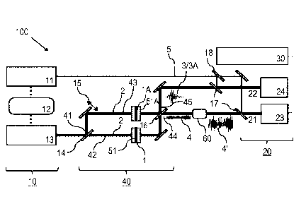
The spectroscopic apparatus 100 of Figure 1 comprises a laser source device
10, including a visible
or Near-Infrared (NIR) femtosecond source 11 for creating a sequence of
initial driving pulses, an
MIR-Infrared (MIR) femtosecond source 13 (including e. g. a LiGaS2 crystal)
for creating a
sequence of MIR pulses based on the driving pulses and a synchronization and
delay unit 12 for a
mutual adjustment of the MIR and driving pulses (e. g. with a delay stage if
the MIR pulses are
generated from the visible or NIR source 11 or with a synchronization and
adjustment of the
repetition rates of the NIR and MIR source). The excitation waves 2 being
provided by the driving
pulses for the interaction with the sample under investigation 1 and the
reference sample 1A are
output from MIR femtosecond source 13.
The excitation waves 2 pulses are split with a 50:50 MIR beam splitter 14,
which provides a first
input port 41 of a Mach-Zehnder interferometer 40, into a first interferometer
arm 42 and a
second interferometer arm 43 of the Mach-Zehnder interferometer 40. The
function of the Mach-
Zehnder interferometer 40 providing an optical adjustment device is described
below. In the first
interferometer arm 42, the sample 1 with the sample container 51 is provided,
including e. g.
biological sample molecules included in water. The reference sample 1A is
included in an identical
Date Recue/Date Received 2021-06-01
CA 03054470 2019-08-23
WO 2018/171869 PCT/EP2017/056705
reference container 51A in the second interferometer arm 43. Preferably, the
sample and
reference containers 51,51A are adapted for low transmission losses in the
whole (mid-)infrared
region (from 2 im to 30 [am). To this end, antireflection coatings can be
provided on the surfaces
of the sample containers 51, 51A for increasing the MIR transmission thereof.
Furthermore, the
sample containers 51, 51A can be arranged with the Brewster angle relative to
the beam paths
along the interferometers arms 42, 43.
Furthermore, a schematically shown delay unit 15 for a mutual adjustment of
the lengths of both
interferometer arms 42, 43 is arranged in the second interferometer arm 43.
The delay unit 15
can be controlled with a control loop (not shown) such that the geometrical
length difference of
the two interferometer arms of the Mach-Zehnder interferometer 40 is
minimized.
Interferometer adjustment can be performed with one or more piezoelectric
transducers (PZT).
By the interaction of the excitation wave 2 with the sample 1 under
investigation and with the
reference sample 1A, the sample wave 3 is created in the first interferometer
arm 42 and the
reference wave 3A is created in the second interferometer arm 43. By the
coherent superposition
of the sample and reference waves 3, 3A at the 50:50 MIR beam
splitter/combiner 16, the dMF
wave 4 is generated at the difference output port 44 (first output port), and
the constructive
coherent superposition of the fingerprint common to both the reference and the
sample wave is
generated at the sum output port 45 (second output port). With the beam
splitter/combiner 16,
the dMF wave 4 is submitted to a first detector channel 21 of the detector
device 20 and the
superposition of the sample wave 3 and the reference wave 3A is submitted to a
second detector
channel 22 of the detector device 20. An optical parametric amplification
device 60 for optical
amplification of the dMF wave 4 (e.g. with optical parametric amplification
(OPA)) and creating an
amplified dMF wave 4 is arranged in the first detector channel 21. Further
details of the optical
parametric amplification device 60 and the function thereof are described
below with reference
to Figures 6 and 7.
The detector device 20 includes electro-optic sampling units 23, 24 each in
one of the detector
channels 21, 22. Parts of driving pulses created with the femtosecond source
11 are submitted as
sampling pulses 5 via MIR-NIR beam combiners 17 and an NIR beam splitter 18 to
the electro-
optic sampling units 23, 24, resp.. The first and second electro-optic
sampling units 23, 24 detect a
temporal amplitude function of the amplified dMF wave 4A and the sum signal
3/3A, resp..
21
CA 03054470 2019-08-23
WO 2018/171869 PCT/EP2017/056705
The calculation device 30 comprises a computer circuit calculating the
spectral response of the
sample under investigation 1 on the basis of a Fourier transformation of the
temporal amplitude
function of the amplified dMF wave 4A detected in the first detection channel
21. It is noted that
the second detector channel 22 is an optional feature of the invention, e. g.
for monitoring or
control purposes.
In practice, the spectroscopic apparatus 100 is adapted for measuring any gas
or liquid of interest.
Furthermore, the applied materials are vacuum compatible (for sample
containers for gases, gas
cells), hard and robust (should not bend when high pressures are applied ¨ for
sample containers
.. for liquid), and/or insoluble materials (against water, acid and solvents).
According to an alternative embodiment of the invention, the spectroscopic
apparatus 100 can be
adapted for SRS measurements based on stimulated Raman scattering of the
sample as described
below with reference to Figures 9 and 10.
In the following, measuring a sample response with the spectroscopic apparatus
100 of Figure 1 is
described. As outlined above, measuring the differential molecular fingerprint
benefits from the
coherent nature of the processes underlying field resolved spectroscopy as
described in WO
2016/102056 Al: (i) the spatio-temporal coherence of electric field
oscillations in the excitation
wave, (ii) excitation of the molecular vibrations in the entire sample volume
in a synchronized
(coherent) fashion by the spatially and temporally coherent excitation wave,
and (iii) re-emission
of coherent radiation (sample wave 3, see Figure 11) by excited molecules
thanks to the perfect
synchronism of their vibrations.
As a direct consequence of (i)-(iii), the electric field oscillations of the
sample wave 3 are perfectly
phase-locked to those of the excitation wave 2. As a result of this coherence,
the sample wave 3
and reference wave 3A emerging from the sample and reference, EGMF(sample)(t),
EGMF(ref)(t), excited by two replicas of one and the same excitation wave 2,
(Eiõ(t))
simultaneously, can be directly compared with each other. In other words, the
GMF from a
sample of interest, EGMF(sample)(t), can be directly referenced to that of a
reference fingerprint,
EGMF(ref)(0, yielding ¨ directly from a single measurement¨the differential
molecular
fingerprint AEGmF(t).
22
CA 03054470 2019-08-23
WO 2018/171869
PCT/EP2017/056705
The preferred implementation of this fundamental concept by means of field-
resolved infrared
absorption spectroscopy consists of the following steps conducted with the
setup of Figure 1.
1) Separate the femtosecond mid-infrared (MIR) pulse (created by the MIR
femtosecond
conversion unit 13 in Figure 1) into two equal parts with the 50/50
beamsplitter 14 (exact
balancing may be achieved with an additional variable attenuator in one of the
two beams after
their splitting).
2) Send one of the MIR excitation pulse (excitation wave 2) through the
reference sample 1A.
Send the other - identical - MIR pulse through the sample 1 under
investigation.
3) Recombine the two transmitted MIR pulses with the beam splitter 16
identical to that used for
the splitting of the beam before the measurement (so that possible minor
residual changes in
waveform imposed by the beam splitter are cancelled upon passing through both
input and
output beam splitter). The setup described under 1)-3) forms the Mach-Zehnder
interferometer
40, the two identical arms 42, 43 of which contain the sample 1 and the
reference sample 1A
(with both being arranged in geometries as identical as possible). As a
consequence, the
dispersion and attenuation of both sample 1/reference sample 1A and sample
containers 51, 51A
are identical except for changes in EGmF(t) caused by differences in molecular
composition.
4) The beam propagation path length in the two interferometer arms 42, 43
preferably are set to
be equal to within one half carrier wavelength of the excitation wave 2 (MIR
pulse). By fine
adjustments of the path length difference within plus/minus half wavelength,
the two pulses
incident on the output beam splitter 16 of the interferometer 40 can nearly
perfectly cancel out
each other, except for differences in their GMF waves rooted in differences in
EGmF(t) between
sample and reference due to their differing molecular composition.
5) Setting the path length difference such that it is minimized, results in
near perfect mutual
cancellation of the excitation pulses carrying approximately 99,9999 % of the
total radiation
energy transmitted through and radiated from the samples. The remaining
approx. 0,0001 % of
the energy is carried in the dMF signal 4 each. If the molecular composition
of the sample 1 and
the reference sample 1A were identical, the sample wave 3 and the reference
wave 3A would be
identical and they also perfectly cancelled out each other. If the molecular
composition of the
sample 1 and the reference sample 1A differ from each other, the sample wave 3
and the
reference wave 3A do not perfectly cancel out but result in a difference
yielding directly
AEGmF(t).
6) Sampling of the electric field of the amplified AEGmF(t) signal 4A with the
electro-optic
sampling unit 23. This can be implemented by the same EOS system used for the
conventional
characterization of individual biomedical samples in Figure 12. The
differential molecular signal
coming without the main pulse offers two significant benefits. First, the [OS
crystal can be
23
CA 03054470 2019-08-23
WO 2018/171869 PCT/EP2017/056705
irradiated with a much higher electric field of the molecular signal, at which
the (much stronger)
excitation wave would irreversibly damage the crystal in the conventional
scheme (Figure 12).
This directly results in a sensitivity increase in addition to that gained
from the differential signal
amplification. Second, the requirement to the dynamic range of the (digital)
electronic system
processing the [OS signal is largely relaxed. The system can be optimized for
detection of the
relevant molecular signal without having to deal with a much stronger
accompanying signal.
7) Fourier transformation of the sampled temporal shape yields the spectral
polarization response
of the sample 1. This can be further processed by the calculation device 30,
e. g. for obtaining
diagnostically relevant information. The spectral features of the polarization
spectrum can be
obtained by subjecting the polarization spectrum to a filtering process.
Specific bands of
compounds characteristic of the health status of a person can be identified.
Furthermore, the
polarization spectrum can be compared with data previously collected with the
same organism
and/or with reference data collected with other, healthy or non-healthy
subjects.
(II) Dispersion compensation of the reference wave
As noted above, the sensitivity of the GMF measurement can be increased if the
GMF signal is
efficiently separated from main pulse (this holds for both reference and
sample waves). This is
due to the background-free detection typical to field-resolved spectroscopy of
WO 2016/102056
Al. compared to other spectroscopic techniques, described in the beginning of
the present
description. An extension of this advantage to the difference GMF can be
obtained, if the
fingerprint common to the reference and sample wave is confined to the
shortest possible time
window, by means of adjusting the chromatic dispersion of the participating
waves accordingly. In
this case, the difference GMF will appear in the sample wave (and in the dMF
signal in the case of
the dMF embodiment) predominantly at the end of the respective wave,
maximizing its
separation from the fingerprint common to the reference and sample waves.
According to this second embodiment of the invention, the adjustment of the
participating waves
includes the temporal separation of the difference GMF from the reference GMF
within the
sample wave by setting the chromatic dispersion in the beam path from the
laser source device
10 to the detector device 20 for compressing the reference wave as illustrated
in Figures 2 to 5.
The temporal separation of the dMF wave from the reference GMF wave can be
provided e. g.
with the embodiment of Figure 1, the SRS measurement of Figure 9 or the
conventional field
resolved spectroscopy of Figure 1.1.
24
CA 03054470 2019-08-23
WO 2018/171869 PCT/EP2017/056705
The temporal separation of the difference GMF from the reference GMF within
the sample wave
preferably is obtained as schematically shown in Figures 2A to 2C and further
exemplified in
Figures 3A to 3F.
Figures 2A to 2C show the second embodiment of the invention without the
interferometric set-
up of Figure 1. The illustrations refer to a variant of the inventive
spectroscopic apparatus 100,
including the laser source device 10 and the detector device 20, wherein only
one single beam
path of the excitation waves 2 is provided, in which the sample or the
reference sample is placed
and the difference of the sample waves and the reference waves 3A is detected
by serial
.. measurements of the sample and reference waves and subsequent calculation
of the difference
thereof. Figures 2A to 2C show the situation, wherein the reference sample 1A
is placed in the
beam path. The laser source device 10 comprises the components 11, 12 and 13
as noted above.
The detector device 20 is adapted for electro-optic sampling of the sample or
reference wave,
using sampling pulses 5 from the NIR femtosecond source 11.
Figure 2A shows the provision of a dispersion adjusting element 53 (optical
adjustment device)
placed after the sample 1. With the MIR femtosecond source 13, excitation
waves 2 are created
being compressed to the Fourier limit. By the reference sample 1A, in
particular the wall material
of the reference container 51A and the reference sample substance included in
the reference
container 51A, the reference main pulse and the reference wave are stretched.
By the effect of
the dispersion adjusting element 53, the reference wave 3A is well-compressed
in time again.
Accordingly, the sensitivity of sensing the dMF wave from the difference of
the sample and
reference waves is increased.
Figure 2B shows the alternative case of providing the dispersion adjusting
element 53 before the
reference sample 1A, while Figure 2C shows the same variant with the sample 1
in the beam path,
instead of the reference sample. Again, the reference wave is well-compressed
in time by the
effect of the dispersion adjusting element 53. As a result, the temporal
compression adjusted to
the reference pulse leads to the dMF signal 4 appearing in the wake of the
sample wave 3. It is
noted that the variants of Figures 2A and 2B are equivalent if the interaction
with the sample or
reference sample is linear. Although in the case of SRS measurement they are
not equivalent, still
both of them can also be implemented for SRS.
CA 03054470 2019-08-23
WO 2018/171869 PCT/EP2017/056705
For an optimized temporal compression of the reference wave, an active and
programmable
dispersion adjusting element 53 can be employed. Examples include acousto-
optic programmable
dispersive filter (or Dazzler) and spatial light modulators.
According to Figure 3A, the excitation wave 2 generally is compressed along
the beam path
towards the detector device 20. This can be done by the effect of the optical
adjustment device
provided by the wall material of the sample container 51, as schematically
shown in Figure 3B,
optionally in combination with the effect of reflective elements 52
introducing negative or
positive dispersion before the reference container 51A (Figure 3C) or after
the reference
container 51A (Figure 3C), or exclusively by the reflective elements 52
introducing negative or
positive dispersion before the reference container 51A (Figure 3E) or after
the reference
container 51A (Figure 3F). The same dispersion setting components are provided
with the beam
path including the sample container (not shown).
The separation effect of shortening the reference wave 3A is schematically
shown in Figure 4,
wherein curve A shows e. g. a 74 fs fwhm bandwidth limited excitation wave 2,
and curves B and C
represent a pulse broadening in a conventional KCI sample container wall
material (10 mm and
100 mm, resp.). Curves B and C strongly overlap the sample GMF of curve D,
thus deteriorating
the detection of the dMF wave 4. With the compression of the reference wave
3A, this overlap is
minimized or excluded.
For optimally compressing the reference wave 3A in time at the field-resolving
detector, the
following two cases can be distinguished:
Firstly, the exciting pulse is already perfectly compressed in time before
entering the
measurement section of the spectroscopic apparatus 100. This would mean that
the components
of the measurement section, like the sample container, mirrors or other
optical components
should not introduce any additional dispersion. This can be accomplished by
the following three
different design strategies.
Design 1: Combine any number of materials and negative and positive group
velocity dispersion.
Thereby the thickness of the individual materials is chosen in a way that the
introduced dispersion
of each material cancels out. The materials might also be used as windows for
a liquid or gas cell
in order to keep the sample of interest in place. Additional anti-reflection
coatings can be applied
to the windows in order to maximize transmission.
26
CA 03054470 2019-08-23
WO 2018/171869 PCT/EP2017/056705
An example for a measurement section based on Design 1, including a liquid
cell sample container
design for a well-compressed laser pulse in time with a central wavelength at
10 j..tm comprises
two 5-mm Germanium windows as walls for the sample container and a 3-mm ZnSe
plate coupled
with one of the Germanium windows. The sample container is arranged under
Brewster's angle
for dispersion compensation. Figure 5 shows the introduced group velocity
dispersion over the
bandwidth of interest. At 10 jim the total GVD is equal to 0.
Design 2: Minimize the total amount of dispersive material. This can be
achieved by either
minimizing the thickness of all transmission windows or dispense them
completely by using freely
streaming liquid jets of the liquid sample of interest. Thereby the produced
liquid film should have
optical surface quality in order to avoid beam distortion and unwanted losses.
The liquid film can
also be placed under Brewster's angle to maximize transmission. Liquid films
with optical quality
have already been demonstrated (see Tauber, M., et al. in "Review of
Scientific Instruments"
74.11 (2003): 4958-4960).
Design 3: Introduce tailored and/or adjustable dispersive elements to
compensate for introduced
dispersion by window materials, optics and/or by components of the sample that
are not of
interest. Those additional dispersive elements could either be chirped
mirrors, spatial light
modulators (SLM) and/or an acousto-optic programmable dispersive filter
(Dazzler).
Secondly, the exciting pulse is chirped before entering the measurement
device. This would mean
that the measurement device must compensate for this chirp to ensure a well
compressed pulse
in time at the field-resolved detector. Similar to case 1, slight variations
of designs 1+3 are
applicable to accomplish this.
Design 1: Combine any number of materials and negative and positive group
velocity dispersion.
Thereby the thickness of the individual materials is chosen in a way that the
introduced dispersion
of each material plus the chirp of the exciting pulse cancels out. The
materials might also be used
as windows for a liquid or gas cell in order to keep the sample of interest in
place. Additional anti-
reflection coatings can be applied to the windows in order to maximize
transmission (requirement
2).
Design 2: Introduce tailored and/or adjustable dispersive elements to
compensate for the chirp
and introduced dispersion by window materials, optics and/or by components of
the sample that
27
CA 03054470 2019-08-23
WO 2018/171869 PCT/EP2017/056705
are not of interest. Those additional dispersive elements could either be
chirped mirrors, spatial
light modulators (SLM) and/or an acousto-optic programmable dispersive filter
(Dazzler).
In general any combination of the above listed designs is adaptable to fulfill
requirement 1 +2 for
the measurement device.
The materials of the container walls, thicknesses of the container walls
and/or dispersion
properties, e. g. of the reflective elements 52, can be selected on the basis
of numerical
simulations of the dispersion along the bam path towards the detector device
20. Sample
container for samples in a liquid sample matrix may comprise e. g. Ge walls
with a ZnSe plate for
dispersion control (having advantages in terms of high transmission and
effective compression), Si
walls with a ZnSe plate for dispersion control, or Thalliumbromidiodide (KRS-
5) walls with a Ge
plate. Sample container for gaseous samples without a sample matrix may
comprise e. g. Ge walls
with a ZnSe plate for dispersion control, KI, Rbl or Csl walls, or KBr, RBr or
CBr walls.
(III) Optimization of the interaction geometry
A further approach for obtaining an optimal access to the sample GMF and/or
the difference GMF
and efficiently use the background-free detection characteristic to FRS
comprises maximizing the
sample GMF wave by optimizing the interaction length with the sample of
interest, as described
in the following.
The optimum interaction length 1 with the sample of interest, in the case of a
strongly absorbing
reference, is
2
1 =
wherein a is the absorption coefficient of the reference sample.
The optimum interaction length 1 is obtained by minimizing the relative error
sa of the retrieval
for a given thickness x and field dynamic range DRE:
2 1 xa
Sa = ¨ * ¨ * e 2
x DRE
The thickness range for which the relative error doesn't deviate more than a
factor 10 compared
to the optimum value yields:
2 1 xa
10 * .s073t = x * DRE *e 2
28
CA 03054470 2019-08-23
WO 2018/171869 PCT/EP2017/056705
1 2 1 xa
* a¨ * e- = - * ¨ * e 2
D RE x DRE
2 xa
10 * a *e1 = - *
The solution of this equation is:
= - Ne __
2W ( 1
) 0.076 xopt
a a 25
2W (-
1 1) 0e 9.78 r
5 x1=
a
Thereby W(x) is product log function.
Thus, an optimum interaction length / in a range from / = 2/25a, to / = 10/a
is obtained.
With more details, the optimum interaction length 1 = -2 is obtained on the
basis of the following
10 considerations. An example of a sample in an absorbing sample matrix
(which constitutes the
reference sample) is constituted by low-concentration solutions of a molecular
species in a
strongly absorbing liquid. Let al be the absorbance of a strongly absorbing
buffer substance and
a2 that of the low-concentration solved molecular species under test. Then,
the intensity of a
certain spectral element is given by:
Is = /0e-a1xe-a2x, (1)
and the "reference" intensity can be considered IR: = . Since electric
fields are measured,
eq. (1) can be written as:
-avc
lE51 = ERle 2 = (2)
Assuming a detector-noise limited sensitivity (as it is the case if the
reference pulse is so short
that it can be efficiently excluded from the time window of the measurement
without losing
significant information on the GMF and if the coupling of intensity and phase
noise via absorbers
in other spectral elements is negligible) and the condition for the minimum
detectable absorption
loss (MDAL) reads:
> NEPE,att y (3)
where NEPE,citt is the noise-equivalent power in the respective spectral
element, after
attenuation through the medium with al:
-a2x 1
1- e 2 > (4)
DRE,att
¨avc
Ct2X
Approximating e 2 by 1 - ¨2 and writing DRE,att = DREe 2 yields:
a2x > __ 2 ¨alx = (5)
DREe 2
29
CA 03054470 2019-08-23
WO 2018/171869 PCT/EP2017/056705
Thus, the MDAL in a2 is reached when the function
aix
e 2
f (x) = ¨ (6)
reaches a minimum. To find this value the first derivative is computed and set
to 0:
ea x (¨ + ¨1 = (1) = 0 .
x2 x 2
The solution is x = 2/a1.
For example, considering water as the buffer substance with al = 600 cm-1. at
9.6 urn an
optimum liquid cell thickness of 33 [inn is obtained. Assuming a dynamic range
of the electric field
measurement of 105, from eq. (5) an MDAL of a2 = 0.0163 crn4 is derived.
(IV) Optical amplification of the sample wave
The difference GMF (the difference of the electric fields of the GMF wave
emitted by the sample 1
and the reference sample 1A, see Figure 1) may be extremely weak. Hence,
before being
measured by electro-optic sampling (or some alternative field sampling
technique), its
amplification would be desirable. According to Figures 6 and 7, optical
parametric amplification
(OPA) is used for this purpose. Efficient OPA requires matching of the phase
velocities of all three
waves involved in the process, the pump wave driving the amplification
process, and the signal
and idler waves being amplified: k, = k, + kJ (with the attributes "signal"
and "idler" being
traditionally connected to the higher- and lower-frequency amplified waves).
If the wave to be
amplified by OPA possesses a super-octave bandwidth, this wave preferably is
the lowest-
frequency "idler" wave in order that the above phase matching condition can be
reasonably well
fulfilled over its entire bandwidth, which is a prerequisite for efficient
amplification without
distortion of the amplified wave. If this phase matching condition is
fulfilled and only the pump
wave and the wave to be amplified (idler) overlap in the OPA crystal, the
latter wave will
(asymptotically) experience an exponential growth.
Ai(z) a Ai(0)e9z
where Ai is the amplitude of the molecular signal (idler wave) upon
propagation along the z
direction in the OPA crystal and g is the OPA gain coefficient proportional to
the amplitude of the
pump wave. A major shortcoming of this simplest implementation of OPA is that
if the amplitude
of the input signal A,(0) is very low it may not sufficiently exceed that of
the radiation emerging
CA 03054470 2019-08-23
WO 2018/171869 PCT/EP2017/056705
spontaneously in the amplifier medium to dominate over this background noise.
If so, the
amplified output would be plagued by intolerable noise. As the present sample
wave 3 to be
amplified indeed may be very weak, the latter problem can be elegantly and
efficiently
circumvented by driving the OPA process not merely with a pump wave but,
simultaneously, with
a signal wave of input amplitude A5(0), which can be easily several orders of
magnitude stronger
than the amplitude of the molecular signal, A5(0) >> Ai (0). Under these
conditions, assuming
again perfect phase matching,
Ai(z) oc As(0)egz
is obtained.
A comparison of the above relationships yields that the amplitude of the
amplified molecular
signal in this latter case is enhanced by
GA5(0)
= _______________________________________
Ai (0)
G can be easily as large as 103 - 105, depending on the initial amplitude of
the molecular wave.
Hence, amplification of the differential molecular signal should be
implemented with a pump-
signal-driven OPA. This is particularly straightforward if the mid-infrared
wave used for
illuminating the molecular systems is generated from the same process. In this
case, the pump
and signal waves leaving the OPA system can be directly recycled for the above
purpose.
This amplification principle is illustrated in Figure 6 showing further
details of the setup of Figure 1
and in Figure 7 showing an alternative embodiment, wherein the amplification
is included in the
detection device 20. According to Figure 6, the sample wave 3 (beam of MIR
pulses after
interaction with sample) is sent to the optical parametric amplification
device 60. The amplified
sample wave 3 is combined with the sampling pulses 5 via the MIR-NIR beam
combiner 17 and
sent to the electro-optic sampling unit 23, which includes an electro-optical
crystal 25, a
Wollaston prism 26 and balanced detectors 27 (as shown in Figures 7 and 12).
In the electro-optic
sampling unit 23, the electro-optical detection is conducted with optical
amplification of the sum-
frequency generated (SFG) signal in the electro-optical crystal 25. The SFG
signal carries the actual
information of the MIR signal (molecular fingerprint signal). According to
Figure 7, the optical
parametric amplification device 60 is included in the electro-optic sampling
unit 23.
31
CA 03054470 2019-08-23
WO 2018/171869 PCT/EP2017/056705
Figure 8 illustrates a variant of the spectroscopic apparatus 100 combining
the interferometric
set-up (embodiment I) of Figure 1 with the dispersion setting (embodiment II)
and the optical
amplification (embodiment IV). In this case, the dispersion adjusting element
53 is placed before
the interferometer 40, and the optical parametric amplification device 60 is
placed in the first
detector channel 21.
(V) SRS embodiment of the spectroscopic apparatus
According to an embodiment of the invention, FRS spectroscopy is used for SRS
measurements.
Figure 9 shows an example of an SRS measurement. It is noted that the
implementation of the
invention is not restricted to this particular set-up but alternatively
possible with modified
variants, in particular with regard to the provision of the pump and Stokes
pulses and the serial
(as shown) or parallel (analogue to Figure 1) measurement of the reference
sample.
According to Figure 9, the spectroscopic apparatus 100 for SRS measurements
comprises the laser
pulse source 10, the detector device 20, and dispersion setting components for
the temporal
compression of the reference wave (embodied by the design of the wall material
of the sample
container 51 or reference container 51A, not shown). The calculation device
for calculating the
spectral response of the sample (see Figure 1) is not shown in Figure 9. The
illustrated
embodiment of the spectroscopic apparatus 100 is adapted for the temporal
separation of the
sample wave from the reference wave. According to an alternative embodiment of
the invention,
the spectroscopic apparatus 100 of Figure 10 can be adapted for an
interferometric separation of
the sample wave from the excitation wave, e. g. according to Figures 1 or 8.
The laser pulse source 10 includes a femtosecond source 11, like a Yb:YAG thin
disk laser creating
driving pulses e. g. with an output energy of 30 ij, a repetition rate of 11
MHz, a centre
wavelength of 1030 nm and a pulse duration of 500 fs (see D. Bauer et al. in
"Opt. Express" 20.9,
p. 9698., 2012; and J. Brons et al. in "Opt. Lett." 41.15, P. 3567, 2016; and
H. Fattahi et al. in "Opt.
Express" 24.21, pp. 24337-24346, 2016). After generation, the driving pulses
are compressed to
their Fourier transform limit. Temporal confinement of the pulses to about 20
fs allows for
detection of molecular free induction decay (FID) with a higher sensitivity
and signal-to-noise
ratio. A femtosecond conversion unit 19 is provided for creating, based on the
driving pulses, a
CEP stable supercontinuum with a spectrum from 450 nm to 2000 nm. The
femtosecond
conversion unit 19 comprises e. g. white light generation in a bulk material
such as quartz. A
portion of the femtosecond conversion unit 12 output is deflected to the first
compression and
32
CA 03054470 2019-08-23
WO 2018/171869 PCT/EP2017/056705
delay unit 13A, including a chirped mirror compressor and a delay unit, for
providing a sequence
of sampling pulses 5 for the electro-optic sampling with the detector device
20.
For the electric field-detection of stimulated Raman scattering, the sample 1
is simultaneously
irradiated with a sequence of e. g. narrowband pump pulses 7 and broadband
Stokes pulses 2 (see
Figure 10) both being created on the basis of the output from the femtosecond
conversion unit
12. The excitation wave is represented by the broadband Stokes pulses 2
supplied to the sample 1
via the second compression and delay unit 13B. The narrowband pump pulses 7
are created with
an acousto-optical modulator 71 (modulating at a MHz frequency) and an ethalon
72, e. g. with a
center wavelength of 1030 nm and a pulse duration of 1 ps. After the
interaction with the sample
1, the modified probe light comprises the pump pulses and the Stokes pulses 8
enhanced by a
vibrational Raman response of the sample 1. Before electro-optic sampling with
the detection
device 20, the enhanced Stokes pulses 8 pass a long pass filter 73 (e. g. 1050
nm) suppressing the
pump light. The enhanced Stokes pulses 8 represent the sample wave 3 described
above. For
implementing the dMF measurement, the sample 1 is replaced by a reference
sample and a
reference wave is detected, including enhanced Stoke pulses excited in the
reference sample.
The detector device 20 for electro-optic sampling is designed as described
above. The sampling
pulses 5 provided by the first compression and delay unit 13A are superimposed
with the sample
waves, e. g. the enhanced Stokes pulses 8, and both are simultaneously
transmitted via the
electro-optical crystal 25 (e.g. BBO crystal), a 700 nm short pass filter and
a )1/4/4 plate, the
Wollaston prism 26 to the balanced detectors 27.
With an alternative embodiment, broadband pump pulses and narrowband Stokes
pulses are
created and the excitation wave comprises the narrowband Stokes pulses,
wherein the probe
light comprises the broadband pump pulses and the modified probe light
comprises the pump
pulses diminished by a vibrational Raman response of the sample. According to
another
alternative embodiment, the spectroscopic apparatus 100 of Figure 9 can be
adapted for an
interferometric separation of the sample wave (enhanced Stokes pulses 8) from
the reference
wave, e. g. according to Figure 1. In particular, the Mach Zehnder
interferometer of Figure 1 can
be provided, including the sample 1 in a first interferometer arm and a
reference sample in a
second interferometer arm. The pump and Stokes pulses are split into both of
the first and second
interferometer arms.
33
CA 03054470 2019-08-23
WO 2018/171869 PCT/EP2017/056705
The field-detection of Stokes pulses according to Figure 9 represents a novel
femtosecond SRS
scheme. The increased sensitivity in this embodiment of the invention is due
to the confinement
of the excitation Stokes pulses in a few femtosecond (fs) temporal window. The
Stokes gain can
be resolved in picoseconds time frame starting from hundreds of fs, and
outside the temporal
window of the excitation pulses. As the molecular response decay exponentially
over time, the
background free measurement allows for higher sensitivity.
In fs SRS the simultaneous interaction of a narrow-bandwidth ps Raman pump
pulse 7 and a
broadband, few-cycle Stokes pulse 2, creates a macroscopic polarization in the
sample. The
narrow bandwidth of the pump pulses 7 provides the high spectral resolution
required for
resolving molecular fingerprint. During the process sharp vibrational gain
features appear on top
of the Stokes envelope and equivalently an exponential decay of the order of
hundreds of ps in
the time domain. The process is shown in Figure 10. The Stokes pulse initiates
vibrational
coherence of molecules in the sample, which are decaying with their
vibrational dephasing time
'vb. This finite duration of the vibrational coherence result in a limited
bandwidth in the frequency
domain and the induced coherent vibrational motion modulates the macroscopic
polarization at
the vibration frequency (Kukura, P. et al. in "Annu. Rev. Phys. Chem." 58.1,
pp. 461-488, 2007).
The entire fingerprint region of a sample can be detected by measuring the
enhanced Stokes
pulses 3 in the time domain.
The features of the invention disclosed in the above description, the drawings
and the claims can
be of significance individually, in combination or sub-combination for the
implementation of the
invention in its different embodiments.
34