Note : Les descriptions sont présentées dans la langue officielle dans laquelle elles ont été soumises.
%
a
CA 03060334 2019-10-17
¨ 1 -
OCT Image Capture Device
The invention relates to an OCT examination device for recording an object by
means of
optical coherence tomography, comprising an OCT radiation source, which emits
OCT
radiation with a wavelength of from 400 nm to 2000 nm and a spectral bandwidth
that
comprises at least a range of from 20 nm to 400 nm, or has a narrow bandwidth
of less
than 20 nm to 400 nm, the radiation source being tunable in such a way that
the narrow
bandwidth forms a wider bandwidth of from 20 nm to 400 nm by time-offset
emission of
waves at different wavelengths, an OCT beam path, comprising an OCT output
direction
of the OCT radiation from the OCT radiation source, and an OCT input direction
of OCT
radiation scattered back by an image object, an OCT radiation receiver for
receiving the
backscattered OCT radiation of the OCT radiation source, a housing, which
contains the
OCT radiation source and the OCT radiation receiver, an exit opening formed in
the
housing for the OCT radiation of the OCT radiation source, an entry opening
formed in
the housing for the backscattered OCT radiation of the OCT radiation source,
an OCT
exit direction of the radiation through the exit opening, an OCT entry
direction of the
backscattered OCT radiation through the entry opening, a control unit, which
is connected
in terms of signal technology to the OCT radiation source and the OCT
radiation receiver
and is configured in order to record a multiplicity of measurement profiles
mutually
separated in a recording period and, within the recording period, to drive the
OCT
radiation source in order to emit the OCT radiation and the OCT radiation
receiver in
order to receive the backscattered OCT radiation.
Optical coherence tomography (OCT) is an examination method in which light
with a
short coherence length is used with the aid of an interferometer for distance
measurement in scattering materials. The OCT examination method is used for
example
in medicine for examination in vivo and in vitro, and besides this medical
application is
also applied in other fields outside medicine in order to examine scattering
materials. In
principle, it is to be understood that the OCT diagnosis device according to
the invention
and the method according to the invention for OCT image capture of an object
may be
used as diagnostic methods which are carried out on the human or animal body,
but may
also be used as analysis methods for a different purpose, i.e. with the
exclusion of those
,
s
CA 03060334 2019-10-17
- 2 -
diagnostic methods which may carried out on the human or animal body. OCT
examination methods are distinguished in that the examined material is not
modified by
the OCT examination process and therefore have neither a surgical not a
therapeutic
effect. It is to be understood that the claimed method for the OCT image
capture of an
object is the subject-matter of this description and the claims in territories
which exclude
diagnostic methods on human bodies from patent protection, with the exclusion
of the
protection of such diagnostic methods on the human body.
OCT systems are measuring systems which, by aligning a measurement beam with
an
area, to be considered in an idealized way as a point, of the object to be
examined, carry
out a point measurement on this object on the basis of the backscattered
radiation. In
order to image the object surface and the depth region lying immediately below
it, with a
typical penetration depth of from 1 to 3 mm, over a sizeable surface region or
as a whole,
OCT measuring systems must therefore move the measurement beam over the
surface
by scanning, which is typically carried out as a line scanning. This scanning
is achieved in
OCT measuring systems by motor-driven deflecting mirrors. High-quality OCT
image
capture is therefore carried out by fixing the object to be examined,
arranging the OCT
measuring device in a fixed position with respect to the object, and then
scanning the
surface of the object by automatically performed scanning by means of the
deflecting
mirror, so as to obtain a representation of the surface and of the region
lying immediately
below the surface by a multiplicity of single-point measurements.
This measurement method has generally been tried and tested, achieves reliable
imaging
qualities and may be carried out with a fast measurement. A disadvantage,
however, is
that fixing the object to be examined is necessary in order to avoid a
relative movement
between the object and the OCT measuring device over the scanning period. This
fixing
is disadvantageous particularly when using the OCT measuring device for
diagnostic
purposes, for example in order to examine the retina of the human eye, since
the outlay
required therefor in order to fix the head or the eye is considerable.
An operation microscope having an OCT examination device coupled in is known
from
US 2017/0027438. The OCT measurement beam is in this case coupled into the
beam
path of the microscope by means of two mobile mirrors and a stationary mirror,
and is
consequently deflected three times. The guide beam paths of the OCT beam are
intended to remain substantially parallel to the optical axis of the
microscope 100 by the
doubly controlled deflection, in order to avoid image distortions. The
technology
previously known to this extent is not suitable for keeping the angle between
the output
direction of an OCT beam from the OCT source and the exit direction of the OCT
beam
from the housing constant, but can only achieve this angle being substantially
constant.
CA 03060334 2019-10-17
- 3 -
Such a configuration, in which the angle cannot however actually be kept
parallel to the
axis of the microscope because of the double deflection and the principle of
scanning the
examination region by means of the double deflection, is not to be understood
in the
sense according to the invention that the angle between the output direction
of the OCT
beam from the OCT source and the exit direction of the OCT beam from the
housing is
kept constant thereby. The device thus previously known captures the
examination region
by means of the OCT device by a scanning process, and therefore requires
fixing of the
device in relation to the object to be examined. Furthermore, the device
occupies
considerable installation space and is therefore unwieldy to use in narrow
spatial
.. situations.
There is therefore a need for an OCT examination device which, for carrying
out the OCT
examination, is simplified overall and is less sensitive to measurement errors
that occur
because of a relative movement between the OCT measuring device and the object
to be
examined.
.. This object is achieved with an OCT examination device of the type
mentioned in the
introduction, in that the control unit is configured in order to keep the OCT
output direction
and the OCT exit direction constant with respect to one another in their
angular
orientation during the recording period.
According to the invention the image capture is also carried out by scanning
the surface
of the object to be examined with a measurement beam in the case of the OCT
examination device according to the invention. This image capture is carried
out during a
recording period which starts with the recording of the first measurement
point in the scan
and ends with the recording of the last measurement point in the scan. Within
this
recording period, a plurality of, i.e. at least two and in general a few
hundred or thousand,
.. measurement points are thus addressed with the measurement beam and the
surface is
thereby scanned, for example in lines or spirally.
While this scan is carried out in the prior art in an automated manner by a
motor-
adjustable deflecting mirror or other beam deflecting devices, so that the OCT
output
direction of the beam from the OCT radiation source is variable in terms of
its angular
orientation relative to the OCT exit direction of the measurement beam from
the OCT
examination device, and is varied during the recording period in order to
carry out the
scanning, in the case of the OCT examination device according to the invention
the
angular orientation of the OCT output direction and of the OCT exit direction
are kept
constant during the entire recording period. The OCT examination device
according to the
.. invention can therefore obviate an adjustment, carried out in a motorized
or other fashion,
,
CA 03060334 2019-10-17
- 4 -
of this angular orientation between the OCT output direction and the OCT exit
direction,
and in particular also an adjustable deflecting mirror, which has the further
advantage that
the OCT examination device according to the invention may be configured more
compactly than previously known OCT examination devices. Instead of the
motorized
adjustments of a deflecting mirror or a correspondingly differently
constructed variable
beam deflecting device according to the prior art, the compact configuration
of the OCT
device makes it possible to carry out the scan of the object surface to be
examined by a
movement of the OCT examination device relative to the object to be examined.
The
relative movement may be carried out as a displacement or tilt, or a
combination thereof,
of the OCT examination device itself, although the object to be examined may
also be
displaced or tilted with the OCT examination device kept spatially stationary,
in order to
produce the relative movement which is required for the scan.
In the OCT examination device according to the invention, the OCT measurement
beam
is directed through an exit opening onto the object to be examined and the
backscattered
OCT radiation re-enters the OCT examination device through an entry opening.
In
principle, it is to be understood that although two separate openings may be
provided in
particular applications, it is particularly preferred for the entry opening
and the exit
opening to be formed by a single housing opening. In particular, the OCT
radiation
coming from the single opening, i.e. the measurement beam and the
backscattered OCT
radiation, may extend coaxially, which makes the OCT examination device
insensitive to
different measurement distances between the entry/exit opening and the surface
to be
examined.
According to another preferred embodiment, the exit opening and the OCT
radiation
source are arranged in such a way that the OCT exit direction and the OCT
output
direction extend parallel to one another, in particular coaxially. According
to this
embodiment, the radiation emerging from the OCT radiation source may be
oriented
directly in the direction of the exit opening and no longer needs to be
deflected in order to
pass from the OCT radiation source through the exit opening. This embodiment
is
suitable in particular for a slim design of the housing, for example in the
form of an
elongate tubular housing, which allows particularly user-friendly handling of
the OCT
examination device.
According to another preferred embodiment, the OCT examination device is
refined by an
observation instrument comprising
an illumination radiation source, which emits light in the visible or infrared
observation
wave range into an illumination beam path passing through the exit opening,
and an
CA 03060334 2019-10-17
- 5 -
observation image sensor, which is sensitive to radiation in the observation
wave range
and which receives reflected light in the visible or infrared observation wave
range from
an observation beam path passing through the entry opening, the observation
beam path
and the OCT exit direction extending parallel to one another, in particular
coaxially. Such
an observation instrument allows a user to observe the examined object
simultaneously
with the OCT image capture by sampling with another examination method, which
images
the surface with light in the visible or infrared range. This observation
instrument on the
one hand makes it possible to carry out a simultaneous examination by means of
two
different imaging methods, and on the other hand by means of the observation
instrument
the alignment of the OCT measurement beam with the object to be measured may
be
monitored while carrying out the OCT examination, and the OCT measurement beam
may be aligned with the aid of the image recorded by the observation
instrument. In
particular, the position of the measurement beam in an image which is recorded
by
means of the observation instrument may be marked. While in the absence of an
observation instrument the user would have to carry out the scan of the object
to be
examined without a direct position check, this may be carried out in a
controlled way in
the presence of an observation instrument and the control unit may, in
particular, be
configured in order to overlay the alignment of the measurement beam of the
OCT exit
direction and the previous scan points of this measurement beam, i.e. the
profile of the
scan, into an image which has been compiled by means of the observation
instrument.
The scan of the examination object is carried out according to the invention
by a relative
movement between the OCT examination device and the object to be examined. In
contrast to the prior art, it is therefore not necessary for the object to be
examined and the
OCT examination device to be constant, or invariant, in their position with
respect to one
another during the recording period and for the scan to be carried out by a
varying
internal deflection or deviation of the OCT measurement beam inside the OCT
examination device, but rather the scan and therefore the relative movement of
the OCT
measurement beam is carried out by a relative movement between the OCT
radiation
source and the object to be examined. This relative movement may in particular
be
carried out manually by a user who guides the OCT examination device by hand.
In
principle, in a simplified form, this may be carried out in such a way that
the user carries
out a scan movement manually and without a visual check, relying on the fact
that such a
line-by-line scan can be carried out reliably in a motorized fashion. The
manual scan may,
however, also be assisted or guided by a visual check, for example by a
movement
direction indicator being displayed to the user on a screen, which indicates
the scan
direction which he should carry out manually by displaying an arrow or the
like, or by the
regions already recorded with the OCT measurement beam being displayed to the
user
by corresponding reproduction of the OCT examination image, so that the
recorded
=
CA 03060334 2019-10-17
- 6 -
surface of the object to be examined is built up simultaneously with the scan
according to
the scan movement which the user is carrying out manually. This allows the
user to check
and correct the scan path which he is carrying out manually with the aid of
the OCT
examination image being built up in real-time, or with a time delay, and
thereby manually
carry out a scan guided spirally or in lines or in another geometrical shape.
In this case, the OCT examination device may furthermore be refined in that
the
electronic control unit comprises an image processing unit, which is
configured in order to
combine a first image, formed by the reflected light received by the
observation image
sensor, and a second image, adjacent to the first image and formed with a time
offset
with respect to this first image from the reflected light received by the
observation image
sensor, in order to form an overall image. This configuration of the
electronic control unit
makes it possible from the scan movement, the recording resulting from the
relative
movement between the OCT examination device, or the OCT radiation source, and
the
examined object, recording of a plurality of images with a time offset with
respect to one
another during the recording period to compile an overall image of the scanned
surface of
the examined object. This combination may be carried out by joining the
individual
images together, overlapping regions with identical image contents
correspondingly being
arranged in an overlapping manner, or these image contents being taken from
only one of
the two joined images, in order to achieve a unique and gap-free compilation
of the
overall image. The electronic control unit may, in particular, be configured
in order to
combine a plurality of images to form an overall image. Furthermore, the
electronic
control unit may be configured in order to mark the regions recorded with the
OCT
measurement beam inside these images. Furthermore, the electronic control unit
may be
configured in order, with the aid of the composed images of the observation
instrument,
also to combine the image values determined with the OCT measurement beam to
form
an overall OCT image. This may, in particular, be carried out in such a way
that with the
aid of the positioning, established by overlap, of the images of the
observation instrument
with respect to one another, the placement of the individual OCT measurement
points
with respect to one another is determined and these are assigned to one
another in their
placement.
Furthermore, it is also preferred for the image processing unit to be
configured in order to
identify an intersection region of the first and second images, in which a
matching image
section is reproduced in the first and second images, and to combine the first
and second
images in such a way that the overall image is composed of the first and
second image
with an overlap of the first and second images in the matching image section.
With the aid
of the matching image regions thus established by the control unit, unique
assignment of
the placement of the two images to be joined in their relation to one another
may be
=
CA 03060334 2019-10-17
- 7 -
carried out, and a gap-free overall image may therefore be compiled. In
particular, the
identification of matching image contents into two or more different images
allows unique
assignment of OCT measurement points in the respective images, so that a
unique
assignment of the placement of these OCT measurement points may also be
carried out
on the basis of such an established overlap.
Furthermore, provision may in this case be made that the image processing unit
is
configured in order to rectify the first and/or the second image, in
particular by the first
and/or the second image being tilted about an image surface normal as a tilt
axis, and/or
the first and/or the second image being scaled, in particular being scaled in
all regions
with a matching scaling factor or with a scaling factor decreasing in one or
two mutually
perpendicular spatial directions. According to this refinement, the image
processing unit is
configured in order, in the course of the identification of matching image
sections, to carry
out rectification which may consist in tilting or scaling, or both. Scaling is
in this case also
to be understood as scaling in only one axial direction, i.e. scaling which
does not keep
the side length ratios of the image constant, but changes them. By such
scaling or tilting
of the image, different recording directions and different recording scales,
as well as
composite distortion effects resulting therefrom, may be compensated for and
two
adjacent images which have a different recording angle or recording scale
because of
such distortion effects may be combined without these distortions occurring as
errors in
the overall image. In particular, the control unit may be configured in order
to carry out a
comparison of two images in the check for matching image components, which
takes
such distortion effects into account by rectification of the images taking
place and the
comparative analysis of the images over matching image sections being carried
out on
the basis of the rectified images.
Furthermore, it is also preferred for the control unit to be configured in
order to process a
measurement profile, compiled from the backscattered OCT radiation received by
the
OCT radiation receiver, and an image recorded simultaneously from the light
received by
the observation image sensor, and to mark a region in the image which
represents the
position of the measurement profile. By the simultaneous processing of the
backscattered
OCT radiation and of the measurement profile compiled therefrom and of the
light
received by the observation image sensor, a spatially unique assignment of the
OCT
measurement profile in relation to the image recorded by the observation image
sensor is
possible, and sequencing of the individual OCT measurement points in the
spatially
correct placement with respect to one another may as a result be carried out
with the aid
of the images recorded by the observation image sensor.
r
,
CA 03060334 2019-10-17
- 8 -
According to another preferred embodiment, the OCT radiation source and the
OCT
radiation receiver are arranged immovably in the housing. By such an immobile
arrangement on the one hand a robust configuration of the OCT examination
device is
achieved, and furthermore, because of this spatially immobile arrangement, the
OCT
examination device may be constructed compactly and fitted in a slim housing.
Furthermore, it is also preferred for the OCT beam path to extend immovably in
the
housing. By such an immobile profile of the OCT beam path, which comprises
both the
emitted OCT radiation from the OCT radiation source as far as the exit opening
in the
housing and also the received backscattered OCT radiation from the entry
opening as far
as the OCT radiation receiver, according to the invention any mobile or
controllable
deflecting means for the OCT radiation inside the examination device are
obviated, which
overall makes a robust and slim construction of the OCT examination device
possible.
Furthermore, it is also preferred for the OCT beam path to be static in
relation to the
illumination beam path and the observation beam path. By such a static, i.e.
immobile
relative to one another, arrangement of the OCT beam path with respect to the
observation and illumination beam paths, according to the invention a clear
assignment,
already established by the structure of the OCT examination device, of the
orientation of
the OCT measurement beam and of the thereby defined position of the OCT
measurement point is achieved in the image obtained by the observation device.
Furthermore, it is also preferred to refine the OCT examination device by a
placement
recording unit, which is configured in order to determine the placement, in
particular the
position and/or the orientation of the OCT radiation receiver in relation to a
static
reference coordinate system, and an image processing unit, which is coupled in
terms of
signal technology to the placement recording unit and is configured in order
to determine
a first measurement profile of the backscattered OCT radiation received by the
OCT
radiation receiver at a first instant, to determine a first placement of the
OCT radiation
receiver at the instant of receiving of the backscattered OCT radiation of the
first
measurement profile from placement data which have been transmitted from the
placement recording unit at the first instant to the image processing unit, to
determine a
second measurement profile, adjacent to the first measurement profile, with a
time offset
with respect to this first measurement profile from the backscattered OCT
radiation
received by the OCT radiation receiver at a second instant, to determine a
second
placement of the OCT radiation receiver at the instant of receiving of the
backscattered
OCT radiation of the second measurement profile from placement data which have
been
transmitted from the placement recording unit at the second instant to the
image
processing unit, to determine the relative placement of the first measurement
profile with
respect to the second measurement profile with the aid of the first and second
CA 03060334 2019-10-17
- 9 -
placements, and to combine the first and second measurement profiles to form
an overall
measurement profile by entering the first and second measurement profiles into
the
overall measurement profile at the previously determined relative placement.
According to
this embodiment, the OCT examination device furthermore comprises a placement
recording unit. This placement recording unit is configured in order to
determine a
placement of the OCT examination device. This placement determination may be
carried
out by the placement recording unit in such a way that a relative change in
the placement
of the OCT examination device takes place between a first and a second
instant, for
example by an acceleration of the OCT examination device or accelerations of
the OCT
examination device along a plurality of axes being recorded by one or
corresponding
more acceleration sensors, which are a component of the placement recording
unit. The
placement recording unit may, as an alternative or in addition, also comprise
sensors or a
sensor which record(s) orientation of the OCT examination device in relation
to the
direction of the force of gravity, so as to record an absolute placement
orientation of the
OCT examination device. The placement recording instrument may furthermore
contain
one or more gyroscopes as sensors, in order to register displacements or tilts
along a
plurality of axes.
The placement recording instrument may be configured in such a way that it
records the
placement of the OCT examination device in relation to a reference system
which is
separate from the OCT examination device and is installed statically in the
environment,
or a correspondingly statically installed reference point.
By the placement recording of the OCT examination device, on the one hand the
placement, i.e. the position and orientation, of the OCT examination device at
the instant
of an image capture may be determined, and consequently with the aid of the
thus
determined placement of the OCT examination device joining of a plurality of
images or
OCT measurement profiles recorded at time intervals and while changing the
position of
the OCT examination device may be carried out. This may on the one hand be
carried out
with the aid of absolutely determined placements of the OCT examination
device, but as
an alternative or in addition also with the aid of relative placement changes
of the OCT
examination device between two images, or OCT measurement points, recorded
with a
time offset. It is to be understood that this joining of OCT measurement
profiles to form an
overall measurement profile may be carried out merely with the aid of the data
of the
placement recording unit, although the data of the placement recording unit
may also be
used in addition to another procedure for joining the individual measurement
profiles to
form an overall measurement profile, for example by the data of the placement
recording
unit in addition to joining the measurement profiles with the aid of the
images recorded by
,
CA 03060334 2019-10-17
- 10 -
the observation instrument, determination of their overlap and the thus
defined placement
of the individual OCT measurement profiles with respect to one another being
carried out.
Another aspect of the invention is a method for OCT image capture of an object
by
means of optical coherence tomography, having the steps:
- emitting OCT radiation with a wavelength of from 400 nm to 2000 nm and a
spectral bandwidth that
o comprises at least a range of from 20 nm to 400 nm, or
o has a narrow bandwidth of less than 20 nm to 400 nm, the radiation source
being tunable in such a way that the narrow bandwidth forms a wider
bandwidth of from 20 nm to 400 nm by time-offset emission of waves at
different wavelengths,
from an OCT radiation source into an OCT beam, comprising
o an OCT output direction of the OCT radiation from the radiation source,
and
o an OCT input direction of OCT radiation scattered back by an image
object,
- receiving the backscattered OCT radiation of the OCT radiation source in
an OCT
radiation receiver,
a housing, which has an exit opening and an entry opening for the OCT beam
path,
containing the OCT radiation source, the OCT radiation receiver,
- guiding the OCT radiation through the exit opening in an
OCT exit direction,
- guiding the backscattered OCT radiation through the entry opening in an
OCT
entry direction,
driving the OCT radiation source and the OCT radiation receiver by means of a
control unit in order to emit the OCT radiation by means of the OCT radiation
source and in order to receive the the backscattered OCT radiation by means of
the OCT radiation receiver over a recording period,
- recording a plurality of mutually separated measurement
profiles from the
backscattered OCT radiation during the recording period, wherein the OCT
output
direction and the OCT exit direction are kept constant with respect to one
another
in their angular orientation during the recording period.
The OCT image capture method according to the invention is distinguished in
that, during
the image recording in which a plurality of OCT measurement points are
addressed and a
plurality of OCT measurement profiles are correspondingly recorded, no change
takes
place between the output direction of the OCT radiation from the OCT radiation
source
CA 03060334 2019-10-17
- 11 -
and the exit direction of the OCT radiation from the OCT examination device.
As
explained in the introduction, the method for the OCT image capture of an
object may be
carried out for any OCT examinations of objects, the method may optionally be
carried
out with the exclusion of use as a diagnostic method for human or animal
bodies.
The method may be refined in that, between the recording of the first and
recording of the
second image, the observation image sensor is moved, in particular displaced
and/or
tilted relative to the image object. The relative movement between the
observation image
sensor and the image object to be examined may in this case either be carried
out by the
observation image sensor being moved, in particular with the entire OCT
examination
device in which the observation image sensor is arranged being moved, with the
object to
be examined being stationary, or by the object to be examined being moved with
the
observation image sensor being fixed. In principle, the relative movement may
also be
carried out by a movement both of the observation image sensor and of the
object to be
examined.
.. According to another preferred embodiment, a first measurement profile and
a second
measurement profile are recorded within the recording period, and the first
and second
measurement profiles are assigned to one another in their spatial placement
and
combined to form an overall measurement profile, in particular by,
simultaneously with the
recording of the first measurement profile, a first image being recorded by
means of
emitting light radiation in the visible or infrared wavelength range and
receiving the light
reflected by an object, simultaneously with the recording of the second
measurement
profile, a second image being recorded by means of emitting light radiation in
the visible
or infrared wavelength range and receiving the light reflected by the object,
the position of
the first measurement profile in the first image being marked, the position of
the second
measurement profile in the second image being marked, the first and second
images
being combined with the aid of image analysis to form an overall image by a
matching
image region of the first and second images being arranged in an overlapping
manner,
and the relative positioning of the first measurement profile with respect to
the second
measurement profile being determined with the aid of their position in the
overall image,
and the first and second measurement profiles being combined with the aid of
the relative
positioning determined in this way to form an overall measurement profile, or
by a first
placement of the housing being recorded simultaneously with the recording of
the first
measurement profile, a second placement of the housing being recorded
simultaneously
with the recording of the second measurement profile, the relative positioning
of the first
measurement profile with respect to the second measurement profile being
determined
with the aid of a relative change in the first placement with respect to the
second
placement of the housing, and the first and second measurement profiles being
combined
CA 03060334 2019-10-17
- 12 -
with the aid of the relative positioning determined in this way to form an
overall
measurement profile.
According to this embodiment, a plurality of measurement profiles are combined
to form
an overall measurement profile, and in this way a corresponding OCT
examination
representation of an examined surface of the object and of an underlying near-
surface
volume region below this examined surface is generated. The joining of the
individual
measurement profiles obtained by the scan to form the overall measurement
profile may
in this case be carried out with the aid of an image analysis with
determination of
matching image contents and corresponding joining of the individual images to
form an
overall image ("stiching") or with the aid of a placement determination of the
placement of
the housing of the OCT examination device and correspondingly of the placement
of the
OCT measurement beam which emerges from this housing, or a combination of
these
two methods may be carried out in to join individual measurement profiles form
an overall
measurement profile.
The implementation and advantages of the invention may be found from the
following
descriptions of the figures. Various exemplary embodiments of the present
invention are
represented in the figures. The figures, the description and the claims
contain numerous
features which have a function individually or in combination. It is to be
understood that all
features are expediently both to be considered individually and to be
understood together
in further appropriate combinations.
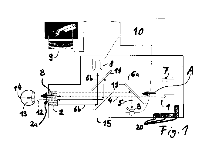
Fig. 1 shows a schematic structure of the OCT device according to the
invention. For the sake of better representability, the OCT device is
represented larger in proportions than an eye to be measured.
Fig. 2 shows a first exemplary embodiment of the invention during use.
Figs 3a and 3b show a second exemplary embodiment, in which the patient is
requested
to follow a moving fixation light with his eye. The fixation light may be
generated and moved by a screen inside an arrangement.
Fig. 4a shows an exemplary representation of a first measurement result
of the
OCT device according to the invention as a single video image with
central marking of the OCT measurement point and an A-scan
associated therewith.
CA 03060334 2019-10-17
- 13 -
Fig. 4b shows an exemplary representation of a second measurement
result of
the OCT device according to the invention with sequenced video images,
a resulting line of the OCT measurement points and an associated B-
scan.
The OCT device 15 for achieving the object comprises a short-coherence
radiation
source (for example SLD) (1) which is distinguished by a corresponding
wavelength (400
nm ¨ 2000 nm) and spectral bandwidth (20 nm ¨ 400 nm) for carrying out optical
coherence tomography (OCT). The OCT device furthermore comprises optics (2),
with
which the measurement beam (4) leaves collimated in the direction of the
object to be
measured.
From the radiation source, the OCT radiation emerges in an OCT output
direction A and
strikes the optics. The OCT radiation passes through the optics and emerges in
an exit
direction B from an exit opening 2a.
Optionally, instead of the broadband light source, a rapidly tunable light
source (so called
swept source) with a smaller bandwidth may be used.
The arrangement furthermore contains an OCT detector (3) for recording the
backscattered OCT radiation (5) of the measurement beam. In the case of the
broadband
light source the OCT detector may be a spectrometer which displays the
backscattered
OCT radiation spectrally decomposed on a linear sensor array (Fourier domain
OCT). In
the case of the tunable OCT radiation source, the OCT detector may consist of
a simple
point light sensor (photodiode) (swept source OCT). In the so-called time
domain OCT
mode, in which the path length of the reference beam is buried during the
measurement,
the OCT detector may likewise consist of a point detector.
Collinearly with the OCT measurement beam, there is an imaging beam path for
video
recording. The imaging beam path (for visible and infrared light) is, for
example, coupled
in through semitransparent mirrors (11). It consists of an illumination beam
(6a) and an
observation beam path (6b). Illumination (7) is, for example, generated by an
LED in the
visible or infrared wavelength range. The reflected light (6b) strikes a light-
sensitive
sensor (8), for example a CCD chip, through imaging optics (2). The image
information of
the CCD chip is on the one hand visualized directly on a monitor (9), and on
the other
hand the image is stored digitally on a data medium in a control computer
(10).
If, for example, the OCT device is placed on the cornea (12) of the eye (13),
the imaging
beam path is designed in such a way that a small section of the retina (14) of
the eye can
CA 03060334 2019-10-17
- 14 -
be represented thereby. Movement of the arrangement (15) relative to the eye
(13), for
instance by tilting or lateral displacement by the hand of the examiner (16),
illuminates a
new area of the retina and correspondingly represents this image on the
monitor and
stores the information at a sufficiently rapid cycle rate on the data medium
in the control
computer (10).
This process is systematically comparable approximately to searching for a
surface
concealed in darkness with a searchlight. If the illuminated area is
remembered, an
arbitrarily large composed image of the searched ¨ scanned ¨ surface is
obtained.
Simultaneously with the freehand scanning of the retina surface by the imaging
beam
path, a depth profile of the retina is produced by means of an OCT measurement
beam at
the center of the illuminated area, or video section. The OCT measurement beam
in this
case penetrates into the position to be measured on the cornea (or another
subject), and
a part of the OCT radiation is reflected or scattered back to the detector.
The
backscattered OCT radiation is superimposed interferometrically with a
reference beam.
This gives rise to individual axial interferograms. A single interferogram
(optical cross-
correlation) of a reference beam and measurement beam gives a linear pattern
that
images the strength of the light-reflecting structures and their relative
optical path length
as an axial depth profile (A-scan or amplitude-mode scan). By moving the
arrangement,
the measurement beam is then guided transversely over the surface of the
retina, so that
a flat tomogram (B scan or brightness-mode scan) or even a three-dimensional
volume
(c-mode scan) may be recorded by scanning.
As alternative to moving the OCT device 15, in another embodiment, with the
OCT device
static, the patient may be requested to follow a moving fixation light (17)
with his eye. The
fixation light may be generated by a screen inside the arrangement and moved
on the
screen. The measurement beam (5), the backscattered light (5) and the imaging
beam
path (6a, 6b) may pass through a central opening (19) onto the eye (13) and
back again
into the measuring arrangement (15). By the movement of the patient's eye, a
scan of the
retina is likewise formed.
Since the freehand movement of the described arrangement (or the movement of
the
eye) and therefore the scanning over the retina often takes place with
insufficient
definition or reproducibility, it is advantageous for the resulting two-
dimensional
photographic surface images of the retina, as well as the individual axial
depth profiles to
be subsequently combined by software in the control apparatus ("stitching").
CA 03060334 2019-10-17
- 15 -
With the aid of the control computer (10), to this end the individually
recorded video
surface recordings (20) are transferred into a virtual coordinate system and
finally
combined to form an overall image (21). The position of the OCT measurement
beam
may be marked as a point (22) in each individual surface recording. According
to the
movement of the arrangement, one or more continuous lines (23) on the overall
recording
(21) are obtained from the individual marking points. Synchronously with the
recorded
line, each individual depth scan of the OCT measurement (A-scan) (24) along
the line
may be plotted graphically in order to obtain a corresponding two-dimensional
tomogram
(B-scan) (25).
The combining of the individual images (20) of the surface to form an overall
image (21)
is carried out in such a way that an overlap of two adjacent images in the
respectively
matching image sections is maximized, and optionally the individual images are
rectified
for a congruent overlap.
In addition, a plurality of, for example three, sensors (30) are fitted in the
arrangement
which register and record the movement of the arrangement in all three spatial
directions.
This may simplify the combining of the individual images and make it more
precise. Such
sensors (30) may, for example, be acceleration sensors which register
translational
and/or rotational movements or register the orientation of the arrangement in
relation to
up and down by means of gravity. Gyroscopes may likewise be used as sensors
(30) in
order to register and track movements of the measuring arrangement. With the
aid of
these sensors, the orientation of the OCT measurement beam and of the imaging
beam
path may be determined at any instant within the recording period of the scan.
With the
aid of the orientation determined in this way, the recorded images may then be
combined
to form an overall image and the recorded OCT measurement profiles may then be
combined on an overall measurement profile.