Note : Les descriptions sont présentées dans la langue officielle dans laquelle elles ont été soumises.
85881057
TWO-PIECE FLOATING JOINT REPLACEMENT DEVICE
WITH A RIGID BACKING MATERIAL
PRIORITY DATA
[0001] This application claims priority to U.S. Provisional Patent
Application Serial
No. 62/538,059, filed on July 28, 2017.
FIELD OF THE INVENTION
[0002] The disclosure generally relates to medical prosthetic devices and
methods.
More specifically, the disclosure relates to prosthetic devices that replace
at least part of
the functionality of the natural soft tissue, such as a meniscus or cartilage,
at joint bearing
surfaces.
BACKGROUND
[0003] A knee has two menisci, a lateral meniscus and a medial meniscus.
Each
meniscus is a crescent-shaped fibrocartilaginous tissue attached to the tibia
at an anterior
and a posterior horn. Damage to the meniscus can cause pain and arthritis.
Further,
damage to cartilage on the bearing surfaces of the tibia and femur may lead to
additional
pain and may cause additional damage to the meniscus. Accordingly, current
practices for
treating patients with damaged knee cartilage are to perform a total knee
replacement.
Alternatively, if the damaged cartilage is limited to one side of the knee, a
unicompartmental knee replacement procedure may be performed where the femur
and
tibia bones are milled off and implants are inserted into both bones to
perform the bearing
function of the knee. In such a procedure, even though cartilage of only one
of the bone
surfaces is damaged, both cartilage surfaces will be removed and replaced with
an
artificial bearing surface. The total or unicompartmental knee replacement
procedures are
invasive and result in significant pain and rehabilitation time for the
patient.
[0004] There remains a need for less traumatic and bone sparing devices
that can
accomplish load bearing and joint function through a range of joint motions.
While
existing devices, systems, and methods have attempted to address these issues,
they have
not been satisfactory in all respects. Accordingly, there is a need for the
improved devices
and methods described herein in accordance with the disclosure.
1
Date Recue/Date Received 2021-06-25
CA 03069234 2020-01-06
WO 2019/023645 PCT/US2018/044196
SUMMARY
[0005] This disclosure is directed to prosthetic joint replacement devices
designed to replace
damaged soft tissue in bone joints, such as, for example, a knee joint, a
shoulder joint, or other
joint. In some aspects, the prosthetic devices disclosed herein may be used to
replace tissue such
as a meniscus or cartilage that may be found between adjacent anatomical (bone
and/or cartilage)
structures in a joint.
[0006] In an example aspect, the present disclosure is directed to a two-
part joint
replacement device for replacing a damaged soft joint tissue. The device may
include a free
floating soft joint tissue replacement component comprising a first tissue-
interface surface
shaped to engage a first anatomical (bone and/or cartilage) structure of a
joint having damaged
soft tissue. The device may also include a free floating rigid base component
comprising a
second tissue-interface surface shaped to engage a second anatomical (bone
and/or cartilage)
structure of the joint. The free floating soft joint tissue replacement
component may be shaped
to slidably interface with the rigid base component. In another aspect, one or
both of the free
floating soft joint tissue replacement component and the rigid base may
include external
passages, including loops, for the passage of suture like materials to loosely
connect the devices
to surrounding tissue while still allowing free floating movement in the joint
space.
[0007] In another example aspect, the present disclosure is directed to a
method for inserting
a two-part joint replacement device inside a joint between a first anatomical
structure and an
adjacent second anatomical structure. The method may include introducing a
free floating soft
joint tissue replacement component and a rigid base component between the
first anatomical
structure and the second anatomical structure of the joint so that the free
floating soft joint tissue
replacement component is disposed to slidingly engage inside the rigid base
component. The
upper surface of the free floating soft joint tissue replacement component may
be positioned to
engage the first anatomical structure. A bottom portion of the rigid base
component may be
positioned to engage the second anatomical structure such that the two-part
joint replacement
device floats between the first and second anatomical structures.
[0008] In another example aspect, the present disclosure is directed to a
two-part floating soft
joint tissue replacement prosthetic device for replacing damaged soft tissue,
such as a meniscus
or cartilage, of a joint. The device may include a free floating first soft
joint tissue replacement
2
85881057
component comprising a first surface for engagement with first anatomical
structure
having damaged soft joint tissue, the first soft joint tissue replacement
component being
formed of a first biocompatible material. The device may also include a rigid
base
component as a second component fixed with the first soft joint tissue
replacement
component and comprising a bottom portion arranged to provide free floating
engagement
with a second anatomical structure. The rigid base component may be formed of
a second
biocompatible material more rigid than the first biocompatible material and
disposed for
direct engagement with anatomical tissue. In another aspect, the free floating
soft joint
tissue replacement device may include external passages, including loops, for
the passage
of suture like materials to loosely connect the device to surrounding tissue
while still
allowing free floating movement in the joint space.
[0008a] According to one aspect of the present invention, there is provided a
two-part
joint replacement device for replacing damaged soft joint tissue, the device
comprising: a
free floating soft joint tissue replacement component comprising an outer
perimeter and a
first tissue-interface surface shaped to engage a first anatomical structure
of a joint having
a damaged soft tissue, wherein the first anatomical structure comprises at
least one of bone
or cartilage; and a free floating rigid base component comprising: a
peripheral wall having
an inner surface shaped to receive the outer perimeter of the free floating
soft joint tissue
replacement component, wherein the peripheral wall is sized to define a space
between the
outer perimeter and the inner surface such that the free floating soft joint
tissue
replacement component floats in the space; and a second tissue-interface
surface shaped to
engage a second anatomical structure of the joint, wherein the second
anatomical structure
comprises at least one of bone or cartilage, and wherein the free floating
soft joint tissue
replacement component is shaped to slidably interface with the free floating
rigid base
component.
10008b] According to another aspect of the present invention, there is
provided a two-
part floating joint replacement prosthetic device for replacing damaged soft
joint tissue,
the prosthetic device comprising: a free floating first soft joint tissue
replacement
component comprising an outer perimeter and a first surface for engagement
with a first
anatomical structure having damaged soft joint tissue, the first soft joint
tissue replacement
component being formed of a first biocompatible material; and a rigid base
second
component comprising: a peripheral wall having an inner surface shaped to
receive the
outer perimeter of the first soft joint tissue replacement component, wherein
the peripheral
3
Date Recue/Date Received 2021-06-25
85881057
wall is sized to define a space between the outer perimeter and the inner
surface such that
the first soft joint tissue replacement component floats in the space; and a
bottom portion
arranged to provide free floating engagement with a second anatomical
structure, wherein
the rigid base second component is formed of a second biocompatible material
more rigid
than the first biocompatible material and disposed for direct engagement with
bone tissue.
BRIEF DESCRIPTION OF DRAWINGS
[0009] Other features and advantages of the disclosure will become apparent
in the
following detailed description of embodiments of the disclosure with reference
to the
accompanying of drawings.
[0010] Fig. 1 is a diagrammatic view of a prosthetic meniscus device
implanted in a
left knee joint between femur F and tibia T, according to an exemplary
implementation.
[0011] Fig. 2 is a diagrammatic perspective view of a prosthetic meniscus
device,
according to an exemplary implementation.
[0012] Fig. 3 is a diagrammatic perspective view of a rigid base component
of a
prosthetic meniscus device according to an exemplary implementation.
[0013] Fig. 4 is a diagrammatic perspective side view of a prosthetic
meniscus device,
according to an exemplary implementation.
[0014] Fig. 5 is a diagrammatic perspective front view of a free floating
meniscus
component of a prosthetic meniscus device, according to an exemplary
implementation.
[0015] Fig. 6 is a diagrammatic cross sectional view of the free floating
meniscus
component of Fig. 5, according to an exemplary implementation.
[0016] Fig. 7 is a diagrammatic cross sectional view of a free floating
meniscus
component in a rigid base component, according to an exemplary implementation.
[0017] Figs. 8A and 8B are diagrammatic perspective top and side views of
the
prosthetic meniscus device, according to an exemplary implementation.
3a
Date Recue/Date Received 2021-06-25
CA 03069234 2020-01-06
WO 2019/023645 PCT/US2018/044196
[0018] Fig. 9 is a diagrammatic perspective view of a free floating
meniscus component
disposed on a tibia according to an exemplary implementation.
[0019] Figs. 10A, 10B, 10C, and 10D are diagrammatic illustrations of an
implanted free
floating meniscus component with the knee articulated through a series of
angles.
[0020] Figs. 11A, 11B, and 11C are diagrammatic cross-sectional side views
of a prosthetic
meniscus device, according to an exemplary implementation.
[0021] Fig. 12 is a diagrammatic perspective view of a free floating
meniscus component
disposed on a tibia according to an exemplary implementation.
[0022] Fig. 13 is a diagrammatic top view of a free floating meniscus
component disposed
on a tibia according to an exemplary implementation.
[0023] Fig. 14 is a flowchart illustrating an exemplary method of
implanting a prosthetic
meniscus device in accordance with an exemplary implementation.
DETAILED DESCRIPTION
[0024] For the purposes of promoting an understanding of the principles of
the disclosure,
reference will now be made to the embodiments illustrated in the drawings, and
specific
language will be used to describe the illustrated embodiments. It is
nevertheless understood that
no limitation of the scope of the disclosure is intended. Any and all
alterations or modifications
to the described devices, instruments, and/or methods, as well as any further
application of the
principles of the disclosure that would be apparent to one skilled in the art
are encompassed by
the disclosure even if not explicitly discussed herein. Further, it is fully
contemplated that the
features, components, and/or steps described with respect to one embodiment
may be combined
with the features, components, and/or steps described with respect to other
embodiments of the
disclosure.
[0025] Fig. 1 is a diagrammatic view of a two-part prosthetic meniscus
device 100 (also
referred to as a joint replacement device) implanted in a joint. The
prosthetic meniscus device
100 may be used to replace tissue such as a meniscus or cartilage that may be
found between
adjacent anatomical (bone and/or tissue) structures in a joint. As used
herein, bone structure on
adjacent sides of a joint is typically not considered to be soft-tissue. In
the example shown, the
joint is a left knee joint and the prosthetic meniscus device 100 is disposed
between femur F and
tibia T. In this example, the prosthetic meniscus device 100 is implanted into
the knee such that
4
CA 03069234 2020-01-06
WO 2019/023645 PCT/US2018/044196
the prosthetic meniscus device floats inside the knee joint. As used herein,
the term "float"
means that the device is not anchored in the joint using a mechanical device
structure, such as a
screw, a fin, a pointed protrusion, or other structure that would penetrate
the bone or soft tissue
like the joint capsule to secure the device in place. Because the prosthetic
meniscus device 100
floats inside the knee joint, the implant may not cause, or may at least
minimize, permanent
damage to the patient's undamaged tibia or other bone and/or soft tissue
structure(s) engaged by
the prosthetic meniscus device 100 in some embodiments. In some instances, the
prosthetic
meniscus device 100 is implanted to alleviate the patient's knee problems
while avoiding
permanent destruction of the patient's anatomy, which may occur if traditional
joint repair
techniques are used, such as cutting or reaming a large opening in the tibia
or anchoring the
prosthetic meniscus device 100 to the soft tissue. Because the surrounding
bone structure may
remain largely or completely intact, in some instances, the prosthetic
meniscus device 100 may
be subsequently removed and replaced with another prosthetic device or
treatment without
adversely affecting the subsequent treatment. While the prosthetic meniscus
device 100 will be
described herein primarily with reference to a knee joint meniscus device that
may be disposed
between a femur and tibia, other implementations of the prosthetic meniscus
device are suitably
shaped and sized for implantation in a shoulder joint, an ankle joint, a hip
joint, or other joint in
the human body.
[0026] In some implementations, the prosthetic meniscus device 100 replaces
some or all of
the function of a natural meniscus and is configured to interact with the
opposing articulating
cartilage surfaces to facilitate movement of a joint with a damaged meniscus.
In the example of
a knee joint, the prosthetic meniscus device 100 device may be disposed
between tibia and femur
surfaces to facilitate movement of a knee joint having a damaged meniscus. In
some
implementations, the prosthetic meniscus device 100 is inserted between tibia
and femur surfaces
of a knee joint and prevents further deterioration of the medial meniscus and
articulating
cartilage surfaces. In another embodiment, prosthetic meniscus device 100
serves as a temporary
implant that is in place while natural meniscus is treated or regrown with a
biologic. In that
regard, the prosthetic meniscus device 100 can be disposed between and in
contact with a lateral
femoral bearing surface or medial femoral condyle in the femur and the natural
lateral tibial
plateau in the tibia. In a further embodiment, the prosthetic meniscus device
100 mimics the
CA 03069234 2020-01-06
WO 2019/023645 PCT/US2018/044196
function of the natural meniscus and redistributes weight load transmitted
across the knee joint,
as well as protect the articulating cartilages.
[0027] As illustrated in Fig. 1, prosthetic meniscus device 100 has been
inserted into the
medial compartment of the native tibial plateau, according to an embodiment.
Unlike
conventional implants, prosthetic meniscus device 100 is not fixed to the bone
or soft tissues of
the knee joint. Instead, prosthetic meniscus device 100 floats inside the
medial compartment
between the femoral bearing surface and the native tibial plateau, and engages
the femoral
bearing surface and the native tibial plateau when the knee is in motion.
[0028] In an embodiment, the prosthetic meniscus device 100 includes a free
floating
meniscus component 102 (also referred to as a soft joint tissue replacement
component) and a
free floating rigid base component 104. The free floating meniscus component
102 has a circular
or a semi-elliptical body. The free floating meniscus component 102 is a
component of prosthetic
meniscus device 100 that redistributes weight load transmitted across the knee
joint while
protecting the cartilage of the medial femoral condyle and protect/delay from
further damage to
the meniscus implant by the native tibial plateau. In an embodiment, the free
floating meniscus
component 102 is made of polycarbonate-urethane (PCU), a similar medical grade
plastic, or a
combination of one or more plastics of same or different densities. Example
plastics are
described in detail below. These plastics allow the free floating meniscus
component 102 to
conform and fit the natural components of the knee joint, and also adapt to
the changes of the
natural components of the knee joint with time and use.
[0029] In the illustrated embodiment, the free floating meniscus component
102 is placed
inside the rigid base component 104, such that the free floating meniscus
component 102 is
surrounded by the rigid base component 104 along its outer portion and bottom
surface area. The
rigid base component 104 is placed inside the native tibial plateau of the
medial compartment
and prevents the free floating meniscus component 102 from being expelled from
the medial
compartment, when, for example, the knee is in motion. Importantly, the rigid
base component
104 is not fixed or attached to the native tibial plateau and is also free
floating inside the medial
compartment. Like the free floating meniscus component 102, rigid base
component 104 can
also be made of polycarbonate-urethane (PCU) or another similar medical grade
plastic which
may be of different density / stiffness from the free floating meniscus
component 102. Typically,
the rigid base component 104 is made up of plastic that is denser than the
free floating meniscus
6
CA 03069234 2020-01-06
WO 2019/023645 PCT/US2018/044196
component 102. In another embodiment, rigid base component 104 may be made of
a bio-
compatible, non-reactive metal, such stainless steel, cobalt chrome, or
titanium, to name a few
examples. In yet another embodiment, the rigid base component 104 may be made
of a bio-
compatible ceramic material.
[0030] In other embodiments (not illustrated here), prosthetic meniscus
device 100 may also
be utilized in other joints about the body. In addition, it may be used in any
of the other knee
bearing surfaces and menisci, such as the right knee medial meniscus, left
knee lateral meniscus,
and/or right knee lateral meniscus. In that regard, the size, shape,
thickness, material properties,
and/or other properties of the prosthetic meniscus device 100 may be
configured for each
particular application, and also to the size and shape of the knee, knee
joints, shoulder, hip,
ankle, compromised and non-compromised menisci, etc., of each patient.
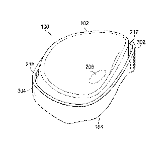
[0031] Fig. 2 is a perspective view of the two-part prosthetic meniscus
device 100, according
to an exemplary implementation. Referring to Fig. 2, the free floating
meniscus component 102
comprises a tissue-interfacing upper surface 202, a lower surface 204 (also
shown in Fig. 7) and
an outer portion 206. In some implementations, the outer portion 206 forms the
outer peripheral
surface of the component 102 that extends between and connects the sides of
the upper surface
202 and the lower surface 204. In some implementations, the outer portion 206
is formed as a
monolithic part of the component 102, and in some implementations, the outer
portion 206 is
formed of a wall structure or peripheral bumper formed or molded about the
central portions
forming the upper surface 202 and the lower surface 204 of the free floating
meniscus
component 102. In some implementations, such as when the free floating
meniscus component
102 is formed of two elements joined together, the peripheral wall structure
or wall may have a
circular or elliptical shape that surrounds and may be attached to the body of
the free floating
meniscus component 102. The outer portion 206 may also comprise of a denser /
stiffer material
than the rest of the free floating meniscus component 102.
[0032] In the illustrated embodiment, the upper surface 202 is shaped and
arranged to face
the medial femoral condyle and may press or engage the cartilage of the medial
femoral condyle
or the femoral surface. In some embodiments, the upper surface 202 may be
custom molded to
shape the cartilage of the medial femoral condyle of the host knee.
[0033] In some implementations, upper surface 202 may be shaped to form a
basin or have a
generally concave shape for the reception of adjacent bone structure forming
the joint. In some
7
CA 03069234 2020-01-06
WO 2019/023645 PCT/US2018/044196
implementations, the upper surface 202 may have one or more bone-relief recess
areas, such as
bone-relief recess area 208. Bone-relief recess area 208 is an indentation in
the upper surface 202
of the free floating meniscus component 102. The bone relief recess area 208
may be
manufactured by any method including molding, machining, etching, or other
method. The
bone-relief recess area 208 limits contact or engagement between the upper
surface 202 and the
bone structure otherwise supported within the basin or concave shape of the
upper surface 202.
For example, when the joint is a knee, the bone-relief recess area 208 may
limit contact or
engagement between the upper surface 202 and a portion of the medial femoral
condyle that is
opposite of the bone-relief recess area 208, while the upper surface 202 still
supports other
portions of the medial femoral condyle. The bone-relief recess area 208 may be
shaped as an
additional divot, depression, or etch formed in the upper surface 202.
[0034] Such limited contact between the upper surface 202 and a portion of
the adjacent
bone may be provided for medical reasons, for general comfort, or for other
reasons. For
example, when certain areas of the meniscus in the bone structure at the
treated joint have been
damaged, further contact with prosthetic meniscus device 100 would exacerbate
the damage or
cause additional pain to the patient. In this case, when the free floating
meniscus component 102
with the bone-relief recess area 208 is inserted into the medial compartment
such that the bone-
relief recess area 208 faces the damaged portion of the femoral bearing
surface, the bone-relief
recess area 208 may limit contact with the damaged surface and prevents
further deterioration of
the femoral bearing surface, while the remainder of upper surface 202 still
provides supportive
contact with the non-damaged cartilage portions the bone structure.
[0035] In another example, limited contact between the prosthetic meniscus
device 100 and
the femoral bearing surface may be necessitated after a patient underwent a
minimally invasive
surgery to replace or repair a portion of the cartilage of the medial femoral
condyle. One way to
replace or repair portions of the cartilage is to insert a biologic or stem
cell paste into the
damaged portions or the cartilage and allow the cartilage to regenerate and
regrow. However,
cartilage does not regenerate at a density required to bear weight in the knee
joint unless pressure
is applied to the cartilage. Hence, in order for the cartilage to regenerate
at a necessary density, a
patient should apply pressure on the knee and on the femoral bearing as the
cartilage regenerates
and regrows. In order for the patient to put pressure on the knee, yet for the
biologic or the stem
cell paste to have limited or no contact with the prosthetic meniscus device
100, the upper
8
CA 03069234 2020-01-06
WO 2019/023645 PCT/US2018/044196
surface 202 includes the bone-relief recess area 208 that faces the portion of
the medial femoral
condyle that has been injected with a biologic or stem cell paste. The bone-
relief recess area 208
may prevent or may limit contact between the prosthetic meniscus device 100
and the portion of
the medial femoral condyle that was injected with a biologic or the stem cell
paste while the
cartilage regenerates. Yet, at the same time, bone-relief recess area 208 also
allows a patient to
apply pressure to the knee that causes the cartilage to regenerate at a
density that supports
pressure on a knee joint.
[0036] In some implementations, rigid base component 104 comprises an outer
portion 214
and a bottom portion 216. Generally, the outer portion 214 is a rigid support
structure or wall
that forms an outer periphery of the base component 104 and has a circular or
an elliptical shape
that imitates or substantially matches the shape of the outer portion 206 of
the free floating
meniscus component 102. The bottom portion 216 is also of a circular or
elliptical shape and
attaches to the lower end of the outer portion 214 on all sides. In some
implementations, this
outer portion 214 and the bottom portion 216 together form a basin or cup in
which the free
floating meniscus component 102 may be disposed. In some implementations, the
surface areas
of an inner surface and an outer surface of the outer portion 214 may be
smooth surfaces.
[0037] In some implementations, bottom portion 216 may be molded to conform
to the shape
of the lower surface 204 and/or the shape of the natural medial tibial plateau
of the host knee.
The bottom portion 216 may include a lower surface or tissue-interfacing
surface that interfaces
with the bone/cartilage tissues of the joint.
[0038] In the illustrated embodiment, the free floating meniscus component
102 is disposed
inside the rigid base component 104, such that the lower surface 204 of the
free floating
meniscus component 102 faces the upper surface of the bottom portion 216 of
the rigid base
component 104. Here, the lower surface 204 and the upper surface of the bottom
portion 216
directly interface. In an embodiment, lower surface 204 may be a smooth
surface that is adjacent
to the smooth surface of the bottom portion 216. In another embodiment, lower
surface 204 may
be a molded surface, in which case, the upper surface of the bottom portion
216 is molded to the
shape of lower surface 204 or vice versa. In yet another embodiment, lower
surface 204 may be a
concave surface and the upper surface of the bottom portion 216 may also be a
concave surface.
In additional embodiments, they are each planar.
9
CA 03069234 2020-01-06
WO 2019/023645 PCT/US2018/044196
[0039] Referring to Fig. 2, when the free floating meniscus component 102
is located inside
the rigid base component 104, the outer portion 214 and the bottom portion 216
surround the free
floating meniscus component 102. Accordingly, the bottom portion 216 provides
a load bearing
surface through which loads on the joint may be passed, and the outer portion
214 may be a
boundary or limit upon the distance that the free floating meniscus component
102 may translate
as it free floats within the rigid base component 104. In the illustrated
embodiment, the upper
surface 202 and upper portions of the outer portion 206 protrude above the
outer portion 214 of
rigid base component 104. This may provide axial separation of the upper bone
structure from
the lower component of the device 100, which in this example is the rigid base
component 104.
[0040] Fig. 2 shows that the prosthetic meniscus device 100 has a height or
thickness 210, a
longitudinal width 212 that may be measured along the largest transverse cross-
sectional length,
and a transverse width 220 that is the smallest cross-sectional length taken
perpendicular to the
longitudinal width 212. The thickness or height 210 may be measured as the
combined height of
the free floating meniscus component 102 and the rigid base component 104.
Also, the thickness
or height 210 may vary depending upon the measured location. For example, the
nonplanar
upper surface 202 of the free floating meniscus component 102 and the
nonplanar lower surface
308 of the rigid base component 104 may impact the thickness or height 210 at
any particular
location of the prosthetic meniscus device 100. Generally, in addition to the
surface variations
and shapes of the upper surface 202, the thickness or height 210 may be
selected to fit within the
available space between the femoral bearing surface and the natural tibial
plateau of a host knee.
In some implementations, the thickness or height 210 may be between 0.5 mm and
15 mm. hi
some implementations, the maximum height 210 measured along the outer edges of
the may be
about 10 mm and the minimum thickness or height 210, which may be measured in
the central
portion of the prosthetic meniscus device 100 may be about 2 mm. Other
thicknesses or heights,
both smaller and larger are contemplated.
[0041] In some implementations, the longitudinal width 212 of prosthetic
meniscus device
100 may be the width of rigid base component 104 since the width of the rigid
base component
will generally be larger than the width of the free floating meniscus
component 102. Generally,
the longitudinal width 212 may be dictated by the available space between the
medial femoral
bearing surface and the natural medial tibial plateau of a host knee. The
longitudinal width 212
may be between 25mm and 70mm, although larger and smaller widths are
contemplated.
CA 03069234 2020-01-06
WO 2019/023645 PCT/US2018/044196
[0042] In some implementations, the transverse width 220 of the prosthetic
meniscus device
100 may be the shortest measurable width of the rigid base component 104 that
is perpendicular
to the longitudinal width 212. Generally, the transverse width 220, like the
longitudinal width
212, may be dictated by the available space between the femoral surface and
the natural medial
tibial plateau of a host knee. The transverse width may be between 20mm and
50mm in some
implementations, although larger and smaller transverse widths are
contemplated.
[0043] In some implementations, the inner dimensions of the outer portion
206 may be larger
than the outer dimensions of the free floating meniscus component 102 so as to
provide a gap or
space 218 therebetween. The space 218 provides clearance between the outer
portion 206 and the
free floating meniscus component 102 so that the free floating meniscus
component 102 may
laterally translate or rotate while disposed in the outer portion 206. The
inner surface of the
outer portion 206 may act as a boundary to limit the amount of translation and
to maintain the
free floating meniscus component 102 within the outer portion 206. The size of
the gap or space
218 may vary depending on the application and the joint to be replaced. In
some
implementations, the space 218 may be between 0.5mm and 3mm, but the
implementation is not
limited to this embodiment. In a different embodiment, the space may so small
such that the
outer portion 214 and inner surface of the outer portion 206 may be
substantially abutting around
the complete outer portion to limit translation in any direction.
[0044] In some implementations, the wall formed by the outer portion 206 of
the rigid base
component 104 prevents free floating meniscus component 102 from being
expelled from the
joint. At the same time, the rigid base component 104 allows free floating
meniscus component
102 to float freely therein, and the prosthetic meniscus device 100 may mimic
functionality of
the natural meniscus. Further, because the rigid base component 104 also
floats within the joint,
the natural meniscus and the supporting femur and tibia may remain intact.
That is, since tissue
penetrating anchors are not employed in some embodiments of the prosthetic
meniscus device
100, additional trauma to the joint may be reduced or minimized when compared
to conventional
devices.
[0045] Fig. 3 is a perspective view of the rigid base component 104 of the
prosthetic
meniscus device 100, according to an example implementation. The rigid base
component 104
may be formed of a rigid, supportive material such as a metal, a plastic,
and/or a ceramic
material. As illustrated in Fig. 3, the outer portion 214 and the bottom
portion 216 together form
11
CA 03069234 2020-01-06
WO 2019/023645 PCT/US2018/044196
a basin or cup defining a containment cavity 217 that is shaped to receive the
free floating
meniscus component 102 (Fig. 2). The outer portion 214 forms the peripheral
wall of rigid base
component 104 and comprises an inner surface 302 and an outer surface 304. The
inner surface
302 of the rigid base component 104 faces the outer portion 206 of the free
floating meniscus
component 102 when the free floating meniscus component 102 is disposed
therein. In some
embodiments, inner surface 302 may be a smooth surface and may be arranged to
provide a limit
or restraint on the distance that the free floating meniscus component 102 may
translate in the
containment cavity 217.
[0046] In some implementations, the outer surface 304 of rigid base
component 104 may be
shaped to be positioned inside the boundaries of the joint, such as, for
example, within a medial
compartment of the knee. This may permit the outer surface 304 to be
surrounded by the
meniscus in the native tibial plateau. In some implementations, the rigid base
component 104
may be positioned within boundaries of the joint, such as the native tibial
plateau such that the
bottom portion 216 is adjacent and conforms to the shape of the meniscus
inside the native tibial
plateau.
[0047] In some implementations, the bottom portion 216 of rigid base
component 104
comprises the upper surface 306 and the lower surface 308. As shown in Fig. 3,
the upper surface
306 of the bottom portion 216 may be molded to have a non-planar, uneven
surface that may be
arranged to match the lower surface 204 of the free floating meniscus
component 102. In some
implementations, the lower surface 308 of the bottom portion 216 may be molded
to fit the
underlying bone structure against which it abuts. For example, when the rigid
base component
104 is a knee implant, the lower surface 308 may be molded to fit a natural
tibial plateau and/or
the meniscus surrounding the native tibial plateau, such that the native
tibial plateau and the
meniscus provide support for keeping the rigid base component 104 in place.
Since the lower
surface 308 abuts directly against and interfaces with bone and/or cartilage
structures, such a
form-fit surface may help maintain the free floating rigid base component 104
in place, even
though free floating displacement may be expected. That is, variations in the
height or thickness
of the surfaces may be selected to match the anatomical features of the
patient in some
embodiments in the form of a natural meniscus.
[0048] In some implementations, the thickness or height 310 of the outer
portion 214 or wall
may vary between a maximum height or thickness in the range of lOmm to 20mm
and may vary
12
85881057
between a minimum height of 2mm to 10mm depending upon the location and/or the
size
of the patient. Height variations may be due to the preformed shape of the
bottom portion
216 to coincide with the lower surface 204 of the free floating meniscus
component 102
and/or with the shaped of the adjacent bone/cartilage structure, such as the
native tibial
plateau. In some implementations, the height 310 of outer portion 214 varies
from a
maximum height of 20mm to a minimum height of 10mm. In other implementations,
the
height 310 varies from a height of 15mm to a height of 5mm. Other amounts are
also
contemplated. In some implementations, the wall thickness 312 of the outer
portion 214
measured between inner surface 302 and the outer surface 304 may be between
0.1mm and
3mm. In one particular embodiment, the wall thickness 312 may be about lmm.
[0049] Fig. 4 is a perspective side view of the prosthetic meniscus device
100 with a
free floating meniscus component 102 disposed inside rigid base component 104,
according to one embodiment. As illustrated in Fig. 4, the lower surface 308
of the bottom
portion 216 in the rigid base component 104 is molded to match the shape of a
bone or
tissue interface, such as the shape of the native tibial plateau. As also
illustrated in Fig. 4,
the height 310 of the outer portion 214 varies because a lower end 215 forming
a
peripheral bottom edge of the outer portion 214 conforms to the nonplanar
variations in
the slope of lower surface 308, while the upper end 223 forming the upper edge
of the
outer portion 214 remains at approximately the same height with respect to
upper edge 219
the free floating meniscus component 102.
[0050] In some implementations, the variation of the surface profile of the
bottom
surface may be measured as a surface variation or height 311 between 0.1mm and
10mm.
This height may be measured as an axial distance along an axis 221 defined by
the surface
forming the outer portion 214 of the rigid base component 104. The height
variations
may be due to the shape of the bottom portion 216 that coincides with the
lower surface
204 of the free floating meniscus component 102 and/or with the adjacent
tissue structure,
such as the native tibial plateau of the host knee.
[0051] Figs. 5 and 6 show a free floating meniscus component 102 of the
prosthetic
meniscus device 100. Some features may be similar to a prior design set forth
in U.S.
Patent No. 8,361,147. The body of free floating meniscus component 102
comprises an
outer body portion 108 (referred to as the outer portion 206 in Fig. 2) and a
central body
portion 110. Generally, the outer body portion 108 has an
13
Date Recue/Date Received 2021-06-25
CA 03069234 2020-01-06
WO 2019/023645 PCT/US2018/044196
increased thickness and height relative to the central body portion 110. In
some instances the
outer body portion 108 has a thickness between 5 mm and 15 mm. In some
instances, the central
body portion 110 has a thickness between 0.5 mm and 5 mm. In one particular
embodiment, the
outer body portion 108 has a maximum thickness of approximately 10 mm and the
central body
portion 110 has a maximum thickness of approximately 2 mm. The height or
thickness of the
outer body portion 108 varies around the perimeter of the prosthetic device in
some instances. In
that regard, the variations in the height or thickness of the outer body
portion 108 are selected to
match the anatomical features of the patient in some embodiments. Similarly,
the height or
thickness of the central body portion 110 varies across the prosthetic device
in some
embodiments. Again, the variations in the height or thickness of the central
body portion 110 are
selected to match the anatomical features of the patient in some embodiments.
In some
embodiments, the free floating meniscus component 102 is inserted in an
insertion configuration
and then loaded, stretched, moved, and/or otherwise transferred to an
implantation configuration.
In some implementations, the insertion configuration has a smaller profile or
shape then the
implantation configuration. In other implementations, the insertion
configuration is simply
different than the implantation configuration in order to accommodate
insertion between the
bones of the joint. In some embodiments the transformation between the
insertion configuration
and the implantation configuration is facilitated through the application of a
loading force of the
free floating meniscus component 102. In such embodiments, the variations in
height or
thickness of the outer and central body portions 108, 110 are selected to
accommodate the
deformation or transformation between the insertion configuration and the
implantation
configuration.
[0052] To this end, the outer body portion 108 of the free floating
meniscus component 102
includes a first portion 112 and a second portion or bridge 114. In some
embodiments, the first
portion 112 substantially matches the shape of a natural meniscus. In some
embodiments, the
outer body portion 108 has a circular or semi-ellipsoidal shape Accordingly,
the first portion
112 extends around a majority of the outer body portion 108. The bridge 114
connects the two
ends of the first portion 112. Thus, where the prosthetic device is configured
for use as a medial
meniscus device, the bridge 114 extends along the lateral side of the device.
Where the free
floating meniscus component 102 is configured for use as a lateral meniscus
device, the bridge
114 extends along the medial side of the device. Accordingly, the outer body
portion 108-
14
CA 03069234 2020-01-06
WO 2019/023645 PCT/US2018/044196
comprised of the first portion 112 and the bridge 114 and having an increased
thickness relative
to the central body portion 110¨completely surrounds the central body portion
110 and serves
to limit movement of the prosthetic device after implantation.
[0053] The height or thickness of the bridge 114 is based on the size of
the femur notch and
the distance to the cruciate ligaments in some embodiments. In some
embodiments, the bridge
114 has a maximum height or thickness that is between 1/4 and 3/4 the maximum
height or
thickness of the first portion 112 of the outer body portion 108. In some
embodiments, the size
and shape of the bridge 114 is selected to achieve an optimal pressure
distribution on the native
tibial plateau in order to mimic the pressure distribution of a healthy
natural meniscus. The
bridge 114 and, more generally, the outer body portion 108 are geometrically
characterized by
anterior, posterior, lateral-anterior, mid-lateral and lateral-posterior
angles and heights as well as
sagittal and coronal radii of curvature. Further, the outer body portion 108
and the central body
portion 110 are shaped and sized such that the free floating meniscus
component 102 is self-
centering within rigid base component 104. That is, the shape and size of the
prosthetic
meniscus device itself encourages the prosthetic device to position or align
itself with a desired
orientation within the knee joint based on the position of the femoral
surface. Accordingly, as
the free floating meniscus component 102 moves through a range of positions
within the knee
joint, it naturally returns to the desired orientation due to the shape and
size of the outer and
central body portion 108, 110. In some embodiments, the outer body portion
and, more
specifically, the bridge 114 alone or together with rigid base component 104
acts as a physical
barrier limiting the movement of the prosthetic device caused by joint
reaction forces. The shape
of the related femoral or tibial bearing component interacting with the self-
centering or self-
aligning mechanism combined with the free floating meniscus component's 102
ability to move
within the knee joint results in improved location of the prosthetic meniscus
device 100 during
typical gait cycles (e.g., flexion-extension angles of 0 to 20 or "heel-
strike" to "toe-off'). The
result is that the free floating meniscus component 102 exhibits a load
pressure distribution
similar to that of a natural meniscus.
[0054] The central body portion 110 defines an upper surface 116 and a
lower surface 118
(referred to as upper surface 202 and lower surface 204 in Fig. 2.). The upper
surface 116 may
interface with the tissue structure of the joint and may foini a part of a
bearing surface. In
particular, the upper surface 116 is configured to movingly engage with a
medial femoral
CA 03069234 2020-01-06
WO 2019/023645 PCT/US2018/044196
condyle of the femur. In that regard, free floating meniscus component 102 can
translate and
rotate with respect to the femur and/or tibia within a range. In some
instances, translation is
possible in both the anterior-posterior and medial-lateral directions. In some
embodiments, the
upper surface 116 includes both a vertical and horizontal surface. To that
end, in some
embodiments the upper surface 116 comprises a concave surface that defines the
vertical and
horizontal surfaces. The thickness of the central body portion 110 between the
upper surface 116
and the lower surface 118 supports stress distribution capability of the
component, while the
increased height of the upper surface 116 as it extends outwardly towards the
outer body portion
108 defines the horizontal surface of the component. Similarly, in some
embodiments the lower
surface 118 includes both vertical and horizontal components. In particular,
in some
embodiments the lower surface 118 comprises a convex surface or a concave
surface that is
molded to the shape of the inside portion of rigid base component 104.
[0055] The thickness of the central body portion 110 between the upper
surface 116 and the
lower surface 118 determines the load distribution capacity of the component,
while the tapered
height of the lower surface 116 as it extends outwardly towards the outer body
portion 108
defines the horizontal component. In some embodiments, the upper surface 116
and/or the lower
surface 118 are shaped such that the component is biased towards a neutral
position in the knee.
For example, the arcuate profiles of the upper surface 116 and/or the lower
surface 118 are
shaped such that the interaction between the surfaces and the femoral surface
encourages the
implant to a particular orientation relative to the surfaces.
[0056] Referring to Fig. 6, shown therein is a diagrammatic cross-sectional
view of free
floating meniscus component 102 taken along an anterior to posterior section
line between
anterior end 113 and posterior end 115. The central body portion 110 is
reinforced by pre-
tensioned fibers 124 wound around the core to inhibit outward deformation
while allowing
inward flexibility. As shown, the anterior end 113 of the outer body portion
108 has an anterior
height or thickness 160 In that regard, the anterior height or thickness 160
of the anterior end
113 is between about 4 mm and immediately adjacent bridge 114 could be as
great as about 15
mm and, in some instances, is between about 5.7 mm and about 9.3 mm. In the
illustrated
embodiment, the anterior height or thickness 160 of the anterior end 113 is
approximately 7.8
mm. In a smaller embodiment, the anterior height or thickness 160 is
approximately 5.7 mm. In
a larger embodiment, the anterior height or thickness 160 is approximately 9.3
mm. The
16
CA 03069234 2020-01-06
WO 2019/023645 PCT/US2018/044196
posterior height or thickness 162 of the posterior end is between about 4 mm
and immediately
adjacent the bridge 114 could be as great as about 20 mm and, in some
instances, is between
about 7.7 mm and about 12.7 mm. In the embodiment, the posterior height or
thickness 162 of
the posterior end 115 is approximately 9.0 mm. In a smaller embodiment, the
posterior height or
thickness 162 is approximately 7.7 mm. In a larger embodiment, the posterior
height or
thickness 162 is approximately 12.7 mm.
[0057] The anterior portion of the upper surface of the anterior end 113
has an anterior radius
of curvature 164. In that regard, the anterior radius of curvature 164 is
between about 10 mm
and about 100 mm and, in some instances, is between about 23.0 mm and about
33.1 mm. In the
embodiment, the radius of curvature 164 is approximately 72 mm. In another
embodiment, the
radius of curvature 164 is approximately 28 mm. In a smaller embodiment, the
radius of
curvature 164 is approximately 23 mm. In a larger embodiment, the radius of
curvature 164 is
approximately 33.1 mm. The posterior portion of the upper surface of the
posterior end 115 has
a posterior radius of curvature 166. In that regard, the posterior radius of
curvature 166 is
between about 5 mm and about 70 mm and, in some instances, is between about
15.2 mm and
about 24.2 mm. In the illustrated embodiment, the radius of curvature 166 is
approximately 30
mm. In a smaller embodiment, the radius of curvature 166 is approximately 15.2
mm. In a
larger embodiment, the radius of curvature 166 is approximately 24.2 mm.
[0058] Further, the anterior portion 113 of the upper surface generally
extends at an anterior
angle 168 with respect to an axis 170 extending substantially perpendicular to
a plane generally
defined by the free floating meniscus component 102, as shown. The anterior
angle 168 is
between about 45 degrees and about 75 degrees and, in some instances, is
between about 62
degrees and about 68 degrees. In the illustrated embodiment, the angle 168 is
approximately 65
degrees. In a smaller embodiment, the angle 168 is approximately 62 degrees.
In a larger
embodiment, the angle is approximately 68 degrees. The posterior end 115 of
the upper surface
generally extends at an posterior angle 172 with respect to an axis 174
extending substantially
perpendicular to a plane generally defined by the prosthetic meniscus device
100, as shown. The
posterior angle 172 is between about 35 degrees and about 70 degrees and, in
some instances, is
between about 55 degrees and about 61 degrees. In the embodiment, the angle
172 is
approximately 58 degrees. In a smaller embodiment, the angle 172 is
approximately 50 degrees.
In a larger embodiment, the angle 172 is approximately 65 degrees.
17
CA 03069234 2020-01-06
WO 2019/023645 PCT/US2018/044196
[0059] The central body portion 110 has a height or thickness 176 between
the articulating
upper surface 116 and the articulating lower surface 118. In some embodiments,
the height or
thickness 176 is the minimal thickness of the central body portion 110 and, in
more specific
embodiments, the minimal thickness of the entire free floating meniscus
component 102. To that
end, the height or thickness 176 is between about 1 mm and about 3 mm and, in
some instances,
is between about 1.2 mm and about 2.1 mm. In the embodiment, the height or
thickness 176 is
approximately 1.5 mm. In a smaller embodiment, the height or thickness 176 is
approximately
1.2 mm. In a larger embodiment, the height or thickness 176 is approximately
2.1 mm.
[0060] Fig. 7 is a cross-sectional view of the prosthetic meniscus device
100 with the free
floating meniscus component 102 disposed inside the containment cavity 217 of
the rigid base
component 104. As illustrated in Fig. 7, the bottom portion 216 of rigid base
component 104 is
adjacent to the outer body portion 108 of the free floating meniscus component
102. As also
illustrated in Fig. 7, the bottom portion 216 of the rigid base component 104
may have a
generally concave shape. In some implementations, the bottom portion 216 is
shaped to form fit
or receive the surface of the outer body portion 108. As also illustrated in
Fig. 7, the bottom
portion 216 may have an edge portion 702 that extends along the outer edge of
the bottom
portion 216.
[0061] As discussed above, the free floating meniscus component 102 and the
rigid base
component 104 may be sized so that the outer portion 206 of the free floating
meniscus
component 102 and the outer portion 214 of the rigid base component 104 may be
separated by
the gap or space 218. This gap or space 218 may permit the free floating
meniscus component
102 to rotate or translate within the rigid base component 104. In one
embodiment, the outer
portion 206 and the outer portion 214 may have outer surfaces generally
parallel to each other
along axis 706 extending substantially perpendicular to a plane generally
defined through the
prosthetic meniscus device 100, as shown. In another embodiment, outer portion
206 generally
extends at angle 708 with respect to axis 706 away from the outer portion 214
and toward the
center of the free floating meniscus component 102. Depending upon the
implementation, the
angle 708 is between 0 degrees and 45 degrees. In some implementations, the
angle 708 is
between 5 degrees and 20 degrees.
[0062] Fig. 7 shows an exemplary implementation of the free floating
meniscus component
102. In this implementation, the free floating meniscus component 102
comprises a bearing
18
CA 03069234 2020-01-06
WO 2019/023645 PCT/US2018/044196
portion 720 cooperatively joined with a peripheral support portion 722. In
this implementation,
the peripheral support portion 722 forms outer body portion 108 described
herein. Here, the
bearing portion 720 comprises the upper surface 202 and the lower surface 204,
and is
configured to interface with tissue at the joint and provide bearing support
for weight at the joint.
[0063] In this implementation, the bearing portion 720 comprises outer
edges 724 that abut
against the peripheral support portion 722. These outer edges 724 comprise
tension apertures
726. In this implementation, the tension apertures 726 extend fully around the
periphery at the
outer edge 724 of the bearing portion 720. In some implementations, the
tension apertures 726
may receive fibers (not shown in Fig. 7), similar to or the same as the pre-
tensioned fibers 124 in
Fig. 6. Such fibers may wind around the bearing portion 720 in the tension
apertures 726 to
inhibit outward deformation while allowing inward flexibility. In other
implementations, instead
of fibers, alternative reinforcement material may be introduced or embedded in
the tension
apertures 726. Some implementations are devoid of tension apertures 726.
[0064] The peripheral support portion 722 may be structurally embedded in a
portion of the
bearing portion 720 so as to be partially enveloped in the bearing portion 720
as shown in Fig. 7.
In some implementations, the peripheral support portion 722 may be formed of a
more rigid
material than the bearing portion 720, and may provide rigidity and strength
to the free floating
meniscus component 102. In the implementation shown, edges of the bearing
portion 720 and
develop an interface with the upper and lower surfaces of the peripheral
support portion 722.
However, other arrangements may be used to securely maintain the peripheral
support portion
722 in place about the bearing portion 720. In some implementations, the
bearing portion 720
and the peripheral support portion 722 are formed of the same material. In one
exemplary
implementation, the peripheral support portion 722 may have one or more
extending ridges,
hooks, or notches that may extend into one or more of the tension apertures
726. In some
implementations, the ridges, hooks, or notches may extend into other grooves
or reception
cavities formed in the outer edge of the bearing portion 720. These types of
arrangements may
provide mechanical interference that prevents the bearing portion 720 from
displacing vertically
relative to the peripheral support portion 722.
[0065] In use, under a bearing load, the bearing portion 720 of the free
floating meniscus
component 102 may be formed to match the profile of the more rigid bottom
portion 216 of the
rigid base component 104. Accordingly, although gaps are shown between the
lower surface 204
19
CA 03069234 2020-01-06
WO 2019/023645 PCT/US2018/044196
of the free floating meniscus component and the upper surface 306 of the rigid
base component,
under load, these gaps may be minimized or reduced. Furthermore, under load,
the concave
cavity of the free floating meniscus component 102 may change shape slightly,
such as the radius
of curvature may be increased as a result of the applied loading.
Additionally, the outer edges
724 of the bearing portion 720 may apply loading on the peripheral support
portion 722, causing
some deformation or expansion of the peripheral support portion 722. As
discussed above, fibers
or other materials may be used to limit, restrain, or control, the amount of
deformation permitted
under a load.
[0066] Figs. 8A and 8B are top and side perspective views of the prosthetic
meniscus device
100 with the free floating meniscus component 102 disposed inside the rigid
base component
104, according to an exemplary implementation. As illustrated in Figs. 8A and
8B, the free
floating meniscus component 102 may have a circular or semi-elliptical shape,
and is disposed
inside rigid base component 104 that may also have a circular or semi-
elliptical shape that
generally conforms to the shape of rigid base component 104. As also
illustrated in Figs. 8A and
8B, gap or space 218 is maintained between components 102 and 104, but may
vary in width 802
in order to allow the free floating meniscus component 102 to float inside
rigid base component
104. In some embodiments, the width 802 of may fall within the range of
between 0.05mm and
3mm. In some implementations, width 802 may change as the free floating
meniscus component
102 floats inside the rigid base component 104 as the knee is in motion.
[0067] As discussed above, the prosthetic meniscus device 100 is a
minimally invasive
implant that floats inside the medial compartment of the knee joint and
prevents further damage
to the meniscus and/or other tissues like cartilage articulating surfaces. The
prosthetic meniscus
device 100 may also protect a structural carrier, such as morsalized bone or a
cartilage matrix,
which may include a biologic, that may be introduced in the medial femoral
condyle to promote
tissue regeneration and regrowth of the damaged cartilage. In some
implementations, the
prosthetic meniscus device 100 may be implanted into the native tibial plateau
of the host knee
such that the free floating meniscus component 102 engages the femoral surface
and redistributes
weight load transmitted across the knee joint, while the rigid base component
104 engages the
natural tibial plateau and prevents the free floating meniscus component 102
from being
unintentionally expelled from the knee joint. As discussed above, the free
floating meniscus
component 102 may be modified to have limited contact with one or more
portions of the
CA 03069234 2020-01-06
WO 2019/023645 PCT/US2018/044196
femoral surface as dictated by the treatment. For example, when the damaged
area of the medial
femoral condyle has been treated with a biologic or stem cell paste to allow
cartilage to
regenerate and regrow, the free floating meniscus component 102 may include
one or more
bone-relief recess areas, such as bone-relief recess area 208 that limits
contact between the
prosthetic meniscus device 100 and the treated areas of the medial femoral
condyle. Depending
upon the implementation, the bone relief recess areas may be custom formed to
match individual
patients or conditions.
[0068] In a further embodiment, the free floating meniscus component 102
with the bone-
relief recess area 208 may be swapped out or exchanged for another free
floating meniscus
component 102 with a different bone-relief recess area 208 or for the free
floating meni scus
component 102 with a smooth upper surface 202. For example, once the medial
femoral condyle
has healed and the cartilage had regrown, the free floating meniscus component
102 with the
bone-relief recess area 208 may be exchanged in a revision surgery for the
free floating meniscus
component 102 with the smooth upper surface 202.
[0069] Fig. 9 shows an example illustration of the prosthetic meniscus
device 100 disposed
upon the tibia T of a knee joint with an injured meniscus 10. The meniscus
includes an outer rim
15 that is anchored to the bone along the posterior rim 20 and the anterior
rim 22. The meniscus
may form a meniscus pocket defined by a sidewall 21 of the meniscus, and in
which the
prosthetic meniscus device 100 may be disposed. The prosthetic meniscus device
100 engages
not only the tibia T, but also the Femur (not shown in Fig. 9.)
[0070] In some implementations, the prosthetic meniscus device 100 may be
implanted in a
two-step process. In the first step, only a temporary free floating meniscus
component 102 may
be implanted into the knee joint. The implanted free floating meniscus
component 102 may
comprise a smooth upper surface 202 or have one or more bone-relief recess
areas, such as bone-
relief recess area 208 formed, such as by etching, on the upper surface 202,
depending on the
treatment. For example, a patient may be required to gradually apply pressure
on the cartilage in
the knee following a minimally invasive surgery in order for the cartilage to
regrow and have
necessary density, as described above. The free floating meniscus component
102 having a
smooth upper surface 202 with the bone-relief recess area 208 opposite the
areas in the medial
femoral condyle where the cartilage is being regrown, allows the patient to
apply pressure across
21
CA 03069234 2020-01-06
WO 2019/023645 PCT/US2018/044196
the entire knee joint, including the areas where the cartilage is being
regrown, yet limits the
physical contact with these areas and the free floating meniscus component
102.
[0071] In some implementations, the second step of the two-step surgical
process may be
perfoimed days, weeks, or months after the first step of the surgical process.
This may allow
some healing to occur prior to the second step. For example, the second step
of the two-step
surgical process may be performed after cartilage has begun growing on the
medial femoral
condyle or other bone structure. In the second step, the free floating
meniscus component 102
may be replaced with a full prosthetic meniscus device 100, including the free
floating meniscus
component 102 and the rigid base component 104. The free floating meniscus
component 102
can be the same or different free floating meniscus component 102 as in the
first step. In some
implementations, the free floating meniscus component 102 may have a smooth
upper surface
202. As indicated herein, the second step generally occurs after the cartridge
has healed or has
been regrown and the prosthetic meniscus device 100 is implanted into the knee
joint for the long
term use by the patient.
[0072] Figs. 10A, 10B, 10C, and 10D show a series of angular positions of
the femur in
relation to the tibia and the correspondent movement of the prosthetic
meniscus device 100 in the
knee joint. In Fig. 10A, femoral axis FA is substantially aligned with the
tibial axis TA. The
prosthetic meniscus device 100 is disposed between the tibia T and the femur
F. In this initial
position, with the axes FA and TA substantially aligned, the outer surface of
the outer portion
214 of the rigid base component 104 may be generally aligned with a posterior
wall of the joint,
referenced by the reference line 550. In this position, the posterior gap or
space 218 between the
outer portion 214 of the rigid base component and the outer body portion 108
of the free floating
meniscus component 102 is indicated by the reference number Dl.
[0073] Fig. 10B illustrates the movement of the prosthetic meniscus device
100 as the femur
F is moved to the position of the angle a' between axis FA and axis TA. A
comparison of Fig.
10B and 10A shows that the rigid base component 104 is maintained
substantially in place, while
the free floating meniscus component 102 has displaced within the containment
cavity 217 of the
rigid base component 104. In this instance, the free floating meniscus
component 102 may have
moved in the posterior direction as far as it is able. That is, it may have
displaced to the point
that the outer body portion 108 of the free floating meniscus component has
engaged the outer
portion 214 of the rigid base component 104. Because of this lateral
translation, the gap or space
22
CA 03069234 2020-01-06
WO 2019/023645 PCT/US2018/044196
218 is shown now on the interior side of the joint. In this instance, the gap
or space 218 is
indicated by the reference number D2, which will equal D1 in Fig. 10A so long
as rotation is
limited. Accordingly, D2 in Fig. 10B is equal to or substantially equal to D1
in Fig. 10A.
[0074] Fig. 10C illustrates the continued rotation of the femur with
respect to the tibia results
in angle a" which is greater than angle a'. A comparison of Fig. 10B and 10C
shows that the
rigid base component 104, which is also free floating, begins displacing in
the posterior direction
a distance D3.
[0075] Fig. IOD illustrates that continued rotation of the femur with
respect to the tibia to
angle a¨, which is substantially 90 degrees, results in further translation to
a distance D3' which
is greater than D3.
[0076] While the foregoing are not limiting, the total translation distance
can range from 3-
20 mm in the anterior to posterior plane, with one embodiment having D1 and D2
of 3mm, D3 of
7mm and D3' of 14mm. Similarly, the rotational angle can range, without
limitation, from 3 to
30 degrees of total angular rotation. An advantage of the free floating system
comes because the
prosthetic device may also rotate in the joint as the angle of the femur and
tibia changes.
[0077] Figs. 11A, 11B, and 11C disclose cross-sectional side views of a
prosthetic meniscus
device 850. Similar to the device 100 described above, the prosthetic meniscus
device 850 may
include a floating meniscus component 852 and a rigid base component 854. In
this
implementation however, the free floating meniscus component 852 and the rigid
base
component 854 have a particular shape that may provide biofeedback such as
tactile feedback to
a patient when the joint articulation begins to exceed a desired amount. In
the implementation
shown, the particular shape provides an abrupt backstop to prevent the free
floating meniscus
component 852 from translating beyond a desired position in the posterior
direction. The
particular shape also provides an increasing or gradual resistance to anterior
displacement of the
free floating meniscus component 852 relative to the rigid base component 854.
[0078] The prosthetic meniscus device 850 may be shaped generally in the
same manner
described herein with reference to the prosthetic meniscus device 100, but
with some
modifications described with reference to Figs. 11A-11C
[0079] Fig. 11A shows a cross-sectional view of the prosthetic meniscus
device 850 with the
free floating meniscus component 852 disposed in the cup-shaped rigid base
component 854. As
can be seen, the peripheral edges of the free floating meniscus component 852
and the rigid base
23
CA 03069234 2020-01-06
WO 2019/023645 PCT/US2018/044196
component 854 are modified from other devices described herein. The free
floating meniscus
component 852 includes a posterior peripheral portion 856 and an anterior
peripheral portion
858. It also includes a sliding bone or tissue interface surface 860 and a
sliding interface 862.
Likewise, the rigid base component 854 includes a posterior peripheral portion
866, and anterior
peripheral portion 868, a tissue interface surface 870, and a sliding
interface 872. As shown in
Fig. 11A, the prosthetic meniscus device 850 has a height HI when the free
floating meniscus
component 852 is disposed such that the sliding interface 862 and the sliding
interface 872 are
engaged at their lowest portions. The height HI may be measured from the
tissue interface
surface 870 to the tissue interface surface 860.
[0080] The posterior peripheral portion 856 of the free floating meniscus
component 852
includes a substantially vertical peripheral edge 871. Likewise the posterior
peripheral portion
866 of the rigid base component 854 includes a substantially vertical
peripheral edge 873. As
such, when the free floating meniscus component 852 slides and abuts against
the posterior
peripheral portion 866, the free floating meniscus component may come to an
abrupt stop. Also,
when the free floating meniscus component 852 is disposed posteriorly in the
rigid base
component 854, the prosthetic meniscus device 850 has the height Hl.
[0081] The anterior peripheral portion 858 of the free floating meniscus
component 852
includes a curved or tapered edge 874. The curved edge 874 extends from an
upper portion of
the free floating meniscus component and tapers inwardly. The anterior
peripheral portion 868
of the rigid base component 854 also includes a curved or tapered edge 876. In
some
implementations, such as the one shown in Fig. 11A, the curved or tapered edge
is disposed on
both the interior portion and the exterior portion of the rigid base component
854. That is, the
interior and exterior profiles of the rigid base component 854 substantially
match. In other
implementations, the curved or tapered edge 876 is disposed only on the
interior portion of the
rigid base component 854. That is, the exterior profile of the rigid base
component 854 may not
match the interior profile of the rigid base component 854. The purpose of the
curved or tapered
edges of the anterior peripheral portions 858 and 868 will become apparent
with reference to
Figs. 11B and 11C.
[0082] Fig. 11B shows the prosthetic meniscus device 850 with the free
floating meniscus
component 852 translated toward the anterior peripheral portion 868 of the
rigid base component
854. In this exemplary embodiment, the profile of the anterior peripheral
portion 858 of the free
24
CA 03069234 2020-01-06
WO 2019/023645 PCT/US2018/044196
floating meniscus component 852 and the profile of the anterior peripheral
portion 868 of the
rigid base component 854 substantially correspond to one another. In other
embodiments, the
profiles of the anterior peripheral portions may not match. In some
implementations, the
location of the free floating meniscus component 852 at the anterior portion
of the rigid base
component 854 may represent a preferred distal position. Thus, in some
implementations, the
rigid base component 854 is sized to permit the free floating meniscus
component 852 to
translate from the location shown in Fig. 11A to the location shown in Fig.
11B. In some
implementations, this may be determined to be a standard or typical amount of
translation as the
knee articulates about the joint.
[0083] Fig. I IC shows the prosthetic meniscus device 850 with the free
floating meniscus
component 852 translated past the position shown in Fig. I I B. Likewise, the
free floating
meniscus component 852 may be translated to a location beyond that found
during normal
acceptable knee articulation. As can be seen, as the free floating meniscus
component 852
begins to translate along the sloped or angled edge 876 of the anterior
peripheral portion 868 of
the rigid base component, the leading anterior edge of the free floating
meniscus component 852
rises, thereby changing the overall height of the prosthetic meniscus device
850. Fig. 11C shows
the original height H1 and the increased height H2 which represents the
overall height of the
prosthetic meniscus device 850 when the free floating meniscus component 852
has moved
beyond a typical or noimal level of displacement on the rigid base component
854.
[0084] As can be seen, posterior motion of the free floating meniscus
component 852 reaches
a hard stop when the posterior peripheral portion 856 abuts against the
posterior peripheral
portion 866. This is because these two components each have substantially
vertical surfaces
causing an abrupt stop when they meet. In contrast, the anterior peripheral
portion 858 and the
anterior peripheral portion 868 provide a gradual stop. The sloped or angled
leading surfaces
cause a vertical displacement of the free floating meniscus component 852
relative to the rigid
base component 854. In this implementation, the sloped or angled leading
surface of the anterior
peripheral portion 868 has a curvature. This curvature provides a nonlinear
increase in height
from H1 to H2. Some implementations use planar ramps that may provide a linear
increase in
height. Other implementations use a series of planar ramps with different
angles.
[0085] In some implementations, this vertical displacement may be felt or
noticeable by the
patient as tactile biofeedback in the knee. This tactile biofeedback, along
with the increasing
CA 03069234 2020-01-06
WO 2019/023645 PCT/US2018/044196
resistance to further motion, may alert a patient to an overextension
condition. Furthermore,
because the prosthetic meniscus device 850 includes a gradual stop rather than
an abrupt stop,
the motion obtained by the prosthetic meniscus device 850 may be more
consistent with the
motion of a natural meniscus, where slightly elastic ligaments gradually
tighten during rotation
to prevent over-rotation, instead of having an abrupt, inflexible stopping
point. Accordingly, the
vertical displacement may of the free floating meniscus component 852 may
tactilely alert the
patient to overextension giving the patient an opportunity to correct the knee
to alleviate the
tension generated by the vertical displacement. So doing may enable the
patient to avoid
inadvertent over displacement of the prosthetic meniscus device 850, which may
help the patient
avoid expulsion of the prosthetic meniscus device 850 from the patient's knee.
[0086] Although shown with having both the free floating meniscus component
852 and the
rigid base component 854 having matching profiles or edges 874, 876 at the
anterior peripheral
portions 858, 868, other implementations have profiles or edges that do not
match. In some
implementations, the rigid base component 854 may have a simple planar slope
as its edge 876 at
its anterior peripheral portion 868. In other implementations, the anterior
peripheral portion 858
of the free floating meniscus component 852 may have a substantially vertical
profile at the edge
874, and the anterior peripheral portion 868 of the rigid base component 854
may have a curved
or sloped edge 876. In some implementations, the vertical displacement between
height H1 and
height H2 may be in the range of lmm to 12mm. Other ranges, both larger and
smaller, are also
contemplated.
[0087] Figs. 12 and 13 show an additional implementation of a prosthetic
meniscus device,
referenced herein by the numeral 900, disposed on a Tibia T. The prosthetic
meniscus device
900 includes the free floating meniscus component 102 and a rigid base
component 904. The
free floating meniscus component 102 may be similar to those described herein,
and those
descriptions will not be repeated here. The rigid base component 904 may
include an outer
portion 906 and a bottom portion 908, together forming a basin or opening for
receiving the free
floating meniscus component 102. The outer portion 906 may be an outer wall
structure
extending about and forming an outer peripheral portion of the rigid base
component. Thus, the
outer portion 906 forms the containment cavity that limits the allowable
translation distance of
the free floating meniscus component 102. This implementation differs from
other rigid base
components described herein because the outer portion 906 extends only
partially about the
26
CA 03069234 2020-01-06
WO 2019/023645 PCT/US2018/044196
bottom portion 908. The bottom portion 908 may be a supporting floor
structure. Fig. 12 shows
the prosthetic meniscus device 900 disposed on a tibia for use. The rigid base
component 904
may include an anterior portion 910, a posterior portion 912, and lateral side
portions 914. In the
implementation shown, the outer portion 906 extends about the anterior portion
910 and the
posterior portion 912, but the lateral side portions 914 are devoid of the
outer portion 906.
Accordingly, the free floating meniscus component 102 may translate or rotate
within the rigid
base component 904. However, the rotation within the rigid base component 904
may be limited
by the outer portion 906 which maintains the free floating meniscus component
102 in the rigid
base component 904. At the same time, the lateral side portions 914 being
devoid of the outer
portion 906 may permit natural lubricating fluids to more easily enter the
sliding interface
between the free floating meniscus component 102 and the rigid base component
904. In some
implementations, the lateral side portions 914 include an outer portion, but
with the outer portion
at the lateral side portions 914 having a height lower than the height of the
outer portion 906 that
is located at the anterior portion 910 and the posterior portion 912. In some
implementations, the
outer portion 906 has a different height at the anterior portion 910 than at
the posterior portion
912. For example, in some implementations the outer portion 906 at the
anterior portion 910 has
a height less than a height of the outer portion 906 at the posterior portion
912. Other
arrangements are also contemplated. The prosthetic meniscus device 900 may
perform in the
same manner described with reference to Figs. 10A-10D.
[0001] Fig. 14 discloses a method 600 of implanting the prosthetic meniscus
devices
according to an exemplary implementation. The method may be performed, for
example, with
respect to any of the prosthetic meniscus devices described herein. It is
understood that
additional steps can be provided before, during, and after the steps of method
600, and that some
of the steps described can be replaced or eliminated from the method 600.
[0088] In some exemplary implementations, the method begins at 602, with a
health care
provider assembling the prosthetic meniscus device prior to implantation in a
patient.
Accordingly, at 601, the health care provider may select a floating meniscus
component from a
plurality of floating meniscus components arranged to fit within a rigid base
component. The
floating meniscus component may be any of the floating meniscus components
described herein.
As described above, in some implementations, a health care provider may be
presented with a
plurality of floating meniscus components, with each having a particular
profile or contour that
27
CA 03069234 2020-01-06
WO 2019/023645 PCT/US2018/044196
may match a particular bone structure. In some implementations, the plurality
of floating
meniscus components may have similar contours, but may be sized differently to
match different
sized patients.
[0089] At 602, the health care provider may introduce the floating meniscus
component into
a cavity of the rigid base component in a manner permitting the floating
meniscus component to
translate within the rigid base component.
[0090] At 606, the health care provider may create an incision at a joint
of a patient having
damaged soft tissue in a manner known in the art. At 608, the health care
provider may
introduce the floating meniscus and rigid base components between opposing
tissue surfaces at a
joint having damaged soft tissue without removing or penetrating bone tissue
at the joint.
Accordingly, in some implementations, the health care provider does not carve,
cut, or introduce
screws, fins, or other anchors into the adjacent bone. As such, the prosthetic
meniscus device
may be free floating within the joint. That is, it may have the ability to
move or slide laterally
within the joint, and may have the ability to rotate within the joint. Flexion
at the joint may
cause the prosthetic meniscus device to translate or rotate. Accordingly, the
prosthetic meniscus
device is devoid of mechanical anchors and is arranged to interface with the
natural tibia plateau
and the femoral surface.
[0091] At 610, the health care provider may position the floating meniscus
and rigid base
components so that the first tissue interface surface receives a portion of
the first bone the second
tissue interface receives a portion of the second bone structure. This may
occur when the first
tissue-interface surface of the floating meniscus component and the second
tissue interface
surface of the rigid base component are shaped to fit contours of the adjacent
bone structure. In
some implementations, this may include a simple concave surface shaped to
receive adjacent
bone structure. In other implementations, the floating meniscus and rigid base
components may
be particularly shaped to match a particular bone surface For example, in some
implementations
the floating meniscus component is shaped to match the contours of a femoral
surface. As such,
the first tissue interface surface may receive contours of the femoral bone.
Likewise, in some
implementations, the rigid base component is shaped to match the contours of a
natural tibia
plateau. As such, the second tissue interface surface may receive contours of
the tibia bone. In
other implementations, the tissue interface surfaces may not have specific
contours, but may be
shaped with concavities or may be relatively planar.
28
CA 03069234 2020-01-06
WO 2019/023645 PCT/US2018/044196
[0092] At 612, the health care provider may close the incision without
mechanically
connecting the floating meniscus or rigid base components to the first or
second bone structure.
This may enable the prosthetic meniscus device to free float within the joint.
[0093] A variety of materials are suitable for use in making components,
such as components
102, 104, 852, 854, and 904 of prosthetic devices described herein. In one
aspect, the flexible
(non-rigid) component forming, such as for example the free floating meniscus
component or the
softer component is formed from a material that will yield/deform under normal
human loading
while the rigid material generally does not deform under normal human loading.
An example
combination may include a flexible component or layer including a material
foimed from a
polycarbonate-urethane having a hardness value of about 3.0 to 9.0 Nimm2 and
the rigid material
being formed of stainless steel material, or alternatively, a rigid
polyurethane, such as
COROTHANE polyurethane 75D, having a hardness in the range of about 45 Nimm2
to 85
N/mm2.
[0094] Medical grade polyurethane based materials suitable for use in the
embodiments
described include, but are not limited to, isolated or in combination, the
materials described or
mentioned in the following paragraphs.
[0095] Bionate , manufactured by DSM, a polycarbonate-urethane is among the
most
extensively tested biomaterials ever developed. Carbonate linkages adjacent to
hydrocarbon
groups give this family of materials oxidative stability, making these
polymers attractive in
applications where oxidation is a potential mode of degradation, such as in
pacemaker leads,
ventricular assist devices, catheters, stents, and many other biomedical
devices. Polycarbonate
urethanes were the first biomedical polyurethanes promoted for their
biostability. Bionate
polycarbonate-urethane is a thermoplastic elastomer formed as the reaction
product of a hydroxyl
terminated polycarbonate, an aromatic diisocyanate, and a low molecular weight
glycol used as a
chain extender. The results of extensive testing encompassing Histology,
Carcinogenicity,
Biostability, and Tripartite Biocompatibility Guidance for Medical Devices
verifies the cost
effective material's biocompatibility.
[0096] Another group of suitable materials are copolymers of silicone with
polyurethanes as
exemplified by PurSilTm, a Silicone Polyether Urethane and CarboSilTm, a
Silicone
Polycarbonate Urethane. Silicones have long been known to be biostable and
biocompatible in
most implants, and also frequently have the low hardness and low modulus
useful for many
29
CA 03069234 2020-01-06
WO 2019/023645 PCT/US2018/044196
device applications. Conventional silicone elastomers can have very high
ultimate elongations,
but only low to moderate tensile strengths. Consequently, the toughness of
most biomedical
silicone elastomers is not particularly high. Another disadvantage of
conventional silicone
elastomers in device manufacturing is the need for cross-linking to develop
useful properties.
Once cross-linked, the resulting thermoset silicone cannot be re-dissolved or
re-melted. In
contrast, conventional polyurethane elastomers are generally thermoplastic
with excellent
physical properties. Thermoplastic urethane elastomers (TPUs) combine high
elongation and
high tensile strength to form tough, albeit fairly high-modulus elastomers.
Aromatic polyether
TPUs can have an excellent flex life, tensile strength exceeding 5000 psi, and
ultimate
elongations greater than 700 percent. These materials are often used for
continuously flexing,
chronic implants such as ventricular-assist devices, intraaortic balloons, and
artificial heart
components. TPUs can easily be processed by melting or dissolving the polymer
to fabricate it
into useful shapes.
[0097] The prospect of combining the biocompatibility and biostability of
conventional
silicone elastomers with the processability and toughness of TPUs is an
attractive approach to
what would appear to be a nearly ideal biomaterial. For instance, in
polycarbonate-based
polyurethanes, silicone copolymerization has been shown to reduce hydrolytic
degradation of the
carbonate linkage, whereas in polyether urethanes, the covalently bonded
silicone seems to
protect the polyether soft segment from oxidative degradation in vivo. DSM
synthesized
silicone-polyurethane copolymers by combining two previously reported methods:
copolymerization of silicone (PSX) together with organic (non-silicone) soft
segments into the
polymer backbone, and the use of surface-modifying end groups to terminate the
copolymer
chains.
[0098] Other applicable materials include PurSillm silicone-polyether-
urethane and
CarboSilIM silicone-polycarbonate-urethane which are true thermoplastic
copolymers containing
silicone in the soft segment. These high-strength thermoplastic elastomers are
prepared through
a multi-step bulk synthesis where polydimethylsiloxane (PSX) is incorporated
into the polymer
soft segment with polytetramethyleneoxide (PTMO) (PurSil) or an aliphatic,
hydroxyl-
terminated polycarbonate (CarboSil). The hard segment consists of an aromatic
diisocyanate,
MDI, with low molecular weight glycol chain extender. The copolymer chains are
then
terminated with silicone (or other) Surface-Modifying End Groups. Aliphatic
(AL) versions of
CA 03069234 2020-01-06
WO 2019/023645 PCT/US2018/044196
these materials, with a hard segment synthesized from an aliphatic
diisocyanate, are also
available.
[0099] Many of these silicone urethanes demonstrate desirable combinations
of physical
properties. For example, aromatic silicone polyetherurethanes have a higher
modulus at a given
shore hardness than conventional polyether urethanes ___________________ the
higher the silicone content, the higher
the modulus (see PurSil Properties). Conversely, the aliphatic silicone
polyetherurethanes have a
very low modulus and a high ultimate elongation typical of silicone
homopolymers or even
natural rubber (see PurSil AL Properties). These properties make these
materials very attractive
as high-performance substitutes for conventional cross-linked silicone rubber.
In both the
PTMO and PC families, some polymers have tensile strengths three to five times
higher than
conventional silicone biomaterials
[00100] Further examples of suitable materials include Surface Modifying End
Groups
(SMEs) which are surface-active oligomers covalently bonded to the base
polymer during
synthesis. SMEs¨which include silicone (S), sulfonate (SO), fluorocarbon (F),
polyethylene
oxide (P), and hydrocarbon (H) groups¨control surface chemistry without
compromising the
bulk properties of the polymer. The result is that key surface properties,
such as
thromboresi stance, biostability, and abrasion resistance, are permanently
enhanced without
additional post-fabrication treatments or topical coatings. This technology is
applied to a wide
range of DSM's polymers.
[00101] SMEs provide a series of base polymers that can achieve a desired
surface chemistry
without the use of additives. Polyurethanes prepared according to DSM' s
development process
couple endgroups to the backbone polymer during synthesis via a terminal
isocyanate group, not
a hard segment. The added mobility of endgroups relative to the backbone
facilitates the
formation of uniform overlayers by the surface-active end blocks. The use of
the surface active
endgroups leaves the original polymer backbone intact so the polymer retains
strength and
processability. The fact that essentially all polymer chains carry the surface-
modifying moiety
eliminates many of the potential problems associated with additives.
[00102] The SME approach also allows the incorporation of mixed endgroups into
a single
polymer. For example, the combination of hydrophobic and hydrophilic endgroups
gives the
polymers amphipathic characteristics in which the hydrophobic versus
hydrophilic balance may
be easily controlled.
31
CA 03069234 2020-01-06
WO 2019/023645 PCT/US2018/044196
[00103] Other suitable materials, manufactured by CARDIOTECH CTE, include
ChronoFlex and HydrothaneTm.
[00104] The ChronoFlex , polycarbonate aromatic polyurethanes, family of
medical-grade
segmented biodurable polyurethane elastomers have been specifically developed
by CardioTech
International to overcome the in vivo formation of stress-induced
microfissures.
[00105] HydroThaneTm, hydrophilic thermoplastic polyurethanes, is a family of
super-
absorbent, thermoplastic, polyurethane hydrogels ranging in water content from
5 to 25% by
weight. HydroThaneim is offered as a clear resin in durometer hardness of 80A
and 93 Shore A.
The outstanding characteristic of this family of materials is the ability to
rapidly absorb water,
high tensile strength, and high elongation. The result is a polymer having
some lubricious
characteristics, as well as being inherently bacterial resistant due to their
exceptionally high
water content at the surface. HydroThaneTm hydrophilic polyurethane resins are
thermoplastic
hydrogels, and can be extruded or molded by conventional means. Traditional
hydrogels on the
other hand are thermosets and difficult to process.
[00106] Additional suitable materials manufactured by THERMEDICS include
Tecothante
(aromatic polyether-based polyurethane), Carbothane (aliphatic polycarbonate-
based
polyurethane), Tecophilic (high moisture absorption aliphatic polyether-based
polyurethane)
and Tecoplast (aromatic polyether-based polyurethane). Tecothane is a family
of aromatic,
polyether-based TPU's available over a wide range of durometers, colors, and
radiopacifiers.
One can expect Tecothane resins to exhibit improved solvent resistance and
biostability when
compared with Tecoflex resins of equal durometers. Carbothane is a family of
aliphatic,
polycarbonate-based TPU's available over a wide range of durometers, colors
and radiopacifiers.
This type of TPU has been reported to exhibit excellent oxidative stability, a
property which may
equate to excellent long-term biostability. This family, like Tecoflex, is
easy to process and does
not yellow upon aging Tecophilic is a family of aliphatic, polyether-based
TPU's which have
been specially formulated to absorb equilibrium water contents of up to 150%
of the weight of
dry resin.
[00107] Additional materials of interest include Tecogel, a new member to the
Tecophilic
family, a hydrogel that can be foimulated to absorb equilibrium water contents
between 500% to
20000/ of the weight of dry resin, and Tecoplastg, a family of aromatic,
polyether-based TPU's
32
CA 03069234 2020-01-06
WO 2019/023645 PCT/US2018/044196
formulated to produce rugged injection molded components exhibiting high
durometers and heat
deflection temperatures.
[00108] Additional potentially suitable materials include four families of
polyurethanes,
named Elast-EonTM, which are available from AorTech Biomaterials. In some
implementations,
the free floating meniscus component may be formed of polyether ether ketone
(PEEK),
polyetherketoneketone (PEKK), polyethylene, and other polymers.
[00109] Elast-EonTm 1, a Polyhexamethylene oxide (PFMO), aromatic
polyurethane, is an
improvement on conventional polyurethane in that it has a reduced number of
the susceptible
chemical groups. Elast-Eon.TM.2, a Siloxane based macrodiol, aromatic
polyurethane,
incorporates siloxane unto the soft segment. Elast-Eon.TM.3, a Siloxane based
macrodiol,
modified hard segment, aromatic polyurethane, is a variation of Elast-Eon.TM.2
with further
enhanced flexibility due to incorporation of siloxane into the hard segment.
Elast-Eonlm 4 is a
modified aromatic hard segment polyurethane.
[00110] Bayer Corporation also produces candidate materials. Texin 4210 and
Texin 4215 are
thermoplastic polyurethane/polycarbonate blends for injection molding and
extrusion. Texin
5250, 5286 and 5290 are aromatic polyether-based medical grade materials with
Shore D
hardness of approximately 50, 86, and 90 respectively for injection molding
and extrusion.
[00111] In some embodiments, the flexible (non-rigid) components of the
prosthetic devices
are a melt mold composite implant composed of two biocompatible materials: DSM
Bionate
Polycarbonate-Urethane (PCU), 80 Shore A, matrix material and ultra high
molecular weight
polyethylene (UBMWPE) reinforcement material (Dyneema Purity). In some
particular
embodiments, a component of prosthetic device formed of PCU and reinforced
circumferentially
with DSM Dyneema fibers results in a desirable distribution of loads on the
underlying
articulation surfaces of the components of the prosthetic device.
[00112] Although described in the context of a knee system, the prosthetic
devices 100 and
1000 described above may be utilized for forming a variety of prosthetic
devices. For example,
in some instances the composite implants are utilized for knee joints
(including meniscus and
total knee joints), hip joints (including acetabular cups), shoulder joints,
elbow joints, finger
joints, and other load and/or non-load receiving prosthetic devices.
[00113] In some implementations, the free floating meniscus component and the
rigid base
component may be formed with a gradient between the soft and hard elements.
This gradient
33
CA 03069234 2020-01-06
WO 2019/023645 PCT/US2018/044196
may be achieved, in some implementations, using 3-D printing for
manufacturing. That is, by 3-
D printing one or both of the free floating meniscus component and the rigid
base component, a
hardness gradient may be generated.
[00114] It should be appreciated that in some instances the prosthetic
meniscus devices 100.
850, and 900 of the disclosure may be formed by other processes than those
described herein.
These manufacturing processes include any suitable manufacturing method. For
example,
without limitation any of the following manufacturing methods may be utilized:
injection
molding including inserting inserts; compression molding including inserting
inserts; inj ection-
compression molding including inserting inserts; compression molding of
prefabricated elements
pre-fot Hied by any of the above methods including inserting inserts;
spraying including inserting
inserts; dipping including inserting inserts; machining from stocks or rods;
machining from
prefabricated elements including inserting inserts; and/or any of the above
methods without
inserts. Further, it should be appreciated that in some embodiments the
prosthetic devices of the
disclosure are founed of medical grade materials other than those specifically
identified above.
In that regard, in some embodiments the prosthetic devices are formed of any
suitable medical
grade material.
[00115] While the principles of the disclosure have been set forth using the
specific
embodiments discussed above, no limitations should be implied thereby. Any and
all alterations
or modifications to the described devices, instruments, and/or methods, as
well as any further
application of the principles of the disclosure that would be apparent to one
skilled in the art are
encompassed by the disclosure even if not explicitly discussed herein. It is
also recognized that
various unforeseen or unanticipated alternatives, modifications, and
variations of the disclosure
may be subsequently made by those skilled in the art. All such variations,
modifications, and
improvements that would be apparent to one skilled in the art to which the
disclosure relates are
encompassed by the following claims.
34