Note : Les descriptions sont présentées dans la langue officielle dans laquelle elles ont été soumises.
DEMANDE OU BREVET VOLUMINEUX
LA PRESENTE PARTIE DE CETTE DEMANDE OU CE BREVET COMPREND
PLUS D'UN TOME.
CECI EST LE TOME 1 DE 2
CONTENANT LES PAGES 1 A 312
NOTE : Pour les tomes additionels, veuillez contacter le Bureau canadien des
brevets
JUMBO APPLICATIONS/PATENTS
THIS SECTION OF THE APPLICATION/PATENT CONTAINS MORE THAN ONE
VOLUME
THIS IS VOLUME 1 OF 2
CONTAINING PAGES 1 TO 312
NOTE: For additional volumes, please contact the Canadian Patent Office
NOM DU FICHIER / FILE NAME:
NOTE POUR LE TOME / VOLUME NOTE:
CA 03069424 2020-01-08
WO 2018/227165
PCT/US2018/036773
Patent Cooperation Treaty
Electrode Cured and Manufactured in the Body,
and Related Methods and Devices
Applicant: Neuronoff, Inc.
Inventors: Manfred Franke, Andrew J. Shoffstall,
John W. Sheets, Jr., Elias Veizi
Related Applications
[001] This application claims priority to, and the full benefit of, the
following US
Provisional Patent Applications: 62/517,082 filed on June 8, 2017; 62/537,294
filed
on July 26, 2017; 62/564,809 filed on September 28, 2017; 62/599,533 filed on
December 15, 2017; 62/643,017 filed on March 14, 2018; and 62/643,543 filed on
March 15, 2018 and incorporates each of them fully as if set forth herein.
This
application also claims priority to, and the full benefit of, PCT application
PCT/U517/65929 filed on December 12, 2017.
Background
[002] Bioelectronic medicine is the application of electronic devices to
address
medical problems. Prior art biocompatible electrodes, however, have many
problems
and limitations which have limited bioelectronic medicine to date. Electrodes
provide
the interface from generally a metallic path for electrical current to the
ionic
environment surrounding a target such as the interstitial fluid inside in a
bodily tissue,
whether in a body or in a sample severed for research purposes. In prior art
electrodes,
regardless of the electrode's material that supplies the mechanical structure,
the metal
at the actual contact with the target in bodily tissue ("the interface") is
comprised of
pre-shaped wires or metallic traces having limited flexibility or ability to
be shaped
to conform to the unique contours of a target in a bodily tissue. The targets
in bodily
tissue vary greatly in size and shape. For example, in the peripheral nervous
system
("PNS") neural plexi are highly irregularly shaped bundles of nerves, an
example of
which is a human brachial plexus as shown in the diagram in Fig. 1A.
Similarly, PNS
CA 03069424 2020-01-08
WO 2018/227165
PCT/US2018/036773
ganglia differ from the cylindrical or oval shape of a PNS nerve like the
median nerve
in the arm, and ganglia have many different shapes. An image of a rat cranial
nerve
ganglion is shown in Fig. 1B. A single peripheral nerve in a limb can be
cylindrical
and fall within a wide range of diameters, from 1 to 25 mm. The range of sizes
of the
same ganglia also varies greatly among individual humans. One study reports
human
superior cervical sympathetic chain ganglia as having an axial diameter of 7.7
mm
+/- 1.8 mm with a range of 4.8 ¨ 13.2 mm., showing very wide variability among
only 53 subjects. (Lee, et al., Superior Cervical Sympathetic Ganglion: Normal
Imaging Appearance on 3T-MRI, Korean J.Radiol 1016 Sep-Oct; 17(5): 657-663) A
cranial nerve can be, at the point of interface, between about 1 and 5 mm and
cylindrical. Thus, there is great variability among the sizes and particular
shapes of a
particular target from individual to individual. A one-size-fits-all neural
interface
electrode has many problems relating to fit.
[003] Implantation of prior art electrodes generally requires a surgical
approach far
more invasive than the injection of a drug by needle. In fact, most prior art
electrodes
designed for a good signal to noise ratio ("SNR") in neural sensing or
selective
stimulation applications for the PNS require the surgeon to have a line of
sight access
to the target in bodily tissue which generally requires a reasonably large
incision,
blunt dissection and release of the nerve from the adjoining tissue. To
describe the
invention herein, it is first helpful to point to several prior art
electrodes, and to set
forth figures showing them.
[004] Most prior art devices for use in bodily tissues do not conform to
the contours
of the target in bodily tissue, and their shapes in fact are sometimes
dictated by the
production processes by which they are made. For example, a flat electrode is
produced by silicon wafer production techniques with needles extending from
the
metal contacts from a planar surface, as shown in Fig. 2A and Fig. 2A from US
patent
no. 5215088. Another prior art planar electrode from U520150367124 is shown in
Fig. 3A and Fig. 3B. These prior art electrodes cannot be easily modified or
adapted
for use on other targets besides the specific location for which they are
designed, and
they are not sized according to the individual, and they are therefore limited
in
adaptability to the many different anatomical shapes and sizes and varied
targets. For
2
CA 03069424 2020-01-08
WO 2018/227165
PCT/US2018/036773
example, PNS ganglia and plexi have a host of irregular shapes whereas the
median
nerve in the arm is linear. Not only does the general size of prior art
devices result in
target mismatch, but the preset locations of the individual electrical
contacts in prior
art devices also present great potential for mismatch in a given implantation.
[005] Another prior art device is a cuff electrode which is generally a
strip of non-
conductive material with wiring to metal electrode contacts and the device is
wrapped
around a PNS target, as shown in the diagram in Fig. 4A from US20060030919 Al
and the image in Fig. 4B (http://www.ardiemmedical.com/wordpress l/wp-
content/uploads/2011/01/C uff-El ectro de. j pg)
[006] Prior art deep brain stimulation electrodes have a generally rod-like
shape, as
shown for example in Fig. 5, which is from US20110191275. Another rod shaped
electrode is Fig. 6 from US8473062. Fig. 5 and Fig. 6 depict rod-shaped
electrode
configurations with one or several electrode contacts aligned linearly.
Electrical field
lines between two contacts on the same electrode and a distal return are not
equidistant and not homogeneous. Attempting to stimulate a neural target next
to the
rod is not an easy task when other neural side targets are close by. One
advantage of
rod shaped electrodes 40 is that they are, compared to other prior art
electrodes, easier
to place through a tunneled approach. That is, the rod shape has a narrow
width and
the surgeon can implant the entire electrode and electrode system through a
keyhole
incision and advance the electrode deep into the body to the neural
stimulation target
structure. There are, however, significant disadvantages. The electrical field
emanating from these electrodes is that of a point source instead of a
homogeneous
field like inside a ring electrode that is placed around a nerve. Fig. 7 shows
the rod-
shaped electrodes 40 in Fig. 5 and electrical contacts which may have a single
electrode contact or a multitude of electrode contacts, here labeled 1 ¨ 4.
Electrical
field lines 73 in group B between contacts 1 and 4 and field lines 73 in group
A
between contacts 2 and 3 on the same electrode and a distal return are not
equidistant
and not homogeneous. Also, field lines 73 in group C are directed in almost
360
degrees, and can have unintended effects. Attempting to stimulate a neural
target
(shaded area in Fig. 7 to the right of the electrode) next to the rod is not
an easy task
when other neural side targets are close by.
3
CA 03069424 2020-01-08
WO 2018/227165
PCT/US2018/036773
[007] The process of encapsulation of the electrode by connective tissue
can migrate
the electrode away from the nerve. This can change the electrical field lines
73 so
much that waveform parameters used for successful stimulation of said nerve
might
not work after a few weeks, or once an encapsulation has formed as a result of
the
normal bodily encapsulation response to a foreign, introduced object. The
point
source will generally depolarize the fascicle(s) inside the nerve that are
mechanically
closest to the electrode. While this may add selectivity, it also can add
unwanted
effects of stimulating all small and large fibers of a fascicle closer to the
electrode
while the large fibers in a more distant fascicle might not be activated, even
though
the goal of the stimulation might be to activate all large fibers in all the
fascicles of a
given nerve. A uniform electrical field as can be provided by a ring of metal
placed
around the nerve as done with a cuff electrode can achieve this equal
activation of
fibers of the same size in a nerve.
[008] There are additional problems as well. The surgical procedure
necessary to
insert a large pre-configured electrode next to a biological target can cause
great
trauma to the target 5 or in the immediate area, causing bleeding and a large
inflammatory response which can lead to excessive growth of connective tissue
between the electrode interface and the target (such as data presented in Fig.
8). The
distance between the prior art device and the target contours can be too
great,
allowing an insufficient transfer of current and providing unneeded space
allowing
growth of connective tissue which has significantly higher impedance than
interstitial
fluid. The fall off of an electric field each contact of a bipolar electrode
is 1/r^2,
where r is the distance from each electrode contact. This means for a unipolar
electrode that the normalized field strength at 100 p.m distance from the edge
of the
electrode and mostly only partly into the nerve, only 10% of the initial field
strength
at the electrode edge may be available. This value drops to about 1/100th for
a tri-
polar electrode. Electrodes that fit more tightly all around the nerve or more
tightly
against a nerve and are able to provide a more uniform field throughout the
nerve
(such as by fully surrounding a nerve in a ring like structure such as a cuff)
are able
to achieve a nerve fiber recruitment profile that is primarily based on fiber
diameter
and less on fiber location with respect to the edge of the electrode, causing
only the
4
CA 03069424 2020-01-08
WO 2018/227165
PCT/US2018/036773
outside fibers in a nerve to depolarize if at all.. Plonsey, R. Quantitative
formulations
of electrophysiological sources of potential fields in volume mixtures. IEEE
Trans
Biomed Eng 31, 868-872 (1984), and Barr, R.C. & Plonsey, R. Propagation of
excitation in idealized anisotropic two-dimensional tissue. Biophys J 45, 1191-
1202
(1984). Post-implantation in chronic usage, prior art devices have great
potential to
cause irritation of surrounding tissues and further inflammatory action. Also,
a prior
art device placed next to a target (without enveloping it) will depolarize the
target
partially but likely not fully (i.e., the areas more distant from the location
of the lead
remain in their pre-implantation voltage states). Many prior art devices are
also not
anchored to the target and so they are pulled away from the target by the
normal
movements of the body in which it is implanted.
[009] Thus a prior art device may have some functionality in the days or
weeks
following surgery, but the inflammatory process may within weeks manage to
wall
the interface off from the target, and thereby reduce or even eliminate the
ability of a
neural interface to control a target tissue.
[010] The surgical procedure for prior art devices itself is an additional
deterrent for
doctors who are aware of risks from surgery such as general anesthesia and
infection,
and time in an operating room is expensive. A patient can also be discouraged
from
undergoing an elective surgical procedure for implantation by his or her less
than
optimum health and also by large insurance co-payments necessitated in
significant
surgery.
[011] The electrical properties of prior art electrodes are also in need of
improvement, in that their charge transfer often may incorporate a significant
resistive current component in addition to the capacitive charge injections
that is fully
reversible, and resistive current is likely to produce corrosive by-products
over
stimulation time.
[012] There is therefore a need for a biocompatible electrode which can be
injected,
cured and molded to surround and conform to the contours of a target in or on
bodily
tissue in a minimally invasive or external procedure, and produce far better
chronic
results at the interface with the target in bodily tissue.
CA 03069424 2020-01-08
WO 2018/227165
PCT/US2018/036773
[013] There is furthermore a need for an electrode interface that can be
used to inject
energies other than electrical energy. Such energies may be, but are not
limited to,
thermal, magnetic, optical, vibration, so the cured electrode includes not
only
stimulation and temporary nerve block any more, but also thermal permanent
nerve
block ("frying") as well as thermal temporary nerve block (cooling), as well
as
electrical permanent nerve block ("chemical ablation" / or pH change near a
nerve /
or direct current nerve ablation etc.), as well as optical temporary nerve
block (laser
onto nerve), as well as vibration/sound temporary nerve block (US can activate
or
has the potential effect of block), as well as the magnetic activation, and
the guiding
of electrical fields to provide a large enough signal that may cause a
temporary
electrical nerve block.
Brief Description of the Figures
Fig. 1A shows the peripheral nervous system ("PNS") neural plexi of a human
brachial plexus.
Fig. 1B is an image of a rat cranial nerve ganglion adjacent to a scale.
Fig. 2A and Fig. 2B depict a prior art electrode with a planar integrated
circuit that
is produced by silicon wafer production techniques with needles extending from
the
metal contacts from a planar surface, as disclosed in US Patent No. 5,215,088.
Fig. 3A is an image of a prior art planar electrode from US Patent Application
Publication No. 20150367124 and Fig. 3B is a perspective drawing of the same.
Fig. 4A is a perspective drawing of a prior art cuff electrode from US Patent
Application Publication No. 20060030919 Al and perpendicular connection to a
wire, as the device is wrapped around a PNS target.
Fig. 4B is an image of a prior art cuff electrode, somewhat similar to that in
FIG. 4A.
The device is being held in a partially open position by an instrument, thus
revealing
the interior side of the device (facing the PNS target) where metal contacts
are
connected by wires. The lead wires to the device contact the device in the
same plane
of the device.
6
CA 03069424 2020-01-08
WO 2018/227165
PCT/US2018/036773
Fig. 5 and 6 depict prior art rod-shaped electrode configurations with one or
several
electrode contacts aligned linearly along the rod. In Fig. 5, from US Patent
Application Publication No. 20110191275, the electrode contacts are
represented by
the darker bands, and dimensions of the electrode contacts and spacing between
them
are depicted. In Fig. 6, from US Patent No. 8,473,062, the electrode contacts
are
represented by pairs of lines.
Fig. 7 contains two duplications of the prior art rod-shaped electrode in the
center of
Fig. 5. Near the left side rod, a shaded circular area to the right represents
the neural
target area, and electrical field lines between electrode contacts are shown,
some of
which run through the neural target area. On the right side rod, the
electrical field
lines near the end of the rod are depicted as scattering in almost 360 degrees
from a
single electrode contact.
Fig. 8 is a chart depicting normalized field strength as a function of
distance in
microns from an electrode for unipolar, bipolar and tripolar electrodes.
Fig. 9 is an image of an embodiment of the cured electrode comprising a
silicone
carrier material injected into chicken meat. The nerve has been pulled
partially out of
the cured electrode, i.e., from the groove in the upper middle of the image,
which is
a portion of the area of the cured electrode in closest contact with the nerve
upon
curing.
Fig. 10 is a conceptual diagram of the distribution of conductive elements
(represented as dark bars) in the carrier material (represented as open ovals)
in a cured
electrode. The empty space represents pores.
Fig. 11 is an image of a portion of a cured electrode including a
nonconductive layer
(right side of image) after the cured electrode was removed from a nerve
target. The
white line is drawn to demarcate the cured electrode from the dark space (left
side of
image) where the nerve target was formerly located before removal of the cured
electrode.
Figs. 12A, 12B and 12C are conceptual diagrams of the liquid conductor/cured
electrode. The black shapes are conductive elements and the circles represent
resorbable carrier material. In Fig. 12A the liquid conductor is outside the
body and
7
CA 03069424 2020-01-08
WO 2018/227165
PCT/US2018/036773
the white background represents air filling any pores. Fig. 12B depicts the
pores after
the liquid conductor has been injected into a body and interstitial fluid
(darkened
background) immediately fills up at least a portion of the pores. Fig. 12C
represents
the cured electrode four to eight weeks post-injection after resorption of
carrier
material.
Fig. 13 is an image of a Transcutaneous Electrical Neural Stimulation (TENS)
system
including a signal generator, a least one cable and a TENS pad electrode.
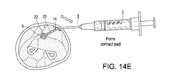
Figs. 14A-14F are cross-section diagrams of a human forearm depicting steps in
the
injection of the liquid conductor around the medial nerve, and connecting it
to a
subcutaneous contact pad, which in turn is in electrical communication with a
TENS
electrode. The bar arrows represent a general direction of movement of the
dispenser
tip.
Fig. 15 is a diagram of the chemical structure of PEG in DuraSeal.
Fig. 16 is a diagram of the chemical structure of Trilysine in Duraseal.
Fig. 17 includes examples of amine-reactive functional groups which can be
substituted for NHS-ester as the active leaving group.
Fig. 18 is a chart of a function depicting the stability of PEG gels based on
the
concentration of elements, i.e., conductive elements.
Fig. 19 is the chemical structure of a PEG with a Hexaglycerol core (8-arm).
Fig. 20 is the chemical structure of a PEG with a Tripentaerythritol core (8-
arm).
Fig. 21 contains diagrams showing steps of amine reactive crosslinker
chemistry
delivering stable conjugates and NHS.
Fig. 22 depicts the chemical structure of carbonyldiimidazole zero-order cross
linker.
Figs. 23-24 are diagrams showing how the hydroxyl moiety can be activated for
coupling ligands.
Fig. 25 illustrates the use of cyanogen bromide to couple an amine ligand.
8
CA 03069424 2020-01-08
WO 2018/227165
PCT/US2018/036773
Fig. 26 is a diagram of the chemical structure showing the interaction between
GLYMO and a silicone as the carrier material and, on the other hand, GLYMO and
silver as the conductive element.
Fig. 27 is a diagram of the mechanism of a cured electrode with low aspect
ratio
conductive elements during bending: as the convex top is bent and conductive
elements move apart slightly and reduce conductivity in the area of the bend,
but
conductive elements at the concave bottom are pressed together and increase
conductivity.
Fig. 28 is an image of a collection of different shapes for a silicone carrier
material.
Fig. 29 is a representation of the function of surfactant to promote
conductivity in a
cyanoacrylate based cured electrode with silver conductive elements.
Fig. 30 shows the final common pathway of coagulation cascade for fibrin glue.
Figs. 31A-31D are images of high-aspect silver flakes manufactured with
various
grain size sand paper wheels using a Dremel tool.
Fig. 32 is another image showing the same high-aspect ratio silver filings as
in Figs.
31A-31D.
Fig. 33 is an image of gold flakes of various aspect ratios produced with a
Dremel
tool.
Fig. 34 contains images of high-aspect ratio conductive elements such as gold
bonding wire bits.
Figs. 35A and 35B are idealized section views of a cured electrode in an
original
linear shape and a subsequent bent position showing, after bending, the high
aspect
conductive elements (35B) maintain connectivity compared to lower aspect ratio
(35A).
Fig. 36 is a diagram of a change of shape for NiTi wire conductive elements.
Fig. 37 is a diagram of a mesh of a cured electrode comprising gold bonding
wire
continuous loops that interconnect with each other, in place around a target.
9
CA 03069424 2020-01-08
WO 2018/227165
PCT/US2018/036773
Fig. 38 is a depiction of two cured electrodes on the same nerve fiber with
different
activation thresholds as a result of proximity to nodes of Ranvier.
Fig. 39 depicts four cured electrodes which have been injected along a nerve
with a
Y-junction, enabling the possibility of selective fascicle stimulation.
Section views
of the cured electrodes at the location of the bar arrows are shown in A-D.
Fig. 40 depicts a selective interface by positioning a cured electrode to
specific
fascicles A and B of a nerve.
Fig. 41 depicts a method of loading the liquid mixture and liquid nonconductor
in a
single chamber dispenser, with the liquid mixture in front (1st) portion
nearest the tip
and the liquid nonconductor in back (2nd) portion.
Fig. 42 is an image of an embodiment of a low viscosity silicone and silver
based
cured electrode dispensed through the dispenser in Fig. 41.
Fig. 43 depicts a cross section of a nerve fascicle surrounded by the cured
electrode
herein in turn surrounded by the nonconductive layer.
Fig. 44 is a diagram of two embodiments of the ring-like portion of a cured
electrode,
and a first side of each being connected with either the anode or cathode end
of a
signal generator and each of the other ends being connected optionally to a
nerve
target.
Fig. 45 depicts a ring like portion of a cured electrode connected to one end
of the
signal generator and also to the nerve (active cathode), or can be placed at
another
location to provide a better electrical interface to the surrounding tissue at
the location
of the distal anode.
Figs. 46A and 46B are the same cross section of a single vertebra, 46A before
injection of a cured electrode, and 46B, after injection, depicting a foramen
transversium as location of the anchor of a cured electrode, here a ring like
portion
around a nerve target.
Fig. 47A contains cross-sections depicting embodiments of a mold for placing
around
a nerve target, comprising an opening through which a wire can be placed and
secured
by crimp hooks, and the wire being in electrical communication with a cured
CA 03069424 2020-01-08
WO 2018/227165
PCT/US2018/036773
electrode dispensed into the space between the hook and the nerve target. The
two
diagrams on the left side depict the mold before insertion, and the two right
side
diagrams depict the hooks after placement. The two lower diagrams depict a
mold
comprising a movable slider capable of sliding out to cover all or a portion
of the gap
in the mold.
Fig. 47B contains perspective views of (I) a straight sock, (II) a curved sock
and (III)
a sock at almost 90 degrees, all at the tip of a dispenser through which the
liquid
mixture is dispensed.
Fig. 48 is a diagram showing a section view of a portion of a prior art cuff
electrode
around a nerve, showing a void between the metal contact of the prior art
electrode
40 (e.g., platinum) and the nerve 5.
Fig. 49A is the same view as in Fig. 48, also showing that a cured electrode
may
function as a bridge between a prior art metallic electrode contact and the
nerve if
liquid mixture is placed onto the contact prior to implantation of the cuff.
Fig. 49B is similar to the view in Figs. 48 and 49A, except that the metallic
electrode
contact is not present, and the space has been filled completely by a cured
electrode.
Figs. 49C and 49D are similar to the view in Fig. 48, except that the void has
been
filled by fibrous tissue. Fig. 49D also shows dispersion of the energy field
lines.
Fig. 49E depicts the energy field lines traveling to the target when a cured
electrode
is placed as a bridge, on the left, on a prior art cuff electrode (as in 49A)
and, on the
right, when the platinum contact is not present (as in 49B).
Fig. 50 depicts a cross section of a needled skin patch electrode with test
electronics
connected to a subcutaneous contact pad. All but one of the needles is in
contact with
the contact pad.
Fig. 51 is a representation of a cross-section of the needled skin patch
electrode
connected electrically to an implantable needle matrix embedded in the contact
pad,
and the needle matrix and the needles from the exterior electrode are
configured to
make electrical connection with one another.
11
CA 03069424 2020-01-08
WO 2018/227165
PCT/US2018/036773
Fig. 52 is an image of a connecting feature for a lead wire to a cured
electrode, here
a helix screw (or, cork screw), held for display by an alligator clip.
Fig. 53 is a representation of a wire loop which is embedded in one portion of
a cured
electrode which also comprises an interface molded and cured around a nerve
target.
Fig. 54 depicts an electrocorticography ("ECoG"1 electrode matrix of the
present
invention in position on human neocortex.
Fig. 55A is an image of a human brain, depicting the sulci and gyri of the
neocortex
and the midline between the two hemispheres.
Fig. 55B is a representation of a section of neocortex and the underlying
white matter
showing the depth (and relative inaccessibility) of the areas within the
sulci.
Fig. 56A is a representation of a portion of the ECoG electrode matrix in Fig.
54
from the top showing the matrix and wires terminating in holes where the wires
make
electrical contact with the liquid mixture (as shown in Fig. 56B) injected
into the
sulci.
Fig. 56B is a cross-section of neocortex and the ECoG electrode matrix
including the
holes allowing injection of the liquid mixture material deep into the sulci,
as shown.
Fig. 57 is a representation of two types of connectors of a neural signal
generator to
enable an excellent mechanical and electrical connection to the cured
electrode.
Fig. 58 is a representation of a neural signal generator encased with a ring-
like portion
of a cured electrode around a target and an anchor in a foramen (shown in Fig.
46A)
for securing the neural signal generator in place. An additional cured
electrode is
connected to the neural signal generator at the end opposite the target.
Fig. 59A, Fig. 59B and Fig. 59C are representations of how a cured electrode
can re-
establish successful electrical connection between a chronically implanted
electronic
prior art electrode and a target, where the prior art electrode has been
walled off by
the body's encapsulation by the body's fibrous tissue. 59A shows encapsulation
of,
and blocking signal from, the prior art electrode, 59B shows reestablishment
of an
electrical connection between the prior art electrode and the target by means
of a
12
CA 03069424 2020-01-08
WO 2018/227165
PCT/US2018/036773
cured electrode, and 59C shows encapsulation of the arrangement in 59B wherein
electrical communication between the prior art electrode and target is
maintained.
Fig. 60 is another example of a prior art rod-shaped electrode carrier/lead
with disk
electrodes as shown in US Patent No. 8,565,894 B2.
Fig. 61 shows a prior art electrode from US Patent No. 8,494,641 B2.
Fig. 62 is a side view of a two-chamber dispenser comprising a syringe body
comprising two coaxial chambers, a first chamber containing liquid conductor
and a
second chamber containing liquid nonconductor, said second chamber encircling
said
first chamber, a first plunger fitted for the first chamber, and a second
plunger fitted
for the second chamber, a coaxial needle with an exit point for both chambers.
Fig. 62A is an enlargement of a coaxial needle tip in cross section, showing
the outer
wall of the needle enclosing an outer needle lumen containing liquid
nonconductor
and extruding it beyond the exit point, the wall of the inner needle lumen
extruding
liquid conductor also beyond the exit point. Additionally, a pattern of
extrusion is
shown.
Fig. 62B is similar to Fig. 62A, except that a wire is also being extruded
from the
inner lumen.
Fig. 62C depicts a two chamber dispenser tip, with each chamber loaded with a
wire
embedded in liquid mixture, and a portion of the same extruded from both
chambers.
Fig. 63 is a side view of an embodiment of the dispenser comprising an
insulated
stimulator wire with an uninsulated electrical stimulator which is near the
exit point
of the dispenser.
Fig. 64A is a diagram of one embodiment of a dispenser as a catheter for
dispensing
liquid conductor or nonconductor.
Fig. 64B is a diagram of another embodiment of the dispenser as a catheter
which is
able to dispense liquid mixture through a vessel wall to the surrounding
tissue.
Fig. 65 depicts the dispenser in one embodiment comprising a light such as an
LED
attached to the needle.
13
CA 03069424 2020-01-08
WO 2018/227165
PCT/US2018/036773
Fig. 66 is a diagram of a conical frustum for graduated diameter decrease for
a
dispenser.
Fig. 67 are images of an auger embedded in a dispenser to provide a
predictable
forward motion of liquid conductor through the dispenser.
Fig. 68 depicts a rollable tube embodiment of the dispenser comprising a
nozzle on
the front end and optional apparatus at the rear to facilitate the rolling of
the tube to
force the liquid conductor to the needle.
Fig. 69A shows a needle system that utilizes an open tip as well as an open
side port.
Fig. 69B shows a needle system that utilizes a closed and rounded needle tip
and a
side port near the tip.
Figs. 70A-Fig. 70C is a sequence of diagrams depicting use of a pre-formed
mold,
here an inflatable balloon, to facilitate placement of a cured electrode.
Fig. 71 depicts a syringe with a wire with a connecting feature at its forward
most
point embedded in the liquid conductor.
Fig. 72 contains four images of one embodiment of a manual mixer. Images A and
B
show two syringes without needles joined by a connector. Image C depicts the
syringes and the connector prior to being joined. Image D is an image of the
manual
mixer comprising a baffle in the lumen of the connector.
Fig. 73 is a schematic of dielectric polarization and heating brought about by
RF
waves.
Fig. 74 contains a larger diagram of staples with prongs inserted into a
connective
tissue plain and the staple heads embedded in cured electrodes surrounding a
nerve
target. Two smaller diagrams are of a staple before insertion (top) and post
insertion
with head embedded in a cured electrode (bottom).
Fig. 75 depicts staples with a connecting head, the prongs of the staples
crimped into
a wall of an organ (e.g., bladder), and the connecting head embedded in the
liquid
conductor/cured electrode.
14
CA 03069424 2020-01-08
WO 2018/227165
PCT/US2018/036773
Fig. 76A shows that by placing the liquid conductor all around the connection
point
of the three side arms forming the Y provides a means to stimulate all nerve
fibers
entering and exiting the Y-junction.
Fig. 76B depicts lacing ring-like portions of the liquid conductor around each
of the
smaller side arms as well as additional liquid conductor around the major
remaining
arm, all surrounded by a single liquid nonconductor/nonconductive layer.
Fig. 77 contains three diagrams showing steps of tying an adjustable hitch
knot
integrated with the cured electrode to allow breakage of the cured electrode
by pulling
on the loop to enable easy removal of the cured electrode.
Figs. 78A-B. A graphic showing shear forces (arrows denoted F) required for
cutting
and/or removing are greater for insulated solid wire Fig. 78A than for the
cured
electrode, Fig. 78B.
Fig. 79 is a diagram illustrating the location of the present invention in an
above the
knee amputation.
Fig. 80A and Fig. 80B are diagrams depicting examples of placement of liquid
mixture "blobs" on prior art electrodes to align field lines through the
target.
Fig. 81A is a diagram depicting homogenous electrical field lines and Fig. 81B
depicts electrical field lines distorted by examples of placement of liquid
conductor
"blobs" to align field lines through a target.
Fig. 82 is a diagram showing liquid conductor blobs injected into a nerve
without
leaving a trace through the epineurium, and cured electrodes outside the
epineurium.
Fig. 83 depicts a liquid conductor blob injected into the nerve while leaving
a wire-
like portion of the cured electrode through the nerve's epineurium, here shown
only
on the left side.
Fig. 84 depicts a nerve target with a chronically-implanted prior art cuff
electrode
with two solid metal contacts on opposite sides of the nerve, and the nerve
encapsulated in fibrous tissue. Electrical field lines scatter through the
nerve and also
around the perimeter (the epineurium) and in the encapsulation.
CA 03069424 2020-01-08
WO 2018/227165
PCT/US2018/036773
Fig. 85, like Fig. 84, contains a chronically-implanted prior art cuff
electrode. Fig.
85, though, illustrates that electrical field lines can be redirected in a
revision
procedure, by placing liquid conductor just underneath the two cuff electrode
contacts on opposite sides of the nerve just inside the cuff electrode, and
also placing
liquid nonconductor in the fibrous tissue to prevent circumferential
electrical field
lines.
Fig. 86 shows electrical field lines through a nerve target between (A) disc
electrodes,
and (B) ring electrodes, either of which may be prior art electrodes or
electrodes
manufactured and cured in situ.
Fig. 87 is a diagram showing a procedure to create a gap in the fibrous tissue
between
the previously implanted prior art cuff electrode's contact pads and then to
inject
liquid conductor to fill that gap, thus bridging the encapsulation.
Fig. 88 is a schematic of a nerve with two electrodes being placed along the
nerve.
Fig. 89 is a schematic of resistive and capacitive impedance components on the
path
from one electrode through interstitial fluid to the axon within a nerve and
back.
Fig. 90 is a schematic of the voltage curve measured during current controlled
stimulation showing the resistive component (solid curve: vertical lines = IR-
drop)
and the capacitive component (dV/dt indicating the charging of surface
boundaries).
Fig. 91A is a schematic of a lab setup for a neurostimulation study with an
LCR meter
and a first and a second steel probe for measuring impedances in various
animal
tissues.
Fig. 91B is similar to 91A, with the addition of a cured electrode in direct
contact
with the first steel probe, but not in direct contact with the second steel
probe.
Fig. 91C is similar to 91B, with the second steel probe being in direct
contact with
the cured electrode to obtain the impedance of the cured electrode(s), and
with the
addition of a third probe not in direct contact with the cured electrode.
16
CA 03069424 2020-01-08
WO 2018/227165
PCT/US2018/036773
Fig. 92 is a schematic of a lab setup for a neurostimulation study with an
oscilloscope
to measure the voltage necessary to apply a current controlled biphasic
waveform
during TENS stimulation on chicken meat, with and without a cured electrode.
Fig. 93A is an image of an oscilloscope readout of 3.8 volts from the setup in
Fig. 92
without a cured electrode injected into the chicken meat.
Fig. 93B is an image of an oscilloscope readout of 1.68 volts from the setup
in Fig.
92 with a cured electrode injected into the chicken meat.
Fig. 94A is an image of a rat brachial plexus.
Fig. 94B is an image of the rat brachial plexus as in Fig. 94A, but with a
cured
electrode on the brachial plexus.
Fig. 94C is an image of a lead wire embedded in the cured electrode in Fig.
94B.
Fig. 94D is an image of a lead wire embedded in a cured electrode formed as a
ring
around a rat bladder neck and some more cured electrode material added for
mechanical matching.
Fig. 95A is an image of a pig brachial plexus and a ring like cured electrode
formed
in open cut down.
Fig. 95B is an image of forming a knot with a suture in a cured electrode and
pulling
on the knot with two surgical clamps.
Fig. 95C is an image after pulling on the knot in 95B with two surgical clamps
and
the pieces of the cured electrode after the suture cut through the cured
electrode.
Fig. 96 is a diagram showing placement of TENS patch electrodes on the outside
of
the skin of a pig, each patch electrode on top of a corresponding cured
electrode as a
subcutaneous contact pad, each contact pad being connected to a ring electrode
attached by a wire acutely to the vagus nerve.
Fig. 97 is an image of the contact pads, from the setup in Fig. 96, next to
coins for
comparison of size.
17
CA 03069424 2020-01-08
WO 2018/227165
PCT/US2018/036773
Fig. 98 is a chart which plots heart rate (bpm) versus time (seconds) observed
from
stimulation of the vagus nerve in pigs in the set up diagrammed in Fig. 96,
under five
different conditions: (1) low amplitude stimulation, (2) mid amplitude
stimulation,
(3) high amplitude stimulation, (4) removal of the subcutaneously placed
contact pad
14 that connected to the cathode to test for leakage driving the HR reduction,
with no
leakage detected, and (5) removal of the subcutaneously placed contact pad 14.
Figs. 99A and 99B are two charts showing a comparison of electrodes and their
capacitive charge injection capabilities: a prior art cuff (Livallova) 99A and
the cured
electrode 99B.
Fig. 100A is an image of the readout of impedance on an LCR meter as 2.328
ohms,
measured across the length of several turns and twists of the extruded very
thin cured
electrodes and wires (<1 mm) as shown in Fig. 100B and 100C.
Figs. 101A and 101B depict differences in impedance spectrometry for a prior
art
device (101A) and the cured electrode (101B) of the present invention.
Fig. 102 shows in A and B that a coil concentrates magnetic field lines and,
additionally, the cured electrodes in B placed near a target induce further
concentration of magnetic energy at the target.
Fig. 103 shows, in dotted line portion A, the top target tissue in an air gap
between
two magnetically cured electrodes with north and south poles. In dotted line
portion
B, the cured electrode acts to shield the bottom target from the magnetic
field. In
dotted line portion C, the effect on magnetic field lines distant from the
cured
electrodes is minimal.
Fig. 104 shows: A, lower magnetic field density at the target with a coil but
without
a cured electrode; B, greater field density at the target by adding a cured
electrode
between the coil and the target; and C, even further concentration than in B
by adding
a second cured electrode and creation of an air gap at the target.
Fig. 105A shows: I, some concentration of magnetic field lines by a coil; and
II,
greater concentration of field lines by adding a cured electrode inside the
coil.
18
CA 03069424 2020-01-08
WO 2018/227165
PCT/US2018/036773
Fig. 105B depicts a headband situated on the circumference of a head, shown
from
the top, said band containing coils which correspond to subcutaneous
magnetically
conductive blobs of cured electrode.
Fig. 106 is a graph showing thermal conductivity of materials.
Fig. 107 depicts a Peltier element embedded between two thermally conductive
cured
electrodes, one surrounding an artery supplying blood to a tissue, with the
Peltier
element's cold side towards the artery and the hot side transferring the heat
away
from the artery and the tissue by means of a second cured electrode.
Fig. 108, somewhat similar to Fig. 107, depicts a Peltier element embedded
between
two thermally conductive cured electrodes, one cured electrode surrounding an
artery
supplying tissue, with the Peltier element's cold side towards the artery and
the hot
side transferring the heat away from the artery to a vein leaving the tissue
by means
of a second cured electrode.
Fig. 109 is a configuration of thermally conductive cured electrodes for
measuring
and controlling temperature in a blood vessel, here an artery.
Fig. 110 is a conceptual representation of how a thin-film lead wire high and
low
structures (A) or holes (B) to allow the liquid mixture to adhere to the lead
wire.
Fig. 111 is a diagram of two cured electrodes surrounding a target connected
to a
diode (D) which is either a voltage or current limiter.
Fig. 112 is a diagram of two cured electrodes surrounding a target connected
to a
diode (D) which is either a voltage or current limiter, also with capacitors
(C).
Fig. 113 contrasts the larger ablation lesion of prior art devices compared to
that from
the cured electrode.
Fig. 114 depicts an embodiment of the cured electrode for use in ablation, in
A, fully
surrounding the target and, in B, partially surrounding the target.
Figs. 115A-C show patch electrodes supplying current, here for ablation, to
the cured
electrode: A, fully surrounding the target; B, partially surrounding the nerve
and C,
19
CA 03069424 2020-01-08
WO 2018/227165
PCT/US2018/036773
using wire-like portions of a cured electrode drawn from the cured electrodes
surrounding the nerve to a subcutaneous contact pad comprising cured electrode
material near each of the patch electrodes.
Fig. 115D depicts transcutaneous transmission of energy to a target surrounded
by a
cured electrode, and the lesion pattern in the tissue surrounding the target.
Fig. 116 contains images taken in sequence for ablation of chicken leg tissue
with a
cured electrode: A, shows placement of an electrode before ablation (note the
return
electrode at top) and B shows the tissue after ablation and removal of the
cured
electrode, revealing the lesion. C is a zoomed view of B.
Figs. 117 A-D are IR images showing temperature in degrees Centigrade from RF
ablation Experiment 1 on chicken tissue with a cured electrode.
Fig. 118 is an image of the setup from RF Ablation Experiment 2 on chicken
tissue
with a cured electrode.
Figs. 119 A-E are six IR images from a video showing time course of the
temperature
changes in RF Ablation Experiment 2.
Figs. 120 A-D are four time stamped images from the same sequence in Fig. 119,
with the time stamps in the lower left corner of each image.
Fig. 121 is an image of the setup of RF Ablation Experiment 3.
Figs. 122 A-E are images from an IR video of the time course of the pork RF
Ablation
Experiment 3.
Figs. 123 A-B are images of pork muscle tissue in RF Ablation Experiment 4
with
cured electrode injected in a cavity (upper) and removed from the cavity
(lower).
Fig. 124 is an image of a cured electrode in RF Ablation Experiment 4 stuck
between
two pieces of pork tissue held during ablation with a toothpick, showing
whitened
tissue ablated on the left in the pattern of the cured electrode on the right.
CA 03069424 2020-01-08
WO 2018/227165
PCT/US2018/036773
Fig. 125 is an image from RF Ablation Experiment 4 with the cured electrode
removed from the tissue ablated (whitened).
Fig. 126 is an image from RF Ablation Experiment 4 with aluminum foil crumbled
and placed between two pieces of pork tissue, where the aluminum foil has been
removed from the whitened spot in the center of the image where it was when
energy
was applied.
Fig. 127 is an image from RF Ablation Experiment 4 with crumbled aluminum foil
(on left) having been removed from the tissue at the arrow, and a cured
electrode (on
right) has been removed from the tissue at the arrow. Note the much greater
ablation
(whitening) of the tissue from the cured electrode on the right.
Fig. 128 A-B are section views of heat transfer (shown by arrows) from a cured
electrode to surrounding tissue, with RF energy in A from a probe and in B
from
dispersed sources.
Fig. 129 is a section view of heat transfer (shown by arrows) from small blobs
of
cured electrodes injected into tissue. Note how the heat emanates from the
blobs
when they receive RF energy from the surrounding.
Fig. 130 is a section view of a cured electrode inserted on one side of a
tumor to stop
its progress, and a probe attached for applying energy, as well as a counter-
electrode.
Fig. 131 A-C are section views of a metal contact on the skin (A) and with a
hydrogel
layer sandwiched between the contact and the skin (b), and a microneedle patch
on
the skin.
Fig. 132 is one embodiment of a waveform for DC ablation.
Fig. 133 is a frontal schematic view of the spinal column and the upper
portion of the
rib cage (front cut-away) and the sympathetic chain running along both sides
of the
spinal column.
21
CA 03069424 2020-01-08
WO 2018/227165
PCT/US2018/036773
Fig. 134 is also a frontal schematic of a portion of the rib cage and the
sympathetic
chain ganglia, showing greater detail (as compared to Fig. 133) of the highly
irregular
shapes of the sympathetic chain ganglia.
Fig. 135 is a drawing showing foramina as exit points for spinal nerves with
placement of liquid conductor or nonconductor in a foramen.
Fig. 136A is a drawing of the basic anatomy of tendons and the Golgi tendon
organs
at the interface to the muscle fibers.
Fig. 136B is a diagram of Golgi tendon organs with four cured electrode
locations.
Fig. 137 is a drawing of placement location for a liquid conductor/cured
electrode on
the brachial plexus in a human (as in Fig. 1A) with a neural signal generator
(not
depicted) implanted to electrically connect to the cured electrode and thereby
fully
depolarize all fibers of the brachial plexus on demand.
Fig. 138 shows a knee joint with multiple thermally cured electrodes cooling
arteries
supplying blood to the knee joint.
Figs. 139, 140 and 141 are drawings of the outer ear. 141 shows some
innervation
patterns from cranial nerves.
Figs. 142 and 143 are images of external cured electrodes placed on a
subject's ear
in different locations.
Fig. 144 contains two images of external cured electrodes, after removal from
the
ear. Note the darkened areas with the greatest concentration of conductive
elements.
Terms
[014] In addition to additional definitions and explanations supplied
throughout this
written description, the following definitions apply.
(1) "Capacitive charge injection" means electrical charge injected from the
interface
into an ionic medium that can be extracted fully without any charge components
causing irreversible chemical reactions.
22
CA 03069424 2020-01-08
WO 2018/227165
PCT/US2018/036773
(2) "Resistive charge injection" means electrical charge injected from the
interface
into an ionic medium that cannot be fully extracted with some charge
components
causing irreversible chemical reactions, thereby likely to change local pH
levels near
the interface and the surrounding or nearby (target) tissue.
(3) "Carrier material" means any biocompatible material comprising a liquid
(or less
viscous) phase curing to a solid or a more viscous phase. A carrier material
is one
selected from a group consisting of a hydrogel, an elastomer such as silicone,
bone
cement, cyanoacrylate, dental amalgam, dental resin, fibrin glue, polyethylene
glycol, hyaluronic acid, or their components and others.
(4) "Collagen" and "gelatin" are synonymous, unless specifically
differentiated.
(5) "Conductive elements" are elements of conductive material which, at the
time of
placement in a body, comprise at least one dimension of at least one micron.
Conductive elements may be produced by a process selected from a group
comprising
cutting, grinding, etching, extruding and conglomeration of smaller elements.
(6) "Conductive" or "conductivity" means the ability to transfer energy
including,
without limitation, electrical, magnetic, thermal, light and vibration
(including
sound).
(7) "Cure" includes, without limitation, polymerizing, crosslinking, going
through
precipitation and/or going through solvent phase inversion, gelation or other
phase
transition to a solid which retains its shape when subjected to shear forces
expected
for a living body in non-extreme conditions. The curing can be substantially
instantaneous, a few seconds or minutes, or may occur over a longer period of
time.
(8) "Elastomer" means any of various elastic substances resembling rubber,
e.g.,
polyvinyl elastomers which comprise a liquid phase and a solid phase,
including
without limitation siloxane.
(9) "Fractal surface" means a volume current injector as a result of a
conglomeration
of smaller pieces which may be roughened as in with a laser (e.g., on Pt foil)
prior to
being shredded, and also through resorption of materials by the body leaving
pores.
23
CA 03069424 2020-01-08
WO 2018/227165
PCT/US2018/036773
(10) "Inject" means introducing into bodily tissue through (a) a dispenser by
means
of a needle or needle-like structure without the need of an incision besides
that of the
needle, (b) a catheter in a blood vessel or other bodily structure with a
lumen, (c) a
pump through a laparoscopic device inserted through a small incision, (d) a
hole that
has been created with a separate incision, or (e) an auger system transporting
the
injectable material inside a lumen from which it is expressed near, into or
around an
interface target.
(11) "Liquid mixture," comprises a carrier material in a liquid phase and
solid
conductive elements dispersed throughout, and the liquid carrier material is
capable
of curing to a solid phase. "Liquid mixture" means not only the liquid carrier
material
but also the solid conductive elements contained within it. "Liquid mixture"
may also
include the carrier material being in a combination of liquid and solid
phases, in
different portions of the same mass of material, and means the same as "liquid
mixture/cured electrode."
(12) "Liquid nonconductor," means a carrier material in a liquid phase without
any
conductive elements, or an insufficient concentration of conductive elements
to
enable energy conductivity. The liquid nonconductor may comprise the same
material as the carrier material in use in the liquid mixture (or not), and
the liquid
nonconductor is also capable of curing to a solid phase and bonding to the
liquid
mixture. A liquid mixture cures to a solid phase termed a "nonconductive
layer."
(13) "Liquid phase," means a state in which liquid or material may flow by,
for
example, injection prior to curing to a later and more solid phase. "Liquid
phase"
includes, without limitation, a paste or other configurations which do not
hold their
shape and do not possess the ability to reestablish an earlier shape (akin to
a pudding)
when subjected to shear forces expected for a living body.
(14) "Network," means an irregular structure comprising numerous conductive
elements of either regular or irregular shape, said conductive elements being
either
touching one another or disposed in very close proximity to one another.
(15) "Nonconductive layer" is liquid nonconductor which has cured to the solid
phase.
24
CA 03069424 2020-01-08
WO 2018/227165
PCT/US2018/036773
(16) "Percolation" means the ability to disperse throughout a mixture while
retaining
direct mechanical contact and thus either an electrical, magnetic, thermal or
optical
path or a combination of those mentioned throughout the mixture.
(17) "Phase transition" includes, without limitation, curing, cross-linking
(chemical,
ionic or other), polymerization, gelation, self-assembly, or
fusion/solidification
(18) "Resistive charge injection" means current transferred by the electrical
interface
into an ionic medium which causes irreversible reactions to occur in the
vicinity of
the electrode/electrolyte interface inside the ionic medium..
(19) "Solid" means a material which has undergone a phase transition away from
the
liquid phase and has substantially polymerized, cross-linked, precipitated,
gelled,
gone through solvent phase inversion, or transitioned otherwise, and retains
its shape
under shear forces expected for a living body in non-extreme conditions at
specific
locations chosen by physicians.
(20) "Solid phase," means a state in which a material has cured substantially
to a
solid and at least partially retains a shape under shear forces expected for a
living
body in non-extreme conditions, either flexible or hard and either hydrous or
anhydrous, or having these qualities partially or in combination.
(21) "Target" includes without limitation nervous tissue including a nerve,
plexus,
ganglion, brain, spinal cord and the like, and any other tissue for which
electrical,
magnetic, thermal, optical or vibratory stimulation (energy) may have an
effect such
as for example, muscle, blood vessels, organs and tumors. The present
invention
provides a preferential energy path to prevent unwanted side effects to non-
target
tissues.
General
[015] The present invention solves the above problems, and provides
additional
advantages unknown in the prior art. The cured electrode 1 of the present
invention,
in one embodiment, first comprises a liquid mixture in a liquid phase which is
capable
of being injected through a dispenser 2 comprising a needle 3 to the target 5
without
a surgical procedure, where it can be pushed from the needle 3 and molded to
the
CA 03069424 2020-01-08
WO 2018/227165
PCT/US2018/036773
contours of the target and is capable of curing to a solid phase which is
capable of
retaining the shape of the contours of the target. The present invention
produces low
impedance values (<100 S2 or even <10 S2 or < 1 S2), low mechanical impedance,
low
optical impedance, low magnetic reluctance, thus providing a simple approach
to
connect electrically to a target in bodily tissue in various locations,
different patients
and within a shorter procedure time when compared to the time needed to place
prior
art electrodes, especially cuff electrodes.
[016] Another advantage of the present invention is that it is injectable
without
surgical dissection of tissue leading up to the target by means of scalpel,
scissors and
the like prior to electrode placement, that is, with little or no disruption
to the target
or surrounding tissues. The present invention has the ability to form a
"negative" from
the "positive" target contours. The novel property of curing to the contours
of the
target not only provides a better
electrical/magnetic/optical/thermal/mechanical
connection to the target, but also a better mechanical adherence to it,
thereby
anchoring it. Anchors 4 for the cured electrode 1 may additionally be achieved
by
injecting either liquid mixture, or liquid nonconductor bonded to the liquid
mixture,
to non-target structures such as bones.
[017] Moreover, through injection the cured electrode of the present
invention can
be placed in hard to reach locations in the body which a surgeon might be
unwilling
to place a prior art device with elective general surgery, e.g., ganglia of
the
sympathetic chain or nerves of the PNS adjacent to major blood vessels and
located
medially in the body which are difficult to access on a direct line from
outside of the
body. See e.g., Fig. 133.
[018] The particular mechanical and structural properties of the cured
electrode 1
can be varied to match the properties of the tissue targeted, by the choice of
the liquid
carrier material 7 or by additives thereto, and by the selection of the
conductive
elements 6. The curing process, i.e., by introducing additional conditions or
energies
during curing such as ultrasound, cooling or heating or radio-frequency
radiation may
furthermore be utilized to change the physical properties of what becomes the
cured
electrode 1.
26
CA 03069424 2020-01-08
WO 2018/227165
PCT/US2018/036773
[019] Additionally, the present invention is generally being put into place
without
the far greater costs of general surgery, and the attendant risks from general
anesthesia and infection. The present invention can be placed by pain
physicians
accustomed to the placement of pharmacological nerve blocks with or without
the
aid of palpation, electrical stimulation (as verification) and ultrasound or
angiography
as means for visualization.
[020] Fig. 9 is an image of an embodiment of the cured electrode 1 with a
silicone
carrier material injected into chicken meat. The silicone was molded against,
and
cured to a solid against a target 5, here a nerve partially on the right side
of the image
and fully on the left side of the image. A few minutes after the injection,
the nerve
was pulled back from the cured electrode 1. Note the 360 degree covering on
the left,
the 180+ degree interface on the right, the groove on the left indicating the
mechanical match between the nerve and the cured electrode and how well the
material matches with the nerve's mechanical structure. This is a fundamental
example showing that it is possible to intentionally encase 180 - 360 degrees
around
a nerve. The impedance of muscle tissue as measured in rats, chicken and pork
is
approximately 500 to 700 S2 at 1 kHz sinusoidal waveforms. Impedance values of
different embodiments of the cured electrode are provided herein. Any material
providing a lower impedance than 100 S2 is thus at least five times more
conductive
and any mixture of < 10 S2 is at least 50 times more conductive than the
surrounding
bulk, not yet taking into account the additional impedance added by the
encapsulation
which encases any electrode placed into the body over time in the chronically
implanted case, i.e. after three to four weeks post implantation. This
difference in
conductance results in a preferential current path for electrical current
preferentially
following the field lines through the low-impedance mixture instead of going
around
the mixture through the high impedance bodily, such as muscle, tissue. Note
how the
pliable cured electrode has conformed around the nerve on the left side. The
nerve
was freed up completely prior to injection in comparison the only partial
encasing of
the nerve on the middle and right side of the image where the nerve had only
been
partially exposed. Note further how the electrically conductive cured
electrode has
reproduced a perfect mechanical imprint from the nerve, indicating how pliable
the
27
CA 03069424 2020-01-08
WO 2018/227165
PCT/US2018/036773
electrically conductive cured electrode is prior to curing and gentle it may
be too
sensitive neural structures. Note as well that the mechanical integration of
the
electrically conductive cured electrode around a nerve may allow for some
slight
movement of the nerve within the cured electrode (especially when silicone
based
carriers are used that don't bond to the biological tissue) and how the
mechanical
integration may be facilitated around a nerve's Y-junction or other anatomical
structures that provide means to form the cured electrode around or into,
thereby
providing a way to mechanically anchor the cured electrode against, into or
around
the biology.
[021] A conceptual diagram of the distribution of conductive elements 6
(represented as rectangles) in the carrier material 7 (represented as ovals)
is shown in
Fig. 10. The conductive elements form a conductive pathway through the carrier
material, either in a liquid phase or a solid phase after curing. The open
spaces
between the conductive elements 6 and the carrier material 7 represent pores
8.
[022] Fig. 11 is an image of a cured electrode 1 including a nonconductive
layer 9
removed from a nerve target. The curvature on the left was produced by the
molding
of liquid mixture/cured electrode against the target (not shown), surrounded
by the
inner cured electrode with silver conductive elements 6, and the outer portion
with
few or no conductive elements is the nonconductive layer 9. Note the white
line
drawn to show how the cured electrode after curing retains the shape of the
neural or
other bodily target.
[023] The fractal structure as in Fig. 11 is formed by the conductive
elements
dispersed within the nonconductive material, here silicone, but may likewise
be
polyethylene-glycol, Hyaluronic acid, or other hydrogels. As water slowly
seeps
into the cured electrode, either by migrating through the non-hermetic
silicone or
other nonconductor such as hydrogel, or by following capillary effects at the
interface between the metal flakes and the silicone/hydrogel carrier, a large
surface
area of metal-to-liquid is forming inside the cured electrode. The cured
electrode
thus becomes a volume interface, where electrical current may transition from
the
metal conductor to the ionic conductor of the liquid inside the cured
electrode, and
28
CA 03069424 2020-01-08
WO 2018/227165
PCT/US2018/036773
the interface with the nerve only along the area where the cured electrode
directly
contacts the nerve. Note the fractal structure of the silver flakes. As the
cured
electrode gets flooded with interstitial fluid in the body, a large surface
area silver-
to-ionic liquid forms. This large surface area allows for essentially the
entire
volume of the electrically conductive cured electrode to conduct electrical
energy
from the metal to the ionic conductor, meaning a volume effect to inject
charge is
used on the level of charge transfer at the interface metal-water, while a
reasonably
small surface area of the cured electrode is exposed to the nerve (hole on the
inside
circle of the depicted cured electrode), allowing the concentration of the
electrical
field lines onto that area. In summary, while a volume effect is used to
transfer
charge from the metal (or i.e. other electrically conductive elements) to the
body's
ionic conductors (interstitial fluid, etc.), an area interface between the
inside of the
cured electrode and the nerve transfers all the ionically conducted charge to
the
nerve. As the conductive elements are in very close proximity to the nerve,
only
small space remains for connective tissue to be grown around the nerve,
further
limiting a chronically large impedance between the cured electrode and the
nerve, a
positive effect. Note the porous structure that forms between the silver (or
other
metal) flakes which allows for capillary forces to drive aqueous liquid all
along the
inside of the electrically conductive cured electrode, resulting in a large
surface area
between the conductive elements of the electrically conductive cured electrode
and
the ionic current along the conductive elements to travel with low impedance
and
the entire bulk volume of metal elements to be on pretty conductor the
human/animal body provides. Note how the metal elements are all touching in
one
or more points, allowing for much the same electric potential. This allows the
electrically conductive cured electrode to spread a potential from one
location
applied to the cured electrode to the entire volume and allows for the
complete
volume to function as a uniform unit, providing a homogeneous electrical field
all
around the nerve.
[024] The cured electrode relying on e.g. only one type of conductive
element, a
surfactant and a nonconductive carrier matrix (that may or may not necessarily
cure
shortly after injection), may thus form a porous interface that fills with
interstitial
29
CA 03069424 2020-01-08
WO 2018/227165
PCT/US2018/036773
fluid from the body, even without the addition of cells or components (e.g.
sugars)
to be absorbed via macrophages. Fractal surface reduces resistance and
increases
capacitance component by, without limitation, (1) reducing thermal noise due
to
low-impedance conductive materials (e.g., metal flakes in contact with each
other);
(2) reducing surface impedance through large fractal surface area; and/or (3)
increasing capacitance through large fractal surface area - not just the
surface
touching the nerve but also the surface of conductive elements within the
cured
electrode.
[025] While the fractal surface reduces the impedance for charge injection
from
the conductive elements to the surrounding interstitial fluid, the current
traveling
between the conductive elements does so as electron based conduction as the
conductive elements/elements are touching each other. This is different from
commonplace hydrogels or conductive mixtures with impedances above the 100
ohm*cm range and especially above the 10 ohm*cm range, as these commonplace
hydrogel conductors generally utilize ionic conduction (achieved or improved
by
doping with ions inside the hydrogel) or semiconductor type conduction.
[026] Stimulation at low current thresholds is furthermore possible with
cured
(electrically conductive) electrodes 1 by providing a directly touching neural
interface that minimizes the gap between the electrode interface and the
nerve. As
such, the current paths between the cured electrode and the target are much
shorter
and more direct than the current path between the contacts of traditional
electrodes,
which are often recessed inside their carrier matrix. Plonsey and Barr
(referenced
elsewhere herein) had shown that one of the primary factors for nerve
activation
current thresholds is the distance between an electrode and the nerve cells of
interest. Furthermore, by providing a complete surrounding of a neural
structure,
the cured electrode 1 has the ability to depolarize the neural target
structure
uniformly. This "cuff effect" of encasing a neural structure all around and as
close
as possible provides a much more predictable neural activation and block
threshold
for said neural structure with a chronically placed cured electrode,
especially when
compared with a traditional electrode that may only be placed "in some
proximity"
to a neural structure, allowing fibrous tissue growth in the distance between
the
CA 03069424 2020-01-08
WO 2018/227165
PCT/US2018/036773
neural target and the traditional electrode to form much more unpredictable
current
paths, thereby less reliable and less reproducible nerve activation and block
thresholds.
[027] The present invention also has another distinct advantage over the
prior art in
its superior qualities as an electrical system for bodily tissue. A wire or
needle tip
(Fig. 2A) or a flat or smooth metal contact (Fig. 4B) has a smaller surface
area to
inject current capacitively than a rough electrode with greater texture. In
one
embodiment of the invention, a carrier material 7 such as a hydrogel that
becomes
porous (partially resorbed between the conductive elements) provides a greatly
expanded surface area for the conductive elements interfacing with the
surrounding
and penetrating interstitial fluid of the body. The charge injection for an
implanted
electrode may consist of both capacitive and resistive current transfer. In a
bodily
tissue, when the intent is to temporarily stimulate or block neural tissue,
the best way
to inject current is via capacitive charge injection which does not lead to
irreversible
chemical reactions which in turn can lead to the dissolution or corrosion of
an
electrode or the change in pH levels near the electrode and the nerve, thereby
damaging the nerve. Cogan, S., Neural Stimulation and Recording Electrodes,
Ann.
Rev. Biomed. Eng. 10:275-309 (2008), Shannon, R.V. (April 1992). "A model of
safe levels for electrical stimulation". IEEE Transactions on Biomedical
Engineering.
39 (4): 424-426, Cogan SF, Ludwig KA, Welle CG, Takmakov P (2016). "Tissue
damage thresholds during therapeutic electrical stimulation," Journal of
Neural
Engineering. 13 (2): 021001 (2016). In order for any electrode to provide a
large
charge injection capacity, two potential pathways are open: (1) increase the
electrode's surface area in contact with the electrolyte, either by enlarging
the
electrode's macro dimensions or by utilizing fractal surface structures (akin
to
platinum black),and (2) use materials that offer a large charge injection
capacity
inherently. In one embodiment the present invention uses the ability of the
body to
dissolve, absorb or resorb the carrier material 7, fully or at least
partially, thereby
leaving the conductive elements 6 (which are not resorbed) to form pores 8 and
a
porous shape to which the electrolyte makes intimate contact while both, the
conductive elements and the electrolyte in intimate contact are encased by
31
CA 03069424 2020-01-08
WO 2018/227165
PCT/US2018/036773
encapsulation of bodily fibrous tissues. This highly increased surface area,
forming a
"charge injection volume", of electrical carrier elements in direct electrical
contact
with each other and in part direct electrical contact with the surrounding
ionically
conductive interstitial fluid, stands in stark contrast to the generally more
or less flat
surface of conductive material (such as a prior art platinum disk or foil)
that only
provides the electrode-electrolyte interface in a more or less planar surface.
The
current may enter this high surface area porous shape through a wire 10 that
is
encased in, and in electrical contact with, the conductive elements 6, thereby
permitting electron transfer as the primary means for current to travel among
the
conductive elements. This in turn provides a significant increase in effective
electrode-to-electrolyte interface area throughout the whole volume of the
conductive
elements. The pores 8, filling with interstitial fluid or otherwise watery
solutions
inside or outside the body during or after the cured electrode placement,
embody a
larger surface area for the charge injection process than is known in the
prior art,
much larger than the surface area for a smooth surface of the same volume's
outer
dimensions.
[028] Calculations have been made of the surface area of the porous cured
electrode
surface area (S/A) assuming a 1 gram cured electrode which is approximately
0.5
cm' and these have been compared to prior art macroelectrodes reported in
Cogan
(2008) cited above. The surface area for the present invention cured
electrodes is up
to eight orders of magnitude greater than reported in Cogan, as shown in Table
One.
Table One
Comparison of Surface Area to Prior Art Electrodes
A
Electrode Flake Hy drogel Powder Conductive m2 lina2
Formulation S/A (g) (g) Elements
(m2/0 wt%
32
CA 03069424 2020-01-08
WO 2018/227165 PCT/US2018/036773
Present 1.0 0.2 0.8 80.0 8.00E-ol 8.00E+13
Invention, Low
S/A
Microflake
Present 4.0 0.35 0.65 65.0 2.60E+00 2.60E+14
Invention,
High S/A
Microflake
Present 7.0 0.5 0.5 50.0 3.50E+00 3.50E+14
Invention,
High S/A
Microflake
Cogan Macro- 1.00E-09 1.00E+05
electrode
Cogan Macro- 1.00E-10 1.00E+04
Electrode
The surface area of the present invention cured electrodes in Columns F and G
in
Table One might be reduced up to 50% to allow for some surface overlap as
these
are only calculated values, not measured. The flakes are highly irregular in
shape and
therefore great compaction is not expected. Even so, the present invention
enables a
vast increase in surface area of the cured electrode interface over the prior
art.
[029] In another embodiment, a carrier material which is not significantly
resorbable after curing (e.g., silicone, bone cement, dental resin or amalgam)
can be
left inside the body chronically which will enable the permeation and in-
creeping of
water and watery solutions along the interface of non-conductive layer and
conductive elements, thereby filling existing pores 8 in the cured electrode
or filling
pores which may form over time as the mixture is subjected to forces from body
movements. That is, pores 8 may form in any cured electrode of the present
invention,
whether resorbable by the body or not. In another embodiment, a carrier
material
33
CA 03069424 2020-01-08
WO 2018/227165
PCT/US2018/036773
which is not significantly resorbable after curing (e.g., silicone, bone
cement, dental
resin or amalgam) can be mixed with other resorbable additives, discussed
elsewhere
herein, which will enable the creation of pores in the cured electrode after
these
additives are resorbed.
[030] An example of pores which are enabled by a resorbable carrier
material is an
embodiment of the invention which comprises conductive elements and a carrier
material (e.g., hydrogel) in the solid phase which is capable of being
resorbed, e.g.,
within an approximate range of four to eight weeks, in extreme cases several
months
leading up to a year for full or close to full resorption.. After curing and
as resorption
occurs during the chronic stage, the cured electrode can be somewhat compacted
but
comprises pores 8 which allow for much larger charge injection capacitance
values
than possible with an outer-surface-only electrode. The mixture in the liquid
phase is
injected at the target in bodily tissue and optionally a connector blob is
attached to,
and then cures to, a solid interface with a wire. The cured electrode thus
includes the
interface molded to the target 5 and the connector blob 26, the interface
being integral
to the connector blob 26. The connector blob ensures better connectivity to
the wire
even as the outside material gets resorbed. This is a system of two or more
components, featuring a blob 26 of conductive elements focused to provide a
stable
interface (and small faradic impedance R) between a wire and the porous
material
that in turn has a large capacitance thanks to its pseudo-fractal interface
surface that
is in contact with the electrolyte composed of bodily fluids. Figs. 12A-C are
diagrams
representing three stages with pores 8 left after resorption creating large
capacitance
values. Fig. 12 represents the mixture placed on a dry surface (outside a
body, not in
a patient, representing composition before injection) and resorbable material
(gray
spheres) which can be resorbed by the body tissues, e.g., macrophages. In Fig.
12 the
liquid mixture has been injected into a body and interstitial fluid
immediately fills up
some pores between conductive elements 6, as indicated by the gray shading,
but
substantial resorbable material 20 is still present. Macrophages (not shown)
begin
digesting the resorbable material. Fig. 12C represents the cured electrode
four to
eight weeks post-injection. Macrophages have eaten the resorbable material
(gray
spheres are gone) and left additional pores 8 for interstitial fluid or other
in-growing
34
CA 03069424 2020-01-08
WO 2018/227165
PCT/US2018/036773
cells (fibrous tissue, etc.) to occupy. The cured electrode's material to
electrolyte
interface has changed fundamentally from two dimensional (only the outer
surface
of an electrode volume) to highly three dimensional (outer surface of an
electrode
volume and all inner surface interface locations between the electron-
conducting
electrode and the ion-conducting bodily fluid (ionic medium) on both, the
outside
and the inside of the cured electrode material.
[031] The same is in principal true for magnetic conduction where
magnetically
permeable material is used instead of electrically conducting material to form
the
conductive (permeable) element-to-element bridge throughout the mixture which
magnetically is placed in parallel to the less permeable surrounding tissue,
thus
forming a preferential path for magnetic field lines to travel from a
generation
location to a target location.
[032] The same is in principal true for thermal conduction where thermally
conducting material is used instead of electrically conducting material to
form the
conducting ( heat transferring) element-to-element bridge throughout the
mixture
which thermally is placed in parallel to the less thermally conducting
surrounding
tissue, thus forming a preferential path for thermal conduction from a heat
generation
location to a target location (for heating; for cooling then the heat
conduction is
preferentially from a target to a heat drain location).
[033] The same is true for vibratory conductive materials which transmit
mechanical waves through a preferential patch from a vibration source to a
target.
[034] The present invention, comprises a variety of material specific
physical
parameters including, without limitation, curing inside or outside the body
with the
ability to adopt and retain the shape of a specific bodily optimal interface
form, from
flexible to stiff and/or rigid post cure, with different conductivities and
the ability to
mechanically interface with nearby locations within the body next to the
target organ
to have additional stress and/or strain relief on both, an organ and on a
cured electrode
post placement.
CA 03069424 2020-01-08
WO 2018/227165
PCT/US2018/036773
[035] As disclosed herein, the needle-based and laparoscopic approach to
placing
liquid mixture 1 resulting in a cured electrode allows for a dorsal surgical
approach
to electrically (or by other means than just pharmacologically) connect to
organs in
novel ways, similar to the ability of connecting to intercostal nerves and
ganglia of
the autonomic nervous system, as further described herein.
[036] Porous electrodes disclosed herein are highly advantageous for
kilohertz
frequency alternating current ("KHFAC") and non-destructive DC nerve block,
i.e.,
charge-balanced direct current ("CBDC") nerve block. Recent preclinical
studies
with focus on reversible electric nerve block have shown that KHFAC nerve
block
can lead to DC contamination which may be more of a problem if an electrode's
charge injection capacitance Qini is small. Other recent preclinical studies
with focus
on reversible electric DC nerve block have shown that a short-term nerve block
using
DC waveforms of several seconds in length is possible as long as the DC is
injected
as capacitive displacement current of the Helmholtz double layer at the
electrode-to-
electrolyte interface. Materials of large surface roughness such as Platinum
Black
have a larger charge injections capacitance Qini and thus allow a DC-nerve
block to
be applied for longer than with a material that has a smaller Qini (such as
Platinum).
[037] Advantages of porous metal electrodes vs. planar metal electrodes
include (1)
larger charge injection capacitance Qini allowing longer duration DC injection
without incurring nerve damage, (2) relatively easy to manufacture via laser
patterning, sputtering or chemical plating of conductive elements 6 that are
then
mixed with a liquid nonconductor 9 (plus potential additional additives) and
thereby
allow the forming of an electrode as described herein, (3) a volume effect vs.
a surface
effect may provide a large increase in charge injection capacitance. Using the
entire
electrode's volume as interface to the electrolyte in the body provides a huge
charge
injection capacitance.
[038] In addition to forming a large electrode-to-electrolyte contact area
throughout
much of the volume of the liquid mixture with the approaches described herein,
the
surface area of the electrode on the outside of the volume can be made porous
as well
by lightly modifying the approaches described (using a variety of sizes for
36
CA 03069424 2020-01-08
WO 2018/227165
PCT/US2018/036773
components that can be resorbed by macrophages) with the goal to create a
surface
porosity that promotes adhesion of advantageous cell types and minimizes the
adherence of non-advantageous cells. This improves the modification of the
fibrous
tissue encapsulation 52 to create either thicker or thinner layers of
connective tissue
around the cured electrode, aiding with the mechanical integration of the
electrode
within the target tissue or the surrounding tissue near a target as well as
with the
option to aid with a modification of the electrochemical effects caused during
an
application of electrical energy to a target via the cured or curing mixture
placed into
the body.
[039] In contrast to prior art electrodes 40, whose microscopic surface
structure and
macroscopic shape is formed ex vivo, the cured electrode 1 disclosed herein
receives
both its microscopic surface structure (the way all the elements within the
mixture
are aligned with each other at time of cure) and macroscopic shape (of the
overall
mixture) in vivo: by forming a "negative impression" of the target similar to
how a
cast forms as a mold around an arm or leg. This is achieved by one or more
processes
of manufacturing the electrode in vivo either inside a living organism or on
the
outside of a living organism. Although the electrode can be formed inside the
body
fully or in part, it may also be formed on the outside without touching the
target
tissue, but instead by adhering the electrode mixture to an electronic lead
wire, with
or without additional supportive structure, with the intent to modify the
final
electrode interface from a lead wire to an optimized neural interface.
Placement And Connection To A Tens Electrode
[040] A Transcutaneous Electrical Neural Stimulation (TENS) system includes
a
signal generator 11, a least one cable 12 and a TENS pad electrode 13 (also
13A-
13B), as shown in Fig. 13. TENS is often used for rehabilitation purposes or
to
provide non-invasive neuromodulation. TENS electrodes 13 are placed onto the
skin
and attempt to push enough current through the skin to a subcutaneous nerve
that is
close enough to the electrode to be depolarized, leading to an action
potential.
Unfortunately, densities of the current passing through the skin are dependent
on the
37
CA 03069424 2020-01-08
WO 2018/227165
PCT/US2018/036773
contact area (thus increasing when the electrode partially detaches) and it is
often
observed that stimulating a subcutaneous nerve requires current densities at
the nerve
which evoke action potentials in nerves and other sensory cells inside the
skin
between the electrode and the subcutaneous nerve which is the target 5,
sometimes
generating unpleasant sensations that limit the applicability of TENS to few
neuromodulation situations where there is very little movement between a
shallow
subcutaneous nerve and an electrode placed on the skin to increase the chance
of
repeatable stimulation of said nerve via TENS. Even then, there may be a
considerable spread in efficacy between patients and on the same patient
between
application days, which in turn may lead to poor patient compliance in the
clinical
reality. Advantages of TENS include the ability to electrically stimulate
subcutaneous nerves that are within the proximity of the TENS electrodes
placed onto
the skin. There is no need for surgery to electrically stimulate these nerves
in close
proximity to the skin. Disadvantages include paresthesia and pain felt in the
skin as
side effects of the neural stimulation and loss of stimulation effects on the
actual
target nerve. Primarily, the current density in the skin underneath the TENS
electrodes, especially in the skin at the edge of the electrodes placed on the
skin, are
significantly larger than the current densities near a targeted nerve, even at
a depth of
0.5 to 1 cm for a target, and even more so 1 to 2 cm in depth away from the
electrodes
placed on the skin (distance measured perpendicular to the TENS electrode
placed
on the skin). The problem is that current densities at the level of the skin
need to be
increased to a level that causes the sensation of paresthesia or even pain in
order to
have large enough current densities (or voltage differentials) at a location
deeper
inside the body (i.e. 0.5 to 2 cm away from the electrode on the skin).
[041] A low-impedance path for the TENS current to pass just below the skin
while
potentially not or only partially passing through the cells that sense
paresthesia or
pain in the vicinity of the outer layers of the skin avoids this problem, by
means of
the liquid mixture/cured electrode disclosed herein. One embodiment of the
cured
electrode comprises a contact pad 14 (just below the last layer of live skin
as disclosed
herein) to make a good connection to the TENS electrodes which is then
connected
through channels to a lower deposit of liquid mixture (uniting all the
channels) which
38
CA 03069424 2020-01-08
WO 2018/227165
PCT/US2018/036773
then is connected to a wire or another line of liquid mixture to reach a nerve
with
high current densities right away. This embodiment may further comprise an
outside
layer of liquid nonconductor/nonconductive layer around the deposit deep
inside the
skin.
[042] Furthermore, placing an electrode via injection around a neural
target and
stimulating said electrode with electrical fields applied from the outside of
the body
to evoke action potentials (or even to cause a temporary temperature increase
interrupting nerve conduction) near a cured electrode offers advantages over
the prior
art. The present invention's minimally invasive delivery, combined with other
abilities like providing electric field shaping or guiding towards a target,
(or away
from an unwanted side target nearby), provide an advance over the prior art.
The
ability to be close to the target nerve offers the advantage of being able to
activate or
block or generally modulate said structure with small current amplitudes or
voltage
thresholds. Being able to guide the electrical energy from a contact pad 14 in
subcutaneous tissue to the target location at e.g. 0.5 to 2cm deep, or even
deeper, by
offering the current a path of <10S2 (or even <1S2) means that current
densities
passing through the skin can be so small to cause no or only minor perceptions
of
paresthesia or pain during their passage through the outer layers of the skin.
The
present invention has the advantage of being able to more reliably activate
neural
tissue in close or far proximity to the outer skin of a person without intense
or
completely without the side effects of unwanted perceptions of paresthesia or
pain in
the skin near the TENS electrode. The present invention has the ability to
further
guide current around off-site targets that are not to be engaged with.
Examples are
multiple nerves running nearby and only one nerve to be engaged with from the
surface of the skin or a nerve running near a ganglion where either the nerve
or the
ganglion is to be stimulated (i.e. electrically / magnetically) but the other
one
(ganglion or nerve) is to be not stimulated at the same time. Guiding the
(i.e. electric
/ magnetic) field lines to the intended target and shielding them away from or
guiding
them around an unintended target are advantages of the present invention.
39
CA 03069424 2020-01-08
WO 2018/227165
PCT/US2018/036773
[043] "TENS electrode" includes, without limitation, an electrode with a
wire
embedded in a hydrogel that separates the wire mechanically from the skin but
provides an electrical connection to the skin. The electrical connection may
further
be emphasized by smaller sized TENS electrodes (size modification), change of
materials (graphene, metals, or metal composites), different optimizations of
the
geometry of the subcutaneously placed contact pad 14 (one line wire, plus
sign,
double cross #, C-shapes, 0-shapes, circles, ovals, partially or fully filled,
entire
networks or mashes formed from liquid mixture to ensure good contact to an
outside
TENS electrode. The TENS electrode may further incorporate small needles that
penetrate the outer layer(s) of the skin and establish an even more directed
energy
path to the formerly injected cured mixture, further reducing energy
requirements on
the signal generator side as well as potential side effects from off-target
stimulation
as paresthesia, pain, etc. (caused from high voltage drop across the outer
layer(s) of
the skin) are minimized or completely avoided. The small needles may further
aid
with the anchoring of the TENS electrode in a specific location and add
mechanical
/ locational and rotational stability of the TENS electrode on the outside of
the body
with respect to the user's body, even in a situation prone to sweat or
movement.
[044] For example, a patient suffering from phantom limb pain after a
traumatic
injury (e.g., amputation) to the median nerve in the forearm is offered TENS
stimulation to treat the pain. See Figs. 14A-F, which are cross-section
diagrams of
the forearm. In order for the TENS signals to reach a PNS target 5 (here,
median
nerve) located in the deep tissue of the forearm, the present invention is
injected
around the median nerve and terminated just below the skin of the patient's
arm to
form a conductive pad. The procedure is conducted as outpatient procedure with
localized anesthesia and in a 10 minute injection time frame. In one
embodiment, the
liquid mixture cures within 30-900 seconds of injection to form a mechanically
compliant material with high electric conductivity form just below the surface
of the
skin to the target nerve but without an opening of the skin once that the
opening has
healed. This is different from percutaneous wires that are placed to remain
reaching
from the outside of the skin to a target nerve, creating a path for bacteria
to follow
from the outside of the skin to the nerve. As the present invention is
completely
CA 03069424 2020-01-08
WO 2018/227165
PCT/US2018/036773
implanted, there is no bacterial path from the outside of the skin to the
nerve. By
placing a TENS electrode on the skin at the location of the contact pad 14,
and using
a TENS electrode 13 connected to a stimulator 15, the patient is able to
achieve pain
relief by stimulating this deep tissue nerve with a surface stimulation
technique. As
the current is routed from just below the skin to the nerve of interest. The
present
invention has additional advantages for the usability and practicality in
activities of
daily living: If the dimensions of the TENS electrode on the outside of the
body are
larger and thus overlapping the dimensions of the contact pad 14 of the cured
electrode, then moving the TENS electrode relative to the contact pad 14 does
allow
for current to reach the nerve even when the outside TENS electrode and the
implanted pad are not 100% concentric in alignment. As the outer layer of skin
is
able to move with respect to the underlying structures, it is not uncommon to
have a
perfectly placed TENS electrode lose contact with a target nerve beneath the
skin
once that a person moves, bends or stretches as the electric field emanating
into the
body from the TENS electrode may be changed significantly by the bent, moved
or
stretched skin between the nerve and the TENS electrode on the outside. By
providing a low impedance path from just beneath the skin to a neural target
of
interest, the electric field lines are still able to primarily take the path
to the nerve
even with movement, bend and stretch of the skin present as long as an
injected
(cured) mixture electrode is providing the low impedance path.
[045] More specifically, the liquid mixture is injected all around the
median nerve
to form the neural interface as well as a conductive path similar to a metal
wire. Fig.
14A is a diagram of a cross-section through the middle of the forearm with a
dispenser 2 (here, a syringe with a needle 3) containing liquid mixture prior
to
injection targeting the median nerve. Fig. 14B shows the dispenser advancing
to the
target. Following the application of localized anesthesia, the dispenser is
advanced
(optionally under ultrasound, angiography or other visual guidance) to the
target 5
median nerve buried deep inside the tissue. The proximity to the nerve can be
verified
by applying electrical pulses from an electrical stimulator 15 the dispenser's
tip 16,
as discussed herein, which is the only de-insulated part of the dispenser) in
the range
of standard neurostimulation pulses (if the connection target is a nerve but
larger or
41
CA 03069424 2020-01-08
WO 2018/227165
PCT/US2018/036773
stronger reactions of organs, such as muscle tissue), connected distally to
the nerve
are to be seen as the dispenser tip 16 comes into closer proximity with the
target. If
the electrical stimulator activates the nerve target, then the physician has
confirmed
electrical contact has been effected with the nerve. The lowest stimulation
threshold
driving the desired activation of the nerve ("activation threshold") confirms
that the
deinsulated tip of the needle's cannula is in close proximity of the nerve. If
the whole
cannula of the needle is completely insulated along the way then the surgeon
can pre-
fill the needle with the conductive mixture and achieve the conductive path at
the tip
of the dispensing needle through the conductive mixture that fills the inside
the of the
cannula and is just about to emerge from the tip. Optimal location for the
electrical
connection to the target can be further confirmed by visualization such as
ultrasound,
x-ray, angiography or MRI where applicable, aided by the skilled physician's
careful
palpation of the anatomy in the vicinity of the target. Fig. 14C is a diagram
showing
dispensing of a ring-like portion 22 of the liquid mixture/cured electrode
around the
target. The dispensing can itself may be used to bluntly separate the target 5
from the
connecting tissue or the liquid mixture can be dispensed in a cavity formerly
formed
around the nerve by blunt dissection. Fig. 14D shows dispensing the liquid
mixture/cured that will form the cured electrode 1 forming a wire-like portion
23 of
the cured electrode from the target 5 to the skin that provides an
electrically
conductive path from the neural target to the skin surface. Fig. 14E depicts
dispensing
the liquid mixture to form a contact pad 14 in the subcutaneous area which, in
one
embodiment, is formed by criss-crossing several lines of liquid mixture just
below
the skin. In this embodiment of the cured electrode, the ring-like / disk-like
portion
22 is electrically connected to the wire-like portion 23 which is also
connected
electrically to the contact pad 14, such that the cured electrode may receive
electrical
current from a TENS electrode on the surface of the skin. Alternatively, the
liquid
mixture/cured electrode can be connected to neural signal generator 17
("signal
generator") including without limitation an implantable pulseform generator
("IPG")
also implanted in the forearm located for example just below or in close
proximity to
the skin. Fig. 14F depicts utilization of a TENS electrode 13 applied to the
skin at
42
CA 03069424 2020-01-08
WO 2018/227165
PCT/US2018/036773
the approximately location of the contact pad 14 to drive electrical current
to a deep
tissue target 5 such as the median nerve.
[046] In one embodiment, the present invention undergoes a phase change
inside
the body at body temperature, with or without the presence of air, water, and
optionally cured by exposure to forms of energy such as ultrasound, UV or
visible
light, and radio frequency waves to form a partially solid, flexible or
inflexible, or
hard material. The carrier material 7 itself may solidify with or without the
addition
of air, water, energy, and it may release energy during the solidification
process of
forming a full or partially solid material. Conductive elements 6 to enable
electrical
conduction, and nonconductive elements to add to dielectric strength, are
added.
Hemostatic agents may be added in another embodiment. The present invention
optionally may have a property to provide visualization inter-operatively via
fluorescence, ultrasound or radio-/angiography, either as an inherent property
of the
liquid mixture, or through the addition of specific audio-, video-, mechano-
or radio-
opaque agents. Radio-opaque materials include, without limitation, platinum
micro-
or nano-elements.
[047] In one embodiment the invention is a eutectic system comprising a
liquid
phase prior to injection and cures to a solid phase at or below body
temperature, even
under anaerobic conditions, the entire mixture forming a cured electrode upon
solidification that provides impedance levels below 100, in some instances
below
1S2, per mm of length and 1 mm2 in diameter.
[048] The present invention also comprises dispensers and systems that
support the
injection process by assisting a physician in finding the target (e.g.,
ability to
electrically stimulate a nerve or sense neural responses) as well as by
dispensing the
liquid mixture or nonconductor.
[049] The injection of the present invention enables formation of an
insulated or
uninsulated wire-like structure in one embodiment, having some similarities to
(1) a
bare wire in that a cured electrode 1 may not comprise a nonconductive layer 9
or (2)
an insulated wire by the cured electrode optionally comprising and being at
least
partially surrounded by a nonconductive layer. Such a cured electrode
comprising a
43
CA 03069424 2020-01-08
WO 2018/227165
PCT/US2018/036773
nonconductive layer may be injected in its first liquid phase optionally
through a
multi-chamber dispenser 2, e.g., a first chamber 18 containing liquid mixture
and the
second chamber 19 containing liquid nonconductor. Or, in another embodiment,
liquid mixture may be injected through one or more dispensers and the liquid
nonconductor may be dispensed through at least one dispenser separate from the
dispenser containing the liquid mixture.
[050] Disclosed herein also are dispensers for pellets or capsules which
are filled
with liquid mixture or nonconductor, allowing the delivery of materials of
different
types at the same time, or to achieve curing which is delayed compared to
liquid
mixture or nonconductor not contained in pellets or capsules.
[051] In one embodiment, a liquid mixture (and the resulting cured
electrode)
comprises resorbable materials 20 (e.g., Figs. 12A-C) interspersed in a
nonresorbable
carrier material including, without limitation, sugars, amino acids, proteins
and
biodegradable materials which macrophages are able to consume any time after
injection and within a period of 100 days, while leaving the external
dimensions of
the cured electrode intact, thereby creating pores 8 that the body is able to
fill with
interstitial fluid, connective tissue and other cells. These pores increase
the electrode-
to-electrolyte surface area as compared to a smooth surface of, for example, a
traditional wire or metal contact, providing means to increase a cured
electrode's
charge injection capacity as the cured electrode "ages." The cured electrode 1
may
further comprise pre-cured components that are manufactured outside the body
with
materials in an already pre-cured component that facilitate partial
resorption. An
implementation may be an already porous structure, itself electrically
conductive,
that may be seeded with cells, nutrients or other eutroph factors that attract
the in-
growth of connective and/or neural tissue (as well as neural support tissue
such as
glia cells and the like), that is electrically connected with the cured
electrode. Similar
to the implementation with non-resorbable carrier material used as described,
a
version with resorbable components such as PEG is feasible in a combination
with
sugars, amino acids, proteins and biodegradable materials which macrophages
are
able to consume any time after injection and within a period of 100 days,
while
leaving the external dimensions of the cured electrode intact, thereby
creating pores
44
CA 03069424 2020-01-08
WO 2018/227165
PCT/US2018/036773
and having both, the biodegradable additives as well as the PEG be replaced by
connective tissue and other bodily cells during the inflammatory and
encapsulation
process. To retain conductive properties for the energy modalities in
question, the
ratio of anon-conducting carrier to conducting components of 50% or less is
required
to ensure that the in-growing / invading cells and the surrounding cells
forming the
connective tissue (bio-fouling) do not severely degrade the charge injection
properties as seen in chemically roughened electrode surfaces.
[052] In one embodiment, the invention is capable of supplying an anodic
current
during the insertion of the dispenser (e.g. needle, cannula, auger, and the
like) into
the tissue and/or during the extraction of the dispenser from the tissue in
order to
achieve electrically mediated vasoconstriction. Anodic (positive) current
activates a
process leading to the constriction of blood vessels, reduces the probability
of small
vessels being ruptured during insertion and reduces bleeding time from small
diameter vessels. Anodic current contracts blood vessels via the release of
nitric
oxide. This may be used in combination with the other modalities of energy
injection
into the body described in the present invention to reduce blood supply during
i.e. the
injection of a nerve block, be it via thermal, electrical (i.e. DC) or other
modes as the
restriction of blood flow to a set of arteries providing oxygen to a nerve is
able to
provide a temporary nerve block via ischemia. Alternatively, this approach of
combining modalities may be used during a tissue ablation procedure to
minimize
pain within a region, organ or specific location of the body.
[053] In contrast to using anodic current alone, higher-level (10V to 50V
amplitude
voltage controlled cathodic first, symmetrical charge balanced pulse trains at
approximately 10 Hz) are capable of stimulating the muscle tissue of blood
vessels
directly and causing blood vessel contraction. This approach may be utilized
to
reduce bleeding not only during the placement process but also, in one
embodiment,
restricts blood flow to an organ. This may be used in combination with the
other
modalities of energy injection into the body described in the present
invention to
reduce blood supply during i.e. the injection of a nerve block, be it via
thermal,
electrical (i.e. DC) or other modes as the restriction of blood flow to a set
of arteries
providing oxygen to a nerve is able to provide a temporary nerve block via
ischemia.
CA 03069424 2020-01-08
WO 2018/227165
PCT/US2018/036773
Alternatively, this approach of combining modalities may be used during a
tissue
ablation procedure to minimize pain within a region, organ or specific
location of the
body.
[054] Achieving a lower access resistance to a nerve in comparison to a
traditional
electrode put next to, adjacent or around a nerve. The access resistance to a
nerve is
directly related to the amount of charge that may be wasted while a nerve is
to be
stimulated: The closer an electrode is to a nerve, and especially the more
tightly it
wraps the nerve in the form of a cuff, the smaller a nerve activation
threshold may
be. See Plonsey/Barr discussed herein.
[055] The cured electrode may be placed into, near, or around a blood
vessel to be
able to electrically stimulate, or block signal transmission in the blood
vessel's cell
wall. The liquid mixture may be injected around the outside of a blood vessel
to
stimulate arterial constriction or relaxation and thereby help to regulate
blood flow
into an organ a cell mass, the skin (to improve blood flow or reduce it to
conserve
body heat). The present invention may, in another embodiment, be placed around
blood vessels to a tumor to prevent or reduce blood flow to a cancerous or
unnecessarily growing or self-replicating site inside the body, thereby
occluding
blood supply and thus reducing the availability of nutrients and oxygen,
leading to a
reduction of the unwanted growth. Organ or tumor growth may be reduced or
reversed (facilitating an intended cell/organ atrophy as medical treatment).
For that,
the liquid mixture may be injected by a dispenser comprising a catheter (Figs.
64A/64B) from the inside of a blood vessel towards the outside of the blood
vessel,
either injecting it into the wall of the blood vessel or outside to the blood
vessel so as
to electrically contact the blood vessel's outside to an implanted wire 10.
Alternatively, another component of the cured electrode may be injected to the
outside of the blood vessel with an approach that comes from further away from
the
blood vessel and comes closer to the blood vessel. The liquid mixture may be
injected
as a ring around a blood vessel by injecting it through at least one needle
that pierce
the blood vessel wall from inside to outside and create either an interrupted
(but
overall connected) or continuous ring around the blood vessel outside. Such a
ring-
like shape portion 22 of the cured electrode may then be contacted by a wire-
like
46
CA 03069424 2020-01-08
WO 2018/227165
PCT/US2018/036773
portion 23 of the cured electrode to facilitate the electrical connection to a
blood
vessel to a specific location inside the body or just below the skin of a
patient. The
wire-like portion 23 is located from outside the blood vessel from a separate
injection.
[056] Utilizing different activation thresholds for nerves and blood
vessels helps to
separate the two when closely aligned or nearby: While nerves will likely be
depolarized at stimulation current amplitudes of 1 mA stimulation current
applied for
a 200 [tsec pulse width, in a symmetrical, cathodic first waveform, blood
vessels will
more likely not react until about 10mA++ are applied based on the fact that
blood
vessel walls are lined with smooth muscle cells whose activation thresholds
are at
least about an order of magnitude higher than that of axons in nerves. It is
feasible
without a nonconductive layer being placed to stimulate and activate a nerve
next to
an artery, but enables stimulation of only the blood vessel (e.g., to
contract) but not
depolarization of a nearby nerve through combinations of various stim and
block
waveforms.
[057] In one embodiment, a cured electrode can be placed on the outside of
a tumor
to ablate existing blood vessels and newly growing blood vessels that may
regrow
nearby the old (ablated) ones with the tumor trying to replace the ablated
ones. With
an existing cured electrode already present only a small incremental amount of
cured
electrode is needed to fully encase new blood vessels the tumor might have
grown,
ablation may be used to heat up the newly and previously placed cured
electrode
when only one point of the entire cured electrode network around the tumor is
touched.
[058] By injecting angiographic contrast agents to blood arterial vessels
that
supply cancerous tissue, a tumor can be visualized against the surrounding
tissue with
increased contrast compared to the surrounding tissue, the contrast being
increased if
combined with other modalities such as contrast assisted PET and CAT scan of
the
cancerous tissue. Once the cancer and its margins have been visualized, a
needle
based delivery of a liquid electrode mix is possible under angiographic
visualization.
This allows the physician to place the liquid electrode in the border region
of the
cancerous tissue. If fluoroscopic contrast agents are used to further
illuminate
cancerous tissue under i.e. UV light, then a laparoscopic approach may be
utilized to
47
CA 03069424 2020-01-08
WO 2018/227165
PCT/US2018/036773
aid the physician in guiding the needle used for the delivery of the liquid
electrode.
The physician may aim to (a) inject the liquid electrode mix into the
cancerous tissue
itself at one or more locations (if i.e. an ablation of the tumor from the
inside outward
is intended), or (b) inject the liquid electrode mix into the cancerous tissue
on the
margins between cancerous and healthy tissue at key locations such as near
vital
arteries or veins that a tumor may not be easily resected from (if e.g. an
ablation of
the tumor at that barrier region is required to avoid spreading or prepare a
later
surgical removal of the tumor following some recovery time between ablation
and
resection as dead tumor tissue may be more easily resected from said vital
tissues or
organs), or (c) inject the liquid electrode mix into the cancer margins
between
cancerous and healthy tissue meaning injecting it into healthy and cancerous
tissue
(i.e. to ensure a wider ablation region and increase the probability of
stopping the
spread of any cancerous tissue), or (d) inject the liquid electrode mix around
the entity
of the cancerous tissue just outside the tumor margins (i.e. with the intent
to ablate
the entire outside and most of the inside of the tumor as well as blood
vessels
supplying it with nutrients, or (e) inject the liquid electrode mix around the
blood
vessels supplying the cancerous tissue with nutrients (i.e. with the intent of
utilizing
ablation of the blood vessels to starve the tumor from nutrients without the
risk of
bursting the blood vessels as the cured electrode may be heated in a more
controlled
manner than a traditional ablation electrode approach would allow).
[059] The present invention, in some embodiments, may be placed into, near,
or
around an organ, especially specific structures of an organ such as internal
blood
vessels or neurons, or an inside or outside wall of the organ to be capable of
electrical
stimulation, or blockage of signal transmission, in the organ, the innervation
or the
blood supply of the organ, for example, the bladder. Organ activity can be
changed
by increasing or decreasing neural communication into and out of the organ,
and
some organ growth and activity can be up- or down-regulated by allowing more
or
less blood enter the organ, such in the case of the gut, the liver, the lungs
or the kidney
which are exchange systems for the body, utilizing a fine mesh of blood
vessels
intertwined with other vessels who either add or extract chemicals in the form
of
dissolved gasses or liquids. The present invention allows for an efficient way
to
48
CA 03069424 2020-01-08
WO 2018/227165
PCT/US2018/036773
contact an organ, such as by injecting the liquid mixture to the outside wall
of an
organ near an innervation point. The conductive elements may in such case
comprise
a biocompatible mesh (not pictured but well known in the art) attached via a
liquid
mixture and/or sutures to the organ, the electrical conduction between mesh
and the
organ being accomplished or improved by the liquid mixture.
[060] Nerves close to the surface of the body that have been shown to
respond to
current injection by thin needle (i.e. electro-acupuncture and similars) can
be targeted
more reliably with a TENS unit once a cured electrode has been placed into
and/or
around the target nerves close to the surface. The physician first verifies
the efficacy
of neural stimulation of a specific nerve via thin needle (i.e. acupuncture),
then map
the nerve's dimension with the needle in the specific location (looking for
smallest
activation threshold), which may be assisted by ultrasound or angiography
visualization. The physician may choose to only place a cured electrode into
the nerve
sheath, or the physician may choose to place a cured electrode as a partial or
full ring
around a nerve target of interest. Then the physician may choose to extend the
liquid
mixture from the nerve target as he/she is retracting the needle towards the
skin,
thereby forming a bulge or a wire-like extension from the bulk of the cured
electrode
near/around the nerve. This extension may be just 1-2 mm in length or it may
be
1 Omm in length or more with the intent to guide electrical field lines in an
anatomically preferential path to the target nerve, the best path electrically
not always
being the shortest path mechanically. By terminating the extension of the
liquid
mixture near or just below the outer layers of the skin the clinician ensures
that the
tissues surrounding the nerve and the skin remain fully supply with blood and
thereby
nutrients, oxygen etc. while allowing a more reliable and interface to the
nerve
formerly only activated with needles from the outside of the body.
[061] The mixture may further contain components that provide a magnetic
interface effect, allowing an easy way to find a subcutaneous cured electrode
with a
magnet placed on the outside of the skin. This approach may aid the patient in
placing
the TENS electrode (then potentially with an added magnetic component via i.e.
rare
earth magnets) always in the correct spot and possibly, if alignment is
important, in
the proper alignment as long as the liquid mixture in the subcutaneous tissue
has
49
CA 03069424 2020-01-08
WO 2018/227165
PCT/US2018/036773
either two magnetic poles, or two locations that are able to interface with
magnets
(e.g. ferro- or ferrimagnetic elements). Furthermore, an electrically
nonconductive
layer 9 but magnetically active mixture may be placed into the subcutaneous
tissue
secondarily to the initial placement of a cured electrode. One such
configuration is
an M-E-M (magnetic - electric - magnetic) design where the electrical
interface is
centered between the two magnetically interfacing cured electrodes. The
corresponding TENS electrode (or a TENS electrode placing device) may utilize
two
rare earth magnets to align the TENS electrode with the center, electric,
interface by
magnetically aligning with the two outer M cured electrodes. This may greatly
enhance user friendliness for finding the subcutaneously placed cured
electrodes and
always optimally placing the TENS electrode on the outside of the body.
[062] Nerves may further be visualized by angiography and injection of
angio
contrast agents into the arterial blood supply of the neural target. The
liquid mixture
may contain contrast agent added to the mixture (same agent as injected
arterially,
platinum components, etc.) to aid with the visualization during cured
electrode
placement. With both, the liquid mixture/cured electrode and the nerves
showing
sufficient contrast against the surrounding tissue, angiography may be
utilized in very
similar ways as done during the placement of a stent during a cardiac
procedure.
[063] The biocompatible liquid mixture comprises conductive elements and
nonconductive carrier material and optionally other elements (affecting curing
times,
integration with the body, inflammatory response, etc.) which is mixed
together by
the physician shortly before placing it inside the body (or thawing it shortly
before
placing it inside the body) and which cures and functions as a conductor
inside the
body, i.e., an aqueous environment with or without the aid of additional
energies. The
liquid mixture has great mechanical stability and homogeneity even though,
prior to
curing in the body, it may flow as a liquid, gel or paste. After curing, the
cured
mixture has conformed to the bodily structures against which it was formed.
The
resulting cured electrode has resistance <10 ohm for a shape of 1 mm diameter
and 1
mm of length (meaning a volume impedance of 10 Ohm*mm) (-). The present
invention is not intended as a thin film, and this application specifically
disclaims
any aspect as a thin film manufactured outside the body.
CA 03069424 2020-01-08
WO 2018/227165
PCT/US2018/036773
Carrier Materials
[064] The carrier material 7 provides the capability of being injected
because it
comprises first a liquid phase and then it cures to a solid phase and, as
such, the liquid
phase carrier material allows injection of the conductive elements 6 which are
interspersed in the carrier material 7. Although curing may begin outside of
the body,
at least some of the curing process is capable of occurring inside the body,
distinguishing the invention from prior art electrodes which are pre-
configured prior
to implantation. The carrier materials include hydrogels, elastomers, tissue
glues,
protein glues tissue adhesives other than glues, tissue sealants, coagulants,
cyanoacrylates, bone cements, dental resins, and dental amalgams. If powders
are
part of an embodiment, then the powder's dispenser allows the formation of a
mechanical structure (with or without the addition of other materials) that
becomes a
less pliable structure after curing. Powders akin to some of the powders used
as
coagulants can form the non-conductive mechanical support structure by first
coagulating bodily fluids and tissues in place co-located with the conductive
carriers,
while limiting the production or aiding with the transmission of excess heat
away
from sensitive tissues such as the neural target tissue.
[065] Fast curing is often optimal, for example, a range of 1 to 5 seconds
as the
body is constantly moving with heart beats, breathing, pulse even in distal
arteries,
moving muscles; in other embodiments it is preferred for the curing to take no
longer
than 900 s. Although the curing time for a specific implementation may exceed
15
minutes (900 s) of time to reach the solid phase, a curing duration of less
than 15
minutes is better in a surgical implementation than a duration of longer than
15
minutes. This curing duration does not include the encapsulation by the body
or the
partial dissolution and/or resorption of components or materials included as
part of
the embodiment of the invention in its liquid phase. Slow curing also has
specific
application for better long term integration to the surrounding tissue.
Forming a good
mechanical bond to the biological tissue is optimal. In the liquid phase the
carrier
material is dispensed via injection. The carrier material may have gel-like
property
as long as it is capable of curing further into a more stable form retaining
the shape
of contours of the target around which it is injected and molded against the
contours
51
CA 03069424 2020-01-08
WO 2018/227165
PCT/US2018/036773
of the target. The carrier material may be a putty-like, amorphous material
(similar
to "Sugru Mouldable Glue" in its mechanical behavior but, in contrast to
Sugru,
biocompatible; and curing fully without the release of toxic or partially
toxic gases
and other substances) that may cure inside the body, retaining some mechanical
flexibility post curing or not. The carrier material may comprise a eutectic
paste. The
carrier material may be doped with the body's own cells to better integrate.
The carrier
material may also be doped with stem cells from the patient or other living
organisms.
It may be doped/mixed with radio-opaque elements or dyes (for example to allow
the
verification of the placement of the carrier material in its liquid phase
around the
nerve or through tissues as well as the ability to detect breaks in the cured
electrode
after years of wear and tear). It may be doped with sugars or other resorbable
materials 20 which the body's macrophages resorb in order to change the
injected
liquid mixture into a porous structure (Figs. 12A-C) as time passes and the
body
partially digests the blob, thereby increasing the active surface area to the
embedded
conductive elements. The carrier material also may comprise fluorescent
elements or
dyes that allow the verification of placement around the nerve or through
tissues
intra-operatively by shining a UV light onto it that does not cure the carrier
material
but instead makes it glow in the dark of the cavity and around the nerve or,
if injected
into the nerve, makes it glow from inside the nerve. The carrier material may
also
comprise pharmacological agents to produce short-term or sustained drug-
delivery
that have complementary action to the cured electrode (e.g. lidocaine to
reduce pain
from operation and/or produce local anesthesia, or other nerve-block agents or
other
pain-alleviating agents that may ordinarily be injected near a neural target).
[066] The viscosity of the liquid mixture affects how readily it will flow
and
distribute itself within a created body cavity. Lower viscosity liquid
mixtures will
flow more easily than higher viscosities, but higher viscosities have greater
ability to
stick to a specific placement location and to hold a specific space filled
without
flowing to unintended spaces.
[067] A low viscosity liquid mixture has an advantage in its greater
capability to be
injected behind or below a nerve but in some embodiments may be used with a
pre-
formed mold (e.g., Figs. 47A and 47B) to be inserted at the target to hold
this space
52
CA 03069424 2020-01-08
WO 2018/227165
PCT/US2018/036773
open during the injection or other placement process. Higher viscosity affords
a
greater capability for the liquid mixture to resist forces from the
surrounding
biological tissue to be pushed out of the cavity, thereby retaining a minimum
ring-
like portion 22 around a nerve when injected without a pre-formed mold.
[068] Among other advantages discussed elsewhere, higher viscosity carrier
materials have the following advantages in aiding: (1) with combatting
separation of
conductive elements from the carrier material as the liquid mixture passes
from the
larger inner diameter of a dispenser to a smaller diameter needle; (2) with
dispensing
as the thicker material sticks in place; and (3) with surgical integration as
the more
viscous liquid mixture may be shaped in place, holding its form and shape to a
certain
degree before curing. Differences in viscosity are primarily achieved by
changing the
ratio of conductive elements vs. silicone carrier material. A secondary way of
changing the viscosity is by adding surfactants, thickening or thinning
agents.
Thinning agents may be selected from a group including water, PEG solutions,
glycerine, and other inactive excipients commonly utilized in the
pharmaceutical
industry found at haps://www.accessdatafda.govlsetiptsledertiigtind ex. dm.
Thickening agents may be selected from a group including inactive polymer
powders
such as polyethylene glycol ("PEG") powder, peptide powders, starches, sugars,
silica powder, and additional metallic and non-metallic fillers that may or
may not
add further elements of high conductivity (graphene being one of them).
[069] A comparison of the hydrogel PEG, fibrin glue and cyanoacrylate as a
carrier
material is useful. PEG becomes mechanically flexible in the solid phase after
curing
with medium to high water content. When PEG is hydrolyzed it dissolves, and
its
stability depends on crosslinking, and dendritic structures create higher
cross linking.
It may be polymerized or cross-linked to the solid phase by different
mechanisms.
Fibrin glue is also mechanically flexible as a solid having a medium water
content. It
can degrade enzymatically in vivo and its stability depends on crosslinking.
Fibrin
requires frozen storage and it may be stored up to two years, and it requires
thawing
before use. Cyanoacrylates have low water content and variable rigidity.
Cyanoacrylates are very stable and hydrolyze over time, though the average
time for
a cyanoacrylate to hydrolyze is to be expected longer than the time needed for
the
53
CA 03069424 2020-01-08
WO 2018/227165
PCT/US2018/036773
body to take over the mechanical stabilization before the cyanoacrylate has
substantially weakened. If the intended location in the body is anticipated to
be under
significant physical stress/strain, e.g. near contracting muscles or joints,
longer
hydrolysis times, at least greater than the time it takes to form a stable
fibrous capsule
around the implant, are desirable. The rate of fibrous capsule formation
itself may be
variable depending on location in the body, and is likely a function of tissue
vascularity. Higher vascularization means a higher mobility of fibroblasts and
macrophages to the site of implantation and thus a higher rate of scar
formation.
Hydrogel
[070] A hydrogel is a network of hygroscopic (water-absorbent) polymer
chains
swollen with water. Hydrophilic gels that are usually referred to as hydrogels
are
networks of polymer chains that are sometimes found as colloidal gels in which
water
is the dispersion medium. One definition of a hydrogel is that of a water-
swollen and
cross-linked polymeric network produced by the reaction of one or more
monomers.
Another definition is that of a polymeric material having the ability to swell
and retain
a significant fraction of water within its structure, but not dissolve in
water.
Hydrogels also possess a degree of flexibility similar to natural tissue
because of their
large water content. The ability of hydrogels to absorb water arises from
hydrophilic
functional groups attached to the polymeric backbone, while their resistance
to
dissolution arises from cross-links between network chains.
[071] A form of hydrogel, cross-linked gelatin forms a cohesive matrix with
tunable
post-curing viscosities. Gelatin easily flows at temperatures exceeding 50
degrees C
and undergoes a reversible transition from solid to gel under specific
conditions.
Gelatin is a naturally occurring, and generally well-tolerated biomaterial.
Gelatin is
an irreversibly hydrolyzed form of collagen. It is an animal collagen
thermally
denatured with a very dilute acid, with many glycine residues (almost one in
three),
proline and 4-hydroxyproline residues. A typical structure is ¨Ala-Gly-Pro-Arg-
Gly-
Glu-4Hyp¨Gly-Pro. While the basic building blocks of gelatin and collagen are
the
same, collagen retains more of its tertiary fibril structure. Conductive
elements may
be mixed with gelatin above its gelation temperature (temperature threshold
for the
formation of a thermoreversible gel), injected into the body, and allowed to
cool. The
54
CA 03069424 2020-01-08
WO 2018/227165
PCT/US2018/036773
resulting cured gel containing conductive elements will be electrically
conductive.
Furthermore, gelatin comprises the processed form of collagen. Gelatin can be
ground up, mixed with conductive elements (and optionally a surfactant and
other
additives) and then added to the carrier material to form a paste that
undergoes the
phase change in the body, and immediately after curing begins a process by
which
the body's inflammatory response starts to exchange, digest, or replace the
gelatin
based elements with the body's own cells, thereby growing into the cured
electrode
or partially digesting the cured electrode, which leaves pores inside the
remaining
cured electrode bulk, thereby creating a porous interface of much larger
surface area
compared to a smooth surface of the same outer dimension.
[072] PEG is a hydrogel, and it has many advantages as a carrier material
for the
liquid mixture and the cured electrode. Hydrolysis of 20kDa cross-linked PEG
is
approximately 4-8 weeks. Higher molecular weight or higher cross-linking
density
may achieve longer hydrolysis times. PEG is hydrophilic and will therefore
adsorb
proteins during and after implantation to the surface, without greatly
denaturing them.
This increases biocompatibility and adherence to surrounding tissues compared
to
silicone and other hydrophobic surfaces. PEG has much greater replacement by
the
body than silicone. PEG provides a regenerative growth substrate for repairing
damaged neurons/axons. PEG's repeating ethylene glycol units provide ample
opportunity for hydrogen bonding, particularly with carboxylic acids in
microenvironments above their pK (-4.5). Importantly, PEG can act as a
chelator or
buffer for bicarbonate, which can locally decrease the pH or presence of
carbonic
acid in the microenvironment which has demonstrated benefits for wound
healing. A
PEG based liquid mixture/cured electrode may be manufactured with an
intentionally
higher impedance than other carrier materials, by adding non-conductive
materials,
elements or elements to the mixture. The resulting insulating PEG cured
electrode
may be used to restrict electrical current flow from certain areas, or as a
liquid
nonconductor it may be used to achieve an insulation around the liquid
mixture/cured
electrode. In some embodiments PEG may be made more nonconductive by adding
elements that make the final cured PEG more attractive to in-growth of fibrous
tissue
thus increasing insulation with the body's own fibrous tissue, in comparison
to the
CA 03069424 2020-01-08
WO 2018/227165
PCT/US2018/036773
PEG based cured electrode that is intended to remain conductive (with
conductive
elements) as the PEG is replaced by the organism.
[073] In yet another embodiment, the addition of gelatin to carrier
materials such as
PEG hydrogels or silicones, is used to intensify the body's inflammatory
response,
on a continuous scale according to the concentration of gelatin added, thereby
increasing the amount of encapsulation 52 that is formed by the body around
the
cured electrode. The cured electrode may also comprise gelatin to thicken
encapsulation, for example, to keep the cured electrode in place and prevent
conductive elements 6 from flaking off, or it may be applied as a second layer
on the
outer aspect of the electrode formed next to, or around, a nerve, to ensure a
thicker
encapsulation to increase the electric impedance towards the outside of the
cured
electrode with the goal to have a low impedance (i.e. thin layer)
encapsulation on the
inside of the ring-like portion 22 that touches the nerve and a large
impedance (i.e.,
thick layer) encapsulation 52 on the outside of the cured electrode against
the
surrounding tissue. This approach may be used to interface selectively with
various
nerves running in parallel or it may be used to minimize muscle fiber
activation or
simply reduce current outflow out of the cured electrode wherever it does not
do any
work stimulating the target. The control of the encapsulation, and thereby the
electrical interface impedance between the cured electrode and the surrounding
tissue
aids in constructing a lower side-effect and more energy-efficient neural
interface
that saves on battery lifetime for signal generators.
[074] PEG is a carrier material which comprises a liquid phase to which
conductive
elements may be added or attached. PEG hydrogels are biodegradable and are
resorbed by the body after injection and after curing to the solid phase of a
cured
electrode, thus allowing the formation of pores.
[075] PEG is a polyether compound and is also called polyethylene oxide
(PEO) or
polyoxyethylene (POE), depending on its molecular weight. The structure of PEG
is
commonly expressed as H¨(0¨CH2¨CH2)n¨OH. PEG, PEO, and POE refer to an
oligomer or polymer of ethylene oxide. The three names refer to the same
compound,
but historically the term PEG is preferred in the biomedical field, whereas
the term
PEO is more prevalent in the field of polymer chemistry. As used herein, PEG
or
56
CA 03069424 2020-01-08
WO 2018/227165
PCT/US2018/036773
polyethylene glycol means any compound comprising the general structure X-
(0¨CH2¨CH2)n-Y where n is a variable number of repeat units and X and Y are
functional groups at the terminal ends. If X = Y, then the PEG is called a
"homo-bi-
functional PEG." If X does not equal Y, then the PEG is called a "hetero-bi-
functional
PEG." If X or Y = -OH and is therefore unmodified, then the PEG compound is a
"monofunctional PEG." Because different applications require different polymer
chain lengths, PEG has tended to refer to oligomers and polymers with a
molecular
mass below 20,000 g/mol, PEO to polymers with a molecular mass above 20,000
g/mol, and POE to a polymer of any molecular mass. PEGs are prepared by
polymerization of ethylene oxide and are commercially available over a wide
range
of molecular weights from 300 g/mol to 10,000,000 g/mol. In one embodiment,
the
PEG suitable for the carrier material is within a range of 1000 g/mol ¨ 50,000
g/mol.
[076] While PEG and PEO with different molecular weights have different
physical
properties (e.g. viscosity) due to chain length effects, their chemical
properties are
nearly identical. PEGs/PEOs come in a variety of molecular weights, with
varying
degrees of polydispersity. Furthermore linear PEG chains may be initiated and
terminated by different functional groups, e.g., -CH3, -OH, -COOH, -SH,
depending
on the initiator, capping agents, and polymerization process used.
[077] PEGs are also available with different geometries. In order to
facilitate
efficient crosslinking, a branched structure is desirable for a carrier
material herein.
The two market leaders for PEG products, Coseal and Duraseal, use 4-arm PEG
which are suitable as carrier materials CoSeal has a MW of 10kDa and DuraSeal
has
a MW of 20kDa. Hyperbranch also provides a dendritic PEG adhesive with much
higher branch numbers which are suitable. Branched PEGs have three to ten PEG
chains emanating from a central core group. Star PEGs have 10 to 100 PEG
chains
emanating from a central core group. Combination PEGs have multiple PEG chains
normally grafted onto a polymer backbone. The numbers that are often included
in
the names of PEGs indicate their average molecular weights (e.g. a PEG with n
= 9
would have an average molecular weight of approximately 400 daltons, and would
be labeled PEG 400.) Most PEGs include molecules with a distribution of
molecular
weights (i.e. they are polydisperse). The size distribution may be
characterized
57
CA 03069424 2020-01-08
WO 2018/227165
PCT/US2018/036773
statistically by its weight average molecular weight (Mw) and its number
average
molecular weight (Mn), the ratio of which is called the polydispersity index
(Mw/Mn). MW and Mn may be measured by mass spectrometry or by gel permeation
chromatograhy. All the above configurations of PEG are suitable as the carrier
material for the present invention.
[078] PEG is soluble in water, methanol, ethanol, acetonitrile, benzene,
and
dichloromethane, and is insoluble in diethyl ether and hexane. It is coupled
to
hydrophobic molecules to produce non-ionic surfactants. If inadequately
purified or
characterized after synthesis, PEGs may potentially contain toxic impurities,
such as
ethylene oxide and 1, 4-dioxane. Ethylene Glycol and its ethers are
nephrotoxic if
applied to damaged skin. It is therefore important that the source of PEG
materials
be rigorously quality controlled, as has been accomplished by a number of
other
manufacturers having FDA-approved PEG adhesive formulations on the market.
[079] PEG and related polymers (PEG phospholipid constructs) are often
sonicated
when used in biomedical applications. However PEG is very sensitive to
sonolytic
degradation and PEG degradation products may be toxic to mammalian cells. It
is,
thus, imperative to assess potential PEG degradation to ensure that the final
material
does not contain undocumented contaminants that may introduce artifacts into
experimental results.
[080] An example of a hydrogel which can be used as a carrier material in
the
mixture is a PEG tissue sealant commercially available called DuraSeal. It
comprises
a 2-part solution system that when mixed forms a synthetic hydrogel coating
that is
biocompatible and degraded in the body over 4-8 weeks. More specifically, it
comprises (1) a 20kDa, 4-arm Branched PEG, terminated with NHS-ester-activated
functional groups, (2) a trilysine crosslinker, and additives including (4) a
preservative: BHT (butylated hydroxytoluene), (5) Dyes ¨ help to ensure mixing
is
complete, FD&C Blue, (6) Buffers- sodium phosphate for PEG, and (7) Buffers ¨
sodium borate for trilysine. The PEG is dissolved at a concentration of 0.5g
in 2.5m1
of sodium phosphate buffered saline (20% w/v or 10.0mM). The tri(L-lysine)
acetate
is dissolved at a concentration of 10.5 mM in 2.5 ml 75m1V1 sodium borate
decahydrate. Fig. 15 is a diagram of the chemical structure of PEG in
DuraSeal. Fig.
58
CA 03069424 2020-01-08
WO 2018/227165
PCT/US2018/036773
16 is a diagram of the chemical structure of Trilysine in Duraseal (showing 4
primary
amines, as well as 2 secondary amines that are not reactive with NHS, which is
an
abbreviation for N-hydroxysuccinimide).
59
CA 03069424 2020-01-08
WO 2018/227165
PCT/US2018/036773
Table Two
Total and relative amount of ingredient per single dosage of Duraseal (-5g).
Per Dosage Delivery
mg/kg
4-arm PEG-NHS 0.5000 7.143
Trilysine 0.0106 0.151
Sodium Borate 75mM 0.0640 0.914
Sodium Phosphate 0.0027 0.038
FD&C Blue 0.0005 0.007
BHT Preservative 0.0001 0.001
Table 2. Cross-linking ratios of Trilysine: PEG
Ratios
PEG Trilysine
Trilysine:PEG
0.5 g PEG 0.01057 g Trilysine 0.021
20000 MW PEG 402.53 MW Trilysine 0.020
0.000025 mol PEG 0.0000263 mol Trilysine 1.050 ***
0.0025 L Buffer 0.0025 L Buffer 1.000
0.010 M PEG 0.0105 M Trilysine 1.050 ***
4 NHS/PEG 4 Primary NH2 1.000
Note: *** 5% Excess of Trilysine: in order to ensure full consumption of NHS
sites
during reaction.
[081] The above
formulation of the PEG sealant is an example of a carrier material
for use in the liquid mixture, with the addition of conductive elements at
high enough
concentration to create a continuous distributed network of separate
conductive
elements (described herein) such that the impedance measures below 100 ohm/cm
for the purpose of curing to a solid electrode in vivo.
CA 03069424 2020-01-08
WO 2018/227165
PCT/US2018/036773
[082] The PEG branching structure may be varied by changing the
polymerization
conditions during preparation of the PEG precursor in order to change the
reaction
kinetics and the ultimate hydrogel mechanical properties. The prototypical PEG
used
in commercially available PEG sealants is a 4-arm branched structure. The PEG
structure of the present invention's carrier material may include, without
limitation,
any of the following structures:
(a) Linear ¨ homo-bifunctionalized PEG provides two reaction groups and is the
minimum required to form a continuous interconnected polymer hydrogel network.
However, given the competing hydrolysis rate of NHS or other activated end
groups,
there will be some terminal PEG molecules, such that the network is likely to
have
some discontinuities in its structure. This may yield a low degree of
crosslinking, and
hence a less stiff or cohesive gel. For temporary cured electrodes or for
anatomies
that are particularly sensitive to stiff materials this may be a particular
benefit.
(b) Branched multi-arm ¨ The most common single-order branching structures of
PEG are 3-arm, 4-arm (pentaerythritol core), 6-arm (dipentaerythritol core)
and 8-
arm (hexaglycerol or tripentaerythritol core). Due to multiple binding sites,
the
multi-arms are more likely to form an interconnected network upon curing than
linear
PEG, and the multi-arm structure is highly suitable as the carrier material.
The
increased number of binding sites will decrease polymer network mobility and
increase stiffness and strength. (c) Multi-level branched (stellate/star) ¨
the most
common PEG dendrimers are generation 1, 2, 3, and 4, and yield
2^(1+generation)
potential functional ¨OH groups available for reactions. Certain dendrimers
with
particularly high cationic surface charge yield toxic side effects upon
degradation,
disrupting biological membranes and resulting in hemolytic toxicity.
(d) Random hierarchy- randomly branched PEG or "hyperbranched" PEGs are
synthesized by random anionic ring-opening multibranching copolymerization of
ethylene oxide with glycidol as a branching agent, leading to poly(ethylene
glycol)
structure with glycerol branching points. The benefit is a higher degree of
branching
and easier rate of manufacture. However, the downside is a stochastically
formed
polymer, which may lead to inconsistencies in polymer viscosities in batch-to-
batch
processing.
61
CA 03069424 2020-01-08
WO 2018/227165
PCT/US2018/036773
[083] Ingredient concentrations in precursor PEG solutions may be varied by
increasing or decreasing the molarity of the solutions and these variations
will change
the reaction rate and system viscosity. For example, increasing the
concentration of
PEG will increase precursor solution viscosity. Increasing crosslinker
concentration
relative to PEG will yield faster curing rates. It will also affect swelling
characteristics. Swelling of Duraseal is ¨98% by volume. Increasing or
decreasing
the viscosity of the precursor solutions has advantages in getting selective
or
consistent suspension of conductive elements. Viscosity also largely
determines the
pressure required to deliver the solutions through syringe/needle devices.
Lower
viscosity solutions (lower concentrations) will mix more easily than higher
viscosity
solutions. Lower concentrations will also cure slower compared to higher ones
according to a molecule-molecule interaction (collision theory).
[084] The PEG molecular weight may be varied by changing the polymerization
conditions, (e.g., the use of varying monomer feed-rates, feed-ratios,
catalyst choice,
catalyst ratio, duration of polymerization as well as the use of capping
agents to
quench the reaction) during preparation of the PEG precursor changes the
reaction
kinetics and the ultimate hydrogel mechanical properties as well as the
viscosity of
the precursor PEG solution to enable selective suspension or precipitation of
conductive filler elements. Suitable PEGs for the present invention are in the
range
5kDa, 10kDa, 20kDa 4-arm branched structure. Higher molecular weight PEG will
take longer to degrade and therefore have longer time for clearance in renal
system.
A hydrogel carrier material of 30 - 50kDa is suitable for the present
invention. At
some point >50kDa, the rate of dissolution of the lyophilized PEG powder with
the
diluent will be a limiting factor. E.g., 100kDa PEG is likely to take over 15
minutes
to reconstitute in aqueous diluent buffer without applying additional heat or
solvents.
This would make clinical implementation challenging.
[085] The amine-reactive functionalization chemistry may be varied by
changing
the active leaving group, for example from NHS to others listed in Fig. 17 in
order
to optimize reaction kinetics to allow for slower/faster curing times and/or
lower
toxicity of reaction byproducts. The change may also resolve compatibility
issues in
62
CA 03069424 2020-01-08
WO 2018/227165
PCT/US2018/036773
the presence of the conductive elements if the conductive elements negatively
interact
with the crosslinking chemistry (e.g. catalyzes undesired reactions)
[086] The amine-containing crosslinker may be varied from trilysine to
other multi-
amine containing molecules selected from a group containing higher order poly-
lysines (quad-lysine, pentalysine) polyamines selected from the group
containing
putresceine, spermindine, or spermine, and other branched polyamines selected
from
a group containing Tris(3-aminopropyl)amine and tetrakis(3-
aminopropyl)ammonium. These crosslinkers may optimize the reaction kinetics to
allow for slower/faster curing times and/or better mechanical properties of
the final
cured system. Furthermore selection of a different amine-containing
crosslinker may
enable different viscosities, allowing for better or more stable suspension of
the
conductive elements. The crosslinker itself may become a surface-modified
conductive element. See herein re covalently bonded agents.
[087] Additives for the PEG hydrogel may also be varied. Other
preservatives, such
as BHT, sucrose, trehalose, glycerin, sodium citrate, poloxamer, CTAB may be
added
to help stabilize the conductive element suspension or resuspension. Dyes may
be
added to allow ultrasound, MRI, or CT imaging, as well as buffers to change
the
reaction kinetics, e.g., high or low pH phosphate or boron buffers (e.g., 50-
100 mM)
as well as other ionic buffers (e.g. hypotonic, isotonic, or hypertonic
saline,
depending on desired swelling properties).
[088] Conductive elements may be surface-modified by covalently conjugating
(or
otherwise associating chemically) moieties on the surface or in order to
improve
chemical or mechanical integration with the carrier matrix material.
[089] A liquid nonconductor which cures in vivo to a nonconductive layer is
also
disclosed, using the same PEG hydrogel as used in the liquid mixture,
described
herein. As described herein regarding the carrier material for the liquid
mixture, the
PEG branching structure may be varied by changing the polymerization
conditions
during preparation of the PEG precursor in order to change the reaction
kinetics and
the ultimate hydrogel mechanical properties in different configurations: (a)
Linear,
63
CA 03069424 2020-01-08
WO 2018/227165
PCT/US2018/036773
(b) Branched multi-arm, (c) Multi-level branched (stellate/star), and (d)
Random
hierarchy.
[090] The liquid nonconductor may also vary the ingredient concentrations
in
precursor solutions by increasing or decreasing the molarity of the solutions
so that
it will change the reaction rate and system viscosity. Higher molarity means
more
viscous. Different ingredient concentrations will also affect swelling
characteristics.
Swelling of "Example Commercial PEG Sealant" is ¨98% by volume. A higher
initial ingredient molarity (e.g., hypertonic with respect to physiological
conditions),
will encourage more water ingress to attempt to balance the ionic and solute
gradients, increasing the post-cure swelling.
[091] Varying the PEG molecular weight of the carrier material by changing
the
polymerization conditions during preparation of the PEG precursor in order to
change
the reaction kinetics and the ultimate hydrogel mechanical properties as well
as
change the viscosity of the precursor PEG solution to enable selective
suspension or
precipitation of conductive elements. The selective suspension or
precipitation of
conductive elements may be used to create a phase-separated electrode, in
which
conductive elements sink to the bottom of the electrode solution confined in a
volume, creating a conductive interface at the bottom, leaving a non- or less-
conductive interface at the top. A lower viscosity suspension that would take
longer
to cure allows for conductive elements to sink to the bottom due to gravity if
surgery
/ injection is done such that the nerve is lower or against a specific
location then one
can have a higher density filler against the nerve and lower density filler
region away
from the nerve - thereby creating an insulating layer on the top.
[092] As with the liquid mixture described herein, it is possible to vary
the amine-
reactive functionalization chemistry by changing the active leaving group in
order to
optimize reaction kinetics to allow for slower/faster curing times and/or
lower
toxicity of reaction byproducts, for example from NHS to other compounds in
Fig.
17. The change may also resolve compatibility issues in the presence of the
conductive elements if the conductive elements (e.g. hypotonic, isotonic, or
hypertonic saline, depending on desired swelling properties) negatively
interact with
the crosslinking chemistry (e.g. catalyzes undesired reactions).
64
CA 03069424 2020-01-08
WO 2018/227165
PCT/US2018/036773
[093] To store a dry PEG carrier material mixture, mix dry PEG powder with
conductive elements, then mix it with solvent when ready for use/injection.
Mixing
may include rapid shaking by a machine akin to a dental amalgam shaker.
[094] Likewise, the PEG carrier material for the liquid nonconductor may
vary the
amine-containing crosslinker from trilysine to other multi-amine containing
molecules, in order to optimize the reaction kinetics to allow for
slower/faster curing
times and/or better mechanical properties of the final cured system.
Furthermore
selection of a different amine-containing crosslinker may enable different
viscosities,
allowing for better or more stable suspension of the conductive elements.
[095] Changes in additives may be made such as preservatives (listed
herein) for
better stability, dyes ¨ allowing Ultrasound, MRI, or CT imaging to change the
reaction kinetics. Glycerine/glycerol slow down the reaction kinetics and
lengthen
the curing time, as shown herein.
[096] Another hydrogel suitable for the carrier material herein are
hyaluronic acid
gels which comprise hyaluronic acid, comprising a chemical formula of
C28H44N2023, and a molecular weight of 776.651 g/mol. It is a natural high-
viscosity
mucopolysaccharide with alternating beta (1-3) glucorinide and beta (1-4)
glucosaminidic bonds. It is found in the umbilical cord, in vitreous body and
in
synovial fluid. Hyaluronic Acid is a glucosaminoglycan consisting of D-
glucuronic
acid and N-acetyl-D-glucosamine disaccharide units that is a component of
connective tissue, skin, vitreous humour, umbilical cord, synovial fluid and
the
capsule of certain microorganisms contributing to adhesion, elasticity, and
viscosity
of extracellular substances.
(https : //pub chem. ncbi . nlm. nih. gov/compound/3 0 8 405 0# s ecti on=Top
)
[097] Variation of the PEG branching structure alters the rate of curing of
the PEG
hydrogel carrier material. "Example Commercial PEG Sealant" is a 4-arm PEG,
but
a 2-arm, 3-arm, 5-arm, etc. are suitable structures for the PEG carrier
material by
synthesizing or obtaining PEGs generated with varying core structures with the
advantage being an increase or decrease in the number of potential cross-
linking sites.
This allows a change in the reaction rate and the strength of the polymer, the
approach
CA 03069424 2020-01-08
WO 2018/227165
PCT/US2018/036773
being focused especially on slowing curing and thereby allowing the physician
to
modify the liquid mixture for optimal fit in the body while or before curing
in part or
completely. Fig. 19 is a diagram of the chemical structure of a PEG with a
Hexaglycerol core (8-arm). Fig. 20 is a diagram of the chemical structure of a
PEG
with a Tripentaerythritol core (8-arm).
[098] Dendrimers are a versatile polymer structure that have been utilized
in the
field of drug delivery, in particular, used for improving solubility and
bioavailability
of poorly soluble drugs. Dendrimers have potential downsides resulting from
biological toxicity related to their degradation byproducts or their cationic
surface
charge. Several strategies to counteract this toxicity have been employed
including
selection of biodegradable cores and other easily metabolized branching units,
as well
as by masking the surface charge by appending a neutrally charged group (e.g.,
PEG,
acetals, carbohydrate or peptide conjugation). Such modified dendrimers do not
exhibit the same degree of biological toxicity as their unmodified
counterparts.
[099] An example of variation of the ingredient concentrations in precursor
solutions is a PEG concentration of 20% w/v or 10mM and a Trilysine
concentration
is 10.5mM. Examples of variation of the PEG molecular weight are disclosed as
follows. Higher and lower viscosity mixtures are possible to enable
homogeneous or
heterogeneous suspensions of conductive elements. For example a 10kDa PEG (20%
w/v) may have an optimal viscosity to homogeneously suspend gold nanowires,
however, gold microelements may sink to the bottom of the solution. On the
other
hand, a 100-300kDa PEG solution (20% w/v concentration) may be optimal for
fully
suspending gold microelement and short microwire segments. "Example
Commercial PEG Sealant" is a 20kDa PEG. Two other PEG-based hydrogels that are
legally marketed surgical sealants are suitable hydrogels: FocalSeal by
Genzyme and
CoSeal by Cohesion Technologies. FocalSeal has a molecular weight of 31,500
Da.
[0100] The amine-reactive functionalization chemistry may be varied. NHS-
Ester
activation chemistry converts hydroxyl (-OH) or carboxylic acid (-COOH) groups
that normally terminate linear or branched PEGs into NHS-ester leaving groups
that
may react with amine (-NH2) functional groups. In the case of "Example
Commercial PEG Sealant", the PEG molecules are first modified to ¨COOH
terminal
66
CA 03069424 2020-01-08
WO 2018/227165
PCT/US2018/036773
groups using succinic anhydride, the intermediate is then reacted with sulfo-
NHS,
EDC, or DCC activators.
[0101] Fig. 21
contains diagrams showing steps of amine reactive crosslinker
chemistry delivering stable conjugates and -- NHS.
-- (Source:
https ://www.thermofisher. com/us/en/home/life-s ci ence/protein-bi ol
ogy/protein-
biol ogy -1 earning-center/protein-bi ol ogy -res ource-library/pi erce-
protein-
methods/amine-reactiv e-crosslinker-chemistry html ) Hydrolysis of the NHS
ester
competes with the primary amine reaction. The rate of hydrolysis increases
with
buffer pH and contributes to less-efficient crosslinking in less-concentrated
protein
solutions. The half-life of hydrolysis for NHS-ester compounds is 4 to 5 hours
at pH
7.0 and 0 C. This half-life decreases to 10 mins at pH 8.6 and 4 C. The extent
of
NHS-ester hydrolysis in aqueous solutions free of primary amines may be
measured
at 260 to 280 nm, because the NHS byproduct absorbs in that range. NHS-ester
crosslinking reactions are most commonly performed in phosphate, carbonate-
bicarbonate, HEPES or borate buffers at pH 7.2 to 8.5 for 0.5 to 4 h at room
temperature or 4 C. Primary amine buffers such as Tris (TBS) are not
compatible,
because they compete for reaction; however, in some procedures, it is useful
to add
Tris or glycine buffer at the end of a conjugation procedure to quench (stop)
the
reaction.
[0102] Other amine-
reactive functional groups may be substituted for NHS-ester
chemistry. A table of several examples is provided in Fig. 17. Furthermore,
other
chemistry linkage types may be substituted, including thiol-based (e.g.
maleimide ¨
SH), click-chemistry, or other common bioconjugation techniques known in the
art.
Fig. 17 depicts the chemical structures of examples of other chemistry linkage
types.
Carbonyldiimidazole (CDT) chemistry is another strategy for linking a
carboxylic
acid or hydroxyl group to a primary amine. The byproduct of the conjugation
reaction
is a urea, which possesses relatively low toxicity and readily cleared by the
body.
Fig. 22 is a diagram of the chemical structure of carbonyldiimidazole zero-
order cross
linker. An additional advantage of CDT is that the hydroxyls of the PEG may be
directly activated as opposed to requiring prior conversion to carboxylic acid
as with
NHS chemistry. The coupling reaction of CDT-PEG proceeds much slower than that
67
CA 03069424 2020-01-08
WO 2018/227165 PCT/US2018/036773
of NHS-PEG, such that the curing time of the electrode may be increased. At
low
temperature (4-deg-C), reaction rate may be extended up to 48 hours. CDT
activation
must be carried out in organic solvents, and the coupling reaction is most
efficient in
alkaline environments (or ¨1pH above the pK value of the amine to be coupled).
CDT-PEG remains active for years if stored in a properly desiccated
environment.
Imidazole carbamates (the reactive intermediate formed with CDT to PEG) have
longer half-lives in water. Whereas the half-life of NHS-PEG in water is on
the order
of minutes due to hydrolysis. The half-life of the imidazole carbamate is on
the order
of hours. The rate of hydrolysis must be balanced with the rate of the
reaction. If
hydrolysis occurs too rapidly once reconstituted, it may be impractical for
use. If
hydrolysis is too slow, it may increase the risk of toxicity side effects.
[0103] Hydroxyl-
containing elements can be activated for coupling ligands using a
number of strategies, which involve either aqueous or nonaqueous reactions.
Epoxy
and vinyl sulfone activation procedures provide reactive groups able to couple
with
amine-, thiol-, or hydroxyl-containing ligands. Cyanogen bromide activation
and the
CDT and DSC methods provide reactive groups for coupling amines. Fig. 23 is a
diagram showing hydroxyl-containing elements use. (Source: Hermanson et al
Bioconjugate Techniques). Additional hydroxyl element activation methods
include
bis-epoxide modification, tosyl activation and tresyl activation methods. The
tosyl
chloride and tresyl chloride activation procedures must be carried out in dry
organic
solvent, but the coupling of an amine-containing ligand can be performed
either in
organic solvent or aqueous buffer. Fig. 24 shows these additional hydroxyl
element
activation methods.
(Source: Hermanson et al Bioconjugate Techniques). Cyanogen bromide can be
used
to activate a hydroxyl element to a reactive cyanate ester, which can then be
used to
couple amine-containing ligands. Fig. 25 illustrates cyanogen bromide use.
(Source:
Hermanson et al Bioconjugate Techniques)
[0104] It is
possible to change the amine-containing crosslinker from lysine by
selecting a molecule from the group consisting of quadlysine, pentalysine, Lys-
tryp-
lys, Polylysine, and Polyarginine. These poly-amine containing molecules may
be
used as a crosslinking agent. Other poly-lysines may be used as a substitute
for tri-
68
CA 03069424 2020-01-08
WO 2018/227165
PCT/US2018/036773
lysine (e.g. poly(lysine)n where n maybe be any number greater than one. Other
multi-amine valent peptides may also be substituted including poly(arginine)n.
Poly
peptides with primary amine functional groups (e.g. lysine or arginine) may
also
include patterned or randomly distributed spacer peptides (e.g. glycine,
tryptophan,
etc.) so as to reduce stereotactic hindrance of amine- crosslinking. Besides
polypeptides, other polyamines may be used, including multi-arm or branched
PEGs
terminated with amine groups, micro- or nano-elements with surface modified
amine
presenting groups, or other polyamine molecules where the presentation of
amines
make them available for crosslinking.
[0105] Preservatives may be added to PEG carrier material to achieve better
solution
stability, particularly surfactants for colloidal (re)suspension. Fig. 18
depicts the
stability of PEG gels based on the concentration of preservative used.
Hydrophobic
elements have a higher propensity for aggregation in aqueous solutions and
will shift
the threshold (1) upward. Threshold (1) shifts downward on the y-axis with
polymers
of higher inherent viscosities, or with the use of surfactants that stabilize
the elements
in suspension. Threshold (2) shifts downward with larger or more hydrophobic
elements. It shifts upward with the use of surfactants. At a greater polymer
concentration (A) the suspension of elements is stable due to high viscosity
of
polymer (e.g., PEG solution). At a decreased polymer concentration (B) the
suspension of elements is unstable as the polymer solution is not viscous
enough to
prevent element aggregation and/or settling; elements settle on bottom of
container.
At a concentration of polymer even further decreased (C), suspension of
elements is
stable due to low concentration of elements, thereby limiting chances for
element
aggregation to occur; this region is only of considerable relevance when
elements are
small (e.g., less than 100 microns). Macro-sized metallic elements (e.g.
greater than
100 microns are unlikely to exhibit much stability in this region without the
use of
surfactants or other viscous stabilizers.
[0106] Buffers may be modified in PEG carrier materials, particularly
increasing or
decreasing the acidity or ionic concentrations of the buffers to change the
reaction
rate kinetics. Phosphate buffers and borate buffers, among others, in the
range of pH
6-8 could may be used.
69
CA 03069424 2020-01-08
WO 2018/227165
PCT/US2018/036773
[0107] DuraSeal (Confluent Surgical, Waltham, MA; Covidien), is a 4-arm 20-
kDa
polyethylene glycol cross-linked with trilysine, used to prevent leakage of
cerebrospinal fluid from dural sutures during spinal surgery; it is hydrolyzed
and
absorbed over a 4-8 week period. A newer formulation using a lower molecular
weight polyethylene glycol, DuraSeal0 Exact, has been reported to provide a
tighter
hydrogel matrix with less swelling than the original formulation. It is
degraded by
hydrolysis and reabsorbed over a 9-12 week period. In both cases, the hydrogel
is
believed to adhere to tissue by mechanical means.
[0108] CoSeal (Angiotech Pharmaceuticals, Vancouver, BC; Baxter), is a
mixture of
a 4-arm PEG tetra-hydroxysuccinimide ester and a 4-arm PEG tetra thiol, each
of
approximate MW 10 kDa, used for arterial and vascular reconstruction. The
resulting
gel comprises thioester linkages that are hydrolytically labile, resulting in
eventual
gel degradation and resorption. Tissue adherence is provided by reaction of
some of
the reactive hydroxysuccinimide esters, and possibly some of the thioester
groups,
with protein amine groups in the tissue. CoSeal is reported to remain
effective at the
application site for 7 days, and is fully degraded after 30 days. It is a
synthetic,
translucent gel for cardiovascular and peripheral vascular surgery
applications. It
consists of two PEGs that rapidly crosslink with proteins in the tissues,
forming a
covalent bond. Also mechanically adheres to synthetic graft materials.
Intended for
adjunctive use to seal areas of leakage.
[0109] Progel (Neomend, Irvine, CA), is a hydrogel which is human serum
albumin
cross-linked with a bifunctional hydroxysuccinimidyl-polyethylene glycol (US
6,899,889 B1), used for intraoperative sealing of pleural air leaks. It is a
hydrogel
sealant made of human serum albumin and PEG. A formulation using a recombinant
albumin, Progel Platinum Surgical Sealant, has been developed. Progel AB is a
hydrogel adhesion barrier sealant that may be sprayed onto general visceral
organs
during surgery to help prevent postoperative adhesions. Approximately 60% of
Progel is degraded after 1 day, and complete degradation is observed after 2
weeks
[0110] FocalSeal-L (Genzyme, Cambridge, MA) is a mixture of a polyethylene
glycol capped with short segments of acrylate-capped poly(L-lactide) and
poly(trimethylene carbonate) with a photoinitiator, eosin Y, and has been used
to
CA 03069424 2020-01-08
WO 2018/227165
PCT/US2018/036773
limit air leak after pulmonary resection. The solution polymerizes upon
exposure to
blue-green light to form a thin film hydrogel. The sealant does not bond
covalently
with tissue, and expands upon contact with bodily fluids over approximately 24
hours. Hydrolysis of the lactide and carbonate linkages allows for gel
degradation
and resorption. FocalSeal has been used as a tissue adhesive.
[0111] Adherus Dural Sealant and Spinal Sealant (HyperBranch Medical
Technology, Durham, NC), a mixture of poly(ethylene-imine) cross-linked with a
bifunctional PEG-hydroxy- succinimidyl ester, used in cranial and spinal
surgery to
prevent cerebrospinal fluid leakage and dural adhesions. Polyethyenimine can
take
different structures including linear or branched, with the general formula X-
(CH2-
CH2-NH)n-Y, where X and Y may be primary amines, methyl or hydroxyl groups,
and where branching may occur off the nitrogen groups, forming a tertiary
amine
structure. Molecular weights that may be used in such applications may range
from
1,000 Da to 50,000 Da.
[0112] OcuSeal Liquid Ocular Bandage (HyperBranch Medical Technology,
Durham, NC), a synthetic hydrogel that is applied directly to the ocular
surface as a
liquid, using a brush applicator.
[0113] Metro hydrogel glue utilizes a modified protein to form a UV-cross-
linking
adhesive. The protein-glue in this way, is similar to fibrin glue, and may be
used as
non-conductive carrier but different in that curing is controlled by UV light.
A simple
way of manufacturing a protein glue with similar characteristics as metro is
using a
poly-1-lysine modified in the same standard way as described in earlier
literatures and
Irgacure 2959. While this may not have the same elastic properties as MeTro
glue, it
is possible to use the same cross-linking mechanism, which allows the
application of
Polylysine, a more standard ingredient.
[0114] There are synthetic hydrogels, some PEG based, that are approved for
use in
the clinic. A combination with electrically conductive elements, wires,
strands,
meshes, fibers, one or more of them being optionally surface modified, and or
optimized for a heightened mechanical integration with the synthetic hydrogel
71
CA 03069424 2020-01-08
WO 2018/227165
PCT/US2018/036773
provides the electrical conductivity needed for them to be applicable in the
field of
neuromodulation.
[0115] The modifications described herein focus on ease of use for the
physician user
who places the liquid mixture with an emphasis on work time (may be as short
as
seconds or may be as long as tens of minutes), viscosity to allow optimal
access
around or into various target structures of interest, mechanical strength and
ability to
integrate with the surrounding tissue, degradability optimized for the
specific tissues
the liquid mixture is placed (i.e. injected) into or around or next to, as
well as other
factors of interest.
[0116] Other modifications of the synthetic hydrogel focus on achieving and
retaining a homogeneous suspension of the electrically conductive elements in
the
PEG base by optimizing the viscosity of the PEG solution prior to (and/or
during a
beginning) curing process. This further facilitates reproducibly homogeneous
conduction of energy, especially electrical energy, across the liquid mixture
or the
cured electrode and ensures an optimal connection between an active
implantable
device and a target in bodily tissue.
[0117] Another aspect is the modification of the synthetic hydrogel to be
resorbed at
a rate that is most optimal for the specific placement location. While a nerve
in a
location that is not subjecting the injectable electrode to shear forces may
allow for a
faster resorption time, most applications will require an injected electrode
to be
mechanically stable (cohesive) for a period of at least two weeks, and most
applications for at least four to six weeks until the tensile strength of the
encapsulating tissue is able to provide structural support. Faster resorption
can be
accomplished by using a lower molecular weight PEG. For example the 10kDa, 4-
arm PEG used in tissue adhesive / sealant applications degrades over 4-8
weeks. A
reduction in molecular weight to 5kDa can reduce resorption time to 2-4 weeks,
whereas an increase in molecular weight to 20kDa can increase the resorption
time
to 8-12 weeks. A liquid mixture may be injected at locations where shear
forces are
present or may be expected. By providing a cured electrode with higher tensile
strength, these shear forces may be resisted better while the body absorbs
and/or
72
CA 03069424 2020-01-08
WO 2018/227165
PCT/US2018/036773
remodels the PEG/hydrogel by replacing it with connective tissue, fibrous
tissue and
or other tissues that may take up the forces.
[0118] Combinations of PEGs and Cyanoacrylates may be used to allow for a
porous
structure that i.e. binds temporarily to bony tissue or other tissues of
higher tensile
strength in the body, while providing the means for the body to grow into the
structure
and replace an overwhelming amount of the total volume of the porous structure
with
its own cells, or the porous structure is filled by interstitial fluid, thus
adding to the
surface area of the conductive elements, as described elsewhere herein.
[0119] An example of a PEG based cured electrode is as follows. A ¨1 mL
volume
nanowire-based liquid mixture has the nanowires suspended non-covalently in a
PEG
hydrogel matrix. Part A and Part B are mixed in a 1:1 ratio, and allowed to
cure to
form a PEG hydrogel-based cured electrode.
Part A:
A. Carrier Material Part A: 0.1g 20kDa 4-arm PEG-NHS at 20% w/v (0.5 ml
total solution) in sodium phosphate buffer, pH 7.4
B. Conductive elements mixed with Carrier Material Part A: Gold conductive
elements (-2nm diameter, ¨5[tm length) at 25-50% weight % (with respect
to PEG + carrier solution, e.g., 50% would be ¨0.5g gold nanowires to ¨0.5g
PEG solution).
Part B
A. Carrier Material Part B: 10mM trilysine (0.5 ml total solution) in 75mM
borate buffer
B. Conductive elements mixed with Carrier Material Part B: Gold conductive
elements (-2nm diameter, ¨5[tm length) at 25-50% weight % (with respect
to trilysine + carrier solution, e.g., 50% would be ¨0.5g gold nanowires to
¨0.5g trilysine solution).
The resulting mixture of Part A and Part B will form a cured electrode in 1-5
minutes.
The conductive elements provide additional mechanical strength.
73
CA 03069424 2020-01-08
WO 2018/227165
PCT/US2018/036773
[0120] An example of a liquid mixture, comprising micrometer size elements
+ PEG
based is described herein. A ¨1 mL volume gold powder-based liquid mixture
that
has gold powder/grains covalently bound in a PEG hydrogel matrix. Part A and
Part
B are mixed in a 1:1 ratio, and allowed to cure to form a PEG hydrogel-based
cured
electrode.
Part A: 0.1g 100kDa 4-arm PEG-NHS at 20% w/v (0.5 ml total solution) in
sodium phosphate buffer, pH 7.4
Part B: 10mM trilysine (0.5 ml total solution) in 75mM borate buffer, or
Modified Conductive filler Mixed with Carrier Part B: Gold conductive
elements (-100-500um major axis width, with aspect ratio 1-5) at 85-99%
weight % (with respect to trilysine + carrier solution, e.g., 99% would be
¨0.99g modified gold elements to ¨0.01g trilysine solution). The elements
will be themselves modified on the surface with a 5kDa linear PEG
terminated at one end with a thiol (-SH) and at the other end an amine
functional group (-NH2). The thiol binds and forms a stable bond to the
surface of gold, exposing a free primary amine that may itself react with the
PEG-NHS carrier in Part A.
The resulting mixture of Part A and Part B will form a cured electrode in 1-5
minutes. The element covalent bonding provides additional mechanical strength.
The higher molecular weight PEG provides additional viscosity allowing the
elements to become fully suspended to form a homogeneous mixture during the
curing process. The conductive elements, having free amines are initially only
suspended in Part B, which has the potential additional benefit of preventing
unwanted reaction of the NHS with the metal surface which may or may not act
as a
catalyst for hydrolysis during storage.
[0121] Another example of a cured electrode comprises PEG and gold
conductive
elements, at least a portion of which form covalent bonds with one another
when
mixed, forming a higher degree of crosslinking between polymer and conductive
elements, improving the mechanical/chemical interface characteristics.
74
CA 03069424 2020-01-08
WO 2018/227165 PCT/US2018/036773
Part A comprises PEG-NHS + Gold-NHS
Part B comprises Trilysine or PEG-NH2 + Gold-NH2
[0122] Another PEG matrix cures rapidly and suspends gold conductive
elements in
solution ¨ pre-cured. This allows gold conductive elements to coalesce and
covalently or ionically interact during hydrolysis of hydrogel matrix. During
hydrolysis, the gold conductive elements coalesce and cross-link
Part A comprises PEG-NHS + gold conductive elements with short (di)sulfide
bridges that will react with the gold wires from Part B to form stable bonds.
Part B comprises trilysine + gold conductive elements
[0123] The impedance of several PEG-silver carrier materials using CoSeal
were
tested, with the silver conductive elements comprising aspect ratios of
approximately
2:1 to 3:1 on average, with major axis as high as 6 microns, and the data is
in Table
Three.
Table Three
Impedance of PEG-Silver Liquid Mixture/Cured Electrode
Mix Ag PEG Glycerol Ag-PEG Impedance (ohms,
(mg) ( L) ( L) lkHz)
Pre- and Post-Cure
One 800 200 80.0% <1
Two 800 200 200 66.6% 10-50
Three 800 200 100 80% <1
Mix one was 80% silver, 800 mg silver and 100 tL each of part A and part B.
Mix
two was 66% silver with 16.6% glycerol, 800 mg silver, 200 IAL glycerol and
100
ML each of part A and part B. Mix three was 73% silver with 9% glycerol, 800
mg
silver, 100 uL glycerol, and 1001.1.1_, each of part A and part B. The curing
times
were: Mix one cured almost instantaneously, with in 3 to 5 seconds; Mix two
cured
CA 03069424 2020-01-08
WO 2018/227165
PCT/US2018/036773
over a long period of time, getting tacky within 30 to 45, and fully curing
within 10
to 15 minutes; and Mix three became tacky within 10 to 15 seconds and fully
cured
within 45 seconds to one minute. The mechanical properties observed were: Mix
one was brittle, chalky and had the most flaking of the three; Mix two was
sticky/slimy, very flexible and Jell-O like once cured. There was some but not
a
large amount of flaking; Mix three was similar to mix two, being flexible,
sticky
and Jell-0 like in consistency. Similar to mix two, there was not as much
flicking in
this formulation compared to silicone. Tearing force was relatively strong for
a gel,
breaking around approximately 3 to 5 g force. Mixes two and three were both
still
electrically conductive after stretching twisting and bending. Mix three was
placed
in water after cured. Its initial weight was 710 mg. After three hours of
soaking, it
had swollen to a mass of 1.2g. A small amount of flaking was observed at the
bottom of the beaker, but not a huge amount. The gel was removed from water
and
it was still conductive and mechanically cohesive.
[0124] An alternative method of delivering a PEG plus optional additives
(cells,
sugars, ..) plus conductive elements (e.g. gold) in a mixture is achieved by
first
hydrolyzing the PEG, then freezing it as one of the components and then mixing
the
frozen components in their respective ratios (ratios mentioned above in this
section).
One method of freezing the liquid components is to supply hydrolyzed PEG under
moderate pressure in a heated syringe with heated nozzle in a freezer
(temperature of
negative 20 degrees C or colder) with the effect of forming PEG snow which
deposits
on a tray within the freezer. This PEG snow must not be compacted to retain
proper
mixing ratios later in the mixing procedure. Similarly, any optional additives
may be
provided in i.e. aqueous solution to allow the generation of optional additive
component (OAC) snow on a second tray (either within the same freezer but
different
compartment or same freezer). Likewise, conductive element (i.e. gold) powder
of
the chosen grain / particle size is cooled to the same temperature as the PEG
and
OAC snow and provided to a third tray within the freezer. A manually
controlled, or
semi or fully automated manipulation unit then collects the appropriate
volumes of
PEG snow, OAC snow and conductive particle powder, and uses the measurement
of each of the component's weight to control the future properties of the
mixture. All
76
CA 03069424 2020-01-08
WO 2018/227165
PCT/US2018/036773
three components are supplied to a blender which may use a rotational motion,
planetary mixing or spatula to blend the three components to a homogeneous
mixture.
Once a homogeneous mixture has been achieved, it is partitioned into syringes
or
other delivery devices, all of which are pre-cooled to avoid any unintentional
melting
of either of the snow components. The syringes may be stored within a freezer
(-
20degC or cooler, better is -80degC) or stored and/or shipped on dry ice
(temperature
approximately -78.5degC) until the liquid electrode mixture is desired. Once
the
liquid electrode is desired, the cold syringe with cold contents may either be
heated
in a warm water bath for a duration lasting from seconds (thin syringe, cold
temperature -20degC) to a few minutes (thick syringe, cold temperature -
78.5degC).
The temperature of the water bath may be between 15 and 42 degreesC, colder
temperatures offering a slower fibrin formation and thus longer work time for
the
mixture prior to achieving full cure. Alternatively, the syringe may be heated
in a
purpose built heating device that measures the temperature of the mixture
inside the
syringe during the application of heat, reporting on the rise in temperature
and
reaching the mixing and later the dispensing temperatures. The purpose built
heating
device may furthermore provide a countdown that indicates the amount of time
available until the mixture inside the syringe is beginning to harden by
itself Optimal
blending of the mixture may further be achieved by agitation of the syringe
via US,
mechanical vibration or by using a mixing nozzle that forces the liquid
mixture
through channels inside the needle, leading to an increase in homogeneity of
the
liquid mixture just prior to injection / placement. The advantage of mixing
frozen
components is to retain the maximal curing time for the physician in the OR,
and
ensuring fresh mixtures of reproducibly high quality.
Silicone
[0125] By combining vinyl terminated siloxane and a polyfunctional silicon
hydride
with a catalyst, silicones may be achieved that do not require moisture to
cure, as
follows:
Si-H + CH2 = CHSi ----> SiCH2CH2Si
77
CA 03069424 2020-01-08
WO 2018/227165
PCT/US2018/036773
The typical by-products of the condensation of such a silicone curing process
is a
small amount of hydrogen gas that may easily dissipate and not cause acute or
chronic
inflammatory responses in stark contrast to industrial silicones that create
either
alcohol or acetic acid as by product of curing. FDA has approved food grade
silicones
for chronic contact with food, and these cure around food and are known to not
leach
significant amounts of toxic by-products into the food before, during or after
curing
near or around food items intended for human consumption. The curing time may
depend on the utilized catalyst and platinum has been shown to provide
advantageous
curing times (<5 minutes) while not causing a heightened chronic inflammatory
reaction.
[0126] Silicone is also used in implanted medical devices, including breast
implants,
wire leads, and device components. It is tough, flexible, soft, and highly
elastic. By
itself it is an electric insulator, but it can be mixed with conductive
elements as
described herein to form a liquid mixture which cures upon injection into a
bodily
tissue. During polymerization it is very self-cohesive and tends to
encapsulate
conductive elements leading to non-percolation of the bulk composite. Addition
of a
surfactant (e.g., 3-Glycidyloxypropyltrimethoxysilane, herein "GLYMO") helps
to
interface the metallic (inorganic) mixture with the polymer (organic) phase as
shown
in the diagram which is Fig. 26 which is a diagram of the chemical structure
of bonds
between, on one hand, GLYMO and a silicone as the carrier material and, on the
other hand, GLYMO and silver as the conductive element. Thus, GLYMO as
surfactant prevents the silicone from completely engulfing the conductive
element,
thereby preserving the liquid mixture/cured electrode's low impedance.
[0127] With two part curing silicone systems, it is possible to incubate
the GLYMO
with conductive elements first, ensuring a full and homogenous coating of the
conductive elements. With silver (e.g., grains sized 50-200um), the weight%
(GLYMO/silver) in one embodiment is approximately 5-15% weight% of final
electrode (e.g., 5% GLYMO, 75% silver, 20% silicone) to achieve uniform
coating
of the conductive elements with GLYMO. Beyond a level of approximately 50%
weight% of the weight of the entire mixture (silicone-silver-GLYMO) the final
silicone-GLYMO-silver element mixture was no longer electrically conductive,
78
CA 03069424 2020-01-08
WO 2018/227165
PCT/US2018/036773
thereby suggesting an upper boundary of approximately 50% weight over which
the
GLYMO fully coats and electrically isolates the conductive elements.
[0128] The GLYMO-silver mix may then be mixed separately with part A and
part
B silicones. In one embodiment the silver-GLYMO-silicone mix required to
achieve
electrical percolation was measured to be at least 65% (silver/silicone) to
achieve
impedance values below 10 S2 for the overall mix. Longer whisker metal
elements
(aspect ratio at least 5:1, or within a range of 5:1 to 10:1) allowed lower
volume
/weight percentages of silver to be present (such as 50-60%) to still provide
sufficient
conductivity (Z<100 S2) for a liquid mixture/cured electrode to be able to
connect to
a nerve at lower impedance values than the surrounding tissues. One embodiment
achieving suitable conductivity comprises 200 mg silver, 50 mg GLYMO, and 100
mg silicone (50 mg part A, 50 mg part B). The precursor materials are mixed as
such,
where the mixing operations within the parentheses are performed first.
Step 1: (100 mg silver + 25 mg GLYMO) + 50 mg Part A silicone
Step 2: (100 mg silver + 25 mg GLYMO) + 50 mg Part B silicone
Step 3: mixture from Step 1 is combined and homogenously mixed with
mixture from Step 2
Different colored dyes are added to Part A and Part B silicones to allow for
visual
confirmation of mix homogeneity. In another embodiment, the silicone used may
be
a one-part room temperature vulcanization ("RTV") curing system, although for
biomedical applications, there are typically concerns over acetic acid buildup
as a
result of the condensation reaction during curing but with small amounts
injected
(e.g., 10-504), the amount of acetic acid is low.
[0129] Table Four is a comparison of Silicone based cured electrodes
outside the
body utilizing gold and silver as conductive elements in various
concentrations. All
impedances were measured with sinusoidal waveforms at lkHz, and may be
understood as volume impedance.
Table Four
Silver-Silicone Impedance
79
CA 03069424 2020-01-08
WO 2018/227165 PCT/US2018/036773
Ag (mg) GLYMO Silicone Ag-Silicone Impedance
(ohms,
(4) (4) lkHz)
100 50 100 50.0%
150 50 100 60.0%
200 50 100 66.7% 2.4
250 50 100 71.4% 2.0
300 50 100 75.0% 1.9
Silicone-Gold Impedance
Au (mg) GLYMO Silicone Au-Silicone
Impedance (ohms,
(4) (4) lkHz)
200 50 100 66.7% <1
150 50 100 60.0% 2.5
[0130] Silicone has an advantage of high flexibility, and it can withstand
elastic
strains up to 50-100%. Due to this flexibility and bendability, the cured
silicone may
bend at very low radii. While cured silicone can withstand this bending
strain, the
cured electrode will undergo compression and tension at the inner and outer
aspects
of the bend, respectively. If the conductive elements comprise a low
concentration or
have a low aspect ratio, the resulting bend may yield a non-conductive surface
on the
outer aspect (Z increases), while the inner aspect may decrease in impedance.
Fig. 27
is a diagram of the mechanism of a cured electrode 1 with low aspect ratio
conductive
elements retaining similar impedance during bending: as the convex top is bent
and
elements move apart slightly, elements at the concave bottom are pressed
together.
While locally the impedance at the top or bottom aspect may change during
bending,
the bulk conductivity along the axis remains relatively consistent.
[0131] Fig. 28 is an image of a collection of different curing capabilities
based on
varying viscosities of a silicone carrier material. Pictured is a blob portion
26 of a
CA 03069424 2020-01-08
WO 2018/227165
PCT/US2018/036773
cured electrode. Reference A shows cured electrodes with embedded wires.
Reference B shows cured electrodes of high viscosity of 4-6 mm in diameter.
Reference C shows cured electrode of high viscosity of 2 mm diameter.
References
D and E are for one cured electrode with a conductive element %weight which
are
low and high respectively. F also shows a low viscosity cured electrode.
Differences
in viscosity are primarily achieved by changing the ratio of conductive
elements vs.
silicone carrier material. A secondary way of changing the viscosity is by
adding
surfactants, thickening or thinning agents. Cured materials in Fig. 28 are all
silicone
based and retain their flexibility post-cure.
[0132] Other surfactants besides GLYMO include the IV injectable PEG
Vegetable
Oil (FDA CAS number 8051352), PEG-40 Castor Oil (61791126), Soybean Oil
(FDA CAS number 8001227), PEG-60 Hydrogenated Castor Oil (61788850) and
optionally the IM injectable Sesame Oil (CAS NUMBER 8008740), Polyoxyl 35
Castor Oil (61791126). Vegetable Oil and Sesame Oil were tested and proved to
provide conductive silicone/silver mixtures at oil to silicone ratios of 1 to
2 and 1.25
to 3 with impedance values <10S2cm for the cured electrodes measured at lkHz.
[0133] An alternative method of delivering a Silicone + surfactant +
optional
additives (e.g., cells, sugars) + conductive element (i.e. gold) mixture is
achieved by
proving the silicone components A and B as frozen granulate, the surfactant as
frozen
granulate, likewise any desired optional additives as granulate of frozen
carrier with
additives and the conductive particles in cooled form. Alternatively, the
conductive
elements may first be mixed with a surfactant or an oil (to prevent surface
interactions
with the silicone during the melting and mixing procedures), then freezing
this
mixture and breaking it up into smaller parts to form a granulate of
conductive
particles covered in surfactant. This frozen granulate is then kept cold and
stored in
a third tray within the freezer, the two respective silicone granulates being
kept in
tray one and two. A manually controlled, or semi or fully automated
manipulation
unit then collects the appropriate volumes of frozen silicone part A
granulate, frozen
silicone part B granulate and conductive particle in surfactant granulate, and
uses the
measurement of each of the component's weight to control the future properties
of
the mixture. All three components are supplied to a blender which may use a
81
CA 03069424 2020-01-08
WO 2018/227165
PCT/US2018/036773
rotational motion, planetary mixing or spatula to blend the three components
to a
homogeneous mixture. Once a homogeneous mixture has been achieved, it is
partitioned into syringes or other delivery devices, all of which are pre-
cooled to
avoid any unintentional melting of either of the snow components. The syringes
may
be stored within a freezer (-20degC or cooler, better is -80degC) or stored
and/or
shipped on dry ice (temperature approximately -78.5degC) until the liquid
electrode
mixture is desired. Once the liquid electrode is desired, the cold syringe
with cold
contents may either be heated in a warm water bath for a duration lasting from
seconds (thin syringe, cold temperature -20degC) to a few minutes (thick
syringe,
cold temperature -78.5degC). The temperature of the water bath may be between
15
and 42 degreesC, colder temperatures offering a slower fibrin formation and
thus
longer work time for the mixture prior to achieving full cure. Alternatively,
the
syringe may be heated in a purpose built heating device that measures the
temperature
of the mixture inside the syringe during the application of heat, reporting on
the rise
in temperature and reaching the mixing and later the dispensing temperatures.
The
purpose built heating device may furthermore provide a countdown that
indicates the
amount of time available until the mixture inside the syringe is beginning to
harden
by itself Optimal blending of the mixture may further be achieved by agitation
of the
syringe via US, mechanical vibration or by using a mixing nozzle that forces
the
liquid mixture through channels inside the needle, leading to an increase in
homogeneity of the liquid mixture just prior to injection / placement. The
advantage
of mixing frozen components is to retain the maximal curing time for the
physician
in the OR, and ensuring fresh mixtures of reproducibly high quality.
Cyanoacrylate
[0134] Cyanoacrylate based materials are also a carrier material for
inclusion in a
liquid mixture. Although offering less flexibility in comparison to silicone
based
mixtures, cyanoacrylates as a carrier material have a variety of advantages,
such as
more ability to resist stress and strain, and excellent integration with bone
and other
tissues. They offer the ability for immediate coagulation and control of
bleeding
under surgical conditions. There are surgical cyanoacrylate variations
available as
FDA approved surgical glue that function as more viscous and less viscous
carriers
82
CA 03069424 2020-01-08
WO 2018/227165
PCT/US2018/036773
in gel form. The gel variety has advantages for delivery via small diameter
needles
where the gel may help with keeping the liquid mixture with e.g. metal
elements more
uniform when subjected to the stress due to passing from large inner diameter
syringe
into small inner diameter needle. Once the cyanoacrylate-surfactant-conductive
element mixture has been injected as a gel mixture and is allowed to cure
inside the
body, the carrier portion gel polymerizes and forms a solid that is able to
provide
structural stability to the cured electrode 1.
[0135] Certain cyanoacrylates are safe for biomedical application,
including
injection into the body as blood-contacting implants. These comprise a first
liquid
phase which is fast-curing within several seconds of application, in
particular on
contact with water. As cured in a second solid phase, it is significantly
stiffer than
soft tissues. It bonds mechanically strongly with biological tissues,
including nerves,
skin, muscle, fat and bone. By itself it has a high impedance and acts as an
insulating
material. When combined with conductive elements at high concentrations, the
resulting liquid mixture/cured electrode is conductive.
[0136] The conductive elements must be mixed with the cyanoacrylate
solution in an
ultra-dry environment. The conductive elements may be pre-treated with inert
gases
(e.g. Argon, Nitrogen), or dry solvents (e.g. isopropyl alcohol) to dry it
fully. The
cyanoacrylate may be mixed homogenously with the conductive elements. However,
the conductive elements may also be intentionally separated into distinct high
and
low concentration regions by use of a thin (low viscosity) cyanoacrylate
solution, in
which the heavy conductive elements selectively sink to the bottom. This may
be
used to achieve low electrical impedance at the bottom interface, while
achieving
high impedance at the top surface after curing, and this process applies to
all carrier
materials and especially for those that are insulative: silicone,
cyanoacrylate and
dental resin. Furthermore, since cyanoacrylate has a high cohesive property
while
curing, it may cure all around the conductive elements 6 leading to complete
isolation
from one another. To overcome this, a surfactant 27 may be added, such as
water
and/or ethanol. Alternatively a 95% ethanol solution mixed with the conductive
filler
material appears to be sufficient to allow for electrical percolation. Ethanol
is mixed
with the conductive elements, and then immediately mixed with cyanoacrylate to
83
CA 03069424 2020-01-08
WO 2018/227165
PCT/US2018/036773
prevent significant evaporation of ethanol. The mechanism of enabling
electrical
percolation is by coating the conductive elements with an ethanol/water layer,
leading
to condensation/polymerization of the cyanoacrylate at the water interface
coating
the conductive element rather than at the metallic interface itself, thereby
allowing
metal-metal contacts to persist throughout the curing process. Water alone
initiates
polymerization of the cyanoacrylate and is not as effective as alcohol to
yield this
effect. Ethanol dissolves the cyanoacrylate and reduces the rate of
polymerization.
Fig. 29 is a representation of the function of the water/ethanol surfactant 27
in a
cyanoacrylate based cured electrode with silver conductive elements 6.Without
any
surfactant present, cyanoacrylate creeps between the conductive elements. If
the
conductive elements have been pre-wetted with surfactant, then the strong
bonds
between cyanoacrylate are not able to pull the conductive elements apart and
isolate
them. As a result, the overall liquid mixture/cured electrode that includes
the
surfactant remains conductive.
[0137] The most commonly used forms of cyanoacrylate for medical
applications
include n-butyl cyanoacrylate and 2-octyl cyanoacrylate, each of which has
some
flexibility after curing. Both are suitable for the liquid mixture herein. In
some
embodiments, cyanoacrylates is functionalized with chemical sub-groups that
allow
the carrier medium itself to become conductive, for example PEDOT:PSS. In that
case a placement of the liquid mixture may be accomplished by spray-on
similarly as
liquid band aid is dispensed on an open wound, this time the liquid mixture
being
sprayed laparoscopically on a nerve, with spray channels both, a
functionalized,
electrically conductive cyanoacrylate as channel 1 and an electrically non-
conductive
cyanoacrylate as channel 2.
[0138] To achieve electrical percolation with silver conductive elements
(in one
embodiment, ¨50-200 micron size distribution) and n-butyl cyanoacrylate, over
85%
weight% (silver/cyanoacrylate) was required, with the silver elements produced
by a
dremel. Lower silver weight% may be attainable with the use of additional
surfactants
or the ethanol (and resulting water phase separation) method discussed herein.
Furthermore, the use of other high aspect ratio conductive elements, such as
84
CA 03069424 2020-01-08
WO 2018/227165
PCT/US2018/036773
microwire rods or whiskers, allow electrical percolation to occur at lower
conductive
element weight% concentrations.
[0139] Omnex (Ethicon, Somerville, NJ) produces a mixture of 2-octyl
cyanoacrylate and butyl lactoyl cyanoacrylate which is used in vascular
reconstructions, and which is suitable for the liquid mixture/cured electrode
herein.
Omnex degrades by hydrolysis over approximately 36 months. While
cyanoacrylates
have also been used as tissue adhesives, for example DermaBond (Omnex), their
use
is limited by toxicity, such as tissue necrosis at the site of application.
[0140] Cyanoacrylate (CA) and its possible derivatives may be processed
similarly
(as described above) in a frozen environment to form a CA-conductive particle
granulate mixture that is transported under dry ice to the application
location where
heating allows for easily reproducible work and curing times for the
physician.
Fibrin Glue
[0141] Fibrin glue (also called Fibrin sealant) is a formulation used to
create a fibrin
clot. It comprises fibrinogen (lyophilised pooled human concentrate) and
thrombin
(bovine, which is reconstituted with calcium chloride) that are applied to the
tissue
sites to glue them together. Thrombin is an enzyme and converts fibrinogen
into
fibrin monomers within 10 and 60 seconds giving rise to a three-dimensional
gel. In
some embodiments, fibrin glue may also contain aprotinin, fibronectin and
plasminogen. Factors that influence dimensional structure of fibrin gel giving
rise to
fine or coarse gel: (1) changing concentration of fibrinogen, (2) changing
concentration of thrombin increasing concentration increases ultimate tensile
strength and Young's Modulus of gel, (3) changing concentration of calcium,
(4) pH,
and (5) temperature.
[0142] Fibrin glue is a human-derived tissue adhesive used for hemostasis
and
sealing of tissues. This biological glue can be manufactured from clotting
factors
taken from donor plasma (fibrinogen, cryoprecipitate and thrombin) or made
intraoperatively out of fibrinogen coming from the patient's own blood. A
mixture of
thrombin and fibrinogen to enhance local surgical hemostasis (arrest of
bleeding) and
to provide effective tissue adherence has long been explored, in 1998 a
commercial
CA 03069424 2020-01-08
WO 2018/227165
PCT/US2018/036773
product (Tisseel) was approved by FDA. Later, a number of other fibrin glue
products
have been developed commercially. (i.e. FloSeal). Also, many fibrin pads,
bandages
and patches have become available that help arrest bleeding.
[0143] Intraoperatively, making fibrin glue, has become state-of-the-art
with the
development of devices for the harvesting of platelet-rich plasma (PRP).
Medtronic's
Magellan, Cell Factor Technologies's GPS System, Interpore Cross's AGF
Processor
and Harvest's SmartPReP are some examples of new technologies available
intraoperatively to process PRP for tissue adhesives, which can be employed to
prepare an autologous fibrin glue for incorporation as a carrier material into
the liquid
mixture/cured electrode of the present invention.
[0144] Fibrin glue is derived from two components. The first component
contains
human fibrinogen and coagulation factor XIII and varying amounts of other
plasma
proteins such as fibronectin and plasminogen. The second component contains
thrombin (of either bovine or human origin). In Europe, both components are
human
derived and supplied in commercial fibrin glue "kits". In the United States,
only the
bovine thrombin component is commercially available, but commercially
manufactured human thrombin and fibrinogen preparations are currently under
development.
[0145] The elastic property, tensile strength, and tissue adhesiveness of
plasma fibrin
glue or sealant has made it an important adjunct in microsurgical techniques,
conventional hemostasis, cardiopulmonary bypass surgery, colostomy closure and
splenic injury repair. On cosmetic surgery, tissue fibrin adhesives have been
used in
lieu of sutures to reduce scar formation and in aiding skin graft fixation in
burn
patients. In various microsurgical techniques, fibrin sealants have been used
not only
to achieve adequate hemostasis but also to attain a fluid or air barrier, to
maintain
tissue adhesiveness and as adjunct in bone and cartilage repair.
[0146] When human tissue is injured, bleeding ensues and then ceases due to
formation of a blood clot. This is the initial mechanism of natural wound
closure. A
clot is formed as a product of the final common pathway of blood coagulation.
Fibrin
glue mimics this coagulation cascade resulting in its adhesive capability.
86
CA 03069424 2020-01-08
WO 2018/227165
PCT/US2018/036773
[0147] Once the coagulation cascade is triggered, activated factor X
selectively
hydrolyses prothrombin to thrombin. In the presence of thrombin, fibrinogen is
converted to fibrin. Thrombin also activates factor XIII (present in the
fibrinogen
component of the glue), which stabilizes the clot, by promoting polymerization
and
cross-gluing of the fibrin chains to form long fibrin strands in the presence
of calcium
ions. This is the final common pathway for both the extrinsic and intrinsic
pathways
of coagulation in vivo, which is mimicked by fibrin glue to induce tissue
adhesion.
[0148] There is subsequent proliferation of fibroblasts and formation of
granulation
tissue within hours of clot polymerization. Clot organization is complete two
weeks
after application. The resultant fibrin clot degrades physiologically.
[0149] The two components of fibrin glue can either be applied
simultaneously or
sequentially, depending on the surgeon's preference.
[0150] Generally, the two components of the fibrin glue may be mixed with
the
conductive elements right before injection/placement into the patient; or one
of the
two components may be pre-mixed by the manufacturer, thereby providing a
situation for the physician where to mix two components together ("component
A,"
by way of example only, being a 15% fibrinogen, 70% gold element mix;
"component B" being the remaining 15% thrombin of the weight of the total
volume
of 100% liquid mixture). In another embodiment, "component A" may contain a
15%
thrombin, 70% gold element mix; "component B" being the remaining 15%
fibrinogen of the weight of the total volume of 100% liquid mixture. These
ratios
may be skewed more towards a 25% fibrinogen, 25% thrombin, 50% gold (or other
metals) liquid mixture ratio or other ratios as needed to be thin enough to be
dispensed
by the means applicable and able to provide sufficient levels of conductivity
inside
the body once cured.
[0151] When simultaneous application is preferred, both the components are
loaded
into two syringes with tips forming a common port (e.g., Duploject syringe).
When
injected, the two components meet in equal volumes at the point of delivery.
The
thrombin converts the fibrinogen to fibrin by enzymatic action at a rate
determined
by the concentration of thrombin. The more concentrated thrombin solution,
87
CA 03069424 2020-01-08
WO 2018/227165
PCT/US2018/036773
thrombin 500, produces a fibrin clot in about 10 seconds and the more dilute
thrombin
solution, thrombin 4, results in a clot in about 60 seconds after glue
application to the
surgical field. As mentioned earlier, both the extrinsic and the intrinsic
mechanisms
of blood coagulation are bypassed but the physiological final common pathway
of
coagulation is replicated. Factor XIII (present in the fibrinogen component of
the
glue) cross-glues and stabilizes the clot's fibrin monomers while aprotinin
inhibits
fibrinolytic enzymes, consequently resulting in a stable clot. The final
common
pathway of coagulation cascade is represented by the diagram in Fig. 30.
[0152] For sequential application, thrombin is first applied to the area of
interest,
followed by a thin layer of fibrinogen. In a minute or two, coagulation starts
and by
two or three minutes, polymerization is complete.
[0153] Alternatively, when apposition is required between opposing
surfaces,
thrombin solution may be applied to one and fibrinogen to the other surface.
[0154] In all of these cases, prior to application of the glue, the
surgical field must be
dried meticulously. After application, the tissue is pressed gently over the
glue for 3
minutes for firm adhesion. At the end of the procedure, pad and bandage is
applied
after instillation of antibiotic drops.
[0155] Fibrin glue prepared from a donor is as safe as other tested blood
products.
Most but not all viruses can be inactivated by solvent or detergent treatment.
[0156] The alternative approach to ensure that fibrin glue is virus free is
by preparing
it from homologous fresh frozen plasma from donors in whom current tests for
viral
markers are negative for at least six months after the donation. This simple
accreditation measure excludes the theoretical possibility of the donors
having been
in the "window period" when they donated blood or plasma. To further ensure
its
safety, most of the proteinaceous products are sterilized by gamma
irradiation.
[0157] If autologous serum is utilized to produce the liquid mixture, then
manufacturer provided conductive elements may be mixed in pre-determined
ratios
by weight to provide e.g. gold-based conductive elements with the patient's
blood to
form the conductive fibrin glue. Such autologous serum based may be very well
88
CA 03069424 2020-01-08
WO 2018/227165
PCT/US2018/036773
tolerated by patients and, not utilizing ingredients such as grass-fed beef,
it is vegan
and thus applicable to a larger patient population.
[0158] Fibrin glue reduces the total surgical time because time required to
place
sutures is saved. The use of glue has been found to lower the risk of post-
operative
wound infection, contrary to conventional suturing. This can be attributed to
accumulation of mucous and debris in sutures which may act as a nidus for
infection.
However, there is no data available to substantiate the low incidence of post-
operative
reaction and infection.
[0159] Mixtures of fibrin glue and antibiotics are being used for local
delivery of
antimicrobial activity. It is well tolerated, non-toxic to the tissue wherever
it is
applied and has some antimicrobial activity. The smooth seal along the entire
length
of the wound edge results in a higher tensile strength, with the bond being
resistant
to greater shearing stress. Fibrin glue is also a useful adjunct to control
bleeding in
selected surgical patients. It has a low incidence of allergic reactions.
However,
anaphylactic reactions following its application have been reported. This
reaction has
been attributed to the presence of aprotinin in fibrin glue.
[0160] Fibrin glue encourages the formation of adhesions when applied to
contaminated tissues. Its use in infected wounds has been reported by two
authors.
This may be possible due to presence of aprotinin which possesses some
antimicrobial activity. Chen et al. Curr Pharm Des. 2002;8(9):671-93, however,
reported that fibrin glue failed to demonstrate any bacteriostatic effects to
either
Gram-ye or Gram+ve bacteria by verifying the size of the bacterial growth
inhibition.
They also detected minimal cytotoxic activity but this was not found to be
significant
clinically.
[0161] Collagen, proteins and other patient-provided, animal-sourced, other-
patient-
sourced or synthetically gathered components may further be part of the
mixture to
advance with tissue integration and wound healing.
[0162] Biodegradable (mesh/suture) strips that have a glue (dispensed) on
them to be
able to attach to themselves in the wound and be filled or coated on the other
side
89
CA 03069424 2020-01-08
WO 2018/227165
PCT/US2018/036773
with conductive, biocompatible mix (fibrin glue and other carrier materials
with
conductive elements).
[0163] Hemaseel (Haemacure Corporation, Montreal, CA), is a fibrin-based
sealant
used between skin grafts and wound sites, and is suitable for use as the
carrier
material in a liquid mixture. As used currently, the use of the fibrin sealant
between
the skin graft and the wound bed interface provides adhesive qualities
allowing
fixation of the graft without the use of staples or sutures and seals the
tissue bed layer,
thereby inhibiting seroma or hematoma formation without compromising the
healing
process, resulting in a higher percentage of graft take with a more acceptable
cosmetic outcome than using mechanical fixation.
[0164] As with two part silicones, each part of the fibrin glue (e.g.,
thrombin-
containing crosslinker and fibrin) may be separately mixed with conductive
elements
and then mixed via a dispenser at the time of application. To improve the
chemical
and mechanical characteristics of the conductive element integration within
the fibrin
matrix, the conductive elements may be surface modified with a tri-amino acid
sequence, arginine-glycine-aspartate ("RGD") peptide or other functional group
that
improves the interface between the two materials. With conductive elements
consisting of gold, the surface may be modified through disulfide chemistry
with the
gold surface.
[0165] The process of injecting a liquid mixture from autologous
ingredients
includes the following steps (1) blood is drawn to extract serum; (2) the
serum is
processed to extract the ingredients to form fibrin glue or a likewise
structure to form
the carrier medium of the liquid mixture, and (3) the carrier medium is then
mixed
with conductive elements to form the liquid mixture to form a cured electrode.
[0166] An alternative method of delivering a fibrin + thrombin + conductive
particle
(e.g. gold) mixture is achieved by first freezing the components and then
mixing the
frozen components in their respective ratios (ratios mentioned above in this
section).
One method of freezing the liquid components is to supply liquid fibrin under
moderate pressure in a heated syringe with heated nozzle in a freezer
(temperature of
negative 20 degrees C or colder) with the effect of forming fibrin snow which
CA 03069424 2020-01-08
WO 2018/227165
PCT/US2018/036773
deposits on a tray within the freezer. This fibrin snow must not be compacted
to retain
proper mixing ratios later in the mixing procedure. Liquid thrombin is
processed
similarly to collect thrombin snow on a second tray (either within the same
freezer
but different compartment or same freezer). Likewise, conductive particle
(i.e. gold)
powder of the chosen grain / particle size is cooled to the same temperature
as the
fibrin and thrombin snow and provided to a third tray within the freezer. A
manually
controlled, or semi or fully automated manipulation unit then collects the
appropriate
volumes of fibrin snow, thrombin snow and conductive particle powder, and uses
the
measurement of each of the component's weight to control the future properties
of
the mixture. All three components are supplied to a blender which may use a
rotational motion, planetary mixing or spatula to blend the three components
to a
homogeneous mixture. Once a homogeneous mixture has been achieved, it is
partitioned into syringes or other delivery devices, all of which are pre-
cooled to
avoid any unintentional melting of either of the snow components. The syringes
may
be stored within a freezer (-20degC or cooler, better is -80degC) or stored
and/or
shipped on dry ice (temperature approximately -78.5degC) until the liquid
electrode
mixture is desired. Once the liquid electrode is desired, the cold syringe
with cold
contents may either be heated in a warm water bath for a duration lasting from
seconds (thin syringe, cold temperature -20degC) to a few minutes (thick
syringe,
cold temperature -78.5degC). The temperature of the water bath may be between
15
and 42 degreesC, colder temperatures offering a slower fibrin formation and
thus
longer work time for the mixture prior to achieving full cure. Alternatively,
the
syringe may be heated in a purpose built heating device that measures the
temperature
of the mixture inside the syringe during the application of heat, reporting on
the rise
in temperature and reaching the mixing and later the dispensing temperatures.
The
purpose built heating device may furthermore provide a countdown that
indicates the
amount of time available until the mixture inside the syringe is beginning to
harden
by itself Optimal blending of the mixture may further be achieved by agitation
of the
syringe via US, mechanical vibration or by using a mixing nozzle that forces
the
liquid mixture through channels inside the needle, leading to an increase in
homogeneity of the liquid mixture just prior to injection / placement. The
advantage
91
CA 03069424 2020-01-08
WO 2018/227165
PCT/US2018/036773
of mixing frozen components is to retain the maximal curing time for the
physician
in the OR, and ensuring fresh mixtures of reproducibly high quality.
Protein Glues, Amino acids, Arginine, Polyamine,
and Other Ionic Conducting Polymers
[0167] Protein glues are suitable carrier materials. One example of a
protein glue
suitable as a carrier material is transglutaminase, also called meat glue that
provides
a carrier medium for the liquid mixture. Transglutaminase is an enzyme that
stimulates a bonding process at the cellular level with the amino acids lysine
and
glutamine in proteins. It is a protein present naturally in both plant and
animal
systems. The product used in kitchens is created from natural enzymes using a
fermentation process. The preparation of the liquid mixture may require
further
processing to ensure proper human biocompatibility.
[0168] Generally, transglutaminase may be any of various enzymes that form
strong
bonds between glutamine and lysine residues in proteins including one that is
the
active form of clotting factor XIII promoting the formation of cross-glues
between
strands of fibrin.
[0169] By doping the protein glue with conductive (biocompatible) materials
before
or during the curing process, electrically conductive tissues may be built
inside the
body to form a liquid mixture.
Dental Resins, Cement and Amalgam
[0170] Dental resins are nonconductors by nature, biocompatible, malleable
when
placed and may be cured with the application of UV or blue light in-vitro.
While
many applications for the liquid mixture/cured electrode may require a
flexible
electrode, there may be situations where an inflexible cured electrode is
advantageous. For these applications, resins that are mixed with a conductive
element
in appropriate mixture ratio (e.g., 70% mixture, 30% resin; or 50 % mixture,
50%
resin; etc.).
92
CA 03069424 2020-01-08
WO 2018/227165
PCT/US2018/036773
[0171] Dental composite resins are types of synthetic resins which are used
in
dentistry as restorative material or adhesives. Synthetic resins evolved as
restorative
materials since they were insoluble, aesthetic, insensitive to dehydration,
easy to
manipulate and reasonably inexpensive. Composite resins are most commonly
composed of Bis-GMA and other dimethacrylate monomers (TEGMA, UDMA,
HDDMA), a filler material such as silica and in most current applications, a
photo-
initiator. Dimethylglyoxime is also commonly added to achieve certain physical
properties such as flow ability. Further tailoring of physical properties is
achieved by
formulating unique concentrations of each constituent.
[0172] Many studies have compared the longevity of composite restorations
to the
longevity of silver-mercury amalgam restorations. Depending on the skill of
the
dentist, patient characteristics and the type and location of damage,
composite
restorations can have similar longevity to amalgam restorations.
[0173] As with other composite materials, a dental composite typically
consists of a
resin-based oligomer matrix, such as a bisphenol A-glycidyl methacrylate
(BISGMA), urethane dimethacrylate (UDMA) or (semi-crystalline polyceram)
(PEX), and an inorganic filler such as silicon dioxide (silica). Compositions
vary
widely, with proprietary mixes of resins forming the matrix, as well as
engineered
filler glasses and glass ceramics. The filler gives the composite wear
resistance and
translucency. A coupling agent such as silane is used to enhance the bond
between
these two components. An initiator package (such as: camphorquinone (CQ),
phenylpropanedione (PPD) or lucirin (TPO)) begins the polymerization reaction
of
the resins when external energy (light/heat, etc.) is applied. A catalyst
package can
control its speed.
[0174] A hand-held wand that emits primary blue light (2max=450-470nm) is
used
to cure the resin within a dental patient's mouth and may be used similarly
near neural
structures in the patient's or proband's body without risk to the neural
structure during
light application. An example of a dental resin liquid mixture comprises (1)
Bis-GMA
or other dimethacrylate monomers (TEGMA, UDMA, HDDMA), and (2) Ag or Au.
It is injected in its liquid form and then cured in the body with blue light
in the way
that it is dispensed around a target, then cured, then dispensing continues,
then curing
93
CA 03069424 2020-01-08
WO 2018/227165
PCT/US2018/036773
continues. This process continues alternating the dispensing of the liquid
mixture and
the curing as needed to mold the desired shape of the electrode around or near
the
nerve.
[0175] A range of resins and cements exists, providing different levels of
mechanical
hardness and stability.
[0176] Glass ionomer cement ("GIC") - composite resin spectrum of
restorative
materials used in dentistry. Towards the GIC end of the spectrum, there is
increasing
fluoride release and increasing acid-base content; towards the composite resin
end of
the spectrum, there is increasing light cure percentage and increased flexural
strength.
[0177] Resin electrodes might allow an integration of the liquid mixture
which then
cures into a bone, mechanical fixation around, near or into a bone, as well as
the
formation of mechanically stiff cured electrode able to resist muscle forces
where
needed.
[0178] GICs are hybrids of glass ionomers and another dental material, for
example
Resin-Modified Glass Ionomer Cements (RMGICs) and compomers (or modified
composites). These materials are based on the reaction of silicate glass
powder
(calciumaluminofluorosilicate glass) and poly alkenoic acid, an ionomer.
Occasionally water is used instead of an acid, altering the properties of the
material
and its uses. This reaction produces a powdered cement of glass elements
surrounded
by matrix of fluoride elements and is known chemically as Glass Polyalkenoate.
There are other forms of similar reactions which can take place, for example,
when
using an aqueous solution of acrylic/itaconic copolymer with Tartaric acid,
this
results in a glass-ionomer in liquid form. An aqueous solution of Maleic acid
polymer
or maleic/acrylic copolymer with Tartaric acid can also be used to form a
glass-
ionomer in liquid form. Tartaric acid plays a significant part in controlling
the setting
characteristics of the material.
[0179] Fissure sealants, which involve the use of glass ionomers as the
materials can
be mixed to achieve a certain fluid consistency and viscosity that allows the
cement
to glue into fissures and pits located in posterior teeth and fill these
spaces which
pose as a site for caries risk, thereby reducing the risk of caries
manifesting.
94
CA 03069424 2020-01-08
WO 2018/227165
PCT/US2018/036773
[0180] Cermets are metal reinforced, glass ionomer cements and they improve
the
mechanical properties of glass ionomers, particularly brittleness and abrasion
resistance by incorporating metals such as silver, tin, gold and titanium. The
use of
these materials with GIC increases compressive strength and fatigue limit as
compared to conventional GIC, however there is no marked difference in the
flexural
strength and resistance to abrasive wear as compared to glass ionomers. This
means
that there are some processes of mixing the dental cements with metal elements
in
place and a substitution of the currently used metals (aimed at mechanical
stability)
for a metal aimed to increase conductivity is desirable (Ag, Au, etc. as well
as non-
metal mixtures such as graphene, carbon nanotubes etc.)
[0181] Eutectic systems, for example dental amalgams, are metal
compositions that
are composed of metals in powder form and at least one metal in liquid form at
the
time of formation. Dental amalgam is one example of such a eutectic system,
where
mercury provides the flux (ability to flow and react) for the said metals to
form a
eutectic structure in an exothermic reaction that creates a hard, durable and
electrically conductive medium. A cured electrode formed as a eutectic system
does
not necessarily need another carrier medium, as the metallic components of the
eutectic system provide high levels of electric conductivity.
[0182] As amalgam assumes the mechanical properties of a paste prior to
curing, so
a simple syringe/needle system may not be sufficient for delivery/injection,
especially a small gauge needle. In these cases, the needle/syringe and the
amalgam
column inside is vibrated at frequencies of 600 to 60,000 Hz. Vibrating the
dental
amalgam can allow more viscous material to achieve a lower effective viscosity
(similar to how sand can flow similar to a liquid when vibrated). Vibration
may be
used to assist in delivery of amalgam and also any other liquid mixture.
[0183] Dental amalgam is a liquid mercury and metal alloy mixture. Low-
copper
amalgam commonly consists of mercury (50%), silver (-22-32%), tin (-14%),
copper (-8%) and other trace metals.
[0184] Basic constituents include (1) Silver, to increase strength and
expansion, (2)
Tin ¨to decrease expansion and strength, and to increases material setting
time, (3)
CA 03069424 2020-01-08
WO 2018/227165
PCT/US2018/036773
Copper ¨ to bond to tin, reduce tarnish, corrosion, creep and marginal
deterioration
and increase strength; (4) Mercury ¨ to activate reaction of the material; (5)
Zinc ¨
to decrease oxidation of other elements, increase clinical performance, and
produce
less marginal breakdown; (6) Indium ¨ to decrease surface tension, reduce
amount of
mercury necessary, and reduce emitted mercury vapor; and (7) Palladium ¨ to
reduce
corrosion. Being electrically conductive, amalgam does not need conductive
elements to increase its conductivity.
[0185] Amalgam may not be applicable for all potential applications, though
there
are locations where high tensile strength, shear strength, or mechanical
stiffness may
be advantageous or not considered a problem. Examples of such implant
locations
are the leg stump of an amputee where there is no muscle activity or where a
nerve
is running very close to a bone and there is little or no lateral motion
between the
nerve and the bone. Placing the amalgam partially into the bone (optionally,
after
creating a hole in the bone) allows for a stable attachment of the amalgam in
one
location. A dispenser may employ a small drill to provide an anchor point for
the
cured electrode in which the carrier material is dental amalgam.
[0186] A variation is a cured electrode of dental amalgam which is both
soft and
hard. One part is hard and e.g. anchored into a bony tissue near the nerve to
be
stimulation to eloquently hold it in place, while the contact to the nerve is
established
though a soft portion which is glued (electrically conductive or non-
conductive) to
the hard portion, allowing for mechanical stability of the entire system and
increased
flexibility of the connection to the nerve.
[0187] An amalgam cured electrode, when encased in a nonconductive carrier
material during or post curing/setting of the amalgam may further provide a
variety
of applications to conduct electrical current inside the body without
unintentionally
stimulating nearby tissue or losing currents to crosstalk and parallel
pathways.
[0188] In yet another embodiment, a cured electrode of amalgam may be
placed
without anchoring it into a bone to be able to move with the surrounding
tissue. As
long as the relative motion between a nerve and an amalgam cured electrode is
minimal, such as not more than 0.5 mm co-axially and not more than 0.2mm
radially
96
CA 03069424 2020-01-08
WO 2018/227165
PCT/US2018/036773
to the nerve, then such a cured electrode may be applicable in a location
where there
is very little or no muscle and/or skin movement near the cured electrode.
Bone Cement
[0189] In another embodiment bone cement, or poly(methyl methacylate)
("PMMA") based materials, may be used as the liquid carrier material. PMMA
bone
cement has been used extensively as an implantable biomedical material. It is
a
rapidly curing polymer and may be mixed with conductive elements to yield a
liquid
mixture. A cold-cure system typically consists of a powder, a cross-linking
agent,
and an accelerator that is typically integrated with the solvent (e.g. N,N-
Dimethyl-p-
toluidine). A conductive filler may be combined homogenously throughout the
powder (PMMA + crosslinking agent) which is then combined with the solvent and
accelerant solution to initiate polymerization.
[0190] PMMA mixtures likewise may be processed similarly (as described
above) in
a frozen environment to form a PMMA-conductive particle granulate mixture that
may be transported under dry ice to the application location where a heating
allows
for easily reproducible work and curing times for the physician.
Conductive Elements
[0191] Very high intrinsic electrical conductivity is the primary property
for the
conductive elements, although intermediate conductivity levels are useful when
resistivity is to be exploited to form electrodes of varying impedance levels.
In one
embodiment element sizes tested are in the p.m range (in a preferred
embodiment
approximately 10 to 300 p.m) as produced by filing metal with a conventional
metal
file and most filings had a diameter of approximately 100 to 200 um. The
nanometer
range shall be avoided for conductive elements as metals in the nanometer
range have
been reported to show characteristics (such as toxicity) which are not
observed in
micron and macroscopic levels. Considering that the conductive elements are
applied
as a device to conduct electricity and not as a "drug" to kill cancer cells
which can be
observed with gold elements in the nanometer range, conductive elements have
at
least one dimension which is one micron or more, and in some preferred
embodiments the conductive elements are in the range between approximately 10
and
97
CA 03069424 2020-01-08
WO 2018/227165
PCT/US2018/036773
300 p.m. Different conductivities are desirable to enable resistive as well as
well
conducting lines in parallel. This partially-conductive material may comprise
a
conductivity between the most conductive and the most insulating material.
Innate
biocompatibility of the conductive elements mixed with the carrier medium is
advantageous, but not absolutely necessary.
[0192] The conductive elements herein may comprise dental amalgam
(comprises
Ag, Ni, Cu, Hg). Although dental amalgam includes elemental mercury
(approximately 50% of the material content as measured by weight in dental
amalgam is Hg), the Hg is bound so well in the eutectic structure of the
amalgam,
that the amount of Hg leached even when mechanical forces (biting) and
chemical
solvents (in saliva as well as acids in fruit and other food) are applied in
combination,
the contamination of the human eating with a Hg-containing tooth filling in
their
mouth is considered safe. Innate biocompatible materials are gold in pure and
in
alloyed form, titanium (pure or alloy), platinum (pure or alloy) and others.
The
conductive elements comprise a metal with appropriate properties selected from
a
group consisting of gold, vanadium, niobium, iron, rhodium, titanium,
tantalum,
gallium, arsenic, antimony, bismuth and platinum. While some of the alloys and
pure
forms of these metals possess innate toxic properties, limiting the metal's
bioavailability is key to its use as implanted materials. The conductive
elements also
may comprise a carbon-based conductive material from a group consisting of
graphite, graphene, diamond and carbon nanotubes. While diamond is an
insulator,
graphite and graphene show highly conductive properties for electrical
current.
Another metal with high conductivity is aluminum (Aluminium internationally).
Carbon Nanotubes, nanometers in diameter but micrometers in length, are highly
conductive for electricity and are very biocompatible, biotolerable or
bioinert in
chronic implantation in both, preclinical and clinical studies and
applications.
Stainless steel is used widely in medical implants and is electrically
conductive.
Alloys such as nitinol (51% nickel, 49% titanium) are being used in heart
valves,
ocular applications and other implant locations of the body. While a patient
may have
an allergic (or otherwise unwanted medical) reaction to a compound (e.g.
nickel) of
an alloy, patient tolerance to the alloy as a whole is significantly improved.
While
98
CA 03069424 2020-01-08
WO 2018/227165
PCT/US2018/036773
toxic to bacteria and living tissue (such as cells of animals or humans),
copper and
silver (in pure form as well as their alloys) are highly electrically
conductive and can
be implanted when bioavailability is limited. Copper (II) ions are toxic for
biological
systems and it is important to shield copper metal from dissolving and its
ions being
able to diffuse or otherwise travel away from the implant location, thereby
becoming
bioavailable. If on the other hand copper (and copper alloys, as well as
similar metals
considered harmful to biological tissue) are coated with another metal that
does not
dissolve in the biological environment under chronic conditions, then copper
can be
used. Corrosion resistance to the chronic implant location is important, not
necessarily for the pure metal as such, but for the implanted system as a
whole: While
aluminum itself is highly reactive with oxygen, it is the oxides of the metal
that allow
aluminum to be practically inert in nature, making it attractive for many
industrial
applications. Furthermore, many metals are present in the human body in bound
form
(referred to as "biometals"), meaning that the human body is able to process
metals
in solution to a certain extent, especially when they are present in chemical
compounds inside the body. One example of a metal alloy is bronze.
Table Five
Conductance For Metals And Carbon Based Substances, In Ohms:
Material Conductivity (1/(2m))
Au (gold) 6 x 10⁷
Ag (Silver) 6.29 x 10⁷
Ti (Titanium) 2.4 x 10⁶
Ni (Nickel) 1.43 x 10⁷
Pt (Platinum) 5.5 x 10⁷
Cu (Copper) 5.95 x 10⁷
Al (Aluminum) 3.77 x 10⁷
Fe (Iron) 1.03 x 10⁷
99
CA 03069424 2020-01-08
WO 2018/227165
PCT/US2018/036773
Carbon nano-tubes 106 to 107
Graphene 2.9 x 10⁷
Conductive polymers 104 x to 107
[0193] The conductive elements may comprise high aspect ratio materials,
although
all conductive elements need not have a high aspect ratio. As used herein,
aspect ratio
of the conductive elements refers to the ratio of the maximum dimension of the
element compared to the minimum dimension. A sphere, by definition has an
aspect
ratio of 1, where as a rod with diameter 1 micron and length 10 microns has an
aspect
ratio of 10. High aspect ratio conductive elements have the advantage that
they may
achieve electrical percolation throughout a composite matrix at a lower weight
percentage than lower aspect ratios.
[0194] Figs. 31A-D are images of silver flakes manufactured with various
grain size
sand paper wheels using a Dremel tool. Fig. 32 is another image showing the
same
high-aspect ratio silver filings as shown in 31A-D. Fig. 33 is an image of
gold flakes
of various aspect ratios produced with a Dremel tool.
[0195] A portion of the metal flakes produced by grinding in this fashion
comprise
shapes that may interlink as a hook and loop fastener holds on and bonds
through
many small connections for both electrical conductivity as well as mechanical
stability of the cured mixture. Thus, the production of flakes through
grinding via
dremel produces inherently non-uniform, high aspect ratio, bent and pointy
metal
elements. Another method of producing conductive elements is use of wet and
dry
sand paper of 600 pitch grain, which produces conductive elements affording
significantly increased the flow rate through a thin needle for liquid
mixture/cured
electrodes of the same weight percent as compared to the non-sandpaper-post-
processed material. In addition to the interlinking properties of metal flakes
described
herein, the conductive elements, metal or otherwise, may be manufactured with
specific features selected from a group consisting of hooks, loops and coils,
so that
these features can interlink with one another, thus improving the connectivity
and
durability of the network of conductive elements. In one embodiment the
conductive
100
CA 03069424 2020-01-08
WO 2018/227165
PCT/US2018/036773
elements are small bits cut from a conductive material comprising fibers of a
shape
found in a steel wool.
[0196] For metal flakes and other conductive elements an aspect ratio of
5:1 and up
to 1000:1 is desirable, although an aspect ratio as low as 2:1 is acceptable
depending
on the application. Conductive elements of less than 2:1 are capable of
conducting
current, but the per cent weight of the conductive elements within the mixture
(comprising the carrier and the conductive elements) would increase. The
aspect
ratios stated herein may be, but are not necessarily, uniform throughout a
liquid
mixture/cured electrode. The conductive elements, as described herein,
maintain
connectivity even under mechanical deformation that a flexible carrier
material can
withstand.
[0197] In one embodiment, gold bonding wire, as used in the semiconductor
industry, is used to manufacture conductive elements is a suitable source for
the
conductive elements. Gold bonding wire ¨ describe diameter/width and any other
relevant information such as a product or manufacturer name. In one
embodiment,
the gold bonding wire may be cut into bits comprising lengths of 10 p.m to 900
p.m,
three images of which are shown in Fig. 34. The shorter wire bits
(approximately 10-
60 p.m) are better for fitting through a tight needle, with a maximum of 20
gauge, and
preferentially 22-26 with <10 micron conductive elements, improve conductivity
even when the cured electrode is stretched or bent. Figs. 35A-B are idealized
section
views of a cured electrode in its original shape and a subsequent bent
position
showing how, after bending, the high aspect conductive elements Fig. 35B
maintains
connectivity compared to lower aspect ratio Fig. 35A. The mechanism of action
of
longer bits providing better conductivity at lower weight percentages,
especially
when non-uniform, bent and with the ability to interconnect is shown in low
aspect
ratio Fig. 35A conductive elements are more likely to lose connection to
neighboring
conductive elements when the cured electrode is bent, not so high aspect (Fig.
35B)
versions.
[0198] When connective elements of high aspect ratio are used, such as
fibers,
whiskers, bonding wire bits, flakes, then these elements can shift in two
dimensions
within the cured electrode without the loss of connectivity. Using the example
of
101
CA 03069424 2020-01-08
WO 2018/227165
PCT/US2018/036773
two bits of bonding wires, these may slide along their axes, twist against
each other,
and slide along each other's axis so that connectivity (by continuing to
touch) is never
lost. In contrast, if low aspect ratio (e.g., a sphere) is shifted against
another sphere
then connectivity is lost virtually immediately. In one embodiment,
advantageous
results are achieved with at least a portion of the conductive elements
comprising an
aspect ratio 2:1 to 20:1, comprising a diameter of 15-50 p.m, and length 15 to
300
pm, maintaining a high likelihood of maintaining contact with movement in two
of
three dimensions.
[0199] Methods of manufacturing conductive elements include: (1) Laser cut:
(plain
cutting or with the goal to round off the cutting edges, essentially forming a
small
ball at each end of the wire; if the ball is larger in diameter than the wire
and a mini
barbell is formed, then these too may interconnect and interlock with each
other,
providing added mechanical strength, while minimizing the risk of puncturing
the
nerve with sharp edges as well as minimizing the electrical field density at
the tip of
the wire on the edge of the liquid mixture/cured electrode at the interface to
the
electrolyte). (2) Electrical cut: burning through the wire at specific points
with high
current; similar results to Laser cut are possible. (3) Scissor cut. (4) Cryo-
Cut
approach: encase gold bonding wires (or the like) into a matrix, then freeze,
and use
a sharp blade to cut the matrix, which increases the ability to mass-produce
similar
length wire bits. Microwires, such as gold bonding wire, may be incorporated
in a
cutting matrix such as an OCT (optimal cutting temperature) compound used for
cryo-histology. The wires may then be cut using a precise microtome (or
vibratome,
cryostat) such that a reproducible length of conductive elements is produced,
which
are collected from the collection pan below the blade, and rinsed on a filter
to remove
the cutting matrix compound. (5) Shaving from a spool: using a file, a knife,
an angle
grinder - because of shaving from a spool, mass production is possible by
essentially
cutting through the spool.
[0200] Gold bonding wire, cut into various lengths as conductive elements,
may be
(1) uniform or varying length within limits (2 sigma within L=100 pm long,
rest 50
<L< 200 pm), (3) with bending (intentionally) or without bending
(intentionally),
and/or (4) with or without tips rounded via electric zap.
102
CA 03069424 2020-01-08
WO 2018/227165
PCT/US2018/036773
[0201] Conductive elements of nitinol wire (or other shape memory
electrical
mixtures) may be processed with aspect ratio, in one embodiment, 10:1 to 30:1.
When
processed from an oblong spool, in one embodiment they can be programmed to be
curved at the edges (hooks at both ends), and flattened during cold
processing, then
they may be injected (easily flow) at room temperature, and then heated above
their
return "shape memory" threshold such that they return to a "hook" shape and
are
more likely to cross and form interlocking features 28, such as coils and hook-
and-
loop-like structures, for conductive elements 6. In one embodiment, nitinol
conductive elements change their shape from body heat when they are injected
into
a body. In another embodiment, they may be extruded as a straight wire and
coiled
afterward with cold-processing, and then cut into small segments such that
they can
be injected with low aspect ratio (coils that flow easier through a needle)
and later be
uncoiled into straight rods with high aspect ratio (better electrical
percolation through
the matrix, more mechanically encapsulated in the material. In another
embodiment,
they may be injected at room temperature as straight bits of wire then, from a
body's
normal heat, change into shapes which interlock and form a mesh. This allows
for an
injectable mesh, which is assembled by the body itself from the nitinol bits
being
exposed to body heat.
[0202] Fig. 36 is a diagram of a mechanism of action for NiTi wire
conductive
elements added to the liquid carrier material to provide a decrease in
impedance. NiTi
wires may come pre-coiled to a small diameter with the drive to straighten
once
subjected to body temperature (transition temperature about 35 degrees C). The
pre-
coiled assembly facilitates the delivery of the small NiTi wire coils 28
through a
smaller diameter bore needle or other dispenser, while the straightening of
the wires
(with or without ends remaining hooks) themselves interconnect within the
carrier
material once inside the body and heated to body temperature but before the
carrier
material of the carrier has cured. Once the liquid mixture has fully cured, a
matrix of
interconnected NiTi wires retains a low impedance value.
[0203] In yet another implementation, the wires are partially un-coiled for
delivery
through a small bore needle or other dispenser. At a transition temperature
just below
body temperature, the wires coil slightly. As the partially coiled wires link
together
103
CA 03069424 2020-01-08
WO 2018/227165
PCT/US2018/036773
more post-delivery (after injection) but pre-curing of the liquid carrier, the
small coils
28 interconnect across the bulk of the mixture, forming an interconnected
network
from small formerly disconnected elements.
[0204] In another embodiment, high aspect ratio conductive elements with
sharp tips
have these advantages: (1) ability to penetrate the epineurium over time and
provide
a better SNR, (2) ability to make electrical contact with the entire nerve,
(3) may be
added with a liquid carrier material as "glue" to existing electrodes just
prior to
implantation to achieve better electric coupling to the nerves, and (4) may be
used to
integrate better into bone and other rough surfaces.
[0205] In another embodiment, low aspect conductive elements may be
interspersed
with those of high-aspect, so as to reduce irritation to tissue in some
applications.
[0206] In another embodiment, the conductive elements may comprise a
network of
conductive mesh 24, forms or filaments of electrically conductive surgical
suture;
and mesh, forms or filaments of other conductive elements; the filaments may
be
made from materials/fibers such as conductive metals or carbon-based materials
or
biocompatible polymers with functionalized groups for conductivity.
Alternatively,
carbon nanotubes (of at least a micron in one dimension) may be used to create
the
mesh, or may comprise individual elements. One embodiment of such a mesh
structure is to form it from one wire instead of dispensing a net of
conductive
elements. A dispenser 2 dispenses a thin (e.g. 15 p.m diameter) bonding wire
covered
with surfactant. This wire 10, in one embodiment, may be dispensed as a
continuous
string through a dispenser comprising multiple chambers and controls on the
dispenser: one dispenses carrier material alone, another dispenses wire alone
and yet
another dispenses carrier material 7 and wire at the same time. The wire may
be
dispensed through a multi-chamber dispenser, each chamber having its own exit
point
29 near the dispenser tip 16, and the wire is pushed by rollers toward the
exit point
29 of one of the chambers, as shown in Fig. 37. Fig. 37 is a diagram of a mesh
of
gold bonding wire continuous loops that interconnect with each other. Even
though
the wire is one continuous wire, the meshing and interweaving of the
surfactant-
covered bonding wire allows for many physical connections between gold wire
loops.
104
CA 03069424 2020-01-08
WO 2018/227165
PCT/US2018/036773
In case one of the wire loops breaks or loses connection to a neighboring
loop, there
are still many others conducting electricity to the target.
[0207] Another material which can be used as a conductive element is
poly(3, 4-
ethylenedioxythiophene) polystyrene sulfonate ("PEDOT:PSS") which is a
conductive polymer. It can be solidified and ground into elements and
dispersed
through a liquid carrier material.
[0208] In another embodiment the conductive elements comprise surface-
covered
Si20 grains using chemical vapor deposition (CVD) or physical vapor deposition
(PVD) to deposit diamond. This conductive diamond covered sand is electrically
conductive and may be used as conductive elements.
[0209] In another embodiment, the surface of conductive elements such as
gold may
be functionalized with a sulfo ¨ PEG ¨ X or disulfide - PEG ¨ X where X is a
¨OH,
-COOH, -NH2 or ¨SH group. Surface functionalization may covalently interact
during cross-linking, e.g. amine functionalized with NHS-PEG, and it may act
as a
surfactant or allow chain-chain interactions with the carrier material (e.g.
PEG-PEG
or PEG-PAA hydrogen bonding)
Other Placement Methods and Devices
[0210] A significant advantage of the present invention electrode is that
the entire
procedure of finding the connection target (i.e., nerve, blood vessel, organ
or alike),
placing the electrode into, next to, around or nearby the connection target,
laying the
connection (similar to a "lead wire") to the connection target and connecting
to
another target (biological or non-biological such as a signal generator)) can
be done
minimally invasively.
[0211] This means that the entire procedure may be done through one small
"keyhole" incision of < 1 cm in length, or even without an incision if a
dispenser
comprising a needle is used in conjunction with a non-invasive visualization:
ultrasound is able to visualize both, organ walls, blood vessels and often
nerves that
generally run alongside an artery. Furthermore, ultrasound is able to
visualize tissue
plains to some degree and separated tissue plains easily, and it can visualize
a metallic
needle or other dispenser for the mixture in the first (liquid) phase placed
into bodily
105
CA 03069424 2020-01-08
WO 2018/227165
PCT/US2018/036773
tissue. Ultrasound is furthermore able to visualize live, without the need for
additional contrast agents and it may visualize the dispensed liquid mixture
material
around, inside and near a target, especially metallic conductive elements.
[0212] In addition to ultrasound, angiography (radiography, video- and
still X-ray)
may be used to visualize the target, a dispenser advancing to the target,
injection of
the liquid mixture at the target, as well as any other structure in the area
such as a
previously implanted electrode or lead wire. Furthermore, visualization after
post-
chronic encapsulation by fibrous tissue may easily be achieved months and
years
post-implantation especially when metals are used as the conductive elements,
such
as silver and platinum. If desired, visualization may be improved by adding
some
platinum or silver powder in sufficient quantity (i.e. 5 to 20% by weight) as
both are
very radio-opaque. A continuous insulation of a cured electrode may be
visualized
post-operatively if the mixture uses platinum elements on a nano-scale level
as long
as these do not become bio-available. Although these kind of nano-elements may
not
intrinsically provide an improvement of conductive properties, they may not
significantly increase impedance either. Even elements that are naturally
electrically
conductive on the micrometer scale, tend to be completely surrounded by the
carrier
material in such a way that the carrier medium interrupts continuous
electrical
connections. Surfactants may be used to aid with the assurance that sufficient
direct
mechanical connections between the conductive elements exist in order to
facilitate
for the whole network of conductive elements to possess an overall low mixture
impedance as described above. Platinum powder on the nanoscale level provides
visualization because of its radio-opaque character, while providing
sufficient
insulation through the carrier and absent electrical connections between the
nanoscale
elements. Visualization may be further improved by utilizing contrast agents
that are
injected into blood vessels near the target (i.e. an artery next to a nerve of
interest; an
artery providing oxygenated blood to the bladder for electrode placements near
or on
the bladder wall) as angiography is used in cardiac and neurosurgery.
[0213] Furthermore, in one embodiment the dispenser itself has the ability
to
electrically stimulate when an insulated wire is included in the dispenser
which is
capable of providing current through a de-insulated tip in contact with the
injected
106
CA 03069424 2020-01-08
WO 2018/227165
PCT/US2018/036773
liquid carrier material around, at, inside or near the connection target.
Finding a nerve
is achieved by providing a repetitive (or intermittent) neurostimulation pulse
(200 us
pulse width, 1 mA current amplitude, cathodic first vs. distal return,
symmetrical
charge balanced for nerves being the connection target; other, likely larger
current
and time values for muscle stimulation, blood vessel stimulation or organ /
muscle
stimulation). As the dispenser's tip 16 (e.g., needle tip or exit point 29)
comes in close
proximity with the target, a response may be visible (e.g., muscle movement),
measured (e.g., muscle EMG, change in blood flow distal or proximal to the
stimulation location measured with Ultrasound-doppler) or otherwise verified.
In one
embodiment the invention has the capability to immediately visualize a
functional
response following the electrical stimulation of the liquid mixture/cured
electrode
placed at, into, near, or around the target allows for immediate documentation
of a
successful placement. Furthermore, as the dispenser is retracted to form a
wire-like
portion 23 of a cured electrode, a successful continuous connection through
the wire-
like portion 23 can be verified by continued intermittent stimulation as only
the intact
connection placed by the electrode will provide conduction. A pressure sensor
inside
the delivery device measuring the pressure during injection/extrusion of the
uncured
material mixture may aid with the assessment of line continuity. Furthermore,
by
adding accelerometers or other types of positioning sensors to the delivery
device
with or without monitoring of or restraining of the target tissue, the
relative position
of the delivery device inside the body / tissue can be calculated by a
processing
system. This allows for relative motion between delivery device and various
bodily
tissues be used to drive the injection/extrusion of the uncured material
mixture in
relative manner to the motion of the delivery device. Such a computer aided
delivery
may utilize location information, pressure data, conductivity data and other
information to assess the injection/extrusion speed, pressure and if pulsatile
delivery
is used, define the specific pressures and timing of injection/extrusion
pulses. The
automated injection/extrusion may be further correlated with expected blood
vessel
density in a specific tissue with the intent to seal off, glue or coagulate
any blood
vessels that may have been partially or completely severed during the
insertion or
manipulation of the delivery device into the body by injecting/extruding
slightly
107
CA 03069424 2020-01-08
WO 2018/227165
PCT/US2018/036773
more volume (5-15%) from the needle than the needle took up, thereby utilizing
the
aid of some residual pressure caused by the material left in the location the
needle (or
delivery device tip) took up when present in the body.
[0214] The combination of electrical functional testing, ultrasound or x-
ray
visualization and a general understanding of the anatomy allows a skilled
physician
to place a cured electrode at or around a target within five minutes or less
measured
from beginning of the injection to having the dispenser removed from the body
of the
patient.
[0215] The placement/injection of the electrode as herein may be
accomplished
under local anesthesia, disabling sensation in only one limb or even only a
part of a
limb. Avoiding general anesthesia means saving lives (local anesthesia has a
much
reduced risk profile in comparison to general anesthesia), cost, operating
room time
and personnel, and reducing recovery times. Conducting the placement of the
electrode under localized anesthesia allows many interventions in
bioelectronics that
have previously required general surgery to become outpatient procedures.
[0216] The ability to place the entire electrode through a needle and
without a large
incision for surgical instruments to place a prior art cuff or electrode such
with further
need to secure it by sutures or encase the indwelling electrode with further
reduces
surgical risk of complications and reduces OR time.
[0217] An example of a patient-physician interaction for placing a neural
connection
would include: Office visit 1: finding the neurostimulation target 5, applying
local
anesthesia and verifying the best placement location via electrical
stimulation
through the dispenser 2. Placement of the liquid mixture/cured electrode 1
through a
needle and verification of good connection from, e.g., a pad formed just below
the
skin to allow an interface for TENS electrodes later. Entire placement
procedure done
in <5 minutes. Office visit 2: One day to one week later, providing the
patient with
the TENS unit, verifying activation thresholds and best stimulation
parameters.
Patient takes TENS unit home; this TENS unit comprises very specific minimum
and
maximum parameters programmed into it to ensure that the TENS unit does not
accidentally over-stimulate the nerve. Office visit 3: One month post implant:
108
CA 03069424 2020-01-08
WO 2018/227165
PCT/US2018/036773
Verification of efficacy and safety, collecting patient feedback, verifying
neural
activation thresholds and adjusting waveforms as needed.
[0218] Visualization may be created or further improved by adding imaging
contrast
agents for use with MRI, CT/x-ray/angiography, and ultrasound, unless radio-
opaque
elements are added to form the PEG based liquid mixture, in which circumstance
the
metal component alone may be sufficient to increase visibility on MRI, CT/x-
ray/angiography, ultrasound.
[0219] Visualization and other properties (stability of the suspension
during the
injection process, ease of injection, ease of placement, improved stickiness,
ability to
coagulate blood vessels and add the element of facilitating hemostasis during
the
injection process) may also be effected and improved by adding other polymers
to
the mixture. These may be in form of soluble materials or as insoluble
suspensions.
Soluble materials may be such as hyaluronic acid, PVA, PVP or other
hydrophilic
biomaterials. The insoluble suspensions may be elements of degradable and non-
degradable biomaterials, including esterified HA, ceramics, polymeric suture
materials and the like.
[0220] More than one of the cured electrodes of the present invention may
be placed
near one another for selectivity and specificity, to yield different nerve
activations. If
one cured electrode herein is placed near but not touching the other, then
stimulating
either one of these leads to an activation of a different set of nerve fibers.
This is
because the fibers within a fascicle as well as the fascicles within a nerve
trunk as
well as the nerves within a set of nerves are not stationary with their
location relative
to the epineurium, the outermost layer of dense irregular connective tissue
surrounding a peripheral nerve. Fascicles change their relative position
within a nerve
trunk in relation to the others down the length of the nerve trunk. As the
probability
function for a nerve fiber to be activated is described by the second spatial
derivative
of electric field potential over time, and the electric field itself decays as
function of
the squared distance from its source, and the probability of axon
depolarization is
especially correlated with the distance of a given nerve fiber to a
depolarizing
electrode. Secondly, as nerve fibers are primarily activated at the nodes of
Ranvier,
and the fact that the nodes of Ranvier for various fiber diameters do no not
necessarily
109
CA 03069424 2020-01-08
WO 2018/227165
PCT/US2018/036773
line up the same way within the distance of a few (e.g., 5 to 10) millimeters,
essentially the width of a given electrode placed around a nerve, it may be
assumed
that any single electrode's interface with a nerve might not line up with the
same
nodes of Ranvier each time.
[0221] Fig. 38 is a depiction of two cured ring-like electrodes 22 on the
same nerve
fiber 5. While the interface of electrode one lines up well with all the nodes
of Ranvier
31 between the myelin sheaths 30 of all fibers, this is not the case with for
electrode
two. As the change in electrical field density is the highest at the edges of
an
electrode, which is equally true for a cured electrode, it is the physical
location of the
cured electrode that defines which nodes of Ranvier will be depolarized at a
given
electrical field strength (i.e., stimulation amplitude in voltage or current).
This causes
a different activation threshold for the fibers of the whole nerve for
electrode one in
comparison to electrode two. Specifically, the activation thresholds for all
nerve
fibers, thin and thick alike, at once for this nerve will be the lowest for
electrode one,
while the expected stimulation thresholds for electrode two will be larger.
This may
be true for all fibers of this nerve and equally true for a subset of nerve
fibers of a
given diameter. Furthermore, not every cured electrode placed around a nerve
is
alike. Some might form a ring 22 around the nerve with a ring width of 2 mm
(not
the diameter, but the width of the ring formed by a 2 mm diameter injection
needle).
Some might form a 1 mm ring width. Some might form an oval shaped object. Some
cured electrodes might be a thicker ring on one side of the nerve than on the
other.
Either way, placing several electrodes along one nerve and connecting them to
different signal generators will likely lead to different nerve activation
thresholds for
each of the electrodes and thereby the option for selective neural stimulation
by
placing a multitude of electrodes on the nerve. Related concepts are presented
herein.
[0222] In one embodiment, a nerve fascicle may be selectively activated
with a cured
electrode injected along a nerve and at a nerve Y-junction, i.e., where a
nerve
branches. Fascicles inside a nerve do not retain their position along a nerve
for an
extended period of time. In fact, especially when nerves branch into two or
more sub
sections, a reorganization of fascicles inside a nerve takes place. This
biological
phenomenon can be utilized to interface with several cured electrodes, placed
around
110
CA 03069424 2020-01-08
WO 2018/227165
PCT/US2018/036773
the whole nerve at specific intervals, in order to achieve fascicle selective
activation.
This allows the specific activation of some fascicle with one electrode at one
location
placed more proximal along a nerve with respect to another electrode placed
more
distally, that cannot activate that fascicle within the same nerve at the same
stimulation amplitude as the more proximally placed cured electrode did. This
is
especially apparent in cases where significant reorganization of fascicle
locations
occur such as around a Y-junction. Fig. 39 is a diagram showing four different
electrodes placed at different locations provide means of fascicle selective
interfacing
with the present invention.
[0223] Fig. 39 depicts four ring-like cured electrodes 22 which have been
injected
along a nerve 5 with a Y-junction. The longitudinal view illustrates the
location of
the four electrodes along the nerve while the transversal (cut through each
electrode
across the nerve) illustrates the location of each specific fascicle 32 in
relation to the
electrode. Each distinct fascicle shape illustrates how the relative position
of fascicles
shifts throughout the nerve over distance. Similar shaped fascicles from one
cross-
section to the next are used to show how one fascicle may be right next to the
outer
rim of the nerve, meaning next to the epineurium of the nerve, while being
more in
the middle of the nerve in another location along the nerve that is surrounded
by an
electrode of the present invention. Proximity of a fascicle to the electrode
may
determine activation thresholds for that specific fascicle, providing a
different
fascicle selective activation for cured electrode "A" from that achieved with
cured
electrode "C." As cured electrode "B" only surrounds the smaller nerve sub
section,
it will provide a different activation of fascicles too. The cured electrode,
"D",
surrounding all fascicles of both nerve sub sections forming the Y-junction,
has the
ability to drive all fascicles.
[0224] In one embodiment a fascicle-selective electrode may be constructed
by using
liquid mixture 1 and liquid nonconductor carrier materials 9 to surround the
whole
nerve or only parts of a nerve. By surrounding only a part of a nerve's
epineurium 33
with the mixture, fascicles 32 with closer proximity to the liquid mixture
will be
activated preferentially to fascicles more distant to the liquid mixture. The
remainder
of the nerve may be surrounded with liquid nonconductor or may be left
uncovered.
111
CA 03069424 2020-01-08
WO 2018/227165
PCT/US2018/036773
For example, using a two-chamber dispenser to deliver two separate carrier
materials,
one with conductive elements and the other without, the physician may
selectively
surround the nerve with either liquid mixture or nonconductor, while still
providing
a structure that encases the entire nerve and provides the mechanical
stability and
anchoring of the cured electrode around the nerve. Fig. 40 depicts a selective
interface to two specific fascicles. Fig. 40 depicts a liquid mixture/cured
electrode 1
and liquid nonconductor/nonconductive layer 9 surrounding a nerve with six
fascicles
32. Only the two fascicles (A) and (B) are preferentially stimulated with this
configuration. The depicted optional surrounding of the liquid mixture with a
nonconductive layer provides additional electrical shielding against the
environmental biological tissue such as adjacent nerves, connective tissue,
blood
vessels or muscle fibers.
[0225] In one embodiment, a nonconductive layer may be added to a cured
electrode
of the present invention after the cured electrode has cured in place at or
around a
target in bodily tissue. First, a liquid mixture is provided and mixed and
loaded in a
dispenser. Then the surgeon injects the liquid mixture in the first phase at
or near a
target in bodily tissue, and then withdraws the dispenser (needle) for at
least 5-10
minutes to allow the liquid mixture to undergo a phase change. In one
embodiment,
a wire is embedded in the first injection. Then the surgeon opens the wound
again
and bluntly separates the cured electrode by vibration, pulsed air or a blunt
needle tip
from the surrounding tissue on the outside of the cured electrode (muscle,
fascia, etc.)
Next, the physician injects the liquid nonconductor of the same type as
contained in
the just-cured cured electrode. If a wire was encased earlier, the liquid
nonconductor
is placed around that wire, adding to the anchoring of the wire with the
surrounding
tissue. Optionally, the physician may make a loop or knot in the wire and
embed that
loop/knot near some structure such as a bone in the nonconductive layer. Next,
the
surgeon withdraws the needle and allows the nonconductive layer to cure around
the
cured electrode.
[0226] An example of the above paragraph is a liquid mixture comprising
silicone as
a carrier material and silver as the conductive elements, which is placed
either by a
112
CA 03069424 2020-01-08
WO 2018/227165
PCT/US2018/036773
needle or in a laparoscopic procedure around a peripheral nerve under
ultrasound or
angiogram visualization.
[0227] Fig. 41 depicts a method of loading the liquid mixture 1 and liquid
nonconductor 9 in the same syringe 2, with the mixture in front (1st) portion
and the
nonconductor in back (2nd) portion. During the placement, the physician may
choose
to place the 1st portion at the neural interface, encasing the nerve and
connecting a
lead wire with the mixture as it cures, or just after curing. Immediately or
after a short
wait the physician may encase the cured electrode with the liquid nonconductor
to
add insulation as well as further improve mechanical attachment to the
surrounding
tissue in the formerly created cavity, but without the risk of introducing new
connective points that are in any way connected to the cured electrode. This
configuration allows dispensing liquid mixture and/or nonconductor around a
target
and wire, then after a brief pause (10 sec to 10 minutes) continue to inject
from same
syringe the back 0.5 cc to insulate the placed liquid mixture against the
other bodily
tissue and improve the mechanical attachment of the overall cured electrode at
the
injection location. Fig. 42 is an image of an embodiment of a low viscosity
silicone
and silver based cured electrode 1 injected through the arrangement depicted
in Fig.
41. The darker portion closest to the needle, shown on the left side of the
image, is
the liquid mixture 1 and is highly conductive due to the high Ag content. The
lighter
portion is the nonconductive material or layer 9 on the right side of the
image has
only sparse amounts of Ag and is inherently insulating.
[0228] The present invention includes a method for minimizing "flaking" of
the
conductive elements from a cured electrode, and preventing mobilization of any
flakes from the cured electrode over a period of chronic use in the body, by
using
chemical bonds to increase the cohesion of the bulk of the cured electrode.
Such
chemical bonds include, without limitation, valence bonds, Van der Waals
bonds,
hydrogen bonds, covalent bonds, and ionic bonds (between the conductive
elements
added to the liquid carrier material and specific functional side groups added
to the
carrier molecules or chains). Surface tension and/or the use of surfactants to
cause
the aqueous environment to drive the conductive elements toward a hydrophobic
bulk
material like silicone (i.e., drive toward lowest energy conformation) may be
used
113
CA 03069424 2020-01-08
WO 2018/227165
PCT/US2018/036773
(Fig. 43). Another method or configuration to minimize any mobilization of
conductive elements that may have formed flakes and became mechanically
detached
from the bulk of the cured electrode, a nonconductive layer 9 may be dispensed
to
keep any flakes in place.
[0229] Fig. 43 depicts covering the liquid mixture/cured electrode 1 with
nonconductive layer 9, an additional layer of mechanical stability may be
provided
to the cured electrode as a whole as well as any conductive elements. In some
embodiments, the nonconductive layer 9 may be seeded with cells, other
biological
or non-biological components to produce a thicker encapsulation of the cured
electrode on the outside, while the inside cured electrode (the mixture in
contact with
the nerve) remains encapsulated by a thin encapsulating layer of fibrous
tissue 52.
The fibrous connective tissue formed by the body as it encapsulates any
foreign
object such as the cured electrode will add yet another layer of mechanical
stabilization and reduce the probability of conductive element mobilization.
The
thickness of the fibrous connective tissue may be modified intentionally by
seeding
the mixture or nonconductive material with elements, cells, other biological
and a-
biotic components to enhance the inflammatory response of the body temporarily
and
cause a thicker outside encapsulation. Reduction of flaking may also be
encouraged
by the shape of the conductive elements themselves. Other embodiments for
reducing
flaking include, without limitation, conductive elements with a high aspect
ratio,
interlocking features 28 at either end (e.g., hook, loop or coil), or a coiled
or similar
structure throughout the length of the conductive elements to improve
mechanical
stability within the cured electrode. More solutions are described elsewhere
herein.
[0230] In one embodiment a signal generator 17, an IPG such as a
miniaturized
BION (e.g., Alfred Mann Foundation, Bioness, Advanced Bionics) or similar may
be
connected to a target in bodily tissue with a cured electrode at or
surrounding the
target. Some of these signal generators may be injected via a large bore
needle and
thus may relatively easily be placed into a patient's body without the need
for a major
surgery. The shortcoming of these very small signal generators is that they
are not
able to depolarize, address, stimulate, or block the whole nerve without the
use of a
cuff-like structure that encases the nerve. Any prior art small metal contact
placed
114
CA 03069424 2020-01-08
WO 2018/227165
PCT/US2018/036773
next to a nerve will not achieve a uniform field or an electrical field that
is more or
less of the same field strength all around the nerve, but the present
invention may
incorporate an IPG or other signal generator 17 to address these issues.
(Figs. 44 &
45). Fig. 44 is a diagram showing an embodiment of the present invention with
each
of two cured electrodes 1, at a first end of each cured electrode at a
specifically
adapted connector 88A, connected with a signal generator 17, and at the other
ends
connected to a nerve to provide a uniform electrical field for the whole
nerve, not just
a strong depolarization signal to the nerve fibers inside the nerve that are
closest to
the signal generator's contacts. Two cured electrodes may be placed, one on
each
contact of the signal generator to utilize two active cured electrodes, one
cathode and
one anode.
[0231] Fig. 45 shows how a cured electrode may also be placed on only one
side to
connect the signal generator to the nerve (active cathode), or may be placed
at another
location to provide a better electrical interface to the surrounding tissue at
the location
of the distal anode.
[0232] In another embodiment, the present invention has the ability to
bluntly
separate tissue plains. That is, it has the ability to be injected into spaces
and crevices
created by blunt dissection. This blunt dissection may be accomplished by
traditional
surgical means with forceps and scissors or it may be achieved by directing
pressurized air, liquid, or a liquid mixture or nonconductor at an interface
between
two tissue plains to separate these two plains. A simple way to encase a nerve
using
the liquid mixture or nonconductor is to inject the material directly around
the nerve
at a 10 to 90 degree angle to cover (1) more nerve tissue longitudinally
(using the 10
degree angle measured vs. the longitudinal axis of the nerve) or (2) a shorter
distance
along the nerve and place more of a thin ring around or at least a C-shape
liquid
mixture/cured electrode behind/next to the nerve (using an angle closer to 90
degrees
as measured vs. the longitudinal axis of the nerve). In one embodiment of the
present
invention, there is a method of blunt dissection which may be aided by
vibrating the
liquid column inside the dispenser or by vibrating the tip of the dispenser or
by
vibrating both, the tip and the liquid column, using the vibration as a means
to have
short moments of higher and lower pressure gently move the tissue plains apart
for
115
CA 03069424 2020-01-08
WO 2018/227165
PCT/US2018/036773
the injection. The vibrating pressure may be applied in bursts or
continuously, it may
be directed in the same direction as the longitudinal axis of the dispenser or
it may
be directed orthogonally to the longitudinal axis of the dispenser. The
vibration may
be along one axis or it may be circular to cover a two-directional movement of
the
dispenser and or dispensed liquid material next to the two tissue plains
intended to
be bluntly separated. In yet another embodiment, injection of the liquid
mixture itself
allows the physician a method of blunt dissection of the tissue around the
target, so
that the liquid mixture itself aids in blunt dissection.
[0233] The present invention also has the ability to form an electrode-to-
nerve
interface in stages, in seconds to hours. Uncured liquid mixture, as long as
it has not
been contaminated with bodily fluids or tissue, may be added to a previously
injected
cured electrode of the same carrier material or, in some combinations, of a
different
carrier material of compatible chemical and mechanical properties. Cured
electrodes,
especially when fully or partially covered by biological tissue, may first
require an
optimized cleaning procedure (including mechanical cleaning and a chemical
deep-
clean or even roughening of the cured electrode surface) prior to continued
electrode
placement/molding/sculpturing in the patient.
[0234] A cured electrode in a cuff-like embodiment around a target may be
injected
as a continuous stream of liquid mixture, or in steps, to cover first the
volume behind
or underneath a nerve, before placing liquid mixture next to the nerve and on
top of
that nerve to close the ring-like portion 22 of a cured electrode.
[0235] A cured electrode also gives the physician the ability to go in a
second time
later and fix a sub-optimal prior art electrode or other device, or even a
prior
implanted cured electrode, without the requirement of explantation of the
previously
implanted device. In so doing, the cured electrode provides an opportunity to
restore
or supplement the function of a previously implanted electronic device.
[0236] The present invention also has the capability to integrate with
boney tissue.
Nerves of the PNS often run close to bones and generally do not move
significantly
relative to these bones, and liquid nonconductor may be used to anchor a cured
electrode used to stimulate a nerve in close proximity near a bone. For that,
the bone
116
CA 03069424 2020-01-08
WO 2018/227165
PCT/US2018/036773
itself may be encased in part, or completely with liquid material; or the bone
skin
(periosteum) may be lifted away from the bone at a location close to the cured
electrode-to-nerve interface to allow the injection of liquid carrier material
into a
pocket between bone skin and bone; or the bone itself may be punctured or
drilled to
form an anchor point for a placement of liquid mixture; all of which may be
done
through a minimally invasive, laparoscopic approach (Figs. 46A and 46B). Fig.
46A
is a section diagram of a vertebra, and 46B is the same view after placement
of liquid
mixture 1 to encase a nerve 5 and attach a lead wire 10, then anchor the
liquid mixture
with liquid nonconductor/non-conductive layer in the foramen transversium 34.
The
anchor 4 for a cured electrode may be done in a hole drilled specifically for
the
purpose of providing space for an anchor 4, or a naturally occurring bony
structure
that may take up mechanical force may be used. An example of an anchoring
point
is a foramen.
[0237] Another embodiment of the present invention further comprises
integration
of a current-limiter within the cured electrode-nerve-interface. A significant
danger
to the nerve in the vicinity of the neural interface is current over-
stimulation that may
lead to temporary nerve damage or permanent nerve damage and scarring. The
lead
wire itself may comprise a fuse component included that may be glued back in
place
using the present invention if the fuse is blown by, e.g., a static shock,
applied
currents of unintentionally high levels, or a shorting caused by improper
electrode
injection/placement during surgery.
[0238] A pre-formed mold 35 may be used to hold the shape of the liquid
mixture/liquid nonconductor temporarily or permanently during or after it is
applied
and cured in the body. The advantage of a pre-formed temporary mold: a
specific
shape for a cured electrode covering a specific volume may be created. The
removal
(including removal by biodegradation if the pre-formed material consists of a
labile
material such as), would then fully expose the cured electrode to the body
tissues. A
permanent pre-formed mold 35 may be used, in one embodiment, which is porous
to
allow free passage of ionic currents. This has the advantage of fully
containing the
liquid mixture or nonconductor during and even after curing. A permanent pre-
formed mold 35 that still allows for proper functioning of the invention, has
the
117
CA 03069424 2020-01-08
WO 2018/227165
PCT/US2018/036773
advantage that it would ensure flakes of the conductive elements 6 do not
migrate
into tissues, and complete removal of the pre-formed mold-encapsulated device
could
be accomplished.
[0239] In one embodiment, a pre-formed mold 35 comprises the shape of hook
36
which may be fit loosely around a nerve with liquid mixture. Once the nerve is
freed
up from surrounding tissue (e.g., in a laparoscopic procedure), a mold in the
form of
a C- (as in Fig. 47A) or 0-shaped hook may be placed around a nerve. In
another
embodiment, an injectable hook (not pre-formed) may be injected in liquid form
to
surround the nerve by 180, 210, 240, 270, 300, 330 or even 360 degrees. The
pre-
formed mold 35 may be in the form of a cuff that is sliced open. It may be in
the
embodiment of a hook comprising a slider to close the hook. These pre-formed
molds, in different embodiments, are electrically conductive, but the
injection of
liquid mixture makes them conductive. The hook, in another embodiment, may
also
comprise a valley running inside the opening around the hook which is filled
with
liquid mixture to ensure a minimum thickness of liquid mixture around the
nerve. In
one embodiment the hook 36 comprises an opening 37 on the opposite side away
from the nerve with means for securing the end of a wire, such as crimp hooks
38 to
which a lead wire may be connected by just sliding it into the hook. The hook,
in one
embodiment, allows the inserted wire 10 to touch the liquid mixture that is
injected
into the opening between nerve and hook (either prior to putting the hook on
the nerve
or after the hook has been placed on the nerve), but the wire 10 is prevented
from
touching the nerve by having designed minimum separation distances between the
nerve and the distal end of the hook, which will correspond with a measure on
the
lead wire that prevents an insertion which is too far from the cured electrode
material.
Fig. 47A is a diagram depicting two embodiments of the hook 36 which enable a
complete covering of the nerve with liquid mixture. Liquid mixture 1 may be
placed
onto or into the hook prior to placing the hook on/around the nerve in a
laparoscopy
or other surgical procedure, or it may be injected into an opening 37 on the
hook or
in a gap between a loosely fitted hook and the nerve. The hook further ensures
that
the lead wire does not touch the nerve and that the lead is integrated with
the liquid
mixture. The hooks may be manufactured from a flexible or a more rigid
material.
118
CA 03069424 2020-01-08
WO 2018/227165
PCT/US2018/036773
The pre-formed mold may be left in place around the liquid mixture/cured
electrode
or, in another embodiment, it may be removed prior to the end of the
procedure, once
the cured electrode is complete. One means for removal comprises the pre-
formed
mold being made in multiple pieces which may be disassembled by the physician
near the end of the procedure.
[0240] In other embodiments of delivery methods for cured electrodes for
neuromodulation or ablation, a mechanical holder or "sock" 96 is provided. The
sock has the ability to curve around a nerve as it is filled with the liquid
mixture.
The liquid mixture is mechanically stabilized by the sock-shaped mesh and
utilizes
the liquid nonconductor simply to aid with the transport of the conductive
elements
from a delivery device (i.e. syringe) through an applicator (i.e. needle) into
the
sock. This sock, in multiple embodiments, may comprise a pre-configured
curvature and differing dimensions to aid with placing the sock in a
particular
location. The mesh openings of the sock 96 must be smaller than the conductive
elements 6. In this way the sock functions akin to a filter, letting the
liquid
nonconductor material pass through but holding in place the conductive
elements
and filling into an optimal shape. The sock can be filled under sufficient
pressure to
retain a tension (sock filled to max), or it can be filled and remain flaccid
(sock
filled to i.e. 70% to 90% of max volume). Fig. 47B depicts different
embodiments
of the sock during filling process with conductive elements in a suspension by
liquid nonconductor. A needle Version (I) is the straight sock, version (II)
is the
pre-configured curvature and version (III) is the sock able to extend at a
perpendicular angle (or other angle if the directing opening has a specific
different
angle than 90 degrees.
[0241] In order for the "sock" to be biocompatible, the same materials as
used in
surgical meshes, such as for hernia repairs or reconstructive work in the body
where
mechanical tissue integrity and / or cohesion is improved by suturing a mesh
to the
bodily tissue. Materials applicable are silk mesh, polypropylene (PP) mesh,
polyethylene-terephthalate (PET) mesh and polytetrafluorethylene (PTFE) mesh
among others.
119
CA 03069424 2020-01-08
WO 2018/227165
PCT/US2018/036773
[0242] The sock can be used to create a neural interface for stimulation,
partial or
full block, temporary or permanent nerve block. It can be used for nerve
ablation. It
can be used to create a neural interface that extends perpendicular from a
needle
introducer 3, which is especially of interest in hard to reach locations
behind
anatomical obstacles or inside the CNS, such as when a DBS approach requires
an
electrode to be extending e.g. perpendicular to its initial insertion path.
The
introducer needle (one embodiment in Fig. 69B with a blunt end 16A and side
opening 64) is inserted into the sock 96 and then the needle and sock are
inserted
into the body. For the sock shown in Fig. 47B III the introducer needle (exit
point
29 at the tip) and sock are then placed into an outer needle (Fig. 69B). For
version
III, the sock 96 is pushed out of the side opening 64 by the liquid mixture
extruded
from the introducer needle 3.
[0243] In another embodiment, the present invention provides a method for
repairing
broken electrode leads for targets, i.e., the wire connections between an
implanted
signal generator and an electrode which is placed on a target. Sometimes these
electrode lead wires break. This is a problem for neural and cardiac
applications alike.
In fact, one of the reasons for revision surgeries in cardiology is to replace
broken
cardiac pacemaker leads that do not deliver the signal from the signal
generator to
the stimulation location inside the heart. The liquid mixture material has the
ability
to "weld" or "glue" cardiac leads with a minimally invasive procedure. The
main
advantage of repairing instead of replacing a broken electrode lead is that
the
interface between the electrode at the end of the lead and the body's tissue
does not
need to be disturbed as is usually the case when a broken electrode lead is
being
removed: A typical technique used in the cardiac space is to simply pull out
the
electrode lead, which may lead to tearing and other unintended damaging of the
heart
muscle, the cardiac valves and other surrounding tissues. In contrast, by
leaving the
electrode lead in place and only fixing the break, the lead is allowed to stay
in place
and the electrode/tissue interface is not injured.
[0244] Another capability of the liquid mixture is to increase the contact
area for
prior art electrodes which have a limited contact area to the electrolyte as
well as the
target 5 in bodily tissue: most prior electrodes provide a planar interface
which is not
120
CA 03069424 2020-01-08
WO 2018/227165
PCT/US2018/036773
perfectly suited to interface with a 3D-object such as neural tissue in the
body. In
fact, most pre-configured cuff electrodes 40 implanted in the body have a pre-
formed
carrier 41 such as a strip of silicone (manufactured outside the body which
holds the
metal contacts (providing the electrode-electrolyte-interface) in place, but
also causes
the electrode contacts to be recessed into the carrier 41 (Fig. 48). Fig. 48
is a diagram
showing a section view of a prior art electrode around a nerve, showing a void
39
between the metal contact of the prior art electrode 40 (e.g., platinum) and
the nerve
5. This void 39 creates additional distance for the electrical current to pass
(thus
reducing stimulation capability) and also fills with fibrous tissue that
causes a
significant change (often 2-5x increase) in stimulation impedance. As a result
of
chronic encapsulation 52 over the time of a month or more inside the body,
this void
fills with connective tissue, increasing the electrode-to-nerve impedance
significantly
and causing a (sometimes large) portion of the current used to stimulate the
nerve
actually shunt around the nerve as the impedance in the interstitial fluid
between
electrode and encapsulation may be significantly smaller than the impedance
electrode-encapsulation-nerve-encapsualtion-back-to-return-electrode. In
contrast,
the injectable liquid mixture 1 allows for a direct interface of the
conductive elements
6 of the liquid mixture material to the electrolyte near the target nerve
without leaving
a void for encapsulation to build up. This results in a smaller electrode-to-
nerve
impedance for chronically implanted cured electrodes in comparison to prior
art cuff
electrodes.
[0245] It is advantageous to cover prior cuff electrodes with liquid
mixture at their
respective electrode contact locations to fill the void 39 marked in Fig. 48.
By using
liquid mixture to fill this void prior to implantation, long term electrode-to-
nerve
interfaces may be provided that have smaller impedances, advantageous for both
stimulation and sensing (Fig. 49A). Fig. 49A depicts the same prior art cuff
electrode
as in Fig. 48, but also shown is the void 39 in Fig. 48 having been filled
with liquid
mixture prior to implantation, so that only a thin film of fibrous tissue may
form
between the cured mixture material and the nerve, providing a better long term
chronic interface. The liquid mixture may be injected/extruded into the void
39 above
121
CA 03069424 2020-01-08
WO 2018/227165
PCT/US2018/036773
the original metal contact 40 (Fig. 49A) or may replace the metal contact
entirely,
providing the connection from the lead wire directly to the nerve, as in Fig.
49B.
[0246] Fig. 49C depicts how fibrous tissue fills the void 39 in Fig. 48
between the
metal contact and the nerve in a traditional electrode. Fig. 49D shows how
electrical
field lines 73 spread because of the fibrous tissue encapsulation 52 being
thick and
filling the gap between the electrode contact as well as lining the inside of
the cuff
electrode. Fig. 49E shows how by filling the void between the Pt contact
bonded to
the lead wire, or just filling the void 39 from the lead wire 10 (replacing
the Pt
contact), with liquid mixture 1, the electrically conductive cured electrode
may allow
for much higher field densities and further concentrate the electrical field
lines 73.
By choosing conductive materials such as gold, platinum, platinum-iridium,
titanium,
graphene, carbon tubes potentially with the additional placement of local anti-
inflammatory medication that facilitate thin bio-encapsulation, the current
spread
may be further limited, thereby allowing smaller stimulation currents and a
lower
noise neural recording interface.
[0247] In another embodiment, the liquid mixture or nonconductor may be
seeded
with stem cells, including the patient's own stem cells, or neurons, glia,
astrocytes,
red or white blood cells, tendon or muscle cells. The resulting cured
electrode may
chronically form a thinner encapsulating layer as well as a spongy bulk form,
allowing for better integration with the surrounding biology of the cured
electrode
recipient. Thicker encapsulation between the cured electrode and the non-
target
bodily tissue is desirable, whereas thinner (preferably, none) encapsulation
between
the target and the cured electrode is desirable
[0248] In another embodiment, needled skin patch electrodes 42 may be
placed on
the skin outside the body. In order for a skin patch electrode to make a
continuous
contact to a deep tissue nerve 5, a continuous electrical connection of low
impedance
throughout is advantageous. The skin provides an impedance of about 500 to
1000 S2
transcutaneously (depending on thickness, sweating) produces a large voltage
drop
if not compensated appropriately. Although the approach of placing a pad of
liquid
mixture/cured electrode subcutaneously in electrical communication with a TENS
unit (e.g. Figs. 14A- to 14F) may overcome the skin impedance, other
embodiments
122
CA 03069424 2020-01-08
WO 2018/227165
PCT/US2018/036773
of the present invention provide additional solutions to the problem of skin
impedance. One embodiment is a needled skin patch electrode 42 comprising
small
needles 43 which form a direct electrical connection to the contact pad 14 and
thereby
are able to reduce the transcutaneous impedance to levels below 10 Q. The
needles
43 may connect to electronics 44 to test and report impedance, in order to
determine
the sufficiency of the electrical connection of the needles 43. The needled
skin patch
electrode 42 itself may or may not be conductive any more as the primary means
of
conducting the electrical energy is to pierce the skin with the needles to
connect to
the subcutaneous cured electrode. If the patch electrode is not conductive,
then it is a
sticky patch without any electrical hydrogel replaced with glue similar to
that on band
aids. For example, electrodes without hydrogel may serve as band aids with
needles,
or, a TENS electrode 13 with micro needles 43 to connect electrically to an
electrical
field connector 15. This allows the test between needles to verify successful
integration into the contact pad 14, allowing the physician to confirm
successful
contact has been established. Fig. 50 is a diagram of a cross section of a
needled skin
patch electrode with test electronics 44 connected to a needle matrix 45
connected to
a contact pad 14, here just below the skin.
[0249] Disclosed is a method of testing the needled skin patch electrode 42
embodiment of the present invention to verify successful connection through
the skin.
If needles 43 penetrates the skin to connect either to a cured electrode in
the shape of
a pad or to a fixture such as a needle matrix 45 (Fig. 51) embedded in a
contact pad
14 in the subcutaneous tissue, then an impedance measurement may be used to
determine the connectivity of the microelectrodes to the cured electrode. This
enables
the physician to ensure that only needles 43 which are in direct connection to
the
contact pad 14 or to a needle matrix 45 in one embodiment) will receive
electrical
energy. Fig. 50 is a representation of a cross-section of the needled skin
patch
electrode 42 with an implantable needle matrix 45 embedded in the contact pad
14,
and the needle matrix 45 and the needles 43 from the outside electrode 42 are
configured to make electrical connection with one another. The implantable
needle
matrix may take 1,000,000 needle injections and not bend, as in the Utah
electrode
array (Figs. 2A, 2B) turned towards the skin and using spring action.
123
CA 03069424 2020-01-08
WO 2018/227165
PCT/US2018/036773
[0250] In Figs. 50 and 51, a subcutaneous contact pad 14 of a cured
electrode 1 may
contact needles 43 inserted from the skin and this connection transfers
current across
the skin. If a set of needles 43 penetrates the skin to connect to either an
contact pad
portion 14 of a cured electrode 1 or a fixture such as a needle matrix 45 in
the
subcutaneous tissue, then an impedance measurement may be used to determine
the
connectivity of said needles 43 to the contact pad 14 or needle matrix 45.
This ensures
that only microelectrodes who are in direct connection to the cured electrode
(or
fixture) will receive electrical stimulation energy. A needled skin patch
electrode 42
with hydrogel or with band aid glue and needled electrodes 43 will achieve
good
direct (continuous) electrical contact by, for example, the needles 43
(conductive
core, partially insulated to pass through sensitive area of the skin) piercing
the
subcutaneously buried cured electrode pad inside the deep tissue. Needles 43
may
come with or without insulation in different embodiments.
[0251] The cured electrodes disclosed herein may be used with a current
limiter to
avoid neural over-stimulation from static shocks or applied currents of
unintentionally high levels. A current-limiter is embedded in the wire-like
portion 23
of the cured electrode, or one current-limiter is added to each of the needles
43 to
provide a safety feature for the nerve. That is, a current limiter is seated
between two
sections of the wire like portion 23, or at the beginning or end of each of
the needles
43. The applications for the current limiter include post-surgical or post-
operative
pain treatment with self-dissolving cured electrodes that allow TENS treatment
for a
deep tissue nerve. The current-limiter is in the needle or in the wire leading
to the
electrode.
[0252] Leads, cables, or connecting wires are continuous metal connections,
generally insulated for most of their length, which allow a direct metal
connection
between, for example, a signal generator 17 and a signal applicator. A typical
signal
applicator in the prior art is an electrode, or a metal connection to a target
5 with
insulating components. In the present invention the wire 10 (i.e., cable or
connecting
wire) may form a direct (i.e. continuous or pure metal connection between, for
example, a signal generator and the liquid mixture/cured electrode 1, which in
turn
connects to the target S. A lead wire 10 may be a helical or double helical
metal wire
124
CA 03069424 2020-01-08
WO 2018/227165
PCT/US2018/036773
encased in silicone to provide insulation against the surrounding biological
tissue.
The function of the lead is to connect the electricity from, e.g., a signal
generator to
a nerve. In the case of the present invention, a prior art cuff electrode may
be replaced
by the liquid mixture/cured electrode. To achieve an optimal mechanical and
electrical contact between the lead and the cured electrode, specific
interfaces are
described herein. One type of lead comprises a connecting feature 46 such as a
helix,
screw or other type of barb at the end (terminal) as interface to the cured
electrode as
shown in Fig. 52 which is an image of a helix screw (or, cork screw) interface
with a
cured electrode, held for display by an alligator clip. Another embodiment of
a
connecting feature resembles the shape of a bird's nest, or a mesh, to
interface with
the cured electrode. In one variation, the connecting feature 46 at the lead
terminal(s)
may be a crumbled up wire, similar to a bird's nest. This may be formed by
continuously (or on button push) dispensing a gold bonding wire (that is
optionally
covered by a surfactant for good electrical conductivity that is not impeded
by having
the entire outside of the wire be covered by the carrier such as silicone,
cyanoacrylate,
fibrin etc. Fig. 37 is a representation of the "bird's nest" or mesh of gold
bonding
wire loops that interconnect with each other. Even though it is one continuous
wire,
it is the meshing and interweaving of the surfactant-covered bonding wire that
allows
for many physical connections between the gold wire loops. In case one of the
wire
loops breaks or loses connection to a neighboring loop, there are still many
others
conducting electricity to the nerve. Another connecting feature 46 for a lead
wire 10
is a loop or a similar shape to increase mechanical adhesion, as compared to a
linear
shape of a wire, to connect to the cured electrode. In some cases, a loop may
be the
most advantageous connection as shown in Fig. 53, a representation of a wire
loop
46 which is embedded in one portion of a cured electrode blob 26 which also
comprises an interface molded and cured as a ring 22 around a nerve target.
[0253] The cured electrode also has embodiments for cortical applications,
connecting to sulcus and gyrus alike, as in Fig. 54 in an electrocorticography
("ECoG"] electrode matrix 47 where the liquid mixture 1 is pushed through at
specific ECoG points such as holes 48, some embodiments of the holes
comprising
structures (e.g., the hole in embodiments in the shape of a frustum open at
the smaller
125
CA 03069424 2020-01-08
WO 2018/227165
PCT/US2018/036773
end nearest the brain, or as shown in Fig. 56B) with the connecting wire 10 of
the
lead exposed and the hole or conduit, in one embodiment the shape of a frustum
48,
allowing for mechanical attachment and integration of cured electrodes in each
of the
holes. De-insulated tips of wires 10 (optionally with the connecting feature
46) are
incorporated into the electrode matrix 47 and extend into the middle of each
of the
holes 48, for the liquid mixture to be injected into through, thereby making
an
electrical connection to the liquid mixture 1 injected to the gyms 50 or
sulcus 49
underneath. A prior art ECoG electrode as placed subdurally and on top of the
arachnoid mater is depicted in the perspective drawing which is Fig. 54. Prior
art
matrices are able to contact only the gyri 50 (hills) of the brain's cortex
but are not
able to penetrate into the sulci 49 (trench/valley) between two gyri. Fig. 55A
is an
image of a brain, of interest here more specifically the sulci of the cortex
and the
midline 51 which is the deep trench between the two hemispheres. The gyri and
sulci
enable the cortex to have a large surface area. Fig. 55B is a representation
of a section
of neocortex and the underlying white matter 25 showing the depth (and
relative
inaccessibility) of the areas within the sulci and midline, and how
stimulation of gyri
only through prior art electrodes is inadequate for any area of the neocortex
not
specifically on a gyms. Prior art ECoG electrodes do not reach into the depths
of the
sulci, but the liquid mixture 1 of the present invention can be injected into
the sulci
as shown in Fig. 56B, and without the risk of injuring the blood brain barrier
as the
liquid mixture 1 in one embodiment is formulated to be molded and cured as
flexible
and pliable against the soft neocortex. Injecting liquid mixture (deep) into
the midline
51 allows mid and deep brain stimulation without injuring the blood brain
barrier.
Fig. 56A is a representation of a portion of an ECoG electrode matrix 47 from
the
top showing the matrix and holes with wires terminating in the holes where the
wires
make electrical contact with the liquid mixture which has been injected into
the hole
to make close with, and to mold and cure against, the neocortex underneath.
The
holes 10 allow the surgeon to place the liquid mixture material deep into the
sulci
(Fig. 56B). On one end the wires 10 at each hole 48 terminate in the open area
of the
holes and, on the other end, terminate at a signal generator 17 and,
optionally, each
wire may be activated separately from each of the other wires, by means of a
126
CA 03069424 2020-01-08
WO 2018/227165
PCT/US2018/036773
controller inserted at the time of the procedure. In contrast to the
traditional ECoG
electrodes that only touch the tip of the cortex, thereby only getting a high
SNR signal
from gyri underneath the contact point of the ECoG electrode matrix, the
combined
liquid mixture-ECoG electrode is able to get signals from the sulci between
the gyri
by injecting the liquid mixture into a sulcus. The ability to interface with
high SNR
to both, a gyrus as well as well as a sulcus allows for better sensory and
stimulating
neural prosthesis. The advantage of being able to press the liquid
mixture/cured
electrode into the deep valleys 49, 51 and letting the liquid mixture mold
against and
retain the shape of the valley minimizes damage to the neocortex, assures a
perfect
fit, and allows for a high fidelity, low-risk neural interface into deep
valleys of the
brain which does not breach the blood brain barrier.
[0254] Aside from injecting the liquid mixture onto a gyrus or into a
sulcus through
the pre-positioned liquid mixture-ECoG matrix 47 sitting on the cortex, yet
another
embodiment uses a laser to display the most probable location of all (e.g.,
20) contacts
of a prior art ECoG electrode as they sit on the brain's cortex to allow the
surgeon to
place the liquid mixture on top of gyri and inject liquid mixture into sulci,
followed
by then placing the prior art ECoG electrode (without the holes described
herein)
onto the cortex. By having liquid mixture placed in contact with the cortex
first at the
specific locations that the ECoG matrix 47 of the present invention has its
electrodes,
a connection to a traditional ECoG electrode can be made with the advantage of
being
able to connect to the deeper structures within the sulci and a better direct
interface
with the gyrus directly beneath each ECoG electrode of the matrix.
[0255] These advantages put the liquid mixture-ECoG approach in a range of
interface fidelity between that of a traditional ECoG placed on top of the
arachnoid
mater and that of a penetrating microneedle-based electrode system (e.g.,
Figs. 2A
and 2B) that breaches the blood-brain-barrier by injuring the arachnoid mater
and the
combination of neural supporting and vascular tissue beneath the arachnoid.
The
present invention allows a novel combination of the liquid mixture/cured
electrode 1
with the ECoG electrode matrix 47 to provide the safety level of the
traditional
subdurally placed ECoG, while achieving a much higher SNR than the traditional
ECoG array placed as a generally planar interface on top of a 3D-object such
as
127
CA 03069424 2020-01-08
WO 2018/227165
PCT/US2018/036773
neocortex. Additionally, as the liquid mixture-ECoG is mechanically adapted
and to
a certain degree mechanically integrated within each sulcus, SNR stays high
even
with brain movements present due to heart beat, breathing, and inertia moving
cortical tissue during walking or other causes of (even abrupt) accelerations
and
decelerations of the brain or the skull. The liquid mixture/cured electrode
makes not
only a good electrical connection to the neocortex, but also a strong
mechanical
connection as the cured electrode in a sulcus acts to fasten the ECoG matrix
electrode
47 in place. The liquid mixture which cures to a solid electrode allows for a
more
flexible neural interface with the cortex and thereby allows the physician to
control
the expected mechanical match between cortical tissue and cured electrode.
Specific
liquid mixture mixtures comprising hemostatic agents (described herein)
further offer
the ability to immediately stop any bleeding, making the liquid mixture an
excellent
choice for brain surgery with an open cortical wound where the blood brain
barrier
is already breached or, similar to a DBS electrode, the liquid mixture may be
injected
into the cortical (or deeper brain) tissue to connect to said structures while
being able
to stop bleeding at the source of the injury by using the liquid mixture as a
blob 26 to
glue any bleeding vessels and then, in so embodiments, supply current. Such a
cured
mass may be chosen as a liquid mixture/cured electrode or a liquid
nonconductor in
order to later connect to e.g. an electrical wire, allowing the liquid mixture
1 (initially
used to stop a bleeding) to be used as electrode for neural stimulation, block
or
sensing applications.
[0256] In another embodiment the liquid mixture 1 may also be placed
through a
small skull bur hole in the skull through which a dispenser, e.g. a flexible
tube, may
dispense the liquid mixture, under ultrasound or angiographic visualization
with the
goal to form an contact pad 14 of liquid mixture 1 epidurally or subdurally.
Such a
contact pad 14, when stimulated by a signal generator 17, may be used to
arrest
seizure activity in patients. In contrast to other electrode technologies, the
liquid
mixture may be placed through a very small bur hole.
[0257] In another embodiment, the present invention comprises a specially
adapted
connector 88A (e.g., clip, hole, matrix, mesh, sponge) for attachment to the
output(s)
of a signal generator 17 to connect mechanically and electrically with the
liquid
128
CA 03069424 2020-01-08
WO 2018/227165
PCT/US2018/036773
mixture, a wire, signal amplifier or any other signal applicator. Fig. 57 is a
representation of two types of specially adapted connectors 88A to enable an
excellent mechanical and electrical connection to the cured electrode.
[0258] Another embodiment of the present invention's ability to encase
other
electrical components achieves a mechanically and electrically stable
interface to a
signal generator 17 in liquid mixture 1 and liquid nonconductor 9 as depicted
in Fig.
58. Fig. 46A shows the anatomical structure including a foramen 34 before
insertion
of the signal generator and the liquid mixture and nonconductor. Fig. 58 is a
representation of a signal generator 17 encased with liquid mixture 1
comprising a
ring-like portion 22 and a nonconductor 9 as an anchor 4 in the foramen 34
(shown
in Fig. 46A) for electrical and mechanical integration with the underlying
neural
tissue.
[0259] Incorporating signal generators as described herein provides yet
another
embodiment of a neural interface system, the signal generator providing the
signal,
and the cured electrode providing the mechanical and electrical integration
with the
anatomy and biology optimized during implantation for each specific patient.
The
present invention thus provides the capability of connection of a signal
generator 17
to internal organs with highly flexible surfaces selected from the group
consisting of
bladder, stomach, gut, heart and liver as well as the ability to connect to
neural plexi
in the abdominal cavity and other locations of the body.
[0260] In another embodiment, the present invention comprises an
electrically
conductive mesh 24 wrapped around or covering a target. The mesh 24 is
configured
and shaped outside the body and does not require curing inside the body and
the
present invention also comprises an electrical and mechanical connection to a
wire
10, allowing for an electrical interface to the target 5 encased in liquid
mixture 1
insulated by the nonconductor 9.
[0261] Disclosed here are further advantages of the present invention
comprising a
liquid mixture injected into the body by optimizing electric lead-cell
communication.
The electrode-electrolyte-cell interface is established primarily between a
liquid
mixture and cured in the close vicinity of a bodily target. The electric
signal of interest
129
CA 03069424 2020-01-08
WO 2018/227165
PCT/US2018/036773
travels as an input or output in relation to a signal application This input
or output,
an electronic lead, is commonly made of metal or another highly conductive
material,
that passes through an opening in a perimeter which is the enclosure of a
synthetic
device capable of either generating an electric output waveform or capable of
sensing
an electric input waveform. As used herein, "waveform" means the change of
voltage
potentials of the lead vs. another lead or the outer shell or another
distantly placed
electrode (distant being a relative term, encompassing electrically the
concept to be
a location that is far enough to provide a common reference point to which an
electronic signal may be measured against).
[0262] There is a difference in the environment in the body between an
acutely and
a chronically placed electrode lead (or other implanted synthetic material),
and the
environment changes over time to become more hostile the implanted object.
Whenever an object is implanted into a living organism that the organism
recognizes
as a foreign object, the living organism will begin a process of attacking,
concealing
and expelling said foreign object. This process is a foreign body (object)
rejection
reaction and it incorporates an acute and a chronic inflammatory reaction of
the living
organism against the foreign object. As the foreign object is first attacked
with
macrophages and encased in fibrous tissue, the electrical interface impedance
between the foreign object (i.e. an electrode, a lead, or the outside wall of
a signal
generator) and a stimulation target in the vicinity of the foreign object may
increase.
[0263] As used herein, the electrode or the lead connected to a signal
generator, other
signal applicator or implant shall be sometimes referred to herein as an
"electronic
interface object" and sometimes as a prior art electrode, both being
referenced as
feature 40 herein. An increase in electrical impedance may result from (1)
Encapsulation of the electrical stimulation (or sensing) sites on the
electronic
interface object with cells that form an added impedance between the e.g.
metallic
surface of the electronic interface object and the target; (2) "Walling off'
of the
electronic interface object by the body through growth of fibrous tissue,
which in
addition to the formation of cells, is further dried out by the body, further
increasing
the mechanical strength of the encapsulation while increasing the electric
impedance
between the cured electrode and the target interface cells of interest, or (3)
Physically
130
CA 03069424 2020-01-08
WO 2018/227165
PCT/US2018/036773
moving the electronic interface object by thickening the encapsulation,
similar to the
process of walling off, but with an active movement in one preferred direction
and
potentially away from the target interface cells of interest.
[0264] In one embodiment, the present invention enables extending an
electronic
interface object towards a cell (electrically and otherwise). As described
herein, the
cured electrode possesses the ability to change the path a neurostimulation
current
takes after an electrode has been in the body and the process of walling off
has begun.
The present invention allows the ability to correct bad electrode placement
(such as
in DBS or other rod-shaped electrodes for the PNS) by creating a better
current path
later on through the injection of the liquid mixture herein, for example, by
placing a
trace of liquid mixture on the opposite side of a stimulation site to re-route
current to
that site. Such an extension may be accomplished during the implantation
procedure
of the electronic interface object. Such an extension may be accomplished a
day after
the implantation procedure of the electronic interface object and thereby
during the
acute phase of the living organism's rejection (i.e. inflammatory) reaction.
Such an
extension may also be accomplished a few days to weeks after the implantation
procedure of the electronic interface object and thereby during the beginning
chronic
phase of the living organism's rejection (i.e. inflammatory) reaction. Or, an
extension
may be accomplished at least three weeks after the implantation procedure of
the
electronic interface object and thereby during the stable chronic phase of the
living
organism's rejection (i.e. inflammatory) reaction. In fact, the extension may
be
accomplished even before the implantation procedure of the electronic
interface
object and thereby in preparation of the implantation of the electronic
interface
object.
[0265] In yet another embodiment, the present invention enables extending a
chronically implanted electronic interface object towards a target 5. For this
embodiment, the implanted electronic interface object shall be understood as
having
been placed several days to a few weeks prior with a stable inflammatory
response
having at least to some degree been walled off the implant from the
surrounding
environment. As a result of the beginning (or stable) chronic stage
encapsulation,
electric communication between the electronic interface object and the target
is
131
CA 03069424 2020-01-08
WO 2018/227165
PCT/US2018/036773
impeded or distorted in its communicated frequency components or otherwise
changed from the level of communication quality that was present on the
implantation day or potentially shortly thereafter. This loss in signal or
communication quality may impact the amount of voltage a signal generator
needs
to provide in order to achieve a consistent or a predictable or a preferential
response
by the electrically interfaced target. This loss in communication quality may
render
the implanted electronic interface object useless or merely unreliable for its
intended
task. To address this problem, liquid mixture/cured electrode may be placed to
extend
the chronically placed electronic interface object electrically, mechanically
(or
otherwise) towards the target, either (1) by pushing the tissue formed by
encapsulation closer to target, or (2) by breaching the tissue formed by
encapsulation
between the electronic interface object and the target, (3) by forming a
bridge through
(or across) the encapsulation between the electronic interface object and the
target,
(4) by pushing the target closer to the encapsulation formed around the
electronic
interface object, or (5) by two or more of the above combined.
[0266] In another embodiment, the invention enables reproducible
stimulation,
especially reproducible selective stimulation (i.e. by fiber type, fiber size
or with
effects of unidirectional activation) as well as partial and/or full nerve
block by
establishing a stable electrical interface between the electronic interface
object and
the target intended to be modulated with stimulation and/or block waveforms.
In
order to achieve an optimal electrical interface, a cured electrode may be
placed by
surrounding the nerves (axons, or nerve fibers as a whole) with liquid mixture
in the
PNS prior to placing a conventional lead (or conventional lead with
conventional
electrodes) next to said nerves, or it may be placed shortly thereafter. In
order to
achieve an optimal electrical interface, the liquid mixture may be placed to
surround
the target (axons, or nerve fibers as a whole) in the PNS after a conventional
lead (or
conventional lead with conventional electrodes) had been placed days or weeks,
or
months, or even years before next to said nerves. The liquid mixture may be
placed
in an open cut-down procedure, in a laparoscopic procedure, in an injection
via
syringe and needle or similar setup utilization based procedure, or otherwise
facilitated by the liquid mixture.
132
CA 03069424 2020-01-08
WO 2018/227165
PCT/US2018/036773
[0267] Figs. 59A-C are representations of how a cured electrode can re-
establish
successful electrical connection between a chronically implanted electronic
interface
object 40 and a target 5, where the electronic interface object has been
walled off by
the body's encapsulation 52 by the body's fibrous tissue. Fig. 59A is a
representation
of an electronic interface object 40, here a prior art electrode from US
8494641 B2
as shown in Fig. 61, surrounded by encapsulation 52. Fig. 61 is another
example of
a prior art rod-shaped electrode carrier/lead with disk electrodes as shown in
US
8565894 B2, which could also be encapsulated.
[0268] Fig. 59B represents a step in which a physician, in a revision
procedure, has
cut away the encapsulation 52, encircled each of the electronic interface
object 40
and the target with ring-like portions 22 of a cured electrode and connected
them with
a wire-like portion 23, thus establishing a good electrical connection between
the
electronic interface object 40 and the target 5. Fig. 59C is the same as 59B,
except
that encapsulation 52 has now surrounded all the portions 22, 23 of the cured
electrode and therefore the encapsulation 52 by the body's fibrous tissue has
now
provided insulation. The solution of placing the liquid mixture/cured
electrode
provides a means for the waveform energy to travel from the signal generator
to the
target nerve again using the path that the cured electrode provides.
[0269] Reproducible stimulation, especially reproducible selective
stimulation (i.e.
by fiber type, fiber size or with effects of unidirectional activation) as
well as partial
and/or full nerve block may require a stable electrical interface between the
electronic
interface object and the neural cells intended to be interfaced / modulated
with
stimulation and/or block waveforms.
[0270] The present invention has beneficial effects of increasing signal
integrity and
preservation. Based on the activating function developed by Frank Rattay, the
further
away an electrode (or open/uninsulated end on a lead) is from a target, the
larger the
voltage must be in order to electrically interface with the voltage gated
channels of
that cell. The activating function is a mathematical formalism that is used to
approximate the influence of an extracellular field on an axon or neurons and
is a
useful tool to approximate the influence of functional electrical stimulation
(FES) or neuromodulation techniques on a target. It predicts locations of
133
CA 03069424 2020-01-08
WO 2018/227165
PCT/US2018/036773
high hyperpolarization and depolarization caused by the electrical field
acting upon
the nerve fiber. As a rule of thumb, the activating function is proportional
to the
second-order spatial derivative of the extracellular potential along the axon.
By
reducing the distance between an electrode (electrode contact/lead
contact/exposed
electrode) intended to electrically interface with a target, various
advantages arise:
(1) Signal preservation
(a) Electrical signals traveling to and from the target are received with a
reduction of distortion, at a higher SNR and likely of higher signal quality
and
integrity. This may be achieved with the aid of an additionally placed cured
electrode.
(b) Signal strength may be preserved better with the aid of an additionally
placed cured electrode.
(2) Lower current densities
(a) Lower current densities at the electrode-electrolyte interface may be
achieved with the aid of an additionally placed cured electrode which allows a
reduction in voltage needed to convey the neuromodulation effect reliably,
thus
requiring lower voltages to be applied when the electrode is used as an output
medium to transmit signals to a target.
(b) Lower current densities passing through tissue in the vicinity of the
(conventional) electrode in order to reach the specific target.
(c) Lower compliance voltages may be needed by an output unit in order to
drive the lower currents needed to achieve the reproducible neuromodulation
effect,
thereby further reducing the probability of high current densities either
through tissue
or at the electrode-electrolyte interface.
(d) Lower levels of charge per phase and lower levels of charge density per
phase may be required if the cured electrode is placed to overcome a distance
and/or
encapsulation issue with chronically placed conventional electrodes /
electrode-lead
combinations etc.
(3) Smaller battery requirements, and less issues with battery life
134
CA 03069424 2020-01-08
WO 2018/227165
PCT/US2018/036773
(a) With a reduction in voltage requirements thanks to decreased electrode-
cell (as well as variations thereof such as electrode-neuron, electrode-axon,
lead-
axon, or lead-neuron) distance, meaning thanks to an increase in proximity
and/or
thanks to a cured electrode bridging through (or across) one or more layers of
encapsulating tissue that may have been in place between the electrode and the
target
(as well as variations thereof), there may be a reduced need for stored charge
in a
battery.
(b) This reduction in stored charge may enable the use of smaller batteries,
it
may enable longer discharging intervals and time spent before a battery may
need to
be re-charged and it may enable longer battery life before a battery reaches
its end of
life due to the overall number of charging/discharging cycles or due to the
depth that
a battery was discharged to (optimal charging levels for typical batteries
used in
implantable devices such as lithium ion batteries are often in the range of
70% to
30%, whereas charging them up to maximum capacity (95+%) or discharging them
to being almost empty (down to i.e. 15% of capacity or less) may be damaging
to the
long term lifetime of the battery).
(4) Smaller coil size needed to provide the inductive charge of a
transcutaneously
powered device
(a) With a reduction in voltage requirements thanks to decreased electrode-
cell (as well as variations thereof such as electrode-neuron, electrode-axon,
lead-
axon, or lead-neuron) distance, meaning thanks to an increase in proximity
and/or
thanks to a cured electrode bridging through (or across) one or more layers of
encapsulating tissue that may have been in place between the electrode and the
cell
(as well as variations thereof), there may be a reduced need for electrical
energy to
be transmitted via coil.
(b) The reduction in electrical energy required to operate an implantable
device (with or without the additional presence of a charge storage device on
board
such as a battery or a capacitor) may enable the use of smaller receiver
(and/or
transmitter) coils.
135
CA 03069424 2020-01-08
WO 2018/227165
PCT/US2018/036773
(c) With smaller coils being used to transmit the energy, smaller form factors
may be possible for implantable devices. The key is a more efficient electrode-
cell
transmission.
(5) Possibility to retain enough power in a capacitor to drive
stimulation or block
(a) With a reduction in voltage requirements thanks to decreased electrode-
cell (as well as variations thereof such as electrode-neuron, electrode-axon,
lead-
axon, or lead-neuron) distance, meaning thanks to an increase in proximity
and/or
thanks to a cured electrode bridging through (or across) one or more layers of
encapsulating tissue that may have been in place between the electrode and the
cell
(as well as variations thereof), there may be a reduced need for stored charge
in a
capacitor.
(b) Capacitors may be used instead of batteries to store the charge in an
implantable device while retaining a long enough application of the device
without
the drawback of degradation of the charge storage over time to the same tune
as is
known from batteries.
(6) Transforming a wire into a cuff
(a) Some implanted neuromodulation devices may utilize electrodes placed
on the outside of a lead, showing the appearance of DBS-style electrodes with
electrodes placed either as circumferential ring or as disk-shaped electrode
next to
other non-disk or non-ring shaped electrodes, e.g. faceted lead technology
(Fig. 60-
61).
(b) All of these electrodes still only sit on the outside of a rod-shaped
structure.
(c) None of these electrodes are encasing, enclosing, cuffing, or otherwise
surrounding a nerve (such as a cuff would around the vagal nerve for example).
(d) As these rod-shaped structures become encapsulated by the body's fibrous
tissue, they may be physically moved, as the body first encases a foreign
object and
then contracts the cells while drying them out, thereby being able to
mechanically
express (expel) a foreign object from the body over time.
136
CA 03069424 2020-01-08
WO 2018/227165
PCT/US2018/036773
(e) All these encapsulation reactions have a high likelihood of decreasing the
SNR, increasing the voltage requirements for a stimulation to occur reliably
and
potentially making it impossible for a successful neuromodulation to occur if
the rod-
shaped lead with electrodes on-board has been walled off sufficiently or moved
away
from the target (neural) cell or both in conjunction.
(f) Furthermore, the rod-shaped (lead with integrated electrodes) structure
itself may have been placed sub-optimally with respect to the target (nerve)
cells. It
may have been originally too far away, it may be at an unfortunate angle, or
it may
be that movement of the body (of the implanted person) may impact the
impedance
between the electrodes and the target cells.
(g) In either way, the cured electrode herein may be placed via injection,
open
cut-down, via a laparoscopic procedure or otherwise to facilitate a low-
impedance
bridge between the electrodes and the target cells of interest. This cured
electrode
may be placed through encapsulating tissue and surround either the nerve, or
the rod-
shaped structure, or both, like a cuff
(7) Achieve KHFAC or neuromuscular junction ("NMJ") nerve block with help
of the cured electrode
(a) While a rod-shaped electrode (Figs. 60 and 61) is able to achieve a
stimulation effect on a neural cell, it is not very likely to achieve a nerve
block effect
as the emitted electric field is neither homogeneous nor stable over time.
(b) By surrounding a nerve that is intended to be blocked with a cured
electrode (i.e. via placing the cured electrode around said nerve), one is
able to ensure
that the electric field applied to the nerve is either uniform, or
homogeneous, or
relatively stable over time (less affected by the effects of encapsulation
than a non-
cuff approach), or two or all of these three in combination.
(8) Interface means for neuromuscular junctions (NMJs)
(a) NMJs generally require a wire to be interwoven (threaded) into the muscle
in close proximity to where the nerve enters the muscle. By placing liquid
mixture,
which cures to an electrode, at that interface (with or without the threading
of the
137
CA 03069424 2020-01-08
WO 2018/227165
PCT/US2018/036773
wire being utilized) a better energy transfer may be achieved at lower
amplitudes
required to achieve neuromodulation. When the body's encapsulation has
insulated a
prior art implant's interface with the bodily target so much that electric
stimulation
waveforms generated by the implant do not achieve the desired response by the
target, placing a cured electrode around or on the contact(s) of the signal
generator
(or generator's lead) and placing the same cured electrode around the target
nerve, or
just in the close proximity of the target nerve (a cuff may not always be
needed) now
allows for the electric stimulation waveforms generated by the implant do not
achieve
the desired response by the target nerve nearby. The newly forming
encapsulation
encases the cured electrode around the nerve and around the prior art
electrode(s) of
the signal generator without again adding so much insulation that
communication
were hindered sufficiently.
Immuno reactivity
[0271] The body is constantly remodeling and therefore presents the unique
challenge as well as opportunity for implanted materials to have differing
properties
over a specified time course to achieve different goals. Furthermore, with
local
release or modification of materials, it may be possible to achieve localized
regional
effects at different locations of the same cured electrode.
[0272] The cured electrode is designed to actively utilize the body's
inflammatory
response for an optimization of its properties. As such, various cells are
being used
based on their ability to grow into the cured electrode, grow around the
electrode,
encase, or even resorb parts or the entire cured electrode depending on
whether the
cured electrode is intended for non-resorbable (permanent) application or if
it is
intended to be in place only for weeks to months by being resorbable. In that
regard,
interactions with macrophages are very important as they are part of the
innate
immunity system. They are attracted to and phagocytose various types of
foreign
molecules. Proteins and protein fragments, or other macrophage chemo
attractants
such as endotoxins may be used to promote a macrophage response, which in-turn
elicits recruitment of other scar forming cell types (e.g. fibroblasts) that
remodel the
surrounding tissue. By making proteins and protein fragments and other
macrophage
chemo attractants such as endotoxins part of the liquid electrode, the
properties of the
138
CA 03069424 2020-01-08
WO 2018/227165
PCT/US2018/036773
cured electrode in the body are modified, allowing for an enhanced the chronic
encapsulation and porosity of the cured electrode, and on the other hand
allows for
an increase in porosity and for resorbable cured electrode an increase their
resorption
rate by the body.
[0273] For a cured electrode in one embodiment, it is possible to
accelerate and
increase remodeling of the local environment to produce or accelerate a
fibrous
encapsulation 52 around the cured electrode 1, thereby forming a naturally
occurring
insulating layer around the cured electrode to isolate it from surrounding
tissues that
may be activated as collateral during stimulation. It should be noted that the
encapsulation of an electronic interface object from fibrous tissue does not
inherently
produce a high impedance, but rather it acts to physically separate the tissue
from the
electrode by a given distance, thereby decreasing the electric field by a
factor of the
inverse square of the distance. Thus, the present invention can produce a
controlled
inflammatory response ("CIR"), which term means an increase of inflammation
leading to a predictable thickness of encapsulation.
[0274] The goal of mediating the inflammatory response may vary but can be
used
to 1) achieve encapsulation 52 for the cured electrode serving as a wire lead,
2)
achieve encapsulation 52 for a cured electrode serving as an contact pad 14 so
as to
prevent collateral activation of nearby subcutaneous c-fibers during
transmission
from the electrical stimulus from an external stimulator, through the contact
pad 14,
3) downregulate the inflammatory response at the intended nerve interface to
prevent
fibrous encapsulation between the nerve and the electrode, 4) for use with a
biodegradable carrier system so as to cause a progressive "tightening" of the
conductive elements 6 as the cured carrier material (e.g., hydrogel) degrades.
The
encapsulation 52 (i.e, scar tissue) thus squeezes the conductive elements of
the cured
electrode together.
[0275] Modulation of encapsulation may be achieved through the addition of
cells
and other inflammatory mediators selected from a group consisting of:(1) cells
(e.g.
mesenchymal stem cells that are known to secrete anti-inflammatory molecules),
(2)
inflammatory mediators (e.g., minocycline or dexamethasone, having precedence
in
139
CA 03069424 2020-01-08
WO 2018/227165
PCT/US2018/036773
the demonstration of lowering the glial scar formation with CNS implanted
devices,
(3) NSAIDs (non-steroidal anti-inflammatory drugs) and the like.
[0276] Nonconductive materials may be coupled to conductive materials, as
described herein. Disclosed is a method of dispensing liquid mixture 1 around
a
target, followed by the dispensing of liquid nonconductor/nonconductive layer
9 with
and without deploying anchors 4 is described, comprising: (1) Connecting the
liquid
mixture (which cures to an electrode) to a target, (2) insulating the liquid
mixture or
cured electrode, using similar material (silicone based liquid mixture is
covered with
silicone based liquid nonconductor; and the same is true for fibrin glue
mixtures,
cyanoacrylate glue mixtures and the like), and (3) optionally, the
nonconductive layer
may be used to further anchor the cured electrode to the target or to
surrounding
structures or just the local anatomy nearby the cured electrode.
[0277] The present invention, in one embodiment, may use current to change
the
carrier material of the liquid mixture/cured electrode, or the neighboring
environment, as follows:
(1) Driving currents to cause partial dissolution of the material by means
of:
(a) Material changes chemical composition, or
(b) Fast cycling with kHz frequency to not cause nerve activation but cause
partial dissolution of the carrier
(2) Changing the thickness of the encapsulation with currents
(a) increasing or decreasing encapsulation with application of kHz frequency,
or
(b) increasing or decreasing encapsulation with application of MHz frequency
Powder Mixtures and Hemostasis
[0278] Combining a hydrophilic polymer and potassium ferrate can provide a
mixture that is able to form a stable scab when applied into a wound first
under
pressure. When this mixture is combined with conductive elements a powder
mixture
results. These powders are available as prescription-free, over the counter
solutions
140
CA 03069424 2020-01-08
WO 2018/227165
PCT/US2018/036773
for small external cuts and bruises. Upon contact with blood (as well as
chicken
meat), the powder forms a sticky compound that keeps mechanically fused
biological
tissues mended as well as blood vessels coagulated. A mixture of hydrophilic
polymer and potassium ferrate can also be added to another carrier material as
a
hemostatic agent.
[0279] Styptics cause hemostasis by contracting blood vessels. Anhydrous
aluminum
sulfate is the main ingredient and acts as a vasoconstrictor in order to
disable blood
flow. The high ionic strength promotes flocculation of the blood, and the
astringent
chemical causes local vasoconstriction. Anhydrous aluminum sulfate powder
mixed
with a conductive metal powder may be seen as yet another embodiment.
[0280] Chitosan hemostats are topical agents composed of chitosan and its
salts.
Chitosan bonds with platelets and red blood cells to form a gel-like clot
which seals
a bleeding vessel. Unlike other hemostat technologies its action does not
require the
normal hemostatic pathway and therefore continues to function even when
anticoagulants like heparin are present. Chitosan is used in some emergency
hemostats which are designed to stop traumatic life-threatening bleeding.
Their use
is well established in many military and trauma units.
[0281] Kaolin and zeolite are minerals which activate the coagulation
cascade, and
have been used as the active component of hemostatic dressings (for example,
in
QuikClot).
[0282] All of the above may be provided in solution or suspension or as
powder and
mixed with conductive elements.
[0283] As powders may express the mechanical behavior of a high-viscosity
paste
prior to curing, a simple syringe/needle system may not be sufficient for
delivery/injection, especially when a small gauge needle is utilized. In these
cases,
the needle, the syringe, the powder column inside the syringe or needle may be
vibrated at frequencies of 600 to 60,000 Hz. Vibrating the structure or the
mixture
can allow more viscous material to achieve a lower effective viscosity
(similar to how
sand can flow similar to a liquid when vibrated). This approach may be
utilized for
both, pure element mixture approaches as well as low-viscosity powder
mixtures.
141
CA 03069424 2020-01-08
WO 2018/227165
PCT/US2018/036773
[0284] The dispenser may not be a basic syringe and needle system for such
a powder
based mixture, but the conductive material may instead come in small capsules
that
are opened at the target for connection or a vibration (similar to the one
explained for
the amalgam) may be utilized in a syringe based dispenser.
Dispensers
[0285] Placing an insulated wire-like structure is feasible with a multi-
chamber
dispenser 2 which can simultaneously or sequentially, or alternating between
the two,
injects liquid mixture and/or nonconductive carrier material and/or a
continuous wire.
Fig. 62 is a diagram of a two-chamber dispenser 2 comprising a syringe body 53
comprising two coaxial chambers 18,19, a first chamber 18 containing liquid
mixture
1 and a second chamber 19 containing liquid nonconductor 9, said second
chamber
encircling said first chamber, a first plunger 54 fitted for the first
chamber, and a
second plunger 55 fitted for the second chamber, a coaxial needle 3 with an
exit point
29 for both chambers. Fig. 62 is an enlargement of the coaxial needle 3 in
cross
section, showing the outer wall of the needle 3A enclosing an outer needle
lumen
containing liquid nonconductor and extruding it beyond the exit point 29, the
wall 3B
of the inner needle lumen extruding liquid mixture beyond the exit point 29.
To the
immediate left of the exit point in Fig. 62A is the pattern of extrusion of
liquid
mixture (inner circle) surrounded by liquid nonconductor (outer circle). 62B
is the
same as 62A, except that wire 10 is being extruded from the inner lumen. In
one
embodiment the inner chamber for the liquid mixture is surrounded by the
chamber
for the nonconductive carrier material, i.e., they are coaxial. In one
embodiment, the
dispensing needle comprises two channels which are coaxial, the inner lumen
being
for dispensing the liquid mixture and the outer lumen for dispensing the
nonconductive carrier material, and the inner channel of the needle
communicating
fluidly with the inner chamber and the outer channel of the needle
communicating
fluidly with the outer chamber. Each plunger may be activated separately or
they may
be activated simultaneously. When the first chamber's plunger is activated
separately, only the liquid mixture is injected into a bodily tissue and, upon
curing,
this material will be a cured electrode without exterior insulation. When the
second
chamber's plunger is activated separately, only the nonconductive carrier
material
142
CA 03069424 2020-01-08
WO 2018/227165
PCT/US2018/036773
will be injected and will cure as a nonconductive structure, such as for
anchoring. If
both plungers are activated simultaneously, then both chambers will dispense
material and the liquid nonconductor will surround the liquid mixture and,
when
cured, will take the form of an insulated wire-like structure, having a
conductive
middle and a nonconductive outer covering.
[0286] In another embodiment, the present invention comprises a dispenser 2
comprising two separate chambers 18, 19, each chamber fitted for a plunger 54,
55
to dispense from one chamber a liquid mixture comprising a carrier material
and
conductive elements and, from the other chamber, a nonconductive carrier
material
which is an insulator. The two chambers can next to one another in any
configuration
or relation to one another.
[0287] In another embodiment, two separate syringes can be filled with
different
materials which can be extruded into a single lumen needle or into a separate
coaxial
needle like the one in Fig. 62-62B.
[0288] In another embodiment a wire is embedded in liquid nonconductor 9 in
each
of at least two chambers 18, 19 which are adjacent but not coaxial. Two or
more
electrically conductive wires are injected into tissue with a needle that
possesses a
bridge in the middle of the needle, as shown in Fig. 62C. Each wire 10 may be
spiral
as in Fig. 62C or insulated (or not). If the wire is insulated then the first
few mm of
the wire are not insulated to be brought into close contact with the neural
tissue. In a
placement in a brain sulcus 49, each wire is ejected from the needle
simultaneously
and optionally with its own angle, e.g., to left and right of needle towards
the neurons
within the depth of the sulcus. Once ejected from the needle and injected into
the
space near the neural tissue (or into the neural tissue), the wires are held
in place more
and more as the liquid nonconductor cures. If insulated wires (with the first
few mm
of the tips being de-insulated) are used then these wires criss-crossing
within the
sulcus is not a problem as the insulation prevents crossing current paths
later for
sensory or stimulation applications. Fig. 62C depicts a selective wire-based
cured
electrode for, e.g., sulcus interfacing.
143
CA 03069424 2020-01-08
WO 2018/227165
PCT/US2018/036773
[0289] In one embodiment, the dispenser further comprises the attachment of
a fiber
that conducts light to the injection site to provide illumination for the
surgeon (during
laparoscopy) or with a different light using e.g. blue or UV to cure the just
dispensed
liquid mixture or nonconductive carrier material.
[0290] Mixing fluorescent or radio-opaque dyes into the insulator enables
the
surgeon to verify that there is no breakage in the insulation around the
mixture.
Utilizing such fluorescent or radio-opaque dye in the liquid mixture can help
the
physician to verify proper application of the glue interoperatively, and even
years
post-op when radio-opaque dye or elements are part of the liquid mixture.
[0291] In one embodiment the dispenser is a device that holds the target in
place or
that is held against the nerve. The dispenser can inject the liquid mixture at
predetermined angles and to predetermined depths into or near the nerve. The
dispenser is further able to dispense from both the inner and outer chambers
while
the dispenser is being extracted from the bodily tissue, thereby sealing or
coagulating
any potentially formerly nicked blood vessels, and also creating a linear
structure
from the target to the subcutaneous region.
[0292] Needle sizes may vary based on the exact composition of the liquid
mixture,
e.g., viscosity and other physical properties of the liquid mixture such as
the size and
shape of the conductive elements, as well as the anatomical environment of
placement. The needle may be designed to have a sharp edge to pierce the
encapsulation that is present around a chronically implanted electrode, or
electrode/lead combination, or electrode/stimulator, or
electrode/lead/stimulator
combination. Or, the needle may be designed to have a blunt edge to minimize
risk
of damaging vital anatomical structures. The needle may also have a
retractable or
otherwise moveable blade to pierce the encapsulation that is present around a
chronically implanted electrode, or electrode/lead combination, or
electrode/stimulator, or electrode/lead/stimulator combination. The needle may
have
an opening on the side to facilitate the placement of liquid mixture or
nonconductor
at an angle to the insertion tract of the needle. The needle may be formed as
a
continuation of the syringe, of the same material or of a different material
and be or
144
CA 03069424 2020-01-08
WO 2018/227165
PCT/US2018/036773
not be detachable. The needle may have elements of these points described
above in
combination.
[0293] In another embodiment, the dispenser comprises an insulated
stimulator wire
54 with an uninsulated electrical stimulator 15 which is near the exit point
29 of the
dispenser as shown in Fig. 63. The stimulator wire's other terminal is
connected to a
power source. The liquid mixture and nonconductive carrier material are
injected and
the electrical stimulator 15 can provide electrical current to the liquid
mixture or to
the target 5, to determine if current flows to a target. The feeding of the
wire 10 may
be similar to a fishing rod feed the fishing line to the hook from a spool
(Fig. 63). In
this embodiment the electrical stimulator 15 may contact the blob 26 of liquid
mixture
or nonconductive carrier material which has been injected, formed and molded
at or
around the contours of the target. The liquid mixture establishes the
connection of
the wire to the target with a large surface are and good mechanical coupling.
Using
the dispenser, the liquid mixture and the stimulator wire can both be guided
to the
connection site at the nerve.
[0294] Fig. 63 depicts one embodiment of the dispenser with a stimulator
wire, i.e.,
a syringe filled with liquid mixture with wire guides 15B attached. Wire 10 is
threaded through wire guides to be able to contact the target directly or an
electrode
placed around or near the nerve that the wire is being contacted to. This
provides the
ability to test the electrical connection of the injected material with the
target. The
stimulator wire may then be similar to the traditional electrode lead.
[0295] Mechanical stability is provided by extruded wire (not the
stimulator wire
described above) while interface to the body is provided by the blobs 26 of
liquid
mixture or nonconductor (similar to how a spider dispenses a web) and then
places
liquid mixture on key points onto the web). Extruded wire with blobs is placed
at
various points, so that it approximates "blobs on a string." Extruded wire can
be fed
external to the needle or through the exit point 29, thus part of the
dispenser. Wire
can be extruded through the same needle tip and only when the liquid mixture
or
nonconductor is dispensed, a blob is formed along the wire. Wire can be
extruded
through the needle of the dispenser, only pulling liquid mixture or
nonconductor
along when pushed out in parallel to the extruded wire. When the wire is
pushed out
145
CA 03069424 2020-01-08
WO 2018/227165
PCT/US2018/036773
alone then no liquid mixture or liquid nonconductor is placed around the wire.
Liquid
mixture or nonconductor is then only placed on locations were the wire is to
be
connected to the tissue electrically and mechanically.
[0296] The dispenser's electrical stimulator 15 is able to provide current
in order to
verify proper liquid mixture or nonconductor flow or placement either before
dispensing, during dispensing or after dispensing. The needle itself may be
conductive and be connected directly to a power source (Fig. 63). The liquid
mixture
itself may have a connection through the syringe/dispenser to verify that the
dispensed liquid mixture is indeed connected and in electrical communication
with
the target. This allows the physician to prevent bad connections during the
dispensing
process. In one embodiment, the dispenser has the ability to verify correct
placement
location near or inside the target before and during injection. The dispenser
in one
embodiment thus has the ability to both sense as well as stimulate a target by
connection to an amplifier, a display, and a signal generator. Optionally, a
secondary
electrode may be placed distally or proximally to the injection site to be
able to listen
to compound action potentials or single fiber activity at the injection site
prior and
during inject. In another embodiment the dispenser can deliver anodic current
to
contract blood vessels such as arteries and arterioles that respond to anodic
current
with contraction, thereby aiding in hemostasis during the needle (or other
dispenser)
injection and extraction process.
[0297] In another embodiment the dispenser automatically dispenses during
retraction from the target. If the dispenser retracts in an automated fashion
from the
tissue into which it is injecting liquid mixture then the thickness of the
blobs or strings
(wires) of liquid mixture may be varied based on the retraction speed of the
needle
(or other dispenser tip) in proportion to the ejection speed of the liquid
mixture. The
retraction may be achieved by the following steps and configurations:
(1) The dispenser comprises a sensor for acceleration (e.g., accelerometer)
and uses
this information to predict extraction speed.
146
CA 03069424 2020-01-08
WO 2018/227165
PCT/US2018/036773
(2) The dispenser comprises a pressure sensor for measuring pressure applied
during
the injection of the liquid mixture while the dispenser is being extracted.
The pressure
information is used to predict extraction speed.
(3) The dispenser comprises a sensor (mechanical or via laser) to determine a
distance
to skin measurement to acquire the information to predict extraction speed.
(4) All or some of the above combined.
(5) A visualization system from the outside is capable of displaying an image
of the
liquid mixture comprising radio-opaque elements. This display may be used to
determine injection speed to allow a sufficiently thick line. The display
information
may be fed to an analysis device running visual signal analysis (e.g. via
ImageJ) able
to determine the thickness of the injected liquid mixture. This information
may be
fed back into the dispenser automatically and control the injection speed.
[0298] If liquid mixture is being placed as the dispenser is being
retracted a specific
path the dispenser may be anchored at or near the location of dispensing to
ensure
that there is no relative motion due to pulsing tissue, heartbeat, breathing
or any other
movement. The physician may select the desired thickness of the blobs or
strings of
liquid mixture. The surgeon may provide the information about the tissue into
which
the liquid mixture is being injected. This matters because fatty tissue for
example
possess significantly less resistance than do tight connective tissue or
various muscle
tissues. In another embodiment, a component providing pressure measurement
during injection is able to help with a heightened accuracy during the
injection.
Injecting liquid mixture into more dense tissue will give different pressure
results
during injection than will more soft tissues. The liquid mixture may be
visualized via
ultrasound, angiography, or MRI as applicable.
[0299] In another embodiment, the dispenser comprises a catheter 56 to
inject the
liquid mixture to a target. This embodiment of the invention comprises:
(1) A catheter with control rods (or other means) inside.
147
CA 03069424 2020-01-08
WO 2018/227165
PCT/US2018/036773
(2) Exit point 29 holds a retractable needle 57 (retractable; may be retracted
into the
shaft when not in use to dispense liquid mixture into target) to dispense the
liquid
mixture.
(3) An electrical stimulator 15 is located near the exit point 29 for
verification of
proper injection location as well as verification of successful modification
of
injection.
(4) Retractable needle 57 must be electrically conductive to verify correct
injection
location with the application of stimulation during the injection process and
needle
communicates with a power source and sensor in the body of the catheter.
(5) Catheter optionally has the ability to electrically stimulate the tissue
prior to and
during placement,
(6) Catheter optionally has means to inject liquid mixture or nonconductor and
other
additives such as resorbable materials, immunoreactive and hemostatic
materials and
the like.
(8) Catheter optionally has the ability to dispense a fluorescent or radio-
opaque dye
to improve visualization of correct injection location prior to, during and
post
injection.
[0300] Fig. 64A is a diagram of one embodiment of a dispenser as a catheter
for
dispensing liquid mixture or liquid nonconductor herein.
[0301] There is also a need to reach neural structures nearby blood vessels
100,
specifically a need to reach neural structures in proximity to blood vessels
but a few
millimeters away from blood vessels. The cured electrode provides means to
service
this need. A system comprising a catheter 56 with balloons 99 to stop blood
flow, a
signal generator to receive RF signals and make contact with liquid mixture
and then
the cured electrode to (a) stop any bleeder post-surgery, and/or (b) seal the
blood
vessel to be able to conduct normal blood flow without leaking, and/or (c)
provide a
fixation, meaning mechanical integration, of the signal generator on the
outside of
the blood vessel, and/or (d) providing a better electrical interface to the
surrounding
neural and blood vessel tissue. Such a system is introduced in Fig. 64B, which
shows
148
CA 03069424 2020-01-08
WO 2018/227165
PCT/US2018/036773
a catheter 56 including an actuator 101 to deliver a signal generator 17
through a
blood vessel wall and seal this wall post-delivery with liquid mixture and
liquid
nonconductor on the outside and, where applicable, the inside of the blood
vessel
post signal generator delivery to assure blood vessel tightness against blood
leaking
from vessel to surrounding tissue. This delivery system has certain advantages
over
the delivery of a signal generator as well as liquid mixture from the outside
of blood
vessels: It allows the signal generator and liquid mixture to reach a location
that was
formerly inaccessible or hard to access with conventional means where a
traditional
cut-down and spreading are needed to deliver said stimulator, or hard to
access with
a laparoscopic approach where e.g. the skull of a person would need to be
opened in
order for the signal generator to be delivered. This system enables the
delivery of
liquid mixture and a signal generator to the cortex of a subject or patient
without the
need to gain access to the delivery site by opening the skull of the patient.
[0302] In another embodiment the dispenser 2 uses vibration to aid with the
dispensing process. Vibration is applied to the column of liquid mixture
and/or
nonconductor which allows the injection of higher density mixtures of liquid
mixture.
Vibration further helps to keep the liquid mixture more uniform provides finer
or less
fine elements during injection. The vibration can be applied throughout the
entire
dispenser, or just the needle, or just to the column of the liquid mixture
(e.g. from the
side or the back of a syringe). The vibration can be tuned to specific liquid
mixture
properties. The vibration, depending on the chosen frequency, can make the
liquid
mixture appear stiffer or more pliable during dispensing. Vibration allows a
very fine
needle to dispense rather highly viscous liquid mixture having large
conductive
elements. Vibration applied at the tip of the dispenser helps to achieve blunt
separation of tissue plains.
[0303] One embodiment of the dispenser enables injection of liquid mixture
or
nonconductor into a nerve. An example of this embodiment uses a smaller
diameter
needle, e.g., 27 gauge (outer diam. ¨0.4 mm), to insert into and place
material inside
a nerve. In one embodiment, the dispenser comprises elements such as e.g. a
rounded
tip or a source for pressurized air for blunt separation of tissue. Another
capability in
one embodiment is a pressure sensor to measure the pressure applied during
injection
149
CA 03069424 2020-01-08
WO 2018/227165
PCT/US2018/036773
to ensure that the blood supply inside the nerve is not being obstructed as
the injected
material increases the pressure inside the nerve and any intra-neural pressure
in the
PNS above 60 mm Hg quenches off blood supply to the structures of the nerve
that
may be distally to the injection site.
[0304] In another embodiment the dispenser is enabled for the injection
around a
nerve, as in a larger diameter needle (see Fig. 14A-F) 12 gauge (outer
diameter ¨2
mm), to inject liquid mixture around a nerve, especially for higher viscosity
material.
The dispenser also comprises elements for blunt separation of tissue. Such
elements
may be spreaders, blunted scissor tips that can be opened and closed with a by-
wire
mechanism (similar to elongated alligator slips)
[0305] An embodiment of the cured electrode is produced by dispensing and
securing
the liquid mixture/cured electrode to a target by covering the target in a
crisscross
fashion similar to how a spider attaches a web to a twig. Spiders need their
webs to
be attached to surrounding structures in a mechanically very stable way in
order for
the web to withstand forces resulting from wind on the web and the
surroundings
(twigs of a tree the web is attached to) as well as the force when an insect
is caught
and decelerated by the web. Spiders crisscross the twigs with their web. The
present
invention is dispensed in vivo to cure in the shape of a mesh, as in Fig. 37.
The liquid
mixture also may comprise a substance which "etches" an insulator off the wire
so
that the system itself becomes one fully insulated wire that then is only de-
insulated
where the blobs are placed.
[0306] In one embodiment the dispenser can dispense pellets or capsules of
liquid
mixture mixed on or near the target inside the body to have the ability to use
materials
that require very little time to solidify (or otherwise transform to form a
mechanically
more stable structure). One embodiment of the present invention provides a
system
that utilizes capsules or pellets that can be applied laparoscopically very
close to the
connection site. Pellets are loaded into a dispenser and then placed where
needed.
Capsules may comprise either one or two components, in the case of the latter
having
a separating wall in-between them and the wall may be crushed or pierced to
initiate
mixing. The pellets or capsules have application, for example, in the CNS,
e.g.,
connecting to a DBS electrode sitting next to the stimulation target and is
able to
150
CA 03069424 2020-01-08
WO 2018/227165
PCT/US2018/036773
stimulate the target correctly when the pellets of liquid mixture connect the
DBS
electrode to the stimulation targets. They also have applications in the PNS
(e.g. to
form a cuff-like conductive structure around the nerve and behind the nerve or
inside
a nerve), or for placement in the abdomen in or near an organ by placing
pellets or
capsules next to each other that then form a conductive path to a wire, a
signal
generator or similar.
[0307] The dispenser also, in another embodiment, possesses the ability to
provide
UV or blue light for curing at the target in bodily tissue. If the material is
a UV/blue
light cured compound (like dental acrylic) then the dispenser may comprise a
syringe
with a UV/blue light LED on the top of the needle, and this can be coupled
with
visualization through an endoscope.
[0308] Fig. 65 depicts the dispenser 2 in one embodiment comprising a light
58 such
as an LED attached to the needle 3. The light is positioned near the exit
point 29 and
can be connected to a power source by means of a wire 10 attached with wire
guides
15B (similar to the manner as described regarding Fig. 63).
[0309] In another embodiment, the dispenser has the ability to provide
blunt
dissection, using either arms that can spread tissue or pressurized water or
pressurized
air to bluntly separate tissue near the tip of the dispenser and hold the
tissue separated
may be an advantage. The dispenser can comprise an element (e.g., rounded tip
16A)
which can provide blunt dissection (thereby opening the path around the nerve)
and
an element that can keep a cavity open for the material to fill around a nerve
(i.e.,
holds open a channel for the material to flow in around the nerve); the blunt
dissection
being provided by blunt tips like on blunt scissors. In another embodiment,
instead
of arms that spread tissue along tissue plains, blunt dissection may be
achieved with
pressurized saline or pressurized air. Once the dissection through muscle
plains and
other tissue plains reaches the nerve, the nerve can be freed from its
surrounding
tissues with such a technique without injuring the nerve. Another embodiment
can
create an air filled cavity near the target by pumping air out near the target
and
blocking the escape path out the keyhole incision with approaches such as a
catheter
56 with at least one balloon 99. Such a catheter comprises a balloon a few (-
10 to
15) centimeters recessed from the tip of the catheter to be expanded and
thereby block
151
CA 03069424 2020-01-08
WO 2018/227165
PCT/US2018/036773
the artery or vein that it is passed inside. If the balloon inflates wide
enough in a small
keyhole incision that was created laparoscopically then it can hold air or
saline inside
the cavity injected from the tip of said catheter. Such a cavity created
around the
nerve the nerve to be freely suspended once freed from surrounding tissues and
thereby provide an easy way to form a molded cuff from injectable material
around
the nerve.
[0310] Another dispenser embodiment comprises a syringe-needle-system with
a
conical frustum 59 near the end of the chamber of the dispenser transitioning
to the
needle 3. As the liquid mixture or nonconductor is being pressed out of a
large
diameter syringe into a smaller diameter needle, pressure points can arise at
each
location where the diameter of the dispensing column decreases. Such high
pressure
points may lead to a separation of the liquid carrier material and the
conductive
elements. Therefore, the ideal flow for most liquid mixtures and nonconductors
involves plug flow, or uniform flow rate, across the entire cross-sectional
area of fluid
being delivered (i.e., flow rate at the wall is the same as that in the
center). By
gradually decreasing the diameter of the dispensing column from the syringe,
or
another version of the primary container during storage and/or dispensing,
diameter
to the needle diameter, steps between the various diameters are avoided. Fig.
66 is a
diagram of a conical frustum 59 for graduated diameter decrease to a needle 3
for a
syringe. The result is a typical decrease in diameter tested successfully at a
decrease
from 5 mm inner syringe diameter to 1.5 mm inner needle diameter over the
distance
of 1.5 cm, and other geometries are also available. The gradual decrease in
diameter
avoids the step function and dispensing of more grainy and thicker liquid
mixtures is
more easily accomplished. This method is further improved when ultrasound or
mechanical vibration are added to the syringe, either to the column of liquid
mixture
or liquid nonconductor inside or to the syringe itself Vibration makes the
conductive
elements behave more as elements of a liquid, allowing the entire composite to
advance without separation from the large inner diameter needle to the smaller
inner
diameter tip of the needle and eventually the syringe.
[0311] In other embodiments the dispenser comprises means for vibration.
Vibration
has been tested and shown to aid in mixing the carrier material with
conductive
152
CA 03069424 2020-01-08
WO 2018/227165
PCT/US2018/036773
elements and keeping the carrier material mixed thoroughly with the conductive
elements while the liquid mixture is in a liquid phase. Such mechanical
vibrations
may come from a sound transducer, an ultrasound transducer or a mass out of
midline
(balance) able to slightly move the carrier material or conductive elements at
a
relatively high frequency (more than 20 times per second, in one embodiment 50
to
100 Hz). This vibrating column is able to pass through smaller diameter
needles and
overcome larger changes in inner diameter over travel distance inside
dispenser and
has even been shown to overcome small step function changes in the dispenser
chamber.
[0312] Controlling viscosity of the liquid mixture also has been shown to
minimize
separation of the conductive elements from the carrier material. As the liquid
mixture
is being pressed out of the larger diameter syringe into the smaller diameter
needle,
pressure points may build up at each location where the diameter of the
dispensing
column decreases. Such high pressure points may lead to a separation of the
less-
conductive carrier medium and the more conductive elements added to the
carrier to
increase conductivity of the overall mixture and increasing the viscosity of
the carrier
medium and/or other components of the liquid mixture.
[0313] In one embodiment, augers 60 (also called extruders) selected from a
group
consisting of a screw conveyor, screw feeder and auger drive. All of these
systems
use a screw inside a hollow tube (e.g., pipe, syringe or needle) that
transports material
along the axis of the hollow tube by turning inside the tube around the same
axis,
pushing materials with its threads. Auger based systems utilize any of: (1) A
screw
on the inside of a hollow tube. (2) A system of a guiding rod placed centrally
inside
a tube and a coil on the outside of the width of an outside tube providing the
driving
motion forward. (3) Two screws on inside of an oval shaped or somewhat eight-
shaped hollow tube. Additionally, and optionally a forward-backward (or random
directional) vibrating motion that may be employed to further avoid clog-up
with the
target to partially transform the transported material into behaving more like
a liquid
than a mixture of solids. Fig. 67 are images of an auger embedded in a syringe
body
53 to provide a predictable forward motion of liquid mixture through the
syringe and
reduce the separation of large-grain elements from low-viscosity carrier media
at the
153
CA 03069424 2020-01-08
WO 2018/227165
PCT/US2018/036773
transition point between syringe and needle. By turning the auger, liquid
mixture is
transported from the entry-hole, located at the 0.5 ml mark, to the front end
of the
syringe. A liquid mixture based on silicone as well as metal and coagulant
were
dispensed from the syringe. The rotational speed determined the amount of
material
transported over time.
[0314] In another embodiment the dispenser comprises a tube 61 which may be
rolled up from the rear to dispense liquid mixture from the nozzle 62, and
note how
the lumen of the tube narrows to the nozzle 61A in a manner consistent with,
and for
the same purposes as, the conical frustum 59 of Fig. 66. In one variation, the
dispenser 2 relies on a tube filled with liquid mixture which is then
compressed by
rollers 63 that are applying pressure onto the tube starting from the back and
moving
forward. Fig. 68 depicts a rollable tube 61 embodiment of the dispenser
comprising
a nozzle on the front end and optional apparatus at the rear to facilitate the
rolling of
the tube to force the liquid mixture to the needle. In one embodiment, the
tube is in
the shape of a pipette, approximately 0.5 mm in inner diameter for the length
of 10
cm, followed by a graduated tip of the length of 2 cm that ends at an inner
diameter
of 2 mm. In another embodiment, the tube is in the shape of a pipette,
approximately
0.5 mm in inner diameter for the length of 10 cm, followed by a graduated tip
of the
length of 2 cm that ends at an inner diameter of 1 mm. The pipette-shaped tube
is
sealed at the back end and may be cut open before dispensing of the liquid
mixture,
causing any pressure that builds up on the inside of the tube by applying
rolls
perpendicular to the axis of the tube to force out liquid mixture at the front
of the
tube.
[0315] As the rollers 63 are compressing the back end of the pipette-shaped
tube and
move forward along the axis of the tube at a linear speed, contents of the
tube are
expressed at a speed which is linear correlated at the tip of the tube: the
speed of
liquid mixture dispensing is proportional to the speed of the rolls advancing
forward
(Fig. 68). In yet another implementation, a plunger is provided at the back of
the tube
instead of the rollers, utilizing more of a syringe approach to dispense the
liquid
mixture or nonconductor.
154
CA 03069424 2020-01-08
WO 2018/227165
PCT/US2018/036773
[0316] In another embodiment a dispenser comprises means for oscillating
pressures
and vibration that are at a continuous or variable rate. A continuously
oscillating
pressure has been investigated as a method of mixing and retaining liquid
mixture
mixed within the delivery. Furthermore, modulated amplitude vibrations have
been
investigated as a method of mixing and retaining material mixed within the
delivery.
Both methods allowed the liquid mixture to behave more similarly to a liquid
than to
a composite of dry elements, noted as effects equally for silicone and
cyanoacrylate
based carriers with silver and/or aluminum flakes, as well as dry silver
flakes with
coagulating powder mixtures. The oscillating pressure as well as the vibration
by
itself did not necessarily allow for a reliable dispensing of the liquid
mixture by itself,
but instead helped with a more uniform and linear flow from the syringe tip
with the
added benefit of a need for smaller pressures to be needed at the back end of
the
syringe to be applied at the plunger to drive liquid mixture from the
dispenser.
[0317] In another embodiment a needle 3 of the dispenser comprises an exit
point 29
on the side instead of at the front. The opening at the exit point 29 may be
of any
shape. In order to combat unwanted injury to the nerve or other tissues during
the
dispensing, different delivery needles were developed. One of these needle
systems,
depicted in Fig. 69A, utilized an open tip 65 at the exit point 29 at the
needle tip as
well as an open side port 64, to be able to dispense liquid mixture at both,
at the tip
and at the side port. Another embodiment of these needle systems, shown in
Fig.
69B, utilized a closed and rounded needle tip 16A and relied only on the open
side
port 64 to be able to dispense only at the side port 64. The open and the
closed (and
rounded) tip allows a blunt dissection of the nerve with the ability to verify
best
needle location without unnecessarily high risk for injury to the nerve. Both
needles
may be insulated throughout except for the electrically conductive end at the
exit
point 29, as an alternate way to deliver current near the exit point, the wire
to the
needle may travel through the walls of the syringe body 53 or through the
walls of
the first chamber 18 in a coaxial dispenser. To be able to verify correct
liquid mixture
placement, needles may be insulated everywhere except at the exit point 29 or
other
location on the tip 16 in order to use electrical stimulation to determine
proximity to
the nerve. Alternatively, the de-insulated exit point 29 or tip 16 in one
embodiment
155
CA 03069424 2020-01-08
WO 2018/227165 PCT/US2018/036773
comprises a sensor to record electroneurography ("ENG") signals as a method to
locate a target nerve. When electrical stimulation was delivered with the
needle
placed blindly during surgery at a location assumed to be close to the target
5 then
the activation thresholds for the target 5 were obtained and verified. The
activation
thresholds as smallest values that activate a sub-section of the nerve of
interest
provided the proper information about the likely best liquid mixture injection
location.
[0318] For most injections of liquid mixture or nonconductor, a needle
gauge smaller
than 0.6mm (> 20 gauge) is desirable. The needle gauge can be modified to
change
the form in which it is extruded. In terms of characterizing the extruded
liquid mixture
or nonconductor, there is a mathematical relationship between the liquid
mixture or
nonconductor volume, the needle gauge, and the extruded paste length.
Table Six
Needle Gauge vs. Extruded Length of Material
4200 IL * 400 A. A. x 800111,
- 16000,
..........¨ ,.........---õ,õõ...r.......¨.......---.... ¨
: ..........
9,M
õ........,õ,õõ_õ, ...õ:õ.õõõõõõõõ, ..õõõõõõõõõõõõ.: ..õõõõõõõõõõõ,
*.:',.(100 . a
,
, X
1 6,000 i
....................................................... , ..
1
,kt :
0 ,,!, MX: 4. i = ,=,=:.µ,.. = ,,= .=:., ..... = =
:.,,,,,,:.,,,,,, = . = ,, . ii:. . .= . = 4. 4. . ... . ....,. . = . .
..... . . . . A. ,,;.4.,,W,,,,..µkkk,kkk.
0
R zoo* ............. .. .
.:. __________________________ :
õ
N
. ...
)
L, tooe
..t. ___ ,..,õ:õ ..,:.:,õ .......... ....
4 .............................................................. ¨
as..
A: a.
20 22 24 1.6 28 30
Needle Gauge
156
CA 03069424 2020-01-08
WO 2018/227165
PCT/US2018/036773
[0319] The smaller the needle bore, the longer the extruded material
becomes,
potentially making the electrode more porous too. The smaller the needle bore
will
also increase the force required to drive the material through.
157
CA 03069424 2020-01-08
WO 2018/227165
PCT/US2018/036773
Table Seven
Volumes of Liquid Mixture or Liquid Nonconductor per Needle
Extruded Length (cm)
Injected Volume uL
Nee- Inner Needle 50 100 200 400 800 1,600
3,200 6,400
die Dia- Vol/cm
Ga. meter Length
(mm) mL/cm
14 1.60 0.020 2 5 10 20 40 80 159
318
16 1.19 0.011 4 9 18 36 72 144 288
575
18 0.84 0.006 9 18 36 72 144 289 577
1,155
20 0.60 0.003 18 35 71 141 283 566 1,132
2,264
22 0.41 0.001 38 76 151 303 606 1,212 2,424
4,848
24 0.31 0.001 66 132 265 530 1,060 2,120 4,240 8,479
26 0.26 0.001 94 188 377 753 1,507 3,014 6,027 12,054
28 0.18 0.000 196 393 786 1,572 3,144 6.288 12,575 25,150
30 0.16 0.000 249 497 995 1,989 3,979 7,958 15,915 31,831
32 0.11 0.000 526 1,052 2,105 4,209 8,418 16,836 33,672 67,345
[0320] The diameter or width of the extruded material can be read from
Table Seven
under the "Inner Diameter" column. To fully form a ring-like portion 22 of a
cured
electrode around a large nerve in a human, approximately 400 - 800 microliters
of
material is required. For gauges 20-24, this yields extruded lengths of 141 -
1060 cm.
[0321] In one embodiment the dispenser is automated based on sensing neural
activity: once the sensor is in proximity to the nerve, changes in impedance
and
158
CA 03069424 2020-01-08
WO 2018/227165
PCT/US2018/036773
electrical activity may be detected. The nerve may then be freed from
surrounding
tissues, with the position of the nerve being mechanically or electrically
stored in
memory. This embodiment may be integrated with ultrasound data as follows: the
initial path of tool insertion may be predetermined from pre-operative
ultrasound
visualization, it may guide the tool path intraoperatively, or may be used at
the tip of
the tool to differentiate tissue types (e.g., nerve, muscle, fat, etc.) in
proximity to the
tool One sensor senses pressure during injection and extraction. Dispensing
occurs at
a pre-defined amount per second by actuation of a button allows a "3-D
printing" of
neural electrodes in vivo. Each actuation of a control may be graded: e.g., a
volume
of 1 mm3 is dispensed, or another kind of actuation dispenses every 0.25
seconds a
volume of 1 mm3, optionally comprising a dial that selects the amount per
click and
the amount of time between click dispenses. In one embodiment, an auger system
is
used to dispense discrete amount with the button push.
[0322] In another embodiment a dispenser for use in general surgery
combines the
ability to throw stitches or place staples into (a) surrounding tissue, (b)
the nerve
itself, (c) an organ wall - with the goal to anchor the liquid mixture better
to the organ
wall, nerve or the surrounding tissue. This embodiment provides another method
for
long term attachment of the liquid mixture if general surgery is needed.
[0323] Dispensers may differ according to the type of material to be
delivered to the
target: (1) auger 60 (screw-in-needle system) to drive higher
density/viscosity
material, (2) syringe for lower viscosity material, or (3) tube 61 to dispense
liquid
mixture of medium viscosity. Herein, "high viscosity" is 100,000-10,000,000
mPa-s
(e.g., toothpaste-like) and "low viscosity" is 1-100 mPa-s (e.g., water-like).
"Mid
viscosity" is 100-100,000 mPa-s (e.g., syrup-like).
[0324] In another embodiment, pre-formed molds 35 may be used by the
surgeon as
stiff or as flexible devices, and may change in one or more dimensions. One
such
example is a balloon 66 for a mold that may be inflated when pushed as a "U"
shape
behind the nerve, then inflated in order to provide a specific cured electrode
thickness
between the nerve and the tissue behind the nerve (Fig. 70A-C).
159
CA 03069424 2020-01-08
WO 2018/227165
PCT/US2018/036773
[0325] Fig. 70A-C is a sequence of diagrams depicting, after a nerve has
been bluntly
separated from the underlying tissue, dispensing a liquid mixture or liquid
nonconductor is possible but consistent thickness may not be easily
guaranteed. By
placing an uninflated U-shaped balloon 66 between the nerve 5 and the
underlying
tissue, as in Fig. 70A and then inflating the balloon 66, a uniform distance
of the
nerve to the underlying tissue may be guaranteed. Once this distance is
established
in Fig. 70B, the liquid mixture 1 may be safely injected below, behind, near
and on-
top of the nerve to form a ring-like portion 22 of a cured electrode of a
guaranteed
minimal thickness, as shown in Fig. 70C. The balloon is mechanically designed
similar to a cardiac stent placement balloon: a u-shaped wire provides the
mechanical
stiffness and is covered with inflatable material, i.e., a balloon 66. When
that material
is filled with air or a liquid, it assumes a predetermined diameter. This
diameter is
equal to the separation distance between the nerve and the underlying tissue.
[0326] In another embodiment the dispenser comprises a magazine and the
liquid
mixture, already mixed, is loaded into the magazine. The dispenser is
connected to a
source of pressurized air, and pressurized air is used to propel small volumes
of the
liquid mixture from the magazine at a pressure that the physician can adjust
to propel
the liquid mixture. The pressurized dispenser allows an even or adjustable
flow to the
target site, and may also comprise a flexible hose for negotiating the tip of
the
dispenser into locations hard to reach by a straight device such as a needle,
such
locations as in the brain's midline and in cortical sulci. See Fig. 55A-B.
[0327] In another embodiment of the dispenser, an automated dispenser uses
ultrasound and a Dispense-Jet, comprising (1) on the input side: (a)
ultrasound to
acquire a live data stream of the anatomical structure and any dispensed
liquid
mixture or pellets, (b) a graphical user interface that is part of input from
the operator
and part of output to the operator, that is, a display of the optimal
placement at the
target location and (c) a mouse or finger pointer to mark the optimal
placement at the
target; (2) on the output side: (a) a pressurized air dispenser to propel
liquid mixture
or pellets to a pre-calculated distance, and (b) a processor to determine the
pressure
and timing needed to dispense the liquid mixture or pellets at the optimal
location.
160
CA 03069424 2020-01-08
WO 2018/227165
PCT/US2018/036773
[0328] In another embodiment shown in Fig. 7L an extruded wire 10
(represented in
dotted line) is integrated within the dispenser, e.g., a syringe, so that the
wire is
coaxial with the liquid mixture. This allows liquid mixture to encase a nerve
first
behind the nerve and then, when the last 1/3 of the liquid mixture leaves the
syringe
the wire with anchoring leaves the syringe too. The liquid material behind the
liquid
mixture may be a liquid nonconductor 9 such as a biocompatible starch,
cellulose or
the like.
[0329] Fig. 71 depicts a syringe with a wire 10 with a connecting feature
46 at its
forward most point embedded in the liquid mixture which enables forward motion
with the viscous mixture. The wire begins in the second half to last third of
the liquid
mixture and continues to the end of the syringe (where the stencil is).
Mixing
[0330] A mixer for the liquid mixture is also disclosed herein. For cases
where four
ingredients form the liquid mixture, an automatic mixer may be used to first
mix
components 1 and 2 together (such as conductive elements and a surfactant),
then
mixing components 3 and 4 together (such as in a 2-part silicone mix or
fibrinogen
mixed with thrombin to form the fibrin mix), followed by mixing the 1/2 with
the 3/4
mixtures. In different embodiments, the mixer may be part of or separate from
the
dispenser. The mixer may use a stirring, revolving or a shaking motion to mix
components. In another embodiment the mixer uses manual action. In one
embodiment the manual mixer is syringe based, with turbulence for improved
mixing
created in part by addition of at least one baffle 68 located within the lumen
of a
connector 67. Two syringes are joined with a connector 67 in the middle, with
the
connector comprising at least one internal baffle 68 to increase turbulence
for
material passing through the connector. Each syringe is filled with one or
more of the
components of the liquid mixture. The at least one baffle 68 causes an
increase in
turbulent flow and speeds up the mixing process as the liquid mixture
components
are being pushed from one syringe into the other and back a few times (Figs.
72A to
72D). For example, a first syringe holds silicone part A and silver flakes
that were
formerly mixed with a surfactant such as PVA, and a second syringe holds
silicone
161
CA 03069424 2020-01-08
WO 2018/227165
PCT/US2018/036773
part B, and silver flakes that were formerly mixed with a surfactant such as
PVA.
Fig. 72A-D are four images of one embodiment of a manual mixer. Images A and B
show two syringes without needles joined by a connector. Image C depicts the
syringes and the connector prior to being joined. Image D is an image of the
manual
mixer comprising a baffle in the lumen of the connector.
[0331] In another embodiment, the liquid mixture or liquid nonconductor
comprises
polymers curing with radio frequency ("RF") or other energy waves. The
physician
uses the dispenser to place this polymer (with or without conductive elements)
which
is subject to curing under a magnetic or RF field. Polar molecules will align
themselves in the presence of an electromagnetic field. Fig. 73 is a schematic
of
dielectric polarization and heating brought about by RF waves.
Surgical Modifications and Anchoring
[0332] In some embodiments, additional surgical modifications and anchoring
may
be used with the liquid mixture and liquid nonconductor described herein. To
ensure
that the cured electrode is mechanically anchored well with the surrounding
tissues
near the target, additional structures may be used. These structures may be
quick and
easy to be placed surgically through a keyhole incision, require only very
little time
to be placed but may provide a significant increase in mechanical integration
with the
surrounding tissue. In one embodiment, prongs of staples 69 may be placed into
the
tissue next to the target, so that the stables provide a mechanical support
(Fig. 74-
75). Fig. 74 is a diagram of staples 69 inserted into a connective tissue
plain 71 with
the nerve target 5 running next to it. The staples have a connecting head 70
(akin to
connecting feature 46) here in a mushroom shape which provides a better
mechanical
connection after being embedded in liquid mixture which cures. The connecting
head
may be any shape akin to a loop which creates additional friction to prevent
the
pulling out of the staple. The ring-like portion 22 of the liquid
mixture/cured
electrode 1 is anchored with a stronger mechanical attachment to the muscle
using
the staples. Staples 69 with a connecting head 70 are shown on the right side
of Fig.
74: the upper view having straight prongs and mushroom-shape embedded in a
cured
electrode 1, the lower view with its ends crimped together post placement into
e.g.
162
CA 03069424 2020-01-08
WO 2018/227165
PCT/US2018/036773
connective tissue 71. Fig. 75 depicts staples 69 with a connecting head 70,
the prongs
of the staples crimped into an wall 72 of an organ (e.g., bladder), and the
connecting
head 70 embedded in the liquid mixture/cured electrode to ensure optimal
mechanical
integration with the cured electrode that is surrounding the bladder at a
location of
nerves 5 entering into or connecting with the organ wall.
[0333] Suture loops provide increased mechanical integration and, in one
embodiment, suture loops may be placed similar to staples into the tissue near
the
target to provide a better mechanical integration with said locations. These
sutures
may be designed to have specific loops that are open for the liquid mixture to
integrate with.
[0334] In another embodiment, injection around nerves at a Y-junction adds
additional mechanical stability, connecting mechanically to at least one of
several
nerve branches as well as supporting structures nearby in the area. Placing
the liquid
mixture/cured electrode at a Y-junction of a nerve provides an excellent
mechanical
integration with the nerve, and additional advantages. There are several
options.
Placing the liquid mixture all around the connection point of the three side
arms
forming the Y provides a means to stimulate all nerve fibers entering and
exiting the
Y-junction as in Fig. 76A. A different option as in Fig. 76B is lacing ring-
like
portions 22 of the liquid mixture around each of the smaller side arms 5 as
well as
additional liquid mixture around the major remaining arm 5, then mechanically
stabilizing these three placements with one liquid nonconductor/nonconductive
layer
9 surrounding all of them allows for a selective stimulation of either one of
the small
side arms as well as the stimulating of all fibers by stimulating the major
arm. One
may further stimulate on one of the small side arms and block on one of the
other two
arms leaving the Y, either to block afferent or efferent activity directly or
to suppress
the resulting reflexive neural traffic coming from the spinal cord a few tens
to
hundreds of milliseconds post initial stimulation.
[0335] Blunt dissection of a nerve from surrounding tissue may be achieved
by
injecting the liquid mixture or liquid nonconductor. Blunt dissection provides
ways
to integrate the liquid mixture with the nerve but stay movable with the
surrounding
tissue, such as integration around a nerve Y junction secures it around the
nerve but
163
CA 03069424 2020-01-08
WO 2018/227165
PCT/US2018/036773
after encapsulation is somewhat movable against the muscle or fascia tissue
around
it. In another method for placing the liquid mixture, blunt dissection using
pulsed air
or water may be used to bluntly separate a nerve from its surrounding tissue.
The air
pressure is to be set to a level that does not overstretch the nerve in case
the nerve is
subjected to the full blast. Pulsed air as well as continuously flowing air
were tested
and pulsed air at approximately 2 to 10 Hz, meaning 2 to 10 air bursts per
second,
proved to be least destructive to the surrounding tissue as well as left the
nerve intact,
while separating the nerve from the underlying connective tissue. Pulsed water
was
tested at the same frequency bandwidth and proved to be efficacious. Water in
contrast to air was able to "split open" muscle cells from each other,
separating the
strings of muscle cells, the open space between these muscle cells or strands
remaining filled with water or air for seconds to minutes following the end of
the
pulsed water application. These gaps between the muscle cells, separated from
each
other but still intact longitudinally, may be filled with liquid mixture or
liquid
nonconductor injections, allowing a direct interface to muscle cells as well
as the
stretch receptors surrounding each of the muscle cells or cell strands. The
pulsed air
may be combined with the delivery of liquid mixture or liquid nonconductor:
first a
strong burst of air separates the tissues along their plains, then a less
intense burst of
air is used to shoot a small amount of liquid material into the void. The void
is then
extended by a stronger burst again, which in one embodiment is followed by an
air
delivered "pellet" of liquid mixture or liquid nonconductor. The process is
continued
until a nerve has been covered all around with liquid mixture or nonconductor.
[0336] Another aspect of the present invention is a cured electrode finder,
say for
example, a tool for use in revision surgery. A device may be used to find the
extent
to which cured electrode is spread below a tissue layer. While this may be
done with
an ultrasound machine or x-ray/angiography, there is the further option to use
a
needle-based system similar to the needled skin patch electrode 42 described
herein
that connect transcutaneously to the buried cured electrode and verify the
existence
of cured electrode in contact with two or more of the needles by measuring the
impedance between the needles.
164
CA 03069424 2020-01-08
WO 2018/227165
PCT/US2018/036773
[0337] In yet another implementation, the method of measuring a change in
capacitance at a distance of e.g. 2 cm may be utilized. The capacitance of
biological
tissue may here be understood as the background "noise" capacitance, which
changes
with cured electrode present within the vicinity of a capacitance reader. Such
a
capacitance reader may comprise an antenna connected to an output stage to
send out
an RF signal and connected to an input stage which is used to measure the wave
reflected from the surrounding dielectric material. As a cured electrode with
its
relatively higher conductance will reflect RF signals differently from the
lower-
conductance biological tissue as well as air, the location of the cured
electrode can
be determined down to a sub-centimeter XYZ accuracy. When this RF based finder
is combined with an accelerometer and moved across a likely cured electrode
location, then a 3D-image of the cured electrode may be obtained using this
device
alone, without any ultrasound or X-ray use.
Removal
[0338] The present invention also comprises an integrated electrode removal
system.
Prior art neural electrodes do not incorporate a removal feature, so that
removal
requires the surgeon to cut into the connective tissue that surrounds any
chronically
implanted electrode followed by cutting the electrode itself Disclosed herein
is a
break feature which, if activated, forces the electrode to break at a specific
location.
This aids with the removal of cured electrodes. A system has been developed
and
tested successfully to break a cured electrode, comprising a suture placed
adjacent to
the target before encasing both the target and the suture with liquid mixture
or liquid
nonconductor which is allowed to cure. Prior to encasement, the suture is tied
in a
knot which may be released later by pulling. Example knots are the adjustable
grip
hitch, the palstek knot and the like. Fig. 77 are diagrams showing steps of
tying an
adjustable hitch knot integrated with the cured electrode to allow breakage of
the
cured electrode by pulling on the loop to support easy removal of the cured
electrode.
The adjustable grip hitch knot allows for a tightening, thereby cutting
through the
cured electrode at a later point in time, even after years of chronic
implantation. Also,
see Figs. 99B-C.
165
CA 03069424 2020-01-08
WO 2018/227165
PCT/US2018/036773
[0339] The temporary cured electrode (e.g. for DBS, SCS, PNS stim/block) is
resorbable over the course of approximately 6 weeks by the body's regular
processes,
and it thus loses its mechanical integrity. The injectable electrode is placed
minimally
invasively in a first surgery using resorbable materials such as liquid
carrier materials
like fibrin glue, proteins, hydrogels and polymers that the body is able to
digest, and
mixtures of these. Conductive elements of iron, graphene and conductive
polymers
such as PEDOT:PSS should be sized no larger than 20 microns to allow
resorption.
Resorption speed may to some degree be controlled by the particle size, with
mixtures
utilizing particles at an average size of lum resulting in cured electrodes at
higher
resorption rates than mixtures utilizing particles at an average size of 10um
or even
20um. Where there will be some resorption of mixtures utilizing particles at
an
average size of 50 or even 100um, one shall expect mixtures of average
particle sizes
above 20um to remain present whereas those below 20um will over time be
resorbed.
Post injection, the electrode is used to e.g. test a neural stimulation target
deemed
likely to be the best location for a therapy. Two outcomes: either inject a
liquid
mixture designed to be permanent in the same location, or find a new location.
In one
embodiment the temporary cured electrode may comprise the patient's own cells
integrated as part of the carrier material. The patient's own fat cells might
be used to
provide a partial resorption.
[0340] Any cured electrode is relatively easy to remove compared to prior
art
devices. The ring-like portion 22 of a cured electrode around a nerve is cut
and
removed. The carrier material for specific embodiments may be designed such
that
the electrodes can be more easily removed. The properties of the tensile
strength of
the mixture and the insulator materials chosen can be modified easier than
those of
standard silicone or polyimide used in traditional electrodes. Furthermore, by
applying thicker injected electrodes around a nerve, a higher total tensile
strength can
be achieved while a thinner application of the material allows for a smaller
tensile
and shear strength. This means that the physician has a direct influence on
the cured
electrode's final tensile and shear strength during his or her electrode
injection
procedure. In contrast to prior art (Case spiral or Huntington spiral) cuff
electrodes
in which the carrier and metal connectors may be difficult to cut once they
have been
166
CA 03069424 2020-01-08
WO 2018/227165
PCT/US2018/036773
around a nerve and have become fully encapsulated, the liquid mixture and
liquid
nonconductor material can be one that has the mechanical tensile strength
similar to
silicone. Furthermore, there are continuous metal wire connections between the
signal generator and the actual electrode contact in a prior art cuff In
contrast, the
metal connections achieved by the cured electrode comprise many small elements
requiring less force to separate than a continuous metal wire (Fig. 78A-B).
The
arrows labeled F in 78A and in 78B indicate the greater force necessary to
break the
prior art structure. The embodiment of the cured electrode may further utilize
the
body's encapsulation through formation of scar tissue to achieve mechanical
stability.
Without a prior art wire core (as in the prior art cuff) a cured electrode may
be
removed more easily, and less invasively.
[0341] The cured electrode can furthermore contain additional materials
that allow
for a long-term modification of the encapsulation. Such materials can be, but
are not
limited to, e.g., metals that cause a heightened buildup of connective tissue
on the
outside of the cured electrode (while the inside of the cured electrode next
to the
nerve is designed to have only a small encapsulation tissue thickness).
[0342] Fig. 78A-B are diagrams comparing the difference in tensile shear
strength
that can be achieved between traditional continuous wire-based conduction of
electricity (78A) and the cured electrode (78B). The cut or shear forces for a
solid
wire connection are much higher and thus it is generally not possible to cut
an
implanted cuff inside the body that has been there for some time and has thus
become
encapsulated fully by the body. It is advantageous to be able to have specific
wire
like connections to and around a nerve that can be more easily cut by a
surgeon.
[0343] The present invitation may be used to relieve phantom limb pain,
pressure,
tickle or paresthesia after amputation. The remaining nerves can form a
neuroma
which can lead to phantom limb pain, the sensation that the amputated limb
hurts.
Fig. 79 is a diagram illustrating the location of the present invention in an
above the
knee amputation. A contact pad 14 under the skin surface collects signal (from
a
TENS electrode 11 as in Fig. 14F), and the current is transmitted on a wire-
like
portion 23 to a ring-like portion 22 around the nerve target 5.
167
CA 03069424 2020-01-08
WO 2018/227165
PCT/US2018/036773
[0344] In one embodiment the liquid mixture is dispensed as a rod-shaped
cured
electrode that may or may not be flexible post cure but will in every case be
electrically significantly more conductive than the surrounding biological
tissue of
the limb. Instead of a portion of the cured electrode comprising a wire-like
structure
23, the cured electrode may also comprise a contact pad 14 below the skin may
terminate in a coil that may receive electrical energy via induction from a
signal
generator held against the skin from the outside, or outside the body from a
TENS
electrode.
[0345] Also disclosed is a method of repairing a broken electrode lead wire
of a
previously implanted electrode. Neural and cardiac stimulators often have the
IPG in
one location A and at least one of the stimulation or sensing electrodes in a
remote
location B. The connection between these two locations A and B is commonly
achieved through a lead wire. If the lead wire breaks due to age, excessive
movement,
force or other causes, then the electrical conduction between point A and B is
interrupted. Liquid mixture as described herein may be used to either contact
the two
ends of the wire directly at the location of the breakage, or it may be used
in
conjunction with a splitter that allows the surgeon to connect a multi-
threaded wire
to a connection board on one end and do the same on the other end.
Connecting to a Prior Art Electrode
[0346] The present invention also comprises a method for electric field
shaping to
correct improperly placed electrode configurations, or ones which have
deteriorated
over time. As discussed herein, rod-like electrode configurations are utilized
in the
CNS for deep brain stimulation or in the PNS to stimulate neural targets from
branches of the trigeminal nerve (Fig. 5 from US20110191275 and Fig. 6, from
patent U58473062 B2) to ganglia such as the sphenopalatine ganglion. They are
primarily used because of their ease of implantation. They have limited
ability to
steer the current field lines as each electrode contact is a "point source"
from a field
geometry perspective. It is hard to stimulate a structure near the rod without
stimulating other adjacent structures unintentionally. The present invention
incorporates methods and capabilities to combine rod-shaped electrode
configurations with the cured electrode, including the ability to (1) change
the path a
168
CA 03069424 2020-01-08
WO 2018/227165
PCT/US2018/036773
current takes after an electrode has been placed chronically, (2) revise bad
electrode
placement (such as in DBS) by creating a better current path later on through
the
injection of liquid mixture, and (3) revise bad DBS electrode implants by
placing a
trace of liquid mixture on the opposite side of a stimulation site to re-route
current to
that site. The present invention also includes the capability to achieve a
better fit for
previously implanted prior art cuff electrodes and thereby increase
selectivity.
[0347] The present invention includes capability to selectively stimulation
and block
of superficial nerves and thereby control muscles with surface stim
selectively or
block pain selectively that may otherwise not be possible with TENS surface
electrodes. Selectivity is achieved through liquid mixture being injected into
the
nerve near specific fascicles. This reduces or eliminates pain formerly caused
by high
current densities in the skin.
[0348] Connecting previously implanted electrode wires to a nerve with a
wire (e.g.,
a plain Pt wire) simply being injected with a 20 gauge needle, each end of the
wire
being connected to a liquid mixture: one blob 26 near (around) the nerve and a
contact
pad 14 in the sub-cutis. This allows the capability to stimulate deep nerves
(in legs,
in abdomen, etc.) with a surface-stim approach. The implant is only a wire 10
and
two blobs of the liquid mixture. Materials needed include a very fine needle
(micro-
needle) for both, PNS and CNS applications, a syringe filled with liquid
mixture, and
a syringe filled with liquid nonconductor (chosen for high impedance). This
approach
includes (1), if a DBS electrode is too far from a neurostimulation target (as
in Fig.
7 on the left side), the present invention provides the ability to guide the
electric
current to the proper location without a major revision surgery that requires
the
ejection and re-insertion of the DBS electrode, (2) using a micro-needle (of 8
to 10
cm length), that is attached to the syringe filled with liquid mixture, a
current path
can be injected into the brain through a series of "blobs" 26. Fig. 80A-B o
are
diagrams depicting examples of placement of liquid mixture "blobs" on prior
art
electrodes to align field lines through the target structure. That is, a
string of blobs
from contacts #1 and 4 makes contact with the target 5, and the electrical
field lines
73 are centered on the target, unlike in Fig. 7, where the current fields A
and B are
not able to electrically stimulate the neural target structure (shaded area)
with or
169
CA 03069424 2020-01-08
WO 2018/227165
PCT/US2018/036773
without simultaneously activating other structures unintentionally. Thus
liquid
mixture may be dispensed to create a path from a prior art electrode to the
neural
target.
[0349] The present invention may be configured as a flexible DBS electrode.
Materials needed include a long micro-needle, a syringe filled with liquid
mixture
(e.g. PEG carrier material mixed with silver conductive elements), and a
syringe
filled with liquid nonconductor (chosen for high impedance). This approach
includes
(1) use of a syringe, and a liquid mixture is placed into the brain from the
GPI-STN
as a string of conductive blobs 26 in the form of a track back out to the
skull, where
a contact point is made, and (2) (optionally) an insulator on the outside of
the
conductive track to avoid accidentally stimulating neighboring structures. An
advantage is that this one cable, in form of pearls making the "cable"
flexible, may
stimulate the nucleus of interest in the brain. This DBS style design allows a
more
minimally invasive approach with the option to later correct the electrode
placement
by imply adding more liquid mixture at the correct location. The carrier
material may
be protein based with a matrix that holds the conductive elements (such as
gold) in
place, ensuring conductivity and keeping the flexible electrode in place. The
mixture
may be injected at the same or a higher rate than the injection needle may be
extracted
with the potential to chemically seal any bleeders that may arise from the
injection of
the needle into brain tissue. If the material is conductive from the point of
injection
onwards (meaning even before a curing period has passed), the conductive
material
may be used to apply an anodic potential that contracts small blood vessels in
the
vicinity of the injected electrode material. This approach is able to hold
ruptured
blood vessels shut for the first few seconds post injection and minimize
bleeding into
the wound channel, thereby reducing the expected neural scarring (glial
scarring)
at/near the injection site, thereby allowing lower neural stimulation
thresholds and
better SNR values for recording setups using the cured electrodes.
[0350] Electric field lines 73 using the present invention may be achieved,
in one
embodiment, by shaping by adding conductive material into the nerve. Using
induced
charge transfer to activate nerve fibers using kHz waveforms to stimulate,
even a
normal stim pulse of 200 ps cathodic and 200 ps anodic charge balancing will
170
CA 03069424 2020-01-08
WO 2018/227165
PCT/US2018/036773
effectively be a 2.5 kHz signal for the moment of stimulation. The liquid
mixture
may be porous for maximal capacitance effects. Electrical field lines 73 pass
preferentially through materials of low impedance. At the location of the
interface of
a good mixture to a bad mixture field lines are most dense. By injecting the
conductive material into the nerve itself and without completely connecting
the liquid
mixture through the nerve's membrane, electric field shaping is possible as
electrical
field lines 73 follow the path of least electric resistance. Fig. 81A-B are
diagrams
showing how placing a material of high conductivity into a medium of lower
conductivity with a homogeneous field that passes through the low-conductivity
medium causes a distortion of the electrical field lines 73. In 81A there are
homogenous field lines 73, but 81B depicts distorted field lines due to a
placement
of a liquid mixture into the electric field lines which are bent towards and
into the
medium of high conductivity. Field lines are able to pass through the medium
of high
conductivity in higher density. Thus, field lines in the medium of low
conductivity
may be bent towards the high conductivity medium, creating hot spots in the
medium
of low conductivity with locally heightened field densities. These higher
field
densities may be utilized by placing them near a stimulation (or block)
target, i.e.,
placing a high conductivity liquid mixture blob 26 near a fascicle 32 with the
fascicle
in line with the liquid mixture blob 26 will cause higher field densities
through that
fascicle while blobs placed near a fascicle on an axis perpendicular to the
field lines
will reduce the field lines through that fascicle. The placement of liquid
mixture blobs
can change the probability for fascicles to be stimulated based on whether the
blob is
placed in line or perpendicular to the field lines.
[0351] At least two configurations result from the foregoing. First the
liquid mixture
1 may be placed inside a nerve 5 without an exit trace. Fig. 82 is a diagram
showing
liquid mixture blob 26 injected into the nerve 5 without leaving an exit trace
through
the nerve's epineurium 33, and the liquid mixture/cured electrode connects
with two
additional cured electrodes just outside the epineurium which in turn connect
to other
wires or devices at 74. Another option is to inject liquid mixture into the
nerve 5 with
a connection left across the epineurium. Fig. 83 depicts a liquid mixture blob
26
injected into the nerve while leaving a wire-like portion 23 of the cured
electrode
171
CA 03069424 2020-01-08
WO 2018/227165
PCT/US2018/036773
through the nerve's epineurium, here shown only on the left side but it is
possible to
do so on both sides. The liquid mixture 23 on the left side exits the
epineurium to
form a faradic bridge and the exit from the nerve can be at a 90 degree angle
(perpendicular to the nerve) or at a very shallow angle leaving a comparably
long
trace inside the nerve. Fig. 83 shows the perpendicular exit of the wire-like
portion
23 of the liquid mixture through epineurium. For these interventions in Fig.
82 and
Fig. 83, the materials and approach include (1) a small diameter needle; (2)
measurement of pressure during injection to avoid occluding blood supply to
distal
structures; (3) use of ultrasound or fluorescent dyes to verify injection into
the nerve
is successful, and (4) depending on a variety of parameters, the liquid
mixture blobs
26 may be injected into the nerve without leaving a continuous stream through
the
epineurium utilizing capacitive displacement current and voltage field shaping
for
the intended effect. In Fig. 83, electrical field lines forming inside the
nerve are
changed from uniform lines to more compacted lines near the injected
conductive
blobs making up the cured electrode.
[0352] Electric field shaping may also be achieved by adding liquid mixture
around
or into the nerve. The current amplitude is always inversely proportional to
the
impedance of a current path. As there is generally more than one current path
in a
biological system, controlling current flow through optimal placement of low
and
high impedances (resistive and capacitive) becomes very important. A prior art
nerve
cuff electrode 40 shown in Fig. 84 (see Figs. 4a-b) for example will rarely
conform
to the contours of a nerve optimally (i.e., without space between the outer
cells of the
nerve's epineurium and the cuffs inner diameter) unless the cuff is intended
to
reshape the nerve, thereby applying an intentional pressure to the nerve from
the
moment of cuff placement. This open space between the nerve and the prior art
cuff
will generally be filled with encapsulation 52 of fibrous tissue which is
relatively dry
and higher in impedance than the surrounding interstitial fluid as well as the
neural
tissue of the nerve to be stimulated. This means that some current (electrical
field
lines 73) will pass from one contact inside a cuff to another other within the
same
cuff without passing through the nerve (as shown by dotted electrical field
lines 73
including those on the circumference of the nerve just inside the epineurium
33), even
172
CA 03069424 2020-01-08
WO 2018/227165
PCT/US2018/036773
when two cuff electrode contacts 40 are diametrically on opposite sides of the
nerve
inside the cuff Fig. 84 depicts field lines 73 through and around a nerve with
two
electrodes placed diametrically on opposite ends. Note how the shortest
current path
is through the nerve but some low impedance paths might be just outside the
nerve
and between the encapsulation 52 of fibrous tissue that has formed between the
nerve
and the cuff 40.Yet without a layer of insulation around the outside of the
electrode
contacts and the nerve, there is even more current spread which is why an
insulating
material helps to provide strong, more uniform electrical fields through a
nerve
instead of non-uniform fields around it, thus increasing the ability to
stimulate or
block the nerve.
[0353] Current shunting around a fascicle is achieved in a manner similar
to the
method for shunting around a nerve when stimulation electrodes are outside the
nerve
or even when inside the nerve and the perineurium 33A around the fascicle 32
is too
dense, so that injecting contacts next to the fascicle of interest can take
care of that
problem (Fig. 85). Fig. 85 is a diagram showing that field lines 73 (compared
to Fig.
84) can be changed even in a chronic cuff electrode placement around a nerve 5
by
placing liquid mixture 1 just underneath the two cuff electrode contacts on
opposite
sides of the nerve just inside the cuff electrode. Also, note that two
insertions of liquid
nonconductor 9 have stopped the electric field lines 73 from going
circumferentially,
as shown in Fig. 84, with the electrical field lines 73 concentrated in the
middle of
the nerve instead of scattered throughout or at the edge.
[0354] Another method allows shaping non-uniform electrical field lines 73
which
current will follow. Another aspect of designing electrical fields 73 that
depolarize
all nerve fibers of a given fiber size within a nerve is to use
circumferential electrode
contacts instead of disc electrode contacts. Field lines 73 around ring
electrodes 75
are not uniform: the closest field lines appear near the edge of the disc
electrodes 74
that is facing the other electrode leading to higher current densities and
thereby larger
induced voltage differentials applied to nerve fibers at that location. As
shown in Fig.
86, (a) disc electrodes represent a point-source electrically and allow higher
selectivity through their ability of activating a nerve's fascicles with a
higher
probability in their proximity, and (b) ring electrodes 74 encircle a target
provide
173
CA 03069424 2020-01-08
WO 2018/227165
PCT/US2018/036773
more uniform electrical field lines and thereby more selectivity based on
fiber size.
Fig. 86 includes two diagrams showing the difference in electrical field lines
between
disc 74 (less uniform) and circumferential ring electrodes 75 (more uniform).
These
field lines can further be changed as needed by placing liquid mixture blobs
26 or
rings 22 around, near or inside a nerve (or other target), as shown in Fig.
81B.
[0355] The present invention also allows a better electrical and mechanical
fit for a
prior art cuff electrode, thus modifying the electrical conduction between a
conventional cuffs electrode contacts and the nerve. As indicated herein, cuff
electrodes are often installed with a void 39 (see Fig. 48) between their
electrode
contacts and the neural target tissue. Fig. 84 is a diagram showing how
encapsulation
52 with connective tissue grows in gaps between the electrodes and the neural
target.
As metallic electrodes often have recesses into the insulating carrier
material
(silicone, polyimide and others), connective tissue encapsulation 52 surrounds
the
nerve with a tight "wall" that is thicker at the location of the electrode (as
it fills the
void 39 between the recessed electrode and the nerve), thereby increasing
stimulation
thresholds and reducing SNR values for sensory applications.
[0356] Fig. 87 is a diagram showing creation of a gap in the tissue between
the prior
art cuff electrode's contact pads and the nerve and then injection of liquid
mixture to
fill that gap, and also a bridging of encapsulation. A liquid mixture 1 may
function
as a bridge between a prior art metallic electrode contact 40 and the nerve 5
if liquid
mixture is placed onto the contact prior to implantation of the cuff, as in
Fig. 49A.
As shown in Fig. 87, this application of liquid mixture may also be placed
post-
implantation of the cuff if a fine needle is used to inject the liquid mixture
1 into and
if the connective tissue right between the cuffs electrode contact and the
nerve is
removed by physical, biological or chemical means (Fig. 87). In Fig. 87 the
electrical
field lines 73 spreading inefficiently around the circumference of the nerve,
will be
redirected by a new application of liquid mixture added after original
implantation
jumps the void 39 and also cuts through the encapsulation 52.
[0357] The present invention may be used for re-establishing a cardiac
conduction at
locations where neural/muscle conduction of control signals to contract the
heart is
interrupted due to illness, injury or alike. A cardiac infarct can lead to the
formation
174
CA 03069424 2020-01-08
WO 2018/227165
PCT/US2018/036773
of scar tissue at a location that is required to transmit electrical signals
from one
location of the heart to another, thereby requiring the implantation of a
cardiac
pacemaker. The cardiac pacemaker senses the depolarization in one location of
the
heart (e.g. atrium) and then transmits this information to another location
(e.g. the
apex) that does not receive the command to contract any more due to injury,
illness
or alike. By injecting liquid mixture e.g. into the scar tissue within the
septum that
may conduct the control signal to the apex, the liquid mixture can reestablish
the
electrical conduction. Considering that cardiac pacemakers are more
complicated
than the re-establishing of conductive pathways, the injection of liquid
mixture in the
heart muscle provides a more reliable and efficient approach for patients than
reinstalling a pacemaker.
[0358] Reducing the IR drop is achievable with the present invention. In
Fig. 88, it
is assumed that two electrodes El and E2 from the signal generator are
connected to
the same signal generator and that a nerve is placed longitudinally between
these
electrodes. Of interest is the voltage between two points P1 and P2 inside the
nerve,
more specifically inside one of the axons of the nerve. Fig. 88 is a schematic
of a
nerve with two electrodes being placed along the nerve. When the voltage
difference
between P1 and P2 changes over a certain threshold at a specific (short) time
then an
action potential is evoked. In order for electric current to flow from point
P1 to P2,
an electrical difference in potential (voltage) must exist between P1 and P2
and a
conductive medium must be present such as a metallic wire (electrons
conducting)
or an ionic liquid such as it is present inside a cell (ions conducting the
electrical
current). There are a several components to the final impedance from the
signal
generator to the electrode to the electrolyte, through the electrolyte to the
nerve,
across the membrane, inside the nerve, back across the membrane, through the
electrolyte towards the electrode, the interface back from electrolyte to the
electrode
and from there back to the opposite end of the signal generator. Fig. 89 shows
the
total impedance from the electrical stimulator to the inside of the nerve and
back. Fig.
89 is a schematic of resistive and capacitive impedance components on the path
from
one electrode through interstitial fluid to the axon within a nerve and back.
In other
words, of the total applied voltage from one side of the signal generator to
the other
175
CA 03069424 2020-01-08
WO 2018/227165
PCT/US2018/036773
side of the signal generator, there is a complex sum of impedances in the
path. The
largest purely resistive component of that path is the ionic conduction of
current
through the electrolyte and the tissue made up of connective tissue between
the
electrode and the nerve's axonal membrane. This more or less purely resistive
component is captured in the "IR-drop" of an applied square wave current-
controlled
signal, shown in the solid line of voltage over time in Fig. 90 which is a
schematic of
the voltage curve measured during current controlled stimulation showing the
resistive component (solid curve: vertical lines = IR-drop) and the capacitive
component (dV/dt indicating the charging of surface boundaries). .Source:
http://iopsciencelop.org/article/10.1088/1741-2560/13/5/056011 via
Google
Images. If the voltage drop through the tissue and electrolyte were subtracted
out,
then the voltage measured to charge the electrode-to-electrolyte interface
(and to a
small degree the capacitance of the axon's membranes) is also shown in Fig. 90
as
the dotted line.
Follower Circuits
[0359] Follower-circuits may be used to pick up an electrical signal in the
radio-
frequency spectrum (e.g., 1 to 10 MHz) and they comprise a receiver coil, a
diode, a
transistor, a capacitor and a resistor, all of them passive components
hermetically
sealed, the product follower-circuit being encapsulated in silicone to provide
some
form of mechanical stability.
[0360] Instead of soldering the electronic components together, they may be
glued
together using liquid mixture, only to then be encased in liquid nonconductor
to
provide the mechanical stability. Such a circuit, constructed truly only from
hermetically sealed components that are connected and encased only in liquid
mixture or liquid nonconductor, offers the advantage of being more
mechanically
stable, less chemically valent (no solder means less metals that may form half
cells
inside an aqueous medium), and be mass produced outside the body as fully
cured
system that may then relatively easily be implanted and then secured inside
the
body using the liquid mixture while connections to nerves may be utilized
using the
same liquid mixture that was used to connect the hermetically sealed
components
176
CA 03069424 2020-01-08
WO 2018/227165
PCT/US2018/036773
earlier to form the follower-circuit. Aside from follower-circuits, other
electronics
such as for sensing, amplification and stimulation can be constructed using
such
manufacturing principles.
Neurostimulation Studies
Impedance - Tissue & Cadaver study
[0361] Tissue impedances of various samples were first measured without the
present invention. Tissue impedances were measured with a LCR meter (DE-5000
Handheld LCR Meter; JET LABS, INC., Westbury, NY) using a lkHz sinusoid by
recording the impedance between two stainless steel wire probes 80, 81
inserted in
animal tissue. Tissues examined were chicken muscle tissue, chicken sub-
cutaneous
tissue, pork muscle tissue, ham (processed pork), beef (muscle) and rat muscle
tissue. First, stainless steel wire (SS 316L, 26 ga, Fort Wayne Metals) was
placed
into the tissue at a distance of 2 cm. The location was chosen such that the
distance
could be varied up to 5 cm. Caution was used to insert approximately 1 cm of
wire
into the tissue for repeatable metal to tissue interface areas. The LCR meter
was
connected to the stainless steel wire probes 80, 81 at distances between 2 and
5 cm
apart. (Fig. 91A). Result: All impedances between 2 and 5 cm distance were
determined to be between approximately 300 and 700 Ohms with the majority of
tissue impedances recorded in the 500 to 700 Ohm range.
[0362] Next, electric field modification and tissue impedances were
observed with
the present invention. Cured electrodes 1 were placed into the meat by needle
injection, originating from the location of one stainless steel wire probe 80
and
bridging the distance to the second stainless steel wire probe 81 with varying
gap
distances between 2mm and 15mm of tissue left un-touched between the end of
the
cured electrode and the second stainless steel wire probe 81. (Fig. 91B).
Result:
impedances across the entire 2 and 5 cm distance were determined to be between
approximately 150 and 270 Ohms and dependent primarily on the length of the
gap
between the end of the cured electrode 1 and the second stainless steel wire
probe.
[0363] Finally, the cured electrode impedance was determined by placing a
third
wire probe 82 directly through the end of the cured electrode 1 closest to the
second
177
CA 03069424 2020-01-08
WO 2018/227165
PCT/US2018/036773
probe 81. The impedance of the cured electrodes did vary by length from about
0.25
to about 0.45 Ohms with smaller impedances correlating with shorter cured
electrode lengths (Fig. 91C).
Voltage drop - Tissue & Cadaver study
[0364] A voltage measurement was taken during TENS stimulation. A
transcutaneous electrical nerve stimulator was applied with TENS electrodes 13
(cut to lcm square) to chicken meat (muscle, approximately lcm thick, 3 cm
wide,
12 cm long). The electrodes 13 were placed approximately 8 cm apart and on
opposite sides of the chicken meat. An oscilloscope was used to visualize the
voltage needed to apply the current controlled biphasic stimulation waveform.
A
diagram of the setup is Fig. 92. The oscilloscope showed the voltage between
the
two TENS electrodes was 3.8 volts (Fig. 93A). The chicken tissue was wrapped
into insulating foil to minimize dry out and parallel current paths through
contacts
on the table.
[0365] A 5cm stainless steel wire 83 (line impedance <0.2 Ohm) with
alligator
clips was clipped to metal pins 84 and inserted through the short axis of the
chicken
and the wire placed into the chicken tissue. One pin 84 was placed in direct
contact
with one of the TENS electrodes 13A ("first electrode"), the other pin 84 was
placed at varying distances along the long axis of the chicken tissue, but
never the
total distance to the second TENS electrode 13B. As the wire 83 produced a
parallel
low-impedance path along the long axis of the chicken tissue, the voltage
measured
by the oscilloscope dropped as driving the same current with the TENS unit was
now possible through a lower impedance parallel path. The drop in voltage
depended primarily on the size of the gap between the second TENS electrode
13B
and pin 84 near it.
[0366] For gap distances larger than 50% of the distance (approximately 4
cm)
between the two TENS electrodes, the voltage (e.g., 3.56 volts) needed to
drive the
current dropped some but not more than 30%. For small gap distances of about
lcm
of the distance between the second TENS electrode 13B and the nearest pin 84,
the
voltage (1.68 volts peak to peak) needed to drive the current dropped to
values of
178
CA 03069424 2020-01-08
WO 2018/227165
PCT/US2018/036773
about 50% of the total voltage needed if no shortening wire 83 was applied, as
shown in the readout on the oscilloscope in Fig. 93B. For very small gap
distances
of <lcm and especially <0.5cm of the distance between the second TENS
electrode
and the pin, the voltage needed to drive the current dropped to values of
about 20%
of the total voltage needed if no shortening wire was applied.
[0367] A cured electrode 1 was placed by needle injection for the distance
of
approximately 3cm into the chicken tissue and the outside TENS electrodes 13A,
13B were repositioned to allow a direct connection of the first TENS electrode
to
the cured electrode 1 while the second TENS electrode remained approximately
0.7cm away from the cured electrode and the results were similar to Fig.
93B.The
voltage needed to drive the same current through the chicken tissue dropped by
about 65%. The voltage needed with the wire placed in parallel, shortening gap
by
approximately 90% resulted in a voltage drop of about 65% from the original
value
of 3.68 volts peak to peak.
Rat Brachial Plexus
[0368] This study was performed on three rats, one animal at a time. Under
deep
anesthetic plane (anesthetic plane monitored using heart rate, breathing
frequency,
and paw withdrawal to toe pinch), the animal's left or right brachial plexus
(sometimes "BP") or both brachial plexi (left and right arm) were exposed with
a
small 5mm incision (Fig. 94A). Fig. 94A is an image of obtaining access to the
brachial plexus, with exposed nerves in the center. (If both BP were tested,
testing
was done sequentially to avoid tissue dry out.) Median, Ulnar and Radial nerve
were freed from surrounding tissue but not from their respective nerve sheath;
in
one case two nerves ran together and it was not clear if median and ulnar had
not
yet separated at the surgical site. Cured electrode material (silicone and
silver
based, line impedance range from 0.2 to 0.4 Ohm * m) was injected behind and
around the nerves, and the brachial plexus was surrounded as a ring-like
portion 22
around the entire plexus (Fig. 94B). A lead wire 10 was sunken into the cured
electrode material prior to curing (Fig. 94C). After the curing time of
approx. 60
seconds, a signal generator 17 was attached to the wire 10 embedded into the
cured
179
CA 03069424 2020-01-08
WO 2018/227165
PCT/US2018/036773
electrode around the brachial plexus. A distal return electrode was achieved
via a
needle that was placed into the sub-cutaneous tissue near the lower back of
the
animal. Stimulation of the brachial plexus was achieved with signal waveforms
of 1
Hz A 0.5mA, 30 Hz A 0.5mA, 30 Hz A 1 mA, 30 Hz A 2 mA, and 30 Hz A 5
mA to differentiate various nerve fiber sizes. Nerve block was tested with 300
Hz
ACh depletion block waveforms since the cured electrode provided a complete
cuff
Parameters for block compared to stim were the same except for the frequency
applied. To ensure stimulating and blocking all fibers, the parameters used
were 30
Hz A 5 mA for stimulation and 300 Hz A 5 mA for block. Immediate block (onset
duration <0.5 sec) was achieved successfully. In two animals, the incision was
widened slightly and a second cured electrode was placed adjacent and more
distally to the first one, about lmm away and without touching the first cured
electrode placed earlier. A lead wire 10 was embedded before curing.
Stimulation
applied to the second cured electrode with the same as well as different
parameters
utilized for stimulation (1 to 30 Hz) showed different effects on lower arm,
wrist,
and paw movement. Stimulating both cured electrodes simultaneously provided
combined movement resulting from the two cured electrodes. Applying
stimulation
waveforms to one of the cured electrodes while applying block waveforms (300
Hz
at high amplitudes) to the second cured electrode led to flaccid paws and
wrists as
long as the block was applied.
Rat Bladder Neck Study
[0369] Another neurostimulation study was on a rat cadaver performed with a
cured electrode formed around the bladder neck (for access to nerves
innervating
the end organ) as shown before in Fig. 94A and after in Fig. 94B, In Fig. 94C
and
Fig. 94D, a lead wire was embedded in the cured electrode formed as a ring 22
and
some more cured electrode material added for mechanical matching, by letting
cured electrode material flow around a moment with slower curing time. The
bladder neck is the primary path for nerves innervating (entering) the bladder
tissue
from the surrounding tissue inside the abdominal cavity. Placing a
mechanically
flexible electrode that conforms to the anatomical shape of the tissue of
interest
around the bladder neck provides a neural interface that can stimulate and
block
180
CA 03069424 2020-01-08
WO 2018/227165
PCT/US2018/036773
neural tissue in locations conventional electrodes do not conform to and thus
do not
perform well. The bladder was filled for demonstration purposes after curing
of the
molded electrode; the molded electrode remained in place and did not show
major
movement. This experiment demonstrated how a cured electrode may be placed
around a flexible tissue composition or an organ at a specific target location
in order
to avoid having to manufacture electrodes outside the body and attempt to fit
such a
pre-manufactured electrode to a target tissue. The advantage of curing the
electrode
inside the body is to adapt to any anatomy of interest and, for specific
mixtures,
retain the ability to deform mechanically while retaining the ability to
interface with
the target tissue by means of energy injection (such as electrical current,
thermal
energy, light or others).
Pig Brachial Plexus
[0370] After the rat brachial plexus data had been obtained, pig studies
were also
performed on the Brachial Plexus in three different designs. Materials used
included: (1) Silicone electrodes: silicone, Kwik-Cast (World Precision
Instruments), silver powder see powder specs below, and the surfactant, GLYMO;
(2) PEG electrodes: CoSeal PEG 8m1 vial, Silver powder see powder specs below,
and glycerol. Silver powder was the same as used in both formulations,
conductive
element sizes ranging from ¨0.6 micron to ¨6 micron, with aspect ratios
ranging
from ¨1 to 6 in a polydisperse system. Number average for the mixture was
approximately between 2-3 aspect ratio, given the larger number of roundish
elements seen. The silicone cured electrodes 1 comprised ¨73% wt% silver
content:
200 mg Kwik-Cast (100 mg each of part A and part B), 800 mg silver powder, and
100 ill GLYMO. Silicone based cured electrodes were also provided a mixture
with
an added layer of Kwik-Cast added to the one surface to act as a selective
insulator.
The PEG cured electrodes 1 comprised ¨73% wt% silver content: 200 mg CoSeal
PEG Reconstituted Using Supplied Syringe system (100 mg each of part A and
part
B), 800 mg silver powder, and 100 ill Glycerol.
[0371] Study 1: This study was performed on 2 pigs, one animal at a time.
The
animals had just expired (defining the situation as tissue study) and allowed
181
CA 03069424 2020-01-08
WO 2018/227165
PCT/US2018/036773
approximately 10 minutes of study time prior to ATP depletion. The animal's
left
brachial plexus (BP) was exposed with a large 10cm incision to allow optimal
visualization for documentation purposes. The nerves of the BP were carefully
exposed and freed from the tissue underneath, and an electrode material mix
was
molded around these nerves to form a cured electrode. The ring-like portion 22
of a
cured electrode was allowed to cure fully within 60 seconds. Fig. 95A. A
handheld
TENS signal generator was used to stimulate the nerves with current controlled
biphasic, charge balanced waveforms. The TENS unit electrode contact
associated
with the cathodic first pulse of the waveform was used to temporarily touch
the
nerves of the BP as well as the cured electrode around said nerves, while the
anodic
first (TENS counter) electrode was placed as distal return by clamping it into
the
open cut down approximately 10 cm away from the cured electrode. Stimulation
of
the brachial plexus was achieved with signal waveforms of 2 Hz applied at a
current
amplitude that did cause the nerve to depolarize and arm muscles to twitch at
2 Hz
when the cured electrode was touched, but not to depolarize when the nerve was
touched with the probe contact coming from the TENS unit directly, either
proximally or distally to the cured electrode. The study confirmed that the
cured
electrode is able to provide a low impedance interface and a concentration of
the
electric waveform energy to the nerve surrounded by cured electrode material
and
that activation thresholds are lowest with such a cuff configuration, lower
than
touching the nerve with the probe contact (probe tip surface area
approximately
1 mm2). Fig. 95A is an image of the pig Brachial Plexus with the cured
electrode
molded during open cut-down. The proximal portion of the nerve is located
south
with respect to the cured electrode in the figure, the distal portions are
north of it.
Pig Cadaver Study
[0372] Following earlier benchtop studies and studies in chicken tissue, a
study was
performed on a pig cadaver to replicate the cutting of a cured electrode with
a
suture. Fig. 95B is an image of forming a knot with a suture 79 and pulling on
the
knot with two surgical clamps. Fig. 95C is an image of pulling on the knot
with two
surgical clamps and checking the path the suture took through the cured
electrode.
Note that pulling the knot split the cured electrode ring-like portion 22 into
two
182
CA 03069424 2020-01-08
WO 2018/227165
PCT/US2018/036773
sections, allowing this now C-shaped cured and cut electrode to be removed by
grabbing it with tweezers and pulling it away from the nerve.
[0373] Following earlier studies on chicken (see above) and rat tissue
demonstrating the ability to reduce voltage needed to bridge a current path
through
tissue by placing a cured electrode in parallel to the tissue, thereby
shortening the
distance between low-impedance elements of a circuit containing animal tissue
in
between opposite electrical potentials, a pig vagal study was performed in two
pigs.
This study was able to replicate the reduction of impedance by placing cured
electrodes into the tissue, bridging distances to the nerve with low impedance
materials (cured electrode and attached wire in this case). The study further
demonstrated the ability to reduce Heart Rate with such a cured electrode and
it
demonstrated the ability to reduce Heart Rate with an external stimulator TENS
unit
that was never in direct contact with metal inside the animal. For the
procedure, an
animal on the table was placed into a deep plane of anesthesia. A vagal cut-
down
was performed to openly expose the vagus nerve. Two prior art cuff electrodes
were placed around the vagus nerve and the lead wire from these cuffs was
connected to a cured electrode placed into the sub-cutaneous tissue of the pig
near
the vagal exposure. TENS electrodes and a TENS stimulator were used to
stimulate
transcutaneously by electrically connecting to the subcutaneously placed cured
electrodes through the skin (without a direct connection through the skin as
the skin
above the cured electrodes was never damaged) which in turn were connected to
the
cuffs around the vagal nerve. The study setup and cut down is diagrammed in
Fig.
96 showing the elements internal to the animal with TENS patch electrodes 13
placed on the outside of the animal above the contact pad 14 just underneath,
allowing vagal stimulation through the skin without damaging the skin. The two
contact pads 14 (image in Fig. 97 next to coins) are placed subcutaneously,
then
connected to cuffs (either prior art cuffs 40 or formed as a ring-like portion
22 of a
cured electrode live in pig). Each cured electrode and a corresponding TENS
patch
Fig. 13A/13B above is diagrammed in Fig. 96.
[0374] Five stimulation tests were performed: (1) low amplitude
stimulation, (2)
mid amplitude stimulation, (3) high amplitude stimulation, (4) removal of the
183
CA 03069424 2020-01-08
WO 2018/227165
PCT/US2018/036773
subcutaneously placed contact pad 14 that connected to the cathode to test for
leakage driving the HR reduction, with no leakage detected, and (5) removal of
the
subcutaneously placed contact pad 14 that connected to the anode to test for
leakage driving the HR reduction, with no leakage detected. The results are
shown
in the chart, Fig. 98, which plots heart rate (bpm) versus time (seconds). The
low
amplitude stimulation (55 sec) provides a first response. The mid (200 sec)
and
large (305 sec) amplitude stimulation provide a strong HR reduction. Once the
subcutaneously placed cured electrode under the cathodic TENS electrode was
removed, stimulation did not result in changes to HR, indicating that the
current
flow had been via the cured electrodes and not via the open cut-down (430 sec
and
forward; control tests).
[0375] A comparison of electrodes was conducted for a Livallova prior art
cuff
(Fig. 99A) versus the cured electrode (Fig. 99B). The present invention had
the
larger capacitive charge injection capabilities. At frequencies of 2 kHz and
above,
the cured electrode was about 1/3 of the impedance of the Livanova cuff (100
ohms
vs. 300 ohms), which saves battery energy for an implanted pulseform generator
due to the lower voltage needed to drive the same stimulation current; one
would
expect a stimulator to require 2/3 less power to drive the same charge into
surrounding tissue when using the present invention. The cured electrode
demonstrated strong capacitive charge injection capabilities for the injection
of
current.
[0376] Very thin cured electrodes and wires (<1 mm) as extruded from a
dispenser
are shown in Fig. 100B and Fig. 100C. The impedance as shown on an LCR meter
was 2.328 ohms, as in Fig. 100A, measured across the length of several turns
and
twists of the extruded electrode in the shape of a wire, further confirming
that the
impedance of each smaller section of the cured extruded shape is smaller than
1
Ohm.
[0377] Fig. 101A reports Impedance Spectroscopy and a Nyquist plot for the
prior
art Livallova cervical vagus cuff electrode, and Fig. 101B reports Impedance
Spectroscopy and a Nyquist plot for a silver/silicone (78%ag) cured electrode
of
184
CA 03069424 2020-01-08
WO 2018/227165
PCT/US2018/036773
same dimensions as the Livallova cuff This data was recorded in PBS using an
Autolab (AUT128N.FRA32M.v PGSTAT128N with FRA32M Module) for the
commercially available Livallova cervical vagus electrode (101A) and compared
to
a silver-silicone (78%Ag) cured electrode (101B) of similar dimensions to
further
validate the AC and DC impedances measured in earlier experiments. The cured
electrode showed comparable results to the Livallova cuff, though the recorded
absolute impedance values for the cured electrode were lower (more favorable).
Magnetic, Thermal, Vibratory and Optical
[0378] Energy is the property of matter and radiation (element/wave
combination)
that manifests as the ability to perform work. By transmitting energy from one
source to a target location, work may be initiated or performed at the target
location. If a target location is neural tissue, then energies may be
transferred along
a waveguide. The cured electrode 1 may be understood as exactly that, an
energy
wave guide cured in vivo at or nearby the target stimulation, block or
ablation site.
The conductive elements 6 may conduct electrical, magnetic, thermal, acoustic
or
vibrational energy, or combinations of these forms of energy, to transmit
energy
from a location to another one inside the body. Such a transfer may happen
from a
location at the surface or just beneath the surface of the skin to a location
several
millimeters or even several centimeters deep inside the body away from the
skin.
Such transfer may also happen from one energy signal generator to another
energy
transformer, which in turn may be connected to another energy transformer or a
biological tissue inside the body. One or more than one type of energy
waveguide
may be used inside a body to achieve a modulation in organ activity, metabolic
activity of tissue and other effects to change clinical and preclinical
research and
treatment paradigms.
[0379] In the case of electrical energy conduction, the cured electrode may
comprise an electrically non-conductive material which is combined or
functionalized with electrically conductive elements or electrically
conductive
functional groups which lowers the impedance of the overall mixture to allow
the
cured electrode to conduct electricity. This cured electrode optionally may be
185
CA 03069424 2020-01-08
WO 2018/227165
PCT/US2018/036773
surrounded at least in part by a nonconductive layer, as an insulator,
stabilizer or
anchor.
[0380] In general, the term "electrically conductive" means impedance
values (for a
specific volume of e.g. lmm high by lmm width by lmm length) of <1 ohm for the
electrically conductive elements themselves (meaning the additive that
increases
conductance for the combined mixture). A cubic volume (e.g. of 1 mm by lmm by
lmm) "mixed or combined electrically conductive cured electrode" has an
impedance value of <100 ohms as a sufficient value, <10 ohms as a good value
and
<1 ohm as an optimal value. This means that an optimal value material would
have
a volume impedance of <1 ohm*cm. Likewise, a cubic volume (e.g. of 1 mm by
lmm by lmm) of an "electrically non-conductive" layer 9 or material 9A has a
minimum impedance value >100 kilo-ohm for the "electrically non-conductive"
carrier as well as, and preferentially > 1 mega-ohm. The electrically
conductive
cured electrode may further provide a large capacitive and relatively small
resistive
interface to a saline electrolyte such as interstitial fluid inside a living
organism or
phosphate buffered saline (PBS) in a representative beaker. A magnetically
conductive cured electrode comprises magnetically non-conductive (having low
ability to form magnetic field lines within itself; being non-preferentially-
permeable
or non-permeable) carrier material which is combined or functionalized with
magnetically highly-permeable (high ability to form magnetic field lines
within
itself) elements (e.g., iron), thereby providing a preferential path for
magnetic field
lines of any magnetic field applied from outside the cured electrode as well
as if it
were applied at least in part from within the cured electrode. There may also
be a
magnetically insulating version of the magnetically conductive cured electrode
that
disperses magnetic fields (diamagnetism for specific frequencies of changing
magnetic fields) to provide a high magnetic impedance while providing similar
mechanical features or an excellent mechanical (and or chemical, biological
and/or
biochemical) integration with the magnetically conductive cured electrode.
[0381] In general, the term "magnetically permeable", or alternatively,
"magnetically conductive" or alternatively, "magnetically guiding" means the
ability of a material to conduct magnetic field lines (giving rise to magnetic
flux)
186
CA 03069424 2020-01-08
WO 2018/227165
PCT/US2018/036773
within itself for a specific volume (of e.g. lmm high by lmm width by lmm
length)
of a specific magnetic reluctance (akin to "magnetic resistance"). The
magnetic
reluctance of a volume (measured in 1/Henry) is dependent on its magnetic
permeability which is the measure of the ability of a material to support the
formation of a magnetic field within itself, especially when a magnetic field
is
applied from the outside, thus guiding the magnetic field lines through the
said
material. It is thus the degree of magnetization that a material obtains in
response to
an applied magnetic field. The absolute permeability of vacuum is p, zero =
4*pi x
10-7 Henry/m which equals the relative permeability of 1. Any material of
significantly larger permeability p, N = N * p. zero preferentially guides
magnetic
field lines through the inside of itself Magnetically conductive elements
providing
the increase in permeability of the whole magnetic waveguide possess relative
permeabilities of at least >=100 (such as carbon steel or nickel alloys),
preferentially >=1000 (such as ferritic stainless steel or electrical steel)
or in
optimal cases values of >= 10000 times the vacuum permeability (such as Adv.
crystalline permalloys Ni80Fe20 and others).
[0382] The overall biocompatible mixture of magnetically liquid
nonconductor
(having a permeability close to vacuum permeability) and magnetically
conductive
elements (having a permeability several orders larger than vacuum
permeability)
offers a resulting permeability that is smaller than the permeability of the
elements
themselves but much larger than the permeability of the carrier or the
biological
tissue that it may be injected into / placed onto. The magnetically conductive
elements may for purposes of increasing their biocompatibility be covered in
part or
completely in other materials that are not significantly affecting the overall
permeability of the mixture, but shield the highly magnetic permeable material
from
the biological environment. One such embodiment are iron microelements that
are
coated in several nanometers of gold, the goal covering providing a bioinert
interface for the cells of the body, while the iron core provides the increase
in
magnetic permeability of the composite element. These composite elements may
then be suspended in a magnetically transparent (non-conductive) carrier such
as
silicone or PEG.
187
CA 03069424 2020-01-08
WO 2018/227165 PCT/US2018/036773
[0383] Examples for magnetic elements include, without limitation, 1) a
sintered
Nd2Fe14B compound of high saturation magnetization (Js ¨1.6 T or 16 kG), a
rare-
earth magnet, meaning a permanent magnet made from an alloy of neodymium,
iron and boron to form the Nd2Fe14B tetragonal crystalline structure, 2)
stainless
steel with ferromagnetic iron components (primarily magnetic variants such as
440
or 420 stainless steel, and 3) ferrite elements in stainless steel. The
following table
is relevant here:
Table Eight
Magnetic Absolute Permeability and Relative Data For Selected Materials
Medium Permeability, itt Relative
(H/m)
permeability, max.,
u/ 0
max.
Metglas 2714A (annealed) 1.26x 100 1000000
Iron (99.95% pure Fe annealed in H) 2.5x10⁻¹ 200000
NANOPERMO 1.0x10⁻¹ 80000
Mu-metal 2.5 x10⁻² 20000
Mu-metal 6.3x10⁻² 50000
Cobalt-iron (high permeability strip 2.3 x10⁻² 18000
material)
Permalloy 1.0x10⁻² 8000
Iron (99.8% pure) 6.3x10⁻³ 5000
Electrical steel 5.0x10⁻³ 4000
Ferritic stainless steel (annealed) 1.26x10⁻³ ¨ 1000¨ 1800
2.26x10⁻³
Martensitic stainless steel (annealed) 9.42x10⁻⁴ ¨ 750 ¨ 950
1.19x10⁻³
Ferrite (manganese zinc) >8.0x10⁻⁴ 640 (or more)
188
CA 03069424 2020-01-08
WO 2018/227165
PCT/US2018/036773
Ferrite (nickel zinc) 2.0x10⁻⁵ ¨ 8.0x10- 16¨ 640
4
Carbon steel 1.26x10⁻⁴ 100
Nickel 1.26x10⁻⁴¨ 100 ¨ 600
7.54x10⁻⁴
Martensitic stainless steel (hardened) 5.0x10⁻⁵ ¨ 1.2x10- 40 ¨ 95
4
Austenitic stainless steel 1.260x10⁻⁶ ¨ 1.003 ¨7
8.8x10⁻⁶
Neodymium magnet 1.32x10⁻⁶ 1.05
Platinum 1.256970 x 10⁻⁶
1.000265
Aluminum 1.256665x10⁻⁶ 1.000022
Wood 1.25663760 x 10⁻⁶ 1.00000043
Air 1.25663753x10⁻⁶ 1.00000037
Concrete (dry) 1
Vacuum 4*pi x 10-7 (0) 1,
exactly
Hydrogen 1.2566371x10⁻⁶ 1
Teflon 1.2567x10⁻⁶ 1
Sapphire 1.2566368x10⁻⁶
0.99999976
Copper 1.256629 x 10⁻⁶
0.999994
Water 1.256627x10⁻⁶ 0.999992
Bismuth 1.25643x10⁻⁶ 0.999834
[0384] A magnetically conductive cured electrode 1 may be interfaced with
electromagnetically in order to enable mechanical (force) interaction between
the
curing or cured electrode and nearby biological tissue with the intent to
compress,
stretch or vibrate the nearby biological tissue, or other non-biological
elements that
in turn may convert mechanical energy to other forms of energy (such as piezo
electronic elements that may be subjected to pressure changes to generate
electrical
differential potentials). A coil on the outside of the body is able to induce
a
electromagnetic field that may interact with the magnetically conductive cured
189
CA 03069424 2020-01-08
WO 2018/227165
PCT/US2018/036773
electrode, setting it in motion and thereby transmitting mechanical forces via
electromagnetic means. In one embodiment, the magnetically conductive cured
electrode may be placed near proprioceptive sensory innervation of the skin to
provide means of communicating with the tactile sensory system of the body by
electromechanical means. In another embodiment, the liquid mixture may be
deployed as an injectable that may pre- and/or post cure transmit mechanical
forces
to the surrounding tissue, in combination with a generator of a time-variant
magnetic field (such as a coil supplied with a pulsed or an alternating
current) and
may be used to convey information to a person by providing an interface that
allows
location specific, amplitude specific and frequency specific means of
information
transfer. In one embodiment, a multitude of magnetically cured electrodes may
be
injected into the subcutaneous tissue just above the skull of a user to be
able to
utilize a multitude of coils placed in a helmet to transmit directional
information,
such as the information about oncoming traffic, a ball within a ball game or
an
approaching heat signature in the middle of the night. By driving larger or
smaller
sinusoidal currents through specific coils placed in a helmet, the location as
well as
the distance of an oncoming or leaving object may be conveyed to the trained
user
within a sub-second interval and without the need of the user to see the
approaching
or distancing object or subject. A helmet for such an application includes,
without
limitation, e a motorcycle helmet, an airline pilot's helmet, a construction
worker's
helmet, a police officer's helmet, a football player's helmet or alike.
[0385] Magnetically conductive elements 6 that are added to a magnetically
non-
permeable (magnetically reluctant) carrier may include ferrites (ferrite in
ceramic
form that by itself is electrically non-conductive but magnetically
conductive),
ferromagnetic elements, ferrimagnetic elements and other, highly permeable
materials. Details of the magnetically conductive cured electrode and
described
below are embodiments in a helmet, a shoe, an example of underwear, a belt and
other implementations. These mixtures (suspensions) of non-magnetically
interacting bio-compatible carrier material 9 combined with magnetically
interacting conductive elements 6 allow for the formation of in-body cured
magnetically interacting composite mixtures ("cured suspensions"). Such a
mixture
190
CA 03069424 2020-01-08
WO 2018/227165
PCT/US2018/036773
may be injected via needle & syringe, then locally forms based on the local
anatomy and may adhere to some bodily tissues providing added mechanical
interaction and minimization of risk due to shear forces exhibited by the
injected
(placed) material.
[0386] Magnetically induced vibration of tactile / proprioceptive sensory
tissues as
they are present in the sole of feet, on hands, or for example in the vicinity
of the
anal or urethral sphincter may be utilized to provide strong sensory input to
the
patient with the intent to activate or interrupt reflexive behavior. One
example is the
placement of the magnetic liquid mixture 1 near (around/adjacent/into) the
external
anal sphincter muscle for the treatment of fecal incontinence. Another example
is
the placement of the magnetically conductive cured electrode near
(around/adjacent/into) the external anal sphincter muscle for the treatment of
urinary incontinence. The cured magnetically interacting composite mixture can
be
vibrated at specific frequencies (e.g. 10 Hz, 20 Hz, burst vibration at 50 Hz
burst
frequency for lsecond on/ 1 second off intervals) can be used to provide a
patient
with a proprioceptive input to activate or strengthen already present
activation of
the sphincter muscles and provide either anal or urinary continence or both.
Placement of a magnetically interacting composite mixture around the anal
sphincter may due to reflexive connections between the anal and the urinary
system
be used to provide not only anal continence but also urinary continence. The
magnetically interacting composite mixture may be activated via coil(s) placed
into
a belt, underwear, outer wear or other devices with the ability to create a
changing
magnetic field (such as a magnet attached to a rod that is rotated by a motor)
to
induce the changing magnetic field that couples with the implanted
magnetically
interacting composite mixture placed in the vicinity of the respective
sphincter.
[0387] Instead of adding elements aimed at the transfer of electrical
current or
magnetic flux, elements optimal for the transfer of heat may be combined with
a
biocompatible carrier medium that is optimal for curing inside the body. Such
an
energy waveguide for thermal energy may as a side effect also transfer
electrical or
other forms of energy besides heat, but the main focus is to transfer thermal
energy.
191
CA 03069424 2020-01-08
WO 2018/227165
PCT/US2018/036773
For that purpose, thermally conductive elements are added to an in-body
curable
carrier medium that by itself does not conduct thermal energy with the same
high
thermal conductivity.
[0388] To achieve long-term stable thermally conductive cured heat
waveguides,
examples for elements added in i.e. powdered form may come in sizes of >lum in
at least one dimension (>1um as a minimum, >20 um as an optimum for increased
long-term stability as macrophages are less likely to engulf >20um elements,
for
extreme examples even >100um in at least one dimension further ensuring long-
term stability). These elements may be composed of various biocompatible
materials such as diamond or graphene, gold, platinum, titanium and other
metals
known to be stable and non-corroding in the body's environment. Examples for
materials are shown in Fig. 106, the most thermally conductive being located
upper
and right.
[0389] Such a thermally conductive cured electrode may be used to transfer
heat
from one location inside the body to another location, or to transfer heat
from one
type of tissue or one organ inside the body to another type of tissue or
another
organ. The thermally conductive cured electrode may be used to transfer heat
from
the inside of the body to a location just below the skin of the body, allowing
for a
cooling of the inside of the body by dissipating heat to the skin of the body
where
the body's sweating process allows for a heat dissipation to the environment.
[0390] The thermally conductive cured electrode 90 may be used to transfer
heat
from an organ to a Peltier element driven at a direct current and thus
operated as
heat pump to drive heat from one side of the Peltier element to the other
side. Such
a Peltier element 90 is hermetically encased e.g. via ceramic can that may or
may
not have an increased heat conduction at specific contact points by employing
thermal vias (metal bridges) that are soldered hermetically into the walls of
the can
in order to withstand the body's inner environment. The thermally conductive
cured
electrode 1 forms a heat bridge with a much higher thermal conductivity than
the
surrounding tissue, thus transferring heat from one location distant to the
Peltier
element 90 to the element on the cooling side of the Peltier element, and
192
CA 03069424 2020-01-08
WO 2018/227165
PCT/US2018/036773
transferring heat from the Peltier element to one location distant to the
element on
the heating side of the Peltier element.
[0391] Thus, the cured electrode may function as a guide for thermal energy
to
conduct heat from one type of tissue to another. One embodiment conducts heat
from one type of tissue to a Peltier element, which uses electricity to heat
one of
two places up while cooling the other plate down. This embodiment may be used
to
conduct heat from a Peltier element to an organ, or from an organ to a Peltier
element. The thermally conductive cured electrode has embodiments which may
facilitate applications that utilize the increase or decrease of metabolic
activity in
various tissues, provide neural block, or change the reflexive behavior of
organs
such as a bladder whose temperature receptors respond differently for cold
urine
than for warm urine. The thermally conductive cured electrode may be used to
conduct heat generated (by e.g. a Peltier element) to a tissue inside the body
that
responds to heat treatment for the reduction of pain. The thermally conductive
cured electrode may further be used to conduct cold generated (also by e.g. a
Peltier
element) to a tissue inside the body that responds to cold treatment for the
reduction
of pain. This cured electrode may contact the tissue directly to transfer heat
(cold)
or it may do so indirectly by directly contacting bodily vessels that
transport i.e.
blood (or interstitial fluid, or cerebro-spinal-fluid CSF, or other fluids) to
the tissue
that is to be heated (or cooled).
[0392] Thermal reduction of metabolic activity in cancerous tissue may aid
with the
reduction of cancer growth and inhibit the cancer's ability to spread via
metastasis
throughout the body. The effect utilized to cool an entire tumor is to cool
blood
vessels supplying a tumor, while sourcing heat from blood entering the tumor.
The
Peltier element may function as a heat pump with the cold side of the element
placed near the blood vessels supplying the tumor. The hot side of the Peltier
element is facing away from the blood vessels and thermally conductive cured
electrode is used to increase the heat dissipating surface area and allows for
interfacing with a variety of tissues and organs inside the body, which
thereby
function as drain for the collected heat pumped from the blood vessels
supplying
the tumor. Similar, the metabolic activity in organs may be reduced for
medical
193
CA 03069424 2020-01-08
WO 2018/227165
PCT/US2018/036773
purposes such as when localized cooling prevents organ damage (i.e. induced
coma
post cardiac arrest or post stroke to preserve healthy cardiac or brain
tissue). Local
cooling of blood vessels from one or more sides or even in the shape of a
blood-
vessel surrounding cuff may help to locally transfer heat out of the supplying
blood,
thereby providing organ or tissue cooling.
[0393] Thermal nerve block may be provided in a similar form to peripheral
nerves
by either cooling said nerves / ganglia / plexi directly or by cooling the
blood
supply to said neural structures. The cured electrode, in another embodiment,
may
function as a guide for optical energy, both in the visible and in the non-
visible
spectrum. This embodiment may be used to conduct light from one type of tissue
to
another. The light may be scattered within and to some degree out of the light
guide
the cured electrode provides and the light may be focused, or concentrated,
close to
or at the target tissue.
[0394] The cured electrode, in another embodiment, is conductive for
acoustic,
Ultrasound and Vibration energy. Ultrasound and sub-ultrasound waves may be
transported (guided) preferentially along a cured electrode in order to
concentrate
the sound (vibrational) energy onto neural or other for activation or block of
said
tissue, or the cured electrode may conduct the sound waves to tissue that a
patient is
reporting as painful. Such tissue may be boney tissue, muscle tissue,
cartilaginous
or joint tissue, that responds to sound, vibration or ultrasound treatment but
requires
very high ultrasound, or sound energies at the outer skin level of a person to
be
effective, which is where the cured electrode helps to direct the vibration,
sound or
ultrasound energy to specific points and thereby allows significantly reduced
energies to be applied on the outside of the skin to achieve the same
sound/vibration/ultrasound energy densities at the target tissue as if the
signal
generator were much closer to the target tissue. The cured electrode may
provide a
focusing effect too.
[0395] The cured electrode, in another embodiment, is conductive for a
combination of magnetic and vibration energy. A magnetic field may be used to
mechanically actuate ( "vibrate") the cured electrode to magnetic fields. This
cured
194
CA 03069424 2020-01-08
WO 2018/227165
PCT/US2018/036773
electrode may be coupled to proprioceptive cells to signal information. This
cured
electrode may also be coupled mechanically to body tissue or to muscle tissue
or
other tissue to alleviate pain.
[0396] Vibration is often used in the clinic to temporarily block pain by
providing a
masking input to e.g. a muscle, together with proprioceptive sensory tissue in
the
muscle, the tendons and the surrounding tissue such as skin sends neural
information back to the spinal cord or the brain, signals that travel on
myelinated
nerve fibers that are faster than c-fibers carrying pain signals, thereby
masking the
pain signal in the spinal cord of brain following the principal of the gate
control
theory of pain. The cured electrode, mechanically excited by magnetic or
electro-
magnetic stimulation or even vibration such as sound or ultrasound, may be
used to
generate such a vibration deep inside the body and thereby provide sensory
input to
the autonomic nervous system (changing the activity of reflexive circuitry),
the
proprioceptive system of the body or tissue innervated by both, c-fibers that
sense
pain and larger nerve fibers that sense motion, tickle, or vibration. The
induced
deep tissue vibration may be used to mask pain on demand for users that have
reoccurring pain in specific regions that respond to vibration. Such a
treatment may
help to acutely reduce the sensation of pain as well as reduce the chronic
perception
of pain by reducing inputs to the spinal cord and brain that trigger
heightened pain
sensitivity with continuous presence of pain.
[0397] In another embodiment, the cured electrode is conductive for a
combination
of thermal and electrical energy. By combining materials and components
conductive for heat and electricity, no-onset nerve block may be applied by
first
cooling neural tissue down, prior to applying electrical nerve block. The
thermal
nerve block may only be applied for a short period of time without unwanted
side
effects but long enough (in seconds to a few minutes) to allow for a fully
established electrical nerve block that may be induced with KHFAC kilohertz
waveforms, synaptic neurotransmitter depletion block waveforms or charge-
balanced non-destructive direct current waveforms. The thermal nerve block and
electrical nerve block may further be alternated to achieve a thermal nerve
block
during periods when the electrical nerve block is impossible or less likely,
such as
195
CA 03069424 2020-01-08
WO 2018/227165
PCT/US2018/036773
during an anodic recharging of the electrode-electrolyte interface that
balances the
introduced charges placed during the cathodic blocking period. Thermal and
electrical nerve block may further be alternated to minimize unwanted side
effects
(such as remaining nerve block caused by electric means after applying the
electric
block too long) while retaining a partial or full nerve block. Both, thermal
and
electrical nerve block applications may be utilized fully as well as
partially.
[0398] In order to achieve a repeatable partial cold nerve block with a
cured
electrode utilized primarily as a thermal conductor, two or more Peltier
elements 90
are needed: one large active Peltier element to provide the heat transfer from
a
neural target, the neural target being connected to the cold side of the large
active
Peltier element via a cured thermal electrode, and a passive (and much
smaller)
Peltier element providing a measurement of the actual temperature of the cured
thermal electrode 1 right next to the neural target. The passive Peltier
element is
connected to a reader (i.e. voltmeter) to determine various temperatures and
correlate these with their specific nerve block effects: the temperature at
which the
first effects of a nerve block are noticed is considered the smallest thermal
nerve
block and may be achieved at temperatures of approximately 15 degrees
centigrade,
thereby recording the first point on a calibration curve. Then temperature at
which
the maximal effects of a nerve block are recorded as the maximal (100%)
thermal
nerve block and may be achieved at temperatures of approximately 5 degrees
centigrade and less, thereby recording the second point on a calibration
curve.
While the temperature to block relationship for each patient may not be a
linear but
instead a sigmoidal one, it is important to record the temperature points that
allow
the desired nerve block effect, such as the reduction or even absence of a
certain
specific pain or spastic muscle contraction, specific organ activity or alike.
This
point, considered the active effect point, is the thermal block that must be
measured
by the small passive Peltier element in order to provide the patient with a
repeatable
nerve block experience.
[0399] The described partial thermal nerve block may be augmented by
electrical
nerve block, be it by providing a thermal block, then an electrical block and
alternating between the two of them or by overlapping the two for a summation
196
CA 03069424 2020-01-08
WO 2018/227165
PCT/US2018/036773
effect that may be more the sum of its parts (holistic effects of the system
going
beyond the sum of its parts).
[0400] The cured electrode 1 has properties which take advantage of the
fibrous
tissue encapsulation to mitigate component migration of the cured electrode.
In
simple terms, encapsulation is the body's response to an implanted object, and
occurs in stages over time. Encapsulation begins within minutes of placing a
foreign object into a living organism. A network of cells (i.e. platelets) and
biological and chemical bonds, connections and elements (i.e. fibrin bonds)
exert
mechanical forces onto the foreign object as a whole as well as components of
the
foreign object as singular entities as well. The cured electrode 1 comprises
conductive elements (such as i.e. micrometer and sub-micrometer (0.1 um to
0.99um) size components of a cured electrode) for the application when its
components are intended for a chronically un-stable cured electrode that may
be
processed in its entirety over an extended period of time of several months.
Where
the chronically stable electrode is to have only the carrier be fully or
partially
replaced, conductive elements and elements are to be from the small (1 to 10
um) to
medium (10 to 20) and large (20 to 500) size micrometer range, the range of 20
to
100 um range elements being understood as too large to be processed and
removed
by macrophages, thus enabling long term stability without sacrificing large
surface
area mixtures that may be injected by small diameter (1mm or less) needles.
These
elements are held in place post-injection by the body's own tissue which
encapsulates these elements. The inflammatory response affects any size
implanted
object within the body with an exposed surface area to the biological tissue
that
initiates an inflammatory reaction leading to an encapsulation of the foreign
object,
such as the cured electrode. Encapsulation 52 creates within one week a
network of
mechanically robust fibers of sufficient tensile strength to hold micrometer
size
elements in place and prevent them from diffusing away from the implantation
site.
Encapsulation of the cured electrode may be enhanced intentionally by adding
mechanical input to a cured electrode (e.g., small vibrations). Encapsulation
may be
enhanced by adding biological or chemical input to a cured electrode (such as
cells
and cell fragments enhancing the inflammatory response). The encapsulation
197
CA 03069424 2020-01-08
WO 2018/227165
PCT/US2018/036773
ensures a reduced bio-availability of the foreign object within the body. The
modulation (increase or decrease of the encapsulation) may change the bio-
availability and chemical availability of the cured electrode as a whole or
its
constituents within the body. A thicker and/or denser encapsulation 52 reduces
bio-
availability, further limiting the body's cells being subject to unwanted
interactions
with the cured electrode, in whole or in part, over time. This process is
discussed
elsewhere herein.
[0401] The invention herein is highly scalable because its shape is
conformable to
any location in the body. Anatomy, the "art of how to cut properly", is the
scientific
study of the structure of organisms that describes the "norm" of how
biological
structures such as organs, tissues, from organ subunits to conglomerates
within a
living body are shaped, aligned and connected. Even though there is a "norm",
there
is great variation among individuals. In fact, even though one's left side is
similar to
one's right, anatomical substructures in something as simple as the neural
structures
in one's left arm are slightly different in shape from those on one's right
arm. As a
result, there is no optimal pre-shaped electrode that will fit on one person's
nerve
trunk X that will fit equally well in another person's nerve trunk X in the
same
location unless the pre-formed electrode (formed outside of the body) has
enough
tolerances built in to allow a sub-optimal fit on both nerve trunks. The cured
electrode 1, being formed and cured (fully or in part) inside the body of the
subject
or patient in question, in contrast provides a perfect fit. This may be
accomplished
by flowing of the liquid mixture into place, massaging it into place,
vibrating it into
place, injecting it into place or pressing it into place. The shape of the
liquid
mixture 1 may be further altered prior to curing with the presence of spacers
placed
temporarily near the injection site such as inflatable balloons, partially or
fully
degradable spacers made from surgical sealants or surgical glue near the
injection
site, or by filling hollow permeable or semi-permeable flexible tubes (made
from
nylon, prolene, or degradable biodegradable material such as surgical
degradable
mesh) with pre-cured electrode material or with surgical sealants or surgical
glue.
This ability to adapt to any anatomical shape, be it the "norm" as described
in
anatomy or non-typical shapes, is one of the many advantages of the cured
198
CA 03069424 2020-01-08
WO 2018/227165
PCT/US2018/036773
electrode improving the interface with biological structures with the aim to
transfer
various forms of energy, either to or from the biological tissue. Another
major
advantage of the cured electrode is its ability to scale from a small to a
large nerve,
either within the same individual or when scaling from a smaller individual of
one
species to a larger individual of the same species, or be it the scaling of
the same or
similar therapy approach from one species (e.g. mouse) to a larger species
(e.g. rat,
or rabbit, or pig) or to bridge the gap between small or large animal to
human. In
that sense, the cured electrode allows adaptability of the energy transmitting
interface from one animal to another animal, as well as to humans and back to
animals to facilitate evaluation of effects in one species as a result of an
interesting
observation in another species, for example. It enables the quicker evaluation
of
effects within one individual, within a species as well as between species.
[0402] The ability to combine various forms of energy transfer within one
embodiment of the cured electrode (including, without limitation, heat and
electricity, or light and electricity, or ultrasound and heat) allows
experimentation
with time courses such as sequential or parallel application of various forms
of
energy transfer. This includes without limitation the transfer of heat from a
neural
target while or just before (or after) transferring electricity to the same
target.
Combinations of energy transfer may be in the same direction (from one energy
from abiotic to biotic and the other form of energy equally from abiotic to
biotic) or
in opposite directions (or in contrast, one form of energy from biotic to
abiotic
while the other form of energy is being transferred from abiotic to biotic
simultaneously. The time courses for energy transfer may also be different,
especially when different forms of energy are being transferred.
[0403] This transfer of various forms of energies may be scaled relatively
easily by
applying larger or smaller volumes of the liquid mixture at the specific
anatomical
interface target compared to another species. For example, the cured electrode
at
the plexus in the abdomen of a rat may be scaled upward or downward to repeat
the
same application of energy in a larger animal (e.g. rabbit, pig, whale) or
smaller
animal (e.g. mouse, fish, spider, sparrow bird), so there is no need to
manufacture
an electrode in a specialized facility (laboratory operating under sterile
conditions)
199
CA 03069424 2020-01-08
WO 2018/227165
PCT/US2018/036773
to fit a specific shape of the anatomical target outside of the body of the
subject's/patient's body: (1) a smaller volume of liquid mixture applied to a
specific
structure (vagal nerve, brachial plexus, sympathetic ganglion) inside a small
animal
(e.g. mouse) may be optimal to encase said anatomical structure; (2) a larger
volume of liquid mixture may likewise by needed to optimally to encase the
same
anatomical structure (vagal nerve, brachial plexus, sympathetic ganglion)
inside a
larger animal (e.g. rat, rabbit, pig); and (3) an optimal ratio of liquid
mixture
volume applied at a specific volume of anatomical or organ tissue. This ratio
will be
different for each specific target structure.
[0404] The liquid mixture 1 may further be easily sterilized with either
water based
steam, gamma radiation, ETO gas or other commonplace technologies depending
on the liquid mixture in use. The primary requirement for choice of
sterilization
approach is that it does not disintegrate the liquid mixture or cause a rapid
acceleration of the curing of the liquid mixture.
[0405] The components for the liquid mixture may further be easily
sterilized with
either water based steam, gamma radiation, ETO gas or other commonplace
technologies depending on the components to form the liquid mixture in use.
Such
sterilization may take place at room temperature or it may be conducted below
the
freezing point of the components of the liquid mixture, such as below zero
degrees
centigrade. The primary requirement for choice of sterilization approach is
that it
does not disintegrate the components and their ability to later on react in
the warm
state of the formed liquid mixture or cause an unexpected ( not reproducible)
change in the expected curing time of the liquid mixture.
[0406] For the case where the components of the liquid mixture are first
sterilized
and then frozen or where they are first frozen and then sterilized, the then
sterilized
frozen components are to be stored in their frozen state for the foreseeable
time to
come. The frozen sterilized components may then be grated or otherwise broken
up
into smaller pieces of optimal diameter (generally: small (1-10) to large (20-
500),
the optimum being in the range of 20 to 100 micrometer in size), the entire
process
being conducted with clean and sterile equipment and in a clean and sterile
200
CA 03069424 2020-01-08
WO 2018/227165
PCT/US2018/036773
environment. If desired, the grated frozen sterile components may be
sterilized
another time post grating. These graded frozen sterile components may then be
mixed in the desired ratios to form the intended mixture, i.e. prior to
dispensing
them into a syringe, everything being done in a cold enough environment to
keep
all components frozen. It is much easier to properly mix and achieve a more
homogenous mixture of all frozen components (in form similar to a granulate)
than
in the case where some components may be liquid and others may by in powder
form. By combining all these frozen components to form the proper mixture
ratio, a
shortened and simplified mixing procedure may be achieved in small and large
quantities, either in a specified mixture beaker or even in small volume
syringes (1
cc in total or less). These fully mixed, homogeneous frozen granulates may, if
so
desired, be sterilized in their shipping container (and still under cold
enough,
meaning frozen condition). The sterilized fully mixed, homogeneous frozen
granulates may then be shipped under frozen conditions (such as under dry ice)
to
the user who will may thaw the homogenous mixture i.e. in the syringe
following a
pre-set heating protocol prior to injection / placement of the then liquefied
now
warm mixture which during warming has formed the liquid electrode material.
[0407] Additional frozen components may be added to the mixture of frozen
components such as steroids and non-steroidal anti-inflammatory medications to
minimize the encapsulation thickness of the fibrous tissue to be expected
around the
chronically placed cured electrode. Such components may be mixed in at the
appropriate ratio prior (e.g. e.g. below 1% to 10%) to the final mixture of
the frozen
granulate version of the future liquid electrode (liquid post warming).
[0408] The electrically conductive cured electrode 1 comprises a liquid
nonconductor (backbone structure) that has been functionalized with
electrically
conductive elements, elements or ions to create an electrically conductive
mixture
that may be cured outside or inside the body. Electrical conductivity is
function of
capacitive, resistive and inductive elements. The electrical impedance Z is
defined
as a function of capacitive, inductive and resistive components to the overall
impedance the moment that an AC signal is applied. Even for quasi-DC signals,
the
resistive and the capacitive components may vary, as the charge injection via
201
CA 03069424 2020-01-08
WO 2018/227165
PCT/US2018/036773
capacitive displacement current is non-damaging to bodily (especially neural)
tissue
in the long run, while resistive injection of charge has been shown to induce
irreversible chemical reactions and change pH levels in the vicinity of an
implanted
object (such as an electrode) that may damage biological tissue in the long
run as
well as change neural conduction in the short run, anticipated and intended or
not
and possibly noticed as a side effect.
[0409] To optimize neural stimulation via electrical current injection, the
transfer of
charge most optimal if conducted capacitively by e.g. charging and discharging
the
Helmholtz double layer at the surface boundary between the electrode and the
electrolyte, and more specifically, between the highly electrically conductive
(low
impedance) elements of the cured electrode (e.g., metallic components) and the
electrolyte bathing the electrode. As the thickness of the Helmholtz double
layer is
defined primarily by the materials used for the conductive elements of the
cured
electrode and the ions in the electrolyte solution, it is the amount of
exposed surface
area of the conductive elements of the electrode in direct contact with the
electrolyte that defines the amount of volume available for capacitive charge
injection into the Helmholtz double layer.
[0410] The electrically conductive cured electrode may thus utilize
elements of
various sizes and irregular or non-spherical shapes to increase the amount of
surface
area available for interaction between the conductive elements and the
electrolyte to
move fundamentally from a surface effect to a volume effect for charge
injection
where a large portion of the internal volume of the cured electrode allows for
capacitive current exchange with the electrolyte, using surface areas inside
the
"blob" of liquid mixture formed around a neural stimulation target as well as
the
surface area in adjacent to the neural stimulation target itself
[0411] The magnetically conductive cured electrode 1 guides magnetic fields
instead of electrical ones. A non-magnetic carrier 9 is supplied with
conductive
elements of high magnetic permeability in order to guide any applied magnetic
fields within the cured electrode. This cured electrode then connects the
magnetic
field to a stimulation target or may guide the magnetic fields through a coil
aimed
202
CA 03069424 2020-01-08
WO 2018/227165
PCT/US2018/036773
to transform the magnetic energy passing through the cross-section of the coil
(via a
path the cured electrode takes through said cross-section) into electrical
energy
within said coil. The cured electrode thus helps to guide magnetic fields
applied
near the skin to deeper tissues to perform work on either the deep tissue
itself or a
transformer coil electrically providing to power a signal generator. The cured
electrode further enables smaller, cheaper, and/or lighter coils to excite
neural tissue
as it helps to focus and further amplify magnetic fields locally. Thereby, the
cured
electrode may enable more mobile and cheaper approaches for transcranial
magnetic stimulation (TMS) treatments.
[0412] In other cases, where electromagnetically induced electric field
lines 73 may
be used to stimulate neural tissue, the electrically conductive cured
electrode 1 may
be used to guide electrical field lines to nerves, then pass them through or
into said
nerves with the intent to activate (stimulate) said nerves. In yet other cases
where
the electrically induced currents are to be expected high enough to achieve a
heating of either the cured electrode or the tissue between two nearby but
discontinuous cured electrically conductive electrodes, these may be used to
achieve a thermal ablation of the neural (or other target tissue) due to the
current
passing through the tissue and heating it due to resistive heating. In these
cases a
coil may be driven by an alternating current waveform, the coil to be
understood as
a primary coil in a transformer, and it may drive a current in the nearby
tissue and
any present electrically conductive cured electrodes placed formerly into the
body.
The induced currents in the tissue and the cured electrode (or electrodes) may
be
enough to electrically stimulate or heat tissue. For that application, the
cured
electrodes placed may be in circular or pseudo-circular (oval, rectangular,
etc.)
shape with continuity throughout the entire cured electrode except for the
location
where the nerve of interest is located, thereby generating voltage
differentials
between the two open ends of the cured electrode right at the location of the
target
tissue. These aspects are related to electromagnetic field lines, though they
concentrate on guiding electric field lines that may be induced by an outside-
the-
body primary coil, utilizing the cured electrode as a guide for electric field
line and
203
CA 03069424 2020-01-08
WO 2018/227165
PCT/US2018/036773
thereby concentrating them at a neural or other tissue targets with the intent
to
either depolarize (stimulate/block) or thermally affect them.
[0413] In electromagnetism, permeability is the measure of the ability of a
material
to support the formation of a magnetic field within itself Hence, it is the
degree of
magnetization that a material obtains in response to an applied magnetic
field. It is
also an indirect measure for a material to be able to concentrate magnetic
field lines
from an externally applied magnetic field. Magnetic permeability is typically
represented by the (italicized) Greek letter II. The opposite of magnetic
permeability is magnetic reluctance. In SI units, permeability is measured in
henries
per meter (H/m or H.m-1), or equivalently in Newton per ampere squared
(NA-2). The permeability constant ( 0), also known as the magnetic constant or
the permeability of free space, is a measure of the amount of resistance
encountered
when forming a magnetic field in a classical vacuum. The magnetic constant has
the
exact (defined) value of
[1.0 = 47( x 10-7 H.m-1 1.2566370614... x10-6 H.m-1 or NA-2). A closely
related property of materials is magnetic susceptibility, which is a
dimensionless
proportionality factor that indicates the degree of magnetization of a
material in
response to an applied magnetic field.
[0414] Guiding magnetic field lines 87 provides the ability to concentrate
magnetic
field lines. An iron core in a transformer guides magnetic field lines
following the
principle that magnetic field lines 87 concentrate when they enter the north-
and
south-poles 89A, 89B of a magnet and do not pass by in parallel to a magnet.
This
principle may be used to guide magnetic field lines from an external magnetic
field
to be entering and exiting from a magnetically conductive cured electrode 1
that is
placed next to a target structure with its north- or south-pole as it is
formed by an
externally applied changing magnetic field. This allows the concentration of
magnetic field lines right next to a neural target chosen for stimulation of
block (or
other target), which in turn experiences larger changes in magnetic field
density and
thereby intensity when the externally applied magnetic field is modulated. The
magnetic cured electrode thus provides a concentration of field lines,
effectively
amplifying the magnetic field intensity right next to the cell membranes of
the
204
CA 03069424 2020-01-08
WO 2018/227165
PCT/US2018/036773
neural (or other) target, as in Fig. 102. In this figure, the cured electrode
guides and
concentrates magnetic field lines from a magnetic field inducing coil to a
nerve.
The magnetic field density to which a target tissue is subjected is directly
affected
by the distance between the coil 86 and that neural target tissue. When the
magnetic
field density / field strength is not enough to cause a desired neural
response (i.e.
depolarization) as a result of a changing magnetic field sent out by a coil
(in A),
injecting a cured electrode (in B) near the target aids in concentrating the
magnetic
field lines from the distant coil, guide them to the neural target tissue,
focus them
around the entire target tissue or focus said magnetic field lines to only a
portion of
the neural target tissue, thereby allowing a relatively small and distant
magnetic
field to depolarize the whole or selectivity only part of the neural target
tissue.
While this is described and depicted specifically for e.g. an axon of a
peripheral
nerve, the neural target tissue may equally be a ganglion, a plexus of nerves,
a
specific nucleus within the CNS or generally any bodily tissue within a human
or
animal, especially if said tissue has neural components within it similar to
nerves
innervating the heart, nerves innervating a gland, neuromuscular end-plates or
even
neural tissue in tendons to provide an access to the proprioceptive (or other
sensory)
portions of the body. The coil depicted in the figure may be an external
device
(outside the skin) or it may be implanted such in a subcutaneous or deep
tissue
location.
[0415] As the externally applied magnetic field (outside the body) is
modulated
(i.e. switched on and off, or sinusoidally modulated from one direction to the
other,
or increased and decreased in intensity, or simply modulated in direction),
virtual
north- and south-poles 89A, 89B are induced at the respective opposite ends of
the
injected magnetic cured electrode placed near the neural interface target,
thereby
concentrating the externally applied magnetic field lines near the neural
interface
target. This concentrated magnetic field is able to depolarize or
hyperpolarize
biological tissue, such as the neural stimulation target tissue, at a much
smaller
applied external magnetic field than without the presence of the cured
electrode
near the target tissue. As a result, lower external magnetic fields may be
utilized to
205
CA 03069424 2020-01-08
WO 2018/227165
PCT/US2018/036773
provide a neural stimulation effect to the tissue in close proximity
(adjacent, or
within 5 mm) to the cured electrode.
[0416] The magnetic cured electrode 1 furthermore is able to provide
selective
stimulation of neural tissue. Neural tissue not nearby an implanted magnetic
cured
electrode will perceive a much smaller changing magnetic field in contrast to
neural
tissue right adjacent to a virtual or real north- or south-pole of the cured
electrode.
Further selectivity may be achieved by placing a magnetic cured electrode as a
"passing cured electrode" near a neural target that may not be magnetically
stimulated (Fig. 103) as the magnetic field lines from externally applied
magnetic
fields are being concentrated through the cured electrode and away from the to-
be-
not-stimulated neural target placed near the cured electrode but far from its
north-
or south-pole. Placing the cured electrode can change the magnetic field
density and
field lines near or through neural target tissue, such as various nerves
forming a
neural plexus. Field densities may be increased dramatically by placing a
magnetically conductive cured electrode close to a neural interface (i.e.
stimulation)
target, but leaving the said neural target in the "air gap" between the cured
electrode, thereby forcing the magnetic field lines out of the upper cured
electrode,
through the neural target and back into the lower cured electrode (A). On the
opposite end of the field shaping spectrum, magnetic field lines are forced
around a
neural target in order to minimize i.e. activation when magnetic fields are
applied
near a neural target. As such, the cured electrode functions as a shield,
guiding
magnetic field lines around a neural target that intentionally is not to be
stimulated /
blocked with the use of an applied magnetic field (B). By placing cured
electrode
far enough from other neural tissue, the effect on magnetic field lines is
minimal
(C).
[0417] In order to guide, concentrate, direct, redirect or optimize
magnetic field
lines 87, thereby affecting the magnetic field densities that result from an
applied
magnetic field, a single magnetic cured electrode, or a plurality of them as a
system
(Fig. 104), may be used in different embodiments. In contrast to placing no
magnetic cured electrode between a coil and the nerve, the magnetic field
lines at
the location of the nerve are distributed over a larger area, resulting in a
small
206
CA 03069424 2020-01-08
WO 2018/227165
PCT/US2018/036773
magnetic field density or magnetic field line density (dotted line circle A).
By
placing one magnetically conductive cured between the coil and the nerve, or
behind the nerve on the axis coil-nerve, magnetic field lines near the nerve
are
concentrated at the location of the nerve (dotted line circle B). By placing
two or
more magnetic cured electrodes between the coil and the nerve, and behind the
nerve on the axis coil-nerve, magnetic field lines near the nerve are further
concentrated at the location of the nerve (dotted line circle C). Fig. 104
depicts two
cured electrodes 1 placed in the vicinity of a nerve to concentrate the
magnetic
fields at the location of the nerve (or any other neural, glandular, muscular
or
otherwise bodily tissue of interest). In contrast to placing no cured
electrode
between a coil 86 and the target 5, the magnetic field lines at the location
of the
nerve may be distributed over a larger area, resulting in a small magnetic
field
density or magnetic field line density (A). By placing one magnetically
conductive
cured electrode between the coil 86 and the nerve 5, or behind the nerve on
the axis
coil-nerve, magnetic field lines near the nerve may be concentrated at the
location
of the nerve (B). By placing two or more magnetically conductive cured
electrodes
between the coil and the nerve, and behind the nerve on the axis coil-nerve,
magnetic field lines near the nerve may be further concentrated at the
location of
the nerve (C).
[0418] A magnetically conductive cured electrode 1 can also provide a
material
core for implanted coils. This core is used to guide magnetic fields
preferentially to
and through the cross section of an implanted coil that may be part of a
signal
receiver, power receiver, signal transmitter or similar within an implantable
signal
or waveform generator. On the one hand, a magnetically conductive cured
electrode may be placed between the subcutaneous tissue or other tissue close
to the
surface of the skin as a starting point and the coil of said implanted pulse
form
generator (IPG) as the end point. On the other hand, magnetic liquid conductor
may
be enclosed in the encasing of the coil of the IPG to provide a better
magnetic field
path from the outside of the skin to the IPG and through the IPG with
minimized
"air gaps" describing the space of low permeability on the magnetic field path
from
the magnetic field generator (i.e. magnet or coil on the outside of the body)
and the
207
CA 03069424 2020-01-08
WO 2018/227165
PCT/US2018/036773
IPG. In yet another implementation, the magnetically conductive material is
placed
as a loop from the subcutaneous tissue to the IPG and back to the subcutaneous
tissue in a loop like structure (Fig. 105A). By placing a magnetically
conductive
cured electrode on the inside of an implanted coil, magnetic field lines are
concentrated and guided through a coil. This increases the coupling factor Q
and
thereby the coupling from an outside to an inside coil. This further allows
for a coil
with pre-cured magnetically conductive material on the inside to be
magnetically
connected to in an implanted location by adding more magnetically conductive
material (perpendicular to the cross sectional area of the coil) and thereby
achieve a
better magnetic coupling between a magnetic field from the outside of the body
to
the implanted coil that may be placed deep inside the body. By pre-curing
magnetically conductive material on the inside of the coil there is no
"magnetic air
gap" when the coil is being connected with magnetically conductive material
during
the surgery. Furthermore, the implanted IPG with coil does not need to be
perfectly
aligned with the outside magnetic field generator as the magnetic field lines
are
guided by the magnetically conductive cured electrode.
[0419] The method of placing such a magnetically conductive cured electrode
1
requires the implanted IPG with coil to have a passage within the coil that
allows
for a needle introducer to be passed through. Said needle introducer is passed
through the opening (passage) in the co, then the pre-cured material mix
forming
the magnetically conductive cured electrode is being dispensed as the needle
introducer is being retracted towards the skin. The dispensing may stop within
millimeters or centimeters after the tip of the needle introducer passes the
hole in
the implanted IPG with coil (Fig. 105A-II), or dispensing may be stopped just
below the surface of the skin, mere seconds before the tip of the needle
introducer
leaves the body of the patient. By providing a magnetic field guide from e.g.
just
below the surface of the skin to an implanted IPG using this approach, the IPG
may
be placed centimeters deep inside the body while retaining good magnetic
coupling
to an outside magnetic waveform generator to drive the implanted IPG.
[0420] The magnetically conductive cured electrode also allows smaller
magnetic
coils, both on the implanted side (the IPG) as well as the excitation coils on
the
208
CA 03069424 2020-01-08
WO 2018/227165
PCT/US2018/036773
outside of the body. As this cured electrode is able to channel (bundle &
concentrate) magnetic field lines, magnetically conductive materials can be
implanted into, for example, cranial locations to provide the patient with the
means
to achieve neural stimulation of deeply located neural tissue using a
reasonably
small external coil which may be fitted into a heat. TMS may be able to
utilize
smaller and potentially portable magnetic coils, lower currents to power these
coils
and still achieve a significant patient benefit by using the injected
magnetically
conductive cured electrode to bundle and concentrate an external magnetic
field.
[0421] A variety of materials (ferromagnetic, etc.) are useable in a cured
electrode
to conduct magnetic fields. Ferromagnetic materials are Iron (including
varieties in
Steel etc.), Cobalt, Nickel, and obviously alloys made from these materials.
Ferromagnetic materials themselves are magnetized by applying an external
magnetic field and they possess the property to retain a magnetic state they
were
subjected in before. This persistence of a magnetic field is described in a
magnetic
hysteresis curve for the given material. In general terms, typical for
ferromagnetic
materials is that all of a material's magnetic ions or atoms may add a
positive
contribution to the overall magnetic field in order for a material to be
considered
ferromagnetic. These materials may have a strong remaining spontaneous
magnetization. Amorphous (non-crystalline) ferromagnetic metallic alloys may
be
made by very rapid quenching (cooling) of a liquid alloy. These have the
advantage
that their properties are nearly isotropic (not aligned along a crystal axis);
this
results in low coercivity, low hysteresis loss, high permeability, and high
electrical
resistivity. One such typical material is a transition metal-metalloid alloy,
made
from about 80% transition metal (usually Fe, Co, or Ni) and a metalloid
component
(B, C, Si, P, or Al) that lowers the melting point. Elements (grains, pellets,
"filings",
flakes) made from ferromagnetic materials, especially from amorphous (non-
crystalline) ferromagnetic metallic alloys, potentially produced by quenching,
are
used to increase the magnetic permeability of a non-permeable carrier medium
without significantly increasing the electrical conductivity to the overall
material
mixture. Such a magnetically conductive cured electrode, featuring a high
magnetic
permeability and low electrical conductivity has different applications from a
209
CA 03069424 2020-01-08
WO 2018/227165
PCT/US2018/036773
magnetically conductive cured electrode that offers high magnetic permeability
combined with high electrical conductivity, providing the user with the choice
to
include electrical neuromodulation with magnetic neuromodulation, or
intentionally, minimizing any electrical conductivity, be it for reasons of
safety. A
magnetically conductive cured electrode close to the surface of the body which
connects magnetic field lines to a location deep inside the body but does not
provide a preferential path for electrical fields to travel increases the
safety for the
tissues and organs deep inside the body that would react negatively when a
high
electrical field were to be in their vicinity. Ferrimagnetic materials are
materials
that possess a major magnetic moment directed in one direction while
spontaneously neighboring magnetic moments within the same material point in
the
opposite direction, resulting in a weaker remaining spontaneous magnetization.
Ferrimagnetic materials have a specific temperature, the magnetization
compensation point, at which the remaining magnetization becomes randomized
and the material as such does not possess a remaining directionality for its
permanent magnetic field. Heating the materials from below to above this
temperature point may be used to "delete" a permanent magnetization. An
implanted magnetically conductive cured electrode which is heated (i.e. with a
changing magnetic field in a specific frequency bandwidth aimed not to use the
magnetically conductive cured electrode to stimulate neural structures but
instead to
warm the magnetically conductive material) may be de-magnetized when
ferrimagnetic materials are used. In certain cases, especially for chronically
placed
cured electrodes, the elements may be further bio-pacified by encapsulating
them
with bioinert or biocompatible materials such as gold, platinum, titanium or
glass
that allow for magnetic field lines to pass through the elements and be
concentrated
by the elements but also minimize the body's inflammatory response.
[0422] Paramagnetic properties describe the tendency of a material to
enhance an
external magnetic field. As such, a magnetically conductive cured electrode
may be
used to amplify a small magnetic field in addition to concentrating a magnetic
field.
[0423] The present invention has the capability of enhancing TMS or other
forms
of magnetic stimulation of the body through focusing magnetic fields in its
vicinity
210
CA 03069424 2020-01-08
WO 2018/227165
PCT/US2018/036773
and thereby causing a change in direction for magnetic field lines (one
embodiment
in Fig. 104, here adopted for deeper tissue). Using this ability, the
injectable
capability of the magnetically conductive cured electrode allows the
concentrated,
preferential application of magnetic field lines to specific neural
structures.
[0424] In one embodiment, the cured electrode acts as a magnetic wave guide
for
DBS applications. The outside is a coil which is implanted into or outside of
the
skull similar to a cochlear stimulation. The skull is sealed with a cap.
Inside is
either (1) a one channel (or multi-channel) cured electrode based on a coil
embedded into the cured electrode which goes to a specific location within the
brain, or (2) one channel only as a magnetic wave guide: ferromagnetic
material
guides the magnetic energy from beneath the skull to the location inside the
brain
that needs magnetic field densities that actually are able to depolarize a
nerve or
nerves.
[0425] In one embodiment, for e.g. DBS applications, ferromagnetic
materials or
ferrimagnetic materials may be mixed as powders with silicone or another
liquid
nonconductor prior to curing to encapsulate a coil that is used to couple in
electromagnetic fields from an outside power- and control unit. One example of
such simple coil- and passive components casted in silicone are implants to
power
electrodes placed on the cortex (vision to the blind) as well as electrodes
driving
sacral roots (bladder voiding), as shown in implants by Donaldson and
Brindley. By
providing a magnetically conductive cured electrode to the center of a coil of
an
implantable signal generator, a better focusing of the externally applied
electromagnetic field is achieved. Generally, the magnetically conductive
cured
electrode may be part of an encasing that is formed around the coil and
circuit
components inside the body produces a nesting or conformity to the local
anatomy
as space is provided and to mechanically attach the entire circuitry with coil
and
other circuit components to specific anatomical structures such as vessels,
fibrous
tissue, or bones.
[0426] In one embodiment, the magnetically conductive cured electrode is
formed
outside the body when the circuit components and the coil are encased with the
211
CA 03069424 2020-01-08
WO 2018/227165
PCT/US2018/036773
same or compatible silicone. In that case, the chronology of the cured
electrode
forming events is important: First, the magnetically conductive or permeable
magnetically conductive material mix of the liquid nonconductor and highly
permeable elements, e.g., iron powder (in one embodiment stainless steel such
as
CRGO steel) is mixed, then injected into the center volume surrounded by the
coil
(these steel elements may again be encapsulated by gold or platinum or glass
or
other bioinert/biocompatible surfaces to enhance their biocompatibility and
long
term life). A strong magnetic field is applied during the curing process to
provide a
pre-magnetization of the entire material inside the coil. Secondly, the coil
and
electrically/mechanically attached circuit components are encased in
magnetically
non-permeable or non-conductive liquid nonconductor material alone with an
emphasis of not covering the cured electrode on the inside of the coil. This
will later
allow the direct attachment/contacting of the cured electrode with non-cured
material during the implantation procedure to cast a channel of magnetically
permeable material from e.g. a location of the subcutaneous tissue to the
magnetically conductive material on the inside of the coil of the signal
generator
that may then be implanted deep inside the body but still receive a strong
magnetic
field when an external magnetic field is applied from the outside of the body
and
near the location of where the magnetically conductive material begins in the
subcutaneous tissue.
[0427] First, the coil's wires are separately encased in a magnetic
nonconductor
(nonpermeable) silicone or other material, meaning for example just encasing
the
wires alone in silicone. Although optional, this step allows to confine the
magnetically conductive cured electrode to be confined to a smaller inner
diameter
than the entire coil diameter if this may be desired.
[0428] A magnetically conductive cured electrode with or without a coil can
be
placed near material which is magnetically conductive (Fe, etc.) and molded
into
not magnetically conductive carrier to direct magnetic field from a location
of
magnetic field generation (coil) to a location of magnetic field application.
212
CA 03069424 2020-01-08
WO 2018/227165
PCT/US2018/036773
[0429] The cured electrode provides a more concentrated or a more
homogeneous
magnetic field in the CNS (by going into the sulcus of the brain) or in the
PNS by
surrounding the nerve. Also, the cured electrode conducts a magnetic field
"around
the corner" of anatomical structures and provides magnetic stimulation to
locations
hidden behind other anatomical structures or locations that would not be able
to
accommodate a coil for magnetic stimulation.
[0430] Magnetically conductive silk can be combined with a liquid
nonconductor.
Magnetically conductive silk may be produced similarly by making the silk
worms
eat ferromagnetic material such as Fe2O3.
[0431] Applying magnetic fields during the curing of the cured electrode
increases
magnetic permeability. CRGO steel elements align themselves in a strong,
externally applied magnetic field. When such a strong magnetic field is
applied as a
non-changing magnetic field during the curing process while the liquid carrier
is
curing, potentially with the addition of vibration to further aid in the
aligning of
CRGO steel elements with the outside magnetic field, then permanent
magnetization remains after the carrier material has cured. CRGO steel is only
one
example, and there are other materials of high magnetic permeability that may
be
used with such an approach as described herein.
[0432] A magnetically conductive cured electrode is able to bring magnetic
field
lines from the skin to an implant and connect it here, and the conductive
elements
may be embedded in the nonconductor- encased coil for the signal generator.
This
cured electrode can bring magnetic field lines from the skin to a target,
powered by
an external magnetic field generator. Optionally, this can be achieved by an
energizer type device as a "Sandwich: cured electrode-nerve-cured electrode"
(nerve in air gap) or as a "surrounding: cured electrode around a target, then
another
cured electrode
[0433] In one embodiment the invention acts as magnetic wave guide and
concentrator for PNS applications. Outside, a coil is applied where stim is
needed,
as magnetic stimulation is not felt in the skin at the applied levels of EMF
energy.
Inside, either a wire-like portion of a cured electrode is placed as an
injection of
213
CA 03069424 2020-01-08
WO 2018/227165
PCT/US2018/036773
magnetically conductive materials (see inside a magnet in a radio), embedded
in a
magnetically-invisible carrier (such as hydrogel, silicone, fibrin and
others). Or,
optionally, crisscrossing said wire-like portion of a cured electrode below
the skin
to have a larger surface area interact with the electromagnetic-field and then
guide
the energy to the nerve or other target. In another embodiment a fuse or a max-
field
element is embedded in the path of the cured electrode to the nerve. The
target
nerve may be just touched or may be surrounded in a cuff-like fashion to
improve
the EMF stim and block effects. The cured electrode serves as an EMF
concentration device.
[0434] In another embodiment, the magnetized ferromagnetic cured electrode
is
placed in a muscle to measure muscle activity, or in surrounding fascia
without
being anchored anywhere else. There is not necessarily any point of touching
of the
cured electrode with other implanted devices, though the cured electrode may
be
placed within the inside or center of an implanted coil. By placing a
magnetized
(ferromagnetic) cured electrode into a muscle it is possible to measure muscle
activity by placing a coil in the vicinity of the muscle on the outside (or
inside) of
the body. For the greatest measureable effect, the cured electrode may be
moved in
a way that the magnetic field generated by the cured electrode or the
magnetized
non-ferromagnetic cured electrode or the magnetized ferromagnetic cured
electrode
is interacting at a 90 degree angle with the coil placed in the vicinity of
the cured
electrode in any of its several magnetically conductive embodiments. One such
embodiment includes the coil to be placed around a muscle with the cured
electrode
being placed into the muscle and within the coil that surrounds the muscle,
allowing
the muscle to contract and move with respect to the coil. The coil 86 may
furthermore be placed laterally at an angle of 90 degrees to the direction of
the
movement of the cured electrode within the muscle as the muscle is contracted,
or
at an angle of 0 degree or at any angle in-between 0 and 90 degrees. This
allows for
a way to measure muscle activity without having to rely on EMG as a way to
determine contractile muscle activity presence (on/off) or quantity
(off/weak/medium/strong/intense). In addition to measuring activity as one-
dimensional data (pull back, release forward), by combining activity from
various
214
CA 03069424 2020-01-08
WO 2018/227165
PCT/US2018/036773
muscle groups that overlay in their directionality with angles unequal to zero
(the
muscles pulling in different directions and one muscle may displace another
one
partially in a non-coaxial manner) then multidimensional movement of a
magnetically conductive cured electrode may be accomplished.
[0435] There is direct contact between magnetically permeable elements 6.
Similar
to the minimization of the air gap in iron cores in transformers, the magnetic
field is
contained best when traveling within a magnetically conductive cured electrode
that
features the magnetically permeable elements touching each other or being in
close
proximity to each other. Similar to the magnetic field within the iron core of
a
transformer, the magnetic field traveling within a cured electrode shows high
magnetic potential differences across air gaps and may cause a concentration
of
magnetic field lines through pathways featuring a more dense and more
continuous
string of magnetically highly permeable elements.
[0436] In contrast to the iron core though, there is fundamentally no "air"
gap
between the elements that make up the high magnetically permeable feature of
the
cured electrode, but instead other low permeability materials such as
interstitial or
other bodily fluid as well as the carrier media of the cured electrode.
[0437] To allow for a magnetically conductive cured electrode 1 of a more
densely
packed high magnetically permeable material, a surfactant similar to the one
in the
electrically conductive cured electrode may be used as a "wetting" agent that
allows
the highly magnetically permeable elements to not be fully encased in the
lower
magnetically permeable carrier medium (such as silicone, cyanoacrylate, fibrin
glue
etc.), thereby having the magnetic domains of the highly magnetically
permeable
material elements physically touch each other. Similar to the crystals in a
CRGO
steel though, touching magnetic domains may only be providing a small increase
over almost-touching domains, meaning that these cured electrodes offer almost
the
same overall magnetic permeability without a surfactant.
[0438] In one embodiment, a magnetically conductive cured electrode 1 is
placed in
body (i.e. subcutis) to convey information via vibration. It is placed as
injection into
the body of a subject as means to be used to utilize the generation of
mechanical
215
CA 03069424 2020-01-08
WO 2018/227165
PCT/US2018/036773
forces in one, two or more directions as well as repetitive forces applied
along one
or more axis to convey a perception of vibration or tickle to the cutaneous
sensory
nerves. Placing it into e.g. subcutaneous tissue allows the transmission of
information as on/off signal, as frequency coded or amplitude coded signal by
applying a magnetic field from the outside of the body that reaches the
vicinity of
the implanted magnetically conductive cured electrode. Such an implementation
has
the application of conveying a "feeling of music" instead of just hearing it
but
utilizing the body's proprioceptive sense of force, motion and vibration to
perceive
motion of an implanted permanent magnet on the subcutis or other tissue (such
as
muscles with stretch receptors, golgi tendon organs in tendons, or auditory
sensory
cells of the ear picking up any vibration that is transmitted to specific
bones near
the ear (such as the skull or foramen of the skull).
[0439] In addition to injecting a mixture of ferromagnetic (and/or
antiferromagnetic) elements 6 mixed with non-electromagnetically permeable
material, entire pellets or other shapes of rare earth magnetic materials
(e.g.
neodymium magnets) can be injected in one embodiment, themselves with a very
high tendency to be only preferentially magnetized along a specific crystal
axis, but
being very difficult to be magnetized in other directions. Generally, one
important
aspect is the tetragonal Nd2Fe14B crystal structure having exceptionally high
uniaxial magnetocrystalline anisotropy (HA ¨7 T ¨ magnetic field strength H in
units of Aim versus magnetic moment in A.m2) as seen in neodymium magnets. If
ferromagnetic material of that or other kinds are implanted in order to be
receivers
of magnetic energy from the outside of the body, then carrier materials that
cure
inside the body may or may not be used to control optimal mechanical
anchoring/hold, position, positioning, angle towards various elements of
anatomy
within the body, as well as help with the control of bleeding, the to be
expected
inflammatory response or other means of bodily reactions to placing a device
such
as a ferromagnetic element into the body.
[0440] In another implementation, the magnetically conductive cured
electrode 1
may be placed in mechanical contact with bones of the body to convey
information
to the carrier/wearer. One of these bony structures may be the skull or may be
the
216
CA 03069424 2020-01-08
WO 2018/227165
PCT/US2018/036773
lower or upper jaw. By applying a sufficiently strong, variable magnetic field
from
the outside of the body, a perception of vibration at different frequencies
and
amplitudes may be conveyed to the carrier/wearer and function as means to
transmit
information.
[0441] In addition to applying the magnetic field from the outside of the
body, the
magnetic field may also be generated by a coil implanted within the body. Such
coil
needs a driver unit strong enough to drive the current needed to create the
magnetic
field that provides a perception of force, vibration or sound to the bone to
which the
cured electrode is mechanically anchored.
[0442] Such a magnetically conductive cured electrode may be placed flat on
the
bone as "disk" or "smear-on" shaped transducer (magnetic field to force or
vibration
or sound), or the cured electrode is placed into a groove, a dimple or hole
(naturally
occurring or man-made during the surgical intervention (which too may be done
laparoscopically) or a foramen that allows the proper mechanical anchoring of
the
cured electrode.
[0443] In yet another implementation, a strong ferromagnetic material of
e.g. a
cylindrical or spherical form such as a neodymium magnet is anchored with
electrically non-conductive material (i.e. hydrogel, cyanoacrylate based
glues,
fibrin-glue, silicone, PMMA based glues, i.e., bone cement, or others) after
manufacturing a cavity (i.e. by drilling a cylindrical hole into the bone of a
slightly
larger inner diameter than is the outer diameter of the cylindrically or
spherically
formed magnet) specifically for that sole purpose of a stable mechanical
anchoring
of said magnet into the bony cavity using nonconductive material 9A such as
hydrogel, a surgical glue such as cyanoacrylate based glue systems, silicone,
hyaluronic acid, PMMA, fibrin glue based systems (fibrinogen + thrombin based
systems). This nonconductive material 9A is placed before or after the
permanent
magnet has been deployed into the bony cavity, or the magnet is pressed into
the
bony cavity to be anchored by a mechanical press fit or tight fit, only to
then
optionally be covered with nonconductive material to improve mechanical
stability
long term, bio-integration and the best possible reception by the subject.
217
CA 03069424 2020-01-08
WO 2018/227165
PCT/US2018/036773
[0444] In one embodiment, a system utilizing a helmet or band is worn to
indicate
direction and distance. The cured electrode is used as a signaling device when
implanted into the body near proprioceptive cells. A coil controlled with an
external
signal excites an implanted/injected cured electrode placed e.g. near tendons
(or
around or into tendons), near muscles (or around or into muscles), near the
outer
skin of a person or other locations that are prone to sense proprioceptive, or
vibrational or movement information.
[0445] In another embodiment an implanted magnetically conductive cured
electrode 1 is configured with a helmet or head band or arm band system
components. In one embodiment, one or more cured electrodes is injected or
otherwise placed just below the skin (Fig. 105B). These placements are in any
shape such as blobs 26 around the circumference of the head at a location
where a
head band or a helmet is worn, and likewise for the arm and the armband. The
arm
or head band contains a plurality of coils 86 providing a magnetic field
independently from each other and allowing the means to activate one or more
implanted cured electrodes that are placed into the subcutaneous tissue. Such
a
system conveys a direction to the subject by vibrating one or more of the
cured
electrode at the same time. The direction may point north. The direction may
indicate the location of a friend or a foe. The direction may indicate an
impending
danger, such as a car approaching from the right behind a bicycle rider who
would
be able to perceive the car thanks to a rear-facing camera system sensing the
car,
intelligent electronics processing and reducing the information and
controlling the
coils on the back of the subject's head band to produce an alternating
magnetic field
that in turn slightly vibrates the implanted cured electrode in the
subcutaneous
tissue, thereby stimulating cutaneous afferents and signaling the user that a
car is
approaching under conditions programmed in a controller outside the body. In
Fig.
105B, the head band with coils, placed around the circumference of the head of
a
person with magnetically conductive cured electrodes placed as small beads, in
one
embodiment, about 1 inch apart from each other. The magnetically conductive
cured electrodes themselves have a tendency to swing in an oscillating
magnetic
field and can indicate to a person which coil is active at a specific (i.e.
resonant)
218
CA 03069424 2020-01-08
WO 2018/227165
PCT/US2018/036773
frequency, optimized for the cured electrodes. In a similar fashion, cured
electrodes
are molded onto an ear, teeth, fingers, toes and other body locations to
magnetically
vibrate the body part, providing a mechanical as well as an acoustic interface
for the
subject. The vibration is used to communicate ayes/no signal, a speed (slower
vs.
faster vibration) or a force or otherwise non-directional information; or may
be used
to communicate a direction if e.g. a specific finger were to be vibrated to
communicate left, or right, or up or down etc.
[0446] The thermally conductive cured electrode also has many uses and
embodiments. In contrast to the primarily electrically conductive cured
electrode
described elsewhere herein, this cured electrode transfers heat from one end
to
another and therefore guides thermal energy, dissipating and equalizing
thermal
energy. In the most simple form, highly thermally conductive materials such as
diamond, graphene, or metal elements may be mixed in with the liquid
nonconductor prior to curing or another thermally more resistive carrier
medium
that provides the mechanical stabilization but not necessarily the thermal
high
conductivity needed to optimally transfer, conduct, direct, dissipate or
concentrate
heat from one location to another. This cured electrode, in several
embodiments, is
used in conjunction with a Peltier element either implanted or external to the
body.
The cured electrode is placed between a location deep inside the body and a
location close to the surface of the body and used in conjunction with an
externally
placed cooler or heater that transfers heat through the skin to the cured
electrode,
and thereby to the deeper tissue surrounding the cured electrode.
[0447] In physics, thermal conductivity (often denoted k,)\,, or x) is the
property of
a material to conduct heat. It is evaluated primarily in terms of Fourier's
Law for
heat conduction. Heat transfer occurs at a lower rate across materials of low
thermal
conductivity than across materials of high thermal conductivity.
Correspondingly,
materials of high thermal conductivity are widely used in heat sink
applications and
materials of low thermal conductivity are used as thermal insulation. The
thermal
conductivity of a material may depend on temperature. The reciprocal of
thermal
conductivity is called thermal resistivity. Examples of a suitable thermal
carrier
include without limitation graphene and diamond. Fig. 106 is a graph showing
219
CA 03069424 2020-01-08
WO 2018/227165
PCT/US2018/036773
thermal conductivity of various materials. Graphene, diamond gold, and other
metals are an excellent thermal conductors, compared to rubber (similar to
silicone)
or water.
[0448] In Fig. 106, three examples of materials stand out: Diamond and
the two
metals, aluminum and copper. While these metals may not be better than
stainless
steel or gold, for example, they suffice to illustrate the superiority of
metallic
thermal conductivity in comparison to polymers. Hence, by adding a sufficient
amount (i.e. 60 to 80% as measured by either weight or by volume) of the
thermally
more conductive material in i.e. powder form to a biocompatible polymer (where
the monomers too are biocompatible), a fibrin glue, a hydrogel or other
carriers of
significantly lower thermal conductivity, then the total thermal conductivity
may be
much closer to the conductivity of the thermal conductor (i.e. metal powder)
added
to the carrier.
Table: Nine
Examples For Thermal Conductivity Under Standard Conditions
(Similar To Body Temperature)
Thermal conductivity
Material [W=m-1=K-11
Acrylic Glass (Plexiglas V045i) 0.170-0.200
Alcohols OR Oils 0.1
Aluminum 237
Copper, pure 401
Diamond 1000
Fiberglass or Foam-glass 0.045
Polyurethane foam 0.020-0.021
Expanded polystyrene 0.033-0.046
Manganese 7.81
220
CA 03069424 2020-01-08
WO 2018/227165
PCT/US2018/036773
Water 0.591
Marble 2.070-2.940
Silica Aero gel 0.02
Snow (dry) 0.050-0.250
Teflon 0.25
[0449] In one embodiment, a Peltier element is used to
transfer heat from one
location to another. In that case, the thermally conductive cured electrode
becomes
the interconnect between one side of the Peltier element and the biological
tissue
that is intended to be either cooled or heated. A Peltier element which has
the
ability to transport heat from one side to the other side when electrical
current is
flowing through the Peltier element. This type of cured electrode is placed in
such a
way around the nerve that it encompasses an entire nerve, thereby distributing
any
thermal energy to and from the nerve in a more or less homogeneous temperature
field, minimizing hot- or cold spots that may be damaging to the nerve, while
simultaneously averaging out the thermal energy that is being applied to a
nerve or
carried away from a nerve by cooling one part of the nerve too far (i.e.
creation of
ice crystals) while other parts remain conductive as may be the case in nerves
of
several millimeters of thickness, such as the human sciatic nerve. A thermally
conductive cured electrode that is placed as a cuff around an entire nerve
ensures
that the temperature throughout the nerve is more or less homogenous, thus
providing more repeatable and reproducible on-target effects with less
unwanted
side- and off-target effects (cooling other tissues). Furthermore, thermally
insulating cured electrodes may be used to encase a formerly thermally
connected
nerve with its nearby Peltier element, providing a thermal insulation vs. the
surrounding tissues and thereby increasing thermal efficacy.
[0450] . The cured electrode thereby can cool or heat
an entire nerve in contrast to
only heating or cooling one side of a nerve (such as by a Peltier element on
its
own), while the other side may remain more or less at the same temperature if
the
heating or cooling were to be supplied by a Peltier element located only on
one side
221
CA 03069424 2020-01-08
WO 2018/227165
PCT/US2018/036773
of the nerve. By placing the thermally conductive cured electrode first around
the
nerve before more thermally conductive material is used to contact the Peltier
element, homogenous cooling or heating may be ensured as thermal energy is
first
transferred (conducted) preferentially inside the cured electrode before the
energy
travels outside the cured electrode to the nerve or other target. This
approach avoids
an unintended heating or cooling of parts of the nerve at locations touching
the
Peltier element, which could unintentionally cause damage to the nerve.
[0451] The thermally conductive cured electrode may also be placed on part
of a
nerve, thereby allowing the intentional cooling or heating of only that part
of the
nerve. This embodiment of the cured electrode may similarly comprise a two-
part
version, one thermally highly conductive cured electrode is contacting the
nerve in
part (or all around) and a thermally non-conducting material 9A is placed
around
the combination of previously placed thermally conductive cured electrode and
the
nerve, the overall combined target + thermally conductive cured electrode +
non-
conductive material representing a mechanically very sound structure.
[0452] In one embodiment, a thermally conductive cured electrode 1 is
furthermore
placed around neural plexi, ganglia, nerves along organs, nerves along blood
vessels, or between a nerve and a blood vessel. In one embodiment, a Peltier
element 90 is placed between a nerve and a blood vessel, both of which were
first
freed from each other carefully, to be able to transfer thermal energy from
the nerve
to the blood vessel or in the opposite direction. Thermally conductive
material
placed around the nerve and/or the blood vessel is used here to ensure both,
an
optimal thermal as well as a stable mechanical interface. The thermally
conductive
cured electrode further provides an element of mechanical cushioning between
the
nerve (or blood vessel) and the Peltier element which by itself is very
mechanically
hard and stiff By being more mechanically flexible, the body is less likely to
grow
connective tissue (which is somewhat thermally insulating) between the
thermally
conductive cured electrode than it would be when in direct contact with the
hard
and stiff Peltier element. Furthermore, as the thermally conductive cured
electrode
has formed like a glove around the anatomical shape of the neutral structure
of
222
CA 03069424 2020-01-08
WO 2018/227165
PCT/US2018/036773
interest, there are less mechanical pressure points than would be on a Peltier
element.
[0453] The thermally conductive cured electrode 1 may be placed on the
outside of
organs (i.e. the stomach) and organ systems, or placed around air filled
vessels
(such as bronchi) or placed around glands (i.e. adrenal gland) to modulate the
metabolic activity within the tissue of the organ or organ system, and thereby
regulate the organ's (system's) output, be it force, hormones, glandular
secretions,
or other bodily fluids or results said organs / organ systems provide.
[0454] The cured electrode can further comprise thermally non- or low-
conducting
carrier mixtures to provide an insulation wherever needed. Examples for such
thermal insulation may be placed as a thin layer on the outside of a nerve-
cured
electrode connection, thereby limiting dissipation of heat from the nerve to
the
surrounding tissue or heat traveling from the outside into the nerve.
[0455] It is essential for the thermally conductive cured electrode to have
a
sufficient content of temperature conducting material components in comparison
to
the not-primarily-heat-conducting conductive elements.
[0456] The conductive elements, in a preferred range, represent
approximately 60
to 80% of the weight and in various cases 60 to 80% of the volume, and in one
preferred embodiment 70%, of the liquid conductor or cured electrode.
[0457] This cured electrode may or may not utilize a surfactant similar (as
described elsewhere herein) to achieve a good electrical conductivity; heat
transfer
is especially optimal when the thermally (more) conductive elements are able
to
move somewhat freely before any curing happens due to i.e. polymerization
effects
of the carrier or due to the body's natural encapsulation.
[0458] Thermal energy (heat) is conducted pre-curing, in one embodiment.
This
effect is utilized to verify optimal placement, effects of applying cooling or
heating
during curing as well as an optimal mechanical adaptation to the surrounding
tissue.
[0459] The thermally conductive electrode modulates the metabolic activity
(rate)
in biological tissue the metabolic activity of tissue and metabolic rate in
tissue is
223
CA 03069424 2020-01-08
WO 2018/227165
PCT/US2018/036773
dependent on the temperature of the tissue. Most biological activity in the
body is
optimized for a very specific temperature range (36 to 37 deg. Celsius or
approximately 97 to 98.5 deg. Fahrenheit). Tissue that is cooled or heated to
temperatures outside that range will experience a change in enzyme activity
inside
the tissue, will show an increased or decreased metabolic rate of processing
calories, changes in energy throughput as well as changes in output of
mechanical
forces, chemical reaction products or the production of hormones and other
bodily
substances to name a few. The cured electrode may be used to intentionally
increase
or reduce the temperature of tissue through local contact with the tissue
whose
temperature is to be modulated, or by modulating the temperature of media
entering
a specific tissue, such as blood, CSF or interstitial fluid. This embodiment
can uses
blood vessels as heat sink. The body's circulatory system, both on the
arterial and
on the venous side, provides an excellent way of draining excess heat from the
hot
side of a Peltier element as blood is continuously flowing by the location of
temperature injection to be distributed throughout the body. The piping, so to
say, is
already present and asking to be utilized as a medium to transport heat (or
cold)
from or to a location of interest. In comparison to other vessels in the body,
the rate
of flow in blood vessels is the highest, thus offering an optimal clearance
option for
heat that is injected (transported) into (or from) the blood vessel. The
ability of a
blood vessel to drain heat injected into the blood vessel wall from the
outside of the
wall without raising the temperature of the blood vessel wall is a direct
function of
the amount of heat injected into the wall, the heat transported through the
wall from
the outside to the inside and the amount of heat transported away from the
inside of
the wall by the blood flowing by. By encasing a blood vessel with thermally
conductive material, the entire vessel's outer wall may be used in the
transfer of
heat from e.g. a Peltier element to the blood flowing inside the blood vessel.
Using
as much of the available surface area of the outside wall ensures the
minimization
of local concentration of heat points and allows for a more even distribution
of heat
from the Peltier element to the outside wall of a blood vessel. Furthermore,
using
the cured electrode is capable of interfacing two or more blood vessels as
well as
other fluid transporting or fluid containing vessels, vesicles or organs with
either a
224
CA 03069424 2020-01-08
WO 2018/227165
PCT/US2018/036773
partial or full encasing of the outer wall of said vessels, vesicles or
organs. An
example of a combination of such vessels, vesicles or organs is blood vessels
entering the bladder, parts of the outer wall of the bladder itself, as well
as one or
two of the ureters transporting urine into the bladder and/or the urethra as a
channel
of urine out of the bladder.
[0460] The cured electrode, in one embodiment, is used to provide a
temperature
bridge to the urine stored inside the bladder, or the urine entering the
bladder
through one or both of the ureters, or the urine on its way out of the bladder
via the
urethra. Cooling urine prior to entry into the bladder or urine present inside
the
bladder induces a urinary contraction, but warming urine in the bladder
prolongs
the time between reflexive bladder contractions.
[0461] The thermally conductive cured electrode is used to sink heat from a
Peltier
element into more than one blood vessel simultaneously utilizing more
volumetric
flow than the volume passing through only one blood vessel, especially if
these
blood vessels are passing along each other (even if one is an artery and the
other is
a vein).
[0462] Various enzymes operating inside specific tissue or specific organs
require a
set temperature within a very narrow band (i.e. 36 to 37 degrees Celsius) to
perform
their intended function. For cases where enzyme activity is too low, raising
the
temperature inside the tissue and/or organ may increase the enzyme activity.
This
may be achieved by heating blood vessels that supply blood to the tissue or
organ.
[0463] The thermally conductive cured electrode, in one embodiment, sinks
heat
into the trachea or bronchi and/or further smaller sub-bronchi of the
breathing
apparatus, thereby heating air entering or leaving the lungs.
[0464] In another embodiment, the thermally conductive cured electrode is
used to
cool bronchial tissue to induce a temporary neural paralysis to reduce the
amount of
mucous production in the bronchi, trachea and connected air vessels lined with
smooth muscle and glandular cells. A temporary, non-damaging and controlled
reduction of neural activity, glandular activity and muscular activity in the
trachea,
bronchi and sub-bronchial structures (smaller air vessels leading to the lung)
may
225
CA 03069424 2020-01-08
WO 2018/227165
PCT/US2018/036773
be used to aid patients with COPD, asthma and other lung disorders by
modulating
the mucous production in the lining of the trachea, bronchi and sub-bronchial
structures as well as modulating (preferentially: reducing) contracting
frequency
and contracting forces of the smooth musculature wherever present in these
structures. By reducing the contraction of smooth muscles and either producing
no
or only thin mucous (low viscosity) inside the trachea, bronchi and sub-
bronchial
structures, this allows for a reduced air flow resistance for the inhalation
and
exhalation in COPD and asthma patients.
[0465] The achieved neural effects may range from activation (heightened
probability to self-fire neural action potentials after temporary heating),
deactivation or block (by cooling or heating outside the temperature window
most
optimal for neural conduction to be provided based on the neural metabolic
activity)
in both, full or partial nerve block.
[0466] The thermally conductive cured electrode is capable of using
arterial blood
vessels as a heat source, thereby cooling the blood vessels and any organs
and/or
bodily tissue which is supplied by the blood passing through the arterial
vessels as
cooling is being applied (meaning heat is being sourced from the blood in the
arterial vessels). Arterial blood that enters an organ (i.e. stomach, heart,
kidney,
adrenal gland) or muscle tissue (i.e. striated muscles such as in the arm or
smooth
muscles such as in the lower intestines) indirectly controls the temperature
of the
organ or tissue it is entering. This method provides the ability to use
convection
cooling of an organ inside a living organism.
[0467] By cooling blood prior to entering a specific organ or tissue to a
temperature
below the preferential operating temperature of the organ and/or tissue,
metabolic
rate (metabolic activity), organ and/or tissue activity as well as
organ/tissue output
(force, heat and other energy, hormones, etc.) may be modulated as needed.
[0468] Various enzymes operating inside specific tissue or specific organs
require a
set temperature within a very narrow band (i.e. 36 to 37 degrees Celsius) to
perform
their intended function. For cases where enzyme activity is too high, lowering
the
226
CA 03069424 2020-01-08
WO 2018/227165
PCT/US2018/036773
temperature inside the tissue and/or organ may decrease the enzyme activity.
This
may be achieved by cooling blood vessels that supply blood to the tissue or
organ.
[0469] Fig. 107 is a depiction of an organ or a tissue whose metabolic rate
is
modulated with temperature adjustments. Generally, blood vessels 100 supply
arterial blood to the organ and transfer venous blood out of the organ (in
addition to
potentially other blood streams entering and leaving such as is the case with
the
heart, the intestines, the lungs, and other organs that add or subtract
chemicals into
or from the blood stream in addition to taking energy from the blood stream to
perform their work). Fig. 107 shows how thermally conductive cured electrodes
1
establish an optimal temperature path from a Peltier element 90 to an artery
with the
goal to provide a heat drain from arterial blood before this blood enters a
target
tissue or organ. The first steps of heat transfer is accomplished by the cured
electrode (1) to the Peltier element, which in turn transports the heat to the
second
cured electrode (2) which may either distribute, carry away to another tissue,
or
simply otherwise function as a drain of heat. The blood passing by cured
electrode
(1) is cooled and then passes into the target organ or tissue of interest
where it
reduces metabolic rate. Similarly, heat may be transferred in the opposite
direction
if an immediate (rapid) warming of the organ or target tissue of interest is
desired or
if the thermally conductive cured electrode and the Peltier element function
as part
of a control system, where the temperature of the blood into the target organ
/ target
tissue of interested is regulated to achieve a specific modulation of i.e.
metabolic
activity over time.
[0470] The thermally conductive cured electrode also can be used to heat or
cool an
organ by differential application of heat between inbound and outbound blood
vessels of the same target tissue or target organ Every organ possesses an
inbound
(arterial) and an outbound (venous) blood vessel. If a thermally conductive
cured
electrode is used to connect thermally one side of a Peltier element 90 to the
inbound (arterial) blood flow, while the other side of the same Peltier
element is
connected thermally to the outbound (venous) blood flow coming from the same
organ, then the excess heat (or cold) that needs to be drained, may be drained
into
the venous blood flow from said organ, as in Fig. 108. Alternatively, the heat
227
CA 03069424 2020-01-08
WO 2018/227165
PCT/US2018/036773
transfer may be draining to blood vessels that connect to other organs on
either the
arterial or the venous side. Furthermore, the thermally conductive cured
electrode
material itself as well as additionally implanted thermal masses (be it made
from
metal for quick thermal energy dissipation within the thermal mass; or be it
made
from a substance of large specific thermal capacitance such as water at 4.184
J*g-
l*K-1; or be it made from thermally conductive cured electrode-like material
that
may combines a large heat storage capacitance by being in part water-based
with
metallic flakes that allow the more rapid transmission of thermal energy along
the
thermally conductive cured electrode material, thereby facilitating quick
dissipation).
[0471] Fig. 108 shows one (preferentially two) or more thermally conductive
cured
electrodes connecting a temperature bridge from an inbound blood vessel to a
Peltier element to an outbound blood vessel. The "inbound" is to be understood
as
delivering blood to or into an organ or tissue that may be temperature
modulated.
The "outbound" is to be understood as delivering blood from an organ or tissue
that
may be temperature modulated. The concept of inbound and outbound in this
context is that they apply to the same organ or tissue. Fig. 108 shows only
the heat
transfer out of the arterial blood vessel and into the venous blood vessel,
but the
process may be reversed, transporting heat from the vein to the artery
delivering
blood to the tissue or organ the vein is fed from.
[0472] In one embodiment, the thermally conductive cured electrode can cool
and
heat blood that is entering cancerous tissue. The target tissue may be, but is
not
limited to, healthy organs such as the stomach to reduce digestive speed
(providing
longer satiety), other components of the digestive system, glands producing
various
hormones, as well as unhealthy collection of cells such as collections of
cancerous
cells whose activity may be intentionally down- or unregulated to aid with
cancer
treatment or minimization of damage to other organs during application of anti-
cancer drugs (regulating other organ activities down and cancerous activity up
to
increase cancer drug uptake in the cancer) or down-regulate temperature into a
cancer to affect inoperable cancers and their spread, metabolic activity and
aggressiveness. Cooling may be applied preferentially to reduce cancer
activity.
228
CA 03069424 2020-01-08
WO 2018/227165
PCT/US2018/036773
This is especially of interest for inoperable cancers in order to reduce their
growth
and spread over time as well as providing the potential to apply analgesia
locally in
the vicinity of the cancer. Heating may be utilized to preferentially increase
cancer
treatment drug uptake in the cancerous tissue during heating.
[0473] The tissue being supplied in Fig. 107 and Fig. 108 can be cancerous
tissue
that may be part of a non-operable cancer may be cooled down to a level
acutely or
chronically with the goal to reduce the cancer's ability to grow, metastasize
or
spread in other ways. Shown here is the cooling of the cancer indirectly by
cooling
blood vessels feeding the cancer. Alternatively, the cancer may be
artificially
heated with or without the additional application of anti-cancer-drugs ("chemo
therapy" or "immunotherapy" drugs) that may be preferentially docking to more
metabolically active cancerous tissue, which may be activated to become more
active as heated blood is provided to the cancer locally. In this case, by
cooling the
blood down as it leaves the cancerous tissue, a preferential binding of a
cancerous
drug would affect the cancerous tissue but not other bodily tissue as the
blood
leaving the cancer would be cooled back to normal or intentionally below to
avoid
unintended treatment of healthy tissue with the anti-cancerous drugs ("chemo
therapy" or "immuno-therapy").
[0474] The thermally conductive cured electrode regulates the temperature
of
neural tissue down by transporting heat away or towards the tissue. By cooling
the
tissue or arterial blood vessels 100 supplying it, the activity of neural
tissue may be
reduced to a level that a partial or full block of neural action potential
activity is
achieved. This is possible by placing a thermally conductive cured electrode
near,
around or into the to-be-cooled neural target tissue, placing a Peltier
element 90 in
proximity to the thermally conductive cured electrode and completing the
temperature bridge with more thermally conductive cured electrode material as
needed to bring the neural target tissue in direct thermal contact with the
cooling
side of the Peltier element. On the heating side of the Peltier element, more
thermally conductive cured electrode material may be used to increase the
surface
area of the heat drain provided to the Peltier element, as well as using the
thermally
conductive cured electrode to make a temperature bridge to other surrounding
tissue
229
CA 03069424 2020-01-08
WO 2018/227165
PCT/US2018/036773
between the hot side of the Peltier element and fatty tissue (that is full of
fluids and
blood vessels, representing a good location to drain excess heat to) or the
outside
skin (by placing the hot side of the Peltier (with or without further added
thermally
conductive cured electrode material) towards the outside of the skin,
potentially
embedding it in the subcutaneous tissue, or by facing the hot side of the
Peltier
element towards a blood vessel or a multitude of blood vessels, which may be
encased with thermally conductive cured electrode material to provide an even
better heat sink between the hot side of the Peltier element and the blood
vessels.
[0475] The source of heat may in this case be considered the neural tissue,
while
the drain of the transported heat ( thermal energy) may be a nearby or distant
blood
vessel, fatty tissue, or other kind of organ or tissue with a reasonably large
heat
capacitance.
[0476] In one implementation, a Peltier element 90 may be placed between a
target
and one or more blood vessel(s) and thermally conductive cured electrode
material may or may not be used to mechanically stabilize the Peltier element
between the blood vessel(s) and the nerve, while increasing the thermal
conductivity for an optimal transfer of thermal energy from the neural tissue.
[0477] In yet another implementation, the heat may be transported to the
fatty
tissue of the body, where it is stored temporarily (seconds to minutes) and
from
where it dissipates via blood vessels (and other vessels such as those for
interstitial
and lymphatic fluid) passing through the fatty tissue as well as convection of
the
heat energy via conduction from fatty to the surrounding tissues.
[0478] In yet another implementation, the heat may be transported from the
deep
tissue location to a location just underneath the skin (subcutaneous tissue),
from
where it passes through the skin of a mammal, user, person or subject. With
additional cooling devices placed on the outside of the skin, this excess heat
is lead
away i.e. via fans into the surrounding air.
[0479] Applications of thermally conductive cured electrodes include nerve
block
and others include: (1) short term block to get rid of onset response; (2)
long term
block; (3) cool down blood going through blood vessel and from there to the
nerve
230
CA 03069424 2020-01-08
WO 2018/227165
PCT/US2018/036773
to produce a cold block via blood cooling the tissue; (4) partial nerve block
to
reduce the perception of pain; and (5) triggering a stimulus change in another
implant made from thermally responsive material (i.e. a device made from shape
memory alloys)
[0480] While a primary Peltier element may provide the heating or the
cooling to
achieve a specific effect to the thermal target interface tissue, a secondary
Peltier
element or a Thermocouple or a thermally resistive element (i.e. thermally
resistive
diode, thermally resistive impedance) may be used to provide the achieved
temperature change to i.e. a blood vessel. Fig. 109 depicts placement of a
temperature sensitive sensor onto a blood vessel or into thermally conductive
cured
electrode material surrounding a blood vessel, information regarding the true
modulation of a blood vessel's temperature may be acquired. These information
may be used in a feedback circuit to increase or decrease the current
amplitude
delivered to a Peltier element (left in the figure), thereby providing a
weaker or
stronger drive to transport thermal energy to or from a blood vessel. A
secondary
temperature measurement similar to the one described downstream (right) to the
temperature controlling thermally conductive cured electrode may be placed
proximal to the temperature-controlling thermally conductive cured electrode
in
order to acquire the temperature of the blood prior to applying a cooling or
heating.
[0481] A feedback circuit may determine the temperature that blood has
prior to
temperature modulation using the thermally conductive cured electrode, apply
sufficient current to a Peltier element to transfer heat from or to a blood
vessel and
then measure the resulting temperature change with a temperature sensor
downstream (distally to the thermally conductive cured electrode temp
intervention). The feedback circuit may be programmed to apply a specific
temperature profile over time (of day or week), or it may be programmed to
apply a
specific temperature change / temperature profile based on additional sensors
placed inside the body or be directed and adjusted by external user input.
[0482] Increasing the thermal conductance of a subject's skin can be
increased by
doping the skin with thermally conductive material, with or without additional
231
CA 03069424 2020-01-08
WO 2018/227165
PCT/US2018/036773
carrier media. One way to achieve and increase in thermal conductance is to
tattoo
the skin with graphene or diamond based thermally conductive cured electrode
mixtures that provide a preferential path for heat energy to travel through
the skin.
This skin may be the outside skin of a person or subject, or it may be the
skin of an
organ, thereby allowing for a thermal bridge across the organ's skin from the
outside of an organ to the inside of an organ without (substantially)
impacting or
even damaging the organ.
[0483] The cured electrode 1 utilizes materials that may be both,
electrically and
thermally conductive. As such, the cured electrode may be utilized to transmit
temperature from one location to another inside the body or apply the same
temperature around an organ. The cured electrode can (1) be around a nerve,
ganglia, plexus, gland, organ part, entire organ, vessels of or inside an
organ, (2)
transport temperature energy from one location to another, (3) affect e.g.
neural
tissue by modifying metabolic rate up or down or stimulating or blocking
neural
activity. Combinations of thermally conductive cured electrode with other
forms of
energy conduction (electric, light, etc.). The neural block, in different
embodiments,
is accomplished either by cooling the nerve directly or by cooling the
arterial blood
that flows to the nerve, thereby indirectly cooling the nerve. A partial
neural block
is achieved by lowering the temperature only by a differential of only 1 to 5
degrees, and full nerve block by lowering the temperature by 5 to 10 degrees
Celsius. The thermally conductive cured electrode may provide a heat
conducting
glue for in-body applications.
[0484] The thermally conductive cured electrode 1 can be applied to
stabilize CNS
structures with cooling temporarily. The thermally conductive cured electrode
may
be injected into the vicinity of the spinal cord or the brain for patients who
suffered
a traumatic injury (spinal cord injury SCI, traumatic brain injury TBI) or
stroke
where neural tissue is preserved if cooled down substantially locally. While
flushing with cold saline may not be applicable, the local application of a
thermally
conductive cured electrode would not introduce dilution to CSF and allow for a
controlled cooling and potentially electrical stimulation and block in
addition to
thermal modulation of metabolic activity.
232
CA 03069424 2020-01-08
WO 2018/227165
PCT/US2018/036773
[0485] The cured electrode 1, in other embodiments, enables methods
comprising
combinations of energies or steps. One is conducted sequentially, such as full
nerve
block without onset. The steps comprise cooling the nerve the down through
means
of a temperature block, then initiating electrical nerve block (charge-
balanced direct
current nerve block, 300 Hz stim depletion block / KHFAC nerve block), then
heating the nerve back up, and retaining block via electric means only.
Another
sequential combination comprises partial nerve block and stimulation without
onset: cooling the nerve down through a temperature block, then Initiating
electrical
stimulation to affect only the nerves that are not cooled enough to provide an
inside
vs. outside fascicle selective stimulation. An alternation of energies is a
full nerve
block without onset comprising switching between thermal and electric nerve
block, cooling nerve down by means of a temperature block, then initiating
electrical nerve block (charge balanced direct current 300 Hz stim / KHFAC
nerve
block), then heating the nerve back up, and retaining block via electric means
only,
then, at the appropriate time (e.g., when the electric block may not be
advised to
avoid unwanted side effects) cooling the nerve down by means of a temperature
block, then initiating electrical nerve block (300 Hz stim / KHFAC nerve
block),
then heating the nerve back up, retaining block via electric means only, then,
at the
appropriate time.
[0486] Thermally conductive cured electrodes 1 can vary in their material
combinations. The nonconductive material 9 can comprise a hydrogel, silicone,
cyanoacrylate, fibrin (fibrinogen + thrombin), PMMA, Hyaluronic acid,
hydrogels
that may or may not polymerize, non-heat conducting, biocompatible polymers
made from biocompatible monomers and others described herein. This liquid
nonconductor may be curing or non-curing inside the body. Thermally conductive
cured electrode heat transmitting media may be the same as for those
conducting
electricity, such as elements composed of Graphene (carbon nanotubes, graphene
powder, etc.), Diamond, various metals such as Gold, Silver, stainless Steel,
Platinum and others
[0487] In another embodiment the thermally conductive cured electrode
reduces the
temperature of glands to reduce glandular activity. Glandular activity may be
233
CA 03069424 2020-01-08
WO 2018/227165
PCT/US2018/036773
reduced in the digestive tract to slow down digestion and provide a user with
a
longer perception of satiety. By reducing the metabolic rate in the stomach or
the
various intestines, food intake will stay longer within these food processing
locations within the body, thereby reducing the drive to eat again. This
approach
may be utilized for a weight loss approach. Similarly, by cooling the stomach
and/or lower intestines during the digestion process, the digestion process
itself may
be slowed down due to reduced enzymatic activity as a result of sub-optimal
temperatures for the digestion process inside the organs. The resulting
prolongation
of the digestion process may lead to a longer feeling of satiety as well as
weight
loss in the process.
[0488] A thermally conductive cured electrode 1 can conduct warm blood to a
patient's arms and legs. The thermally conductive cured electrode may be
injected
around blood vessels in the periphery of the body of patients who suffer
suboptimal
activity in their autonomic control of blood flow to their limbs and digits.
An
example are patients with Raynauds syndrome
(https://www.niams.nih.gov/Health Info/Raynauds Phenomenon/ raynauds ffasp).
The thermally conductive cured electrode may help to either induce thermal
input to
the blood vessels in the periphery, triggering a reflexive response to
normalize the
action, possibly in combination with electrical stimulation or block of neural
activity along the blood vessels, thereby opening the contracted blood vessels
back
up (the smooth muscles of a blood vessel may be blocked by temperature (cold)
block as well as electrical block). Here the thermally conductive cured
electrode
increases blood flow to the periphery. The thermally conductive cured
electrode
also helps to heat up blood flowing towards the periphery. The warm side of a
Peltier element is used to connect with thermally conductive cured electrode
material to a blood vessel, thereby allowing for the transfer of heat into the
pathologically smaller flow of blood which in turn would still be able to
provide
more of a heating effect into the periphery than no intervention at all.
[0489] The thermally conductive cured electrode enables a temporary cold
block
anesthesia to an organ. The thermally conductive cured electrode may be
injected
around blood vessels in the limbs/periphery of the body of patients or around
blood
234
CA 03069424 2020-01-08
WO 2018/227165
PCT/US2018/036773
vessels providing the blood supply to an organ that is to be receiving surgery
to
allow for a reduced metabolic rate in the limbs or organ of interest,
especially if
such reduction of metabolic rate is advantageous for post-surgical recovery.
The
thermally conductive cured electrode may here function to provide a localized
temporary "come-like state to the limb or organ, allowing less anesthetic
drugs
during and aiding post-surgery.
[0490] The light conductive cured electrode 1 is another embodiment of the
invention herein. In general terms, muscle tissue and many other tissues in
the body
filter a large spectrum of wavelengths and permit only a longer wavelengths to
pass
through the body (red light and infrared light spectrum). To manufacture a
light
conductive cured electrode, light conducting elements are mixed with a less-
light
conducting carrier., the overall mixture being significantly more conductive
for the
same red to infrared light spectrum, but in addition to that offering the
ability to
conduct wavelengths even up into the green, blue, and violet spectrum, in
extreme
cases even ultraviolet as long as the light conducting elements are packed
dense
enough and themselves highly transmissive (conductive) for the wavelength(s)
of
interest. Examples are clear diamond, polished glass in form of beads, round
or
oval, or cubic in shape, or metallic elements such as germanium based alloys
that
can conduct specific wavelengths of the visible and non-visible spectrum of
light.
Such a light conductive cured electrode may be placed as a seal of a skull bur
hole
to allow an external light generator to be placed on the skull when needed and
thereby transfer energy to a deeper structure inside the skull, such as a
sulcus next
to gyri of the cortex, or even deeper brain structures.
[0491] The light conductive cured electrode is applied in optogenetic
modification
of PNS nerves, ganglia or CNS nuclei. This cured electrode functions as light-
waveguide and allows the same intensity of light to penetrate one or more
structures
as either directed light or as a diffused light to be provided from one source
to one
or more neural target structures.
[0492] To achieve an optimal conduction of light, a minimum number of phase
boundaries need to be present throughout the volume of the light conductive
cured
235
CA 03069424 2020-01-08
WO 2018/227165
PCT/US2018/036773
electrode. In order for elements with high optical conductance (clearance) to
be able
to touch each other and conduct light without excessive scatter, a surfactant
(described elsewhere herein) is used to minimize the encasing of the highly
optical
conducting (clear) elements with the less conducting mechanical carrier and
stabilization medium (i.e. fibrin).
[0493] As used throughout herein, the term "vibratory cured electrode"
means a
cured electrode comprising elements whose conductive capacity includes that of
vibration energy including without limitation acoustic and ultrasound.
[0494] The electrically conductive cured electrode utilizes materials that
may be
both, electrically and conductive to light in specific wavelengths. As such,
the cured
electrode may be utilized to transmit light, photonic energy, of one or more
wavelengths from one location to another inside the body or apply the same
photonic energy around a nerve, an organ or any other specific anatomical
structure
of interest inside the body. The application of sounds or mechanical forces
such as
mechanical vibrations has been shown to evoke action potentials when applied
to
axons in the periphery. Ultrasound (US) is one form of mechanical vibration
that
may be conducted by various media. Some media function as good, others as bad
conductors to US energy. The combination of good-conducting and bad-conducting
materials allows the formation of US-lenses that may focus US energy.
[0495] By combining materials (i.e. metals, graphene and others) that are
characterized with a high conduction velocity and slow damping factor for
sound
waves with a surfactant it is possible to make these conducting materials
(i.e. as a
powder) still have contact throughout an cured electrode when a sound-damping
material is used as a carrier (i.e. silicone). While silicone alone would
function as a
damper of US waves, it is possible to change the conducting properties of the
silicone by adding the elements and either form a reflective or conductive
mixture.
If the elements have only small amount of direct mechanical interactions with
each
other but large amount of mechanical interaction surface with the silicone
(achieved
at smaller concentrations of elements, such as 5% to 40% of the volume is
formed
from elements, the remainder from silicone (and optionally surfactant), then
the
236
CA 03069424 2020-01-08
WO 2018/227165
PCT/US2018/036773
overall mixture appears more as a reflector of US energy. In contrast, if
concentrations of approximately 60 to 80% elements, 0 to 10% surfactant and
the
remainder silicone (or other carrier) are used then the elements have a large
number
of mechanical interface points that are not damped by a thin silicone layer
between
the elements, allowing the mix to function as a conductor of US from one
location
to another.
[0496] Guiding and focusing US energy is used, in one embodiment, to
transmit US
energy from the outside of the body and to increase energy conversion efficacy
at
the implant. Some recent developments utilize ultrasound (US) to transmit
power to
implantable electronics such as signal generators to stimulate neural tissue
or
biopotential or bio-signal recording sensors that may acquire and then
transmit
biosignals back out from the body. These devices when powered by US require a
certain ultrasound energy density to be applied at the device in order to
convert US
to electrical energy which then in turn powers the implant. The vibratory
cured
electrode captures, concentrates and/or guides US energy from a larger cross-
sectional area (or volume inside the body) to the relatively small volume of
the
implant. This may be accomplished by utilizing materials that conduct US
preferentially to the surrounding biological tissue, offer a path with less
reflections
of US energy along the path and potentially utilize reflection of US energy
from the
edges of the path back into the path, thereby functioning as a US waveguide.
In
certain cases, the US conducting cured electrode may be used to cover a bur
hole in
the skull, allowing a better transmission of US energy to implanted electronic
devices that utilize US energy as their primary power source. The US
conducting
cured electrode reduces the losses of US energy between the skin and the
deeply
implanted US powered devices. This may be further aided by utilizing small
needles (diameter <0.5mm) to transmit the US energy from the outside device
through the outer layers of the skin into the US conducting cured electrode,
from
where the US energy is transmitted towards the US powered electronics
implanted
more deeply inside the body.
[0497] One implementation of such a vibratory cured electrode comprises the
physical form of a cylinder made from a liquid mixture comprising elements
with
237
CA 03069424 2020-01-08
WO 2018/227165
PCT/US2018/036773
the capability to conduct, for example, US sent into the body to be captured
into the
vibratory cured electrode and transmitted to the implant which touches the
vibratory
cured electrode on one end. The other end of the vibratory cured electrode, in
one
embodiment, comprises a coating comprising a US reflective material, thereby
offering a closed end to a standing US wave while the open end of the
vibratory
cured electrode represents an output location of captured US energy to the
implant
to be powered.
[0498] In one embodiment, the vibratory cured electrode conducts US energy
from
the skin and transports it preferentially to a nerve. This cured electrode is
able to
conduct US energy around corners and to focus the US around a whole nerve;
having reflective properties for the US to depolarize the whole nerve instead
of just
a part of it. In another embodiment the vibratory cured electrode can also
transduce
and conduct non-US vibrations from inside or outside the body.
[0499] It is possible to focus US energy with a US lens made from vibratory
conductive material. In one embodiment, the vibratory cured electrode has a
shape
(e.g., a cone) from which sound energy is reflected off the edges and focuses
towards a specific target located for example at its tip.
[0500] The US conducting (vibratory conducting) cured electrode may further
have
ferromagnetic elements included and the magnetically conductive cured
electrode
are combined. That is, a cured electrode in one embodiment comprises elements
which conduct vibratory and magnetic energy. A mechanical signal or waveform
can be conveyed by an implanted ferromagnetic element mix with a liquid
nonconductor (e.g., hydrogel, cyanoacrylate or silicone or PMMA (Polymethyl
methacrylate), or pellet (of 0.1mm<diameter<l0mm), or marble (of
0.1mm<diameter<10mm), or cylinder (of 0.1mm<diameter<10mm), star-shaped or
i.e. T20 torx bit shaped implant (may have different T-values) or otherwise
logical
implantable structure easily deployed into a cylindrical hole drilled prior to
implanting the described device. In another embodiment, the magnetized
ferromagnetic cured electrode is placed in a muscle to measure muscle
activity. In
one embodiment a magnetically conductive cured electrode is placed in a body
(i.e.,
238
CA 03069424 2020-01-08
WO 2018/227165
PCT/US2018/036773
subcutis) to convey information via vibration for more information and a
better
understanding. The materials which are suitable for vibratory conductive
elements
are hard and dense, such as biocompatible metals including without limitation
gold,
silver, platinum, platinum-iridium, titanium, titanium oxide and iron and also
graphene and diamond.
[0501] The present invention also comprises optimized wires, leads and
connectors
to interface with the cured electrode. For example, with the electrically
conductive
cured electrode, there is a need to avoid a galvanic cell between the
materials
present in a cured electrode (especially one for conducting electricity) and
the
connecting lead wire conducting electrical (or other) energy into the cured
electrode
with the goal to have the cured electrode transfer that energy to i.e. a
neural target
tissue.
[0502] The present invention also comprises capabilities of avoiding the
formation
of a galvanic cell with the cured electrode inside the body. When a metals is
submerged in an aqueous solution then a chemical half-cell is formed between
the
ions in the aqueous solution and the atoms in the metal. This half-cell
features a
specific half-cell potential between the metal and the solution. When two
different
metals are submerged in an aqueous solution then two half-cells are formed
between the ions in the aqueous solution and the specific atoms in the two
bulk
metals. The two half cells in turn form a whole cell, or galvanic cell,
between each
other, the basis for any battery, with a metal-metal specific chemical cell
potential
between the two metals. If an electrical connection is made between the two
metals
that allows for electrons to flow from one metal to the other, then reversible
and
irreversible chemical processes are initiated at the surface boundaries
between each
metal and the ionic aqueous solution. These chemical reactions may dissolve
one
metal and deposit it on the other one with metal atoms from what is called the
less
noble metal going into solution, traveling as ions towards the more noble
metal, and
depositing themselves as metal atoms again on the other metal. This process is
used
industrially to electroplate one metal onto another (such as covering
stainless steel
with zinc to further add to the minimally corrosive nature of a stainless
steel rod for
applications in contact with nature). In the body, the formation of a Galvanic
cell as
239
CA 03069424 2020-01-08
WO 2018/227165
PCT/US2018/036773
a result of combining two different metals that are connected through an
electronic
circuit on the one hand and the body's interstitial fluid on the other hand is
to be
avoided. This minimizes the corrosion of electrodes, lead wires, contacts and
implant housings in general, and, equally importantly, minimizes or prevents
the
formation of metal ions and corrosive reaction products from going into
solution
and causing a prolonged inflammatory response around a neural implant in the
long
run.
[0503] The electrically conductive cured electrode provides the electronic
bridge as
the direct interface between a signal generator's signal traveling on metallic
conductors (e.g., lead wires) and the neural or other target tissue. Such an
electrically conductive cured electrode can be an injected cuff surrounding a
nerve,
ganglion or plexus on the one end and a lead wire on the other hand. If the
lead wire
were to be made simply from stainless steel and the electrically conductive
cured
electrode mixture were gold flakes as conductive elements in a fibrin, or a
silicone,
or a cyanoacrylate carrier matrix, then a galvanic cell may develop between
the lead
wire's stainless steel (an Iron Fe alloy) and the more chemically stable, more
noble, gold of the cured electrode mix. To avoid the development of a galvanic
cell,
the lead wire is optimized to achieve a gold-to-gold interface between the
cured
electrode mix and the lead wire, both of which may be in contact to some
degree
with the aqueous solution of the interstitial fluid and/or other fluids in the
body.
[0504] Optimized wires for the cured electrode, in one embodiment, comprise
metal plating (electro-plating, heat-plating, etc.). In one embodiment, steel
(or other
metal) wire is electroplated or hot-dip galvanized on the outside with gold to
a
sufficient thickness (e.g., >I um) to allow a mechanically stable
metallurgical bond
between the gold atoms on the steel wire matrix even when subjected to forces
between the cured electrode and the wire. For cured electrode embodiments
combining, for example, metal flakes and a somewhat flexible carrier, relative
motion between the lead wire and the cured electrode may cause abrasive
effects as
the metal flakes apply shear forces against the lead wire. In order for the
metal
plating to retain an excellent interface and not be seared off, a sufficient
thickness
and metal-to-metal bond such as metal plating (hot plating, electro plating)
is
240
CA 03069424 2020-01-08
WO 2018/227165
PCT/US2018/036773
utilized. Wire with electroplated / heat-plated gold include those as
described
herein. Gold (or other metals) is heat-plated or electroplated onto another
wire (e.g.,
steel) for the last few mm to cm of the wire, the length of the overall wire
that will
be in direct contact with the cured electrode plus a reasonable distance (i.e.
5 mm)
to allow the encasing of the wire with a pacifying agent that increases the
impedance to the high kOhm or even MOhm range and is adhering to both of the
metals of the wire in such a way that water does not easily penetrate along
the
metal-to-pacifier boundary. In one embodiment, the pacifier comprises layers
of
parylene C or other substances that provide a close to hermetic covering of
the two
metals of the wire interfacing prior to encasing the overall wire in i.e.
silicone to
finalize the lead. CVD / PVD covering with gold and/or other metals or
materials
are deposited on the outside of a carrier-wire (e.g., steel based) to achieve
a strong
metal-to-metal bond.
[0505] In another embodiment, a wire "core" is placed inside another wire
"tube" to
allow for the interface to the cured electrode to be of the same metal as the
metal
elements used in the cured electrode. "Hiding" the metal to metal transition
beneath
insulating structures. The transition point from one metal to another metal is
covered with insulating and potentially hermetic structures (i.e. polyimide
with
parylene C deposition around the metal to metal interface) to expose only the
same-
metal-interface metal (e.g., gold) to the cured electrode. Such transitions
are
manufactured with thin film, lithographical or even thick film technology to
some
degree, allowing mass manufacture of small, flexible lead wires of high
reproducibility.
[0506] A thin film lead wire with grated surface structure for optimal
mechanical
interface is used with the cured electrode herein in one embodiment, To
achieve an
optimal mechanical integration / interface between the lead wire and the cured
electrode, the lead wire is flattened or manufactured with thin film
technology
approaches using i.e. polyimide or silicone carrier (cured around the wire)
that
encases a metal conductor and then is pressed into a shape that has openings
similar to a grated structure, examples of which are shown in Fig. 110. This
figure
contains photographs of two examples for grated structures that allow a strong
241
CA 03069424 2020-01-08
WO 2018/227165
PCT/US2018/036773
mechanical bond. Conceptual representation of how a thin-film lead wire may
have
high and low structures (A) or holes (B) to allow the liquid mixture to bond
more
securely as it cures to a solid.
[0507] Another embodiment of the cured electrode herein combines hard and
soft
material in one or more cured electrodes. Combining hard, less flexible, with
soft,
more flexible, materials allows formation of a cured electrode (or combination
of
cured electrodes) with a graded transition from a hard, inflexible structure
to
integrate better with bone, to a softer structure that may better integrate
mechanically with soft tissue (such as various neuromodulation target
tissues). In
one embodiment, a soft, more pliable cured electrode in a first step is formed
around a neural target structure with a more flexible material such as
hydrogel,
silicone or fibrin. In a second step, the lead wire is placed onto the softer
material
and encased with softer material. Then, in a third step, some less-pliable
material
including, without limitation, cyanoacrylate is added to form either a shell
on the
outside or, at least in part, seep into the softer material, or a mixture or
softer
material with less-pliable material to encase the formerly placed softer
material.
The resulting cured electrode features qualities of both, the more pliable and
softer
material towards the neural interface tissue, while offering a harder shell
and
mechanically stronger interface to e.g. a lea wire or a boney structure to
which the
now harder-shelled cured electrode may be bonded to reasonably well.
[0508] In a further embodiment a current and/or voltage limiter and
"predetermined
mechanical breaking point" is provided. The wire / lead can comprise a
mechanical
weak point, or predetermined breaking point to ensure that the cured electrode
around a neural or otherwise interface structure is not inflicting damage to
said
structure when the wire is being pulled or pushed on, i.e. from the outside of
the
skin such as i.e. on an arm. In another embodiment, the lead / wire comprises
a
current limiting circuit and/or a voltage limiting circuit. Diodes may be
placed to
function as a short for high voltages applied from the body as shown in Fig.
111
depicting an over voltage limiter using two diodes. Fig. 112 depicts an over
voltage
limiter and DC current limiter using diodes (D) and capacitors (C).
242
CA 03069424 2020-01-08
WO 2018/227165
PCT/US2018/036773
[0509] Other embodiments of optimizations of the mechanical integration
between
the lead wire intended to connect with a liquid electrode prior to curing and
thereby
forming a mechanically strong bond with the cured electrode is to utilize
grates,
holes or mesh-like structures at the end of the wire right at the interface
location
between the lead wire and the liquid electrode prior to becoming the cured
electrode. Fig. 110 shows two examples, (A) for a grated structure that allow
a
strong mechanical bond and (B) a flattened metal with holes to allow the
particles
and carrier material of the liquid electrode to mechanically interface better
prior to
curing and achieve a better mechanical integration of the cured electrode with
the
lead wire.
[0510] Non-curing carrier materials enable a cured electrode for special
circumstances, and are a sub-class of electrode variants which do not require
a
curing of the electrode inside the body. This is similar to the electrically-
conductive
powder-only based cured electrode disclosed herein in which the powder itself
is a
carrier as well as other sections where only the conductive elements is placed
into/around/into-the-vicinity-of the neural target tissue using for example
pellets
filled with the powder that are deployed (implanted as resorbable pellet
leaving
only the powder that integrate over time or the pellet being opened during the
implantation procedure to release the powder or an auger system to deploy the
powder or powder mix). In addition to these implementations described herein
and
before, the carrier material itself can be non-curing inside the body. In that
sense,
the carrier material only functions as a delivery vehicle of the energy-
conducting
elements or mixtures through the channel of the delivery device (i.e. syringe
with
needle) that would not be able to dispense/deliver a powder by itself, but is
able to
deliver a powder that is suspended in a viscous solution such as a hydrogel, a
gel in
general forms, a sugar solution (including complex less convertible sugars
such as
trehalose), or any other form of pharmacologically inactive ingredient to
temporarily suspend the powder. Post-delivery to the target tissue, the target
tissue
itself provides the mechanical stabilization that the non-curing carrier does
not
provide. Such a cured electrode has the advantage that a variety of FDA
cleared
inactive ingredients is used to deliver a non-curing material allowing the
conduction
243
CA 03069424 2020-01-08
WO 2018/227165
PCT/US2018/036773
of specific energies to or from neural target tissue. (See FDA Database of
Approved
Pharmaceutical Excipients).
[0511] Two aspects allowing a cured electrode to be formed inside the body
with a
non-curing (i.e. non-polymerizing) material, be it for electrical, thermal,
magnetic
or other means of energy transfer include: (1) the body provides the temporary
mechanical stabilization during the material placement and for the first few
days
until a strong encapsulation has been provided by the growing fibrous tissue.
Such a
"temporary mechanical stabilization" is provided by injecting the liquid
mixture
into the nerve sheath surrounding a nerve, nerves, or ganglia within the body.
This
temporary anchoring is aided by the bleeding at the cured electrode placement
site,
forming a conglomerate of blood and electrically conductive liquid mixture;
and (2)
The body encapsulates the injected material with fibrous tissue within days to
weeks to mechanically stabilize the cured electrode in its temporary location
and
make the temporary placement location a permanent one.
[0512] Additional aspects for embodiments with noncuring carrier materials:
(1) a
hydrogel as a carrier helps with delivery of the metal elements, (2) the gel
can be
electrically conductive by adding ions, electrically conductive elements such
as e.g.
gold flakes, (3) injection can be into the nerve sheath so the body absorbs
the gel
while metal elements stay in place, (4) FDA cleared hydrogels are suitable
including without limitation PLGA, PEG, NIPAM and do not swell much or are
already swollen, (5) they can be mixed with gold elements, (6) they augment
the
process of pushing elements through the syringe & (thin) needle, (7) the
mechanical
stabilization is primarily provided by nerve sheath, (8) it is polarized, may
be any
viscous solution or sugar such as trahalose, a complex sugar not immediately
taken
up by cells and which is used in pharmaceuticals as a stabilizer, (9) this may
be a
viscous liquid suspending the gold elements, and which is enzymatically broken
down over time into glucose, fructose, sucrose, and (10) and applications
include
without limitation, around or into DRG, around nerve stump for neuronal axons,
etc.
244
CA 03069424 2020-01-08
WO 2018/227165
PCT/US2018/036773
[0513] The noncuring elements include some of those on the FDA's approved
list
of inactive ingredients at
https://www.fda.gov/drugs/informationondrugs/ucm113978.htm. A ZIP file of all
inactive ingredients is at:
https://www.fda.gov/downloads/Drugs/InformationOnDrugs/UCM080154.zip Of
these ingredients, only a few may be called out as practical, depending on
their
viscosity (carriers may preferentially be more viscous than i.e. water),
inert,
thermal, electrical, optical or magnetic conductance, as well as other
properties.
Inactive ingredients are included to serve as competitive molecules for
impeding
the speed of crosslinking reaction, serve as surfactants for the organic-to-
inorganic
interfaces, plasticizers, coloring agents, imaging contrast, or imparting
other useful
properties to the precursor or cured materials. Inactive ingredients must be
biocompatible (bio-inert), and be either stable and cleared by excretion or
degradable/metabolized into non-toxic byproducts.
[0514] Electric Epi-Pen is one of the embodiments of the present invention.
Allergies are chronic conditions, and often a standard intervention to an
allergic
reaction is the increase of sympathetic activity by injecting epinephrine
(adrenaline). By stimulating either elements of the sympathetic chain or by
electrically stimulating the adrenal gland, the concentration of epinephrine
in the
blood stream is increased. In contrast to the drug-based version, the electric
Epi-pen
is turned off (turn off stim, quick stop of the induced epinephrine release).
A
feedback circuit measures heat-rate and blood pressure, indicating heightened
stress
levels and allows the user to operate a push-button system to electrically
increase
adrenaline in the blood stream and thereby allow the user to avoid perceiving
heightened stress for too long automatically. In one embodiment, differing
levels of
the electric epi pen are achieved by stimulating the sympathetic chain at
various
locations along the spine.
[0515] As described elsewhere herein, the cured electrode interfaces with
the PNS
at locations formerly not feasible with prior art electrodes, such as behind
structures
245
CA 03069424 2020-01-08
WO 2018/227165
PCT/US2018/036773
which the needled-based or laparoscopic approaches can go through and/or
around
structures, and through a laparoscopic approach with air opening up a cavity
and
visualization via camera.
[0516] The cured electrode interfaces with the Adrenal Glands, which sit on
top of
or next to the kidneys, through the celiac and renal plexus. Delivery
approaches
include without limitation contacting plexi and innervation points with the
cured
electrode and wires/BION like signal generator, and encasing the whole Adrenal
Gland with liquid nonconductor to provide mechanical stability and
integration, all
done with laparoscopic approach. The Adrenal Gland secretes Adrenalin
(Epinephrine) and Ghrelin (growth hormone; works with Dopamine and others on
happiness; works on metabolism). Injection of a cured electrode in one
embodiment
can be into the Adrenal Gland to the middle of the gland where the Ganglion
sits.
Or, the cured electrode can be injected to surround the ganglion by injecting
into
Adrenal Gland and using the gland as the mold for the liquid mixture that
surrounds
the Ganglion. The adrenal gland is innervated by celiac plexus and renal
plexus
which stimulate the plexi and release epinephrine, dopamine and nor-
epinephrine.
One may choose to stimulate directly via efferent innervation, or indirectly
via
reflex drive, especially with the utilization of sympathetic and
parasympathetic
fibers. One may choose to increase the effect by combining sympathetic
stimulation
of nerves heading to the gland, running nearby the gland or even sympathetic
neural
tissue distant from the gland, while modulating (stimulating and/or blocking
partially or fully) the parasympathetic innervation to the gland or globally
at one or
more locations in the body. The effect will be increase in epinephrine
(adrenaline)
in the blood stream as well as generally heightened sympathetic activity and
side
effects such as increased blood pressure, heart rate, alertness, energy and
metabolism throughout the body.
[0517] Interfacing the cured electrode with the Renal Plexus of the
autonomic
nervous system allows a number of applications to modulate both,
parasympathetic
and sympathetic activity locally as well as throughout the entire body. Renal
activity may be modulated directly by stimulating efferent nerves and via
reflex
pathways stimulating afferent nerves. Nearby plexi may be activated as needed
to
246
CA 03069424 2020-01-08
WO 2018/227165
PCT/US2018/036773
control gut and intestinal activity. In contrast to traditional electrodes,
the cured
electrode is flexible and the specific neural interface may be crafted /
designed by
the surgeon during the procedure. Interfacing with the renal plexus allows for
means to interface with the ovarian plexus in females, providing a direct
neural
interface that may use electrical stimulation and/or block as well as heat or
cold
block to modulate ovarian activity over time. A direct access to the ovarian
plexus
may be chosen, though reflexive activity modulating other SNS and ANS
responses
may be avoided by additional stimulation and / or block of neural activity at
the
location of the renal plexus.
[0518] The renal plexus is formed by filaments from the celiac ganglia and
plexus,
aorticorenal ganglia, lower thoracic splanchnic nerves and first lumbar
splanchnic
nerve and aortic plexus. The nerves from these sources, fifteen or twenty in
number,
have a few ganglia developed upon them. It enters the kidneys on arterial
branches
to supply the vessels, renal glomerulus, and tubules with branches to the
ureteric
plexus. Some filaments are distributed to the spermatic plexus and, on the
right
side, to the inferior vena cava. The ovarian plexus arises from the renal
plexus, and
is one of two sympathetic supplies distributed to the ovary and fundus of the
uterus.
[0519] Another application for the cured electrode is stimulation of the
Ovarian
Plexus with Stim and Block waveforms, to limit fertility in women, reduce
abdominal pain during monthly period. This application can use a thermally
conductive cured electrode to apply temperature block, alternatively or in
conjunction with an electrically conductive cured electrode.
[0520] The large Surface Area leads to large charge injection capacity and
low
impedance, such as disclosed elsewhere herein. The large charge is due to
large
metal-to-electrolyte interface inside the electrically conductive cured
electrode, and
the low resistance is due to metal to metal direct contact throughout the
electrically
conductive cured electrode and to the location right next to the nerve without
the
several hundreds of microns of space the electrodes are separated from a nerve
in
traditional (i.e. cuff) electrodes. This provides the optimal electrode
interface for
both sensing and stimulation applications, and is suitable for charge-balanced
direct
247
CA 03069424 2020-01-08
WO 2018/227165
PCT/US2018/036773
current (CBDC) block. Carbon based conductive elements may be used alone or in
conjunction with metal elements. also: Silicone/carbon based liquid mixtures
curing
inside the body
[0521] Delivering a line of liquid mixture epi-durally through a small
skull bur-hole
allows applications for seizures. In this instance, there is a need for a line
of stim or
a blob of stim at a cortical location. There are options to go sub-durally or
epi-
durally: sub-durally transmits to the sulci and fill around gyri, and epi-
durally: more
safety and use of flexible material. The advantages include (1) delivery can
be via
flexible tube, (2) during delivery, the liquid mixture is very malleable and
thus
optimized for the anatomy below. Post- cure the cured electrode can be
flexible or
somewhat stiff as needed (3) one bur-hole allows addressing a large
neocortical
area, (3) a signal generator may be implanted into the bur-hole as a cap, (4)
the
liquid mixture may be used to form the cap with PMMA and a gold wire
(roughened) going through, preventing bacteria to travel along the gold wire
but
allowing the conduction of electricity from the outside via a TENS like device
and
(5) the liquid mixture may be used to form the cap and may include a current-
limiter, voltage-limiter or an embedded signal generator.
[0522] The cured electrode enables control of the ANS by simultaneously
stimulating and blocking the sympathetic and parasympathetic systems. At a
local
level:
= Sympathetic nerves innervating an organ are accessed with cured electrode
1
(with or without implanted stimulator or Peltier element or an externally
placed signal generator)
= Parasympathetic nerves innervating an organ are accessed with cured
electrode 2 (with or without implanted stimulator or Peltier element or an
externally placed signal generator)
= Stimulation may be provided by a magnetically conductive cured electrode
an external coil as well
= A control unit decides
248
CA 03069424 2020-01-08
WO 2018/227165
PCT/US2018/036773
o if the sympathetic innervation to the organ should be stimulated, while
the parasympathetic innervation is stimulated in parallel (but maybe
both at either same or different frequencies for different levels of
activation) and what the time course of the stimulation and block with
respect to each other are (at same time or one then the other)
o if the sympathetic innervation to the organ should be stimulated, while
the parasympathetic innervation is blocked in parallel (maybe both at
either same or different frequencies for different levels of activation)
and what the time course of the stimulation and block with respect to
each other are (at same time or one then the other)
o if the sympathetic innervation to the organ should be blocked, while
the parasympathetic innervation is stimulated in parallel (maybe both
at either same or different frequencies for different levels of
activation) and what the time course of the stimulation and block with
respect to each other are (at same time or one then the other)
o cured electrode 1 may then stimulate and/or block as needed; similarly
cured electrode 2
= An external or implanted sensor may be used to close the loop for a
closed-
loop-control system
= The user or physician may fine tune the levels of stimulation and block
(within limits set by the physician in conjunction with a technician)
[0523] At the global level:
249
CA 03069424 2020-01-08
WO 2018/227165
PCT/US2018/036773
= Sympathetic nerves in the body (i.e. on the sympathetic chain) are
accessed
with an cured electrode One (with or without implanted stimulator or Peltier
element or an externally placed signal generator)
= Parasympathetic nerves in the body (i.e. the vagal nerve and/or the
sacral
plexus providing parasympathetic access) are accessed with an cured
electrode Two (with or without implanted stimulator or Peltier element or an
externally placed signal generator)
= Stim may be provided by a magnetically conductive cured electrode with an
external coil as well
= A control unit determines:
o if the sympathetic innervation to the organ should be stimulated, while
the parasympathetic innervation is stimulated in parallel (but maybe
both at either same or different frequencies for different levels of
activation) and what the time course of the stimulation and block with
respect to each other are (at same time or one then the other)
o if the sympathetic innervation to the organ should be stimulated, while
the parasympathetic innervation is blocked in parallel (maybe both at
either same or different frequencies for different levels of activation)
and what the time course of the stimulation and block with respect to
each other are (at same time or one then the other)
o if the sympathetic innervation to the organ should be blocked, while
the parasympathetic innervation is blocked in parallel (maybe both at
either same or different frequencies for different levels of activation)
and what the time course of the stimulation and block with respect to
each other are (at same time or one then the other)
o if the sympathetic innervation to the organ should be blocked, while
the parasympathetic innervation is stimulated in parallel (maybe both
at either same or different frequencies for different levels of
250
CA 03069424 2020-01-08
WO 2018/227165
PCT/US2018/036773
activation) and what the time course of the stimulation and block with
respect to each other are (at same time or one then the other)
o cured electrode two may then stimulate and/or block as needed;
similarly cured electrode two
= An external or implanted sensor may be used to close the loop for a
closed-
loop-control system
= The user or physician may fine tune the levels of stimulation and block
(within limits set by the physician in conjunction with a technician)
[0524] The present invention provides custom-designed energy interfaces for
implantable electrical devices. Implantable signal generators, implantable
data
acquisition systems and other forms of implantable electrical devices that may
interface with the body by transferring energy into the body or recording
electrical,
electro-chemical, biological or mechanical data from the body come in many
different form factors, sizes and appearances. Currently, all of them have a
pre-
configured form or shape. Many do not allow a modification of the interface in
vivo
during the implantation procedure or beyond that. Most electrically
interfacing
devices, such as cardiac pacemakers, neuromodulation signal generators or
implanted drug supplying pumps do not make use of technologies that would
allow
a better electrical, mechanical, magnetic, thermal or otherwise energy-
specific
coupling with the body.
[0525] Described herein is the modification of a device that is finished
(completely
manufactured) outside the body and that interfaces in a more optimal form when
an
cured electrode, either as electrically conductive cured electrode,
magnetically
conductive cured electrode, thermally conductive cured electrode, vibratory
cured
electrode or other energy based cured electrode is placed in direct mechanical
contact with the implant and cures inside the body to form an energy waveguide
that better transfers the specific energy of the implant to the body.
[0526] One implementation of such a combination product is to use an
electrically
conductive cured electrode to optimize the electrical interface of an implant,
such as
251
CA 03069424 2020-01-08
WO 2018/227165
PCT/US2018/036773
a small signal generator or a small signal recorder that itself has only a
small
electrode (or electrodes) but would benefit from a larger electrode interface
with the
body that shows the typical electrically conductive cured electrode features
such as
a high charge capacitance and low added resistance of the complex impedance
between the signal generator's small electrode and the body. An electrically
conductive cured electrode may be used to optimize the electrical
characteristics of
the electrode to body interface, thereby allowing for a more controlled
direction of
the current by controlling the bio-response and resulting inflammation around
the
electrode.
[0527] The present invention allows the cooling down of blood flow into a
neural
structure such as a ganglion
[0528] The cured electrode has the capability for sensory applications. In
one
embodiment, the electrically conductive cured electrode improves contact
impedance and the large volume instead of surface area interface allows the
whole
volume of the cured electrode to pick up and transduce the voltage information
near
the nerve, thereby reducing the interface impedance.
[0529] Compression of the electrically conductive cured electrode may lead
to a
reduction of electrical or impedance magnetic permeability that can be used as
a
measure for mechanical pressure applied to an electrically conductive cured
electrode.
[0530] The cured electrode can be used for intermittent stimulation (and/or
block)
of the sympathetic nervous system. The cured electrode may be used, as
described
herein, to stimulate or block as well as selectively change firing and
metabolic
activity in the sympathetic nervous system by interfacing with the nerves and
ganglia of the sympathetic chain. One application resulting from such an
interaction
is the reduction of pain, the reduction of the perception of chronic pain, as
well as
the reduction of the perception of short-term acute pain. As such, the cured
electrode may be used for palliative care in the treatment of terminal cancer
with
the goal to give the patient a better control over their pain in general as
well as a
252
CA 03069424 2020-01-08
WO 2018/227165
PCT/US2018/036773
reduction in drug dosage and choice of drugs used for their treatment of pain,
offering the option to use drugs with a lesser habit-forming potential.
[0531] In very general terms, pain and the response of the Sympathetic
Nervous
System (SNS) are inter-connected. The perception of pain raises SNS activity
and
SNS activity may influence how a painful stimulus may be perceived by a
patient.
Fundamentally, there are two forms of SNS activity: Acute, short term
activity, a
temporarily higher firing rate and duration of a production of short bursts
followed
by extended periods of low-level SNS neural firing activity on the one hand;
and
chronically elevated neural firing rates with fewer pauses between neural
spikes and
fewer bursts or just continuous, repetitive bursting of neural activity,
meaning the
average action potential count per unit of time is either not as low as during
low-
level SNS activity periods following the acutely raised activity or it is
generally
higher. In simplified terms, acutely modulated SNS activity from very low
firing
rates during extended periods of time interrupted by moments of high firing
rates
are acute responses of the SNS and an indication of a healthy SNS. Chronic,
repetitive, somewhat tonic SNS activity may manifest itself similar to neural
firing
patterns observed in deep brain structures during Alzheimer's disease or in
auditory
structures following the development of tinnitus.
[0532] Similar to heart rate in a healthy patient, acute high variability
of SNS
activity may be interpreted as a sign of a healthy individual, whereas
chronically
raised activity (without the low lows and high highs in activity) represents a
reduction in SNS variability and is a sign of a less healthy individual.
Acutely
raised SNS activity may be the result of an acute stimulus such as a painful
stimulus, hearing a beautiful song (and feeling the urge to dance), seeing a
beautiful
individual (causing a fight or flight response) or being challenged to perform
a
physically demanding activity such as in a sports competition. Chronically
high
sympathetic activity is correlated with the perception of chronic stress,
chronic
pain, potentially fear, continuous tiredness, lower heart rate variability but
generally
increased heart rate compared to the healthy norm as well as the inability to
respond
as well to acute stimuli to the SNS.
253
CA 03069424 2020-01-08
WO 2018/227165
PCT/US2018/036773
[0533] Chronically elevated SNS activity changes the overall perception of
pain:
small stimuli may be over-interpreted to require a major response by the body,
somewhat similar to an allergic reaction where a small stimulus may result in
a
major response and as a result may lead to an anaphylactic shock.
[0534] The results of this observation is in stark contrast to the acute
suppression of
pain by temporary activation of sympathetic activity, which is a natural
response of
the central and autonomic nervous system to acute, short painful stimuli.
[0535] Similarly how a state of oncoming or established anaphylactic shock
may be
treated with the administration of epinephrine, electrical or other
stimulation of the
sympathetic nervous system may result in a change of chronic over activity in
the
SNS and rebalance the system. Short term, acute, high intensity stimulation of
the
SNS by activating neural fibers or ganglia of the sympathetic chain, followed
either
by non-stimulation of said neural fibers or ganglia or followed by intentional
block
of neural activity in the neural fibers or ganglia may be enough to push the
autonomic nervous system out of the chronic over activity and into a more
response-driven acute activity production, thereby normalizing the state of
the SNS
from a less to a more healthy level.
[0536] Similarly, acute stimulation of the adrenal gland, releasing
epinephrine and
having other glandular and neural feedback effects back to the CNS (here
composed
of spinal cord, brainstem and brain with added sympathetic chain along the
spine),
causes an acute increase in SNS activity, which later results in a
parasympathetic
counter activation, thereby reducing the overall SNS activity during the time
of
absent neural- or neuro-glandular stimulation. The acute, short term, high
intensity
stimulation of the SNS (with or without followed block or reduction of neural
activity and/or glandular activity) thereby normalizes the SNS activity from
an
elevated neural firing tone of low variability to a more variable neural
firing activity
with lower lows (more relaxation) and higher highs (more SNS bursts when
needed). The modulation of the SNS activity may be achieved by
= electrical means, electrically stimulating the SNS neurons, ganglia and
plexi
of i.e. the sympathetic chain and connected peripheral nerve fibers directly,
254
CA 03069424 2020-01-08
WO 2018/227165
PCT/US2018/036773
= electrical means, electrically blocking the SNS neurons, ganglia and
plexi of
i.e. the sympathetic chain and connected peripheral nerve fibers directly,
= thermal means by reducing or increasing metabolic activity and thereby
increasing the firing probability of SNS neurons, ganglia and plexi of i.e.
the
sympathetic chain and connected peripheral nerve fibers directly,
= electro-magnetic means by increasing the firing probability of SNS
neurons
with the application of changing magnetic fields in the direct proximity of
said SNS neurons, ganglia and plexi of i.e. the sympathetic chain and
connected peripheral nerve fibers directly,
= electro-magnetic means by stimulating SNS neurons with the application of
changing magnetic fields in the direct proximity of said SNS neurons, ganglia
and plexi of i.e. the sympathetic chain and connected peripheral nerve fibers
directly,
= optical means by increasing the firing probability of SNS neurons with
the
application of optical energy used to flood the neuronal tissue of said SNS
neurons, ganglia and plexi of i.e. the sympathetic chain and connected
peripheral nerve fibers directly,
= optical means by increasing the firing probability of SNS neurons with
the
application of optical energy used to flood the neuronal tissue that may have
been modified with optogenetic means to react to light with the production of
action potentials in said SNS neurons, ganglia and plexi of i.e. the
sympathetic
chain and connected peripheral nerve fibers directly,
= magnetic means by increasing the firing probability of SNS neurons with
the
application of magnetic energy used to flood the neuronal tissue that may
have been modified with magneto-genetic means to react to changing
magnetic fields with the production of action potentials in said SNS neurons,
ganglia and plexi of i.e. the sympathetic chain and connected peripheral nerve
fibers directly, and/or
255
CA 03069424 2020-01-08
WO 2018/227165
PCT/US2018/036773
= acoustic means by increasing the firing probability of SNS neurons with
the
application of acoustic energy used to flood the neuronal tissue pressure
changes with the production of action potentials in said SNS neurons, ganglia
and plexi of i.e. the sympathetic chain and connected peripheral nerve fibers
directly.
= vibration affecting proprioceptive cells in the body; the vibration
transduced
by electro-magnetic means of moving an implanted/injected thermally
conductive cured electrode with an oscillating magnetic field and thereby
activating proprioceptive receptors in muscles, tendons or the skin of inner
organs or the outer skin of the body.
[0537] It is also possible, in one embodiment to place a cured electrode on
the
external surface of the ear to achieve therapeutic results. The auricular
nerve allows
for an interface with the autonomic nervous system. Current technologies rely
on
gluing metal electrodes to the ear with sticky tape or, to some degree, using
TENS
electrodes that are further fixed with tape to specific locations on the ear.
There is a
need for an electrode that conforms to the ear's anatomy in a way that itself
may
support the mechanical stability of an electrode assembly on the ear. The
cured
electrode, especially the electrically conductive cured electrode, may provide
such
an interface when cured outside, not inside of the body, and in this case as a
prime
example location the ear. The ear offers various crevices, bumps and dents
that
allow for a unique mechanical fixation as long as the cured electrode is
conforming
well to the anatomy and is able to cure, partially or fully, to provide a
structure that
may hold itself up (i.e., support itself against the forces of gravity and or
other
forces of acceleration) while allowing to transfer electrical energy to the
skin of the
organ (here the ear) with the specific goal of providing enough of a current
density
outside the skin that the nerves just below the skin are activated as
required. A mold
may be used during the curing process, where liquid mixture is placed onto the
organ's skin outside of the body (i.e. ear) and the mold is (and or additional
tools
may be or the physician's fingers may be) used to push liquid mixture into the
various crevices, to ensure both, an excellent mechanical as well as optimal
256
CA 03069424 2020-01-08
WO 2018/227165
PCT/US2018/036773
electrical interface to the body. A surgeon may choose to massage the liquid
mixture into place during the placement, while only partially using a mold;
another
surgeon may only want to rely on a mold. A mold may be used to shape where
(electrically) conductive and where non-conductive (non-permeable) liquid
mixture
is placed to form the final shape of the cured electrode on the outside skin
of said
organ.
[0538] It is also possible, in one embodiment, to spray-on a cured
electrode
comprised of fibrin either by (1) paste on / paint on / sprinkle conductive
elements
(metal grains, graphene, etc.), or: (2) wire conductive matrix / mesh around
the
nerve and then spray on the Fibrin. Delivery speed of metal (or other
elements) vs
fibrin needs to be controlled and optimized for each specific application
location.
By providing cooled fibrinogen and thrombin as a cooled mixture, the reaction
rate
can be reduced so that conductive particles or wires may be embedded to create
a
continuous electro-conducting path in the fibrin/thrombin mixture before it
fully
reacts to fibrinogen. Alternatively, by dispensing fibrin snow and thrombin
snow
with conductive elements/matrices/meshes/wires interwoven, the fibrin forming
reaction is only initiated after melting, an approach that is only feasible as
long as
the underlying tissue does not get frozen and damaged in the process. It may
be
surgeon controlled or it may be automatic as speed of delivery of one being a
function of the delivery speed of the other. In one embodiment, pulsed air is
used to
push elements out from a reservoir with an auger moving conductive elements
into
position to be sprayed. Spraying on the fibrin is advisable before wiring the
mesh
around the nerve / pasting on the metal / using air to spray on the metal
powder.
[0539] In a second approach, fibrin is sprayed on electrically or
magnetically or
optically or acoustically conductive powder is sprayed / thrown on, and is
repeated
until desired thickness is achieved.
[0540] A cured electrode guide reaches underneath nerve, holding it up
while
delivering cured electrode mix. A delivery device is described with elements
of a
"hand" or "guide" that can be used to reaching underneath" a nerve, hold the
nerve
away from other structures and inject liquid mixture around the nerve without
257
CA 03069424 2020-01-08
WO 2018/227165
PCT/US2018/036773
injecting it onto other surrounding structures, similar to a mold. This mold
or
"hand" or "guide" bends back when it is retracted away from the neural
interface
target, either with liquid mixture coming out beneath the nerve while the
retraction
happens or after the liquid mixture has been injected around (beneath) the
nerve and
has at least partially cured. Such a flexible mold is temporarily flexible by
threading
a wire as a guide into a flexible sheath, whereas the wire may be retracted
from the
sheath, thereby making the sheath more flexible (up to the point where it is
floppy)
which aids with the subsequent retraction of the sheath.
[0541] Delivery methods of the pre-mixed liquid mixture include, without
limitation:
1) Two separate components in two syringes that are to be combined. Similar to
the
way Fibrin is sold as fibrin and thrombin, just that we add
silver/gold/graphene or
other conductive elements as well as a surfactant. That means that, in the
embodiment with silicone, the conductive elements are first mixed with the
surfactant. The surfactant ensures the conductive elements will be able to
touch in
the final mix, improving conductivity. This conductive element-surfactant-mix
is
then split in half, with one half being mixed, as with fibrinogen and the
other half
with thrombin. For a silicone based system that uses a two component silicone,
then
the mixing follows a similar approach: 1/2 of conduct elements with one part
of
silicone premixed, similarly the other 1/2 of the conductive elements mixed
with
the other part of the silicone; in the OR both mixtures are combined allowing
about
2 minutes until the complete silicone/Ag/surfactant mix hardened).
Example for two-component systems that are non-conductive but feasible as
carriers:
http://www.ethicon.com/healthcare-professionals/products/biosurgery/evicel-
fibrin-
sealant-human and https://www.wpiinc.com/products/top-products/kwik-sil-kwik-
sil-adhesive/
2) Three components are mixed together: The conductive elements come pre-mixed
with the surfactant as "part 1", then the other two parts (e.g. silicone part
1, silicone
part 2) are added all together.
258
CA 03069424 2020-01-08
WO 2018/227165
PCT/US2018/036773
3) Similar to how amalgam is mixed in a vibrating mixer, components may come
separated in a two-part or three-part capsule. The parts of the capsule are
separated
by a thin layer that is damaged when the capsule is shaken rapidly. Hence the
vibrating mixer: it breaks the barrier(s) inside the capsule and then mixed
the
components. The capsule may then be loaded in a dispensing device that empties
the contents as needed by the surgeon or the capsule may be opened and the
mixed
contents may be extracted by hand to be dispensed / deposited onto the target
(neural etc.) tissue.
4) If light cured polymers are used as a carrier then the final mix of carrier
and
conductive elements (+ potentially surfactant) may come completely pre-mixed
or
in parts as well. The complete mix is dispensed onto the target tissue prior
to
illumination with visible, IR or UV light.
[0542] Those are a few versions of how the cured electrode may be delivered
that
are practical in the OR. The basic principle is to save the surgeon time and
stress as
much as possible, thus anything we may do to partially or fully automate the
mixing
/ preparation of the cured electrode without impacting shelf life is
advantageous.
[0543] Vibration is a method to compact the conductive elements in a liquid
mixture. This allows the use of lower viscosity liquid mixture (lower mix,
such as
50% Au or Ag or similar instead of 70%) as the compacting happens around the
nerves only but not in the syringe. This facilitates thinner syringe / needle
diameters
and allows a delivery of lower viscosity ( "more liquid") mixtures. This too
allows
the use of carriers that are rather liquid as long as there is a stirring
happening
inside the syringe to keep the elements more or less equally in suspension.
The user
can place low viscosity liquid mixture around the nerve and helical wire end
near
nerve. Next the user must make sure the nerves and wire are at the lowest
point of
the setup and that vibration will not make the liquid mixture "flow away" off
to the
side. If need be, rotate the subject / animal. Then vibrate the mix by
touching
nearby tissue, the liquid mixture itself or other mechanically connected
structures
(table the animal is on etc.). Care should be taken to ensure the more dense
mixture
fully encapsulates the nerves and the wire. If target nerves or wire appear to
be left
259
CA 03069424 2020-01-08
WO 2018/227165
PCT/US2018/036773
"out in the open" without proper contact to the conductive elements, then the
user
must dispense more liquid mixture on top and resume vibration. The positive
side
effect is that the conductive liquid mixture that encapsulates the nerves and
wire,
electrically (thermally, mechanically, etc.) connecting both is encased by a
layer of
non-conductive material that floats on-top. This ensures a stronger impedance
(likely in the MS2 range) against the sensory fibers in the skin or other
tissues above
the cured electrode.
[0544] Vibration may be used during curing to just condense conductive
elements
but also to speed up curing itself
[0545] Electrically conductive Silk can be combined with a liquid
nonconductor
material to produce another embodiment of the cured electrode. There are
reasonably simple ways to manufacture electrically conductive silk. The
electrically
conductive silk may further be used to conduct thermal energy along the path
of the
cured electrode. Electrically conductive silk may be combined with a
nonconductive carrier in two ways to form a cured electrode in at least the
following ways:
1. The electrically conductive silk is spun into the carrier before it is
deployed
at the target tissue, and the silk represents a substantial part of the volume
of
the cured electrode as it is being delivered (injected/extruded) into or onto
the
patient or subject.
2. The electrically conductive silk is "shot" (e.g. rolled off a spool) into
the
carrier with a sufficient enough velocity that a curling or meandering of the
silk happens inside the carrier during the injection of the liquid mixture or
encapsulation of target tissue with the liquid mixture.
[0546] Gelatin¨methacrylamide/hyaluronic acid¨methacrylate (GelMa/HAMa)
hydrogel provides a liquid nonconductor suitable for a cured electrode. A
hydrogel
formed from two components, one being gelatin¨methacrylamide, the other one
being hyaluronic acid¨methacrylate, allows for the combined GelMa/HAMa
hydrogel to be cross-linked with 365 nm UV light. As all of the components of
this
260
CA 03069424 2020-01-08
WO 2018/227165
PCT/US2018/036773
hydrogel lack a high magnetic permeability, a high electrical conductivity,
and a
high thermal conductivity, an excellent sound wave conductivity, and an
optimal
optical transmittance, but specific elements may be added to optimize the
various
capabilities of interest. Magnetic permeability may be increased by adding
ferromagnetic, ferrimagnetic or otherwise magnetically permeable material
containing elements in the shape of fine dust (i.e. stainless steel dust),
micrometer-
size grains or flakes, microspheres (1 to 500 um in diameter), oval and round
structures with similar dimensions or high aspect ratio volumes shaped similar
to a
rod or a wire. Electrical conductivity may be increased by adding electrically
highly
conductive material containing elements in the shape of fine dust (i.e. gold
dust),
micrometer-size grains or flakes, microspheres (1 to 500 um in diameter), oval
and
round structures with similar dimensions or high aspect ratio volumes shaped
similar to a rod or a wire. Thermal conductivity may be increased by adding
thermally conductive material containing elements in the shape of fine dust
(i.e.
gold dust), micrometer-size grains or flakes, microspheres (1 to 500 um in
diameter), oval and round structures with similar dimensions or high aspect
ratio
volumes shaped similar to a rod or a wire. Mechanical or sound wave
conductivity
may be increased by adding mechanically conductive material containing
elements
in the shape of fine dust (i.e. gold dust), micrometer-size grains or flakes,
microspheres (1 to 500 um in diameter), oval and round structures with similar
dimensions or high aspect ratio volumes shaped similar to a rod or a wire.
Optical
transmittance may be increased by adding optically conductive or reflective
material containing elements in the shape of self-aligning fine dust (i.e.
platinum
dust), micrometer-size grains or flakes, microspheres (1 to 500 um in
diameter),
oval and round structures with similar dimensions or high aspect ratio volumes
shaped similar to a rod or a wire.
[0547] Graphene microfibers (5-7 um in diameter, 100 um long), single or
wound
together as a cord, may be used to form thermally conductive cured electrode
and
electrically conductive cured electrode material mixtures.
[0548] Electrically conductive elements are, in one embodiment, optimized
with
PVD, CVD or electroplating. Electroplating is used to put a coating of a
highly
261
CA 03069424 2020-01-08
WO 2018/227165
PCT/US2018/036773
conductive metal on a less conductive metal or non-conductive non-metal
substrate.
Physical Vapor Deposition (PVD) may be used to put a coating of a highly
conductive metal on a less conductive metal or non-conductive non-metal
substrate.
Chemical Vapor Deposition (PVD) may be used to put a coating of a highly
conductive metal on a less conductive metal or non-conductive non-metal
substrate.
The highly conductive metal may be gold or another noble metal. These elements
may further be coated in a surfactant, then washed to rinse off any excess
surfactant
before being added to a liquid nonconductor to form the electrically
conductive
cured electrode mix.
[0549] Although described with a focus on the applications for the
electrically
conductive cured electrode, other means of transferring energy with the cured
electrode but the electrical pathway optimization may be achieved with this
method,
while providing a more noble, chemically more inert (fractal) surface on the
inside
of an cured electrode.
[0550] In another embodiment a metal-blood based electrically conductive
cured
electrode and thermally conductive cured electrode are used in combination.
Whole
blood as well as simply plasma may be used as a carrier and mixed with the
specific
elements to achieve the desired properties. A silver-blood based electrically
conductive cured electrode was constructed and evaluated for electrical
conductivity. Impedance values of less than 10 ohms were measured. Generally,
putting silver powder alone into a wound may make the wound electrically and
thermally conductive. Similar results may be obtained by adding gold powder,
or
carbon nanotubes. Such a metal based electrically conductive cured electrode
would
likely equally be a thermally conductive thermally conductive cured electrode
and
could be used to provide the combined temperature and electric nerve block. As
the
blood in the cured electrode gets resorbed over time, the electrically
conductive
cured electrode may feature an increasingly porous structure that is filled in
part
with fibrous tissue, making the whole cured electrode more mechanically
flexible as
time (in weeks) passes by.
262
CA 03069424 2020-01-08
WO 2018/227165
PCT/US2018/036773
[0551] The present invention enables modulation of the Inflammatory
Response to
add impedance at the cured electrode. Encapsulation is the body's response to
an
implanted object, and Encapsulation happens in stages over time. Encapsulation
creates within minutes of placing a foreign object into a living organism a
network
of cells (i.e. platelets) and biological and chemical bonds, connections and
elements
(i.e. fibrin bonds) that are able to exert mechanical forces onto the foreign
object as
a whole as well as parts/elements/components of the foreign object as singular
entities as well. If a foreign object is made up of subunits (such as i.e.
micrometer
and sub-micrometer (0.1um to 0.99um) size components of an cured electrode)
then
these subunits experience mechanical forces holding them in place of their
implantation location post implantation if they were to having become
dislodged
from the bulk of the cured electrode placement location, even if the distance
between the main bulk cured electrode location and the dislodged component
(such
as an Au element for example) is only a few micrometers or even if the
distance is
several millimeters or centimeters. The inflammatory response effects any size
implanted object within the body with an exposed surface area to the
biological
tissue that initiates an inflammatory reaction leading to an encapsulation of
the
foreign object.
[0552] Encapsulation creates within 1 week a network of mechanically
somewhat
robust fibers of sufficient tensile strength to hold micrometer size elements
in place
and prevent them from diffusing away from the implantation site. Encapsulation
may be enhanced by adding mechanical input in the form of small vibrations to
a
cured electrode. Encapsulation may be enhanced by adding biological or
chemical
input to a cured electrode (such as cells and cell fragments enhancing the
inflammatory response).
[0553] The encapsulation ensures a reduced bio-availability of the foreign
object
within the body. The modulation (i.e. increase or decrease of the
encapsulation)
may change the bio-availability and chemical availability of the foreign
object (i.e.
cured electrode or components of the cured electrode) within the body. A
thicker
and/or denser encapsulation may lead to a reduced bio-availability, further
limiting
263
CA 03069424 2020-01-08
WO 2018/227165
PCT/US2018/036773
the body's cells being subject to unwanted interactions with the liquid
mixture,
cured electrode components or the cured electrode as a whole over time.
[0554] Modulation of the cured electrode may be accomplished by:
= Thickness of encapsulation affects the impedance for electrical effects
on
tissue
= Modifying inflammation changes the thickness of the body's encapsulation,
affecting Impedance
= Corticosteroids to minimize inflammation; dexamethasone or others
= Collagen and other cells to increase inflammation
= Modulating inflammation to affect electrical impedance, magnetic
impedance, light impedance as well as temperature impedance
[0555] The thickness of encapsulation affects the impedance for electrical
effects
on tissue. The impedance of the encapsulating tissue is significantly higher
than the
impedance of the surrounding, moister and less dense tissue (i.e. muscle
tissue). As
such, the body's reaction to the implanted foreign object (i.e. the cured
electrode)
may be used to provide an impedance modulation by achieving a thinner or
thicker
encapsulation. By adding autologous cells (e.g. cartilage collected earlier
and
purified) to the liquid mixture, a thick encapsulation may be achieved around
the
cured electrode. This thicker encapsulation provides a higher electrical
impedance
and stronger mechanical strength than a thin encapsulation would. As such, the
thick encapsulation is to be used on the outside facing part of the cured
electrode if
an cured electrode is employed in a two-step approach, where a thin-
encapsulation
liquid mixture may be placed towards the neural target tissue and the thicker,
stronger encapsulating liquid mixture (all other components the same but added
i.e.
collagen or cartilaginous cells) on the outside.
[0556] This multi-step cured electrode may be provided in one applicator
such as
syringe with the frontal part (injected first) of the chamber being filled
with liquid
mixture that is intended to provide a thin encapsulation and the following
(back)
264
CA 03069424 2020-01-08
WO 2018/227165
PCT/US2018/036773
past of the chamber (of i.e. a syringe) being filled with the liquid mixture
that
incorporates an additional component that increases the inflammatory response.
[0557] Reducing the bio-availability of cured electrode components by
modified
encapsulation is a capability of the cured electrode. In a similar approach to
the
cured electrode facilitating a thicker encapsulation for a stiffer mechanical
integration or thicker (and thereby higher) electrical impedance, the
encapsulation
is used to control the timed release of drugs or other components from the
liquid
mixture. In that case, the combined cured electrode uses a component that
causes a
quick growth of encapsulating tissue around the cured electrode which can
later be
resorbed to allow a long-term thin encapsulation. Modulating the inflammatory
response with volatile components in the cured electrode can facilitate such
inflammation profile over time.
[0558] Additives to the cured electrode can reduce post-injection pain and
other
effects To further aid with the integration of the cured electrode post-
injection and
combat pain, various agents may be added to the liquid mixture that are
released
slowly, allowing a control of post-injection pain, inflammation, and other
effects.
[0559] Pain controlling additives include Lidocaine, Marcaine,
Carbamazepine,
Topiramate, Lamotrigine, Oxcarbazepine, Gabapentin, Morphine derivatives,
Hydrocodone/acetaminophen, Ibuprophene and Propofol. These and other pain
medicines may be added to the liquid mixture in combined or further
encapsulated
form to allow a slow release profile over time. Steroids including steroids,
corticosteroids, glucocorticoids (dexamethasone) may be added to the liquid
mixture to minimize post injection pain, reduce local inflammation, tissue and
neural swelling in the vicinity of the injection / placement location and have
other
positive side effects on localized wound healing and patient satisfaction.
Antibiotics
may be added to the liquid mixture to minimize post injection infections,
pain,
reduce local inflammation, tissue and neural swelling in the vicinity of the
injection
/ placement location and have other positive side effects on localized wound
healing
and patient satisfaction.
265
CA 03069424 2020-01-08
WO 2018/227165
PCT/US2018/036773
[0560] The present invention also comprises leads, wires and connections to
other
energy guiding materials in addition to the cured electrode itself Avoiding
corrosion between the cured electrode and the electrical lead wire is very
important
to a chronically successful connection between them. Silver or gold plating of
a e.g.
stainless steel or copper core wire are one solution. Thin film leads also
have the
capability to change metal within an isolated part of the lead:
o Interface to the cured electrode conductive elements comprises the
same material (i.e. silver, gold, etc.) as the conductive elements within
the cured electrode.
o Polyimide (with or without parylene C coating) on the outside, at the
transition point between the metals (silver, or gold connected to
stainless steel, copper, or others) on the inside of a sandwiched
micromachined structure
o The structure may be manufactured using photo-lithographical
techniques
o Method of manufacturing the sandwich comprises the following
steps:
= Use a Polyimide substrate.
= Place photoresist, expose and etch to leave the area for the later
wire open.
= Deposit metal 1 (i.e. steel or copper) via physical vapor
depositioning PVD or chemical vapor depositioning (CVD)
for a distance of just over the first half of the open "later wire
opening".
= Deposit metal 2 (i.e. gold or silver depending on the flake
material in the cured electrode) via PVD or CVD for a distance
of just over the second half of the open "later wire opening".
266
CA 03069424 2020-01-08
WO 2018/227165
PCT/US2018/036773
= Etch remaining Photoresist.
= Cover with more Polyimide.
Seal everything in at least 4, or as many as 8 to 10 layers of
Parylene C to achieve a status close to hermeticity.
267
CA 03069424 2020-01-08
WO 2018/227165
PCT/US2018/036773
RF Ablation
[0561] Radiofrequency (RF) Ablation utilizes the application of alternating
current
in the high kilohertz range (350 to 400 kHz) to heat and destroy tissue and,
in the
case of nerves, prevent the tissue from conducting signals such as pain.
Although
damaged (ablated) nerves grow back, there is a permanent effect on the nerve
that
may last for months until a conduction, often reduced, may be reestablished by
the
body as nerves in the periphery have the ability to grow back. Nerves in the
periphery often grow back in their original tracks or along other neural
tissue which
is why nerve grafts (from the same person or other people) are often used to
provide
a path that a person's own injured/damaged/severed nerve can grow back on or
in.
[0562] The cured electrode described elsewhere herein, with and without
modifications, functions as an Ablation interface: RF energy applied to the
cured
electrode is dispersed throughout the cured electrode and generates heat at
the
surface boundary between the low impedance electron-conductor cured electrode
and the higher impedance ionic conductor bodily tissue. By placing the cured
electrode as an injection near and/or around a nerve, or other bodily tissues,
uniform heating of the nerve or other tissue can be achieved during ablation
application.
[0563] Applications range from a neuromodulation interface using pulsed RF
to
temporarily heat a nerve and reduce or increase neural conduction (i.e. as
pain or
spasticity treatment); to a neuromodulation interface using RF ablation aimed
to
permanently destroy neural tissue (with the understanding that Peripheral
nervous
system nerves will grow back, at least in part) that allows for an easy re-
ablation
thanks to the improved visibility of the cured electrode near and/or around a
nerve;
to a tissue ablation interface that can be used to affect non-neural tissue
such as
cartilage, muscle, blood vessels, glia; and the ability to use this interface
at, near,
inside and/or around collections of cancerous cells to ablate tumors in
varying
stages, shrinking them, minimizing their growth or preventing them from
growing
beyond a certain barrier. Applications are also found in the realm of treating
blood
vessels that supply other unwanted tissues inside the body such as fatty
tissue and
268
CA 03069424 2020-01-08
WO 2018/227165
PCT/US2018/036773
others. Applications are also found in using the cured electrode to deliver
either
direct current with the intent of destroying tissue or RF energy with the
intent of
heating and destroying tissue, such as neural tissue, that innervates muscles,
thereby
providing the means for a muscle relaxation similar to a phenol or botulinum-
toxin
nerve block. Applications may thus range from the medically necessary to the
aesthetically desired.
[0564] While a prior electric lead 40 placed next to a nerve will not be
able to fully
depolarize the nerve but generally only cause a full depolarization of fibers
near the
lead and an incomplete depolarization of the nerve more distant from the
location of
the lead, the cured electrode in its way to encase the nerve at low impedance
values
(<100S2 or even <10S2) provides a simple surgical approach to connect to
neural
tissue in various locations, different patients and both within a short
application /
implantation / injection time. For some ablation applications, the optimal
impedance of such an cured electrode may be in the range of 50 to 250, for
some
other ablation applications, the optimal impedance of such a cured electrode 1
may
be in the range of 5 to 10 kS2, all impedance measured either at 1 kHz for
compatibility to earlier patent applications or at 100 kHz to be in the
similar range
as RF ablation frequencies.
[0565] Radiofrequency Ablation in general utilizes the application of
alternating
current in the high kilohertz range (350 to 400 kHz). Radiofrequency ablation
of the
neural tissues is a method using electrical energy transmitted through an
electrode
inside a RF needle which is broad into contact with the tissue inside the body
that is
to be ablated or broad into contact with tissue that is close (2 to 3 mm) to
the tissue
inside the body that is intended to be ablated. During the ablation procedure,
the
energy transmitted from the signal generator causes heat around the tip of the
needle. The increase in temperature close to the needle tip to above 80
degrees
Celsius usually is sufficient to induce protein denaturation and tissue
necrosis. In
many cases, temperatures above 65 degrees are enough as long as they are
applied
for long enough. Generally, the higher the applied temperature, the shorter
the
necessary ablation duration and vice versa. The volume of tissue necrosis is
proportional to the size of the active needle tip. Close proximity to a nerve
can
269
CA 03069424 2020-01-08
WO 2018/227165
PCT/US2018/036773
cause the equivalent of a neurotomy. Neurotomy of the sensory nerve fibers
innervating the capsule of a joint is thought to inhibit pain signals
traveling to brain
and result in pain relief
[0566] Traditional RF probes are insulated needles with a 5 or 10 mm active
tip.
An electrode is placed inside the RF needles to transmit the electrical
current. The
RF needle is positioned close to the targeted nerve and it produces an oval
lesion
along the active tip. The RF ablation of the medial branches that innervate
the facet
joint produces a significant improvement in pain intensity for 6 months on
average
since it is hypothesized that the nerves regrow to bridge the gap. Repetitive
lesions
have shown to be equally effective.
[0567] Needle gauges 16g ¨ 20g are typically used along with lessoning
durations
of 90s to 150s and at temperatures of 60-80 degC, depending on the
specifications
of the electrode used and the lesion volume desired. Prior art RF ablation
systems
include (1) cooled RF (CRF) (17 g), "V" shaped active cannula and protruding
electrode (PE) (18 g and 20 g), and monopolar RF (16 g, 18 g, and 20 g). These
systems when tested in chicken breast tissue were found to be capable of
producing
lesion volumes ranging from approximately 143mm3 (monopolar 20g, 90s A
80degC) to 595mm3 (CRF 17g, 150s A 60degC). Information on prior art ablation
devices is found in "Pain Physician: September/October 2017: 20:E915-E922"
which is incorporated in its entirety by reference herein.
[0568] The cured electrode 1 has great advantages for ablation over the
prior art.
Prior art ablation devices require minimally invasive repetitive procedures,
especially for the case where insufficient nerve ablation is achieved during a
procedure or when the nerve has recovered after e.g. months post a traditional
procedure. Each time a patient must undergo an ablation procedure, the
physician
must map out the anatomy, verify the proper ablation needle placement and can
only then apply the RF energy. The cured electrode 1 on the other hand is
placed
under US or fluoroscopy visualization for the first time and repeat procedures
are
applied wirelessly, transcutaneously, or with a simplified needle-based
procedure.
Prior art ablation devices confront anatomical challenges for accessing the
nerves,
270
CA 03069424 2020-01-08
WO 2018/227165
PCT/US2018/036773
but the cured electrode has the ability to be placed near or around nerve
structures
and conform to their geometry. For the prior art, the needle geometry and
consequently the lesion geometry does not allow for appropriate and easy
targeting
making the procedure less effective, i.e., the oval shape lesions 93
originating from
the side of the nerve 5 are not optimized for the specific lesion volume
desired, but
the cured electrode lesion 93 (right side) is confined to a smaller area. Fig.
113
depicts the ability of the cured electrode (right side) to confine the lesion
volume to
a smaller region than prior art monopolar electrodes 40 (left side). The cured
electrode, being highly thermally conductive allows greater energy transfer
from
the probe to the cured electrode and from there diffusion into the surrounding
tissues. This enables a shorter duration of lesioning, which injects an
overall smaller
amount of energy into the tissue. A smaller RF ablation tip is shown for the
cured
electrode combination since the primary mode of action is transfer from the
electrode to the cured electrode to the tissues, concentration of heat in
center of the
cured electrode in case of donut/cuff shape.
[0569] Additional advantages of the cured electrode for ablation include
(1)
implanted mini-electrodes delivered in selected patient by diagnostic
procedure as
per standard of care to avoid any false positive, (2) the possibility for self-
administered pulses on demand, and (3) no need for repeated minimally invasive
procedures, no discomfort and no need for hospital or outpatient surgery
center
resources. The cured electrode's lesion is smaller and closer to the targeted
tissue
and exact location is quite significant. This is especially important in
treatment of
chronic pain due to osteoarthritis of the lumbar facet joints, sacroiliac
joints or
event hip and knee joints. Delivery of the "cured electrode" is performed
under
fluoroscopic guidance. The "cured electrode" is opaque which makes it easy for
specific delivery. The cured electrode can be placed at many locations
including
without limitation (1) Facet joint location of medical branches in the lumbar
spine,
(2) sacroiliac joint either along the sacral articular surface or at the 6, 9
and 12
o'clock position of the foramina Si, S2 or S3, (3) hip articular branches of
obturator
nerve, (4) knee genicular nerve medial and lateral as well as recurrent, (5)
genitofemoral nerve, (6) Ilioinguinal nerve, (7) suprascapular nerve, (8)
greater
271
CA 03069424 2020-01-08
WO 2018/227165
PCT/US2018/036773
occipital nerve, (9) vagal nerve and trigeminal nerve. The cured Electrode is
able to
conform/molded according to the anatomical location it is delivered. Once
implanted the cured electrode is available for multiple uses: (A) RF ablation
to
deliver heat multiple times, 2 pulses every 20 seconds, (B) Pulsed
Radiofrequency,
to deliver the heat not just around the electrode but multiple times, 2 pulses
every
minute - thereby less temperature increase, and (c) Neuromodulation,
stimulation or
block,
[0570] The energy to the cured electrode 1 can be transmitted wirelessly
and
transcutaneously with a patch electrode with a transmitter with a needle
(small
gauge) insulated that touches the cured implanted electrode and connects with
an
energy source via a "hook" connector" or via magnetic or microwave energy
transfer by using a filler that has a ferromagnetic core (e.g., Fe304, iron
(II, III)
oxide). The shape of the cured electrode and the targeted ablation volume
further
determines the delivery tool, be it a needle with an insulated tip, a V-shaped
one or
other. Control of the procedure can be performed (1) via a mobile Phone "app"
application platform what controls a small IPG that can be charged regularly
in
conventional manner of pulsed-RF potentially used this way to heat the nerve
to
provide neuromodulation and not ablation or RF-ablation (non-pulsed) with such
a
system to be used by a physician, (2) by a patient therapy device comprising
the
battery and the patch electrode placed on the skin above the cured electrode,
and/or
(3) software and electrical pulse design. The present invention is introducing
a
novel approach to ablation and neuromodulation via the cured electrode. It
includes
a new device, delivery instruments, application, method of treatment, software
and
hardware.
[0571] Fig. 114A-B depicts an embodiment of the contact-based Ablation
approach
described herein. The cured electrode 1 in (A) fully surrounds the nerve 5
whereas
the cured electrode in (B) only passes by the nerve or partially surrounds the
nerve.
By touching the cured electrode during RF application heat is generated either
inside the cured electrode 1 or at the interface between the cured electrode 1
and the
surrounding bodily fluid and bodily tissue (the effect can be steered by
choosing a
lower or higher impedance cured electrode 1, as described further herein).
272
CA 03069424 2020-01-08
WO 2018/227165
PCT/US2018/036773
[0572] The cured electrode enables Ablation with patch electrodes from the
outside
of the skin. By overlaying energy signals applied from the outside of the skin
aiming at a common crossing point inside the body at the cured electrode,
ablation
can be applied without breaking the skin with a needle, or the needle can be
only
very superficially breaking the skin to induce optimal electromagnetic
coupling to
the body. Fig. 115A-C depicts patch electrodes 13A/13B with this contact-based
ablation approach: The cured electrode in (A) fully surrounds the nerve
whereas the
cured electrode in (B) only passes by the nerve or partially surrounds the
nerve, and
whereas the energy transmission can be further aided as depicted in (C) by
drawing
the liquid mixture out closer to a contact pad 14 in the subcutaneous tissue
(shown
to its full possible extent, or exaggerated). By transferring the energy to
the cured
electrode as electromagnetic waves during RF application heat is generated
either
inside the cured electrode material or at the interface between the cured
electrode
material and the surrounding bodily fluid and bodily tissue (the effect can be
steered by choosing a lower or higher impedance cured electrode material mix,
described herein). The energy applied to the cured electrode which
concentrates the
energy on the target tissue (be it neural or non-neural, healthy or cancerous)
to
achieve an ablation effect of the tissue or at the minimum a heating effect of
the
tissue (such as with pulsed RF).
[0573] Fig. 115D depicts wireless or transcutaneous coupling with a cured
electrode. Energy may be transferred transcutaneously, whether electrically,
electromagnetically, RF, or US coupled.
[0574] An electrically conductive cured electrode may be used for DC
ablation of
superficial peripheral efferent and/ or afferent nerves. For efferent nerves
providing
neural input to muscles (i.e. facial muscles) and for afferent nerves that
provide
sensory input from i.e. a painful region such as in the face via trigeminal
afferents.
One method for doing so comprises the steps of (1) placing the electrically
conductive liquid mixture by needle injection around or near the target nerve,
(2)
optionally while extracting the needle extending the liquid mixture towards
the skin
but not leave the skin as the needle exits the puncture wound, (3) waiting for
the
liquid mixture to cure, (4) optionally waiting for the cured electrode to be
273
CA 03069424 2020-01-08
WO 2018/227165
PCT/US2018/036773
encapsulated by the body for the period of 2 to 8 weeks, (5) placing a
hydrogel or
metal electrode on top of the skin overlaying the injected cured electrode and
a
distant return electrode, (6) applying stimulation current through these
electrodes to
verify the stimulation threshold for the nerve by increasing the current or
voltage
amplitude of current or voltage controlled, 200 .is pulse width wide charge
balanced stimulation pulses at e.g. 10 Hz stimulation frequency while
observing for
muscle contractions (to ablate an efferent nerve) of the target muscle or
while
observing for reports of pain perception by the patient (to ablate an afferent
nerve)
and measuring said stimulation threshold for a later comparative measurement,
the
(7) applying a DC signal through the same electrode with an amplitude
sufficient
enough to ablate the tissue of approximately 5 to 50 milliampere depending on
nerve diameter for a period of tens of seconds, and (8) measuring the
current/voltage stimulation threshold for the nerve in the same way as done
before
(in "6" above) to verify that the stimulation threshold has increased
considerably. A
considerable increase may be understood as a doubling (2x) or quadrupling (4x)
of
the stimulation threshold, but in certain cases and incorporating the
clinician's
experience, a targeted post-ablation threshold of 10x or more may be desired.
Note
that the application of DC (in step 7 above) may comprise a ramping of the DC
in
such a way that the patient does not experience a painful perception during
the DC
ablation procedure, enabling the application of said method without the need
to
apply any analgesia or anesthesia and further enabling a graded or stepped
ablation
procedure protocol. If the physician or patient chooses to only target a
partial
ablation of the nerve and intents to verify the success by targeting a 2x
increase of
the stimulation threshold post DC ablation, but then notices that the clinical
(medical / aesthetic) outcome of the ablation was insufficient (muscle still
contracts
partially and enough to be of concern, or the pain from an afferent nerve is
still too
strong), then an immediate re-application of the DC ablation is possible with
the
intent to subject the nerve with a longer cumulative DC ablation and target a
post-
second ablation threshold to be a 4x of the initial threshold measured.
Figuratively,
the 4x stimulation threshold may be understood as more tissue having been
ablated
274
CA 03069424 2020-01-08
WO 2018/227165
PCT/US2018/036773
than for the case where a 2x threshold has been measured, as more current is
needed
to activate nerve bundles on the edge of the ablated tissue volume.
[0575] The materials for the thermally conductive elements and the liquid
nonconductor of the thermally conductive cured electrode as described
elsewhere
herein include without limitation, PEG, Silicone, Cyanoacrylate, PMMA, other
Hydrogels and the conductive elements include without limitation Silver / Gold
/
Alloys of one or both, Platinum and platinum alloys such as Platinum-Iridium,
Titanium and titanium alloys, Stainless steel (less electrically conductive
but good)
which dissipates heat well and has higher impedance than silver and is cheaper
than
gold or silverõ and other metals that are bioinert or biocompatible and
sufficiently
electrically and/or thermally conductive
[0576] Different experiments were performed with different mixtures. In one
embodiment, mixtures of resulting impedances between 4.1 S2 (at 100 kHz) and
400
S2 (at 100 kHz) were optimal, producing at least a millimeter of denatured
(cooked)
tissue as identified by whitening when compared against the pink original
tissue.
Mixtures of 1.2 kS2 at 100 kHz were effective but not as optimal as the lower
impedances mentioned before. Mixtures of up to 34.5 kS2 (at 100 kHz) were
useable
in that they were able to cause whitening of the tissue during application.
Mixtures
less than 10 S2 (at 100 kHz) were found to be effective but a heating of the
current
feeding wires from the signal generator to the applicator was observed.
Conductive
element size and shape of finished cured electrode were significant variables -
1/4
HF wavelength etc. to selectively heat and selectively disperse, form an
antenna
optimized for specific frequencies, and/or a shield or a reflector of specific
frequencies and thereby had the ability to concentrate heat generation more on
one
side if this side is concave.
[0577] A Peltier element may be present either in close proximity or in
direct
contact with the liquid mixture, allowing a readout of the temperature during
the RF
ablation procedure. It is important that the Peltier element is electrically
insulated
against the RF energy, but thermally connected well enough to the mixture.
This
may be achieved by placing the Peltier element in a ceramic case that is in
275
CA 03069424 2020-01-08
WO 2018/227165
PCT/US2018/036773
mechanical contact with the conductive finished mixture on one side (the "hot"
side) of the Peltier element and the other side being exposed to the
surrounding
tissue. A real time feedback control of the ablation process is enabled by
providing
such a temperature readout from the conductive finished mixture during the
application of RF energy, providing the operator data that may be used to
direct
how much and/or long ablation energy is applied. The regulation of the
ablation
energy may be done by an operator directly or by electronic circuitry and
processing unit utilizing the temperature data over time.
[0578] Experimental data has been obtained via Coagulation Ablation and
Microwave Ablation. A surgical coag unit (Stryker Serfas Energy RF Generator
#279-000-000) with a corresponding hand piece (Stryker 279-351-250 Endoscopy
Serfas Energy 50-S Hand Piece 3.5mm) was used to apply 200 kHz RF energy to
tissue and initiate an ablation reaction with a cured electrode present in
tissue.
[0579] Figs. 116 - 127 show RF Ablation Experiment data including images
for
ablation results using the cured electrode as an Ablation interface, either in
chicken,
or pork muscle tissue as well as in saline to show the dispersion of heat. The
cured
electrode was either applied as a pre-cured block of material that was placed
into
contact with the cadaver tissue (Experiment 1), or as pre-cured block of
material
placed into contact with saline (Experiment 2), or as uncured cured electrode
material that was placed as a slow-meandering structure on-top of cadaver
tissue
(Experiment 3), or as uncured cured electrode material that was placed as a
meandering mesh on-top or into of cadaver tissue (Experiment 4).
[0580] Fig. 116A-C are visible light images of experiment 1, ablation of
chicken
tissue with pre-cured cured electrode attached to a wire. Note the return
electrode
on the opposite side of the leg. A before ablation and B (C is a zoom view of
B) is
after, with re-extracted cured electrode on a wire, in which the light colored
(meaning ablated) tissue all around the interface area with the cured
electrode and
note that there is no such whitening near the return electrode on the opposite
side of
the leg. Note the light colored (meaning ablated) tissue all around the
interface area,
both on the sides as well as deep inside the hole.
276
CA 03069424 2020-01-08
WO 2018/227165
PCT/US2018/036773
[0581] Fig. 117A-D are infrared (IR) images from RF Ablation Experiment 1
also
shown in Figs. 116A-C: Ablation of chicken tissue with pre-cured cured
electrode.
Images were taken with a Flir Infra-Red temperature device, the images
acquired
from an IR video. Temperature in Fig. 117A before ablation was 11.5 degrees C.
117B shows the temperature of the chicken tissue during the application of the
ablation procedure after a few seconds is 32.7 degrees C, a difference of
about 20
degrees C. This temperature is not capable of ablating tissue, but it provides
an
appreciation of the surrounding volume of tissue that is heated during the
ablation
procedure. Fig. 117C shows the temperature of the chicken tissue during the
application of the ablation procedure after a few more seconds is 56.6 degrees
C, a
difference of about 45 degrees C vs. the initial temperature. The ablation
process
has begun and could be kept at this temperature if low-temperature ablation is
targeted. Fig. 117D shows the temperature of the chicken tissue during the
application of the ablation procedure after more time is 70 degrees C, a
difference
of almost 60 degrees C vs. the initial temperature. The ablation process is
happening inside the tissue surrounding the cured electrode material.
[0582] RF Ablation Experiment 2 included temperature (IR) recording of
active
cured electrode and counter electrode, both immersed in saline. This
experiment
assessed the generation and dissipation of thermal energy at the surface
boundary
between the cured electrode and the surrounding higher-impedance material, in
which the active pre-cured electrode was placed into 0.9% saline, as shown in
Fig.
118. A counter electrode (approximately 1/2 TENS electrode) was immersed in
the
same saline. A Flir IR video camera was used to record the temperature change
over time. Saline cover applied was approximately 5mm, thus fully immersing
both
the electrodes. Fig. 119 contains six photographs from before (upper left)
application of the ablation RF energy to during the application of RF energy
(consecutive images). The temperature readings went from 20.1 deg C (upper
left)
to 26.6 deg. C (upper mid) to 41.3 deg C (upper right) to 50.8 degrees C
(lower left)
to 63.2 deg. C (lower middle) to 73.7 degrees Centigrade (lower right) for a
maximum delta temperature of 53.6 degrees centigrade. To assess the time
course,
the images in Fig. 120 provide time stamps from the same video that showed the
277
CA 03069424 2020-01-08
WO 2018/227165
PCT/US2018/036773
progression of the heat application shown in Fig. 119. Fig. 120A: Time stamp
of
the IR video is 6 seconds. No RF ablation energy has been applied at this
point. The
temperature is room temperature approximately 20.1 degrees C. for the entire
experiment. Fig. 120B: Time stamp of the IR video is 8 seconds. The RF
ablation
energy has been applied for two seconds at this point. Fig. 120C: Time stamp
of the
IR video is 18 seconds. The RF ablation energy has been applied for twelve
seconds
at this point. Ablation is achieved at this temperature if the saline is
substituted with
tissue. Fig. 120D: Time stamp of the IR video is 23 seconds. The RF ablation
energy has been applied for 17 seconds at this point. At this temperature,
ablation is
expected to occur rapidly.
[0583] ExpeAment 3 was to investigate temperature generation and tissue
ablation
along the cured electrode. The ablation effect follows the shape of the cured
electrode placed into tissue. A meandering snake could thus be placed just as
well
as a line of cured electrode material. As the setup is depicted in Fig. 121, a
simple
line of cured electrode material was placed below saline in a beaker with a
standard
TENS electrode as a counter. Figs. 122A-E are taken from a video that has been
recorded with an IR sensitive camera (Flir One) which unfortunately does not
perfectly align the heat signature it records with one IR camera and the black
and
white edge-detected image it overlays the IR signal on. There is thus a delta
in
space of about 1 cm due to the close proximity of the IR camera to the beaker.
The
images from the video still bring the data across that heat is released all
along the
line of cured electrode material into saline and then the heat dissipates
through the
beaker as the warm saline moves. An observation of experiment 3 is that the
generated heat is along the entity of the cured electrode line. Fig. 122A: IR
recording of the cured electrode Line. Image taken before RF ablation is
applied.
Note the similar temperature of 18.6 degrees C all around the cured electrode
line
(placed just to the left and below the temperature crosshairs. Figure 122B:
image
taken 2 seconds post initiation of RF ablation application. Note the
temperature
generation just to the right and above the temperature reader crosshairs and
that the
temperature generation of 19.4 degrees C is linear in nature. Due to the
misalignment of the IR camera and the black-and-white edge detection camera
278
CA 03069424 2020-01-08
WO 2018/227165
PCT/US2018/036773
there is a delta between the cured electrode location and the heat signature
(a
common problem of the Flir One when recording IR from targets closer than one
meter; here the distance was about 20 cm and thus there is misalignment). Fig
122C: Image taken 3 seconds post initiation of RF ablation application. Note
the
temperature generation of 19.5 degrees C just to the right and above the
temperature
reader crosshairs and that the temperature generation is linear in nature
(again, note
the misalignment). Fig. 122D: Image taken about 3.4 seconds post initiation of
RF
ablation application. Note the temperature generation of 20.2 degrees C just
to the
right and above the temperature reader crosshairs (misaligned) and that the
temperature generation is linear in nature. Fig. 122E: Image taken 5 seconds
post
initiation of RF ablation application. Note the temperature generation of 59.4
degrees C just to the right and above the temperature reader crosshairs and
that,
although the temperature generation was previously linear, it is now beginning
to
float around with the saline in the beaker.
[0584] RF Ablation Experiment 4 was to investigate whether a cured
electrode
generates heat sufficient for ablation from microwave energy application. (2.4
GHz)
energy without ablating tissue from outside of the section of interest.
[0585] Pork muscle tissue was sectioned in a horizontal plane, then incised
in a
sagittal plane to form a pocket. This pocket was filled with cured electrode
material
via syringe (inner diameter 0.8 mm), hence the intentional filling here with a
"worm-like" structure. The cured electrode material had a linear impedance of
approximately 50 S2 per cm while the pork muscle tissue had a linear impedance
of
about 500 S2 per cm. To prove that there is an optimal range for the cured
electrode
impedance, experiments were repeated with crumbled aluminum foil which has a
much lower (<1 S2) impedance than the cured electrodes that showed the
strongest
ablation effect. Fig. 123 shows the experimental set up: Pork muscle tissue
with
cured electrode material placed (injected) into a cavity (top) and removed
from the
cavity (bottom). Both images were taken post microwaving for approximately 10
seconds at 10% of maximum energy (1200W max output, 10% being 120W). Note
how only the tissue in close proximity has denatured from pink (here: grey) to
white
while the surrounding tissue remained pink (here: grey), indicating that the
energy
279
CA 03069424 2020-01-08
WO 2018/227165
PCT/US2018/036773
applied to ablate tissue was limited to a few millimeter near the placed cured
electrode. Fig. 124 shows a cured electrode mix stuck between two pieced of
pork
muscle tissue. Note how the tissue in contact turned white (meaning: was
denatured
-or- ablated) while the surrounding tissue was not. The two pieced of pork
tissue
were held in place by a wooden tooth pick. Fig. 125: another cured electrode
mix
stuck between two pieces of pork muscle tissue. Note how the tissue in contact
turned white (meaning: was denatured -or- ablated) while the surrounding
tissue
was not. The pork chop was sliced in half post ablation right through the
ablated
area with care to not cut the cured electrode material too much.
[0586] Fig. 126 and Fig. 127: For comparison, aluminum foil was crumbled
and
placed into sliced pieces of pork muscle tissue or in-between pieces of pork
chop
prior to subjecting them to the same microwave energy. Only very minimal
ablation
was observed (the thin white s tripe in the middle) even at microwaving
durations
that led to much more visible denaturation/ablation effects when cured
electrodes
were used instead of the crumbled aluminum foil. Fig.126: Pieces of pork
muscle
tissue were microwaved with crumbled aluminum foil and a cured electrode
present, the latter showing far more ablation as shown by the much greater
whitened area visible clearly in Fig. 127. The much greater ablation effects
from
the cured electrode in Fig. 127 comparing crumbled aluminum foil (left) vs.
the
cured electrode (right) with both of them present during the microwaving
process at
the same time. It was concluded that the aluminum foil which had an impedance
between any two points 1 cm apart of <0.2S2 (measured with an impedance meter
at
sinusoidal frequency of 100 kHz), most of which may have been the contact
impedance between the probes and the aluminum foil, was too good of a
conductor
to heat up as quickly and efficiently as the electrically conductive cured
electrode
subjected to the same microwave energy at the same time, likely due to an
impedance that was in the range of 1<X<100 Ohms. The impedance of the cured
electrode at any given location of the meandering structure depended, among
other
factors, primarily on the length of each meandering loop that was measured,
the
thickness of next connecting points, and the diameter of the meander itself
Generally, the cured electrode was at least 5 times larger in impedance than
the
280
CA 03069424 2020-01-08
WO 2018/227165
PCT/US2018/036773
comparative measurement in aluminum foil, and at least 5 times smaller than
the
impedance of the animal tissue. This lead to a concentration of the current in
the
cured electrode (when compared to the animal tissue) and the current inside
the
cured electrode generated enough heat to warm the surrounding tissue beyond
the
point of whitening in a large area indicating a significant heating of the
cured
electrode (when compared to the aluminum foil).
[0587] Impedance values for RF and DC ablation cured electrodes have some
common principles but some differences. The optimal impedance range and
thermal
conductance range for cured electrodes used for ablation depends on the mode
of
ablation that will be used. In general terms, low impedance cured electrodes
(<10
Ohm) are more favorable to conduct electrical energy to the ablation location
without having significant off-target ablation effects. This is especially
true when
the cured electrode for ablation utilizes the approach of forming a cuff
around the
target (nerve/blood vessel/etc.) and then extending a contacting wire-like
structure
towards the skin that terminates in the sub-cutaneous tissue in a contact pad.
[0588] For RF ablation, both thermal and electrical parameters matter and
depend
on the type of cured electrode interface that is placed. (Additional
discussion of
these same parameters for DC ablation are discussed elsewhere herein). On the
one
hand there is the cuff placed around a target with a wire-like extension from
the
cuff to a subcutaneously placed contact pad (same setup as for the DC ablation
described below). This specific implementation is contacted by an RF ablation
needle to drive the RF current from an outside-the-body device through the
skin-
penetrating needle into the cured electrode, from where the RF energy is
transmitted into the target surrounded by the cuff. For this approach, the
high
frequency electrical impedance of the cured electrode is to be as low as
possible,
meaning the optimum for electrical conductivity is again located at the higher
end
of the spectrum of potential parameters (<100hm). The thermal conductivity
shall
be on the high end as well, though not as critical as the electrical
conductivity as
large currents and voltages are transmitted at kilohertz frequency into the
tissue.
Larger off-target effects than with DC ablation may be expected with this
approach
if the same geometry cured electrode is utilized. On the other hand, there is
the
281
CA 03069424 2020-01-08
WO 2018/227165
PCT/US2018/036773
approach of placing a cuff around a target structure without any extensions
and
apply RF from two or more sources as radiofrequency injection into the tissue
(read: radio transmission into the body), which may be further added by
overlapping nearby frequencies similar to interferential current. Without
relying on
the wire-like extension to guide the electrical or electromagnetic energy into
the
cuff and thereby into the target, the cured electrode cuff may be, but does
not need
to be, formed from higher electrical impedance material (feasible: <10 Ohms as
well as 10 Ohms <Z < 100 Ohms). The thermal conductivity shall be on the high
end to allow for an even distribution of the heat generated inside the cured
electrode
cuff as well as the heat generated on the interface between the cured
electrode
material and the interstitial tissue to be distributed all along the interface
by the
cured electrode material for an even ablation effect to happen.
[0589] Following a variety of experiments and mixture optimizations, the
optimal
parameters for ablation are that the cured electrode's impedance values must
be
within a specific range for optimal effects, aiming for an impedance match
(all
values seen for a line impedance, S2cm or S2m; all measured at 100 kHz):
= optimally to be greater than >1 S2, the impedance of a Copper Wire
(connecting the signal generator to the waveform applicator; if the cured
electrode impedance is similar to that of the connecting wire (which is often
made out of copper) between the signal generator and the actual applicator
electrode to be inserted into the body to make contact with the cured
electrode by touching it then the wire too heats up in the process, an
unwanted side effect) and larger than the impedance of very low conductors
that may be implanted inside the body in form of pacemaker leads or
similar. This was confirmed by the absence of significant heating of the
aluminum foil (electrically in parallel to the cured electrode) in the
microwave experiments and the copper wire connecting the return electrode
(electrically in series with the cured electrode) in the RF ablation 200kHz
experiments.
282
CA 03069424 2020-01-08
WO 2018/227165
PCT/US2018/036773
= 1 S2 <X < 100 S2 cured electrode = first optimal range as it is larger
than
the copper wire (see above) but still reasonably low impedance to achieve
good conduction throughout the cured electrode without significant voltage
drop across the cured electrode material before the voltage drops across the
cured electrode - bodily fluid interface, ensuring that the heat generation is
at that interface and thus heating up the surrounding bodily fluids with a
rather homogeneous heat distribution around the entire cured electrode. This
is especially important for cured electrodes of large dimension but
potentially smaller cross sections, meaning that the impedance across the
cured electrode is still significantly smaller than the impedance from cured
electrode to bodily fluids and impedance across bodily tissues, primary goal
being to generate and radiate heat homogeneously with the entire cured
electrode being a heat point source. This ensures that touching the cured
electrode even at a remote spot with the energy applicator ( inserted needle)
allows a very small temperature gradient across the entire cured electrode
and a large temperature gradient from anywhere on the cured electrode to
and into the bodily tissue surrounding the cured electrode.
= 100 S2 <X < 500 S2 cured electrode = second optimal range this impedance
being somewhat larger than the first optimal range but still smaller than the
typical impedance of bodily tissue (usually about or >500 S2 for bodily
tissue) as well as smaller than the impedance drop when the current takes
the path from the electron conducting cured electrode to the ionic conductor
bodily fluid. This impedance range has the advantage of larger heating in
the direct close proximity of the cured electrode, achieving a faster heating
and more intense heating right at the interface. This impedance range for
cured electrode material can be more optimal than the ones above for the
application with patch electrodes that send electrical fields into the body
without a direct mechanical metal (needle) to metal (cured electrode)
connection.
= 500 S2 < X <10,000 S2 cured electrode = third optimal range this
impedance being larger than the impedance across bodily tissues but still
283
CA 03069424 2020-01-08
WO 2018/227165
PCT/US2018/036773
allowing a heating inside the cured electrode as well as in the directly
adjacent bodily fluids and adjacent tissues. This impedance range too is
suitable for both, direct connecting to the cured electrode with a needle
applicator as well as contactless heating of the implanted cured electrode
from the outside via patch electrode that do not break the skin (or breaks the
skin to a depth of <5 mm with very fine needles that allow a better coupling
and have an outer diameter or less than 500 pm).
[0590] The impedance across the cured electrode is optimally in a window
characterized on the low end of being significantly larger (2x-10x) than the
wire
impedance of the RF applicator (wire from RF generator to applicator), yet on
the
high end of the window being significantly smaller (2x-10x) than the impedance
to
and/or across bodily tissue. The impedance is optimally measured at
frequencies in
the same range as the RF that is applied (i.e. 100 kHz measurement frequency
as a
representation of 200 Hz RF to 400 Hz RF). Other aspects of impedance values
have been covered elsewhere herein.
[0591] The cured electrode embodiments for neuromodulation comprise lower
impedances than for RF ablation. Optimal impedance for neuromodulation with
the
cured electrode is <1 Q. The preferred impedance is as low as possible to not
lose
electrical energy to bridge the path to the nerve. The goal is to transfer
current
without major voltage loss across the cured electrode, the cured electrode
being
either a contact point to the nerve or a cuff around the nerve or even
including a
wire that is drawn out from cured electrode material inside the body to
electrically
connect the contact point or the cuff to a more distant location without
having to
place a wire in the traditional sense but instead drawing it out (with or
without wire
core inside or adjacent to the drawn out cured electrode). The cured electrode
used
for neuromodulation is traditionally used to apply energy to the neural tissue
that is
not necessarily heat but instead electrical or magnetic fields). Optimal
impedance
for Ablation with the cured electrode, however, is >10 S2, and <100 S2 or even
<1
ka Impedance can be larger to allow heating of the cured electrode material
itself
during the RF application because, up to a point, the higher the impedance the
more
heat is generated. Impedance can be larger as long as there is large enough
284
CA 03069424 2020-01-08
WO 2018/227165
PCT/US2018/036773
impedance jump between the cured electrode and the ionic conductor of the
biological tissue. The goal is to achieve a heating at the location of the
cured
electrode either by heating the tissue or bodily fluid in direct proximity to
the cured
electrode or by heating the cured electrode itself and have heat generated
inside the
cured electrode then radiate to the tissue or bodily fluid in direct proximity
to the
cured electrode, thereby again providing an ablation effect. The cured
electrode
used as an ablation tool (or applicator) is intended to heat the surrounding
tissue to
either change their metabolic rate (see elsewhere herein) of cells inside the
body or
affect neural activity inside the body via heat, cooling, pulsed RF heating or
RF
ablation.
Tumor and Cancer treatment
[0592] While the cured electrode can be applied to temporarily or
permanently
block neural communication by heating neural tissue, the RF ablation used of
the
cured electrode go beyond neural applications. Cancer and Tumor treatment is
one
of the primary goals of this ablation technology. The cured electrode enables
the
shrinking of tumors from the Outside-In or Inside-Out. Ablation can be applied
to
cancerous cells by using the cured electrode to heat a tumor from multiple
points
(voxels) to achieve a joint heating of the tumor. The heating can be applied
to blood
vessels supplying nutrients to the tumor. The ablation of these blood vessels
and
subsequent starving of the tumor may enhance the physician's control over
cancerous tissue inside the body that has already grown very large. While
traditional surgery and/or traditional ablation may not be an option, ablating
tumors
from the outside inward (or inside outward) and at its supply lines can be a
very
efficient and very minimally invasive path to destroying a tumor. Fig. 128A-B
depicts use of the cured electrode to treat cancer from the Outside-In or from
the
Inside-Out. Approach A is a representation of the direct touching of an cured
electrode volume that has been placed in direct proximity to the cancerous
cells,
whereas Approach B uses wireless means such as RF transmitters 94 to heat the
cured electrode mass and thereby ablate cancerous and/or surrounding non-
cancerous tissue before the cancer is able to infiltrate those tissues. The
effect is to
285
CA 03069424 2020-01-08
WO 2018/227165
PCT/US2018/036773
seal off the cancer from tissues it may want to invade, to cut of nutrient and
energy
supply and to start ablating the cancer inward.
[0593] If a tumor is reasonably small then applying sufficient heat can be
achieved
by seeding the tumor with cured electrode blobs 26 prior to applying thermal
energy to the tumor for a larger volume heating of the tumor than is possible
with
traditional RF alone (inside-out approach). Fig. 129 shows use of the cured
electrode to treat cancer from the Inside-Out. The heating is here shown via
the
contact-less approach like RF transmitters 94 and applied without breaching
the
skin. The RF is induced from the outside and heats the cured electrode blobs
26
which are injected inside the tumor, with the option to place it near blood
vessels
adjacent to the tumor and further increase effectiveness.
[0594] The cured electrode can also generate mechanical barriers for tumor
cells.
Tumor growth can be slowed down by placing new cells or ablated cells in the
pathway of an existing tumor, thereby reducing its spread beyond a certain
border.
Such a border can be generated by placing a line or a sheet of cured electrode
material at the edge of a tumor, then applying an RF ablation of the tissue
and
thereby generating a wall in the tumor's path. Fig. 130 depicts the cured
electrode
as a therapy to prevent cancer from growing beyond a certain "line in the
sand."
[0595] Delivery devices, systems, methods and associated devices for use in
ablation using the cured electrode have been covered elsewhere herein.
External
electrodes used in the four experiments with ablation reported herein were
standard
TENS electrodes. Other (e.g. full-metal electrodes instead of hydrogel
electrodes)
can also be used. The electrodes here were either simple wires touching the
cured
electrode material, or was cured electrode material that was drawn out to be a
wire
(conducting either thermal or electrical energy to the target location). Fig.
131A-C
shows the various types of electrodes which can be used for the approach that
injects energy from the outside of the skin inward. The electrode can be a
metal
contact 40 placed on the skin (A) or with an external hydrogel layer 95 (B) or
with
additional micro needles 43 that may aid in communicating the electromagnetic
field for the RF energy through the first hundred microns of skin and aid with
286
CA 03069424 2020-01-08
WO 2018/227165
PCT/US2018/036773
injecting a larger energy density into the body without heating the outer
layer of the
skin (C).
DC Ablation
[0596] Direct current (DC) has been shown to temporarily block nerves by
depolarization or hyperpolarization of neurons. Using a (very) large charge
injection capacitance, several second long DC nerve block has been shown to
achieve a temporary interruption of neural conduction without permanently
affecting neural conduction, thus being a fully reversible nerve block. On the
other
hand, DC applied with prior art electrodes has also been shown to permanently
destroy nerve cells when the applying electrode did not feature a large enough
charge injection capacity or the applied charge exceeded the capacitance that
can be
stored in the Helmholtz double layer of the water in direct contact with the
electrode.
[0597] In one embodiment, the present invention enables a temporary nerve
conduction block without any neural onset response, followed by the
intentional
continuation of DC injection with the goal of destroying neural tissue
conduction.
Such a waveform is presented herein. Furthermore, a gradual and slow, then
potentially accelerating and larger charge injection with the intent to
chemically
burn and thus ablate tissue in any location and of any type of tissue has not
been
described to our knowledge. The cured electrode disclosed herein allows for
the
mechanical and fundamental electrical means to achieve such a DC nerve block,
as
well as DC based tissue ablation, and this application describes electrical
waveform
specifications to achieve the intended effects.
[0598] The invention herein enables magnetically induced heating to (1)
first place
an interface in a non-solid and very flexible form in contact with a ablation
target,
then (2) have that non-solid cure to a solidified structure that may or not
may be
absorbed by the body over time and can be heated by a changing magnetic field.
This field can be applied from the outside or the inside of the body.
287
CA 03069424 2020-01-08
WO 2018/227165
PCT/US2018/036773
[0599] Furthermore, as disclosed elsewhere herein, the cured electrode can
be used
to convert US energy into heat at the location of the implanted (injected)
cured
electrode within tissue. The heat that is generated can be used to ablate
tissue.
[0600] In another embodiment for a system comprising one or more cured
electrodes, and the system further comprises "cage" for a cured electrode that
allows deploying the biocompatible, electrically conductive elements with the
use
of a non-conductive but biocompatible carrier into a volume that is pre-formed
in
the expected measurements of the target tissue, thereby allowing the cured
electrode
to assume a pre-determined shape and allowing the carrier to not undergo phase
transition to hold the conductive carrier elements in place as this function
is to be
performed by the cage.
[0601] There is a need for an injectable neural interface that can be
casted, or
formed, at runtime during a medical procedure, by a minimally, yet sufficient
skilled physician and within a short period of time to improve the ease of
tissue
ablation, especially repeated applications of ablation.
[0602] By forming a cuff shaped or donut shaped interface with the liquid
mixture
which cures in place around a tubular or circular target structure, the
physician only
needs to find the ablation target structure once, whether peripheral nerves,
arteries
supplying healthy tissue, arteries supplying cancerous tissue, tissue margins
of a
cancer or other tubular or circular ablation targets. The procedure to
optimally place
the liquid mixture may be done with the aid of US or fluoroscopy / angiography
as
the liquid mixture may have added or inherent elements that function as
contrast
agent in the specific visualization modality. The physician may further use
commonplace contrast injections into the arterial supply of the target tissue
of
interest to further increase visibility vs. the background while placing or
verifying
proper placement of the liquid and curing electrode. Once the electrode has
cured in
a shape that fully surrounds / encases a circular or tubular target structure
in the
shape of a cuff or a donut (the circular or tubular target structure in the
center), an
ablation procedure as described elsewhere herein allows for the concentration
of
energy in the center of the cuff/donut, shortening the time the ablation
energy may
288
CA 03069424 2020-01-08
WO 2018/227165
PCT/US2018/036773
need to be applied to provide an effect. With the cured electrode or at a
minimum
(following the body's inflammatory response to the cured electrode) the
majority of
the conductive elements of the cured still in place, the cured electrode may
still be
very easily visualized on US or angiography and thereby ease a repeated,
needle
based RF ablation months later, or may ease a non-contact ablation (RF / DC /
others as described).
[0603] Offering a characteristically different impedance from the
surrounding
tissue, a cured electrode may be found and its location verified with a needle
that
features two contacts: once inserted into the body, the impedance between the
two
contacts drops characteristically once both contacts are touching the
electrically
conductive cured electrode.
[0604] The injectable direct current and magnetism tissue heating and
ablation
interface with optional cage -or- simply cured electrode provides such an
interface:
While a conventional electric lead placed next to a nerve will not be able to
fully
depolarize the nerve but generally only cause a full depolarization of fibers
near the
lead and an incomplete depolarization of the nerve more distant from the
location of
the lead, the cured electrode in its way to encase the nerve at low impedance
values
(<100S2 or even <10S2) provides a simple surgical approach to connect to
neural
tissue in various locations, different patients and both within a short
application /
implantation / injection time. For some DC and/or magnetic ablation
applications,
the optimal impedance of such a cured electrode can be in the range of 50 to
250,
for some other ablation applications, the optimal impedance of such an cured
electrode can be in the range of 5 to 10 kS2, all impedance measured either at
1 kHz
for compatibility to earlier patent applications or at 100 kHz to be in the
similar
range as RF ablation frequencies.
[0605] A significant advantage of the cured electrode is that it conforms
to the
anatomical structures present at a given location of interest. While
traditional
electrodes impose forces on the neural tissue to achieve a mechanical
"holding" or
"anchoring" effect (e.g. a cuff electrode rolled around a nerve in the
periphery), it's
the unique ability of the cured electrode to envelope a nerve with or without
289
CA 03069424 2020-01-08
WO 2018/227165
PCT/US2018/036773
additional blunt dissection of said nerve prior to the cured electrode
placement. The
cured electrode has the ability to form a "negative" from the "positive" forms
of the
biological tissues of interest. With some of the tissues being excellent
candidates to
become mechanically interface, such as muscles, fibrous tissue, bone near the
nerves intended to be electrically stimulated or blocked, it is the cured
electrode that
features specific abilities such as adhering to muscles or connective tissue
which
enable the cured electrode to connect both electrically where needed and
mechanically where advantageous to have a stable interface at one location
while
not putting strain on another such as a nerve.
[0606] Disclosed herein is the use of the cured electrode as an interface
around
neural tissue that may or may not be simple or uniform in nature, that may or
may
not be easily accessible and that may or may not need repeat applications of
RF
ablation to achieve the most optimal outcome for a patient.
[0607] Short term and long term permanent pain-free DC tissue ablation can
be
achieved with the cured electrode
[0608] This embodiment applies earlier technologies and, where not yet
described,
defines specific optimizations to achieve ablation where ablation was not
achieved
before, or neuromodulation where that was not achieved in that form before as
well
as metabolic modulation if not stated otherwise.
[0609] Direct current has been reported to permanently destroy nerve
conduction
Direct current has also been reported to temporarily interrupt nerve
conduction, yet
presented herein are efforts first to block nerve conduction temporarily and
then
achieve a permanent DC induced chemical ablation of the tissue of interest, be
it
neural or otherwise tissue inside the body. Also presented are the steps to
first place
a liquid electrode near the innervated target tissue of interest, then
temporarily
block nerve conduction in the innervation to the target tissue while
potentially
reducing metabolic activity as a side effect in the target tissue (which too
may be
neural) and then achieve a permanent DC induced chemical ablation of the
tissue of
interest, be it neural or otherwise tissue inside the body.
290
CA 03069424 2020-01-08
WO 2018/227165
PCT/US2018/036773
[0610] Temporary DC nerve block functions by either slowly depolarizing or
hyperpolarizing a nerve. This can be achieved without an onset response,
meaning a
nerve block is possible without an initial activation period. Such an initial
activation
period in the prior art is inducing or initiating neural action potentials
when e.g.
kilohertz frequency alternating current (KHFAC) nerve block is applied to
reduce
neural firing following the initial onset response. Prior art direct current
nerve block
uses a slow ramp to initiate a short term nerve block, but requires the
current
injection in the opposite direction to achieve a charge balancing and avoid
chemical
ablation following the application of the DC nerve block. By using the cured
electrode with its high charge injection interface, however, a partial and
eventually
full temporary nerve block can be achieved without an onset response. This
method
can use either charge balance for the injected charge or continue the charge
injection to switch from a temporary nerve block to a permanent effect. In
contrast
to prior art methods, the DC nerve ablation method of the present invention
achieves a chemical ablation of the nerve without the need for the application
of a
chemical nerve block agent prior to initiating the DC nerve block. Moreover,
this
DC nerve block can be applied near or around any target tissue (especially any
neural target tissue) and can be applied without the need for prior analgesics
(pain
blocks such as lidocaine or marcaine).
[0611] In one embodiment needle gauges of 16g ¨ 20g are used to contact the
previously-placed cured electrode in order to inject the DC charge first to
temporarily block nerve conduction without the initiation of a neural onset
response, next followed by continuing DC injection to eventually destroy the
neural
tissue. In other embodiments, the DC current can be applied directly from the
skin
when the placed cured electrode uses the "cured electrode + sub-cutaneous
contact
pad" approach as described elsewhere herein.
[0612] While the waveform and method DC block is described in the context
of the
cured electrode, it can be used without the cured electrode to first
temporarily and
then permanently block nerve conduction with conventionally placed neural
electrodes which have been placed by other means than a needle injection,
where
applicable.
291
CA 03069424 2020-01-08
WO 2018/227165
PCT/US2018/036773
[0613] A method of short term and permanent pain-free Ultrasound tissue
ablation
can be administered using the cured electrode. As described elsewhere herein,
Ultrasound can be guided by an ultrasound-transparent cured electrode to a
neural
or a different bodily tissue target. While elsewhere herein there is
disclosure of
stimulation of neural and or other tissue or on temporary heating of said
tissue to
temporarily change their metabolic activity or to induce a short term nerve
block
(such as controlled short term heating of neural tissue to approximately 42+
degrees
C), we are also disclosing a method of using the Ultrasound energy to generate
heat
to ablate tissue. As long as the generated heat is causing a tissue heating in
the
range of 50 to 90 degrees centigrade, more optimally 60 to 80 degrees
centigrade,
then tissue ablation is achievable.
[0614] The cured electrode may function both, as a guide as well as a
reflector of
Ultrasound energy and thereby provide the means to concentrate ultrasound
energy
at a location of interest that is to be ablated. A guide type transduces
(transfers,
guides) ultrasound energy from a location near the outside of the skin (but
the cured
electrode still below the skin, e.g. in the sub-cutaneous tissue) to an
ablation target.
A reflector type either blocks ultrasound energy from reaching tissues, or it
concentrates the ultrasound energy by placing the cured electrode reflector in
a
concave shape, thereby functioning like a lens. In addition, the reflector
type may
help to generate heat right next to the cured electrode volume on one side
while the
other side remains more or less temperature neutral.
[0615] Short term and long term or permanent pain-free magnetic tissue
ablation
can be obtained by using the cured electrode. As described elsewhere herein,
magnetic energy can be passed by a magnetically conductive cured electrode to
a
tissue target. While elsewhere herein there is disclosure of stimulation of
target
tissue or on temporary heating of said tissue to temporarily change metabolic
activity or to even induce a short term nerve block (such as controlled short
term
heating of neural tissue to approximately 42+ degrees centigrade), the
following
method describes using the magnetic energy to generate heat to ablate tissue.
As
long as the generated heat is causing a tissue heating in the range of 50 to
90
292
CA 03069424 2020-01-08
WO 2018/227165
PCT/US2018/036773
degrees centigrade, more optimally 60 to 80 degrees C, then tissue ablation is
achievable.
[0616] The cured electrode may function both, as a guide as well as a
reflector of
magnetic energy and thereby provide the means to concentrate magnetic energy
at a
target to be ablated. A guide type transduces (transfers, guides) magnetic
energy
from a location near the outside of the skin (but the cured electrode still
below the
skin, e.g. in the sub-cutaneous tissue) to an ablation target. A reflector
type helps
either to block magnetic energy from reaching tissues, or to concentrate the
magnetic energy by placing the cured electrode reflector in a concave shape,
thereby functioning like a lens. In addition, the reflector type generates
heat right
next to the cured electrode volume on one side while the other side remains
more or
less temperature neutral. Furthermore, the cured electrode, in one embodiment,
contains iron oxide to heat in changing magnetic fields and allow a
temperature
increase up to the 60 to 80 degrees C range.
[0617] No matter which energy is use to ablate tissue with the cured
electrode, the
application of the energy can be graded to achieve an onset-free nerve block
preventing the patient from feeling any effects from the ablated or
neighboring
tissue even without a chemical nerve block (e.g., lidocaine, marcaine).
[0618] At least some methods of ablation with the cured electrode comprise
the
steps of (1) the energy applied is chosen such that first a patient
stimulation
threshold (with whichever energy is used) is recorded by (2) applying the
energy at
a level that is expected to induce a neural response of the tissue to be
ablated or the
nerves innervating the tissue to be ablated, then (3) a slow ramp of the same
energy
is applied to drive the tissue and/or innervating nerve fibers into a onset-
free nerve
block and then (4) the ramp is continued to a plateau of applied energy of
sufficient
time to achieve a permanent tissue ablation.
[0619] Other embodiments of ablation methods comprise steps of (1)
selecting a
first blocking form of energy (different than the energy for the actual nerve
ablation) to first find a patient sensory stimulation threshold by applying
said
energy in a pulsed fashion, then (2) inducing a nerve block by ramping up said
first
293
CA 03069424 2020-01-08
WO 2018/227165
PCT/US2018/036773
blocking energy until a full nerve block from the target tissue is expected or
verified
(e.g. by overlaying additional electrical pulses), after which (3) the
ablation energy
is applied (either as a slow ramp, or pulsatile application, or other) of
sufficient time
to achieve a permanent tissue ablation.
[0620] The methods for ablation and the waveforms comprise the steps of
= First, a cured electrode is placed on, nearby or around an ablation
target.
= Secondly, stimulation pulses are applied to find an activation threshold
"I Act." that causes some form of biological response, be it sensation of the
stimulation, a muscle twitch, or other reflex or response by the body. See
Fig. 132.
= Thirdly, the current amplitude of the stimulation pulses is increased
further
beyond at least 150% of the current needed to elicit any changes in that
biological response, indicating a saturation threshold beyond which no more
neural fibers are being activated. The ablation current amplitude shall be
defined as 150% of this saturation threshold, thereby ensuring that all fibers
will be activated during the DC nerve block. Fig. 132 is a depiction of one
representative shape of a nerve block waveform in one embodiment of the
ablation method first to lame and then to ablate tissue.
[0621] Fig. 132 depicts the application of unidirectional direct current,
meaning
there is no application of charge balancing with the intent of ablating the
nerve with
DC. At first, the current amplitude is raised to the level of the nerve
activation
threshold which allows the recording of said threshold. Then the current
amplitude
is raised higher to record the current value at which no more change in i.e.
muscle
activity results from increasing the unidirectional DC stimulation amplitude.
At this
point, all nerve fibers are being recruited by the stimulation pulses (of i.e.
200 us
pulse width). Once the clinician / researcher / experimenter has found the
activation
and saturation thresholds, ablation current will be applied at or above the
saturation
threshold equaling the ablation threshold. The current for the ablation is
ramped
slowly, either in a linear or in an exponential or in a quasi- stepped form by
using a
294
CA 03069424 2020-01-08
WO 2018/227165
PCT/US2018/036773
linear increase but increasing the rate of linear increase over time (Fig. 132
shows
two linear rates, first a smaller and then a larger rate of current amplitude
change
over time). Once the ablation current reaches the ablation threshold that was
determined earlier, the ablation current may be held constant for a desired
period of
time, i.e. 10 to 100 seconds, to provide the intended amount of nerve
ablation. After
the application of the desired amount of unidirectional current has been
delivered,
the nerve's ability to conduct neural signals is tested by stimulating the
nerve, i.e. at
the initial activation threshold, or as shown in Fig. 132 at the ablation
threshold. If
there is no response from neural stimulation at the specified amplitude, the
procedure may be considered a success and finished. If a stimulation at a
specific
amplitude (i.e. ablation threshold) shows an organ response (connected muscle,
or
pain sensed by the patient) then the ablation procedure may be repeated with
the
same or higher targeted current amplitudes for stimulation and ablation.
[0622] There are additional embodiments of cured electrode placements and
applicator electrode placements. Cured electrodes 1 can be placed (e.g.,
injected)
around an ablation target, near an ablation target or into an ablation target.
DC
energy applicator electrodes are then placed in direct contact with these
previously
placed cured electrodes to achieve DC ablation. In contrast to RF ablation,
the
cured electrode is not subjected to significant changes in temperature during
the DC
application and thus allows for different mixture formulations that do not
require a
thermal stability beyond 42 degrees centigrade (normal body temperatures being
35
to 42 deg. C). Fig. 114A-B illustrates embodiments of the contact-based DC
Ablation approach. The cured electrode in 114A fully surrounds the nerve
whereas
the cured electrode in 114B only passes by the nerve or partially surrounds
the
nerve. By touching the cured electrode during DC application chemical species
(acids, bases) are generated either inside the cured electrode material or at
the
interface between the cured electrode material and the surrounding bodily
fluid and
bodily tissue.
[0623] In another embodiment the DC Ablation method with the cured
electrode
further comprises the steps of applying patch electrodes from the outside of
the
skin. By overlaying DC current signals applied from the outside of the skin
aiming
295
CA 03069424 2020-01-08
WO 2018/227165
PCT/US2018/036773
at a common crossing point inside the body at which the cured electrode
material
shall be located, ablation can be applied without breaking the skin with a
needle, or
the needle can be only very superficially breaking the skin to induce optimal
electromagnetic coupling to the body. Fig. 115A-C illustrates embodiments of
non-
contact-based DC Ablation methods. The cured electrode in 115A fully surrounds
the nerve whereas the cured electrode in 115B only passes by the nerve or
partially
surrounds the nerve, and whereas the energy transmission can be further aided
as
depicted in 115C by drawing the cured electrode material out closer to the
subcutaneous tissue (shown to its full possible extent, or exaggerated). By
transferring the energy to the cured electrode as electromagnetic currents
during DC
application chemical species (acids, bases) that damage bodily cells are
generated
either inside the cured electrode material or at the interface between the
cured
electrode material and the surrounding bodily fluid and bodily tissue.
[0624] The methods and materials described elsewhere herein for
formulating,
injecting and curing electrodes for neurostimulation and block and for energy
wave
guides are similar to those for Ablation cured electrodes.
[0625] For the DC based ablation, the cured electrode comprises materials
providing optimal impedances in the range of <10 S2, preferably <1 S2 measured
across the material. The charge injection capacitance does not need to be very
large
and can be in the range of a single wire being placed near, next to or around
a
nerve, though the placement of the cured electrode for ablation purposes is
understood to be made of either many small elements held in place by a
nonconductive carrier, a mechanical holder ("cup" or "mesh" or "sock"), or a
very
thin wire such as a gold bonding wire that is injected or placed onto, into,
or around
a nerve to form a meandering continuous string in the direct vicinity of the
cells
intended to be ablated. Materials used can be more iron or steel based as very
little
charge injection capacitance is needed for this technology to function
properly.
[0626] For DC ablation, the current must travel through the skin from the
outside of
the body into the contact pad 14, though the wire-like extension 23 from the
contact-pad to the cuff around the target structure, where it affects the
target
296
CA 03069424 2020-01-08
WO 2018/227165
PCT/US2018/036773
structure. The electrical return path may be a second cuff around the same
target
structure or another wire-like extension from a second subcutaneously placed
contact pad a few centimeters separated from the first contact pad. For an
effective
DC ablation to occur within a time that is feasible in an in-office or out-of-
office
but not at-home procedure, currents of about 10mA may be required to achieve a
fast and long lasting (¨weeks to 3 months) DC ablation of i.e. a nerve or
other
metabolically very active tissues. For at-home applications, where the DC
current
may be supply by a patch on the outside of the body that carries i.e. a 3.7 to
20V
battery supply on the inside, smaller currents such as 100uA over extended
periods
of time may be supplied to the target to achieve a sufficient effect.
Additionally, if a
nerve is to be blocked or ablated, then low current amplitudes (i.e. 50 to 200
uA)
that may be below the nerve's activation threshold (i.e. 100 to 500 uA) may be
used
to block temporarily (minutes to days) or ablate more permanently (weeks to
months) a nerve without the patient experiencing the sensation of nerve
activity
during the blocking and ablation application. Patients may be given patches
that
have different "strengths" by applying a variety of driving voltages
(electrically: a
voltage limiter) and having pre-set maximum current values (electrically: a
current
limiter) to allow for a continuous and reproducible nerve block perception or
ablation effect. The specific currents driven by the specific voltages needed
depend
on each patient's anatomical specifics (i.e., a large nerve that needs to be
blocked
requiring larger currents etc.) as well as how deep the nerve is located and
how far
the contact pad is located below the skin, both factors defined in the
implantation/injection procedure that vary from one physician to the next as
well as
from one procedure to the next. The outside-the-skin worn patch electrodes, an
active device with battery supply, optional current and voltage regulating
elements
as well as optional microelectronics to allow for non-constant current
delivery such
as pulsed or ramped DC, account for these variabilities. In either case, the
electrical
conductivity of the cured electrode shall be on the higher end of the spectrum
to
achieve a minimum of voltage drop from the subcutaneous contact pad through
the
wire-like extension to the cuff around the target. The thermal conductivity in
this
configuration does not matter for the efficacy of the DC ablation effect.
297
CA 03069424 2020-01-08
WO 2018/227165
PCT/US2018/036773
[0627] An injectable electrode consists of a liquid nonconductor and
conductive
elements. Here the conductive elements are themselves "Super-elements" or
larger
elements formed of a mixture of smaller conductive elements. The super-
elements
provide a temporary scaffold to hold together the percolated network of
conductive
material that forms the injectable electrode. The superelements are of a size
that is
too large to allow for element migration (e.g. <100 microns). However, the
individual conductive elements that comprise the super-elements are
individually
each smaller than 5 microns allowing them to be phagocytosed and cleared or
isolated by macrophages.
[0628] These super-elements have the following advantages compared to solid
metal elements of the same size: less precious metal material per given
volume,
better element mechanics/flexibility, lower percolation threshold for bulk
injected
electrode.
[0629] An example system consists of superelements made by conventional
alginate bead encapsulation techniques. In brief, the metallic elements (less
than 5
microns diameter) are added above their percolation threshold to a sterile 1%
sodium alginate solution. The alginate-metal mixture is then dropped (or blown
or
otherwise processed) into a cross-linking solution such as calcium chloride.
The
super-elements are then thereby formed and cured for 1-2 hours until fully
gelled.
These super-elements are then used in combination with any of the previously
described liquid nonconductors to create an injectable electrode.
[0630] Magnetized elements can form an interconnected network, particularly
in
the embodiment of Gold Coated Magnetic Iron Oxide as Conductive elements.
[0631] Since element migration may cause tissue reactions in some people as
shown in metal-on-metal hip implants, it is beneficial to comprise the
conductive
elements of the injectable electrode out of magnetic (or magnetically induced)
elements such that each conductive element is attracted to one another,
thereby
limiting diffusion of individual elements away from the bulk device. The
magnetic
elements will be sized such that they may still flow easily through a standard
minimally invasive needle or cannula. The magnetic elements will then
naturally
298
CA 03069424 2020-01-08
WO 2018/227165
PCT/US2018/036773
self-assemble into the most energetically stable formation upon injection into
the
body cavity. If placed in areas of moderate to low motion or externally
applied
forces, the magnetic force between elements is sufficient to keep the device
bulk
integrity intact.
[0632] In the case of need for surgical removal of the device (or its shed
elements),
a strong magnet can be used to retrieve the device or its shed elements.
[0633] Advantages of the magnetically conductive cured electrode include
that they
are a low Cost and Effective Conductive Filler, and 2) Magnetic Elements are
Attracted to One Another Prevent Individual Element Migration Away from the
main bulk of the cured Electrode. Magnetic iron oxide (Fe304), or Iron (II,
III)
oxide, nano elements have been used as imaging contrast agents for decades,
specifically for enhancing T2-weighted MRI imaging applications. Others have
shown that it is possible to coat magnetic iron oxide elements with tens of
nanometer thick gold to increase their stability and inertness. Multiple
approaches
have been outlined by Silva et al Chem. Commun. 2016, 52, 7528. At the same
time, Bastus et al Langmuir 2011, has shown that gold Nano elements can be
grown
in a controlled fashion from seed templates. The seeds they showed were
themselves gold Nano elements. Combining (1) and (2), and making modifications
to the protocols accordingly, we propose to coat magnetic iron oxide flakes
with a
gold layer ¨ 50 nm thick. Example Materials: Black iron oxide, Alpha
Chemicals,
Natural = 40 um avg size; Synthetic = 300 nm avg. size; 7 m2/g surface area
compared to 1-5 m2/g micron-sized silver flakes used for conductive paste
applications). 25.99 and 11.99 respectively for 101bs and llb respectively
[0634] Magnetic elements such as gold-coated magnetic nanoelements (AuMNPs)
have been used for multiple biomedical applications, including as imaging
contrast
agents. These contrast agents are generally well tolerated with intravenous
administration and are cleared from the blood stream rapidly. Ultimately
clearance occurs primarily in the liver and spleen and may take extended
periods of
time (> 1 year). However, the process appears to be well-tolerated given the
clincial
experience with these agents. The elements remained confined in lysosomal
299
CA 03069424 2020-01-08
WO 2018/227165
PCT/US2018/036773
compartments in the cells of these organs. Coating of the MNPs with a thin
layer of
gold further increases the biocompatibility and circulation time of the MNPs.
[0635] Gold coating can be accomplished in several ways including a direct
coating
method and an indirect method. The direct method uses the iron core as a
"seed"
substrate and attaching gold atoms to the seed with reducing agents such as
sodium
citrate and sodium borohydride. The indirect method utilizes a third material
as a
"glue" to generate a coating around the MNP core, which can then form
interactions
with a gold shell. In both cases expansion of the shell thickness can be
accomplished by a layer-by-layer growth procedure that uses a heated solution
of
sodium citrate and a gold salt (e.g. HAuC14) to deposit sequential layers of
gold.
Size of the elements can be initially measured by UV-vis spectroscopy and
confirmed by TEM for verification.
[0636] Gold-coated elements have the additional advantage of providing an
easily
modifiable surface via thiol-chemistry. Gold surfaces form a complex bond with
thiol and di-thiol ligands. Typical modifications include pegylation to
enhance the
hydrophilic properties and anti-fouling properties of the surface or attaching
proteins or enzymes for the purpose of biosensor applications.
Additional Applications
[0637] The injectability of the liquid mixture allows interfacing with the
PNS at
locations formerly not feasible with prior art devices. Many nerves of the PNS
originate on the ventral side of the spine, running along the bones of the
ribcage as
intercostal nerves or diverge into the abdominal cavity. Most of these nerves
may
not be interfaced with current technologies, such as common cuff electrodes,
unless
a major surgery first grants access to such a deep tissue nerve, generally
only
possible from the ventral (abdominal) side, and then places the cuff electrode
around the nerve with the need to then find a place to anchor the cuff
electrode via
suturing it to tissue that does not move much in relation to the nerve the
cuff was
placed on. Prior art devices do not allow a dorsal access as the surgical
field
required to gain access to a nerve in order to place a cuff electrode is
simply not
available between the bones of the rib cage 78 or the muscles of the back
without
300
CA 03069424 2020-01-08
WO 2018/227165
PCT/US2018/036773
causing major damage to the movement and stabilization apparatus of the
patient or
subject in the process. Yet, many nerves of the autonomic nervous system
("ANS"),
especially the majority of sympathetic nerves are located on the ventral side
of the
spine 77 and run along as the ganglia of the sympathetic chain (Fig. 133). The
sympathetic chain 76 and its ganglia are depicted as positioned near to both
sides
laterally of the vertebrae at the rear of the thoracic cavity. To reach the
sympathetic
chain ganglia ventrally in this area would be massive surgery going around the
heart and the lungs and the largest and most critical blood vessels, so the
sympathetic chain is not reachable ventrally. Dorsally, though, through a thin
needle may be placed through the muscles of the back through the ribs and so
deliver injectable liquid mixture/cured electrodes to the sympathetic chain
ganglia,
instead of requiring an thoracic/abdominal surgical procedure. The present
invention thus allows access to many locations formerly believed impossible to
access such as, for example: (1) pre-ganglionic fibers exiting the spinal cord
before
synapsing in the ganglia of the sympathetic chain. (2) ganglia of the
sympathetic
chain. The liquid mixture/cured electrode may be injected to encase a specific
ganglion allowing for the complete and or selective/partial depolarization
using
uniform electrical fields achieved by fully encapsulating the ganglion with
the
cured electrode and stimulation versus a distal return electrode. (3)
connecting
fibers between adjacent ganglia of the sympathetic chain. (4) post-ganglionic
fibers
that exit the ganglion of the sympathetic chain and travel to the inner
organs, organ
systems and neural ganglia or plexi inside the abdomen and other locations
inside
the body. (5) Foramen that Ganglia are located in. These foramen may function
as
mold pre-cure as well as added mechanical protection of the cured electrode.
(6)
Foramen that function as passage ways for nerves and nerve bundles. These
foramen may equally function as mold pre-cure as well as added mechanical
protection of the cured electrode. (7) tissue plains between muscle bundles
that
have nerves and nerve bundles pass in-between may become a mold to form a
cured
electrode in and attach a cured electrode to. The placement of the cured
electrode
does not require the same number of surgical steps needed to achieve a blunt
separation of the nerve tissue from surrounding other tissue as well as may
not
301
CA 03069424 2020-01-08
WO 2018/227165
PCT/US2018/036773
require separate steps to anchor an electrode as the liquid electrode may
provide
this as an innate property, interfacing with the surrounding as well as the
encapsulated neural tissue.
[0638] The present invention enables interface for ganglia in the PNS as
never
before. Ganglia are intersections of nerves in specific locations of the body.
These
intersections may be formed of afferent only, efferent only or combined
afferent and
efferent nerves. Ganglia contain axons and cell bodies of neurons and
represent small
processing units, somewhat similar to neural plexi, in the periphery of the
body,
meaning computational units that may perform signal analysis, combination,
reduction and processing outside the central nervous system. Ganglia thus
represent
a highly desirable target for neural interfacing to stimulate or block
activity within
them fully or partially.
[0639] The prior art method of interfacing with a ganglion is to stab it,
meaning
injecting a sharp electrode, such as a microelectrode, into the ganglion. This
unfortunately only allows for the interfacing with a few of the neurons
passing
through or connecting inside the ganglion. If a comparably large electrode
were to be
stabbed into a comparatively small ganglion then irreparable compression
damage is
to be expected for the ganglion, and injury from irritation during body
movement.
[0640] Fig. 134 shows greater detail of the highly irregular shapes of the
sympathetic
chain ganglia 76 at the back of the rib cage 78. Cuff electrodes for ganglia
in contrast
do not exist as such, for cuffs form a cylindrical volume when deployed. Cuff
electrodes are not very desirable, but they can form a circular structure
around a
cylindrical nerve. They do not interface at all, however, with a ganglion that
is
irregularly shaped and has more than two neural entry points. If a cuff
electrode were
to exist for a ganglion, it would require substantial surgical access around
the
ganglion to encompass all entering and exiting nerve endings. Such a procedure
would require more than a simple blunt dissection lasting less than 5 minutes.
Prior
art cuffs often require a suturing of the cuff around the nerve or to
neighboring tissues
which require the supporting mechanical biological environment as well as
additional
surgical time.
302
CA 03069424 2020-01-08
WO 2018/227165
PCT/US2018/036773
[0641] The advantages of the present invention in comparison include,
without
limitation, the ability to (1) provide a blunt dissection by placing the
liquid mixture
next to, behind and around a biological structure which reduces surgical time
needed
to free up a target from its surrounding tissue, (2) encase a irregularly
shaped target
inside the body with a liquid mixture that flows initially and, after
undergoing a phase
change, flows less (e.g. malleable) or even ceases to flow (fully cures) and
allows to
place a neural interface all or partially around a ganglion with the
expectation of
having a reliable mechanical interface in place, (3) place a connecting wire
into the
liquid mixture at any location within the liquid mixture to fit the anatomy of
the
patient, (4) place liquid mixture via needle, with added instruments using a
laparoscopic approach and under ultrasound or angiographic/x-ray
visualization,
allowing minimally invasive placement of liquid mixture on/around ganglia not
surgically accessible before, e.g., deep inside the abdomen such as the
ganglia of the
sympathetic chain adjacent to the spine and on the ventral (abdominal) side of
the
spine.
[0642] Dorsal root ganglia (DRG) at the spinal cord may be interfaced in a
similar
fashion, (a) requiring a blunt dissection of the DRG first, (b) or using the
blunt
separation abilities of the liquid mixture placement to save time on the OR
table. The
DRG may be encased with liquid mixture, then also fully or partially encased
with
liquid nonconductor anchored elsewhere to raise mechanical stability or
improve
selective neural stimulation of the DRG.
[0643] The present invention has the capability to provide electrical
stimulation of
ganglia and connecting nerves of the sympathetic chain. The sympathetic chain
innervates virtually every organ in the body and, additionally, is connected
to blood
vessels throughout the body, allowing a coordinated, body-wide effect with one
or
more neural interfaces that stimulate or block either ganglia or the
connecting nerve
fibers between ganglia of the sympathetic chain or the nerves connecting
ganglia with
the organs in the body. The sympathetic chain is commonly understood to
regulate
the "fight-or-flight" response. Some of the applications resulting from
stimulating the
sympathetic chain with the present invention include, without limitation: (1)
an
"electronic caffeine," i.e., waking a person within seconds, (2) a "boost of
energy" to
303
CA 03069424 2020-01-08
WO 2018/227165
PCT/US2018/036773
the subject, including a raised heart rate, respiratory rate, sweat
production,
modulation of blood pressure and modulations of the iris diameter, (3) an anti-
depressant and mood regulator, (4) combating the sensation of hunger, (5)
raising
the body's base metabolism and metabolic rate when a subject exercises, (6) an
electric analgesic, (there is a directly correlation between parasympathetic
over
activity and perception of pain and modulating sympathetic activity can reduce
the
duration and intensity of pain) (7) modulation of sympathetic activity
indirectly leads
to a modulation of parasympathetic activity in the body. By temporarily
blocking
sympathetic activity, parasympathetic activity will decrease as a response due
to the
body's own regulatory pathways, allowing a reduction of parasympathetic
activity by
both, fully stimulating, partially stimulation, partially blocking, fully
blocking as well
as partially blocking and partially activating specific ganglia and connecting
nerve
branches of the sympathetic chain. The present invention provides all three, a
medical
diagnostic, a medical treatment and an academic research tool to directly
interface
with the sympathetic chain of the human body, as well as animal preparations,
while
relying on a minimally invasive surgery to place the cured electrode.
[0644] The present invention enables neuromodulation of all the organs
connected
to the sympathetic chain, by attaching one or more embodiments of the present
invention to the sympathetic ganglia. The ability of the present invention to
encase a
ganglion completely with all entry and exit nerve branches allows for the
application
of nerve block waveforms uniformly to depolarize or hyperpolarize fibers and
cell
bodies within the ganglion similar to the uniform depolarization that may be
achieved
with a 360-degree encased cuff on a PNS nerve trunk: while a partially encased
nerve
trunk may be partially depolarized and hyperpolarized with fibers close to the
electrodes perceiving the effects of the electrical field first, it is the 360-
degree
encapsulation that allows for a uniform field (of rotational symmetry) within
the
nerve trunk inside the 360-degree coverage of the liquid mixture/cured
electrode to
provide the uniform depolarization and hyperpolarization. It is this ability
to produce
the uniform depolarization and hyperpolarization that is key to a controlled
and
reproducible nerve block, especially with chronically placed electrodes.
304
CA 03069424 2020-01-08
WO 2018/227165
PCT/US2018/036773
[0645] The ability of the present invention to adhere very closely to a
neural
stimulation or block target of interest in combination with the ability to
fully encase
said target is vital to achieving a controlled and reproducible (partial or
full) nerve
block using waveforms such as kHz-frequency for KHFAC nerve conduction block,
200 Hz (range: about 150 to 900 Hz) for neurotransmitter depletion block to
anodic
block without damaging the ganglion by penetrating it with an electrode. The
present
invention thus provides the only interface needed for a controlled, partial or
full and
repeatable nerve block without the need to breach the membrane of the
ganglion. The
present invention achieves this for every ganglion with a proper surgical
placement
around the ganglion, with a partial or a full covering/encasing of the
ganglion to
achieve the ability to block as described.
[0646] In one embodiment, the present invention is a much more feasible
alternative
to prior art methods of sympathetic ablation that uses an endoscopic approach
with a
dorsal access. Some patients receive a surgical cauterization/dissection of
the
sympathetic chain to treat autonomic disorders or intractable pain. While this
may
treat the initial underlying condition, sympathetic ablation will generally
yield a non-
reversible result for the patient: it may not be undone and any resulting side
effects
caused by ablating the wrong or too much neural tissue of either the chain
links
between the ganglia or the ganglia itself may not be reversed. In contrast,
the cured
electrode provides a means to deliver a therapy (with a dorsal or ventral or
combined
dorsally-ventral approach) to reversibly stimulate or block neural tissue of
the
sympathetic chain or ganglia in the periphery or neural plexi in the abdomen
as well
as other peripheral locations. Use of the cured electrode is fully reversible
by
switching the waveforms off and, further, may be adapted very specifically to
the
patient since waveforms may be changed by the physician by noninvasive means.
Additionally, the present invention's cured electrodes may be removed more
easily
through a minimally-invasive procedure if the patient or physician desires so,
an
advantage over prior cuff-like electrodes or electrodes that would penetrate
the
membrane of a ganglion, thereby leaving an indentation and scar tissue inside
the
ganglion when removed.
305
CA 03069424 2020-01-08
WO 2018/227165
PCT/US2018/036773
[0647] The present invention enables stimulation of spinal nerves formerly
not
accessible. The ability of the cured electrode to be delivered via needles
placed
through keyhole incisions is vital to a new treatment paradigm presented to
the neural
engineering community. Surgery to place a prior art electrode on a neural
target in
the PNS required a comparatively large incision as well as a significant
amount of
spreading of tissue inside the body to gain access to the nerve and have
enough space
left to place the electrode into or around the nerve, the liquid mixture/cured
electrode
is capable of providing a minimally invasive procedure to access and interface
neural
tissue of interest for stimulation and block.
[0648] The present invention provides the first reliable interface for deep
tissue
nerves in the PNS at hard to reach locations such as the spinal nerves exiting
the
spinal foramen and running along the intercostal space between the ribs or
along other
bony structures of the body.
[0649] The present invention enables a neural interface for a foramen 34
(plural,
foramina) which are naturally occurring openings, holes, or passage ways for
arteries,
veins, nerves and alike in or through bony tissue. Foramen come in many forms,
shapes and sizes and are different from one human to another. As such, it is
very
complicated to provide a one-size fits-all" pre-made electrode that may be
placed into
a foramen to interface with a nerve. On the other hand, a foramen may
represent an
ideal mechanical anchor point for a neurostimulation electrode to nerve
interface, as
electrodes that integrate with the foramen do not experience any movement in
relationship to the nerve passing through the foramen. The presence of bony
tissue
around an electrode and nerve further adds protection for the fragile neural
interface.
One method of placing and securing a liquid mixture/cured electrode is the
injection
of liquid mixture around a nerve and also into a foramen. The procedure of
placing a
connecting wire into a foramen, injecting liquid mixture around that wire and
the
nerve, encasing both, in some embodiments, alleviates removal of bone near the
cured electrode location (e.g., a laminectomy). Fig. 135 is a drawing showing
foramina 34 as exit points for spinal nerves with placement of liquid
nonconductor 9
as an anchor 4 in a foramen. The anchor 4 is connected to a ring-like cured
electrode
306
CA 03069424 2020-01-08
WO 2018/227165
PCT/US2018/036773
22 surrounding a target 5. The needle 3 of a dispenser is able to reach these
locations
in the lower spine.
[0650] The present invention enables interfacing sacral nerves and branches
for
parasympathetic and bladder control. The human body has two primary interface
points to the parasympathetic nervous system: (a) several cranial nerves
(vagal,
trigeminal, occipital, auricular and others), and (b) nerves of the sacral
level spinal
cord. For lack of easy surgical access to the nerves to the sacral spinal
nerves, most
current neuromodulation technologies aiming to utilize a modulation of
parasympathetic activity focus on interfacing primarily with the vagal nerve
and to
some degree with the trigeminal, occipital and auricular nerve. Current
interfaces,
such as the Medtronic Interstim device for bladder modulation or the Brindley
Vocare
System for bladder control in spinal cord injured individuals, require a major
surgery
to gain access to the sacral nerves (and/or nerve roots), in part requiring a
laminectomy and removal of spinal bone structure to place prior art
electrodes. In
contrast, the present invention provides a minimally invasive approach that
allows
the injection of the primary interface as a liquid mixture around the target.
This liquid
mixture may then either connect to an injected signal generator, or it may
encase the
de-insulated tip of a lead wire that in turn may connect to an implantable
signal
generator, or it may be connected to a subcutaneous connection pad as
described
herein.
[0651] Additional embodiments for the present invention include, without
limitation,
(1) Stimulating parasympathetic fibers from the sacral level and modulate HR,
BP
etc. (2) producing calm under stress by being able to stimulate
parasympathetic fibers
(sacral and cranial level), (3) creating alertness when needed, (4) reducing
hunger
sensation even with food volume deprivation by stimulating the sympathetic
chain,
and (5) stimulation of the duodenum to provide the sensation of satiety for
low-
volume eating.
[0652] The present invention enables a novel neural interface for neural
plexi in the
PNS and CNS. A neural plexus is a collection of nerves in the same location,
often
with crossovers and interfacing between said nerves. Ganglia may be part of a
neural
plexus. All of these structures are shaped differently, have different nerve
thicknesses
307
CA 03069424 2020-01-08
WO 2018/227165
PCT/US2018/036773
and often slightly different contours and locations for nerve entrances and
exits. It is
virtually impossible with conventional technologies to interface in a simple
and
straight forward way with the nerves of a neural plexus and utilizing the
exact same
technology across multiple patients, each having unique anatomy. An example of
the
above application is for the brachial plexus, a collection of nerves near the
clavicle
bone towards the arm pit and from there innervate the entire arm, as shown in
Fig.
1A. The majority of nerves that innervate the arm are very easily accessible
at the
brachial plexus. The advantage of using the liquid mixture/cured electrode, as
contrasted with prior art electrodes, is the capability of encasing one or
more or
nerves here. A patient suffering from chronic arm pain due a traumatic nerve
injury,
or from phantom limb pain, will benefit from the placement of a cured
electrode on
the nerves of the brachial plexus (image below). Especially for cases where
the entire
arm was lost due to traumatic injury it may be advantageous to utilize a
neural
interface that may stimulate all the nerves involved at one central interface
location.
The cured electrode, placed as injection at a perpendicular angle to the
longitudinal
axis of the involved nerves, aiming to inject the liquid mixture below and
behind the
nerves, offers an interface that may be adapted by the operating physician at
runtime
in the ER or and under localized anesthesia at the injection site alone.
Wherever
needed, nerve or fascicle specific selectivity may be increased by combining
liquid
mixture and liquid nonconductor.
[0653] Fig. 137 is a drawing of placement location for a liquid
mixture/cured
electrode on the brachial plexus in a human (shown without a cured electrode
in Fig.
1A) with an IPG implanted to electrically connect to the cured electrode and
thereby
fully depolarize all fibers of the brachial plexus on demand.
[0654] Other targets for the present invention include the cervical,
thoracic,
abdominal, and pelvic plexi. Prior art electrodes are designed to interface,
for
example, with a cylindrical nerve fiber (via a cuff) or a nerve nucleus (via a
needle).
The distribution of many fine nerves connected together and forming a neural
network in the physical structure of a mesh requires a different type of
interface. The
cured electrode provides such an interface by allowing the physician to spray,
paint,
308
CA 03069424 2020-01-08
WO 2018/227165
PCT/US2018/036773
inject below, through and on-top of the body's irregular structures. For
example, the
sacral plexus and other plexi in the abdominal cavity, especially within the
pelvic
region, represent neural stimulation targets for specific organs (e.g.,
bladder, bowel,
sexual organs) or the dualism between the sympathetic and parasympathetic arm
of
the autonomic nervous system.
[0655] Another example of applications for the present invention is
stimulating
baroreceptors in the abdominal and thoracic cavity. The body utilizes
baroreceptors
in two locations to drive sympathetic inhibition and parasympathetic
activation with
the goal of causing a combination of bradycardia and vasodilation to lower
blood
pressure when needed: one group of baroreceptors sits on the carotid sinus,
the
second one on the aortic arch. The aortic arch catches system high blood
pressure
(body wide) whereas the carotid sinus catches a stronger component of blood
pressure differences heading cranially.
[0656] A selective stimulation (and/or potential temporary partial or full
nerve block)
of the baroreceptors or innervating nerve fibers connecting to said
baroreceptors of
the Aortic arch may provide a simple and more effective way to lower systemic
blood
pressure as compared to stimulation (and/or potential temporary partial or
full nerve
block) of the baroreceptors or innervating nerve fibers connecting to said
baroreceptors of the carotid sinus.
[0657] The aorta may be surrounded with a cured electrode, focusing the
liquid at
the aortic arch and liquid nonconductor as mechanical stabilizer without
impeding
the ability of the aorta to stretch and contract as cardiac contractions push
pressure
waves through the aorta. Such a cured electrode is one again achieved under
ultrasound or angiographic visualization in a laparoscopic approach using
sterile,
minimal-invasive technique.
[0658] Additionally, stimulation of the baroreceptors is possible at
tissues
connected to the sternum. Sternal thrusts are commonly performed on patients
for
whom breathing has stopped and conventional means of resuscitation are
unavailable, impractical, or exhausted. The inferior vena cava which extends
from
309
CA 03069424 2020-01-08
WO 2018/227165
PCT/US2018/036773
the heart and connects to the liver through the diaphragm, may stimulate the
baroreceptor reflex when stretched.
Anchoring
[0659] The present invention prevents or minimizes migration of
miniaturized prior
art neural implants. Similar to the approach of securing a signal generator to
a bone
or other biological structures passing through or nearby a foramen, small
neural
signal generators may be secured very easily with liquid mixture or liquid
nonconductor at their specific neural interface location. Liquid nonconductor
may
be used to provide an optimal mechanical anchoring of the small neural signal
generator, whereas the active electrode area to the neural target of interest
or the
distant return electrode (which may just be 5 to 25 mm away from the neural
target
of interest on the other end of the small signal generator) may be formed
using the
liquid mixture which may use the same carrier medium as used to form the
liquid
nonconductor to provide an optimal mechanical integration of the small neural
signal generator. A neural signal generator, anchored with liquid
nonconductor,
and/or liquid mixture, retains its mechanical position better than when held
in place
by only one suture or by conforming to anatomical structures of the
implantation
target. This is true for placement locations near single small nerves, single
large
nerves, and many collinear running nerves, near or inside a foramen, near or
inside
a neural plexus.
Cardiac Applications
[0660] Many cardiac applications range from arrhythmias (heart not at
correct
rhythm, incorrect timing of contractions or of partial contractions of the
heart), to
bradycardia (slow heart), to tachycardia (fast heart), to bundle branch block
before
heart failure develops. In general, hearts age with each person/patient and as
the
muscle (and neuro-muscle) tissue in the heart undergoes small damages,
fibrosis
sets in and healthy heart tissue slowly becomes non-conductive and needs to be
electrically bridged. Cardiac resynchronization pacemakers and other
pacemakers
as well as cardiac defibrillators are implanted to combat effects caused by
this loss
of conductivity inside the heart.
310
CA 03069424 2020-01-08
WO 2018/227165
PCT/US2018/036773
[0661] The present invention further comprises a dispenser and methods for
improving electrical connectivity for the above cardiac pathologies:
(1) A dispenser is provided which comprises a catheter comprising a needle to
sense electrical signals and stimulate cardiac muscles, with the dispenser
being
connected to a controller outside the body.
(2) A physician advances the catheter needle into the septum to a point where
it
senses the natural progression of the neural stimulation signal still
reaching, and the
physician location is marked as P1 on the controller.
(3) The catheter is advanced into the septum at a point a few millimeters
(2..5 mm)
distally to a point where stimulation, coordinated in time with the sensed
signal
earlier at P1, provides normal cardiac rhythm, this location being marked as
P2
(4) The physician then actuates a flow of liquid mixture between P1 and P2
through
the catheter needle to establish the electrical connection again
(5) The physician uses ECG data to verify electrical connectivity between P1
and
P2
[0662] In addition to injecting liquid mixture through the catheter into
the heart, the
physician may also dispense liquid mixture from the outside of the heart
through
another dispenser adapted from a syringe, to establish electrical connectivity
in a
manner analogous to that described above in the ER/OR at runtime.
Alternatively,
the liquid mixture may be dispensed to temporarily block nerve conduction for
open
heart surgery comparable to current techniques where a fork is used to bypass
electrical conduction for a given region of the heart, rendering it still and
allow a
surgeon to complete a procedure.
[0663] A further optional goal is to combat arrhythmias by utilizing a
similar
approach of injecting liquid mixture into the heart at locations of broken
conductivity to prevent or combat arrhythmias, essentially providing parallels
to the
Purkinje fibers or hiss bundles and the like, using a fine needle to inject
the liquid
mixture, providing a conductive bypass around areas which should conduct but
do
not.
311
CA 03069424 2020-01-08
WO 2018/227165
PCT/US2018/036773
Post-surgical Pain
[0664] Many surgeries are associated with longer-lasting deep tissue pain,
especially when bony structures, tendons and muscle pathways were re-aligned
as
part of the procedure. Examples are hip replacements, fixing and stabilizing a
broken femur bone, knee and/or ankle surgeries, or procedures on the lower
back
such as fusing vertebrae or fixing a herniated disk. This pain may last
several weeks
as a result of a successful surgery and months to years as a result of a
suboptimal
surgical outcome. While post-op care generally involves the supply of opioids
to
the patient for follow-up pain treatment, the present invention provides to
the
localized pain block instead of systemic opioid use.
[0665] Some liquid mixture embodiments described herein provide a temporary
cured electrode resorbed by the body over time. The temporary cured electrode,
when placed as part of the surgery (likely towards the end of the procedure)
and
connected to nerves, tendons, or larger tissue groups within the surgical
wound
itself, does not require an excessive amount of surgeon time while providing
the
option for a local pain block relying on e.g. a transcutaneous stimulation
paradigm
later on with the added benefit that the waveforms needed for the specific
patient's
needs may more easily be designed and adjusted with a device outside the body.
[0666] In yet another implementation, liquid mixture not temporary and
described
herein is placed into the wound to connect to the same tissues when the
physician
determines that a long term electrical neural interface is required.
[0667] There is a need for a stable mechanical interface to organs and/or
organ
systems which may flexibly or rigidly move with the organ within the body
without
putting excessive strain on the organ's walls.
[0668] Pain treatment via the cured electrode connected to tendons and
muscles for
proprioception. Tendons are innervated with Golgi Tendon Organs, reporting
information on the tendon strain and thereby the strain (and in part the
stretch) of
the muscle connected to the tendon. Similarly, muscles are innervated by
nerves
connecting to muscle spindles that measure the amount of stretch of the
muscle.
Together, Golgi tendon organs and muscle spindles are the primary sensory
input
312
DEMANDE OU BREVET VOLUMINEUX
LA PRESENTE PARTIE DE CETTE DEMANDE OU CE BREVET COMPREND
PLUS D'UN TOME.
CECI EST LE TOME 1 DE 2
CONTENANT LES PAGES 1 A 312
NOTE : Pour les tomes additionels, veuillez contacter le Bureau canadien des
brevets
JUMBO APPLICATIONS/PATENTS
THIS SECTION OF THE APPLICATION/PATENT CONTAINS MORE THAN ONE
VOLUME
THIS IS VOLUME 1 OF 2
CONTAINING PAGES 1 TO 312
NOTE: For additional volumes, please contact the Canadian Patent Office
NOM DU FICHIER / FILE NAME:
NOTE POUR LE TOME / VOLUME NOTE: