Note : Les descriptions sont présentées dans la langue officielle dans laquelle elles ont été soumises.
CA 03075654 2020-03-11
WO 2019/055569 PCT/US2018/050753
OPTICAL SYSTEMS AND METHODS FOR EXAMINING A TOOTH
CROSS-REFERENCE TO RELATED APPLICATIONS
[0001] This application claims the benefit of U.S. Provisional
Application No.
62/557,648, filed on September 12, 2017; U.S. Provisional Application No.
62/569,260, filed on
October 06, 2017; U.S. Provisional Application No. 62/570,037, filed on
October 09, 2017; U.S.
Provisional Application No. 62/571,081, filed on October 11, 2017; U.S.
Provisional Application
No. 62/584,638, filed on November 10, 2017. Each of the above referenced
application is
incorporated herein by reference in their entireties.
Background
Field
[0002] This disclosure generally relates to optical systems and methods
to detect
caries, cracks, tooth defects and oral pathologies.
Description of Related Art
[0003] Dental caries, also known as tooth decay or a cavity, is one of
the most
common chronic diseases in the world. Caries is an infection that causes
demineralization of the
hard tissues (e.g., enamel, dentin and cementum) and destruction of the
organic matter of the
tooth, often by production of acid by hydrolysis of the food debris
accumulated on the tooth
surface. If demineralization exceeds remineralization from saliva, or from
other factors such as
the use of calcium and fluoridated toothpastes, these tissues may
progressively break down,
producing dental caries (e.g., cavities or holes in the teeth). If left
untreated, the disease can lead
to pain, tooth loss and infection. While caries may be directly visible, the
caries and its extent of
destruction may be detected and evaluated by imaging, e.g. radiographs, as
well as by tactile
inspection. Caries may form and develop anywhere on the tooth, e.g., occlusal
surfaces (pits and
fissure caries), proximal and cervical surfaces (smooth surface caries), root
surfaces, etc.
[0004] Caries or cavities may progress in various stages. For example,
early stage
caries may be non-cavitated, in which decay has progressed within the enamel,
but not below the
enamel into dentin. If the caries do not progress any further, then no or
minimal treatment may
1
CA 03075654 2020-03-11
WO 2019/055569 PCT/US2018/050753
be adequate. However, if there is further progression into the enamel, then
treatments, such as
the application of a sealant and/or antimicrobial or fluoride agents, may be
desirable. If the
decay progresses below the enamel and into the dentin, but not reaching the
pulp, then a clinician
can treat the tooth by restoring the tooth and applying antimicrobial and/or
fluoride agents. For
caries that progress into the pulpal cavity, endodontic treatment is often
advised. Endodontic
treatement can include removal of inflamed or infected pulp, cleaning and/or
disinfection of the
pulpal cavity followed by filling and sealing of a rubber-like material.
100051 Dental caries generally contain bacteria and their byproducts,
food remnants,
healthy tissue and decayed tissue, and may include other organic and/or
inorganic materials.
Organic material (or organic matter) includes organic substances typically
found in healthy or
diseased teeth or root canal systems such as, for example, soft tissue, blood
vessels, nerves,
connective tissue, cellular matter, pus, microorganisms, bacteria, biofilms,
and plaque, whether
living, inflamed, infected, diseased, necrotic, or decomposed. Inorganic
matter includes calcified
tissue and calcified structures, calculus, tartar, etc., which are frequently
present in or on teeth.
[0006] Non-invasive and non-destructive methods of examining a tooth
and/or the
pulpal cavity can be advantageous to detect caries, cavities, cracks and/or
determine
concentration of bacteria in the pulpal cavity.
SUMMARY
[0007] Embodiments described herein have several features, no single
one of which
is solely responsible for their desirable attributes. Without limiting the
scope of the invention as
expressed by the claims, some of the advantageous features will now be
discussed briefly.
[0008] Optical techniques utilizing the differences between the
interaction of
electromagnetic radiation with different types of biological tissue can be
used to obtain
information about the presence of diseases and precursors to diseases. This
application
contemplates using various optical techniques to identify diseases, defects
and/or morphological
changes in various dental tissues. The optical techniques contemplated in this
application can be
used to obtain information regarding efficacy of a treatment For example, the
optical techniques
contemplated in this application can be used to compare pre-treatment and post-
treatment
conditions of various dental tissues. The optical techniques contemplated in
this application can
be used to the etiology of various diseases affecting dental tissue. This
application also
contemplates determining one or more characteristics representative of the
dental tissue based on
2
CA 03075654 2020-03-11
WO 2019/055569 PCT/US2018/050753
an analysis of the information obtained by the optical techniques described in
this application.
Additionally, this application contemplates a system (e.g., an automated
electronic system) that
can detect caries, crack, cavities and/or changes to the morphology of a
dental tissue based on an
analysis of the information obtained by the optical techniques described in
this application.
Furthermore, this application contemplates a system (e.g., an automated
electronic system) that
can provide clinical feedback prior to or during an endodontic treatment
procedure. This
application also contemplates a visualization system that can display one or
more characteristics
representative of a defect, a change in the morphology of the tooth and/or an
amount of bacteria
in different portions of the tooth.
100091 Various embodiments of optical systems and methods described
herein
provide the ability to optically interrogate root canals, carious lesions,
cracks in enamel of the
tooth and/or periodontal pathology. Various embodiments described herein
comprise optical
systems and methods that can be positioned to direct light onto one or more
teeth of a patient and
receive light emitted by, reflected from or scattered by different portions of
the one or more teeth
of the patient. The received light emitted by, reflected from or scattered by
different portions of
the one or more teeth of the patient can be used to detect caries and tooth
defects (e.g., cracks),
estimate the amount of bacteria in the root canal of the one or more teeth of
the patient, measure
the canal working length, characterize the structure and architecture of inner
canal wall structure
of the one or more teeth, visualize and diagnose periodontium pathologies
and/or visualize apical
region of a root canal.
[0010] This application contemplates a device capable of measuring the
depth and
bacterial load of a periodontal pocket This application contemplates a device
capable of
detecting and imaging caries on a sequence of teeth, with visual display for
the user by color-
coding the tooth surface according to pathological, metabolic or morphological
state. This
application contemplates a device capable of detecting cracks. This
application contemplates a
device capable of measuring root canal working length. This application
contemplates a device
capable of providing cross-sectional images of root canals.
[0011] The systems, methods and devices disclosed herein each have
several
innovative aspects, no single one of which is solely responsible for the
desirable attributes
disclosed herein. A variety of example systems and methods are provided below.
3
CA 03075654 2020-03-11
WO 2019/055569 PCT/US2018/050753
[0012] Embodiment 1: A dental system to optically examine a dental
structure, the
system comprising:
a mechanical assembly sized and shaped to be inserted into a mouth of a
patient;
an optical assembly configured to provide an illumination beam to a portion of
the dental
structure and to collect light from the portion of the dental structure, the
optical assembly
mounted on the mechanical assembly; and
an electronic processing system configured to:
analyze the information recovered from the collected light; and
determine a characteristic representative of the portion of the dental
structure based on the
analysis.
[0013] Embodiment 2: The dental system of Embodiment 1, wherein the
mechanical
assembly is configured to provide a controllable path for moving the optical
assembly in the
mouth of the patient.
[0014] Embodiment 3: The dental system of any of Embodiments 1-2,
wherein the
mechanical assembly comprises a track or a rail.
[0015] Embodiment 4: The dental system of any of Embodiments 1-3,
wherein the
optical assembly comprises an optical fiber.
[0016] Embodiment 5: The dental system of any of Embodiments 1-4,
wherein the
optical assembly can be moved with respect to the dental structure.
[0017] Embodiment 6: The dental system of any of Embodiments 1-5,
wherein the
optical assembly is a part of a fluorescence spectroscopy system.
[0018] Embodiment 7: The dental system of any of Embodiments 1-5,
wherein the
optical assembly is a part of a Raman spectroscopy system.
[0019] Embodiment 8: The dental system of any of Embodiments 1-5,
wherein the
optical assembly is a part of an optical coherence tomography (OCT) system.
[0020] Embodiment 9: The dental system of any of Embodiments 1-5,
wherein the
collected light is in a spectral region between a wavenumber of 420 cm-1 and a
wavenumber of
450 cm-I.
[0021] Embodiment 10: The dental system of any of Embodiments 1-5,
wherein the
collected light is in a spectral region between a wavenumber of 570 cm-1 and a
wavenumber of
610 cm-1.
4
CA 03075654 2020-03-11
WO 2019/055569 PCT/US2018/050753
[0022] Embodiment 11: The dental system of any of Embodiments 1-5,
wherein the
collected light is in a spectral region between a wavenumber of 940 cm-1 and a
wavenumber of
980 cm-1.
[0023] Embodiment 12: The dental system of any of Embodiments 1-5,
wherein the
collected light is in a spectral region between a wavenumber of 1020 cm4 and a
wavenumber of
1065 cm-1.
[0024] Embodiment 13: The dental system of any of Embodiments 1-5,
wherein the
collected light is in a spectral region between a wavenumber of 2920 cm-1 and
a wavenumber of
2960 cm-1.
100251 Embodiment 14: The dental system of any of Embodiments 1-5,
wherein the
information recovered from the collected light comprises a ratio of peak
intensities in a first
spectral region between a wavenumber of 420 cm11 and a wavenumber of 450 cm11
and a second
spectral region between a wavenumber of 2920 cm4 and a wavenumber of 2960 cm4
.
[00261 Embodiment 15: The dental system of any of Embodiments 1-5,
wherein the
information recovered from the collected light comprises a ratio of peak
intensities in a first
spectral region between a wavenumber of 940 cm11 and a wavenumber of 980 cm11
and a second
spectral region between a wavenumber of 2920 cm4 and a wavenumber of 2960 cm-
1.
[0027] Embodiment 16: The dental system of any of Embodiments 1-15,
wherein
the electronic processing system is configured to generate a heat map of the
portion of the dental
structure based on the determined characteristic.
[0028] Embodiment 17: A dental system to optically examine a dental
structure, the
system comprising:
a mechanical assembly sized and shaped to be inserted into a mouth of a
patient;
an optical assembly configured to provide an illumination beam to a portion of
the dental
structure and to collect light from the portion of the dental structure, the
optical assembly
mounted on the mechanical assembly, the mechanical assembly configured to move
the optical
assembly relative to the dental structure to obtain a plurality of optical
signals representative of a
condition of the dental structure.
[0029] Embodiment 18: The dental system of Embodiment 17, wherein the
mechanical assembly comprises a track or a rail configured to facilitate
movement of the optical
assembly in the mouth of the patient.
CA 03075654 2020-03-11
WO 2019/055569 PCT/US2018/050753
[0030] Embodiment 19: The dental system of Embodiment 17, wherein the
optical
assembly is laterally moved along an outer surface of the teeth to scan a
plurality of locations on
the outer surface of the teeth to detect caries.
[0031] Embodiment 20: The dental system of Embodiment 17, wherein the
optical
assembly comprises an optical fiber configured to be inserted into a root
canal or a periodontal
pocket.
[0032] Embodiment 21: The dental system of Embodiment 20, wherein the
optical
fiber is moved along a longitudinal axis of the root canal to scan a plurality
of locations along a
length of the root canal.
100331 Embodiment 22: The dental system of any of Embodiments 20-21,
wherein
the optical fiber is rotated about a longitudinal axis of the root canal to
scan a plurality of
locations along the inner surface of the root canal.
[0034] Embodiment 23: The dental system of any of Embodiments 20-22,
wherein
light emitted from the optical fiber is directed along a direction transverse
to the longitudinal axis
of the root canal.
[0035] Embodiment 24: The dental system of any of Embodiments 20-22,
wherein
light emitted from the optical fiber is directed along a direction parallel to
the longitudinal axis of
the root canal.
[0036] Embodiment 25: The dental system of any of Embodiments 20-24,
wherein
mechanical assembly is integrated in a mouthguard sized and shaped to be
inserted into the
mouth of the patient
[0037] Embodiment 26: The dental system of any of Embodiments 20-25,
further
comprising an electronic processing system configured to determine a
characteristic
representative of the condition of the dental structure and generate a heat
map of the dental
structure based on the determined characteristic.
[0038] Embodiment 27: The dental system of any of Embodiments 20-26,
further
comprising a display device, wherein the electronic processing system is
configured to display
the heat map on the display device.
[0039] Embodiment 28: A dental system to optically examine a dental
structure, the
system comprising:
6
CA 03075654 2020-03-11
WO 2019/055569 PCT/US2018/050753
an optical assembly configured to provide an illumination beam to a portion of
the dental
structure and to collect light from the portion of the dental structure; and
an electronic processing system configured to:
analyze the information recovered from the collected light;
determine a characteristic representative of the portion of the dental
structure based on
the analysis; and
render the determined characteristic on a display device as a heat map, the
heat map
displaying different values of the determined characteristic with different
indicia on the display
device.
100401 Embodiment 29: The dental system of Embodiment 28, wherein the
optical
assembly comprises an optical fiber.
[0041] Embodiment 30: The dental system of any of Embodiments 28-29,
wherein
the optical assembly comprises a beam-steering system configured to scan the
illumination beam
across the portion of the dental structure.
[0042] Embodiment 31: The dental system of any of Embodiments 28-30,
further
comprising a movement assembly configured to move the optical assembly from a
first position
in a mouth of a patient to a second position in the mouth of the patient.
[0043] Embodiment 32: The dental system of Embodiment 31, wherein the
movement assembly comprises a track or a rail placed in the mouth of the
patient.
[0044] Embodiment 33: A dental system to optically examine a dental
structure, the
system comprising:
an optical source configured to emit light;
an optical system configured to condition the light emitted from the optical
source to generate an
illumination beam;
an optical delivery system configured to deliver the generated illumination
beam to a portion of a
dental structure;
an optical collection system configured to collect light from the portion of
the dental structure
illuminated by the generated illumination beam;
an optical receiving system configured to receive the collected light, the
optical receiving system
configured to recover information from the collected light; and
an electronic processing system configured to:
7
CA 03075654 2020-03-11
WO 2019/055569 PCT/US2018/050753
analyze the information recovered from the collected light; and
determine a characteristic representative of the portion of the dental
structure based on
the analysis.
[0045] Embodiment 34: The system of Embodiment 33, wherein the dental
structure
comprises a portion of a root canal of a tooth.
100461 Embodiment 35: The system of Embodiment 34, wherein the optical
collection system comprises an optical fiber.
100471 Embodiment 36: The system of Embodiment 35, wherein the dental
system
is configured to cause the optical fiber to translate along a length of the
root canal.
100481 Embodiment 37: The system of Embodiment 35, wherein the dental
system
is configured to cause the optical fiber to rotate about an axis of the root
canal.
[0049] Embodiment 38: The system of any of Embodiments 34-37, wherein
the
collected light comprises a fluorescence signal from the root canal of the
tooth.
[0050] Embodiment 39: The system of Embodiment 38, wherein the
information
recovered from the collected light comprises an intensity of the fluorescence
signal from the root
canal of the tooth at a plurality of wavelengths.
[0051] Embodiment 40: The system of any of Embodiments 34-39, wherein
the
characteristic representative of the portion of the dental structure comprises
an amount of
bacteria in a portion of the dental structure or a cleanliness of a portion of
the dental structure.
[0052] Embodiment 41: The system of any of Embodiments 34-39, wherein
the
characteristic representative of the portion of the dental structure comprises
an identification of a
bacteria in a portion of the dental structure.
[0053] Embodiment 42: The system of any of Embodiments 34-41, wherein
the
collection system is configured to collect light from a plurality of locations
in the root canal of
the tooth.
[0054] Embodiment 43: The system of Embodiment 33, wherein the dental
structure
comprises a root canal of a tooth, a periodontal pocket or an enamel of the
tooth.
[0055] Embodiment 44: The system of Embodiment 43, wherein the optical
system
comprises an optical splitter configured to split the light emitted from the
optical source along a
reference arm and a signal arm.
8
CA 03075654 2020-03-11
WO 2019/055569 PCT/US2018/050753
[0056] Embodiment 45: The system of Embodiment 44, wherein the
collected light
is configured to optically interfere with the light in the reference arm to
generate an optical
coherence tomography (OCT) image.
[0057] Embodiment 46: The system of any of Embodiments 43-45, wherein
the
dental structure comprises the enamel of the tooth and the characteristic
representative of the
portion of the dental structure comprises caries, cavities, or cracks in the
enamel of the tooth.
[0058] Embodiment 47: The system of any of Embodiments 43-45, wherein
the
dental structure comprises the root canal and the characteristic of the
portion of the dental
structure comprises a length of the root canal.
100591 Embodiment 48: The system of any of Embodiments 43-45, wherein
the
dental structure comprises a periodontal pocket and the characteristic of the
portion of the dental
structure comprises a morphology of the periodontal pocket.
[0060] Embodiment 49: The system of Embodiment 33, wherein the dental
structure
comprises an enamel of the tooth.
[0061] Embodiment 50: The system of Embodiment 49, wherein the
collected light
comprises Raman scattered light.
[0062] Embodiment 51: The system of Embodiment 50, wherein the optical
collection system comprises an optical filter having a passband that
attenuates the illumination
beam.
[0063] Embodiment 52: The system of Embodiment 51, wherein the
characteristic
of the portion of the dental structure comprises demineralization of the
enamel.
[0064] Embodiment 53: The system of Embodiment 33, further comprising a
display device, wherein the electronic processing system is configured to
display the determined
characteristic on the display device.
[0065] Embodiment 54: The system of Embodiment 53, wherein the
electronic
processing system is configured to display the determined characteristic on
the display device as
a heat map, the heat map displaying different values of the determined
characteristic with
different indicia on the display device.
[0066] Embodiment 55: The system of Embodiment 54, wherein the
electronic
processing system is configured to overlay the heat map over an image of the
examined portion
of the dental structure.
9
CA 03075654 2020-03-11
WO 2019/055569 PCT/US2018/050753
[0067] Embodiment 56: The system of Embodiment 33, further comprising a
mounting assembly and a housing configured to be attached to the mounting
assembly, wherein
the housing comprises at least one of the light delivery system or the light
collection system.
[0068] Embodiment 57: The dental system of any one of Embodiments 33 to
56,
further comprising a treatment system configured to clean the dental
structure.
100691 Embodiment 58: The dental system of Embodiment 57, wherein the
treatment system comprises a pressure wave generator configured to generate
pressure waves in
a treatment fluid having energy sufficient to clean the dental structure.
[0070] Embodiment 59: The dental system of Embodiment 58, wherein the
pressure
wave generator comprises a liquid jet device.
[0071] Embodiment 60: The dental system of any one of Embodiments 58-
59,
wherein the treatment system comprises a fluid platform configured to position
a distal end of
the pressure wave generator within a chamber of a tooth, the pressure wave
generator configured
to clean a root canal of the tooth.
[0072] Embodiment 61: The dental system of any one of Embodiments 58-
59,
wherein the treatment system comprises a fluid motion generator configured to
generate a
swirling flow profile of fluid to clean the dental structure.
[0073] Embodiment 52: The dental system of any one of Embodiments 58-
59,
wherein the treatment system comprises a chamber configured to be positioned
against a tooth
over a carious region on an exterior surface of the tooth, the pressure wave
generator configured
to clean the carious region.
[0074] Embodiment 63: The dental system of any one of Embodiments 57-
62,
further comprising a console, the treatment system and the electronic
processing system disposed
in or on the console.
[0075] Embodiment 64: The dental system of any of Embodiments 33-63,
wherein
the collected light is in a spectral region between a wavenumber of 420 cm -I
and a wavenumber
of 450 cm'.
[0076] Embodiment 65: The dental system of any of Embodiments 33-63,
wherein
the collected light is in a spectral region between a wavenumber of 570 cm-1
and a wavenumber
of 610 cm-1.
CA 03075654 2020-03-11
WO 2019/055569 PCT/US2018/050753
[0077] Embodiment 66: The dental system of any of Embodiments 33-63,
wherein
the collected light is in a spectral region between a wavenumber of 940 cm4
and a wavenumber
of 980 cm-1.
[0078] Embodiment 67: The dental system of any of Embodiments 33-63,
wherein
the collected light is in a spectral region between a wavenumber of 1020 cm-1
and a wavenumber
of 1065 cm'.
[0079] Embodiment 68: The dental system of any of Embodiments 33-63,
wherein
the collected light is in a spectral region between a wavenumber of 2920 cm-1
and a wavenumber
of 2960 cm4
.
100801 Embodiment 69: A method of determining a characteristic of a
portion of a
dental structure based on an optical examination of the portion of the dental
structure, the method
comprising:
directing an illumination beam to a portion of a dental structure from an
optical source included
in an optical system;
receiving light from the portion of the dental structure at an optical
receiver included in the
optical system;
analyzing the received light using an electronic processing system in
electrical communication
with the optical system;
determining a characteristic representative of the portion of the dental
structure, using the
electronic processing system; and
providing an output based on the determined characteristic on an output
device.
[0081] Embodiment 70: The method of Embodiment 69, wherein the optical
system
comprises a fluorescence spectroscopy measurement system.
[0082] Embodiment 71: The method of Embodiment 70, wherein the portion
of the
dental structure comprises a root canal.
[0083] Embodiment 72: The method of Embodiment 71, wherein the
determined
characteristic comprises at least one of an amount of bacteria in the root
canal, an identification
of bacteria in the root canal, or a metric associated with cleanliness of the
root canal.
[0084] Embodiment 73: The method of Embodiment 69, wherein the optical
system
is a Raman spectroscopy measurement system.
11
CA 03075654 2020-03-11
WO 2019/055569 PCT/US2018/050753
[0085] Embodiment 74: The method of Embodiment 73, wherein the portion
of the
dental structure comprises an enamel.
[0086] Embodiment 75: The method of Embodiment 74, wherein the
determined
characteristic comprises a demineralization index.
[0087] Embodiment 76: The method of Embodiment 69, wherein the optical
system
is an optical coherence tomography system.
[0088] 'Embodiment 77: The method of Embodiment 76, wherein the portion
of the
dental structure comprises an enamel, a root canal or a periodontal pocket
[0089] Embodiment 78: The method of Embodiment 77, wherein the
determined
characteristic comprises a length of the root canal.
[0090] Embodiment 79: The method of Embodiment 77, wherein the
determined
characteristic comprises caries, cavities or cracks in the enamel.
[0091] Embodiment 80: The method of Embodiment 77, wherein the
determined
characteristic comprises a morphology of the enamel, the root canal or the
periodontal pocket.
[0092] Embodiment 81: The method of Embodiment 69, wherein the output
comprises the determined characteristic, and the output device comprises a
display device.
[0093] Embodiment 82: The method of Embodiment 71, further comprising
displaying the determined characteristic on the display device as a heat map.
[0094] Embodiment 83: The method of Embodiment 82, further comprising
overlaying the heat map over an image of the examined portion of the dental
structure.
[0095] Embodiment 84: The method of any of Embodiments 69-83, wherein
directing an illumination beam to a portion of a dental structure comprises
scanning the
illumination beam across the portion of the dental structure.
BRIEF DESCRIPTION OF THE DRAWINGS
[0096] The following drawings and the associated descriptions are
provided to
illustrate embodiments of the present disclosure and do not limit the scope of
the claims.
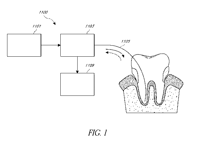
[0097] FIG. 1 schematically illustrates an embodiment of an optical
system
configured to excited fluorescence in the protoporphyrin and/or porphyrin
molecules.
[0098] FIG. 2 shows spectra collected from different internal and
external locations
on a freshly extracted diseased tooth.
12
CA 03075654 2020-03-11
WO 2019/055569 PCT/US2018/050753
[0099] FIG. 3 shows the fluorescence spectra for F. nucleatum bacteria
when
illuminated by an excitation light
[0100] FIG. 4 shows the fluorescence spectra for P. intermedia bacteria
when
illuminated by an excitation light.
[0101] FIG. 5 illustrates an optical system configured to perform a
Raman
spectroscopic examination of a tooth.
[0102] FIG. 6 illustrates an embodiment of an optical coherence
tomography (OCT)
system configured to perform an optical examination of a tooth.
[0103] FIG. 7 schematically illustrates an embodiment of an OCT system
configured
to obtain cross-sectional images (in the radial-axial plane) of the surfaces
of a root canal.
[0104] FIG. 8 schematically illustrates an embodiment of an OCT system
that can be
used to visualize the periodontal space.
[0105] FIG. 9 schematically illustrates an example heat map showing the
bacterial
load in various portions of a root canal.
[0106] FIGS. 10A ¨ 10C depict an example of a mounting assembly.
[0107] FIG. 11 is a schematic system diagram of a dental system.
[0108] FIG. 12 schematically illustrates an example of a treatment
system for treating
(e.g., cleaning) a tooth with a pressure wave generator.
[0109] FIG. 13 and FIG. 14 are graphs that schematically illustrate
possible examples
of acoustic power that could be generated by different embodiments of the
pressure wave
generators disclosed herein.
[0110] FIG. 15 schematically illustrates an example of a treatment
system for treating
(e.g., cleaning) a tooth with a fluid motion generator that comprises a
pressure wave generator,
according to various embodiments.
[0111] FIG. 16 schematically illustrates an example of a treatment
system for treating
(e.g., cleaning) a carious region on an exterior surface of a tooth.
DETAILED DESCRIPTION
[0112] Although certain preferred embodiments and examples are
disclosed herein,
inventive subject matter extends beyond the specifically disclosed embodiments
to other
alternative embodiments and/or uses of the inventions, and to modifications
and equivalents
thereof. Thus, the scope of the inventions herein disclosed is not limited by
any of the particular
13
CA 03075654 2020-03-11
WO 2019/055569 PCT/US2018/050753
embodiments described below. For example, in any method or process disclosed
herein, the acts
or operations of the method or process may be performed in any suitable
sequence and are not
necessarily limited to any particular disclosed sequence.
101131 For purposes of contrasting various embodiments with the prior
art, certain
aspects and advantages of these embodiments are described. Not necessarily all
such aspects or
advantages are achieved by any particular embodiment. Thus, for example,
various
embodiments may be carried out in a manner that achieves or optimizes one
advantage or group
of advantages as taught herein without necessarily achieving other aspects or
advantages as may
also be taught or suggested herein.
101141 Various embodiments described in this application include
optical systems
and methods to examine a portion of a dental tissue. As used herein, dental
tissue can include
the different components or layers of a tooth including but not limited to
enamel, dentin,
cementum and pulp. As used herein, dental tissue can also include gingivae or
gums. The
optical systems and methods described herein can detect caries and tooth
defects (e.g., cracks),
estimate the amount of bacteria in the root canal of one or more teeth of the
patient, measure the
working length of a root canal, characterize the structure and architecture of
inner canal wall
structure of the one or more teeth, visualize and diagnose peridontium
pathologies and/or
visualize apical region of a root canal. The systems and methods described
herein can have
several advantages. For example, the optical system and methods described
herein can provide a
non-destructive and a non-invasive method for examining dental tissue. As
another example, the
systems and methods described herein can examine the one or more teeth quickly
as compared to
other methods. For example, optical measurement of one or more teeth of a
patient can be
obtained in 10 seconds or less using the optical system and methods described
herein. The
optical systems and methods described herein can be configured to be compact
by using
miniaturized optical components and/or optical fibers.
Optical interrogation of a root canal
101151 Root canal bacteria can endogenously bio-synthesize
protoporphyrin and/or
porphyrin molecules after consuming root canal pulpal tissue. The
protoporphyrin and/or
porphyrin molecules are located wherever bacteria are present inside a root
canal. The optical
systems and methods described herein can be used to optically detect the
amount of
protoporphyrin and/or porphyrin molecules and/or the bacteria present in the
root canal. For
14
CA 03075654 2020-03-11
WO 2019/055569 PCT/US2018/050753
example, the protoporphyrin and/or porphyrin molecules can be detected via
spectroscopic
methods. Without any loss of generality, the cleanliness of a root canal
depends on the amount
of bacteria (or bacterial load) present in the root canal. A root canal with a
lower bacterial load
can be considered to be cleaner than a root canal with a higher bacterial
load. Accordingly, the
root canal of a patient's teeth can be optically interrogated to determine the
cleanliness of a root
canal.
[0116] Systems and methods employed to optically interrogate a root
canal can have
several advantages. For example, the entire root canal all the way down to the
apex can be
interrogated using optical techniques. As another example, optical techniques
of interrogating a
root canal can provide a non-destructive and a non-invasive method for
examining the root canal.
Additionally, optical examination of a root canal can done quickly. For
example, measurements
that are related to cleanliness of the root canal can be obtained in less than
10 second (e.g., as
little as 1-2 seconds). Optical fiber based systems and methods of examining
root canals can be
compact since optical fibers can have a small diameter (e.g., diameters
between approximately
100 microns and approximately 300 microns). Thus, optical fiber based systems
and methods of
examining root canals can work with non-instrumented canals. As used herein, a
non-
instrumented canal refers to a root canal that is prepared without removal of
endogenous tissue
(e.g. dentin). Accordingly, a tooth with non-instrumented canal can have
higher structural
integrity as compared to an instrumented canal which is prepared by removal of
endogenous
tissue (e.g. dentin) by filing or some other method of removing tissue.
Additionally, the systems
and methods of optically interrogating the root canal discussed herein can
provide quantitative
measurements that can be used to classify the cleanliness of the root canal.
[0117] One method of determining the bacterial load in the root canal
comprises
fluorescence spectroscopy. The protoporphyrin and/or porphyrin molecules
synthesized by the
bacteria in the root canal can fluoresce when illuminated by light having
wavelengths (e.g.,
wavelengths in the range between about 280 nm and about 650 nm). Without any
loss of
generality, the fluorescence process involves elevation of the protoporphyrin
and/or porphyrin
molecules to a higher energy state as a result of absorption of the light at
the excitation
wavelength followed by a spontaneous decay to a distribution of rotational and
vibrational
energy states within the same or lower energy state. When decaying from the
higher energy
CA 03075654 2020-03-11
WO 2019/055569 PCT/US2018/050753
level to the lower energy level, the difference in the energy between the two
energy levels is
released in the form of broadband emission of light in the visible or near
infrared spectrum.
[0118] The presence and/or amount of bacteria present in the root canal
can also be
detected via Raman spectroscopy which is described in greater detail below.
Without any loss of
generality, Raman spectroscopy is an inelastic scattering phenomena and is
capable of providing
biochemical and morphological information. Scattering is predominantly an
elastic phenomena,
whereby the scattered light has the same frequency as the incident light;
however, a portion of
the scattered light can be attributed to inelastic scattering which has a
different frequency from
the incident light. By detecting and analyzing the scattered light at a
different frequency from
the incident light, information about the scattering material can be obtained.
[0119] One embodiment of an optical system 1100 configured to excited
fluorescence
in the protoporphyrin and/or porphyrin molecules is depicted in Fig. 1. The
optical system 1100
can include a combination of fiber optic and freespace optical elements. The
optical system
1100 comprises an optical source 1101. The optical source 1101 can comprise a
laser, one or
more laser diodes and/or one or more light emitting diodes (LEDs). The optical
source 1101 can
be operated in a continuous (CW) mode or a pulsed mode. In some embodiments,
the optical
source 1101 can comprise a monochromatic source that outputs light having a
single wavelength.
In some embodiments, the optical source 1101 can comprise a broadband source
that outputs
broadband light in a spectral range. In some embodiments, the optical source
1101 can comprise
a tunable laser whose output wavelength can be controlled. The optical source
1101 can be
configured to output light having a wavelength in one or more of the
ultraviolet spectral range,
the visible spectral range and/or the near-infrared spectral range. For
example, the optical source
1101 can be configured to output light having a wavelength between about 250
nm and about
430 nm, between about 400 nm and about 510 nm, between about 500 nm and about
550 nm,
between about 520 nm and about 610 nm, between about 580 nm and about 650 nm,
between
about 630 nm and about 780 nm, between about 650 nm and about 980 nm or any
range/sub-
range defined by any of these wavelength ranges.
[0120] The light output from the optical source 1101 can be conditioned
by an optical
system 1103 comprising a plurality of optical components. For example, the
optical system 1103
can comprise a short-pass dichroic filter, configured to transmit the laser
light and reflect the
longer fluorescence wavelengths. As another example, the optical system 1103
can comprise
16
CA 03075654 2020-03-11
WO 2019/055569 PCT/US2018/050753
one or more collimating and/or focusing lenses configured to focus the light
from the optical
source 1101 onto a portion of a tooth. As yet another example, the optical
system 1103 can
comprise a dynamic optical component, such as, for example, a galvanometer or
a micro-electro
mechanical system (MEMS) based device that can be controlled to focus light
from the optical
source 1101 on different locations of the tooth. In various embodiments of the
optical system
1100 configured to examine the root canal, the light from the optical source
1101 can be coupled
into an optical fiber 1105 using one or more lenses (e.g., a focusing lens).
The spectrum of the
light from the optical source 1101 can be tailored using a band-pass filter
that transmits the light
output from the optical source 1101 and reduces or eliminates any tails in the
spectrum of the
light output from the optical source 1101 prior to being coupled into the
optical fiber 1105. The
optical fiber 1105 can be configured as a flexible cable, which is capable of
being inserted into
the root canal and is capable of being navigated along the length and the
curvature of the root
canal. To estimate the cleanliness of the root canal, the optical fiber 1105
can be positioned at
one or more locations within the root canal. At each or some of the one or
more location, the
user (e.g., dentist/dentist's assistant) can activate the optical source 1101,
for example, by
pressing a button or some other way to excite fluorescence. In some
embodiments, the optical
fiber 1105 can be configured to output light along a direction parallel to the
optical axis of the
optical fiber 1105 (or the axis of the root canal) which can extend along the
length of the optical
fiber 1105. In such embodiments, the light output from the optical fiber 1105
is emitted from the
end face of the optical fiber that is inserted into the root canal along a
direction normal to the end
face. In some embodiments, the light output from the optical fiber 1105 can be
directed in a
radial direction (e.g., normal or at an angle with respect to the optical axis
of the optical fiber
1105 or the axis of the root canal). This configuration can be referred to as
"side-firing." One
advantage of the side-firing configuration is the ability to improve optical
sampling on the root
canal walls, where bacteria typically reside in the form of biofilm on the
dentinal walls and
inside dentinal tubules. In an example embodiment comprising the side-firing
configuration
includes an optical fiber having a 400 micron core diameter that achieves side
firing via total
internal reflection on the distal end (the end closer to the root canal). In
some embodiments, the
light emitted from the optical fiber 1105 can be directed laterally with
respect to the axis of the
canal. Lateral direction of light can be achieved by providing a lens having a
beveled surface at
the distal end of the optical fiber 1105 (the end inserted into the root
canal) or by providing a
17
CA 03075654 2020-03-11
WO 2019/055569 PCT/US2018/050753
reflection surface orientated at an angle with respect to the axis of the
optical fiber 1105 (or axis
of the root canal). This configuration can be similar or identical to the side-
firing configuration.
An advantage of directing the light along a direction lateral to the axis of
the canal is that the
lateral features of the tooth structure, such as dentin tubules or lateral
canals, can be interrogated
for bacterial presence.
101211 Any fluorescence, excited by the activation of the optical
source 1101, is
collected by the optical fiber 1105 and directed towards the optical system
1103. The optical
fiber 1105 can be configured to have a high numerical aperture to efficiently
collect as much of
the fluorescence signal as possible. For example, the distal end of the
optical fiber 1105 (e.g.,
the end inserted into the root canal) can comprise a lens configured to
enhance the collection
efficiency. The lens can be a ball lens or a half-ball lens. The lens can have
high refractive
index. Additionally, the lens can comprise a high impact resistance material
such as ruby or
sapphire. The collected fluorescence signal can be directed by a dichroic
filter in the optical
system 1103 along a receive path towards an optical receiver 1109. The
dichroic filter in the
optical system 1103 can be configured to transmit light from the optical
source 1101 and reflect
the fluorescence signal. In order to collect fluorescent signal corresponding
to a particular
wavelength region, with a demonstrable ability to provide an indication of
bacterial presence, the
collection optical train can comprise a specialized multi-bandpass filter
disposed in the receive
path. The optical receiver 1109 can comprise a photodetector and/or a
spectrometer. In various
embodiments, the fluorescent signal can be collected using other types of
photodetectors such as
photomultiplier tubes (PMTs) or photodiodes. In various embodiments, the
optical receiver 1109
can provide a digital signal of fluorescent intensity as a function of
wavelength.
[0122] The optical system 1100 can be operated in "integrated" or
"single shot"
mode. In this mode, the optical source 1101 can be pulsed optical source that
emits optical
pulses at fixed or regular time intervals. Fluorescence signal can be
collected from many
different locations while the fiber optic probe is positioned at different
portions inside the root
canal. In some embodiments, different pulses can have different spectral
contents. The
fluorescence spectra obtained for a plurality of the pulses can be averaged to
provide an
integrated measurement of the entire tooth's cleanliness. In some embodiments,
the user (e.g.,
the dentist/dentist's assistant) can operate the device such that a reading is
provided each time
18
CA 03075654 2020-03-11
WO 2019/055569 PCT/US2018/050753
the laser is activated, thereby providing the user with spatial discretization
of cleanliness within
the tooth.
[0123] Some embodiments of the optical system 1100 can be entirely (or
substantially entirely) fiber-optic based design with the filters miniaturized
and placed inside
fiber-optic housing. Various embodiments of the optical system 1100 can
comprise an optical
source 1101 that can emit a plurality of wavelengths to induce fluorescence
from multiple
bacterial species. The excitation and collection sub-systems can be
synchronized to sequentially
excite and collect fluorescence from different bacterial strains.
[0124] The optical receiver 1109 can comprise an electronic processing
system
configured to analyze the obtained fluorescent spectra. The electronic
processing system can be
further configured to estimate the bacterial load in various portions of the
root canal and/or
provide a metric associated with the cleanliness of the root canal. The
electronic processing
system can be configured to increase the signal-to-noise ratio of the obtained
fluorescence
spectra by using various signal processing techniques. For example, the signal-
to-noise ratio of
the obtained fluorescence spectra can be increased by using digital filtering
algorithms such as a
Savitsky-Golay filter and/or smoothing of the spectra using simple methods
such a window
average. Improving the signal-to-noise ratio of the obtained fluorescence
spectra can increase
the reliability of detecting the presence of bacteria in the root canal and
estimating the
cleanliness of the root canal.
[0125] Spectral analysis can be performed by signal processing
algorithms to remove
background signal due to autofluorescence from endogenous species (dentin,
enamel, etc) or
species present post-root canal treatment (RC1), such as, for example, EDTA,
water, sodium
hypochloride, etc. The result of this analysis can be a "background corrected"
spectrum,
representative of the photonic signal due only to the porphyrin fluorescence.
[0126] In various embodiments, it may be desirable to reduce the data
size via
software binning which can potentially reduce hardware-related costs, reduce
data rates and/or
reduce computational processing time associated with the optical detection
methods disclosed
herein. As fluorescence spectra can be broadband, spectral binning can be
implemented with
reduced loss in data accuracy.
19
CA 03075654 2020-03-11
WO 2019/055569 PCT/US2018/050753
Bacteria Identification
[0127] Using linear algebraic mathematical operations, a measured
spectra,
representing the linear superposition of multiple fluorophores, can be
decomposed into
individual spectral profiles corresponding to one of the multiple fluorophores
to determine the
presence of different bacteria. Spectral decomposition can yield relative semi-
quantitative
concentrations of bacterial populations; in-vitro calibration of fluorescent
intensity as a function
of bacterial concentration can be used for true quantification. Various
spectral decomposition
methods are discussed below.
[0128] The fluorescence spectra obtained by the optical system 1100 can
be a
combination of all possible interrogated species that generate fluorescence at
wavelengths of the
excitation light. These could be many different strains of bacteria, enamel,
dentin, restorative
material, obturation material. The following method describes a way to
deconstruct the
measured, integrated spectra into its "basis" or "constituent" components in a
technique known
as spectral decomposition.
[0129] The system can be modeled by equation (1)
M = SeCT + e (1)
[0130] Where M = measured spectrum with dimension equal to the number,
n, of
wavelength datapoints, which depends on the number of x-axis pixels and the x-
bin setting, n can
be given by equation (2) below:
n1367/ (2)
101311 In equation (1), S is the basis spectra matrix with dimension n
x d, where d is
the number of species, c is the concentration vector with dimension d xl, and
e is the noise. The
measured spectrum can also be corrected by subtracting any background signal
that was present.
[0132] The Gauss-Markov theorem states that a linear regression model:
y = XI) + 6,
where a is the noise then f =(XT XT is the function of X and b that
minimizes the sum of
the squared residuals.
[0133] Initially if the noise is ignored:
CA 03075654 2020-03-11
WO 2019/055569 PCT/US2018/050753
(sTs)'sTm = (STS)' (ST*
(STS)' (STS) = /
.". c = (SS)' STm
[0134] The quantity K given by equation (3) is the pseudo-inverse of
&b., and can
be pre-computed.
K = (ST Sr ST (3)
[0135] Thus, quantification may be performed in realtime by using
equation (4)
CT = K MbI,, (4)
[0136] The above described spectral decomposition technique can be
applied even if
Mbin contains multiple spectra, for example, a plurality of columns with each
column
representing a spectrum.
Experimental Results
101371 Fig. 2 shows spectra collected from different internal and
external locations on
a freshly extracted diseased tooth. The healthy hard tissue spectrum (curve
2101) is a broadband,
monotonically decaying signal, which contrasts with spectra collected from
locations with
pathology with well-defined, prominent peaks superimposed onto a broadband
signature. For
example, curve 2103 corresponds to the spectrum obtained from the periodontal
region, curve
2105 corresponds to the spectrum obtained from occlusal caries, curve 2107
corresponds to the
spectrum obtained from root canal. These peaks and the associated fluorescence
profiles are
from porphyrins, which are organic compounds occurring as digestive products
of endodontic
bacteria such as Enterococcus Faecalis and Prevotella Intermedia.
[0138] The differences in spectral shape provide encouraging visual
evidence of the
diagnostic capabilities of the fluorescence spectroscopic method of estimating
bacterial load in
the root canal. An overview of a diagnostic protocol used to estimate the
cleanliness of the root
canal is described below:
[0139] 1. Basis spectra can be measured for known species that
fluorescence at the
excitation laser wavelength of choice. Examples of these species include
Protoporphyrin IX,
Coproporphyrin III, healthy enamel and healthy dentin.
21
CA 03075654 2020-03-11
WO 2019/055569 PCT/US2018/050753
[0140] 2. Based on characterization studies previously performed,
identify the limits
of detection for each basis species in the presence of other species,
corresponding to the minimal
signal that can be detected to determine the presence of a particular species.
[0141] 3. A spectrum, or series of spectra, are collected from
locations inside or
outside a tooth. These spectra are then processed using the mathematical
linear algebra analysis
described above to assign contributions from the basis components using the
limit of detection
values.
101421 4. The assigned contributions are used to provide output to the
user
designating if the measured location is "clean" or "dirty", and the level of
infection quantified
into qualitative categories such as "high", "medium" or "low".
In Vitro Experiments
[0143] Many bacterial strains can be present in vital root canal pulp
tissue and
theoretically any of these bacterial strains can present pathological concern
to cause pulp
infection and possible periapical infection. However, in practice only a few
bacterial species are
present in infected root canals. Previous studies have demonstrated that the
root canal
microbiology is typically dominated by anaerobic bacteria, corresponding to a
commensurate
reduction in facultative species. In terms of fluorescence detection, the
species found in infected
root canals can be simplistically described by two main categories: those that
emit fluorescence
in the green portion of the visible spectrum and those that emit fluorescence
in the red.
[0144] The results of an in vitro study conducted to demonstrate the
ability to detect
both types of fluorescence using the systems and methods discussed above are
presented. Two
species, Fusobacterium nucleatum and Prevotella Intermedia, were cultivated on
agar plates and,
after an incubation period, placed into a water solution for measurement with
the fiber-optic
probe. These species are the two most frequently isolated in root canals,
found in 48% and 34%
of root canals respectively. The spectra were collected with the prototype by
immersing the
probe in vials containing the solution and acquiring data at clinically
relevant exposure times
(100ms). Spectra were also obtained of the solvent alone and served as
background. The
background spectra was then subtracted from the raw spectra, yielding a
corrected spectra. Fig.
3 shows the results for F. nucleatum and Fig. 4 shows the results for P.
intermedia. In Fig. 3,
curve 3101 represents the raw fluorescent spectrum, curve 3103 represents the
background
fluorescence and curve 3105 represents the corrected fluorescent spectrum
obtained by
22
CA 03075654 2020-03-11
WO 2019/055569 PCT/US2018/050753
subtracting the background fluorescence from the raw spectrum. In Fig. 4,
curve 4101 represents
the corrected fluorescent spectrum obtained by subtracting the background
fluorescence from the
raw spectrum.
[0145] In the case of P. intermedia, the red fluorescence peak is
clearly observed in
the curve 3105. In the case of the "green" species, F nucleatum, a less
intense, broader "hump-
like" structure 4103 can be observed in curve 4101. The shape of the spectra,
with a small peak
superimposed onto a spectrally broad signal, may suggest that other sources of
fluorescence are
present such as auto-fluorescence generated by the fiber-optic glass. This
auto-fluorescence can
be eliminated via material selection in more refined prototype version.
Summary
[0146] The optical system described above utilizes fluorescence
spectroscopic
methods to optically interrogate a root canal and determine the cleanliness of
a root canal. In
other embodiments, the fluorescence spectroscopic methods can optically
interrogate other
treatment regions (such as caries on an outer surface of the tooth, gingival
pockets, etc.) to
examine the cleanliness of the treatment region. Additionally, the
fluorescence spectroscopic
methods can be used to estimate the bacterial load in different portions of
the root canal and/or
identify the various bacteria present in the root canal. The optical system
configured to
determine the cleanliness of the root canal can be integrated with a device
that is configured to
clean root canals to determine the efficacy of the cleaning process. The
system to obtain
fluorescence spectra from different portions of the root canal can comprise a
tunable laser
configured to be operated in a CW mode or a pulsed mode. For example, the
tunable laser can
be configured emit a first pulse train having a first wavelength at a first
time interval and a
second pulse train having a second wavelength at a second time interval. The
first pulse train
can be configured to excite protopophyrins synthesized by a first bacteria and
the second pulse
train can be configured to excite protopophyrins synthesized by a second
bacteria. Fluorescence
from the protopophyrins synthesized by the first bacteria can be collected in
the first time
interval and fluorescence from the protopophyrins synthesized by the first
bacteria can be
collected in the second time interval. Thus, the fluorescence data collection
from different
wavelengths can be synchronized to the duration of the pulses for the
different wavelengths. In
some embodiments, the optical source can be operated in the continuous mode.
Fluorescence
23
CA 03075654 2020-03-11
WO 2019/055569 PCT/US2018/050753
data can be collected from different heights along the length of the root
canal to determine the
cleanliness along the length of the root canal.
Raman Spectroscopy
[0147] Various optical systems and methods to examine a tooth
contemplated by this
application to comprises an optical system configured to perform a Raman
spectroscopy (RS) on
the tooth. An optical system configured to perform Raman spectroscopy can
advantageously
detect early carious lesions. For example, demineralization of the
hydroxyapatite matrix, which
is initiated by acids produced by oral bacterial, alters chemical bonds which
can be detected
using the Raman spectroscopic technique. Demineralization can eventually lead
to clinical
presentation associated with caries such as white spot lesions and cavitation
(holes) as the acid
continues to deteriorate the matrix. Without subscribing to any particular
theory, Raman
spectroscopy can measure the phenomena of inelastic scattering, in which
scattered light
undergoes an energy shift relative to the incident light. The energy shift
that results from
inelastic scattering can translate to a change in wavelength, which can be
detected and measured.
Without subscribing to any particular theory, the energy shift can occur as a
result of excitation
of molecules in various portions of the tooth being examined to higher
vibrational and/or
rotational energy levels.
[0148] FIG. 5 illustrates an optical system 5100 configured to perform
a Raman
spectroscopic examination of a tooth. The system 5100 comprises an optical
source 5101
configured to emit an optical beam comprising light at one or more
wavelengths, conditioning
optics 5103 configured to condition the optical beam and output a conditioned
illumination beam
5107 to at least a portion of a tooth 5109. In various implementations, the
conditioned optical
beam 5107 can be directed to an entire tooth or a portion of the tooth (e.g.,
enamel, a portion at
or below the gumline surrounding the tooth, a periodontal pocket, or any other
portion of the
tooth). In the illustrated embodiment, the conditioned optical beam 5107 is
directed at an
exterior surface of the tooth 5109 to detect or monitor a progression of
caries on an external
surface of the tooth 5109. In other embodiments, the conditioned optical beam
5107 can be
directed at inner surfaces of the tooth 5109 (e.g., the pulp chamber, root
canal(s), etc.) after
cleaning to determine the cleanliness of the tooth. The Raman scattered light
5111 from one or
more portions of the tooth 5109 can be collected by the collection optics
included in the
conditioning optics 5103 and directed towards an optical receiver system 5113.
The light
24
CA 03075654 2020-03-11
WO 2019/055569 PCT/US2018/050753
received at the optical receiver system can be analyzed by an electronic
processing system 5115
configured to determine one or more characteristics that is representative of
a constituent of the
teeth and/or a morphology of a constituent of the teeth.
[0149] The optical system 5100 illustrated in FIG. 5 can be used in
facilities that
provide dental care. The optical system 5100 can be configured to be mobile
and easily portable.
The system can be used and operated by dentists/dental hygienists to obtain
Raman
spectroscopic data of at least a portion of a tooth of a patient. In some
embodiments, the optical
system 5100 can be automated and designed in such a manner that it can be
operated by an
approximately untrained operator. In some embodiments, the optical system 5100
can be setup
and operated in a relatively short period of time.
[0150] The optical source 5101 can comprise a near-infrared (NIR)
source of
radiation. For example, in various embodiments, the optical source 5101 can be
configured to
emit radiation in a wavelength range between approximately 700 nm and
approximately 1.5
micron. For example, the optical source 5101 can be configured to emit
radiation in a
wavelength range between approximately 700 nm and approximately 850 nm,
between
approximately 800 nm and approximately 900 nm, between approximately 850 nm
and
approximately 980 nm, between approximately 900 nm and approximately 1.1
micron, between
approximately 1.0 micron and approximately 1.3 micron, or any wavelength in a
range/sub-range
defined by any of these values. Various embodiments of the optical source 5101
can comprise a
laser, a laser diode, a light emitting diode (LED) or any other source capable
of emitting
radiation in the MR wavelength range. The optical source 5101 can be operated
in continuous
mode or in pulsed mode. In some embodiments, electric power to the optical
source 5101 can be
supplied from an electrical power supply line. In various embodiments,
electrical power to the
optical source 5101 can be supplied by a voltage regulator. In some
embodiments, electrical
power to the optical source 5101 can be supplied by a battery pack. In various
embodiments, an
electronic control system can be used to control the optical source 5101. The
electronic control
system can be configured to switch the optical source 5101 on or off, and/or
control one or more
parameters (e.g., average optical power, spot size, output wavelength, etc.)
of the output optical
beam 5107. In some embodiments, the electronic control system can be used to
alternate
between continuous and pulsed mode of operation. In various embodiments, the
electronic
CA 03075654 2020-03-11
WO 2019/055569 PCT/US2018/050753
processing system 5115 can comprise the electronic control system configured
to control the
optical source 5101.
[0151] As discussed above, the light emitted from the optical source
5101 can be
conditioned by conditioning optics 5103. The conditioning optics 5103 can
comprise one or
more optical elements (e.g., lenses, optical beam splifters, dichroic mirrors,
reflectors,
polarization controllers, polarizers, retarders, prisms, wavelength filters,
spatial filters, such as,
for example, a pin-hole or an aperture, galvanometer, resonant or micro
electromechanical
(MEMs) mirror system, optical attenuators, or a combination of any of these
components). The
conditioning optics 5103 can be configured to tailor the light emitted from
the optical source
5101 to produce a light beam having a desired wavelength and a desired spot
size on at least a
portion of the tooth 5109.
[0152] The light beam at the output of the conditioning optics 5103 is
delivered to at
least a portion of the tooth 5109. The light beam output from the conditioning
optics 5103 can
be delivered to at least the portion of the tooth in freespace. As discussed
above, the
conditioning optics 5103 can comprise a beam-steering system (e.g., a
galvanometer or a MEMS
based scanning system) that can be used to scan the light beam across the
surface of a portion of
the tooth 5109. In some embodiments, an optical probe can be connected to the
output of the
conditioning optics 5103 to deliver the light beam. The optical probe can be
flexible or rigid.
The optical probe can comprise a resilient material that can be bent into one
or more desired
shapes. In various implementations, the optical probe can comprise metal,
plastic, silica, or
polymer. In various implementations, the optical probe can comprise an optical
fiber.
[0153] In various embodiments, the elements of the conditioning optics
5103 can be
configured to produce a light beam 5107 having desired optical properties
(e.g., a desired optical
spot size, a desired wavelength or a desired range of wavelengths, a desired
optical power, etc.)
on the portion of the tooth 5109 to be examined. The light beam 5107 can
optically interact with
the portion of the tooth 5109 to be examined. For example, the light beam 5107
incident on the
portion of the tooth 5109 being examined can be scattered non-elastically
(e.g., by the
phenomenon of Raman scattering) by the constituents of the portion of the
tooth 5109 being
examined as scattered beam 5111. The wavelength of the scattered beam 5111 can
be different
from the wavelength of the incident light beam 5107 due to the Raman frequency
signal shift
26
CA 03075654 2020-03-11
WO 2019/055569 PCT/US2018/050753
discussed above. The scattered beam 5111 can be collected by the conditioning
optics 5103 and
directed towards the optical receiver system 5113.
[0154] The conditioning optics 5103 can comprise an optical beam
splitter (e.g., a
dichroic mirror) that directs the scattered beam 5111 towards the optical
receiver system 5113
along a receive path. The optical beam splitter (e.g., the dichroic mirror)
can be configured to
transmit the light from the optical source 5101. The optical receiver system
5113 can comprise
one or more optical filters (e.g., optical bandpass filters) and one or more
photodetectors. The
one or more optical filters can be configured to transmit a portion of the
collected light in one or
more spectral regions corresponding to the spectral regions where the
information from the
optical spectrum can be correlated to the constituents of the teeth. For
example, in some
implementations, the one or more optical filters can be configured to transmit
a portion of the
collected light in one or more spectral regions where the information from the
optical spectrum
can be correlated with the mineralization level of enamel, such as the bending
and/or stretching
mineralization index. For example, the one or more optical filters can be
configured to transmit
light in a spectral region between a wavenumber of 430 cm-1 and a wavenumber
of 2941 cm-1
corresponding to the mineralization level of enamel. As another example, the
one or more
optical filters can be configured to transmit light in a spectral region
between a wavenumber of
960 cm-1 and a wavenumber of 2941 cm11 corresponding to bending and/or
stretching
mineralization index. In various embodiments, the one or more optical filters
can comprise a
notch filter, an edge filter and/or a band-pass filter to attenuate any
portion of the incident light
beam 5107 that may be received at the optical receiver system 5113.
[0155] The one or more photodetectors can comprise photodiodes
sensitive to light in
a wavelength range between 700 nm and 1.5 micron and/or photo multiplier tubes
(PMTs). In
various embodiments, the optical receiver system 5113 can comprise a single
broadband
photodetector. In such embodiments, a series of bandpass optical filters can
be disposed in the
receive path before the single broadband photodetector to transmit light in
different wavelength
regions. In some embodiments, the optical receiver system 5113 can comprise a
photodetector
that is sensitive to wavelengths each of the plurality of filtered spectral
regions. In such
embodiments, each optical bandpass filter can be associated with the
photodetector that is
sensitive to the light transmitted through that optical bandpass filter. In
various embodiments,
27
CA 03075654 2020-03-11
WO 2019/055569 PCT/US2018/050753
the optical receiver system 5113 can comprise a Rainan spectra appropriate
spectrograph, such
as, for example, a holographic grating.
[0156] The Raman spectra can include peaks associated with the
constituents of the
tooth 5109 as well as peaks associated with morphological changes in the
molecular structure of
the constituents of the tooth 5109. Thus the Raman spectra can provide
valuable information
regarding not only the constituents of the tooth but also associated with the
diseased state of the
tooth.
101571 The spectral information obtained by the optical receiver system
5113 can be
analyzed using an electronic processing system 5115 as discussed above.
Without any loss of
generality, the electronic processing system 5115 can be configured to analyze
the peaks at
wavenumbers in a range between about 420 cm-1 and about 450 cm-1 (e.g., 431 cm-
1), between
about 570 cm4 and about 610 cm-1 (e.g., 590 cm-1), between about 940 cm4 and
about 980 cm-1
(e.g., 959 cm-1) and/or between about 1020 cm-1 and about 1065 cm11 (e.g.,
1043 cm-1) of the
spectral information obtained by the optical receiver system 5113 to examine a
tooth or a portion
thereof. These peaks can correspond to excitation of the compound P043- which
is a constituent
of the enamel to higher vibrational/rotational energy levels.
[0158] In various implementations, the electronic processing system
5115 can be
configured to analyze the ratio of the intensities (e.g., maximum intensities)
of a peak in a range
between a wavenumber of about 420 cm-1 and a wavenumber of about 450 cm-1
(e.g., 430 cm-1)
and a peak in a range between a wavenumber of about 2920 cm-1 and a wavenumber
of about
2960 cm-1 (e.g., 2941 cm-1). These peaks can be associated with bending modes
of the molecules
of the constituent of enamel.
[0159] In various implementations, the electronic processing system
5115 can be
configured to analyze the ratio of the intensities (e.g., maximum intensities)
of a peak in a range
between a wavenumber of about 940 cm-1 and a wavenumber of about 980 cm-1
(e.g., 960 cm4)
and a peak in a range between a wavenumber of about 2920 cm-1 and a wavenumber
of about
2960 cm4 (e.g., 2941 cm-1). These peaks can be associated with stretching
modes of the
molecules of the constituent of enamel.
[0160] The ratio of the intensities (e.g., maximum intensities) of
peaks at a
wavenumber of 430 cm-1 and at a wavenumber of 2941 cm-1 and the ratio of the
intensities of
peaks at a wavenumber of 960 cm-1 and at a wavenumber of 2941 cm-1 can be
representative of a
28
CA 03075654 2020-03-11
WO 2019/055569 PCT/US2018/050753
mineralization index of the enamel. For example, a first value of the ratio of
the intensities (e.g.,
maximum intensities) of peaks at a wavenumber of 430 cm-1 and at a wavenumber
of 2941 cm-1
can be representative of a first mineralization index and a second value of
the ratio of the
intensities (e.g., maximum intensities) of peaks at a wavenumber of 430 cm-1
and at a
wavenumber of 2941 cm can be representative of a second mineralization index.
Similarly, a
first value of the ratio of the intensities (e.g., maximum intensities) of
peaks at a wavenumber of
960 cm' and at a wavenumber of 2941 cm-1 can be representative of a third
mineralization index
and a second value of the ratio of the intensities (e.g., maximum intensities)
of peaks at a
wavenumber of 960 cm' and at a wavenumber of 2941 cm-1 can be representative
of a third
mineralization index.
[0161] Other metrics can be calculated from the Raman spectra in
addition to or
instead of peak locations and the ratio of the intensities of the peaks. These
other metrics may be
representative of the constituents of the tooth and/or changes in the
morphology of the
constituents of the tooth.
[0162] The electronic processing system 5115 can be configured to
employ statistical
analysis techniques (e.g., principal component analysis, factor analysis,
hierarchical cluster
analysis, linear discriminant analysis or analysis of variance) on the
obtained Raman spectra. In
some embodiments, the electronic processing system 5115 can be configured to
employ spectral
processing such as smoothing of spectral peaks via digital filtering, such as,
for example,
Savitsky-Golay or a moving average method. In various embodiments, the
electronic processing
system 5115 can be configured to increase the signal-to-noise ratio of the
obtained Raman
spectra. For example the electronic processing system 5115 can be configured
to increase the
signal-to-noise ratio of the obtained Raman spectra by taking multiple
acquisitions per spot and
averaging the result. As another example, the electronic processing system
5115 can be
configured to increase the signal-to-noise ratio of the obtained Raman spectra
by subtracting a
background to improve the signal-to-noise ratio.
[0163] The optical system 5100 can comprise one or more optical fibers.
For
example, the conditioning optics 5103 can comprise optical fiber based
mirrors, filters and/or
lenses. As another example, the optical probe 5105 can comprise an optical
fiber. In such
implementations, one or more optical elements can be included at the output of
the optical fiber
to manipulate the diverging light exiting from the optical fiber and converge
it to create an
29
CA 03075654 2020-03-11
WO 2019/055569 PCT/US2018/050753
illumination spot on at least a portion of the tooth and/or to collect the
scattered radiation from at
least a portion of the tooth. The optical fibers used in the optical system
5100 can have an inner
diameter between about 1 micron and about 1 mm and an outer diameter between
about 5 micron
and about 1.5 mm. The optical fibers used in the optical system 5100 can be
single mode fiber
or multimode fiber. Various embodiments of optical fibers employed in the
optical system 5100
can be coated with a material to provide flexibility and abrasion resistance.
Various
embodiments of the optical system 5100 can comprise polarization maintaining
fibers.
101641 Various embodiments of the optical system 5100 can be configured
as a point-
based scanning system. An incident beam 5107 having a spot size between about
5 microns and
about 500 microns can be generated using one or more optical elements (e.g.,
one or more
lenses) of the conditioning optics 5103. The spot size of the incident beam
5107 can be based
upon imaging resolution requirements. It is noted that the surface of the
tooth can be non-
uniform. The non-uniformity of the tooth surface can change the focal distance
of the incident
beam 5107. Accordingly, the spot size can change as the beam is scanned from
one location to
the next. The non-uniformity of the surface of the tooth can also change the
distance between the
output of the conditioning optics 5103 and the tooth surface. This can change
the beam diameter
of the light beam 5107.
[0165] The conditioning optics 5103 can comprise one or more dynamic
optical
components configured to steer the incident beam 5107 to a desired location of
at least a portion
of the tooth. The one or more dynamic optical components can comprise a
galvanometer or a
resonant or micro electromechanical (MEMs) mirror system. The one or more
dynamic optical
components can be controlled by the electronic control system to scan
different portions of the
tooth and/or gum-line. For example, in one embodiment, the one or more dynamic
optical
components can be controlled by the electronic control system to generate
closely spaced line
scans of at least a portion of the tooth or gum-line.
[0166] At each scanned location, a measurement can be obtained. The
measurement
can comprise one or more of locations of the peaks in the Raman spectra and/or
ratios of
intensities (e.g., maximum or peak intensities) of different peaks in the
Raman spectra. After the
scanning is completed, there are a series of unique values 1:1 mapped to the
spatial location,
from which an image can be generated. Different indicators can be associated
with the
individual measurements or a range of measurements. Different spatial
locations of tooth can be
CA 03075654 2020-03-11
WO 2019/055569 PCT/US2018/050753
associated with the different indicators depending on the corresponding value
of the
measurement. An image of a tooth in which different spatial locations are
indicated by different
indicators based on the corresponding value of the measurement can be referred
to as a hat map.
101671 Various embodiments of the optical system 5100 can be configured
as an
imaging system configured to image the entire tooth 5109 including the portion
of the gum line
surrounding the tooth and/or a selected portion of the tooth 5109 or the gum
line. In such
embodiments, the conditioning optics 5103 can be configured to generate an
incident optical
beam 5107 having a larger spot size, such as, for example, a spot size between
1 mm and 10 mm.
The optical receiver system 5113 can comprise a charge coupled device (CCD)
array or an array
of cameras that are configured to image the portion of the tooth 5109 being
illuminated by the
incident optical beam 5107. The image of the portion of the tooth 5109 being
illuminated by the
incident optical beam 5107 can comprise a plurality of image pixels
corresponding to various
regions of the portion of the tooth 5109 being illuminated by the incident
optical beam 5107.
The electronic processing system 5115 can be configured to employ various
algorithm and data
processing techniques to assign a color level to each image pixel based on a
value of a metric
obtained from the Raman spectra of the corresponding region of the portion of
the tooth 109
being illuminated. In this manner, the electronic processing system 5115 can
generate a "Raman
signal" map of the portion of the tooth being examined. The electronic
processing system 5115
can be further configured to display the generated Raman signal map on a
display device
associated with the electronic processing system 5115.
[0168] Various embodiments of the optical system 5100 can comprise two
optical
paths. The illumination beam can be directed along a first optical path
towards the tooth or a
portion therof. The illumination beam 5107 can be wide enough to illuminate
the desired portion
of the tooth. Accordingly, the illumination beam can be wide enough to span
across the entire
surface of the tooth. The Raman scattered light 5113 is directed along a
second optical path
toward an optical receiver for image generation. One or more bandpass filters
can be disposed in
the second optical path to collect light in one or more selected range of
wavenumbers. The
acquisition can be synchronized so that the one or more bandpass filters are
sequentially
deployed and multiple images collected, each corresponding to Raman-shifted
light in a different
range of wavenumbers. These individual wavenumber images could then be
overlayed onto each
31
CA 03075654 2020-03-11
WO 2019/055569 PCT/US2018/050753
other to create a multispectral or hyperspectral (depending on the number of
wavenumber
ranges) image.
[0169] Endogenous dental species, such as enamel, can be birefringent.
Thus, the
index of refraction of the endogenous dental species, such as enamel, can be
polarization
dependent. Without subscribing to any particular theory, the birefringence can
be attributed to
the tight, preferred orientation of the enamel rods within the hydroxyapatite
matrix.
Demineralization can reduce this level of alignment, which can be detected by
a polarization-
sensitive method. Accordingly, various embodiments of the optical system 5100
can be
configured to be sensitive to polarization. For example, the optical system
5100 can comprise
one or more polarization altering optical components (e.g., half or quarter
wave plates, retarders,
polarizers, or combinations thereof) at different locations in the transmit
path and the receive
path. The one or more polarization altering optical components can manipulate
the excitation
light and select Raman scattered photons based on their polarization.
[0170] As discussed above, the optical system 5100 can be configured to
be mounted
on a track that facilitates positioning the optical system 5100 with respect
to one or more teeth of
the patient. In some embodiments, the optical system 5100 could be contactless
and placed in
the mouth above a desired location and mechanically held in place via an
articulating arm. The
arm can be operated remotely or directly by a user (e.g., a dentist or a
dentist's assistant) for
desired placement with respect to a tooth. The placement could be achieved
manually by the
user using a joystick or similar function to adjust the optical system's
position via control of
mechanical motion linear stages. Alternatively, the placement could be
automated by the user
selecting the desired tooth for measurement and the position adjusted via a
feedback between a
camera identifying the current position and the mechanical motion devices. In
another
alternative, the user can position the arm using a camera system that provides
feedback regarding
the placement of the arm.
[0171] In conclusion a Raman spectroscopic system configured to perform
dental
exam is described herein. The Raman spectroscopic system can comprise a
scanning system that
is configured to generate a Raman map of the patient's teeth. Embodiments of
the Raman
spectroscopic system described herein can be configured to form an image of
the patient's teeth
with different portions of the patient's teeth associated with a metric
obtained from the Raman
spectra.
32
CA 03075654 2020-03-11
WO 2019/055569 PCT/US2018/050753
Optical Coherence Tomography
[01721 Various optical systems and methods to examine a tooth
contemplated by this
application comprise an optical coherence tomography (OCT) system. The OCT
system is an
interferometry-based imaging technique. This application contemplates the use
of OCT for a
variety of dental applications including but not limited to the detection of
caries, identification of
tooth defects such as cracks, as a tool for measuring the true root canal
working length, to
characterize the structure and architecture of inner root canal wall
structure, for the visualization
and diagnosis of peridontium pathologies, and/or visualization of the apical
region of a root canal
[0173] FIG. 6 illustrates an embodiment of an OCT system 6100. The OCT
system
6100 comprises an optical source 6101. The light from the optical source 6101
can be divided
between a signal arm and a reference arm using an optical splitter 6105 (e.g.,
a 90-10 splitter).
Light from the signal arm of the optical splitter 6105 (e.g., the output of
the optical splitter
through which 90% of the input light is emitted) is input into a first port of
an optical circulator
6109 and output through a second port of the optical circulator 6109. Light
output from the
second port of the circulator 6109 is directed towards one or more teeth to be
examined along a
transmit path comprising a collimator 6111, a beam splitter 6113 and an
optical train 6115. In
various embodiments, the beam splitter 6113 can comprise a dichroic mirror.
The optical train
6115 can comprise a plurality of optical components, such as, for example, one
or more focusing
lenses, one or more collimating lenses, one or more beam steering optical
components or
combinations thereof. The beam splitter 6113 is configured to direct a first
portion of the light
reflected or scattered from the one or more teeth to a video camera 6131 to
assist the user (e.g., a
dentist or a dentist's assistant) to direct the optical signal to a desired
location on the one or more
teeth. A second portion of the reflected or scattered light from the one or
more teeth is directed
by the beam splitter 6113 towards the second port of the optical circulator
6109 through the
collimator 6111. Light received at the second port of the optical circulator
6109 is output
through a third port of the optical circulator 6109 and input into a first arm
of a second optical
coupler 6125.
[0174] Light from the signal arm of the optical splitter 6105 (e.g.,
the output of the
optical splitter through which 10% of the input light is emitted) is input
into a first port of an
optical circulator 6119 and output through a second port of the optical
circulator 6119 towards a
reflector 6123 via a collimator 6121. Light reflected from the reflector 6123
is input into the
33
CA 03075654 2020-03-11
WO 2019/055569 PCT/US2018/050753
second port of the optical circulator 6119 and output through a third port of
the optical circulator
6119 and coupled into a second arm of the second coupler 6125. The optical
signal from the
second coupler 6125 is provided as an input to a balanced optical detector
6127. The electrical
signal output from the balanced optical detector 6127 can be acquired by a
high-speed digital
acquisition (DAQ) system 6129 to obtain the OCT signal. Polarization
controllers 6107 and
6117 can be provided at the output of the signal arm and the reference arm
respectively.
[0175] Without any loss of generality, the OCT signal is generated by
the optical
interference of the optical signal reflected/scattered by the one or more
teeth and the optical
signal reflected from the reflector 6123. The interference condition can be
varied by varying the
difference in the optical path length between the optical signal reflected or
scattered by the one
or more teeth and the optical signal reflected from the reflector 6123. In
some embodiments, the
interference condition can be varied by moving the reflector 6123. In some
other embodiments,
interference condition can be varied by varying the wavelength of light
emitted from the optical
source 6101 with time. Implementations of an OCT system in which the
wavelength of light
output from the optical source 6101 can be referred to as swept source (SS)
OCT.
[0176] The OCT system 6100 depicted in Fig. 6 is configured as SS-OCT
system.
Accordingly, the optical source 6101 can comprise a tunable laser or a tunable
laser diode which
can be tuned over a plurality of discrete wavelengths over time. The SS-OCT
system depicted in
Fig. 6 can comprise a k-clock 6103 that provides a clock output whose
frequency is proportional
to the wavelength output by the optical source 6101. The output of the k-clock
6103 can be used
as a sampling clock for the DAQ 6129. In SS-OCT systems, the wavelength of the
optical
source can be tuned to a plurality of discrete wavelengths to generate a
spectral scan. As spectral
information is not collected simultaneously, there is no need for a
spectrometer. Accordingly,
SS-OCT systems can be compact as compared to other OCT systems.
[0177] The OCT systems that can be used for dental application
including the SS-
OCT system depicted in Fig. 6 can be optical fiber based, including the k-
clock, reference and
sample circulators 6119 and 6109 respectively and the balanced detector 6127.
The reference
collimation arm comprising the collimator 6121 and the signal arm comprising
the optical train
6115 can be configured as free space sections to facilitate scanning across
the surface of the one
or more teeth and to focus light onto a surface of the one or more teeth.
34
CA 03075654 2020-03-11
WO 2019/055569 PCT/US2018/050753
[0178] In some embodiments, the OCT system can be configured as a fully
fiber-
based system as shown in Fig. 7. The fiber-based OCT system can comprise a
fiber configured
to be inserted into a root canal, with lateral (radial) illumination yielding
cross-sectional images
(in the radial-axial plane) of the surfaces of the canal. In Fig. 7, light
from the optical source
7101 is output from an optical system 7103. In various embodiments, the
optical system 7103
can comprise optical components that form the reference arm and the signal arm
of the OCT
system. Light from the optical system 7103 is input into an optical fiber 7105
that can be
inserted into a root canal. Light received from the root canal is input to an
optical detector
included in the optical system 7103. In some embodiments, the light received
from the root
canal can be input to the optical detector along with light from the reference
arm of the OCT
system. The output of the optical detector can be analyzed by an electronic
processing system
7107 to image to obtain an image (e.g., an OCT image) of the root canal. For
example, a cross-
sectional image of the root canal showing the different layers/strata of the
root canal (e.g.,
biofilm, dentin, enamel, cementum, etc.) can be obtained. Different (e.g.,
each) cross-sectional
images can be obtained by scanning the laser along the inner canal wall for a
certain length. A
cross-sectional, panoramic image can be formed by stitching together these
individual images. A
length of the root canal (e.g., a working length of the root canal) can be
measured or estimated
from the one or more cross-sectional images and/or the panoramic image.
Additionally,
information regarding the morphology of the various layers of the root canal
can be obtained
from the one or more cross-sectional images and/or the panoramic image.
Additionally, for each
axial length segment, the laser could be scanned to provide images in the
circumferential/azimuthal direction such that a 3D "down-the-pipe" view is
obtained.
[0179] The generation of cross-section, panoramic images can be used to
calculate
the working length of the canal and the geometry of the apex including the
apical constriction
and apical foramen diameters.
[0180] The optical source 6101 or the optical source 7101 can be
configured to
output different excitation wavelengths depending on the tissue of interest.
For example,
wavelengths in the range between about 1500 nm and about 1600 nm can be used
to image hard
tissue (enamel, dentin, bone) while wavelength in the range between about 750
nm and about
1000 nm can be used to image soft oral tissue (mucosa or gingiva) so that the
light is not
absorbed by water or blood. Accordingly, in various implementations, the
optical source 6101 or
CA 03075654 2020-03-11
WO 2019/055569 PCT/US2018/050753
the optical source 7101 can be configured to output wavelengths in a range
between about 1500
nm and about 1600 nm.
[0181] In another fiber-based embodiment, the fiber can be configured
to be inserted
into a periodontal space, as shown in Fig. 8, to visualize the periodontal
space by scanning the
external gingival surface. The alveolar bone can be imaged, and in the areas
of the mouth where
bone is not too thick, the OCT system can be used to visualize the apex
itself. In various
embodiments, image processing algorithms can be implemented to analyze the OCT
images for
biological feature such as cracks and caries.
[0182] An OCT system can be used in combination with a mechanical
device for
analyzing a plurality of teeth, for example the scanning mechanical devices
described herein. An
OCT system in combination with such a mechanical device can provide for
measurement and
analysis of all tooth surfaces (e.g., occlusal, buccal, inter-proximal,
lingual, palatial) and/or of all
types of fully erupted or partially erupted teeth (e.g., incisors, pre-molars,
molars, 3rd molars).
In some embodiments, a scanning device can be used to create a 2D pattern. The
scanning device
can comprise a galvanometer or a MEMs mirror. An OCT system in combination
with a
mechanical device for analyzing a plurality of teeth can provide one or more
of the following: a
depth-of-field ranging from 50 microns to 4 mm, a lateral resolution (in
enamel) ranging from 2-
12 microns, and an axial resolution (in enamel) ranging from 2-60 microns. In
some
embodiments, a swept source laser using FDML (Fourier domain mode locking)
laser, vertical
cavity semiconductor laser (VCSEL) or micro-electro mechanical systems (MEMs)
based
technologies can be used. In some embodiments, to account for a possible non-
linear scanning
operation, algorithms capable of "de-warping" the distortion created by non-
linear scanning can
be used. In some embodiments, a visual imaging system can be used to assist
with visual
alignment. Such an imaging system can include a camera, an achromatic lens
system, dichroic
components, or any other imaging system components known in the art. In some
embodiments,
an OCT system in combination with a mechanical device for analyzing a
plurality of teeth can
include one or more features for depth adjustment, such as a zoom lens system,
to account for
varying tissue depths of interrogation. In some embodiments, an OCT system in
combination
with a mechanical device for analyzing a plurality of teeth can be configured
to generate a voxel
volume of a tooth structure in under 5 seconds. In some embodiments, the
visual imaging
system can have the following features: distortion of less than 1% and field
of view between
36
CA 03075654 2020-03-11
WO 2019/055569 PCT/US2018/050753
6mm to 12mm. In some embodiments, the imaging system can be diffraction
limited or near
diffraction limited (for example, 0.5 to 3 waves of PV wavefront error) across
wavelengths in the
visible range, such as, for example, between 380 nm and 700 rim.
101831 The above-described OCT system is configured to detect caries,
cavities
and/or tooth decays. The OCT system described herein is configured to obtain
an OCT image of
the morphology of various constituents of the patient's teeth and determine
one or more
characteristics representative of a condition of the patient's teeth based on
an analysis of the
OCT image. For example, information regarding the thickness and/or morphology
of various
layers/strata of the teeth and/or gums can be obtained from the OCT image. As
another example,
information regarding dimensions (e.g., length, width and/or depth) of carious
lesions or cracks
in the enamel of the tooth can be obtained from the OCT image. As another
example,
information regarding depth of a periodontal pocket can be obtained from the
OCT image. In
some embodiments, information regarding the bacterial load in the periodontal
pocket can be
obtained from the OCT image. For example, the OCT system can be combined with
the
fluorescence spectroscopy system to obtain an OCT image of the periodontal
region or the root
canal as well as estimate the bacterial load in the periodontal region or the
root canal. As yet
another example, information regarding the length or working length of a root
canal can be
obtained from the OCT image. The OCT system can be configured to obtain an OCT
image of
the periodontal space and determine morphology of the periodontal space based
on an analysis of
the OCT image.
Light Scattering Based Techniques
[0184] This application contemplates optical systems and methods that
can spatially
quantify the scattering properties across a dental surface to identify carious
lesions or tooth
defects.
Spatial Frequency Domain Imaging
101851 The level of scattering and absorption of a surface of a tooth
or gum can be
determined after mathematically analyzing the collected back-scattered signal
from the surface
of a tooth or gum illuminated with a structural pattern, in a technique known
as spatial frequency
domain imaging (SFDI). An optical system configured for SFDI comprises a light
source, a
spatial light modulator and focusing optics on the delivery side and an
imaging system on the
collection side. SFDI comprises projecting a sinusoidal pattern onto a surface
of a tooth or gum
37
CA 03075654 2020-03-11
WO 2019/055569 PCT/US2018/050753
at multiple phases for different wavelengths of light emitted by the
illumination source. The
illumination source can comprise a laser or an LED. To illuminate the sample
at different
wavelengths, multiple LEDs or lasers can be used or alternatively a white
light source coupled
with a tunable optical filter can be used.
[0186] As hard dental tissue will have significant specular reflection,
crossed linear
polarizers can be disposed on the collection side prior to the imaging surface
to reject the
specular light and collect only those photons that have experienced diffuse
scattering events
within the tissue. The collection of specular reflection can also be reduced
by illuminating at a
slightly off-axis (to the vertical). To account for the topography of the
measured dental surfaces,
height profile data can be determined via illumination at a single spatial
frequency.
[0187] The penetration depth can be altered by varying the spatial
frequency,
providing the capability of selective depth sampling. In one embodiment, each
dental imaged
area is illuminated with two spatial frequencies: one to measure subsurface,
superficial layers
and the other to measure deeper layers.
[0188] Enamel structure comprises enamel rods, which include densely
packed,
highly orientated hydroxyapatite crystals. The enamel rods are cylindrical in
shape and each rod
is orientated roughly perpendicular to the dentin enamel junction at which it
terminates. This can
result in any section of enamel having very homogeneous enamel structural
orientation and
alignment. This anisotropy of the enamel rods results in an optical property
known as
birefringence, whereby the index of refraction is higher in the axial
direction than along the cross
section. Specifically, the birefringence of enamel rods is known as a
birefringence of form and is
exhibited by many other biological tissues including collagen, the cornea,
retina and bone. The
birefringent properties of a material can be exploited by modifying the
polarization of incident
light For example, light linearly polarized parallel to the enamel rod major
axis will have a
higher index of refraction (slower phase velocity) than light linearly
polarized orthogonal to the
major axis (higher phase velocity).
[0189] Accordingly, for dental applications, this application
contemplates combining
SFDI with polarized incident light to exploit the birefringence of enamel and
identify the
presence of any effect that would cause the "scrambling" of the highly aligned
enamel rods such
as caries or cracks. An optical system that combines SFDI with polarized
incident light (PSFDI)
can comprise an illumination source; a digital micromirror device (DMD) that
is configured to
38
CA 03075654 2020-03-11
WO 2019/055569 PCT/US2018/050753
spatially modulate the incident light into sinusoidal patterns; an optical
system to project the
structured illumination onto the surface of the tooth or the gum; a polarizer
configured to alter
the polarization state of light prior to being projected onto the surface of
the tooth or the gum;
and an imaging system. The polarizer can comprise a linear polarizer mounted
on a rotational
stage. The polarization state of the spatially modulated incident light can be
changed before
being projected onto the surface of the tooth or the gum. Light reflected from
the surface of the
tooth or the gum can pass through a polarizer (e.g., linear polarizer) before
being imaged by the
imaging system. The imaging system can comprise a camera. The polarization
angle of the
spatially modulated light can be changed from 0 to 180 degrees at discrete
intervals (for
example, 5-degree steps) and at each polarization angle, the imaging system
can be configured to
obtain an image for each polarization state. The obtained images can be
analyzed using an
electronic processing system to diagnose a disease state of the teeth or the
gum.
Reflectance Imaging
[0190] Scattering properties of dental tissue can be measured using a
technique
known as reflectance spectroscopy. For enamel, the wavelength-dependent
reduced scattering
coefficient can be described by a power law relationship:
its = A (24-13
0
(5)
101911 Where X0 is a reference wavelength, A is proportional to the
density of the
enamel, B is the exponent that is empirically determined and related to the
size of the enamel
rods. The reduced scattering coefficient can be a metric for caries
identification as
demineralization will reduce enamel density. Thus, there can be significant
differences in values
of A and B for different levels of demineralization. For enamel, the
scattering coefficient (which
is related to the reduced coefficient coefficient), ranges from values of
approximately 8 cm to
1-2 cm-1 across the visible wavelength range and is inversely proportional to
X.3.
[0192] The reduced scattering coefficient is determined by using a
calibration look up
table that is a database of reflectance spectra for different scattering
coefficient values. The
database could be empirically measured by collecting a large sample of ex-vivo
reflectance
spectra measurements from healthy enamel and carious enamel. Alternatively,
and more
practically, the database is formed by measuring artificial samples, known as
phantoms, which
have been designed with clinically relevant scattering properties. The
reflectance is defined as:
39
CA 03075654 2020-03-11
WO 2019/055569 PCT/US2018/050753
/sample /background (A)
Rdiffuse (1) (6)
standard /background x 1 00 / Rs,andard
[0193] Where Rdiffuse is the reflectance, Isatnple is the raw collected
spectra,
Ibackground is the background spectrum without the light source on (spectra
due to background
light and the noise on the CCD) and Rstandard is the reflectance spectra of a
calibrated standard.
101941 For the reflectance spectra collected from a location with
unknown scattering
properties, numerical fitting algorithms (e.g. non-linear least squares) can
be used to determine
the scattering properties.
[0195] An embodiment of an optical system configured to obtain
reflectance spectra
of a portion of the tooth can comprise an optical source (e.g., a white light
illumination source),
an optical system for focusing the light from the optical source into a fiber-
optic cable; collection
of the reflected light by a multi-fiber bundle; and coupling of the fiber
bundle into a spectrometer
from which the reflectance spectra can be calculated using the reflectance
equation (6) above.
[0196] The delivery and collection cables can be included with a single
fiber-optic
probe. The arrangement of the collection fibers relative to the delivery fiber
can take any
suitable form; however, the most space efficient configuration is the delivery
fiber located at the
center with the collection fibers surrounding it in a circular pattern, or
series of circles depending
on the number of collection fibers used. In another embodiment, spectroscopy
can be combined
with imaging functionality (e.g., spectral imaging (SI)), which can provide
several advantages
including but not limited to high spatial and spectral resolution.
[0197] The device for spectral imaging can be configured as either a
color imaging
system or a spectral imaging system, which are methods for combining spectral
and spatial
information.
[0198] Depending on the number of spectral data points required, the
system can
either be multi-spectral imaging (MSI) with 5-20 spectral bands or hyper-
spectral imaging (HSI)
with 100+ spectral bands. For spectral imaging, a spectral cube or stack of
data can be
generated, with 2 spatial dimensions and 1 spectral dimension. Therefore, each
element in the
cube corresponds to a particular spatial location at a certain spectral band.
[0199] For the case of spectral scanning, the imaging wavelength can be
chosen by
mechanically moving filters where each filter corresponds to one of the
spectral bands, for
CA 03075654 2020-03-11
WO 2019/055569 PCT/US2018/050753
example a rotating disk containing different filters and the rotational speed
of the filter is
synchronized with the spatial scanning protocol. In another embodiment, the
wavelength
selection can be achieved with non-moving optical components that are
controlled by voltage or
acoustic signals such as liquid crystal tunable filters, acousto-optic tunable
filters and liquid
crystal Fabry-Perot filters.
102001 In another embodiment, the 3D spectral cube can be formed
without any
spectral or spatial scanning by using, optical components, such as, for
example dichroic or
polychroic optics (a color camera), multiple Wollastone prism stages, multi-
aperture systems
utilizing a microlens array with bandpass filters in front of each microlens,
computed
tomography imaging spectrometer method, and/or coded apertures.
41
CA 03075654 2020-03-11
WO 2019/055569 PCT/US2018/050753
Visual Display
102011 The various optical systems described above (e.g., 1100, 5100,
6100) can be
in communication with a display device. The display device may comprise a
monitor or a touch
screen. The electronic processing system associated with the various optical
systems described
above can be configured to process the data obtained from optical
interrogation or examination
of the teeth using one or more of the optical techniques discussed above, such
as, for example,
fluorescence spectroscopy, Raman spectroscopy, OCT, SFDI/PSFDI, reflectance
spectroscopy,
hyper-spectral and/or multi-spectral imaging and generate "heat maps" of
diagnostically relevant
parameters obtained by a particular technique which can be displayed visually
as a "heat map"
on the display device. For example, a metric associated with the cleanliness
of the root canal
estimated from fluorescence spectra obtained at various locations along the
length of the root
canal can be displayed as a heat map. As another example, the bacterial load
estimated from the
fluorescence spectra obtained at various locations along the length of the
root canal can be
displayed as a heat map. As another example, the peak locations, peak
intensities, ratios of
maximum or peak intensities, Raman indices and/or reduced scattering
coefficients at selected
wavelengths obtained from the Raman spectra of a tooth or a portion of the
tooth can be
displayed as a heat map. As yet another example, spatial identification of
different
morphological regions such as a carious lesion, enamel, dentin, cementum,
bone, biofilm, dentin
mud etc. obtained from an OCT image can be displayed as a heat map. As another
example,
automated quantification of regional geometry such as thickness (i.e. enamel
or biofilm
thickness), depth (lesion depth into enamel or periodontal pocket depth or
crack depth), length
(working length) or width (crack width) obtained from analyzing the OCT images
can be
displayed as a heat map.
[0202] Without any loss of generality, the heat map can be generated
via calibration
of a diagnostic metric or a qualitative category to a legend (e.g., a color-
coded legend). For
example, different values of a diagnostic metric can be associated with
different
indicias/indicators. The indicators can be a color, a tone level, a texture,
etc. Similarly, different
qualitative categories can be associate with different indicias/indicators. In
some embodiments,
the diagnostic metric or the qualitative category can be a numerical
diagnostic metric including
but not limited to, Raman peak intensities or Raman indices or reduced
scattering coefficients at
selected wavelengths. In some embodiments, the diagnostic metric or the
qualitative category
42
CA 03075654 2020-03-11
WO 2019/055569 PCT/US2018/050753
can be associated with the cleanliness of the root canal. In some embodiments,
the diagnostic
metric or the qualitative category can be associated with the demineralization
index. In some
embodiments, the diagnostic metric or the qualitative category can be
associated with a
thickness, a depth, a length or a width of morphological features of the tooth
or gum.
[0203] The heat map can be overlaid over an image (e.g., a color image)
of the
corresponding portion of the tooth or gum that was examined to obtain the data
used to generate
the heat map. In various embodiments, the image of the examined portion of the
tooth or gum
can comprise a plurality of image pixels. When overlaying the heat map over
the image of the
examined portion of the tooth or gum, each element of the heat map can be
registered with a
corresponding pixel of the image of the examined portion of the tooth or gum
which contributed
to the data from which that element of the heat map was generated. For
example, a heat map of
the metric associated with a cleanliness of a root canal can be overlaid over
an image of the root
canal that was examined as shown in Fig. 9. In Fig. 9, the cleanliness of a
root canal 9100 is
examined using a fluorescence spectroscopic method as discussed above. The
region 9101 of the
root canal having low bacterial load/concentration and thus considered to be
"clean" can be
displayed as green colored region. The region 9103 of the root canal having
moderate bacterial
load/concentration can be displayed as yellow colored region. The region 9105
having high
bacterial load/concentration can be displayed as red colored region. In
addition, a remark may
be associated with the various regions. For example, an annotation or a remark
"monitor" may
be associated with the yellow colored region 9103. As another example, an
annotation or a
remark "immediate treatment" may be associated with the red colored region
9105. In various
embodiments, the remark may be displayed along with the heat map. The heat map
and/or the
remarks/annotations may be configured to allow the dentist/dentist's assistant
to easily identify
portions of one or more teeth that may need further attention or treatment. In
various
embodiments, the electronic processing system may be configured to
automatically alert the
dentist/dentist's assistant in response to detecting caries, cracks, regions
of high bacterial
load/concentration, or other defects/pathologies. In In some embodiments, the
electronic
processing system associated with the various optical systems described above
and/or the display
device may be configured to allow the dentist/dentist's assistant to include
remarks associated
with the various regions. For example, display system may be configured to
allow the
43
CA 03075654 2020-03-11
WO 2019/055569 PCT/US2018/050753
dentist/dentist's assistant to select a region (e.g., 9101, 9103, 9105), add a
remark associated with
that region and/or display the added remark.
[0204] The electronic processing system associated with the various
optical systems
described above (e.g., 1100, 5100, 6100) and/or the display device can be
configured to allow the
user (e.g., dentist/dentist's assistant) to manipulate the digital display by
changing the field of
view (zoom in or out) via a touch screen or manual control (keyboard or mouse
operation). The
electronic processing system associated with the various optical systems
described above and/or
the display device can be configured to allow the user (e.g.,
dentist/dentist's assistant) to
manipulate the digital display by changing the angle (rotation) via a touch
screen or manual
control (keyboard, mouse operation or joystick). When a series of teeth are
imaged, the
electronic processing system associated with the various optical systems
described above and/or
the display device can be configured to allow the user (e.g.,
dentist/dentist's assistant) to
manipulate the digital display by selecting the tooth of interest.
[0205] The generated heat maps, the data associated with the generated
heat maps
and/or the image of the corresponding portion of the tooth or gum that was
examined to obtain
the data used to generate the heat map can be stored electronically (e.g., in
an electronic memory
device) and retrieved at a later stage for visual and/or quantitative
comparison. The associated
remarks added by the dentist or the dentist's assistant can also be stored
electronically.
Mounting Assemblies
[0206] The various optical systems described above or portions thereof
can be
mounted to a mechanical construction that can be disposed over, adjacent, or
otherwise
proximate a plurality of teeth. In various embodiments, the mechanical
construction can provide
a controllable path along which the light delivery system and the light
collection systems can be
moved to optically examine one or more teeth. In various embodiments, the
mechanical
construction can comprise a track or a rail. Figs. 10A ¨ 10C depict an example
of a housing
10103 comprising an optical assembly. The optical assembly can be a part of
any of the optical
systems described above that can be mounted to a track 10101. The track 10101
shown in Fig.
10A has a curved shape and overlaps with a plurality of teeth. In various
implementations, the
track 10101 can comprise a contiguous section covering all mandibular or
maxillary teeth. In
other embodiments, the track 10101 can comprise a shorter section that covers
only a single
tooth or a pair of teeth. In some implementations, the track 10101 can be
segmented for specific
44
CA 03075654 2020-03-11
WO 2019/055569 PCT/US2018/050753
types of teeth (molar, pre-molars or anteriors). The housing 10103 can be
slidably mounted to
the track 10101 (or otherwise engaged with the track so as to move along a
pathway defined by
the track 10101) such that the housing 10103 can be moved from one tooth to
the next to serially
examine various teeth. Accordingly, the housing 10103 can be positioned over a
first tooth to be
examined and moved to a second tooth to be examined after examination of the
first tooth is
complete. The housing 10103 can be moved along the track manually by the user
(e.g.,
dentist/dentist's assistant). In some embodiments, the movement of the housing
10103 can be
controlled by an automated motion controller which can control the operation
of an actuator or
motor that moves the housing 10103 along the track 10101. The automated motion
controller
can be configured to move the housing 10103 in response to signals from the
user (e.g.,
dentist/dentist's assistant). For example, the user (e.g., dentist/dentist's
assistant) can control the
motion of the housing 10103 using a joystick or buttons. In other embodiments,
the automated
motion controller can be programmed with a map such that the housing 10103 can
automatically
move along the track 10101 without user control or intervention.
[0207] In various embodiments, the housing 10103 can be mounted on a
controllable
motion stage attached to the track 10101. The controllable motion stage can be
manually
controlled or electrically controlled. The controllable motion stage can have
multiple degrees of
freedom (e.g., linear and rotational degrees of freedom). For example, the
controllable motion
stage can be configured to move along a direction parallel to the x-axis, y-
axis and/or the z-axis
(or along the x-y plane, the x-z plane, or the y-z plane). The controllable
motion stage can also
be configured to be rotated about the x-axis, the y-axis and/or the z-axis. In
various
embodiments, the controllable motion stage may comprise a gross motion
controller and a fine
motion controller. The controllable motion stage with the housing 10103 can be
positioned over
a tooth to be examined using the gross motion controller. The position of the
translation stage
with the housing 10103 can be locked by a locking mechanism. The position of
the interrogation
light beam 10105 can be adjusted in the lateral and the vertical directions
using the fine motion
controller of the translation stage. In some embodiments, the fine motion
controller can be used
to scan the interrogation light 10105 across the surface of the tooth. As
discussed above, the
translation stage can be moved manually or using an automated motion
controller. In various
embodiments, the housing 10103 can be oriented to scan across the occlusal
surface, the lingual
CA 03075654 2020-03-11
WO 2019/055569 PCT/US2018/050753
surface and/or the buccal surface of the tooth to image the outer surface
and/or side surfaces of
the tooth.
[0208] In embodiments of the housing 10103 configured to direct light
into a root
canal, the housing 10103 can be configured to be moved along the z-axis and/or
rotated about the
z-axis. For example, in such embodiments, the housing 10103 can be translated
up and down
along the z-axis to image along the length of the root canals spaces and/or
the pulp chamber. In
such embodiments, the housing 10103 may be rotated (for example, in
embodiments that utilize
a side-fire optical fiber) to circumferentially examine the wall(s) of the
root canal(s).
[0209] In various embodiments, the track 10101 can be supported at the
patient's
mouth in any suitable manner. For example, in some embodiments, the track
10101 can be
integrated with a mouth guard, which can be worn by the patient The track
10101 can comprise
a stand (e.g, a translation stage) to which the housing 10103 can be attached
in other
embodiments. The stand can comprise one or more locking mechanisms, such as,
for example,
screws or clips that can be engaged to fix the position of the stand on the
track.
[0210] The housing 10103 can include an optical assembly comprising one
or more
components of the various optical systems described above. For example, the
10103 can
comprise the delivery optics that deliver the light for optically examining
the tooth and the
collection optics that collect light reflected, emitted, and/or scattered by
the tooth being
examined. Other components of the various optical system described above such
as the optical
source, the optical receiving system, the display device and/or the electronic
processing system
can be disposed outside the housing 10103 (for example, within a console of
the system).
Accordingly, as shown in Fig. 10C, the housing 10103 and the track 10101 can
be disposed in
the mouth of the patient and the optical source 10107 including a dichroic
filter and one or more
optical components, the optical receiving system 10109 including the optical
detectors and the
electronic processing system and the display device 10111 can be disposed
outside the mouth of
the patient.
[0211] The housing 10103 can be in optical and/or electrical
communication with the
optical source 10107 including a dichroic filter and one or more optical
components. In various
embodiments, the housing 10103 can comprise freespace optical elements that
can direct the
interrogation light 10105 to a desired portion of the tooth or gum through an
opening in the
housing 10103. In various embodiments, the housing 10103 can comprise an
optical fiber
46
CA 03075654 2020-03-11
WO 2019/055569 PCT/US2018/050753
configured to deliver the interrogation light 10105 to a desired portion of
the tooth or gum
through an opening in the housing 10103.
[0212] In various embodiments, the mounting assembly can comprise a
first track
configured to engage with a lower jaw with a first housing having an opening
facing the teeth of
the lower jaw that can be used to optically examine teeth of the lower jaw,
and a second track
configured to engage with an upper jaw with a second housing having an opening
facing the
teeth of the upper jaw that can be used to optically examine teeth of the
upper jaw. In some
embodiments, the mounting assembly can comprise a single track that is used to
optically
examine teeth of the lower jaw and upper jaw. In such embodiments, the housing
can be rotated
or flipped such that the opening faces the teeth of the upper or lower jaw
that are being
examined.
[0213] In some embodiments, the housing can be positioned in the mouth
of the
patient without a track. In such embodiments, the housing 10103 can be placed
in the mouth
above a desired location and mechanically held in place via an articulating
arm. The arm can be
operated remotely or directly by a user (e.g., a dentist or a dentist's
assistant) for desired
placement with respect to a tooth. The placement could be achieved manually by
the user using
a joystick or similar function to adjust the optical system's position via
control of mechanical
motion linear stages. Alternatively, the placement could be automated by the
user (e.g., a dentist
or a dentist's assistant) selecting the desired tooth for measurement and the
position adjusted via
a feedback between a camera identifying the current position and the
mechanical motion devices.
In another alternative, the user can position the arm using a camera system
that provides
feedback regarding the placement of the arm.
[0214] The optical systems described herein can advantageously allow
rapid analysis
of a plurality of teeth. The optical systems described herein can facilitate
measurement of
various tooth surfaces (occlusal, buccal, inter-proximal, lingual, palatial).
All types of fully
erupted or partially erupted teeth (incisors, pre-molars, molars, 3rd molars)
can be examined
and'or image using the optical systems and methods described herein. The
optical system
described herein can comprise a scanning device (e.g., a galvanometer or a
MEMs mirror) to
create a 2D pattern. Various embodiments of the optical systems described
herein can have a
depth-of-field ranging from about 50 microns to about 4 mm. Various
embodiments of the
optical systems described herein can have a lateral resolution (in enamel)
ranging from 2-12
47
CA 03075654 2020-03-11
WO 2019/055569 PCT/US2018/050753
microns. Various embodiments of the optical systems described herein can have
an axial
resolution (in enamel) ranging from 2-60 microns. Various embodiments of the
OCT systems
described herein can comprise a swept source laser. The swept source laser can
include a FDML
(Fourier domain mode locking) laser, a vertical cavity semiconductor laser
(VCSEL) or a laser
based on MEMs technology. Various embodiments of the optical systems described
herein can
include algorithms capable of "de-warping" the distortion created by non-
linear scanning.
Various embodiments of the optical systems described herein can include a
visual imaging
system to help the user (e.g., dentist/dentist's assistant) with visual
alignment. The visual
imaging system can include components, such as, for example, camera,
achromatic lens system
and/or dichroic optical element. Various embodiments of the optical systems
described herein
can include a zoom lens to facilitate optical interrogation at various tissue
depths. Various
embodiments of the optical systems described herein can be capable of
generating a voxel
volume of a tooth structure in under 5 seconds. The visual imaging system can
be configured to
have less than 1% distortion, field of view in the range between 6-12 mm
and/or can be
diffraction limited or close to diffraction limited. For example, the visual
imaging system can
have 0.5 to 3 waves of PV wavefront error) across wavelengths in the visible
spectral range.
48
CA 03075654 2020-03-11
WO 2019/055569 PCT/US2018/050753
Examples of Dental Treatment Systems
[0215] Each of the optical or examination systems and techniques
disclosed herein
can be used in conjunction with any suitable dental treatment procedure or
system. For example,
Fig. 11 is a schematic system diagram of a dental system 1. The dental system
1 can comprise a
treatment system 2 and an examination system 4. The examination system 4 can
comprise one
or more optical systems 1100, 5100, 6100 described above. The dental treatment
system 2 can
be configured to clean a root canal of a tooth, to clean a carious region from
an exterior surface
of a tooth, to clean gums and periodontal pockets, and/or to clean deposits
and plaque from an
exterior surface of the tooth. The treatment system 2 can comprise any
suitable type of dental
treatment system (e.g., a cleaning system, an obturation system, etc.). As
explained herein, in
various embodiments, the treatment system 2 can comprise a pressure wave
generator configured
to generate pressure waves at the treatment region having sufficient energy to
clean the treatment
region, and/or configured to generate fluid motion to improve the removal of
material from the
treatment region.
[0216] The examination system 4 can comprise any of the examination or
optical
system described herein. In various embodiments, the examination system 4 can
be configured
to detect a progression of diseased tissue (for example, a carious region on
the outer surface of
the tooth) before a cleaning procedure is performed. In other embodiments, the
examination
system 4 can be configured to monitor a cleaning procedure during cleaning of
the treatment
region (e.g., during the cleaning of a root canal space or a carious region on
an external surface
of the tooth). In still other embodiments, the examination system 4 can be
configured to detect
the cleanliness of a treated region (e.g., root canal, exterior surface of the
tooth, periodontal
pocket, etc.) after a cleaning procedure.
[0217] Beneficially, the clinician can utilize the examination system 4
to monitor in
real-time whether the monitored region of the tooth is sufficiently clean.
Furthermore, the
system 1 shown in Fig. 11 can advantageously include both a treatment system 2
and an
examination system 4, which can enable the clinician to use a common console
for treating and
examining or imaging a tooth. In some embodiments, the treatment system 2 and
the
examination system 4 can be housed in a common console with one or more
controllers and a
user interface configured to control the operation of the dental system 1. In
some embodiments,
the one or more controllers of the console can control the operation of both
the treatment system
49
CA 03075654 2020-03-11
WO 2019/055569 PCT/US2018/050753
2 and the examination system 4. In other embodiments, however, the treatment
system 2 and the
examination system 4 can be provided in separate housings or consoles. The
treatment system 2
and examination system 4 can be in data communication with one another in
various
embodiments, such that the treatment system 2 can receive information about
the cleanliness of a
region of the tooth.
102181 Fig. 12 schematically illustrates an example of a treatment
system 2 for
treating (e.g., cleaning) a tooth 10 with a pressure wave generator 64,
according to various
embodiments. The treatment system 2 of Fig. 12 is configured to clean a root
canal 30 of the
tooth 10. The tooth 10 shown in Fig. 12 comprises a molar tooth, but the
system 2 can treat
other types of teeth, such as pre-molars, anteriors, etc. An endodontic access
opening can be
formed into the tooth 10, for example, on an occlusal surface, a buccal
surface, or a lingual
surface. The access opening provides access to a portion of the pulp cavity 26
of the tooth 10.
The system can include a fluid retainer 66 and the pressure wave generator 64.
The pressure
wave generator 64 can be electrically connected to a source of electrical
power by an electrical
lead 63.
[0219] The fluid retainer 66 can comprise a cap 70 and a flow
restrictor 68 that
inhibits flow of fluid from the tooth 10. The flow restrictor 68 may also
inhibit flow of air into
the tooth 10. The cap 70 may be formed from a sufficiently durable,
biocompatible substance
such as metal or plastic. The flow restrictor 68 may include a sponge, a
membrane (permeable
or semi-permeable), or a vent. The flow restrictor 68 may limit fluid pressure
in the tooth 10
such that if the fluid pressure rises above a threshold, fluid can leak or
flow from the tooth
chamber through the flow restrictor 68. The use of a flow restrictor 68
advantageously may
prevent fluid pressure in the tooth chamber (e.g., in the pulp chamber 28 or
at the apex 32 of the
tooth) from rising to undesirable or unsafe levels. Fluids as described herein
generally means
liquids, and the liquids may include a certain amount of dissolved gas. For
example, a fluid can
include water (having a normal dissolved gas (e.g., air) content as can be
determined from
Henry's law for the appropriate temperature and pressure conditions) or
degassed water, which
can have a reduced dissolved gas content as compared to water with a normal
dissolved gas
content. In various embodiments, for example, the treatment fluid can be
substantially degassed,
e.g., so as to have less than about 5% dissolved gases by volume, less than
about 1% dissolved
CA 03075654 2020-03-11
WO 2019/055569 PCT/US2018/050753
gases by volume, less than about 0.5% dissolved gases by volume, or less than
about 0.1%
dissolved gases by volume.
[0220] The fluid retainer 66 may include a handpiece (not shown) by
which a dental
practitioner can apply or maneuver the fluid retainer 66 relative to the tooth
10 during treatment.
In some implementations, the fluid retainer 66 can be applied to the tooth
with a mechanical
clasp or clamp, a dental adhesive, or by pressure applied by the patient by
biting on the retainer.
[0221] As schematically illustrated in Fig. 12, a distal end of the
pressure wave
generator 64 can be disposed in the fluid in a tooth chamber 65 in the tooth
(sometimes the tooth
chamber 65 may be referred to herein as a tooth cavity). The tooth chamber 65
may include at
least a portion of any space, opening, or cavity of the tooth 10, including
any portion of spaces,
openings, or cavities already present in the tooth 10 (either by normal or
abnormal dentin and/or
tissue structure or by degeneration, deterioration, or damage of such
structure) and/or any portion
of spaces, openings, or cavities formed by a dental practitioner during a
treatment. For example,
the tooth chamber 65 may include at least a portion of the pulp chamber 28 and
may also include
at least a portion of one or more of the following: an access opening to the
tooth, a root canal
space 30, and a tubule. In some treatments, the tooth chamber 65 can include
some or all of the
root canal spaces 30, accessory canals, and tubules in the tooth 10. In some
treatments, the
access opening can be formed apart or separately from the tooth chamber.
[0222] The distal end of the pressure wave generator 64 may be disposed
in the tooth
chamber, for example, in the pulp chamber 28. The distal end of the pressure
wave generator 64
may be sized or shaped to fit in the tooth chamber. For example, the distal
end of the pressure
wave generator may be sized to fit in or through an endodontic access opening
formed in the
tooth. In some treatment methods, the distal end of the pressure wave
generator 64 may be
disposed within a few millimeters of the floor of the pulp chamber 28. In
other methods, the
distal end of the pressure wave generator 64 can be disposed in the fluid
retained by the fluid
retainer 66, but outside the pulp cavity 26 (e.g., beyond the occlusal surface
of the tooth). In
some implementations, the pressure wave generator 64 (in addition to or as an
alternative to the
fluid retainer 66) may be coupled to a handpiece or portable housing that may
be maneuvered in
the mouth of the patient so as to position or orient the pressure wave
generator 64 relative to a
desired tooth under treatment.
51
CA 03075654 2020-03-11
WO 2019/055569 PCT/US2018/050753
[0223] The distal end of the pressure wave generator 64 may be
submerged in fluid in
the tooth chamber during at least a portion of the endodontic procedure. For
example, the distal
end of the pressure wave generator 64 may be disposed in the tooth chamber 65
while there is
little or not liquid in the tooth chamber. Fluid can be added to the tooth
chamber such that a
fluid level rises above the distal end of the generator 64. The pressure wave
generator 64 may
then be activated for at least a portion of the endodontic procedure. During
other portions of the
procedure, the generator 64 may be inactive and/or above the fluid level in
the tooth chamber 65.
Although not shown in Fig. 12, in various embodiments, the fluid platform can
comprise a fluid
outlet or suction port in fluid communication with a vacuum pump. The vacuum
pump can be
activated to remove fluid and organic material from the treatment region. In
addition, in some
embodiments, one or more vents can be provided in the fluid platform and
exposed to ambient
air. The one or more vents can beneficially be used to regulate the pressure
of the system 2.
[0224] In various implementations, the pressure wave generator 64 can
include a
liquid jet device. In some embodiments, the liquid jet device comprises a
positioning member
(e.g., a guide tube) having a channel or lumen along which or through which a
liquid jet can
propagate. The distal end portion of the positioning member may include an
impingement
surface on which the liquid jet impinges and is deflected into jets or spray.
The distal end
portion of the positioning member may include one or more openings that permit
the jet to
interact with the fluid in the surrounding environment (e.g., fluid in the
tooth chamber) and also
permit the deflected liquid to exit the positioning member and interact with
the surrounding
environment in the tooth 10 (e.g., the tooth chamber and the fluid in the
tooth chamber). The
result of these interactions can be generation of pressure waves and fluid
circulation in the tooth
chamber 65, which can at least partially clean the tooth. In some treatment
methods, the
openings disposed at or near the distal end portion of the positioning member
are submerged in
fluid retained in the tooth 10 by the fluid retainer 66. In some embodiments
the liquid jet device
may function as a fluid inlet to the tooth chamber 65 and may deliver fluid to
at least partially fill
the chamber. Accordingly, in some such embodiments, the liquid jet device
functions as a
pressure wave generator 64 and as a fluid inlet
[0225] In some embodiments, the pressure wave generator 64 may include
a sonic,
ultrasonic, or megasonic device (e.g., a sonic, ultrasonic, or megasonic
paddle, horn, or
piezoelectric transducer), a mechanical stirrer (e.g., a motorized propeller
or paddle or
52
CA 03075654 2020-03-11
WO 2019/055569 PCT/US2018/050753
rotating/vibrating/pulsating disk or cylinder), an optical system that can
provide optical energy to
the tooth chamber 65 (e.g., an optical fiber that propagates laser light into
the tooth chamber), or
any other device that can cause a pressure wave to be generated in the tooth
or in a propagation
medium in the tooth (e.g., the fluid retained in a tooth chamber).
[0226] In some embodiments, the cap 70 is not used. For example, the
flow restrictor
68 may be applied to the occlusal surface of the tooth 10 around or over the
access opening, and
the distal end of the pressure wave generator 64 can be inserted into the
tooth chamber 65
through the flow restrictor 68 (or an opening in the flow restrictor).
Examples of Acoustic Cavitation Produced by the Pressure Wave Generators
Disclosed Herein
102271 The pressure wave generator 64 can be configured to generate an
acoustic
wave 67 that can propagate through the tooth and/or the fluid in the tooth
chamber 65 and can
detach or dissolve organic and/or inorganic material from dentinal surfaces
and/or dissociate
pulpal tissue. The fluid in the tooth chamber 65 can act as a propagation
medium for the
acoustic wave 67 and can help propagate the acoustic wave 67 toward the apex
32 of the root
canal space 30, into tubules, and into other spaces in the tooth where organic
matter may be
found. The acoustic wave 67 may cause or increase the efficacy of various
effects that may
occur in the tooth 10 including, but not limited to, acoustic cavitation
(e.g., cavitation bubble
formation and collapse, inertial cavitation, microjet formation), acoustic
streaming,
microerosion, fluid agitation, fluid circulation, vorticity, sonoporation,
sonochemistry, and so
forth. The acoustic energy may be sufficient to cause organic and/or inorganic
material in the
tooth to be detached from surrounding dentin. It is believed (although not
required) that the
effects caused (or enhanced) by the acoustic energy may lead to a cleaning
action that
delaminates or detaches the pulpal tissue from the root canal wall, dentinal
surfaces, and/or
tubules, and may further break such tissue down into smaller pieces.
[0228] Without subscribing to or being limited by any particular theory
or mode of
operation, the acoustic field generated by the pressure wave generator 64 may
generate a
cavitation cloud within the fluid retained in the tooth chamber 65. The
creation and collapse of
the cavitation cloud (and/or the jet impacting the impingement surface) may,
in some cases,
generate a substantial hydroacoustic field in the tooth 10. This acoustic
field may generate
pressure waves, oscillations, and/or vibrations in or near the canal spaces of
the tooth and/or
interior dentinal surfaces, which are filled with dentinal tubules. Further
cavitation effects may
53
CA 03075654 2020-03-11
WO 2019/055569 PCT/US2018/050753
be possible, including growth, oscillation, and collapse of cavitation bubbles
formed in or near
the tubules (e.g., possibly at the high surface-energy sites of the tubules).
These (and/or other)
effects may lead to efficient cleaning of the pulp chamber 28 of the tooth.
[0229] Additional details of the system 2 shown in Fig. 12 and
components thereof
may be found throughout U.S. Patent Nos. 9,675,426 and 9,492,244, the entire
contents of each
of which are hereby incorporated by reference herein in their entirety and for
all purposes.
Examples of Acoustic Power Generated by Pressure Wave Generators
102301 Fig. 13 and 14 are graphs that schematically illustrate possible
examples of
acoustic power that could be generated by different embodiments of the
pressure wave
generators disclosed herein. These graphs schematically show acoustic power
(in arbitrary units)
on the vertical axis as a function of acoustic frequency (in kHz) on the
horizontal axis. The
acoustic power in the tooth may influence, cause, or increase the strength of
effects including,
e.g., acoustic cavitation (e.g., cavitation bubble formation and collapse,
microjet formation),
acoustic streaming, microerosion, fluid agitation, fluid circulation,
sonoporation, sonochemistry,
and so forth, which may act to dissociate organic material in the tooth 10 and
effectively clean
the pulp cavity 26 and/or the canal spaces 30. In various embodiments, the
pressure wave
generator 64 may produce an acoustic wave 67 including acoustic power (at
least) at frequencies
above: about 0.5 kHz, about 1 kHz, about 10 kHz, about 20 kHz, about 50 kHz,
about 100 kHz,
or greater. The acoustic wave 67 may have acoustic power at other frequencies
as well (e.g., at
frequencies below the aforelisted frequencies).
[0231] The graph in Fig. 13 represents a schematic example of acoustic
power
generated by a liquid jet impacting a surface disposed in a tooth chamber 65
and by the
interaction of the liquid jet with fluid in the tooth chamber. This schematic
example shows a
broadband spectrum 190 of acoustic power with significant power extending from
about 1 kHz
to about 1000 kHz (e.g., the bandwidth may about 1000 kHz). The bandwidth of
the acoustic
energy spectrum may, in some cases, be measured in terms of the 3-decibel (3-
dB) bandwidth
(e.g., the full-width at half-maximum or FWHM of the acoustic power spectrum).
In various
examples, a broadband acoustic power spectrum may include significant power in
a bandwidth in
a range from about 1 kHz to about 500 kHz, in a range from about 10 kHz to
about 100 kHz, or
some other range of frequencies. In some implementations, a broadband spectrum
may include
acoustic power above about 1 MHz. In some embodiments, the pressure wave
generator 64 can
54
CA 03075654 2020-03-11
WO 2019/055569 PCT/US2018/050753
produce broadband acoustic power with peak power at about 10 kHz and a
bandwidth of about
100 kHz. In various embodiments, the bandwidth of a broadband acoustic power
spectrum is
greater than about 10 kHz, greater than about 50 kHz, greater than about 100
kHz, greater than
about 250 kHz, greater than about 500 kHz, greater than about 1 MHz, or some
other value. In
some cleaning methods, acoustic power between about 20 kHz and 200 kHz may be
particularly
effective. The acoustic power may have substantial power at frequencies
greater than about 1
kHz, greater than about 10 kHz, greater than about 100 kHz, or greater than
about 500 kHz.
Substantial power can include, for example, an amount of power that is greater
than 10%, greater
than 25%, greater than 35%, or greater than 50% of the total acoustic power
(e.g., the acoustic
power integrated over all frequencies).
[0232] The graph in Fig. 14 represents a schematic example of acoustic
power
generated by an ultrasonic transducer disposed in a tooth chamber 65. This
schematic example
shows a relatively narrowband spectrum 192 of acoustic power with a highest
peak 192a near the
fundamental frequency of about 30 kHz and also shows peaks 192b near the first
few harmonic
frequencies. The bandwidth of the acoustic power near the peak is about 5 to
10 kHz, and can be
seen to be much narrower than the bandwidth of the acoustic power
schematically illustrated in
Fig. 13. In other embodiments, the bandwidth of the acoustic power can be
about 1 kHz, about 5
kHz, about 10 kHz, about 20 kHz, about 50 kHz, about 100 kHz, or some other
value. The
acoustic power of the example spectrum 192 has most of its power at the
fundamental frequency
and first few harmonics, and therefore the ultrasonic transducer of this
example may provide
acoustic power at a relatively narrow range of frequencies (e.g., near the
fundamental and
harmonic frequencies). The acoustic power of the example spectrum 190 exhibits
relatively
broadband power (with a relatively high bandwidth compared to the spectrum
192), and the
example liquid jet may provide acoustic power at significantly more
frequencies than the
example ultrasonic transducer.
[0233] It is believed, although not required, that acoustic waves
having broadband
acoustic power (see, e.g., the example shown in Fig. 13) may generate
cavitation that is more
effective at cleaning teeth than cavitation generated by acoustic waves having
a narrowband
acoustic power spectrum (see, e.g., the example shown in Fig. 14). For
example, a broadband
spectrum of acoustic power may produce a relatively broad range of bubble
sizes in the
cavitation cloud, and the implosion of these bubbles may be more effective at
disrupting tissue
CA 03075654 2020-03-11
WO 2019/055569 PCT/US2018/050753
than bubbles having a narrow size range. Relatively broadband acoustic power
may also allow
acoustic energy to work on a range of length scales, e.g., from the cellular
scale up to the tissue
scale. Accordingly, pressure wave generators that produce a broadband acoustic
power spectrum
(e.g., some embodiments of a liquid jet) may be more effective at tooth
cleaning for some
endodontic treatments than pressure wave generators that produce a narrowband
acoustic power
spectrum. In some embodiments, multiple narrowband pressure wave generators
may be used to
produce a relatively broad range of acoustic power. For example, multiple
ultrasonic tips, each
tuned to produce acoustic power at a different peak frequency, may be used.
Additional Treatment Systems
102341 Fig. 15 schematically illustrates an example of a treatment
system 2 for
treating (e.g., cleaning) a tooth 10 with a fluid motion generator 5 that
comprises a pressure wave
generator, according to various embodiments. The system 2 shown in Fig. 15 is
illustrated
during cleaning of a pre-molar or anterior tooth. In other embodiments, the
system 2 can be
configured to clean a molar tooth.
[0235] As shown in Fig. 15, a treatment instrument or cap 3 can be
configured to be
applied to (e.g., pressed against or attached to) a treatment region of the
tooth 10. A fluid motion
generator 5 (which may be a pressure wave generator) can be activated to clean
the treatment
region. The fluid motion generator 5 or pressure wave generator may generate
broadband
pressure waves, similar to the spectrum shown in Fig. 13. The system 2 can
include a console 8
configured to control the operation of the system 2 and one or more conduits 7
that provide fluid
communication (and/or electrical or wireless/electronic communication) between
the cap 3 and
the console 8. The console 8 can include one or more fluid pumps and
reservoirs that can supply
treatment liquids to the treatment region of the tooth 10. The console 8 can
also comprise a fluid
removal system including a suction pump and a waste reservoir for removing
liquids and waste
materials from the tooth 10 by way of the conduit(s) 7. The console 8 can also
include one or
more processors that are configured to electronically control the operation of
the evacuation
and/or delivery pumps to control and the delivery of liquid to the tooth and
the removal of liquid
from the tooth.
[0236] The cap 3 can comprise a chamber 6 defined at least in part by
an upper wall
232 and a side wall 220 that extends transversely from the upper wall 232.
When coupled to the
tooth 10 (e.g., pressed against the tooth or attached to the tooth), the
chamber 6 can retain liquid
56
CA 03075654 2020-03-11
WO 2019/055569 PCT/US2018/050753
and other materials during a treatment procedure. The upper wall 232 and side
wall 220 may be
integrally formed as a single component in some embodiments; in other
embodiments the upper
wall 232 and side wall 220 may comprise separate components that are connected
or joined
together. The side wall 220 can extend annularly relative to the upper wall
232 to at least
partially define the chamber 6. It should be appreciated that the upper wall
232, as used herein,
refers to the wall near the proximal end of the chamber 6; thus, during some
treatments (such as
those of upper teeth), the upper wall 232 may be disposed in a downward
orientation.
102371 In addition, the cap 3 or chamber 6 can include a distal portion
227 configured
to contact the treatment region of the tooth (or a portion thereof). The
distal portion 227 can
define an access port 231 that provides fluid communication between the
chamber 6 and the
treatment region of the tooth 10 (e.g., a root canal 13). In various
arrangements, the distal
portion 227 can taper radially inwardly towards a central axis Z of the cap 3
and/or chamber 6.
The central axis Z can be perpendicular to and comprise a central axis of the
access port 231.
For example, the side wall 220 can comprise a substantially conical taper that
continuously and
substantially linearly tapers inwardly and distally. Thus, as shown in Fig.
15, a proximal portion
of the chamber 6 can have an inner diameter D3 (or other major dimension) and
the access port
231 of the distal portion 227 can have an inner diameter Di (or other major
dimension) that is
smaller than D3. The chamber 6 may also have a height h. The height h of the
chamber 6 can be
less than about 5 cm in various embodiments, e.g., less than about 2 cm.
Moreover, although not
illustrated in Fig. 15, a sealing member can be disposed about the chamber 6
and cap 3. The
sealing member can comprise a compressive material (such as a foam) that can
seal the treatment
region when pressed against the tooth by the clinician. When pressed against
the tooth, the cap 3
can be urged into the tooth such that the sealing member is proximal the
distal end of the cap 3.
[0238] For root canal treatments, as shown in Fig. 15, the distal
portion 227 can be
inserted into or onto an access opening of the tooth 10 to provide fluid
communication with the
root canal 13. A sealing material 225 may be applied between the distal
portion 227 and the
tooth 10 to create or enhance a fluid seal such that liquid, air, and/or
debris does not escape to or
from the chamber 6 and/or the tooth 10. As shown in Fig. 15, the distal
portion 227 can be
tapered such that the taper extends from an intermediate or proximal portion
of the cap 3 to the
distal-most end of the cap 3. For example, as shown in Fig. 15, the side wall
220 of the cap 3
can comprise a generally straight or cylindrical portion 203 (along which the
diameter D3
57
CA 03075654 2020-03-11
WO 2019/055569 PCT/US2018/050753
remains substantially constant) and a tapered or conical portion 204 that
tapers inwardly and
distally from the straight portion 203 such that the inner diameter Di
decreases along the distal
direction (e.g., towards the tooth 10 in Fig. 15). The tapered portion 204 can
be disposed distal
the straight portion 203 and can include the distal portion 227 and the distal-
most end of the cap
3. Tapering the cap 3 as shown in Fig. 15 can advantageously enable the
clinician to conduct
treatment procedures on teeth of any size, including very small teeth or teeth
that have very small
root canal spaces, e.g., the smallest human tooth that would be treated by the
system 2. For
example, the distal portion 227 can be sized to treat teeth with endodontic
access openings
having sizes (e.g., diameters or other major dimension) in a range of about
0.5 mm to about 5
mm.
[0239] The fluid motion generator 5 (which may also be a pressure wave
generator,
as described above) can be disposed on and/or through the side wall 220 of the
cap 3. The fluid
motion generator 5 can be disposed eccentrically relative to a central axis of
the cap 3. The fluid
motion generator 5 can supply liquid 221 to the chamber 6 so as to generate
rotational liquid
motion in the chamber 6. The supplied liquid 221 can comprise a degassed
liquid as explained
herein. The supplied liquid 221 can be any suitable type of treatment fluid,
including, e.g.,
water, EDTA, bleach, obturation material (for filling procedures), etc. For
example, a fluid inlet
61 can supply pressurized liquid 221 to the chamber 6. In Fig. 15, the
pressurized liquid 221 can
be passed through a nozzle 210 at a location in the side wall 220 of the cap 3
(e.g., a sealing cap)
at a location near the top wall 232. The nozzle 210 can be disposed offset
from the central axis
of the chamber 6. Thus, the fluid motion generator 5 may be off-center or
asymmetric relative to
the cap 3. For example, the fluid inlet 61 and the nozzle 210 can be offset
relative to the central
axis Z of the cap 3. As shown in Fig. 15, the central axis Z can pass distally
along the height h of
the cap 3 through the center of the access port 231, e.g., the central axis Z
can be transverse to
the access port 231at or near the center of the access port 231. The central
axis Z can also define
the central longitudinal axis of the conical shape of the cap 3, e.g.,
transverse to the radial
direction of the conical shape.
[0240] The pressurized liquid 221 supplied by the fluid motion
generator 5 can
induce liquid circulation in the chamber 6 of the cap 3. For example, the
fluid motion generator
(e.g., the inlet 61 and/or nozzle 210) can generate a swirling, rotational
motion of influent
liquid 222 about the central axis Z of the chamber, which can be transverse to
(e.g., substantially
58
CA 03075654 2020-03-11
WO 2019/055569 PCT/US2018/050753
perpendicular to in some arrangements) the X axis along which the liquid is
introduced into the
cap 3. In some arrangements, rotational or circulatory motion can also be
induced about other
directions, e.g., about an axis parallel to the direction of fluid
introduction. As shown in Fig. 15,
the influent liquid 222 can introduce rotational flow near and/or along walls
205 of the canal
spaces 13 as the rotating liquid 222 enters the canal spaces 13.
102411 In some embodiments, the pressurized liquid 221 can pass through
the nozzle
210 and can emerge as a coherent, collimated liquid jet, which can act as a
fluid motion
generator and/or pressure wave generator, as explained above. In various
embodiments of the
nozzle 210, an orifice or opening in the nozzle may have a diameter di at an
inlet or a diameter
d2 at an outlet that may be in a range from about 5 microns to about 1000
microns. Other
diameter ranges are possible. In various embodiments, one or both of the
diameters di or d2 of
the nozzle opening may be in a range from about 10 microns to about 100
microns, a range from
about 100 microns to about 500 microns, or range from about 500 microns to
about 1000
microns. In various other embodiments, one or both of the orifice diameters di
or d2 may be in a
range of about 40-80 microns, a range of about 45-70 microns, or a range of
about 45-65
microns. In one embodiment, the orifice diameter di is about 60 microns. The
ratio of axial
length Li to diameter di, the ratio of axial length L2 to diameter d2, or the
ratio of total axial
length Li + L2 to diameter di, d2, or average diameter (di+d2)/2 may, in
various embodiments, be
about 50:1, about 20:1, about 10:1, about 5:1, about 1:1, or less. In one
embodiment, the axial
length Li is about 500 microns. Additional examples of nozzles may be found in
U.S. Patent
Publication No. US 2011/0117517, which is incorporated by reference herein.
[0242] In some embodiments, the liquid 221 may comprise a stream of
liquid that is
not a jet, or that is not a circular jet. After entering the chamber 6, the
liquid 221 can impact the
side wall 220 of the cap 3. In some arrangements, the jet may impact an
impingement surface
before entering the chamber, e.g., a surface in the inlet path leading to
chamber 6. The angle of
the jet at the impact may be adjusted such that the impact leads to minimal
loss of momentum.
The fluid motion generator 5 can be angled such that, upon impingement of the
liquid 221
against the wall 220, a rotating sheet of influent liquid 222 is generated in
which the sheet of
influent liquid 222 rotates in a swirling motion about the central axis Z and
travels distally along
the side wall 220 in the chamber 6 towards the opening 227. The rotating sheet
of influent liquid
222 can continue downward along the inner walls 205 of the root canal(s) 13
towards the apical
59
CA 03075654 2020-03-11
WO 2019/055569 PCT/US2018/050753
opening 15 of the tooth 10. The rotating liquid 222 can effectively and
efficiently clean the
entire root canal space 13. For example, the rapid, bulk fluid motion of the
influent liquid 222
can interact with diseased matter in the root canal 13 and can dislodge or
otherwise remove the
diseased matter from the root canal 13.
[0243] Furthermore, in the embodiment shown in Fig. 15, when the liquid
jet
emerges from the nozzle 210, the jet can interact with treatment liquid in an
interaction zone 230
near the interface between the nozzle 210 and the chamber 6. As explained
herein, the liquid jet
can pass through the liquid and can generate pressure waves 23 that propagate
through the liquid
in the chamber 6 and root canal 13 of the tooth 10. As shown in Fig. 15, and
as explained above,
the pressure waves 23 can propagate from the interaction zone 230 distally
into the canal 13 of
the tooth 10. The pressure waves 23 can comprise multiple frequencies that can
cause liquid to
flow into small spaces, cracks, and tubules of the tooth 10 to substantially
clean the tooth 10. In
some arrangements, the bulk flow of influent liquid 222 or large scale fluid
motion may act to
remove larger amounts of diseased material from relatively large spaces of the
tooth, and the
pressure waves 23 can flow into smaller spaces that may not be exposed to the
bulk flow of
liquid 222 or large scale fluid motion. The combination of rotating influent
liquid 222 and
pressure waves 23 can act to substantially clean the tooth, including large
and small spaces of the
tooth that may include different types and sizes of organic and inorganic
matter.
[0244] It can be important to enable the influent liquid 222 to be
removed from the
treatment region to ensure that waste materials (e.g. dislodged debris, etc.)
are irrigated from the
tooth 10 and/or to enhance the fluid rotation at the treatment region.
Accordingly, a fluid outlet
62 can be provided in and/or through the top wall 232 of the cap 3. The fluid
outlet 62 can
comprise a suction port 233 defining an opening between the chamber 6 and an
outlet passage
209 (which may be one of the conduit(s) 7 described above) that conveys
outgoing fluid to the
waste system by way of a suction pump. The suction pump can apply suction to
the outlet
passage 209 and outlet 62 to draw fluids out of the chamber 6 and towards a
reservoir outside the
cap 3.
[0245] The outlet 62 and chamber 6 can be configured such that the
influent liquid
222 turns back proximally at a return location to be drawn out of the chamber
6. The treatment
liquid can turn back towards the cap 3 in an outgoing fluid path 224. The
outgoing fluid path
224 may be different from the flow path or pattern of the influent liquid 222.
For example, the
CA 03075654 2020-03-11
WO 2019/055569 PCT/US2018/050753
returning or outgoing flow 224 path can comprise rotational (or semi-planar)
flow near the center
of the canal spaces and/or within the swirling influent flow path 222. In some
embodiments, the
outgoing flow 224 can comprise a spiral flow path that passes inside the
rotating influent liquid
222. The induced outward flow 224 can be carried outside the treatment region
to carry waste
and other matter away from the treatment region (e.g., outside the canal 13
and tooth 10).
Moreover, the suction provided by the outlet 62 and/or the rotating influent
liquid 222 can
provide a negative pressure at the apical opening 15 in which treatment liquid
and/or waste is
prevented from passing through the apical opening 15, which can reduce the
risk of infection
and/or pain to the patient. The outgoing liquid 224 can pass through the
suction port 233 and can
be drawn to the waste reservoir through the outlet line 209 by the suction
pump. In addition,
although not illustrated in Fig. 15, a vent assembly can be provided to
enhance the removal of
waste fluids from the system. For example, one or more vents can be provided
through the cap 3
downstream of the suction port 233. In addition, in some embodiments, an
auxiliary port can be
provided on the cap 3. Examples of vent assemblies can be found in, e.g., U.S.
Patent
Publication No. 2012/0237893, which is incorporated by reference herein in its
entirety.
Furthermore, additional examples, of the system 2 shown in Fig. 15 may be
found throughout
U.S. Patent Publication No. US 2016/0095679, the entire contents of which are
incorporated by
reference herein in their entirety and for all purposes.
[0246] Fig. 16 schematically illustrates an example of a treatment
system 2 for
treating (e.g., cleaning) a carious region 115 on an exterior surface of a
tooth 110. In particular,
Fig. 16 is a schematic side cross-sectional view of the system 2 having a
fluid platform 101
coupled to a treatment tooth 110 and that covers, or is positioned proximate
to, a relatively small
carious region 115 on the tooth 110. The carious region 115 illustrated in
Fig. 16 may include a
non-cavitated caries, e.g., a caries in which decay has progressed within the
enamel, but not
below the enamel into dentin. The carious region 115 of Fig. 16 may be formed
in a side surface
107 of the tooth 110, such as a buccal or lingual surface of the tooth 110, as
shown in Fig. 16. It
should be appreciated, however, that the carious region 115 can be on other
surfaces of the tooth
110. In the illustrated embodiment, the carious region 115 may be formed on
the side surface
107 above a gum line 109 of the tooth 110.
[0247] The system 2 can include a handpiece 108, a fluid retainer or
cap 102
configured to be attached to the tooth 110 over the carious region 115, and a
pressure wave
61
CA 03075654 2020-03-11
WO 2019/055569 PCT/US2018/050753
generator 105, which may generate broadband pressure waves as described above.
The
handpiece 108 can be provided to assist the clinician in positioning and
coupling the cap 102 to
the tooth 110. For example, the clinician can manipulate the handpiece 108
such that the cap
102 is disposed over and/or encloses the carious region 115. Furthermore, the
handpiece 108 can
be used by the clinician to position the cap 102 and the pressure wave
generator 105 relative to
the carious region 115 such that the pressure wave generator 105 is capable of
generating
sufficient acoustic energy to clean the carious region 115. For example, the
clinician can use the
handpiece 108 to position the cap 102 such that a distal portion of the
pressure wave generator
105 is suitably spaced apart from and/or angled relative to the carious region
115 of the tooth
110. For example, the clinician, for various treatment reasons, may want to be
able to position
the pressure wave generator 105 at a particular distance from the carious
region 115 and/or at a
particular angle relative to the carious region 115 in order to achieve
desirable treatment
outcomes. In addition, as explained herein, the handpiece 108 can also, in
some arrangements,
include various inflow and outflow conduits to permit the transfer into and
out of the cap 102 of
suitable treatment fluids and/or waste fluids.
[0248] The cap 102 can be coupled to, or integrally formed with, the
handpiece 108,
e.g., at a distal portion of the handpiece 108. The cap 102 can be sized and
shaped to retain fluid
in a chamber 104 of the cap 102 when the cap 102 is attached or coupled to the
tooth 110. In
various embodiments, the chamber 104 of the fluid platform 101 can be at least
partially filled
with a liquid during treatment of the tooth 110. In some embodiments, for
example, the chamber
104 can be substantially filled with liquid during treatment. For example, the
chamber 104 can
be filled above about 30% of the volume of the chamber 104, above about 50% of
the volume of
the chamber 104, above about 60% of the volume of the chamber 104, above about
75% of the
volume of the chamber 104, above about 90% of the volume of the chamber 104,
about 100% of
the volume of the chamber 104, etc. The cap 102 can be configured to maintain
a sealed liquid
connection between the carious region 115 of the tooth 110 and the handpiece
108. For example,
the cap 102 can be attached to the tooth 110 using an adhesive or sealant (not
illustrated in Fig.
15). The adhesive or sealant can act to couple the cap 102 to the tooth 110
and/or to provide a
liquid seal between the tooth 110 (e.g., the carious region 115) and the
handpiece 108. In
various embodiments, described below, treatment fluid can be introduced by way
of one or more
inlets from the handpiece 108 to the chamber 104 of the cap 102. In some
embodiments, when
62
CA 03075654 2020-03-11
WO 2019/055569 PCT/US2018/050753
the pressure wave generator 105 is a liquid jet, for example, the pressure
wave generator 105 can
introduce liquid into the chamber 104. in still other embodiments, a separate
fluid introducer can
be provided to introduce fluid into the chamber 104. The connection created
between the cap
102 and the tooth 110 can be flexible such that the interface between the cap
102 and tooth 110
can accommodate movements in the handpiece 108 relative to the chamber 104,
while
maintaining the sealed connection. For example, the sealed connection between
the cap 102 and
the tooth 110 can allow the clinician to adequately position a distal portion
of the pressure wave
generator 105 relative to the carious region 115 of the tooth 110. The cap 70
can be formed from
a sufficiently durable, biocompatible substance such as metal or plastic.
102491 The pressure wave generator 105 can be coupled to the cap 102,
and at least a
portion of the pressure wave generator 105 can be disposed in the chamber 104.
For example, a
distal portion of the pressure wave generator 105 can be disposed in the
chamber 104. The
pressure wave generator 105 can be activated inside the chamber 104 of the cap
102 to clean the
carious region 115 using generated acoustic waves 103. In some embodiments,
the distal end
portion of the pressure wave generator 105 can be submerged in the fluid
inside the chamber
104. In other embodiments, the distal end portion of the pressure wave
generator 105 can be
disposed outside the fluid in the chamber 104.
[0250] The pressure wave generator 105 can generate the acoustic or
pressure waves
103 within the liquid inside the chamber 104 in some embodiments. The pressure
waves 103 can
propagate throughout the liquid inside the enclosed volume formed by the
chamber 104 and the
cap 102, which can be sealed or attached to the tooth 110. Without being
limited by theory, it is
believed, although not required, that by applying sufficiently high-intensity
pressure waves 103,
acoustic cavitation may occur. The collapse of cavitation bubbles may induce,
cause, or be
involved in a number of processes such as, e.g., sonochemistry, tissue
dissociation, tissue
delamination, sonoporation, etc. The pressure wave field by itself may also be
involved in one
or more of the abovementioned processes. In some arrangements, the generation
of pressure
waves may or may not create or cause cavitation.
[0251] The pressure wave generator 105 can be any suitable pressure
wave generator.
For example, in some embodiments, the pressure wave generator 105 can include
a liquid jet
device. In particular, a coherent, collimated liquid jet can be formed by an
orifice near a
proximal portion of a guide tube. The jet can pass through a channel of the
guide tube and can
63
CA 03075654 2020-03-11
WO 2019/055569 PCT/US2018/050753
impact an impingement surface in some arrangements. The impact of the jet on
the impingement
surface can create the pressure waves 103 shown in Fig. 15. In some
embodiments, the pressure
waves 103 can propagate through the fluid that at least partially or
substantially fills the chamber
104 of the cap 102. The pressure waves 103 can interact with the carious
region 115 of the tooth
to substantially remove decayed tooth matter, e.g., the caries. In some
embodiments, the liquid
that at least partially or substantially fills the chamber 104 can be a
degassed liquid, which can
improve cavitation and reduce the presence of gas bubbles inside the caries in
some treatments.
In other embodiments, the pressure wave generator 105 of Fig. 15 can include a
mechanical
pressure wave generator, an ultrasonic generator, an electromagnetic pressure
wave generator
(e.g., a laser), or a piezoelectric pressure wave generator. In still other
embodiments, the
pressure wave generator 105 can include a generator that transfers energy to
particles within the
treatment liquid that in turn creates pressure waves (e.g., photo-induced
cavitation).
[0252] As explained herein, various conventional dental techniques may
leave non-
cavitated caries, such as the small caries shown in Fig. 15, untreated, or may
only minimally treat
the caries. Advantageously, the embodiment of Fig. 15 can detect and clean the
non-cavitated,
carious region 115 without harming the enamel or the underlying dentin.
Furthermore, the
system 2 of Fig. 15 can detect and clean such small carious regions 115 that
may otherwise go
undetected or untreated using conventional dental techniques. By detecting and
cleaning even
small caries (whether non-cavitated or cavitated), the system 2 disclosed
herein can prevent
further progression or worsening of the caries and can improve the overall
health of the tooth
110. Additional details of the system 2 shown in Fig. 15 are disclosed
throughout U.S. Patent
Publication No. US 2015/0044632, the entire contents of which are incorporated
by reference
herein in their entirety and for all purposes.
[0253] A wide variety of other variations are possible. Components can
be added,
removed, and/or rearranged. For example, in some embodiments, the optical
system does not
include a thermally conductive housing or a thermoelectric controller. In some
embodiments,
the optical fibers can be oriented to direct light to the flow cell without
the use of lenses or other
optical elements. Other variations are also possible. Similarly, in any method
or process
disclosed herein, steps or operations can be added, removed, and/or
rearranged.
[0254] Reference throughout this specification to "some embodiments,"
"certain
embodiments," or "an embodiment" means that a particular feature, structure or
characteristic
64
CA 03075654 2020-03-11
WO 2019/055569 PCT/US2018/050753
described in connection with the embodiment is included in at least some
embodiments. Thus,
appearances of the phrases "in some embodiments" or "in an embodiment" in
various places
throughout this specification are not necessarily all referring to the same
embodiment and may
refer to one or more of the same or different embodiments. Furthermore, the
particular features,
structures or characteristics may be combined in any suitable manner, as would
be apparent to
one of ordinary skill in the art from this disclosure, in one or more
embodiments.
[0255] As used in this application, the terms "comprising,"
"including," "having,"
and the like are synonymous and are used inclusively, in an open-ended
fashion, and do not
exclude additional elements, features, acts, operations, and so forth. Also,
the term "or" is used
in its inclusive sense (and not in its exclusive sense) so that when used, for
example, to connect a
list of elements, the term "or" means one, some, or all of the elements in the
list.
[0256] Similarly, it should be appreciated that in the above
description of
embodiments, various features are sometimes grouped together in a single
embodiment, figure,
or description thereof for the purpose of streamlining the disclosure and
aiding in the
understanding of one or more of the various inventive aspects. This method of
disclosure,
however, is not to be interpreted as reflecting an intention that any claim
require more features
than are expressly recited in that claim. Rather, inventive aspects lie in a
combination of fewer
than all features of any single foregoing disclosed embodiment.
[0257] Although the inventions presented herein have been disclosed in
the context
of certain preferred embodiments and examples, it will be understood by those
skilled in the art
that the inventions extend beyond the specifically disclosed embodiments to
other alternative
embodiments and/or uses of the inventions and obvious modifications and
equivalents thereof.
Thus, it is intended that the scope of the inventions herein disclosed should
not be limited by the
particular embodiments described above.
[0258] It will be appreciated that each of the processes, methods, and
algorithms
described herein and/or depicted in the figures may be embodied in, and fully
or partially
automated by, code modules executed by one or more physical computing systems,
hardware
computer processors, application-specific circuitry, and/or electronic
hardware configured to
execute specific and particular computer instructions. For example, computing
systems can
include general purpose computers (e.g., servers) programmed with specific
computer
instructions or special purpose computers, special purpose circuitry, and so
forth. A code
CA 03075654 2020-03-11
WO 2019/055569 PCT/US2018/050753
module may be compiled and linked into an executable program, installed in a
dynamic link
library, or may be written in an interpreted programming language. In some
embodiments,
particular operations and methods may be performed by circuitry that is
specific to a given
function.
[0259] Further, certain embodiments of the functionality of the present
disclosure are
sufficiently mathematically, computationally, or technically complex that
application-specific
hardware or one or more physical computing devices (utilizing appropriate
specialized
executable instructions) may be necessary to perform the functionality, for
example, due to the
volume or complexity of the calculations involved or to provide results
substantially in real-time.
For example, specifically programmed computer hardware may be necessary to
generate a heat
map based on one or more metrics representative of a characteristic of the
examined portions of
the teeth, overlap the heat map on one or more images of the examined portions
of the teeth such
that the heat map is registered with the pixels (or voxels) of examined
portions of the teeth
and/or display the heat map and the one or more images of the examined
portions of the teeth in
a commercially reasonable amount of time. As another example, specifically
programmed
computer hardware may be necessary to process the OCT images, the fluorescence
images
and/or the Raman maps of the examined portions of the teeth and extra
information regarding
condition of the examined portions of the teeth in a commercially reasonable
amount of time.
[0260] Code modules or any type of data may be stored on any type of
non-transitory
computer-readable medium, such as physical computer storage including hard
drives, solid state
memory, random access memory (RAM), read only memory (ROM), optical disc,
volatile or
non-volatile storage, combinations of the same and/or the like. The methods
and modules (or
data) may also be transmitted as generated data signals (e.g., as part of a
carrier wave or other
analog or digital propagated signal) on a variety of computer-readable
transmission mediums,
including wireless-based and wired/cable-based mediums, and may take a variety
of forms (e.g.,
as part of a single or multiplexed analog signal, or as multiple discrete
digital packets or frames).
The results of the disclosed processes or process steps may be stored,
persistently or otherwise,
in any type of non-transitory, tangible computer storage or may be
communicated via a
computer-readable transmission medium.
[0261] Any processes, blocks, states, steps, or functionalities in flow
diagrams
described herein and/or depicted in the attached figures should be understood
as potentially
66
CA 03075654 2020-03-11
WO 2019/055569 PCT/US2018/050753
representing code modules, segments, or portions of code which include one or
more executable
instructions for implementing specific functions (e.g., logical or
arithmetical) or steps in the
process. The various processes, blocks, states, steps, or functionalities can
be combined,
rearranged, added to, deleted from, modified, or otherwise changed from the
illustrative
examples provided herein. In some embodiments, additional or different
computing systems or
code modules may perform some or all of the functionalities described herein.
The methods and
processes described herein are also not limited to any particular sequence,
and the blocks, steps,
or states relating thereto can be performed in other sequences that are
appropriate, for example,
in serial, in parallel, or in some other manner. Moreover, the separation of
various system
components in the embodiments described herein is for illustrative purposes
and should not be
understood as requiring such separation in all embodiments. It should be
understood that the
described program components, methods, and systems can generally be integrated
together in a
single computer product or packaged into multiple computer products.
67