Note : Les descriptions sont présentées dans la langue officielle dans laquelle elles ont été soumises.
CA 03079169 2020-04-15
DESCRIPTION
Tide of Invention: ULIRASONIC DIAGNOSTIC DEVICE COVER, AND ULIRASONIC
DIAGNOSTIC DEVICE WITH COVER
Technical Field
[0001] The present invention relates to an ultrasonic diagnostic device cover
and an ultrasonic diagnostic
device with cover for improving acoustic coupling between a sensor of the
ultrasonic diagnostic device and a
part to be examined.
Background Art
[0002] As an apparatus for visualizing the inside of a patient's body, an
ultrasonic diagnostic device as
disclosed in PTL 1 has been known, for example. The ultrasonic diagnostic
device includes a probe capable of
transmitting and receiving ultrasonic waves, and the probe is connected to a
monitor via an image processor. In
the ultrasonic diagnostic device configured as described above, the probe is
applied to the epidermis of a part to
be visualized (i.e., part to be examined), and the probe emits ultrasonic
waves. Then, the ultrasonic waves are
reflected from the part to be examined, and the reflected waves are received
by the probe. The image processor
performs image processing on the basis of the received reflected waves, and an
image obtained by the image
processing, that is, a cross section of the part to be examined, is displayed
on the monitor.
Citation List
Patent Literature
[0003] PTL 1: JP 2017-042188 A
1
Date Recue/Date Received 2020-04-15
CA 03079169 2020-04-15
Summary of Invention
Technical Problem
[0004] In the ultrasonic diagnostic device of PTL 1, an echo gel is applied to
a body surface to improve
acoustic coupling between the probe and the epidermis, and the probe is
applied to the body surface through the
echo gel. Incidentally, when the probe is directly applied to the body surface
to which the echo gel is applied, the
probe needs to be washed every time the patient changes because the echo gel
adheres to the surface of the probe.
For this reason, it is conceivable to use the probe in a plastic bag or the
like, so that the echo gel does not adhere
to the surface of the probe. However, if the probe is placed in a plastic bag,
the plastic bag is interposed between
the body surface and the probe, which may affect acoustic coupling.
[0005] Hence, an object ofthe present invention is to provide an ultrasonic
diagnostic device cover capable of
improving acoustic coupling without requiring a plastic bag interposed between
a body surface and a probe.
Solution to Problem
[0006] An ultrasonic diagnostic device cover of the present invention is an
ultrasonic diagnostic device cover
that covers an ultrasonic diagnostic device including a casing and a probe
arranged in one end portion in a
predetermined direction of the casing and having a sensor that receives
ultrasonic waves reflected from a part to
be examined, the ultrasonic diagnostic device cover including a cylindrical
body that is formed in a cylindrical
shape having an opening on both ends, and that allows the ultrasonic
diagnostic device to be inserted from one
of the two openings, and a probe cover body that closes the other opening, and
that is provided in the cylindrical
body so as to come into contact with one end portion in a predetermined
direction of the ultrasonic diagnostic
device, the part coming into contact with the one end portion in the
predetermined direction being made of a gel
material that enables acoustic coupling between the sensor and the part to be
examined.
[0007] According to the present invention, one end portion in a predetermined
direction of an ultrasonic
diagnostic device inserted from one opening of a cylindrical body can be
brought into contact with a portion of a
probe cover body provided so as to close the other opening of the cylindrical
body, the portion being made of a
2
Date Recue/Date Received 2020-04-15
CA 03079169 2020-04-15
gel material. In this state, a sensor arranged at one end in the predetermined
direction is applied to a part to be
examined through the aforementioned portion. Hence, a plastic bag does not
need to be interposed between the
part to be examined and the probe. Hence, excellent acoustic coupling can be
achieved.
Advantageous Effects of Invention
[0008] According to the present invention, excellent acoustic coupling can be
achieved without requiring a
plastic bag interposed between the body surface and the probe.
Brief Description of Drawings
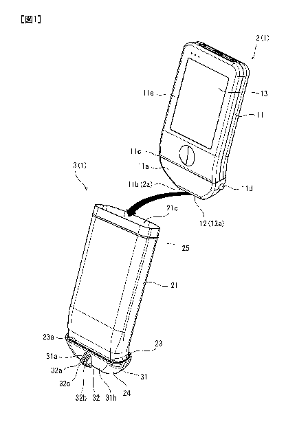
[0009] Fig. 1 is a perspective view of an ultrasonic diagnostic device cover
of a first embodiment ofthe present
invention.
Fig. 2 is a front view showing an ultrasonic diagnostic device with cover in
which the ultrasonic
diagnostic device of Fig. 1 is inserted into the ultrasonic diagnostic device
cover.
Fig. 3 is an exploded perspective view showing a disassembled state ofthe
ultrasonic diagnostic device
cover of Fig. 1.
Fig. 4 is a cross-sectional view of the ultrasonic diagnostic device cover of
Fig. 2, cut along cross-
sectional line IV-IV.
Fig. 5 is a front view showing an ultrasonic diagnostic device with cover in
which the ultrasonic
diagnostic device is inserted into an ultrasonic diagnostic device cover of a
second embodiment of the present
invention.
Fig. 6 is a front view showing how the ultrasonic diagnostic device is
inserted into the ultrasonic
diagnostic device cover.
Fig. 7 is a plan view of a bracket with guide shown in Fig. 5.
Fig. 8 is a cross-sectional view of the bracket with guide of Fig. 7, cut
along cross-sectional line VIII-
Vffl.
3
Date Recue/Date Received 2020-04-15
CA 03079169 2020-04-15
Fig. 9 is a side view of the bracket with guide of Fig. 7.
Fig. 10 is an exploded sectional view of the bracket with guide of Fig. 8.
Fig. 11 is an exploded perspective view showing a disassembled state of an
ultrasonic diagnostic
device cover of another embodiment.
Fig. 12 is a side view showing another configuration example of a pair of
locking pieces ofthe bracket
with guide of the second embodiment
Description of Embodiments
[0010] Hereinafter, ultrasonic diagnostic devices with cover 1 and 1A
according to embodiments of the
present invention will be described with reference to the drawings. Note that
the concepts of directions used in
the following description are used for convenience in the description, and do
not limit the directions and the like
of configurations of the invention. Additionally, the ultrasonic diagnostic
devices with cover 1 and 1A described
below are merely an embodiment of the present invention. Hence, the present
invention is not limited to the
embodiments, and additions, eliminations, or modifications can be made without
departing from the spirit of the
invention.
[0011] [First Embodiment]
The ultrasonic diagnostic device with cover 1 shown in Fig. 1 is used when
puncturing a needle or the
like while visualizing a blood vessel or the like under the skin, and is used
when placing a catheter in a blood
vessel in dialysis treatment, for example. The ultrasonic diagnostic device
with cover 1 having such a function
includes an ultrasonic diagnostic device 2 and an ultrasonic diagnostic device
cover 3, and the ultrasonic
diagnostic device cover 3 covers the ultrasonic diagnostic device 2 during
use. Hereinafter, an example of a
configuration of the ultrasonic diagnostic device 2 and the ultrasonic
diagnostic device cover 3 will be described.
[0012] <Ultrasonic diagnostic device>
While being applied to the body surface, the ultrasonic diagnostic device 2
emits ultrasonic waves to a
part to be examined (i.e., subcutaneous tissue), and receives ultrasonic waves
reflected from the part to be
4
Date Recue/Date Received 2020-04-15
CA 03079169 2020-04-15
examined (i.e., reflected waves). Additionally, the ultrasonic diagnostic
device 2 performs image processing on
the basis of the received reflected waves, and displays a cross section of the
part to be examined. The ultrasonic
diagnostic device 2 having such a function has a casing 11, a probe 12, and a
monitor 13 (also refer to front view
of Fig. 2). For example, the casing 11 is made of a synthetic resin such as
polycarbonate, ABS (acrylonitrile
butadiene styrene) resin, SAS (silicon acrylonitrile styrene), and
polypropylene, and is a box having a
substantially rectangular shape in front view and with a flat cross section.
Additionally, a tip end side portion 11a,
which is a lower end portion (i.e., one end side portion) of the casing 11, is
formed in a trapezoidal shape tapered
toward the tip end, and a probe 12 is arranged in a tip end portion lib of the
casing 11. That is, the ultrasonic
diagnostic device 2 has the probe 12 at its tip end portion 2a. Additionally,
the casing 11 is curved so as to warp
rearward at an intermediate portion, and is inclined rearward from a curved
portion 11c to abase end side portion.
Moreover, the monitor 13 is arranged on a front surface lie of the casing 11,
and the monitor 13 displays an
image. Hereinafter, the probe 12 and the monitor 13 will be described in more
detail.
[0013] The probe 12 is configured to transmit and receive ultrasonic waves,
and has a sensor 12a. The sensor
12a is configured of a transducer such as a piezoelectric element and an
acoustic lens, for example. The sensor
12a configured as described above is mounted so as to be embedded in the tip
end side portion 11 a of the casing
ii as also shown in Fig. 2. The sensor 12a is arranged in the casing ii so
that a lower end surface of the sensor
12a is flush with the tip end portion llb of the casing 11. Additionally, the
sensor 12a is connected to a control
device (not shown) accommodated in the casing 11, and emits ultrasonic waves
in response to a command from
the control device. Additionally, the sensor 12a receives reflected waves
reflected from the part to be examined,
and outputs a signal corresponding to the received ultrasonic waves to the
control device. That is, the transducer
of the sensor 12a vibrates due to the reflected waves reflected from the part
to be examined, and outputs a signal
corresponding to the vibration to the control device. The control device has
an image processing function, and
performs image processing on the basis of the signal to output image data to
the monitor 13.
[0014] The monitor 13 as an example of an image display portion is a liquid
crystal monitor and an organic
FT. monitor, for example, and displays an image corresponding to image data.
That is, the monitor 13 displays
Date Recue/Date Received 2020-04-15
CA 03079169 2020-04-15
an image created on the basis of reflected waves received by the sensor 12a
such as a cross section of a part to be
examined. The monitor 13 having such a function is arranged on the front
surface lie of the casing 11 as
described above. More specifically, the horizontal width of the monitor 13
extends from the vicinity of the left
end of the casing 11 to the vicinity of the right end of the casing 11, and
the vertical height of the monitor 13
extends from the curved portion 11c of the casing 11 to the vicinity of the
base end of the casing 11.
[0015] The ultrasonic diagnostic device 2 configured as described above
applies the probe 12 to a body
surface of the part to be examined, and emits ultrasonic waves from the
transducer of the sensor 12a of the probe
12 in this state. The emitted ultrasonic waves are reflected from the part to
be examined, and the reflected waves
are received by the transducer of the sensor 12a. Then, the transducer of the
sensor 12a vibrates according to the
reflected waves, and the transducer outputs a signal con-esponding to the
vibration to the control device. The
control device performs image processing on the basis of the signal, whereby a
cross section of the part to be
examined is displayed on the monitor 13.
[0016] The ultrasonic diagnostic device 2 having such a function is used in
such a manner that the probe 12
scans the body surface ofthe part to be examined. For this reason, when
sterilization processing is not performed
on the ultrasonic diagnostic device 2, the ultrasonic diagnostic device 2 is
covered with the ultrasonic diagnostic
device cover 3.
[0017] <Ultrasonic diagnostic device cover>
The ultrasonic diagnostic device cover 3 covers the entire ultrasonic
diagnostic device 2 so that blood
or the like does not adhere to the ultrasonic diagnostic device 2, or to
prevent attached blood or the like from
scattering The ultrasonic diagnostic device cover 3 having such a function
includes a cylindrical body 21, an
inner bracket 22, a probe cover body 23, and a bracket with guide 24. The
ultrasonic diagnostic device 2 can be
inserted and stored in the cylindrical body 21.
[0018] The cylindrical body 21 is a sterile soft body that has been subjected
to sterilization processing, and
specifically, is a film body that has no elasticity. As shown in Fig. 3, the
cylindrical body 21 is a generally
rectangular soft body that has open upper and lower ends and is vertically
long in front view More specifically,
6
Date Recue/Date Received 2020-04-15
CA 03079169 2020-04-15
the cylindrical body 21 is a highly transparent soft body made of a material
such as polyethylene, polypropylene,
polyethylene terephthalate, and polyvinyl chloride. An external shape of the
cylindrical body 21 in front view is
similar to the ultrasonic diagnostic device 2. That is, the cylindrical body
21 is a cylindrical body having a flat
cross section, and a lower end side portion 21a of the cylindrical body 21 is
formed in a trapezoidal shape tapered
toward a lower end 21b. Additionally, the cylindrical body 21 has an upper end
(i.e., base end) and a lower end
21b (i.e., tip end) that are open, and openings 21c and 21d formed at the
upper and lower ends, respectively. Since
the cylindrical body 21 is a soft body, it is easy to accommodate the
ultrasonic diagnostic device 2 in the
cylindrical body 21, and since the cylindrical body 21 has high transparency,
it is easy to check the monitor 13 of
the ultrasonic diagnostic device 2 accommodated inside the cylindrical body 21
from the outside.
[0019] The upper opening 21c, which is the opening 21c on the upper side, is
formed so that the ultrasonic
diagnostic device 2 can be inserted from its tip end portion 2a. On the other
hand, the lower opening 21d, which
is the opening 21d on the lower side, has a smaller diameter than the upper
opening 21c, so that after the ultrasonic
diagnostic device 2 is inserted from the upper opening 21c of the cylindrical
body 21, the lower opening 21d
naturally comes into contact with the periphery ofthe tip end side ofthe
ultrasonic diagnostic device 2 or the inner
bracket 22, and supports the ultrasonic diagnostic device 2 so as not to fall
out through the lower opening 21d.
Additionally, the lower opening 21d is formed slightly larger than the outer
edge shape of the tip end portion 2a
of the ultrasonic diagnostic device 2 in a state where the ultrasonic
diagnostic device 2 is inserted into the
cylindrical body 21, and allows the tip end portion 2a of the ultrasonic
diagnostic device 2 (i.e., portion of the
sensor 12a of the probe 12) to project from the lower opening 21d. Since the
tip end portion 2a of the ultrasonic
diagnostic device 2 projects from the cylindrical body 21, the probe cover and
the tip end portion 2a of the
ultrasonic diagnostic device 2 can be brought into close contact without
having to form the shape of the probe
cover on the contacting side in a special shape.
[0020] The cylindrical body 21 having such a shape is formed longer than the
ultrasonic diagnostic device 2
in the vertical direction (i.e., predetermined direction), so that
substantially the entire ultrasonic diagnostic device
2 fits therein. More specifically, in the cylindrical body 21, the ultrasonic
diagnostic device 2 is accommodated
7
Date Recue/Date Received 2020-04-15
CA 03079169 2020-04-15
with its tip end portion 2a pmjecting from the lower opening 21d, and the
cylindrical body 21 is formed long in
the vertical direction, so that the upper opening 21c is located higher than
the upper end of the ultrasonic
diagnostic device 2 in this state. Additionally, a zip seal 25 is attached to
an inner peripheral surface of the
cylindrical body 21 over the entire circumference in the vicinity of the upper
opening 21c. The zip seal 25 has a
male zip portion 25a and a female zip portion 25b, and the two zip portions
25a and 25b are brought into contact
to be engaged with each other. For example, the male zip portion 25a and the
female zip portion 25b are provided
on a front portion and a rear portion of the inner peripheral surface of the
cylindrical body 21 so as to face each
other, and are brought into contact to be engaged with each other to close the
upper side of the cylindrical body
21. The inner bracket 22 is provided in the cylindrical body 21 configured as
described above, as shown in Fig.
4.
[0021] The inner bracket 22 is a cylindrical member provided in the
cylindrical body 21, and has a function
of guiding the ultrasonic diagnostic device 2 inserted into the cylindrical
body 21 to a predetermined position and
positioning the ultrasonic diagnostic device 2 in the predetermined position.
More specifically, the inner bracket
22 is made of a material harder than the probe cover body 23 described later
in detail, such as synthetic resin like
polycarbonate, ABS (acrylonitrile butadiene styrene) resin, SAS (silicon
acrylonitrile styrene), and
polypropylene. Additionally, as shown in Fig. 3, the inner bracket 22 is a
cover-shaped member having a
substantially trapezoidal shape in front view and having open upper and lower
ends, and has a flat cross section
when viewed from above. More specifically, an outer peripheral surface of the
inner bracket 22 is formed in
substantially the same shape as an inner peripheral surface of the lower end
side portion 21a of the cylindrical
body 21. Alower end side portion 22a ofthe inner bracket 22 pmj ects from a
lower opening 21d ofthe cylindrical
body 21.
[0022] On the other hand, since the lower end side portion 21a ofthe
cylindrical body 21 is formed in a tapered
shape tapered toward the tip end, the inner bracket 22 fits into the lower end
side portion 21a of the cylindrical
body 21 such that its lower end side portion 22a pmjects from the lower
opening 21d of the cylindrical body 21
and the remaining portion does not project to the outside. Additionally, an
inner peripheral surface of the inner
8
Date Recue/Date Received 2020-04-15
CA 03079169 2020-04-15
bracket 22 is formed in substantially the same shape as an outer peripheral
surface of the tip end side portion 11 a
of the ultrasonic diagnostic device 2, so that the ultrasonic diagnostic
device 2 inserted into the cylindrical body
21 can be placed inside and mounted to the inner bracket 22 from the upper end
of the inner bracket 22.
Additionally, the probe 12 of the ultrasonic diagnostic device 2 projects from
the lower end of the inner bracket
22.
[0023] As described above, the inner peripheral surface of the inner bracket
22 having such a shape is formed
in substantially the same shape as the outer peripheral surface of the tip end
side portion 11 a of the ultrasonic
diagnostic device 2, that is, formed in a tapered shape tapered toward the
lower end. For this reason, when the
ultrasonic diagnostic device 2 is inserted into the inner bracket 22, the
inner bracket 22 guides the ultrasonic
diagnostic device 2 to a predetermined position where the probe 12 projects
from the lower end of the inner
bracket 22. The inner bracket 22 having such a function is formed in a shape
having a flat cross section as
described above, and a pair of grooves 22b, 22b is formed in side surface
portions positioned on both sides in the
width direction, respectively (also refer to Fig. 4).
[0024] The pair of grooves 22b, 22b extends in the vertical direction and
penetrates the inner bracket 22 in the
inside-outside direction (i.e., width direction). Additionally, the pair of
grooves 22b, 22b is arranged apart from
each other in the longitudinal direction, thereby forming a flexible piece 22c
between the pair of grooves 22b,
22b. The flexible piece 22c warps in the width direction from its base end
portion. Additionally, a fitting hole
22d is formed in the flexible piece 22c. That is, a pair of fitting holes 22d
is formed in the inner bracket 22 so as
to be separated from each other in the width direction. Additionally, fitting
protrusions 11d, lid corresponding
to the pair of fitting holes 22d are formed on side surfaces of the tip end
side portion 11 a of the casing 11.
[0025] The pair of fitting protrusions lid, lid is foimed in a substantially
hemispherical shape, and protrudes
in the width direction from the side surfaces ofthe tip end side portion ha.
When the ultrasonic diagnostic device
2 is inserted into the inner bracket 22, the pair of fitting protrusions 11d,
lid each comes into contact with the
corresponding flexible pieces 22c, and when the ultrasonic diagnostic device 2
is pushed in further, the pair of
fitting protrusions lid, lid pushes and spreads the corresponding flexible
pieces 22c radially outward. With this
9
Date Recue/Date Received 2020-04-15
CA 03079169 2020-04-15
spreading, the ultrasonic diagnostic device 2 can be inserted to a
predetermined position in the inner bracket 22.
Additionally, when the ultrasonic diagnostic device 2 is inserted to the
predetermined position, each ofthe pair of
fitting protrusions lid, lid fits into the con-esponding fitting hole 22d. As
a result, the ultrasonic diagnostic device
2 is positioned and held at the predetermined position. The probe cover body
23 covers the inner bracket 22
configured as described above to close a lower opening 22e of the inner
bracket 22.
[0026] The probe cover body 23 has a bottomed cylindrical shape having a
bottom, a side surface, and an
opening, and is provided in the cylindrical body 21 such that it covers the
inner bracket 22 to close the lower
opening 22e ofthe inner bracket 22 and comes into contact with the sensor 12a
ofthe ultrasonic diagnostic device
2 projecting from the lower opening 22e. That is, the probe cover body 23 can
be fixed to the ultrasonic diagnostic
device with nothing interposed between the tip end portion 2a ofthe ultrasonic
diagnostic device 2 and the probe
cover body 23. More specifically, the probe cover body 23 is a bag body having
a substantially trapezoidal shape
tapered in front view and having a flat cross section. Additionally, the probe
cover body 23 is closed on the tip
end side (i.e., lower side), and has an opening 23a on the base end side
(upper side). Additionally, an inner
peripheral surface and an outer peripheral surface ofthe probe cover body 23
are formed in substantially the same
shape as the outer peripheral surface of the inner bracket 22, and the inner
peripheral surface of the probe cover
body 23 is formed slightly smaller than the outer peripheral surface of the
inner bracket 22.
[0027] The probe cover body 23 having such a shape is made of a styrene-based,
urethane-based, or silicone-
based elastomer gel (styrene-based elastomer gel in the embodiment), for
example, and is configured to be
stretchable. Hence, by pushing and spreading the opening 23a outward, the
probe cover body 23 allows the
lower end side portion 22a of the inner bracket 22 to be inserted into the
probe cover body 23. Additionally, the
probe cover body 23 contracts inward with the inner bracket 22 inserted
therein, and presses itself against the
inner bracket 22 to prevent the probe cover body 23 from slipping off the
inner bracket 22. Moreover, the probe
cover body 23 extends further upward than the lower end side portion 22a ofthe
inner bracket 22 projecting from
the cylindrical body 21, and covers the lower opening 21d of the cylindrical
body 21. As a result, the lower
opening 21d ofthe cylindrical body 21 and the vicinity thereof are sandwiched
between the probe cover body 23
Date Recue/Date Received 2020-04-15
CA 03079169 2020-04-15
and the inner bracket 22, so that the cylindrical body 21 is not easily
detached from the probe cover body 23 and
the inner bracket 22. Note that the probe cover body 23 may have a weak
adhesive property on its surface. In
this case, the weak adhesive effect can prevent the probe cover body 23 from
being displaced from the cylindrical
body 21.
[0028] Thus, the probe cover body 23 is provided on the cylindrical body 21 so
as to cover the inner bracket
22, and the probe cover body 23 is provided on the cylindrical body 21 to
close the lower opening 21d of the
cylindrical body 21. In this closed state, when the ultrasonic diagnostic
device 2 is inserted into the cylindrical
body 21 to prci ect the tip end portion 2a of the ultrasonic diagnostic device
2 from the lower opening 21d of the
cylindrical body 21 and the lower opening 22e of the inner bracket 22, the tip
end portion 2a can be brought into
contact with the probe cover body 23. Moreover, when the ultrasonic diagnostic
device 2 is pushed from this
state to a predetermined position, the entire sensor 12a can be pressed
against and brought into close contact with
the probe cover body 23. As a result, it is possible to prevent a small gap
from being formed between the sensor
12a and the probe cover body 23.
[0029] The probe cover body 23 configured as described above is made of a
material that improves acoustic
coupling between the sensor 12a and the body surface of a part to be examined.
By applying the probe cover
body 23 to the body surface ofthe part to be examined with the sensor 12a in
close contact therewith, it is possible
to display a cross section of the part to be examined on the monitor 13 in a
favorable state. Furthermore, the
bracket with guide 24 covers an outer surface of the probe cover body 23
configured as described above. Note
that in the embodiment, in order to display a cross section of a part to be
examined on the monitor 13 in a more
favorable state, a liquid gel such as an echo gel, physiological saline, and a
liquid material such as a disinfectant
solution (hereinafter referred to as "liquid gel and the like") are applied to
the body surface of the part to be
examined so that an air layer is not formed between the part to be examined
and the sensor 12a. However, the
liquid gel and the like may be applied to the outer surface of the probe cover
body 23.
[0030] The bracket with guide 24 has a bracket main body 31 and a guide
portion 32. The bracket main body
31 is made of a material harder than the probe cover body 23, such as a
synthetic resin like polycarbonate, ABS
11
Date Recue/Date Received 2020-04-15
CA 03079169 2020-04-15
(acrylonitrile butadiene styrene) resin, SAS (silicon acrylonitrile styrene),
and polypropylene. Additionally, the
bracket main body 31 is a substantially trapezoidal cylindrical member in
front view, and the inner space thereof
is formed to match the shape of the outer peripheral surface of the inner
bracket 22. More specifically, the inner
space of the bracket main body 31 is formed slightly larger than the inner
bracket 22, so that the bracket main
body 31 can be fitted and mounted to the inner bracket 22 over the probe cover
body 23 and the cylindrical body
21.
[0031] The bracket main body 31 configured as described above is formed in a
substantially trapezoidal shape
tapered toward the lower end as described above. By fitting the bracket main
body 31 to the inner bracket 22, it
is possible to securely press the probe cover body 23 against the inner
bracket 22 and stabilize the close-contacting
state between the sensor 12a and the probe cover body 23. Note that the size
ofthe gap formed between the inner
peripheral surface of the bracket main body 31 and the outer peripheral
surface of the inner bracket 22 can be
made smaller than the thickness of the probe cover body 23 by the wedge effect
of the bracket main body 31.
Consequently, the probe cover body 23 is pulled so as to be extended and
lifted toward the base end side of the
casing 11. Hence, the probe cover body 23 is pressed more securely against the
sensor 12a than when the bracket
main body 31 is not mounted, and can be brought into closer contact with the
sensor 12a. That is, since the probe
cover body 23 can be securely brought into close contact with the sensor 12a
without pressing the probe 12
against the body surface, it is possible to display a cross section of the
part to be examined on the monitor 13
simply by applying the probe 12 instead of pressing it against the body
surface. As a result, it is possible to display
the veins and the like near the epidermis on the monitor 13 without crushing
them. The bracket main body 31
having such a function further has a fitting prci ection 3 la.
[0032] The fitting projection 31a is integrally provided on a front surface 3
lb ofthe bracket main body 31 (i.e.,
surface on same side as the front surface 1 1 e of the casing 11). The fitting
prqjection 31a is arranged at the center
in the width direction on the front surface 31b of the bracket main body 31,
and is formed so as to protrude
frontward from the front surface 3 lb of the bracket main body 31. The guide
portion 32 is fitted to the fitting
projection 31a formed in this manner. The guide portion 32 guides a needle 41
for puncturing and determines
12
Date Recue/Date Received 2020-04-15
CA 03079169 2020-04-15
the puncture direction of the needle 41 when puncturing the subcutaneous
tissue with the needle 41 (see two-dot
chain line in Fig. 4) of a needle assembly such as an indwelling needle. The
guide portion 32 has a substantially
trapezoidal block shape in side view The guide portion 32 is provided on the
front surface side of the cylindrical
body 21 (on the monitor 13 side when the ultrasonic diagnostic device 2 is
accommodated in the cylindrical body
21). Accordingly, it is possible to make the puncture while watching the
monitor 13 of the ultrasonic diagnostic
device 2. Additionally, since the distance from the guide portion 32t0 the
sensor 12a is short, the puncture length
can be shortened. This can improve stability of the puncturing operation and
reduce the burden on the patient.
Additionally, by providing the guide portion 32 in a position overlapping the
sensor 12a in side view, it is possible
to reduce the distance from the guide portion 32 to the sensor 12a even more.
Note that while the guide portion
32 is provided on the front surface side of the cylindrical body 21 having the
open tip end and base end, the
cylindrical body used in the invention in which the guide portion 32 is
provided on the front surface side of the
cylindrical body 21 having the open tip end and base end does not need to be
the cylindrical body 21 having an
open tip end. The configuration and arrangement of the guide portion 32
described herein are useful in all forms
of fixing a guide portion to an ultrasonic diagnostic device.
[0033] The guide portion 32 having such a shape has a front end surface 32a at
a predetermined angle a (e.g.,
20 degrees < a <80 degrees) with respect to the front surface 3 lb of the
bracket main body 31. The front end
surface 32a of the guide portion 32 is inclined with respect to the front
surface 3 lb of the bracket main body 31.
Additionally, a guide hole forming portion 32b is formed in the front end
surface 32a at a center portion in the
horizontal direction. The guide hole forming portion 32b projects from the
front end surface 32a in a direction
perpendicular to the front end surface 32a, and extends obliquely upward and
frontward along the front end
surface 32a. Additionally, a cross section of the guide hole forming portion
32b (i.e., cross section perpendicular
to the front end surface 32a) is formed in a substantially semicircular shape.
The guide hole forming portion 32b
has a guide hole 32c formed along its axis. As a result, a guide hole 32c
extending obliquely upward and
frontward is formed in the guide portion 32. The guide hole 32c penetrates the
guide portion 32 obliquely upward
and frontward, so that the needle 41 of the needle assembly can be inserted
from above and a needle tip 41a can
13
Date Recue/Date Received 2020-04-15
CA 03079169 2020-04-15
be drawn out from below. Note that an outlet 32d extending in the axial
direction of the guide hole 32c is formed
on a front surface of the guide hole forming portion 32b, and the needle 41
can be pulled frontward and pulled
out from the outlet 32d.
[0034] <Mounting and puncturing>
The ultrasonic diagnostic device 2 is placed in the ultrasonic diagnostic
device cover 3 when used as
described above, and is used while being sealed inside the ultrasonic
diagnostic device cover 3. More specifically,
in order to maintain a sterile state, the ultrasonic diagnostic device cover 3
is placed in a bag such as a sterile pack
(not shown) in an already assembled state. Then, the ultrasonic diagnostic
device cover 3 is taken out ofthe sterile
pack at the time of use. After being taken out, first, the upper opening 21c
of the cylindrical body 21 of the
ultrasonic diagnostic device cover 3 is opened, and the ultrasonic diagnostic
device 2 is inserted into the upper
opening 21c from the tip end portion 2a.
[0035] Thereafter, when the cylindrical body 21 is set up straight so as to
accommodate the entire ultrasonic
diagnostic device 2 in the cylindrical body 21 or when the ultrasonic
diagnostic device 2 is pushed in, the
ultrasonic diagnostic device 2 moves through the inside ofthe cylindrical body
21 toward the lower opening 21d.
When the ultrasonic diagnostic device 2 is pushed even further after being
accommodated, the tip end portion 2a
ofthe ultrasonic diagnostic device 2 eventually enters the inner bracket 22.
When the ultrasonic diagnostic device
2 is pushed even further, the pair of fitting protrusions lid, lid comes into
contact with the upper end of the inner
bracket 22 (more specifically, upper ends of the flexible pieces 22c). When
the ultrasonic diagnostic device 2 is
pushed from above in this state, the pair of fitting protrusions 11d, lid
pushes and spreads the corresponding
flexible pieces 22c, and the ultrasonic diagnostic device 2 can be pushed
further downward. By continuing to
push, the ultrasonic diagnostic device 2 reaches a predetermined position
while being guided by the inner bracket
22. When reaching the predetermined position, each of the pair of fitting
protrusions 11d, lid fits into the
corresponding fitting hole 22d, and the ultrasonic diagnostic device 2 is
positioned and held inside the cylindrical
body 21. Additionally, when the ultrasonic diagnostic device 2 reaches the
predetermined position, the sensor
12a can be projected from the lower opening 21d, whereby the sensor 12a comes
into close contact with the
14
Date Recue/Date Received 2020-04-15
CA 03079169 2020-04-15
probe cover body 23.
[0036] When the ultrasonic diagnostic device 2 is accommodated in the
ultrasonic diagnostic device cover 3
in this manner, next, the male zip portion 25a and the female zip portion 25b
of the zip seal 25 are brought into
contact and engaged with each other to seal the cylindrical body 21. As a
result, it is possible to prevent the liquid
gel and the like and blood and the like from adhering to the ultrasonic
diagnostic device 2 and to prevent the
attached blood and the like from scattering Thus, the ultrasonic diagnostic
device cover 3 is mounted so as to
cover and seal the entire ultrasonic diagnostic device 2. By mounting the
ultrasonic diagnostic device 2, the
ultrasonic diagnostic device 2 becomes usable. Hence, puncturing is performed
while displaying an image of a
part to be examined, for example. Hereinafter, puncturing using the ultrasonic
diagnostic device with cover 1
will be described.
[0037] In the ultrasonic diagnostic device with cover 1, after the ultrasonic
diagnostic device cover 3 is
mounted to the ultrasonic diagnostic device 2, the probe 12 is pressed against
the body surface through the probe
cover body 23. As a result, a cross section of the part to be examined is
displayed on the monitor 13. Note that a
liquid gel and the like may be applied to the body surface depending on the
state of acoustic coupling to improve
the acoustic coupling. When the cross section of the part to be examined is
displayed on the monitor 13, the user
searches the position of the blood vessel while looking at the monitor 13, and
when the position of the blood
vessel is confirmed, the user inserts the needle 41 of the needle assembly
into the guide hole 32c of the bracket
with guide 24 to puncture the blood vessel with the needle tip 41a of the
needle 41. Note that the angle a of the
guide hole 32c is set in advance, so that the needle 41 inserted therein
overlaps the monitor and passes through a
predetermined position in the displayed image. The state of the needle rip 41a
used for puncturing can be
confirmed on the monitor 13.
[0038] When it is confirmed that the blood vessel has been punctured with the
needle tip 41a, in order to
remove the ultrasonic diagnostic device 2 from the needle 41 while keeping the
needle 41 as it is, the needle 41
is pulled out from the guide hole 32c through the outlet 32d. Specifically,
the needle 41 is pulled out from the
guide hole 32c through the outlet 32d by tilting the ultrasonic diagnostic
device 2 rearward so as to separate the
Date Recue/Date Received 2020-04-15
CA 03079169 2020-04-15
ultrasonic diagnostic device 2 from the needle 41. As a result, the needle 41
can be left in this position, and
subsequent treatment (e.g., treatment such as dialysis, liquid injection, and
blood collection) can be performed.
[0039] In the ultrasonic diagnostic device with cover 1 having such a
function, by merely inserting and
pushing the ultrasonic diagnostic device 2 into the cylindrical body 21 of the
ultrasonic diagnostic device cover
3, as described above, the probe cover body 23 can achieve excellent acoustic
coupling between a part to be
examined and the sensor 12a, that is, the ultrasonic diagnostic device 2 can
be brought into a usable state. Hence,
the ultrasonic diagnostic device 2 can be easily mounted to the ultrasonic
diagnostic device cover 3. Additionally,
since it is only necessary to insert the ultrasonic diagnostic device 2 into
the ultrasonic diagnostic device cover 3,
the ultrasonic diagnostic device 2 does not come into contact with the outer
surface of the ultrasonic diagnostic
device cover 3. Hence, the ultrasonic diagnostic device cover 3 can be kept in
a sterile state.
[0040] Additionally, in the ultrasonic diagnostic device with cover 1, by
pushing the ultrasonic diagnostic
device 2 into the inner bracket 22, the ultrasonic diagnostic device 2 is
guided to a predetermined position and
held at the predetermined position by the inner bracket 22. At the
predetermined position, the tip end portion 2a
of the ultrasonic diagnostic device 2 can be prci ected from the inner bracket
22, and the entire sensor 12a can be
brought into close contact with the probe cover body 23. Hence, excellent
acoustic coupling can be achieved
merely by inserting and pushing the ultrasonic diagnostic device 2 into the
cylindrical body 21 of the ultrasonic
diagnostic device cover 3. Additionally, by adopting a structure in which the
pair of fitting protrusions lid, lid
is fitted and held in the con-esponding fitting holes 22d, it is possible to
curb displacement of the ultrasonic
diagnostic device 2 with respect to the inner bracket 22. As a result, the
entire sensor 12a can be securely brought
into close contact with the probe cover body 23 (i.e., contacting state can be
stabilized), and excellent acoustic
coupling can be maintained.
[0041] Additionally, in the ultrasonic diagnostic device with cover 1, by
placing the inner bracket 22 inside
the probe cover body 23, it is possible to curb deformation of the probe cover
body 23. Hence, it is easy to insert
the tip end portion 2a of the ultrasonic diagnostic device 2, or more
specifically, the tip end side portion 11 a of the
casing 11, into the probe cover body 23. Accordingly, it can be made even
easier to mount the ultrasonic
16
Date Recue/Date Received 2020-04-15
CA 03079169 2020-04-15
diagnostic device 2 to the ultrasonic diagnostic device cover 3. Moreover, in
the ultrasonic diagnostic device with
cover 1, the probe cover body 23 covers not only the inner bracket 22 but also
the lower opening 21d of the
cylindrical body 21 and its vicinity. Hence, the probe cover body 23 can close
the lower opening 21d of the
cylindrical body 21. Additionally, since the probe cover body 23 covers the
lower opening 21d of the cylindrical
body 21, the cylindrical body 21 is sandwiched between the probe cover body 23
and the inner bracket 22. Hence,
it is possible to curb displacement of the cylindrical body 21 with respect to
the inner bracket 22 during use.
[0042] [Second Embodiment]
The configuration of an ultrasonic diagnostic device with cover lA of a second
embodiment is similar
to that ofthe ultrasonic diagnostic device with cover 1 ofthe first
embodiment. In the following, the configuration
of the ultrasonic diagnostic device with cover 1A of the second embodiment
will be described mainly with
respect to differences from the ultrasonic diagnostic device with cover 1 of
the first embodiment. The same
configurations will be assigned the same reference numerals, and descriptions
will be omitted.
[0043] <Ultrasonic diagnostic device cover>
The ultrasonic diagnostic device with cover 1A of the second embodiment
includes an ultrasonic
diagnostic device 2 and an ultrasonic diagnostic device cover 3A as shown in
Fig. 5, and the ultrasonic diagnostic
device cover 3Ais configured as follows. That is, the ultrasonic diagnostic
device cover 3A includes a cylindrical
body 21A, a bracket with guide 24A, and a probe cover body 23A. The
cylindrical body 21A is made of an
inelastic soft film body having high transparency as in the first embodiment
and has been subjected to
sterilization processing. The cylindrical body 21A made of such a material is
a cylindrical body having a flat
cross section as in the case of the cylindrical body 21 of the first
embodiment. When the cylindrical body 21A is
assembled with the bracket with guide 24A and the probe cover body 23A, a
lower end side portion 21a is formed
in a trapezoidal shape tapered toward a lower end 21b. On the other hand, an
upper end side portion 21e of the
cylindrical body 21A is formed in a trapezoidal shape expanding toward an
upper end 21f (i.e., upper opening
21c).
[00/H] In the cylindrical body 21A having such a shape, when inserting the
ultrasonic diagnostic device 2, the
17
Date Recue/Date Received 2020-04-15
CA 03079169 2020-04-15
upper end side portion 21e is folded as shown in Fig. 6. That is, the upper
end side portion 21e of the cylindrical
body 21A is folded outward with respect to a middle portion 21g connected to
the lower side of the upper end
side portion 21e, and the vicinity of a zip seal 25 (more specifically, part
below zip seal 25) is folded upward.
Additionally, the cylindrical body 21A is folded such that the upper opening
21c is positioned on the lower end
side of a folded portion (i.e., portion obtained by folding the upper end side
portion 21e with respect to the middle
portion 21g) 21h ofthe cylindrical body 21A. By folding in this manner, when
inserting the ultrasonic diagnostic
device 2 into the cylindrical body 21A from above, it is possible to keep the
ultrasonic diagnostic device 2 from
hitting an outer surface of the upper opening 21c of the cylindrical body 21A.
That is, it is possible to keep the
inserted ultrasonic diagnostic device 2 from coming into contact with the
upper opening 21c of the cylindrical
body 21A and contaminating the upper opening 21c.
[0045] Additionally, by folding the upper opening 21c as described above, the
upper opening 21c can be
opened and the upper opening 21c can be also be closed in the following
manner. That is, fingers such as the
thumb and forefinger of both hands are inserted between the middle portion 21g
of the cylindrical body 21A and
the upper opening 21c from below, so as to sandwich the middle portion 21g
from both right and left sides. Then,
by lifting the fingers upward in this stare and stretching the upper end side
portion 21e upward, the upper end side
portion 21e can be brought back to the spread out state as shown in Fig 5.
Moreover, by bringing a male zip
portion 25a and a female zip portion 25b (not shown) of a zip seal 25 into
contact and engaged with each other,
the upper opening 21c can be closed with the ultrasonic diagnostic device 2
accommodated in the cylindrical
body 21A The cylindrical body 21A configured as described above is provided
with a bracket with guide 24A
for attaching the probe cover body 23 to a lower opening 21d.
[0046] The bracket with guide 24A has a bracket main body 31A and a guide
portion 32A. The bracket main
body 31 is made of a material harder than the probe cover body 23A described
later, such as a synthetic resin like
polycarbonate, ABS (acrylonitrile butadiene styrene) resin, SAS (silicon
acrylonitrile styrene), and
polypropylene. The bracket main body 31A made of such a material is formed in
a substantial plate shape in
front view as shown in Fig. 5, and is formed in a horizontally long, flat and
annular shape in plan view as shown
18
Date Recue/Date Received 2020-04-15
CA 03079169 2020-04-15
in Fig 7. Note that in Fig 7, the guide portion 32A is omitted for convenience
of explanation. The same applies
to Fig. 9 described later
[0047] The bracket main body 31Ahaving such a shape has an inner hole 31c, and
an inner peripheral surface
defining the inner hole 31c is formed in a tapered shape tapered downward as
shown in Fig. 8. Additionally, the
inner hole 31c is formed horizontally long as in the case of the shape of the
outer peripheral edge at a tip end
portion 2a ofthe ultrasonic diagnostic device 2. The inner hole 31c allows
insertion ofthe tip end ofthe ultrasonic
diagnostic device 2 therethrough. More specifically, the inner peripheral edge
of the inner hole 31c of the bracket
main body 31A is larger than the outer peripheral edge of the tip end of the
ultrasonic diagnostic device 2 (i.e.,
outer peripheral edge of tip end of the tip end portion 2a), and smaller than
the outer peripheral edge of a curved
portion 11 c (outer peripheral edge ofbase end of the tip end portion 2a). As
a result, when inserted into the inner
hole 31c of the bracket main body 31A, the tip end portion 2a of the
ultrasonic diagnostic device 2 fits into the
inner hole 31c of the bracket main body 31 at a middle portion thereof and
does not project further downward.
More specifically, the inner hole 31c of the bracket main body 31A keeps the
tip end portion 2a of the ultrasonic
diagnostic device 2 from projecting for a predetermined length or more from
the bracket main body 31A (more
specifically, from the lower opening 21d of the cylindrical body 21A). A pair
of locking pieces 33 is integrally
formed with the bracket main body 31A configured as described above to hold
the ultrasonic diagnostic device
2.
[0048] The pair of locking pieces 33, which is engaging portion, is plate-like
member extending in the vertical
direction as shown in Fig. 9, and is integrally provided on an upper surface
of the bracket main body 31A. More
specifically, the pair of locking pieces 33 is separated to the right and left
such that their main surfaces face each
other. Since the pair of locking pieces 33 is provided not on the front side
but on the right and left, the pair of
locking pieces 33 does not hinder puncturing. Additionally, the pair of
locking pieces 33 extends diagonally
outward and upward so as to separate from each other, following the side
surface shape of the tip end portion 2a
of the ultrasonic diagnostic device 2. The tip end portion 2a of the
ultrasonic diagnostic device 2 is designed to
be inserted between the pair of locking pieces 33. Additionally, a locking
hole 33a is formed in each main surface
19
Date Recue/Date Received 2020-04-15
CA 03079169 2020-04-15
of the pair flocking pieces 33. The locking hole 33a corresponds to each of a
pair of fitting protrusions lid, lid
(engaged portions) of the ultrasonic diagnostic device 2. That is, the locking
holes 33a are arranged so that the
pair of fitting protrusions lid, lid fits therein when the ultrasonic
diagnostic device 2 is inserted into the bracket
main body 31Ato a predetermined position. The ultrasonic diagnostic device 2
is locked and held by the bracket
main body 31A by fitting the pair of fitting protrusions 11d, lid in the
locking holes 33a. The ultrasonic
diagnostic device 2 is fixed to the bracket main body 3 lA at a position where
it deforms the flat probe cover body
23A. Specifically, the ultrasonic diagnostic device 2 is fixed to the bracket
main body 31A at a position where it
stretches the probe cover body 23A. For this reason, it is possible to prevent
a gap from being formed between
the probe cover body 23A and the tip end portion 2a of the ultrasonic
diagnostic device 2.
[0049] Note that a predetermined position is a position where the tip end
portion 2a ofthe ultrasonic diagnostic
device 2 fits into the inner hole 31c of the bracket main body 31A and cannot
project any further, and the
ultrasonic diagnostic device 2 is locked to the bracket main body 31 in such a
predetermined position.
Additionally, as in the case of the flexible pieces 22c of the first
embodiment, when the tip end portion 2a of the
ultrasonic diagnostic device 2 is pushed in between the locking pieces 33, the
locking pieces 33 are pushed and
spread by the pair of fitting protrusions lid, 11d. The locking pieces 33
elastically return to their original positions
when the pair of fitting protrusions 11d, lid enters the corresponding locking
holes 33a. Consequently, the
bracket main body 31 is locked and mounted to the tip end portion 2a of the
ultrasonic diagnostic device 2.
[0050] The bracket main body 31A configured as described above is provided in
the lower opening 21d of
the cylindrical body 21, as shown in Fig 5. That is, the shape of an outer
peripheral surface of the bracket main
body 31A is horizontally long, flat, and annular in plan view The entire lower
opening 21d of the cylindrical
body 21Ais fixed, or more specifically, welded, along the outer peripheral
surface ofthe bracket main body 31A.
The probe cover body 23A is fixed to the bracket main body 31A attached as
described above, and the probe
cover body 23A is fixed to the cylindrical body 21Athrough the bracket main
body 31A.
[0051] The probe cover body 23A has a substantially flat plate shape and is
formed in a horizontally long
substantially rectangular plate shape in plan view The contour shape of the
probe cover body 23A in plan view
Date Recue/Date Received 2020-04-15
CA 03079169 2020-04-15
is similar to the shape of the inner hole 31c of the bracket main body 31A.
More specifically, the probe cover
body 23A is formed slightly larger than the inner hole 31c so as to be fixed
to the bracket main body 31A, as
described later. The probe cover body 23A having such a shape is made of the
same material as the probe cover
body 23 of the first embodiment, and is formed to be expandable and
contractible. The probe cover body 23A
configured as described above is fixed to the bracket main body 31A. In order
to fix the probe cover body 23,
the bracket main body 31A is configured of upper and lower two members, that
is, a first bracket portion 34 and
a second bracket portion 35, as shown in Fig. 10.
[0052] The first bracket portion 34 and the second bracket portion 35 are both
horizontally long, flat, and
annular in plan view, and have substantially the same shape. Additionally, the
pair of locking pieces 33, 33 is
formed on an upper surface of the first bracket portion 34, and the first
bracket portion 34 is superimposed on the
second bracket portion 35 so that a lower surface of the first bracket portion
34 abuts against an upper surface of
the second bracket portion 35. Moreover, on an outer peripheral surface of the
second bracket portion 35,
multiple engagement projections (six engagement prijections in the embodiment)
35a are formed at intervals in
the circumferential direction. On an outer peripheral surface of the first
bracket portion 34, multiple engaging
portions 34a con-esponding to the multiple engagement projections 35a are
formed. An engagement hole 34h is
formed in each of the engaging portions 34a, and a con-esponding engagement
projection 35a fits into to the
engagement hole 34h. By fitting in this way, the con-esponding engaging
portions 34a and engagement
projections 35a are engaged with each other, so that the two bracket portions
34 and 35 do not separate or become
displaced from each other. That is, the two bracket portions 34 and 35 are
locked to each other by being
superimposed in the vertical direction which is the insertion direction On the
upper surface ofthe second bracket
portion 35 formed in this manner, a recess 35b is formed on the inner
peripheral edge thereof
[0053] The recess 35b is formed over the entire inner peripheral edge of the
second bracket portion 35 in the
circumferential direction, and is recessed to be lower than the remaining
portion. Hence, when the first bracket
portion 34 is covered on the recess 35b, the recess 35b forms an accommodation
space 31d over the entire inner
peripheral surface ofthe bracket main body 31Ain the circumferential
direction. The accommodation space 31d
21
Date Recue/Date Received 2020-04-15
CA 03079169 2020-04-15
is a space that is recessed outward, and can accommodate the outer peripheral
edge of the probe cover body 23A
over the entire circumference. That is, the shape of the outer peripheral edge
of the probe cover body 23A is
formed to match the shape of the recess 35b. By placing the outer peripheral
edge of the probe cover body 23A
on the recess 35b and superimposing and mounting the first bracket portion 34
to the second bracket portion 35,
the outer peripheral edge of the probe cover body 23A is sandwiched between
the two bracket portions 34 and
35. Thus, the probe cover body 23A is fixed to the bracket main body 31, and
in the fixed state, closes the inner
hole 31c. That is, the probe cover body 23A is provided on the cylindrical
body 21A through the bracket main
body 31A so as to close the lower opening 21d of the cylindrical body 21A
[0054] Additionally, in the recess 35b, a lower prcjection 35c is formed over
the entire circumference. The
lower projection 35c is formed in an annular shape in plan view to match the
shape of the recess 35b, and
protrudes toward the lower surface of the first bracket portion 34.
Additionally, an upper projection 34c
corresponding to the lower projection 35c is formed also on the lower surface
of the first bracket portion 34. The
upper projection 34c is formed in an annular shape in plan view to match the
shape of the lower projection 35c,
and protrudes toward the upper surface of the second bracket portion 35. The
two projections 34c and 35c
arranged in this manner are arranged so as to face each other in the vertical
direction with a slight gap
therebetween, so as to sandwich the outer peripheral edge of the probe cover
body 23A therebetween. With the
two projections 34c and 35c described above, the probe cover body 23A can be
sandwiched firmly and held.
The guide portion 32A is provided on the bracket main body 31A holding the
probe cover body 23A as shown
in Fig 5. The guide portion 32A is for guiding a needle 41 of a needle device
such as an indwelling needle and
determining the puncture direction, and is formed in a substantial block shape
as in the case of the guide portion
32A of the first embodiment.
[0055] <Mounting>
Hereinafter, an operation of covering the ultrasonic diagnostic device 2 with
the ultrasonic diagnostic
device cover 3A to use the ultrasonic diagnostic device 2 will be described.
That is, in order to maintain a sterile
state, the ultrasonic diagnostic device cover 3A is placed in a bag such as a
sterile pack (not shown). Then, the
22
Date Recue/Date Received 2020-04-15
CA 03079169 2020-04-15
ultrasonic diagnostic device cover 3A is taken out of the sterile pack at the
time of use. The taken out ultrasonic
diagnostic device cover 3A has an upper end side portion 21e of the
cylindrical body 21A folded in advance as
shown in Fig. 6, and the user opens the cylindrical body 21A by inserting
fingers of both hands between the
folded upper end side portion 21e and a middle portion 21g from below. The
ultrasonic diagnostic device 2 held
by another person is inserted into the widened opening from the tip end
portion 2a.
[0056] Thereafter, when the cylindrical body 21 is set up straight so as to
accommodate the ultrasonic
diagnostic device 2 in the cylindrical body 21 or when the ultrasonic
diagnostic device 2 is pushed in, the tip end
of the ultrasonic diagnostic device 2 enters the inner hole 31c of the bracket
main body 31A, and the tip end
further comes into contact with the probe cover body 23A When the ultrasonic
diagnostic device 2 is pushed in
further, the probe cover body 23A stretches according to the tip end shape of
the ultrasonic diagnostic device 2,
and the tip end portion 2a of the ultrasonic diagnostic device 2 projects from
the bracket main body 31A. When
the ultrasonic diagnostic device 2 is pushed further in this state, the pair
of fitting protrusions lid, lid eventually
comes into contact with the corresponding locking pieces 33, 33, and the
locking pieces 33, 33 are pushed
outward by the corresponding fitting protrusions lid, 11d. When the ultrasonic
diagnostic device 2 is pushed in
further and positioned in a predetermined position, the pair of fitting
protrusions lid, lid is fitted into the locking
holes 33a, 33a, and the locking pieces 33, 33 elastically return to their
original positions. Thus, the ultrasonic
diagnostic device 2 is mounted by being engaged with the bracket main body 31A
(see Fig. 5).
[0057] As described above, in the ultrasonic diagnostic device cover 3A, the
probe cover body 23A is held so
as to close the inner hole 31c of the bracket main body 31. Hence, the tip end
portion 2a of the ultrasonic
diagnostic device 2 can be pressed against the probe cover body 23A and
brought into close contact therewith,
merely by placing the tip end portion 2a inserted in the cylindrical body 21A
in the inner hole 31c of the bracket
main body 31 and pushing. That is, the tip end portion 2a can be brought into
close contact with the probe cover
body 23A with a simple operation of inserting in the inner hole 31c. Hence,
acoustic coupling between a part to
be examined and the sensor 12a can be improved even more.
[0058] Additionally, since at least a part ofthe inner hole ofthe bracket is
formed larger than the tip end portion
23
Date Recue/Date Received 2020-04-15
CA 03079169 2020-04-15
2a of the ultrasonic diagnostic device 2, the tip end portion 2a of the
ultrasonic diagnostic device 2 can be pressed
against the bracket to be extended. Then, the sensor 12a can be pressed
against the probe cover body 23A with
the sensor 12a in close contact with the probe cover body 23A. As a result, it
is possible to curb entry of air
between the sensor 12a and the probe cover body 23, and improve acoustic
coupling between the part to be
examined and the sensor 12a even more. Moreover, in the ultrasonic diagnostic
device with cover 1A, as
described above, the protruding length of the tip end portion 2a of the
ultrasonic diagnostic device 2 with respect
to the bracket main body 31A is limited to a predetermined length depending on
the shape of the inner hole 31c
and the formed position of the locking holes 33a, 33a. For this reason, it is
possible to curb excessive stretching
of the probe cover body 23A when the tip end portion 2a of the ultrasonic
diagnostic device 2 is pressed
thereagainst and extended. Hence, it is possible to prevent the probe cover
body 23A from being excessively
extended and damaged.
[0059] When the ultrasonic diagnostic device 2 is inserted to the
predetermined position in this manner, the
user then lifts the thumb and forefinger of both hands to stretch the upper
end side portion 21e upward. When
the entire upper end side portion 21e has been stretched, the zip seal 25 is
positioned in an upper portion of the
ultrasonic diagnostic device 2. Hence, the male zip portion 25a and the female
zip portion 25b of the zip seal 25
are brought into contact and engaged with each other to close the upper
opening 21c. By closing the upper
opening 21c in this manner, the entire ultrasonic diagnostic device 2 is
sealed inside the ultrasonic diagnostic
device cover 3A As a result, the ultrasonic diagnostic device 2 becomes
usable, and therefore puncturing is
performed while displaying an image of a part to be examined, for example.
Puncturing using the ultrasonic
diagnostic device with cover lA is the same as that using the ultrasonic
diagnostic device with cover 1 of the first
embodiment, and detailed description is omitted. In addition, the ultrasonic
diagnostic device with cover lA has
similar effects as the ultrasonic diagnostic device with cover 1 of the first
embodiment.
[0060] [Other Embodiments]
In the ultrasonic diagnostic device cover 3 of the first embodiment, the inner
bracket 22 is provided in
the cylindrical body 21. However, the inner bracket 22 is not always
necessary, and an ultrasonic diagnostic
24
Date Recue/Date Received 2020-04-15
CA 03079169 2020-04-15
device cover 3B as shown in Fig. 11 is also conceivable. That is, in the
ultrasonic diagnostic device cover 3B, a
probe cover body 23 covers a lower end side portion 21a of a cylindrical body
21 so as to close a lower opening
21d. Since the probe cover body 23 covered in this manner has elasticity, when
the ultrasonic diagnostic device
2 is placed in and pushed into the lower end side portion 21a of the
cylindrical body 21, the probe cover body 23
stretches so as to follow the shape of an outer peripheral surface of a tip
end side portion 11 a of a casing 11 of the
ultrasonic diagnostic device 2. Hence, the probe cover body 23 can accommodate
the ultrasonic diagnostic
device 2. Additionally, the lower end side portion 21a of the cylindrical body
21 is formed in a tapered shape, so
that the remaining portion except the tip end portion 2a of the ultrasonic
diagnostic device 2 does not project to
the outside. For this reason, as in the case where the inner bracket 22 is
provided, the ultrasonic diagnostic device
2 can be placed in a predetermined position and brought into a usable state
only by inserting the ultrasonic
diagnostic device 2 into the cylindrical body 21.
[0061] Additionally, in the ultrasonic diagnostic device covers 3 and 3B, the
bracket with guide 24 covers the
probe cover body 23. The inner peripheral surface of the bracket with guide 24
is formed slightly larger than the
outer peripheral surface of the tip end side portion 11 a of the casing 11 and
has a substantially similar shape. For
this reason, when the tip end portion 2a of the ultrasonic diagnostic device 2
is inserted into the probe cover body
23, it is guided and positioned by the bracket with guide 24, and the bracket
with guide 24 serves as a positioning
member.
[0062] Note that in the ultrasonic diagnostic device covers 3 and 3B, the
bracket with guide 24 covers the
probe cover body 23 to guide the needle 41 when performing puncturing.
However, the bracket with guide 24
is not necessarily required, and does not have to cover the probe cover body
23. Additionally, the probe cover
body 23 may be simply covered, or at least a part ofthe inner surface thereof
may be joined to the outer peripheral
surface of the cylindrical body 21 so as not to come off. Additionally, the
ultrasonic diagnostic device covers 3
and 3B are not necessarily limited to those used for puncturing, and are also
used when only making a diagnosis
by displaying a cross section of a part to be examined on the monitor 13.
Additionally, in the ultrasonic diagnostic
device 2, the probe 12 and the monitor 13 are provided integrally with the
casing 11. However, the probe 12 and
Date Recue/Date Received 2020-04-15
CA 03079169 2020-04-15
the monitor 13 do not necessarily have to be integrated, and the probe 12 and
the monitor 13 may be formed in
separate casings.
[0063] Moreover, in the ultrasonic diagnostic device covers 3 and 3B, the
entire probe cover body 23 is
configured of an elastomer gel that can provide excellent acoustic coupling
between a part to be examined and
the probe 12, that is, a styrene-based, urethane-based, or silicone-based
elastomer gel. However, the probe cover
body 23 does not necessarily have to be configured in this manner. In the
probe cover body 23, it is only necessary
that at least a portion located between the part to be examined and the probe
12 is made of the aforementioned
elastomer gel. Note that while the probe cover body is provided on the outer
surface of the cylindrical body in
the embodiment, the probe cover body may be provided on the inner surface.
[0064] Additionally, in the ultrasonic diagnostic device covers 3 and 3B, the
zip seal 25 is provided to close
the upper opening 21c of the cylindrical body 21. However, it does not
necessarily have to be a zip seal, and may
be a hook-and-loop fastener or an adhesive tape.
[0065] Additionally, while the cylindrical bodies 21 and 21A in the
embodiments are soft cylindrical bodies
made of a material such as polyvinyl chloride and have a length that covers
the entire ultrasonic diagnostic device
2, the cylindrical body is not limited thereto. For example, a hard
cylindrical body having an inner surface shape
mountable to the tip end side surface ofthe ultrasonic diagnostic device 2 may
be adopted. The probe cover body
23, 23A may be fixed to the tip end of the cylindrical body 21, 21A, so that
the probe cover body 23, 23A comes
into close contact with the ultrasonic diagnostic device 2 when mounted to the
ultrasonic diagnostic device 2.
Such a specification can be manufactured, for example, by a method of
sandwiching a part of the probe cover
body 23, 23A by a cylindrical body having irregularities mountable to the
ultrasonic diagnostic device 2 on the
inner surface thereof, and another cylindrical body, and integrating the two
cylindrical bodies 23, 23A by
ultrasonic welding or the like. Note that a soft cylindrical body is
preferable because the ultrasonic device can be
easily inserted therein.
[0066] Additionally, while the probe cover body 23A of the ultrasonic
diagnostic device cover 3A of the
second embodiment is formed in a substantially flat plate shape, the shape is
not necessarily limited to such a
26
Date Recue/Date Received 2020-04-15
CA 03079169 2020-04-15
shape. For example, the probe cover body 23A may have a bag shape like the
probe cover body 23 of the first
embodiment. Additionally, the cylindrical body 21A does not necessarily need
to be formed in a trapezoidal
shape in which the upper end side portion 21e expands toward the upper end 21f
and may be formed in a shape
similar to the middle portion 21g. That is, the upper end side portion 21e may
extend straight.
[0067] Moreover, in the ultrasonic diagnostic device cover 3A ofthe second
embodiment, the cylindrical body
21A is welded to the outer surface of the bracket main body 31A. However, the
cylindrical body does not
necessary have to be to be attached in this manner. For example, the lower end
21b of the cylindrical body 21A
may be fixed to the bracket main body 3 1 A such that the lower end 21b ofthe
cylindrical body 21A is sandwiched
between the two bracket portions 34 and 35 as in the case of the probe cover
body 23A. Additionally, the lower
end 21b ofthe cylindrical body 21Amay be welded to the inner peripheral
surface ofthe bracket main body 31A.
Additionally, the guide portion 32A does not necessarily need to be provided
on the bracket main body 31A.
Additionally, the shape of the upper opening of the cylindrical body 21A is
not limited to the shape of the
embodiments, and a suitable opening shape may be adopted. For example, the
upper part of the cylindrical body
may be closed before use so as to maintain a sterile state until immediately
before use, and the upper part of the
cylindrical body may be opened through a cutout line or a notch, or by
scissors or the like.
[0068] Moreover, the pair of locking pieces 33 in the second embodiment may be
provided with a finger hook
for unlocking. For example, as shown in Fig 12, a finger hook 33b extending
rearward from the upper ends of
the pair of locking pieces 33 may be provided. A gap increasing portion where
the gap between the ultrasonic
diagnostic device 2 and the locking piece 33 increases is formed between the
ultrasonic diagnostic device 2 and
the finger hook 33b. When the pair of locking pieces 33 is warped outward in
the second embodiment a finger
is inserted into the gap between the pair of locking pieces 33 and the
ultrasonic diagnostic device 2. However, it
may be difficult to insert the finger deeply due to the presence of the
cylindrical body 21A. Further, even if the
finger can be inserted, since a large portion of the cylindrical body 21A is
involved, it may be difficult to warp the
pair of locking pieces 33 outward. However, in the pair of locking pieces 33
of Fig 12, the finger hook 33b
extending rearward does not face the ultrasonic diagnostic device 2, and the
need to insert a finger into the gap
27
Date Recue/Date Received 2020-04-15
CA 03079169 2020-04-15
between the pair of locking pieces 33 and the ultrasonic diagnostic device 2
can be eliminated or reduced. Since
the finger hook 33b is formed above the pair of locking pieces 33, the locking
piece 33 can be warped with a
small force, and also, since the finger hook 33b extends rearward, it hardly
hinders the puncturing. Note that
while Fig. 12 is shown as an example of the finger hook 33b, the shape is not
limited to that in Fig. 12. For
example, by forming the pair of locking pieces 33 into an inverted triangular
shape, the tops thereof can be formed
as finger hooks. The finger hook may be formed in any shape as long as the
finger can be easily hooked.
Additionally, the gap increasing portion may be achieved by providing a groove
in the ultrasonic diagnostic
device 2. The finger hook 33b may be elongated to such an extent that it has a
part that does not face the ultrasonic
diagnostic device. Additionally, while a pair of locking pieces 33 each having
a finger hook 33b for unlocking is
fixed to the cylindrical body 21A having an open tip end and base end, the
cylindrical body used in the invention
provided with the finger hook 33b for unlocking does not need to be a
cylindrical body having an open tip end.
Additionally, the bracket need not be assembled with the probe cover body and
may just include a needle guiding
function.
[0069] The above-described embodiments include multiple independent
inventions, and each characteristic
portion can be realized as an independent invention. For example, the bracket
in the present application has a gel
sheet inside, and the gel sheet is stretched when the ultrasonic diagnostic
device and the bracket are assembled.
Hence, air bubbles are hardly generated between the gel sheet and the probe.
Additionally, the bracket of the
present application has a pair of locking pieces provided with finger hooks.
Hence, it is possible to achieve both
easy detachment and hard detachment of the bracket from the ultrasonic device.
The function and effect of these
features do not depend on whether or not a plastic bag is interposed between
the body surface and the probe, but
solves problem independent of the problem of the present application.
Accordingly, whether or not a plastic bag
is interposed between the body surface and the probe is not an essential
requirement for making these features
independent inventions, and it is not necessary to provide an echo cover.
Reference Signs List
[0070] 1, 1A ultrasonic diagnostic device with cover
28
Date Recue/Date Received 2020-04-15
CA 03079169 2020-04-15
2 ultrasonic diagnostic device
2a tip end portion (one end portion in predetermined direction)
3, 3A, 3B ultrasonic diagnostic device cover
11 casing
11 a tip end side portion
lid fitting protrusion (engaging portion)
12 probe
12a sensor
13 monitor (image display portion)
21,21A cylindrical body
21c upper opening (one opening)
21d lower opening (other opening)
22 inner bracket (guide member)
22d fitting hole
23, 23A probe cover body
24, 24A bracket with guide
31A bracket main body
31a inner hole
32A guide portion
33 locking piece
33b finger hook
34 first bracket portion
35 second bracket portion
41 needle
29
Date Recue/Date Received 2020-04-15