Note : Les descriptions sont présentées dans la langue officielle dans laquelle elles ont été soumises.
CA 03082549 2020-05-13
WO 2019/104084 PCT/US2018/062100
ACID-MEDIATED ASSAY FOR ANALYZING LIGAND-DRUG CONJUGATES
CROSS REFERNCE TO RELATED APPLICATIONS
[0001] This application claims the benefit of U.S. Patent Application
62/590,169 filed
November 22, 2017, the disclosure of which is hereby incorporated in its
entirety for all
purposes
BACKGROUND
[0002] Ligand-drug conjugates (LDCs) are the focus of increasing interest for
targeted
therapy. LDCs are comprised of a cytotoxic agent, typically a small molecule
drug with
a high systemic toxicity, and a highly selective ligand for a tissue or cell-
specific
antigen (e.g. an antibody in the case of antibody-drug conjugates (ADCs)),
linked
together through a linker that is relatively stable in circulation, but
releases the
cytotoxic agent in the targeted environment. Antibody-drug conjugates (ADCs)
hold
great promise, especially in oncology, as the next generation of targeted
therapies.
Leveraging the immunologic specificity of antibodies to deliver highly potent
cytotoxic
agents to diseased tissue both improves antitumor activity and limits off
target
toxicities. This approach has now been used successfully in two FDA-approved
ADCs,
namely brentuximab vedotin and ado-trastuzumab emtansine (Verma et at., 2012,
Younes et al., 2010), and is the focus of numerous preclinical studies and
clinical trials.
[0003] Intense research effort has been directed towards improving
pharmacokinetic
profiles, toxicity and chemical stability of LDCs. Most LDCs are heterogeneous
mixtures of variably drug-loaded ligands, meaning a variable number of drug or
drug-
linker molecules can be linked to one ligand. Once an LDC is placed in a
biological
environment, biotransformations, such as loss of drug or drug-linker can
occur,
resulting in further heterogeneity. While majority of ADCs use amide and
thioether
chemistry to link potent cytotoxic agents to antibodies via endogenous lysine
and
cysteine residues and maleimide-cysteine conjugation has been used for many
clinical
stage ADC programs, maleimides have been shown to exhibit some degree of post-
conjugation instability. Thus, there is a need for LDCs with an improved
stability of the
drug-antibody linkage to ensure target specific delivery of a drug and limit
off target
toxicities.
[0004] Such development of improved LDCs typically requires multiple
bioanalytical
assays. Biotransformations, and drug or drug-linker stability, may be assayed
by
1
CA 03082549 2020-05-13
WO 2019/104084 PCT/US2018/062100
measuring the concentration of drug that is stably conjugated to the ligand
over time, or
after exposure to the biological environment using various analytic methods.
Such
assays require means of releasing the drug or a portion thereof for subsequent
measurement. This may be done by enzymatic cleavage. However, some drugs and
drug-linkers are not cleavable by enzyme. Therefore, there is a need for
alternative
means of cleaving drugs and drug-linkers from LDCs, which are suitable for use
with
appropriate analytic methods for detection and quantitation of released drugs
or portions
thereof
SUMMARY
[0005] The present disclosure provides methods of measuring, analyzing and
quantifying LDC in a sample, thereby determining the amount of a drug
conjugated to a
ligand. Specifically, the methods use an LDC comprising an analytic target
that can be
released from the LDC by treatment with acid, e.g., aqueous trifluoroacetic
acid (TFA).
Further provided includes the methods of determining the amount,
concentration, and
stability of an LDC based on the measurement of the analytic target released
from the
LDC. The method of analyzing an LDC provided herein can be an essential tool
for the
development of a novel LDC with a better stability and less toxicity.
[0006] More specifically, in one aspect, the present invention provides a
method of
analyzing a ligand-drug conjugate (LDC) in a sample, comprising the step of:
(a)
providing the sample comprising the LDC, wherein the LDC comprises a ligand
and an
analytic target, wherein the analytic target comprises a drug molecule or a
portion
thereof; and (b) contacting the sample with aqueous trifluoroacetic acid (TFA)
at a
concentration between 1 to 30% (v/v), thereby inducing release of the analytic
target
from the LDC.
[0007] In some embodiments, the method further comprises the steps of: (a)
measuring
the amount of the analytic target released from the LDC; and (b) determining
the
concentration of the drug molecule or the portion thereof in the sample using
the
amount of the released analytic target.
[0008] In some embodiments, the step of measuring the amount of the analytic
target
released from the LDC comprises subjecting the analytic target to liquid
chromatography-mass spectrometry (LC-MS). In some embodiments, the step of
measuring the amount of the analytic target released from the LDC comprises
2
CA 03082549 2020-05-13
WO 2019/104084 PCT/US2018/062100
subjecting the analytic target to liquid chromatography tandem mass
spectrometry (LC-
MS/MS).
[0009] In some embodiments, the method further comprises the steps of: (a)
measuring
the amount of the ligand in the sample; and (b) determining the concentration
of the
drug molecule or the portion thereof in the sample by using the measured
amount of the
ligand.
[0010] In some embodiments, the method further comprises the step of
collecting the
LDC from the sample prior to the step of contacting the sample with aqueous
trifluoroacetic acid (TFA). In some embodiments, the step of collecting the
LDC is
performed by affinity chromatography, size exclusion chromatography, ammonium
sulfate precipitation, ion exchange chromatography, immobilized metal chelate
chromatography, or immunoprecipitation.
[0011] In some embodiments, the step of measuring the amount of the analytic
target
released from the LDC is performed by using a standard curve of the LDC.
[0012] In some embodiments, the method further comprises the steps of: (a)
adding to
the sample a fixed amount of an internal standard, wherein the internal
standard
comprises the ligand and a second analytic target, wherein the second analytic
target is
a labeled derivative of the LDC; (b) contacting the sample with aqueous
trifluoroacetic
acid (TFA) at a concentration between 1 to 30% (v/v), thereby inducing release
of the
analytic target from the LDC and the second analytic target from the internal
standard;
(c) measuring the amount of the second analytic target released from the
internal
standard; and (d) measuring the amount of the analytic target released from
the LDC
based on the amount of the second analytic target released from the internal
standard.
[0013] In some embodiments, the second analytic target has a different
molecular
weight than the analytic target. In some embodiments, the internal standard
comprises
an isotopically labeled version of the LDC. In some embodiments, the isotopic
label is
stable or non-stable. In some embodiments, the isotopic label is deuterium or
carbon 13.
[0014] In some embodiments, the method further comprises the step of:
collecting the
LDC and the internal standard from the sample prior to the step of contacting
the
sample with aqueous trifluoroacetic acid (TFA). In some embodiments, the step
of
collecting the LDC or the internal standard is performed by affinity
chromatography,
size exclusion chromatography, ammonium sulfate precipitation, ion exchange
3
CA 03082549 2020-05-13
WO 2019/104084 PCT/US2018/062100
chromatography, immobilized metal chelate chromatography, or
immunoprecipitation.
In some embodiments, the ligand is an antibody or a functional fragment
thereof and the
LDC or the internal standard are extracted from the sample by contacting the
sample
with a resin selected from a Protein A resin, a Protein G resin and a Protein
L resin.
[0015] In some embodiments, the sample is contacted with aqueous
trifluoroacetic acid
(TFA) at a concentration of 10% (v/v).
[0016] In some embodiments, the drug molecule is monomethyl auristatin E
(MMAE)
or monomethyl auristatin F (MMAF). In some embodiments, the drug molecule is
monomethyl auristatin F (MMAF).
[0017] In some embodiments, the analytic target comprises a tetra-peptide, Val-
Dil-
Dap-Phe.
[0018] In another aspect, the present invention provides a method of
determining
stability of the ligand-drug conjugate (LDC), comprising the steps of: (a)
obtaining a
first sample and a second sample from a single source at different time points
after
exposure to the LDC; (b) analyzing the LDC in the first sample and the second
sample
by the method provided herein, thereby determining the amounts of the analytic
target
released form the LDC in the first sample and the second sample; and (c)
determining
stability of the LDC by comparing the amounts of the released analytic target
in the first
sample and the second sample.
[0019] In some embodiments, the method further comprises the steps of: (a)
measuring
the amounts of the ligand in the first sample and the second sample; and (b)
determining
the ratios of the amount of the released analytic target and the ligand in the
first sample
and the second sample.
[0020] In some embodiments, the sample, the first sample, or the second sample
is a
biological sample derived from mammalian tissues or aqueous mammalian fluids.
In
some embodiments, the biological sample is obtained from one of the following:
plasma, serum, blood, tissue, tissue biopsy, feces, and urine. In some
embodiments, the
biological sample is obtained from plasma. In some embodiments, the plasma was
treated with the LDC. In some embodiments, the plasma is from a human subject
that
has been treated with the LDC.
[0021] In yet another aspect, the present invention provides a method for
quantifying an
LDC in a sample, comprising the steps of: (a) providing a sample comprising
the LDC,
4
CA 03082549 2020-05-13
WO 2019/104084 PCT/US2018/062100
wherein the LDC comprises an analytic target, the analytic target comprising a
drug
molecule; (b) adding to the sample an internal standard, wherein the internal
standard is
a labeled derivative of the LDC and comprises a second analytic target; (c)
extracting
the LDC and the internal standard from the sample; (d) contacting the LDC and
the
internal standard with aqueous TFA at a concentration between 1 to 30% (v/v),
wherein
the TFA releases the analytic target from the LDC and the second analytic
target from
the internal standard; (d) determining the amount of the analytic target
released from
the LDC and the second analytic target released from the internal standard,
wherein the
amount of the analytic target released from the LDC correlates with the amount
of LDC
in the sample.
[0022] In some embodiments, the amount of the analytic target released from
the LDC
is determined by using the amount of the second analytic target released from
the
internal standard, wherein the amount of analytic target released from the LDC
correlates with the concentration of the drug molecule conjugated to an
antibody in the
LDC in the sample.
[0023] In some embodiments, the amount of the analytic target released from
the LDC
is determined by using a standard curve of the LDC.
[0024] In some embodiments, the drug molecule is monomethyl auristatin F
(MMAF)
or monomethyl auristatin E (MMAE). In some embodiments, the analytic target
comprises MMAF or tetra-peptide Val-Dil-Dap-Phe. In some embodiments, the
analytic
target comprises mcMMAF. In some embodiments, the analytic target and the
second
analytic target comprises tetra peptide Val-Dil-Dap-Phe and the second
analytic target
is isotopically labeled with 6 or more carbon and 13 or 6 or more deuterium.
In some
embodiments, the analytic target and the second analytic target comprises a
pegylated
linker DPR-PEG-gluc-carbamate -MMAE. In some embodiments, the analytic target
and the second analytic target comprises MMAE and the second analytic target
is
isotopically labeled with 6 or more carbon and 13 or 6 or more deuterium.
[0025] In some embodiments, the LDC and the internal standard are contacted
with the
aqueous TFA concentration at a concentration of 10% v/v.
[0026] In one aspect, the present invention provides a kit for determining the
amount of
an LDC in a sample, comprising: (a) an internal standard for the LDC, wherein
the
internal standard is a labeled derivative of the LDC, and comprises a drug
molecule;
CA 03082549 2020-05-13
WO 2019/104084 PCT/US2018/062100
and (b) aqueous trifluoroacetic acid TFA for application at a selected
concentration
between 1 to 30% (v/v). In some embodiments, the internal standard is
isotopically
labeled.
[0027] In another aspect, the present invention provides a kit for determining
the
amount of an LDC in a sample, comprising: (a) a labeled linker-drug complex
and a
ligand, wherein the labeled linker-drug complex can be conjugated to the
ligand,
thereby forming an internal standard; and (b) aqueous trifluoroacetic acid TFA
for
application at a selected concentration between 1 to 30% (v/v). In some
embodiments,
the internal standard is isotopically labeled.
BRIEF DESCRIPTION OF THE DRAWINGS
[0028] FIG. 1 provides the ex vivo stability profile of two mAb-mcMMAF ADCs.
Citrated rat plasma was spiked with the ADCs, and the samples were analyzed at
each
time point. ADCs were captured on Protein A affinity resin, and the drug was
released
using 10% aqueous TFA. The released drug was then quantified by LC-MS/MS. Each
time point reflects the percent of the conjugated drug that was observed at
to.
[0029] FIG. 2 illustrates the change in drug loading over time for an ADC from
patient
samples. Clinical samples from patients treated with mAb-mcMMAF ADC every 3
weeks (q3w) or every 6 weeks (q6w) were analyzed. After Protein A affinity
capture,
10% TFA ¨ mediated release, and drug quantification by LC-MS/MS, the samples
were
further analyzed for antibody concentration using ELISA. TFA treatment
released the
tetrapeptide Val-Dil-Dap-Phe, which was quantified by LC-MS/MS. Results are
plotted as drugs per antibody over time.
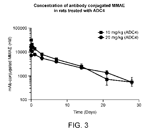
[0030] FIG. 3 provides the in vivo stability profile of a mAb-MMAE ADC. The
acid
release product MMAE was analyzed according to the described method and
plotted as
amount of conjugated drug over time.
[0031] FIG. 4A shows predicted molecular structures with sites selected for
conversion
to cysteine near the hinge region of the antibody CH2 domain. Sites were first
identified
on the Fc fragment proximal to the hinge between the Fc and the Fab (left
panel). These
sites coincide with the CD16 binding sites as shown in the co-crystal
structure 1E4K
(center panel). Relative orientations of the Fc, Fabs, and CD16 can be seen in
the
model generated from docking CD16 onto the intact antibody crystal structure
1HZH
(right panel). FIG. 4B shows solvent accessibility of conversion sites
calculated using
6
CA 03082549 2020-05-13
WO 2019/104084 PCT/US2018/062100
1HZH as a template FIG. 4C provides electrostatic potential calculated for the
modeled
in silico mutants projected on the molecular surface. These sites showed no
consistent
trend in either highly acidic or basic elements near the engineered site of
conjugation.
[0032] FIG. 5 illustrates drug conjugation sites confirmed by proteolysis and
mass
spectrometry. Wild-type (WT Fc), engineered cysteine antibodies (S239C) and
ADCs
(S239C + Drug) were digested with endoproteinase GluC (cleavage at position
E233
and C-terminal to the hinge disulfide bonds (FIG. 5, left)) followed by
subsequent
analysis of the Fc fragment using time-of-flight mass spectrometry. When a
wild-type
ADC is digested, the resulting Fc fragment has a mass of 24,054 Da (top panel)
showing no signs of conjugation, consistent with all of the conjugation sites
being on
the N-terminal side of position 233. Digestion of an S239C antibody results in
an Fc
fragment with an additional 16 Da in mass, 24,070 Da total, corresponding to
the
difference in mass between serine and cysteine (center panel). The digestion
of a S239C
pure 2-loaded ADC results in an Fc fragment with an additional 942 Da in mass,
24,995
Da total, corresponding to the differing masses of serine and cysteine and the
addition
of the drug linker (bottom panel).
[0033] FIG. 6 shows in vivo activity of naked antibody, native 4-loaded ADC
and
engineered cysteine antibodies (K326C, E269C, A327C, and S239C). Antibodies
were
tested for activity in a single 10 mg/kg dose 786-0 xenograft experiment. The
2-loaded
S239C engineered cysteine outperformed the native 4-loaded and all other
engineered
cysteine mutant ADCs.
[0034] FIGs. 7A-B provides data representing ADC maleimide stability in
plasma.
FIG. 7A provides a schematic where step 1 shows the reversible Michael
addition used
to conjugate antibody and drug linker. Step 2 illustrates a potential
hydrolysis reaction
that stabilizes the conjugate and prevents loss of the drug linker. FIG. 7B
shows time
course stability of drug-linker conjugate. The data shows loss of the
conjugated drug via
the retro-Michael reaction during incubation of the ADC with rat plasma. The 2-
loaded
5239C engineered cysteine is more stable than the native 4-loaded and all
other
engineered cysteine mutant ADCs. Terminal % drug load relative to t = 0 hr for
each
construct is shown in Table 2.
[0035] The figures depict various embodiments of the present invention for
purposes of
illustration only. One skilled in the art will readily recognize from the
following
discussion that alternative embodiments of the structures and methods
illustrated herein
7
CA 03082549 2020-05-13
WO 2019/104084 PCT/US2018/062100
may be employed without departing from the principles of the invention
described
herein.
DETAILED DESCRIPTION
Definitions
[0036] Unless defined otherwise, all technical and scientific terms used
herein have the
meaning commonly understood by a person skilled in the art to which this
invention
belongs. As used herein, the following terms have the meanings ascribed to
them
below.
[0037] A "ligand-drug conjugate" or "LDC" refers to a ligand (e.g. an
antibody)
conjugated to a pharmaceutical agent, e.g. to a cytotoxic or cytostatic drug.
"Ligands"
include, but are not limited to, polymers, dendrimers, oligonucleotides,
proteins,
polypeptides, peptides, including cyclic peptides and glycopeptides, or any
other cell
binding molecule or substance. More specifically, ligands include aptamers
(oligonucleotides or peptides), as well as various proteins, such as
interferons,
lymphokines, knottins, adnectins, anticalins, darpins, avimers, Kunitz
domains, and
centyrins. Additional ligands include hormones, growth factors, colony-
stimulating
factors, vitamins, and nutrient transport molecules. Suitable ligands include,
for
example, antibodies, e.g. full-length antibodies and antigen binding fragments
thereof
Antibodies also include bispecific antibodies and multi specific antibodies.
[0038] An "antibody-drug conjugate" or "ADC" refers to an antibody, antigen-
binding fragment, or engineered variant thereof conjugated to a pharmaceutical
agent.
Typically, antibody-drug conjugates bind to a target antigen (e.g., CD70) on a
cell
surface, followed by internalization of the antibody-drug conjugate into the
cell and
subsequent release of the drug into the cell. The antibody or antigen-binding
fragment
thereof may be covalently or non-covalently bound to the pharmaceutical agent.
In
specific embodiments, the drug in LDCs and particularly that in ADCs, is
conjugated to
the ligand, or more particularly the antibody, through a linker. The linker
typically
comprises residues resulting from conjugation to the drug and conjugation to
the ligand
separated by a chemical spacer. The chemical spacer may simply be a
hydrocarbon
chain, an alkenylene, (e.g., -(CH2)n-, where n is a selected integer, or n is
2-10), or a
heteroalkenylene chain containing one or more oxygens, carbonyls (C=0),
sulfurs, or
amino groups (e.g., NH or Nalkyl). The linker may be structurally more
complex, for
example, the linker may be substituted¨with a PEG (polyethylene glycol) group,
or
8
CA 03082549 2020-05-13
WO 2019/104084 PCT/US2018/062100
other hydrophilic group or may contain a cleavable group, e.g, a 13-
glucuronide that is
cleavable by 13-glucuronidase., such that cleaving the group, cleaves the
linker.
[0039] The linker is a chemical species linking the ligand to the drug.
Typically, the
LDC is formed by two conjugation steps. A precursor to the linker, which is a
heterobifunctional species, having two different reactive groups most often
separated by
a spacer and optionally substitued is most often reacted with the drug
molecule to form
a linker-drug combination which retains one of the reactive groups. A
heterobifunctional linker precursor contains the spacer between the two
reactive groups
with different reactivity. For example, a heterobifunctional linker precursor
may
contain an amine-reactive group at one end and a thiol reactive group at the
other end.
In another more specific example, a heterobifunctional linker precursor may
contain a
carbonate for reaction with an amine of the drug to form a carbamate. In other
more
specific examples, a heterobifunctional linker precursor may contain an azide
or a N-
hydroxysuccinimide ester (NHS ester or a sulfo-NHS ester) for reaction with an
amine
of the drug to form an amide. Each of such amine reactive groups can be paired
in a
linker precursor with a maleimide group, which under selected known
conditions, is
selective for reaction with thiols. After conjugation to the drug, one of the
reactive
groups remains in the linker-drug combination.
[0040] The linker-drug combination retaining the reactive group can then be
used as a
reagent for conjugation of the drug to the ligand. For example, a ligand
conjugation
reagent can contain a maleimide group for reaction with thiol groups on a
ligand. More
generally, the ligand conjugation reagent can contain any appropriate reactive
groups
for conjugation to groups on the ligand. The reactive groups may react, for
example,
with amine groups, with carboxylate groups, with thiol groups or with hydroxyl
groups.
[0041] An "analytic target" refers to a drug or a portion thereof that is
released or
cleaved from a ligand-drug conjugate, and which is detected or measured
(quantitated)
by one or more known analytic techniques, e.g. mass spectrometry. The analytic
target
contains at least the drug or a portion thereof and may in addition contain a
portion of
the linker. The amount of analytic target is representative of the amount of
the ligand-
drug conjugate from which it is released or cleaved. More specifically the
analytic
target is the drug of the LDC or a portion of the drug of the LDC. In specific
embodiments, where the drug is an auristatin, the analytic target can be a
tetrapeptide
released from the drug.
9
CA 03082549 2020-05-13
WO 2019/104084 PCT/US2018/062100
[0042] When an internal standard is used, an analytic target can be a drug or
a portion
thereof that is released or cleaved from the internal standard. In typical
embodiments,
an analytic target released from an internal standard can be differentiated
from an
analytic target released from a ligand-drug conjugate, for example, by having
a different
molecular weight and/or by being labeled.
[0043] The term "antibody" denotes immunoglobulin proteins produced by the
body in
response to the presence of an antigen and that bind to the antigen, as well
as antigen-
binding fragments and engineered variants thereof Hence, the term "antibody"
includes, for example, intact monoclonal antibodies (e.g., antibodies produced
using
hybridoma technology) and antigen-binding antibody fragments, such as a
F(ab')2, a Fv
fragment, a diabody, a single-chain antibody, an scFv fragment, or an scFv-Fc.
Genetically, engineered intact antibodies and fragments such as chimeric
antibodies,
humanized antibodies, single-chain Fv fragments, single-chain antibodies,
diabodies,
minibodies, linear antibodies, multivalent or multi-specific (e.g.,
bispecific) hybrid
antibodies, and the like, are also included. Thus, the term "antibody" is used
expansively to include any protein that comprises an antigen-binding site of
an antibody
and is capable of specifically binding to its antigen.
[0044] The terms "extract", "extracted", "extraction", and "extracting" refer
to
isolation of an LDC or ADC from a heterogeneous sample comprising several
proteins
and other molecules. Any appropriate method or material known in the art that
can
selectively extract an LDC or ADC from a heterogeneous sample, particularly a
biological sample, can be employed in the methods herein. Extraction, for
example, can
include: affinity chromatography, size exclusion chromatography, ammonium
sulfate
precipitation, ion exchange chromatography, immobilized metal chelate
chromatography, and immunoprecipitation.
[0045] Binding of LDC or ADC to a resin which contains a species to which the
ligand
or antibody binds can be used for extraction. Antibody binding proteins can be
used for
extraction of ADCs. For example, extraction of an ADC from a sample may
involve
running the sample over a protein A column or contacting the sample with a
protein A
resin and thereafter removing the resin from the sample in order to capture
the antibody,
thereby extracting the ADC from the sample. With respect to ADC's, surface
proteins
protein A, protein G or protein L may be used for extraction. The structural
requirements for binding of a given antibody to protein A, protein G or
protein L are
CA 03082549 2020-05-13
WO 2019/104084 PCT/US2018/062100
known in the art and one of ordinary skill in the art can select from among
them, the
appropriate surface protein for use with a given antibody. Materials useful in
extractions using these proteins include resins, e.g., beaded agarose, or
magnetic beads,
or similar support material to which the protein A, protein G or protein L is
covalently
immobilized.
[0046] The terms "intracellularly cleaved" and "intracellular cleavage" refer
to a
metabolic process or reaction inside a cell on a ligand-drug conjugate (e.g.,
an antibody-
drug conjugate), whereby the covalent attachment, e. g, the linker between the
drug
moiety and the ligand unit is broken, resulting in free drug, or other
metabolite of the
conjugate dissociated from the antibody inside the cell. The cleaved moieties
of the
drug-linker-ligand conjugate are thus intracellular metabolites.
[0047] The terms "release", "released", and "releasing" refer to extracellular
cleavage
of an analytic target from an LDC by the acid-mediated cleavage method
described
therein. For a given LDC carrying (i.e., conjugated with) a given number of
linker-drug
combinations, the amount of analytic target released will typically vary with
acid
concentration (see below) used in the release reaction, the temperature and
pressure of
the reaction (see below) and the reaction time employed. For consistency of
results
from sample to sample, the same acid concentration and reaction conditions
should be
employed. Treatment with acid as described herein need not release all
analytic target
from the LDC. All that is needed is to release an amount of analytic target
that is
sufficient for obtaining an accurate and precise measurement of the analytic
target in
view of the analytic method employed.
[0048] The terms "contact", "contacted", and "contacting" refer to adding acid
or
reagent to a sample, which may be a test sample or a control sample( including
biological samples), so that the components of the sample are made available
to the acid
or reagent, and a reaction can thus occur. The reaction associated with acid
addition in
the method herein is release of an analytic target from an LDC or more
specifically an
ADC.
[0049] A "cytotoxic effect" refers to the depletion, elimination and/or
killing of a target
cell. A "cytotoxic agent" refers to a compound that has a cytotoxic effect on
a cell,
thereby mediating depletion, elimination and/or killing of a target cell. The
term
includes radioactive isotopes (e.g., 211At, 1311, 1251, 90y, 186Re, 188Re,
153sm, 212Bi, 32p,
60C, and radioactive isotopes of Lu), chemotherapeutic agents, and toxins such
as small
11
CA 03082549 2020-05-13
WO 2019/104084 PCT/US2018/062100
molecule toxins or enzymatically active toxins of bacterial, fungal, plant or
animal
origin, including synthetic analogs and derivatives thereof. In certain
embodiments, a
cytotoxic agent is conjugated to an antibody or administered in combination
with an
antibody. Suitable cytotoxic agents are described further herein.
[0050] "Cytotoxic activity" refers to a cell-killing, a cytostatic or an anti-
proliferative
effect of a ligand-drug conjugate compound or an intracellular metabolite of a
ligand-
drug conjugate. Cytotoxic activity may be expressed as the ICso value, which
is the
concentration (molar or mass) per unit volume at which half the cells survive.
[0051] The term "patient" or "subject" includes human and other mammalian
subjects
such as non-human primates, rabbits, rats, mice, and the like and transgenic
species
thereof, that receive either prophylactic or therapeutic treatment.
[0052] The term "standard curve" or "calibration curve" refers to a graph used
as a
quantitative research technique. To generate the standard curve, multiple
samples with
known properties are measured and graphed, which then allows the same
properties to
be determined for unknown samples by interpolation on the graph. The samples
with
known properties are the standards, and the graph is the standard curve.
Standard
curves are of particular use when measuring the amount or concentration of an
analyte
in a sample that may contain an unknown amount of the analyte. The use of a
standard
curve alone represents the use of an external standard. As is understood in
the art, the
standard curve of a given analyte (i.e., the LDC) to be quantitated should
generally span
the concentration range of the analyte expected in the samples. Again as is
understood
in the art, samples used for preparing the standard curve are processed by the
same
steps as test samples and any control samples in which the analyte is to be
measured. A
standard curve can also be employed in combination with the use of an internal
standard. In this case, a constant (or fixed) amount of the internal standard
is added to
each sample used to generate the standard curve of known analyte
concentrations. The
same constant amount of internal standard is added to each test sample and to
any
blanks or control samples. The details of use of standard curves (calibration
curves) as
an external standard and a combination of the use of a standard curve with
addition of
internal standard for quantitation of analytes by analytic methods, including
MS, LC-
MS and LC-MS/MS methods, is well known in the art. One of ordinary skill in
the art
understands how to use such analytic methods in the determination of
concentrations of
analytes in a variety of samples, including biological samples as discussed
herein.
12
CA 03082549 2020-05-13
WO 2019/104084 PCT/US2018/062100
[0053] An "internal standard" is a chemical species that behaves in a selected
assay
similarly to the chemical species to be quantitated (i.e., LDC), but which is
distinguishable from that chemical species in the analytic method being used.
Typically,
the internal standard is labeled to distinguish it from the chemical species
to be
quantitated, but the label employed does not significantly differentially
affect its
behavior compared to that of the chemical species to be quantitated.
Preferably,
anything that affects the measurement of the chemical species to be
quantitated (e.g.,
analyte peak area) will also affect the measurement of the internal standard
similarly.
The ratio of the measurements of the chemical species to be quantitated and
its internal
standard preferably exhibits less variability than the measurement of the
chemical
species in a test sample. For use in mass spectrometry methods, the internal
standard
has a molecular weight that is different from the chemical species to be
quantitated.
[0054] Most often labeling with stable isotopes, such as deuterium (2H) and
carbon 13
('3C) is employed. Labeling must allow separate measurement of analyte and
internal
standard. Preferably, an isotopically labeled internal standard differs in
molecular
weight from the chemical species to be quantitated by at least 3 amu (i.e.,
labeling with
3 or more 2H or nC). More specifically, labeling results in a difference in
molecular
weight of 6 amu or more. Internal standards can also be surrogates of the
chemical
species to be quantitated. Surrogate internal standards differ structurally
from the
chemical species to be quantitated by substitution of an atom or chemical
group by a
different group, for example the substitution of a methyl group or other small
alkyl for a
hydrogen, or the substitution of a halogen, e.g., a fluorine, for a hydrogen.
Such
surrogates may be of particular use where it is not possible to readily obtain
an
isotopically labeled internal standard.
[0055] The terms "determine", "determined", and "determining" refer to the
ascertaining of the concentration or amount of a particular analyte based on a
measurement of the amount of an analytic target and the known amounts of one
or more
correlative factors. As is understood in the art, an analyte concentration can
be
combined with the results of other measurements to determine other structural
and
physical properties of an analyte.
[0056] When trade names are used herein, the trade name includes the product
formulation, the generic drug, and the active pharmaceutical ingredient(s) of
the trade
name product, unless otherwise indicated by context.
13
CA 03082549 2020-05-13
WO 2019/104084 PCT/US2018/062100
Other interpretational conventions
[0057] Ranges recited herein are understood to be shorthand for all of the
values within
the range, inclusive of the recited endpoints. For example, a range of 1 to 50
is
understood to include any number, combination of numbers, or sub-range from
the
group consisting of 1, 2, 3, 4, 5, 6, 7, 8, 9, 10, 11, 12, 13, 14, 15, 16, 17,
18, 19, 20, 21,
22, 23, 24, 25, 26, 27, 28, 29, 30, 31, 32, 33, 34, 35, 36, 37, 38, 39, 40,
41, 42, 43, 44,
45, 46, 47, 48, 49, and 50.
[0058] Unless otherwise indicated, reference to a compound that has one or
more
stereocenters intends each stereoisomer, and all combinations of
stereoisomers, thereof.
Assay for analyzing a ligand-drug conjugate (LDC)
[0059] In one aspect, the present invention provides a method of analyzing a
ligand-
drug conjugate (LDC) in a sample, comprising the step of: (a) providing the
sample
comprising the LDC, wherein the LDC comprises a ligand and an analytic target,
wherein the analytic target comprises a drug molecule or a portion thereof;
(b)
contacting the sample with aqueous trifluoroacetic acid (TFA) at a
concentration
between 1 to 30% (v/v), thereby inducing release of the analytic target from
the LDC.
In some embodiments, the method can comprise the steps of (a) providing a
sample
comprising the LDC, wherein the LDC comprises an analytic target, the analytic
target
comprising a drug molecule; (b) adding to the sample an internal standard,
wherein the
internal standard is a labeled derivative of the LDC and comprises a second
analytic
target; (c) extracting the LDC and the internal standard from the sample; (d)
contacting
the LDC and the internal standard with aqueous TFA at a concentration between
1 to
30% (v/v), wherein the TFA releases the analytic target from the LDC and the
second
analytic target from the internal standard; (e) determining the amount of the
analytic
target released from the LDC and the second analytic target released from the
internal
standard, wherein the amount of the analytic target released from the LDC
correlates
with the amount of LDC in the sample.
A sample comprising ligand-drug conjugate (LDC)
[0060] The present invention provides a method of analyzing a ligand-drug
conjugate
(LDC) in a sample. An LDC is a complex comprising a ligand and an analytic
target.
The analytic target comprises a drug molecule or a portion thereof Various
samples
comprising an LDC or suspected to comprise an LDC can be subject to analysis
using a
method provided herein. In particular biological sample can be analyzed.
14
CA 03082549 2020-05-13
WO 2019/104084 PCT/US2018/062100
Sample
[0061] An LDC in a biological or non-biological sample can be analyzed by the
methods provided herein. In preferred embodiments, the sample is a biological
sample
derived from a mammalian subject. Specifically, in some embodiments, the
biological
sample is obtained from one of the following: plasma, serum, blood, tissue,
tissue
biopsy, feces, and urine.
[0062] In some embodiments, the sample is a biological sample contacted with
an LDC
in vivo. For example, the sample can be a biological sample derived from a
subject
exposed to an LDC. In some embodiments, the sample is obtained at a specific
time
point after administration of an LDC. In some embodiments, the sample is
obtained at
multiple time points after administration of an LDC. In some embodiments, the
sample
is obtained before administration of an LDC.
[0063] In some embodiments, the sample is a biological sample contacted with
an LDC
ex vitro. In some embodiments, the sample is contacted with an LDC for a
specific time
period. In some embodiments, a plurality of samples contacted with LDC for
different
periods are subject to analysis. In some embodiments, the sample is obtained
before
exposure to an LDC.
Ligand-drug conjugate (LDC)
Ligand
[0064] In some embodiments, the ligand is a protein having specific affinity
to a target
molecule. In some embodiments, the ligand is an antibody. Useful polyclonal
antibodies are heterogeneous populations of antibody molecules derived from
the sera
of immunized animals. Useful monoclonal antibodies are homogeneous populations
of
antibodies to a particular antigenic determinant (e.g., a cancer cell antigen,
a viral
antigen, a microbial antigen, a protein, a peptide, a carbohydrate, a
chemical, nucleic
acid, or fragments thereof). A monoclonal antibody (mAb) to an antigen-of-
interest can
be prepared by using any technique known in the art which provides for the
production
of antibody molecules by continuous cell lines in culture.
[0065] Useful monoclonal antibodies include, but are not limited to, human
monoclonal
antibodies, humanized monoclonal antibodies, or chimeric human-mouse (or other
species) monoclonal antibodies. The antibodies include full-length antibodies
and
antigen binding fragments thereof. Human monoclonal antibodies may be made by
any
of numerous techniques known in the art (e.g., Teng et al., 1983, Proc. Natl.
Acad. Sci.
CA 03082549 2020-05-13
WO 2019/104084 PCT/US2018/062100
USA. 80:7308-7312; Kozbor etal., 1983, Immunology Today 4:72-79; and Olsson et
al., 1982, Meth. Enzymol. 92:3-16).
[0066] The antibody can be a functionally active fragment, derivative or
analog of an
antibody that immunospecifically binds to target cells (e.g., cancer cell
antigens, viral
antigens, or microbial antigens) or other antibodies bound to tumor cells or
matrix. In
this regard, "functionally active" means that the fragment, derivative or
analog is able to
elicit anti-idiotype antibodies that recognize the same antigen as the
antibody from
which the fragment, derivative or analog is derived. Specifically, in an
exemplary
embodiment the antigenicity of the idiotype of the immunoglobulin molecule can
be
enhanced by deletion of framework and CDR sequences that are C-terminal to the
CDR
sequence that specifically recognizes the antigen. To determine which CDR
sequences
bind the antigen, synthetic peptides containing the CDR sequences can be used
in
binding assays with the antigen by any binding assay method known in the art
(e.g., the
BIA core assay) (See, e.g., Kabat et al., 1991, Sequences of Proteins of
Immunological
Interest, Fifth Edition, National Institute of Health, Bethesda, Md.; Kabat E
et al., 1980,
J. Immunology 125(3):961-969).
[0067] Other useful antibodies include fragments of antibodies such as, but
not limited
to, F(ab')2 fragments, Fab fragments, Fvs, single chain antibodies, diabodies,
tribodies,
tetrabodies, scFv, scFv-Fv, or any other molecule with the same specificity as
the
antibody.
[0068] Additionally, recombinant antibodies, such as chimeric and humanized
monoclonal antibodies, comprising both human and non-human portions, which can
be
made using standard recombinant DNA techniques, are useful antibodies. A
chimeric
antibody is a molecule in which different portions are derived from different
animal
species, such as for example, those having a variable region derived from
murine
monoclonal and human immunoglobulin constant regions. (See, e.g., U.S. Pat.
No.
4,816,567; and U.S. Pat. No. 4,816,397, each of which is incorporated herein
by
reference in its entirety.) Humanized antibodies are antibody molecules from
non-
human species having one or more complementarity determining regions (CDRs)
from
the non-human species and a framework region from a human immunoglobulin
molecule. (See, e.g., U.S. Pat. No. 5,585,089, which is incorporated herein by
reference
in its entirety.) Such chimeric and humanized monoclonal antibodies can be
produced
by recombinant DNA techniques known in the art, for example using methods
16
CA 03082549 2020-05-13
WO 2019/104084 PCT/US2018/062100
described in International Publication No. WO 87/02671; European Patent
Publication
No. 0 184 187; European Patent Publication No. 0 171 496; European Patent
Publication No. 0 173 494; International Publication No. WO 86/01533; U.S.
Pat. No.
4,816,567; European Patent Publication No. 012 023; Berter et al., 1988,
Science
240:1041-1043; Liu et al., 1987, Proc. Natl. Acad. Sci. USA 84:3439-3443; Liu
et al.,
1987, J. Immunol. 139:3521-3526; Sun et al., 1987, Proc. Natl. Acad. Sci. USA
84:214-
218; Nishimura et al., 1987, Cancer. Res. 47:999-1005; Wood et al., 1985,
Nature
314:446-449; and Shaw et al., 1988, J. Natl. Cancer Inst. 80:1553-1559;
Morrison,
1985, Science 229:1202-1207; Oi et al., 1986, BioTechniques 4:214; U.S. Pat.
No.
5,225,539; Jones et al., 1986, Nature 321:552-525; Verhoeyan et al., 1988,
Science
239:1534; and Beidler et al., 1988, J. Immunol. 141:4053-4060; each of which
is
incorporated herein by reference in its entirety.
[0069] Completely human antibodies are particularly desirable and can be
produced
using transgenic mice that are incapable of expressing endogenous
immunoglobulin
heavy and light chains genes, but which can express human heavy and light
chain
genes.
[0070] Antibodies include analogs and derivatives that are either modified,
i.e., by the
covalent attachment of any type of molecule as long as such covalent
attachment
permits the antibody to retain its antigen binding immunospecificity. For
example, but
not by way of limitation, derivatives and analogs of the antibodies include
those that
have been further modified, e.g., by glycosylation, acetylation, pegylation,
phosphorylation, amidation, derivatization by known protecting/blocking
groups,
proteolytic cleavage, linkage to a cellular antibody unit or other protein,
etc. Any of
numerous chemical modifications can be carried out by known techniques
including,
but not limited to, specific chemical cleavage, acetylation, formylation,
metabolic
synthesis in the presence of tunicamycin, etc. Additionally, the analog or
derivative can
contain one or more unnatural amino acids.
[0071] Antibodies can have modifications (e.g., substitutions, deletions or
additions) in
amino acid residues that interact with Fc receptors. In particular, antibodies
can have
modifications in amino acid residues identified as involved in the interaction
between
the anti-Fc domain and the FcRn receptor (see, e.g., International Publication
No. WO
97/34631, which is incorporated herein by reference in its entirety).
17
CA 03082549 2020-05-13
WO 2019/104084 PCT/US2018/062100
[0072] Antibodies immunospecific for a cancer cell antigen can be obtained
commercially or produced by any method known to one of skill in the art such
as, e.g.,
chemical synthesis or recombinant expression techniques. The nucleotide
sequences
encoding antibodies immunospecific for a cancer cell antigen can be obtained,
e.g.,
from the GenBank database or a database like it, the literature publications,
or by
routine cloning and sequencing.
[0073] In certain embodiments, useful antibodies can bind to a receptor or a
receptor
complex expressed on an activated lymphocyte. The receptor or receptor complex
can
comprise an immunoglobulin gene superfamily member, a TNF receptor superfamily
member, an integrin, a cytokine receptor, a chemokine receptor, a major
histocompatibility protein, a lectin, or a complement control protein. Non-
limiting
examples of suitable immunoglobulin superfamily members are CD2, CD3, CD4,
CD8,
CD19, CD20, CD22, CD28, CD30, CD70, CD79, CD90, CD152/CTLA-4, PD-1, and
ICOS. Non-limiting examples of suitable TNF receptor superfamily members are
CD27, CD40, CD95/Fas, CD134/0X40, CD137/4-1BB, TNF-R1, TNFR-2, RANK,
TACT, BCMA, osteoprotegerin, Apo2/TRAIL-R1, TRAIL-R2, TRAIL-R3, TRAIL-R4,
and APO-3. Non-limiting examples of suitable integrins are CD11 a, CD11b, CD11
c,
CD18, CD29, CD41, CD49a, CD49b, CD49c, CD49d, CD49e, CD49f, CD103, and
CD104. Non-limiting examples of suitable lectins are C-type, S-type, and I-
type lectin.
[0074] In some embodiments, the ligand is a receptor ligand. The receptor
ligand can
have a binding partner that is enriched in a specific cell type, tissue or
organ. The ligand
can be a naturally occurring agonist or antagonist of a receptor, or a
synthetic molecule
that has an affinity to the receptor. The receptor ligand can be a protein,
nucleic acid or
other receptor ligand such as a peptide, vitamin, and carbohydrate. In one
embodiment,
the ligand is folate that has affinity to a folate receptor.
[0075] In some embodiments, the ligand is a targeting moiety that has been
used and
developed for targeting a drug to a target organ or tissue. Such site-specific
ligands
known in the art can be used and adopted in the method provided herein.
Drug
[0076] The drug of the LDC can be any cytotoxic, cytostatic or
immunosuppressive
drug also referred to herein as a cytotoxic, cytostatic or immunosuppressive
agent. The
drug has a functional group, such as an amino, alkyl amino group or
carboxylate that
can form a bond with an appropriate reactive group of a reagent precursor
containing
18
CA 03082549 2020-05-13
WO 2019/104084 PCT/US2018/062100
the linker, such as an amine group, a carboxylic acid group, a sulfhydryl
group, a
hydroxyl group or an aldehyde or ketone group. In an embodiment, the drug is
conjugated to a linker to generate an amide or a carbamate. In an embodiment,
the drug
is conjugated to a linker by an amide bond. In an embodiment, the drug
contains a
single amide bond. In an embodiment, the drug is conjugated to the linker by a
carbamate and the drug contains an amide bond. In specific embodiments, TFA
treatment, releases the drug or a portion thereof by cleavage of the amide
bond to the
linker or an internal amide bond in the drug.
[0077] Useful classes of cytotoxic or immunosuppressive agents include, for
example,
antitubulin agents, auristatins, DNA minor groove binders, DNA replication
inhibitors,
alkylating agents (e.g., platinum complexes such as cis-platin,
mono(platinum),
bis(platinum) and tri-nuclear platinum complexes and carboplatin),
anthracyclines,
antibiotics, antifolates, antimetabolites, chemotherapy sensitizers,
duocarmycins,
etoposides, fluorinated pyrimidines, ionophores, lexitropsins, nitrosoureas,
platinols,
pre-forming compounds, purine antimetabolites, puromycins, radiation
sensitizers,
steroids, taxanes, topoisomerase inhibitors, vinca alkaloids, or the like.
Particularly
useful classes of cytotoxic agents include, for example, DNA minor groove
binders,
DNA alkylating agents, and tubulin inhibitors. Exemplary cytotoxic agents
include, for
example, auristatins, camptothecins, duocarmycins, etoposides, maytansines and
maytansinoids (e.g., DM1 and DM4), taxanes, benzodiazepines (e.g.,
pyrrolo[1,4]benzodiazepines (PBDs), indolinobenzodiazepines, and
oxazolidinobenzodiazepines) and vinca alkaloids. Select benzodiazepine
containing
drugs are described in WO 2010/091150, WO 2012/112708, WO 2007/085930, and
WO 2011/023883.
[0078] In an exemplary embodiment, the drug is a peptidic drug containing one
or
more, two or more, three or more or four or more amino acid groups. In an
exemplary
embodiment, the drug is a peptidic drug containing an N-terminal, N-methylated
amino
acid group. In a further exemplary embodiment, the drug is a peptidic drug
having an
N-terminal, N-methylated amino acid with an alkyl side group. In a further
exemplary
embodiment, the drug is a peptidic drug having an N-terminal, N-methylated
alanaine,
N-methylated isoleucine, N-methylated leucine or N-methylated valine. In a
further
exemplary embodiment, the drug is a peptidic drug having an N-terminal, N-
methylated
valine.
19
CA 03082549 2020-05-13
WO 2019/104084 PCT/US2018/062100
[0079] In a preferred embodiment, the drug is an auristatin. Auristatins
include, but are
not limited to, AE, AFP, AEB, AEVB, MMAF, and MNIAE. The synthesis and
structure of auristatins are described in U.S. Patent Application Publication
Nos. 2003-
0083263, 2005-0238649 2005-0009751, 2009-0111756, and 2011-0020343;
International Patent Publication No. WO 04/010957, International Patent
Publication
No. WO 02/088172, and U.S. Pat. Nos. 7,659,241 and 8,343,928; each of which is
incorporated by reference herein in its entirety and for all purposes.
Exemplary
auristatins of the present invention bind tubulin and exert a cytotoxic or
cytostatic effect
on the desired cell line. In an embodiment, exemplary auristatins contain an N-
terminal,
N-methylated amino acid. More specifically, exemplary auristatins contain an N-
terminal N, N-methylated amino acid with an alkyl side chain, such as alanine,
isoleucine, leucine, or valine. Yet more specifically, exemplary auristatins
contain an
N-terminal, N-methylated valine.
[0080] Other individual cytotoxic or immunosuppressive agents include, for
example,
an androgen, anthramycin (AMC), asparaginase, 5-azacytidine, azathioprine,
bleomycin, busulfan, buthionine sulfoximine, calicheamicin, camptothecin,
carboplatin,
carmustine (BSNU), CC-1065, chlorambucil, cisplatin, colchicine,
cyclophosphamide,
cytarabine, cytidine arabinoside, cytochalasin B, dacarbazine, dactinomycin
(formerly
actinomycin), daunorubicin, decarbazine, docetaxel, doxorubicin, etoposide, an
estrogen, 5-fluordeoxyuridine, 5-fluorouracil, gemcitabine, gramicidin D,
hydroxyurea,
idarubicin, ifosfamide, irinotecan, lomustine (CCNU), maytansine,
mechlorethamine,
melphalan, 6-mercaptopurine, methotrexate, mithramycin, mitomycin C,
mitoxantrone,
nitroimidazole, paclitaxel, palytoxin, plicamycin, procarbizine, rhizoxin,
streptozotocin,
tenoposide, 6-thioguanine, thioTEPA, topotecan, vinblastine, vincristine,
vinorelbine,
VP-16 and VM-26.
[0081] Suitable cytotoxic agents also include DNA minor groove binders (e.g.,
enediynes and lexitropsins, a CBI compound; see also U.S. Pat. No. 6,130,237),
duocarmycins (see U.S. Publication No. 20060024317), taxanes (e.g., paclitaxel
and
docetaxel), puromycins, vinca alkaloids, CC-1065, SN-38, topotecan, morpholino-
doxorubicin, rhizoxin, cyanomorpholino-doxorubicin, echinomycin,
combretastatin,
netropsin, epothilone A and B, estramustine, cryptophysins, cemadotin,
maytansinoids,
discodermolide, eleutherobin, and mitoxantrone.
CA 03082549 2020-05-13
WO 2019/104084 PCT/US2018/062100
[0082] Examples of anti-tubulin agents include, but are not limited to,
taxanes (e.g.,
Taxol® (paclitaxel), Taxotere® (docetaxel)), T67 (Tularik) and vinca
alkyloids (e.g., vincristine, vinblastine, vindesine, and vinorelbine). Other
antitubulin
agents include, for example, baccatin derivatives, taxane analogs (e.g.,
epothilone A and
B), nocodazole, colchicine and colcimid, estramustine, cryptophysins,
cemadotin,
maytansinoids, combretastatins, discodermolide, and eleutherobin. Maytansine
and
maytansinoid are another group of anti-tubulin agents. (ImmunoGen, Inc.; see
also
Chari et al., 1992, Cancer Res. 52:127-131 and U.S. Pat. No. 8,163,888).
[0083] Exemplary auristatin drugs have the following formula or a
pharmaceutically
acceptable salt thereof wherein the wavy line indicates site of attachment to
the linker:
H3c cH3
CH3 H3C
LN
Ny=VN
NCOOH
CH3 0 CH3 OCH3 0 OCH3
H3C CH3
(monomethyl auristatin F) and
H3CCH3 H3C4i OH
0 CH3 H3C
N
CH3
CH3 0 CH3 OCH3 0 OCH3 0
H3C CH3
(monomethyl auristatin E).
[0084] Alternative auristatin drugs for conjugation to a ligand through a
linker have the
following formula or a pharmaceutically acceptable salt thereof, where the
wavy line
indicates the site of attachment to the linker:
21
CA 03082549 2020-05-13
WO 2019/104084 PCT/US2018/062100
H30 0H3
0 0H3 H30 0
N
CH3 0 CH3 OCH3 0 OCH3 0
H3C CH3
or
H3c CH3 H3C4N44.
0 CH3 H3C 0
H
N
HN
CH3 0 CH3 OCH3 0 OCH3
H3C CH3
[0085] Additional cytotoxic compounds useful for the preparation of LDCs and
particularly useful for the preparation of ADCs are those described in U.S.
patent
6,884,869, which is incorporated by reference herein in its entirety,
particularly for
descriptions of cytotoxic compounds. Additional description therein describes
preparation of drug conjugates with the cyctotoxic compounds described.
Linker
[0086] General procedures for linking a drug to linkers are known in the art.
See, for
example, U.S. Pat. Nos. 8,163,888, 7,659,241, 7,498,298, U.S. Publication No.
U520110256157 and International Application Nos. W02011023883, and
W02005112919.
[0087] The linker can be cleavable under intracellular conditions, such that
cleavage of
the linker releases the therapeutic agent from the ligand in the intracellular
environment
(e.g., within a lysosome or endosome or caveolea). The linker can be, e.g., a
peptidyl
linker that is cleaved by an intracellular peptidase or protease enzyme,
including a
lysosomal or endosomal protease. Intracellular cleaving agents can include
cathepsins B
and D and plasmin (see, e.g., Dubowchik and Walker, Pharm. Therapeutics 83:67-
123,
1999). For example, a peptidyl linker that is cleavable by the thiol-dependent
protease
cathepsin-B, which is highly expressed in cancerous tissue, can be used (e.g.,
a linker
comprising a Phe-Leu or a Val-Cit peptide). The linker can also be a
carbohydrate
22
CA 03082549 2020-05-13
WO 2019/104084 PCT/US2018/062100
linker, including a sugar linker that is cleaved by an intracellular
glycosidase (e.g., a
glucuronide linker cleavable by a glucuronidase).
[0088] The linker also can be a non-cleavable linker, such as a maleimido-
alkylene- or
maleimide-aryl linker that is attached to the ligand via a sulfur (thiol) and
released by
proteolytic degradation of the antibody.
[0089] An antibody can be conjugated to one or more linker via any appropriate
reactive group, e.g., via an amine group (for example, an N-terminal amino
group or an
amine group of an amino acid side group, such as lysine), a thiol group (-SH,
for
example, that of a cysteine residue), a carboxylate (for example, a C-terminal
carboxylate, or that of an amino acid side chain, such as glutamic acid) or a
hydroxyl
group (for example of a serine residue), of the antibody.
[0090] In exemplary ADCs, monomethyl auristatin E is conjugated through a
protease
cleavable peptide linker to an antibody, monomethyl auristatin F is conjugated
to an
antibody through the linker maleimidocaproic acid (mc). The linker may, in
addition,
contain chemical groups that modulate solubility or pharmacokinetics. For
example, an
exemplary linker is pegylated. Specific exemplary linker-drug combinations
are:
0 H3c cH3
0 0 cH3 H3C
NM7
N COOH
0 CH3 0 CH3 OCH3 0 OCH3 0
*
H3C CH3
*
*
mc-MMAF, wherein the maleimide group of the linker can react with thiol groups
of a
ligand and particularly of an antibody; or
23
CA 03082549 2020-05-13
WO 2019/104084 PCT/US2018/062100
0 H3c.õ.........õõcH3 0 OH
CO3H CH3 N3C
H H
0 0
OCH3 0 CH3
HOO H3 0 H3c....cH3 H3 OCH3 0
OH
0NI-I
0 0
0
0
1,1H2
DPR-PEG-gluc-carbamate-MMAE, wherein the linker is pegylated and contains a
glucuronic acid (cleavable by glucoruonidase) and wherein the maleimide group
of the
linker can react with thiol groups of a ligand. In LDCs containing the above
linker-drug
combinations, treatment with acid as described herein releases the tetra
peptide Val-Dil-
Dap-Phe (where Dap is dolaproline) from mc-MMAF, and the entire drug MMAE from
DPR-PEG-gluc-carbamate-MNIAE. Internal standards for LDCs and ADCs can be
prepared by labeling of such linker-drug combinations, wherein the label is
released on
treatment with acid as described herein. Exemplary internal standards for LDC
and
ADC conjugated to mc-MNIAF, include those that are deuterated or labeled with
'3C in
the tetrapeptide released. Exemplary internal standards for LDC and ADC
conjugated
to mc-MMAF, include those that are deuterated or labeled with '3C in the
1\4:MAE
released. In the above structures, sites for possible '3C labeling or
deuterium labeling
are shown by "*."
[0091] Quantitation methods herein generally employ the release of a fragment
of a
LDC, designated as an analytic target herein, which represents the entire LDC,
and
which analytic target is quantitated. Quantitation of the analytic target
allows one to
measure the amount of analytic target released, the amount of analytic target
in the LDC
in a sample and/or the amount of LDC in a sample. In some determinations, it
is
necessary to know or to determine, by appropriate known methods, the amount of
ligand in a sample or to know or to determine, by appropriate methods, the
number (or
average number) of drug molecules conjugated to a given LDC. More
specifically, the
analytic target herein is the drug molecule of the LDC or a portion of the
drug molecule
of the LDC. Drugs are conjugated to the ligand in an LDC by a linker species,
so an
24
CA 03082549 2020-05-13
WO 2019/104084 PCT/US2018/062100
analytic target may also include a portion of or the entire linker in addition
to the drug
or portion thereof In specific embodiments, herein the analytic target is the
drug
conjugated to the LDC. In specific embodiments, herein the analytic target is
a portion
of the drug conjugated to the LDC. In specific embodiments herein, the drug is
a
peptide or derivative thereof and the analytic target is the peptide drug or a
peptide
portion of the peptide drug. In specific embodiments, where the drug is a
peptide or
derivative thereof, the analytic target is a dipeptide or derivative thereof,
a tripeptide or
derivative thereof, or a tetrapeptide or derivative thereof.
Cleavage mediated by trifluoroacetic acid (TFA)
[0092] The method of the present invention comprises the step of contacting a
sample
with aqueous trifluoroacetic acid (TFA) at a concentration between 1 to 30%
(v/v), to
induce release of an analytic target from LDC. Solutions of TFA in
acetonitrile can also
be employed.
[0093] The TFA concentration employed can be 1-20%, 1-10%, 2.5-30%, 2.5-20%,
2.5-10%, 5-15%, 7-13%, 9-11%, or 9.5 to 10.5%, v/v with all ranges inclusive.
The
TFA concentration is about 1%, about 2%, about 3%, about 4%, about 5%, about
6%,
about 7%, about 8%, about 9%, about 10%, about 11%, about 12%, about 13%,
about
14%, about 15%, about 16%, about 17%, about 18%, about 19%, about 20%, about
21%, about 22%, about 23%, about 24%, about 25%, about 26%, about 27%, about
28%, about 29%, or about 30%, all %v/v. In a preferred embodiment, the TFA is
10%
(v/v).
[0094] The TFA concentration may be a result of dilution of 100% TFA in water,
sample mixture, or any other acceptable solvent. The TFA may be diluted before
it is
added to the sample, or diluted in the sample mixture itself.
[0095] The TFA reaction may be performed under variable time and temperature
conditions. For example, the reaction may be performed at between 20 and 80 C,
such
as about 20 C, about 21 C, about 22 C, about 23 C, about 24 C, about 25 C,
about
26 C, about 27 C, about 28 C, about 29 C, about 30 C, about 31 C, about 32 C,
about
33 C, about 34 C, about 35 C, about36 C, about 37 C, about 38 C, about 39 C,
about
40 C, about 41 C, about 42 C, about 43 C, about 44 C, about 45 C, about 46 C,
about
47 C, about 48 C, about 49 C, about 50 C, about 51 C, about 52 C, about 53 C,
about
54 C, about 55 C, about 56 C, about 57 C, about 58 C, about 59 C, about 60 C,
about
61 C, about 62 C, about 63 C, about 64 C, about 65 C, about 66 C, about 67 C,
about
CA 03082549 2020-05-13
WO 2019/104084 PCT/US2018/062100
68 C, about 69 C, about 70 C, about 71 C, about 72 C, about 73 C, about 74 C,
about
75 C, about 76 C, about 77 C, about 78 C, about 79 C, or about 80 C.
[0096] The TFA reaction is typically performed at ambient pressure. It will be
apparent
to one of ordinary skill in the art that the pressure of a reaction may be
varied in such a
reaction without significant detriment. It will be appreciated that a change
in pressure
may require a change in temperature. A reaction conducted at a higher pressure
may
permit a lower reaction temperature to be used. It will be appreciated that
concentration
of acid, time of reaction and the reaction temperature may be varied within
ranges
described herein along with the pressure of the reaction to achieve a desired
level of
release of analytic target.
[0097] The reaction may be performed for a period of about 12-24 hours, 10-20
hours,
or 15-17 hours. However, any combination of acid concentration, temperature,
time,
and pressure that allows the selected analytical method to give a measurement
of the
desired accuracy and precision may be used. As noted elsewhere, for
consistency of
results in a given experiment or quantitation, the reaction conditions used
should be the
same for all test samples (unknowns), all controls and all calibration samples
for a given
experiment or quantitation. In an exemplary embodiment the cleavage reaction
is
performed using TFA 10%, at 70 C, and at ambient pressure for a period of
about 16
hours.
[0098] Other acids may be used in the disclosed methods, such as, but not
limited to
other fluorinated acids, organic or mineral acids. Specific alternative acids
include
trifluoromethane sulfonic acid. Acids that are volatile are generally
preferred over
mineral acids, such as HC1.
Measurement of analytic target
[0099] In some embodiments, the method further involves the step of measuring
an
analytic target in a sample. An analytic method appropriate for quantitation
of the
analytic target in the concentration range that is expected to be encountered
in samples
can be used.
[0100] In some embodiments, an LDC or an internal standard is extracted from
the
sample prior to the measurement of the analytic target. The analytic target
can be
collected by affinity chromatography, size exclusion chromatography, ammonium
sulfate precipitation, ion exchange chromatography, immobilized metal chelate
26
CA 03082549 2020-05-13
WO 2019/104084 PCT/US2018/062100
chromatography, or immunoprecipitation. In some embodiments, an LDC or an
internal
standard includes an antibody or a functional fragment as a ligand. In those
cases, the
LDC or the internal standard can be collected by contacting the sample with a
resin
selected from a Protein A resin, a Protein G resin and a Protein L resin.
[0101] In some embodiments, the analytic target is detected and quantified
using
liquid chromatograph/mass spectrometry (LC/MS) methods. More specifically,
tandem
mass spectrometry (MS/MS) methods are employed. In MS/MS methods, one or more
fragment ions of a selected parent ion of the analytic target are monitored. A
parent ion
of the analytic target is selected as known in the art in a first MS step and
that parent
ion is subjected to fragmentation, typically collision-induced fragmentation,
to generate
one or more fragment ions each of which can be quantitated by measurement, for
example, of the ion current associated with each fragment to generate ion
current peaks
as a function of mass (m/z). Integrated peak areas of a fragment can be
measured for
quantitation of the chemical species from which the parent ion and one or more
fragment ions thereof derive. In application to measurement of analytic target
herein,
the one or more fragments derive from the parent ion of the released analytic
target.
[0102] Any MS/MS method can be employed for quantitation of analytic targets
herein, but methods employing a triple quadrupole or a quadrupole-ion trap are
more
typically employed. Mass spectrometers used in the methods herein can be
operated to
monitor the entire mass spectrum of a sample, or more typically a selected
portion
thereof of interest. Particularly in MS/MS methods, the signal (e.g., ion
current) from
one or more fragment ions of a selected parent ion may be monitored. Selected
reaction
monitoring (SRM) operation can be used in which a single fragment ion
generated from
a selected parent ion is monitored. Alternatively, multiple reaction
monitoring (MRM)
operation can be used in which more than one fragment ion generated from a
selected
parent ion is monitored. The use of the term fragment ion relates to ions
generated in
MS/MS by the dissociation or fragmentation of a selected ion. It will be
appreciated
that methods are known in the art and used for quantitation of analytes that
involve
reacting selected parent ions to more generally generate product ions which
include
fragment ions as well as other product ions that are not fragment ions. MS/MS
methods
which generate all such product ions can be analogously employed in the
methods
herein.
27
CA 03082549 2020-05-13
WO 2019/104084 PCT/US2018/062100
[0103] In some embodiments, a liquid chromatography method appropriate for use
in
quantitation of analytic targets in various samples is used.
[0104] In some embodiments, the method involves use of standard curves
(calibration
curves) as an external standard and a combination of the use of a standard
curve with
addition of internal standard for quantitation of analytes by MS, LC-MS and LC-
MS/MS methods. In some embodiments, the standard curves can be used to
determine
concentrations of analytes in a variety of samples, including biological
samples as
discussed herein. Specifically, the amounts of analytes from internal standard
can be
used to determine the amounts of analytes from an LDC. In particular
embodiments,
the amounts of analytes from internal standard are used to generate standard
curves for
use in determination of amounts of analytes from an LDC. In these embodiments,
analytes from internal standard and analytes from an LDC can be differentiated
by
labeling.
Concentration assay
[0105] In some embodiments, the method further comprises the step of
determining
the concentration of an LDC in a sample. The present invention also provides a
method
for determining, in a sample, the concentration of a drug that is conjugated
to a ligand
in an LDC.
[0106] The quantitation analysis preferably includes calibration within the
assay. A
standard curve can be generated, for example, by preparing a series of at
least 6 samples
with increasing concentrations of LDC. The internal standard is added to the
standard
curve samples, which are then processed by the protein A and LC-MS/MS methods
described above. The peak area for each standard is divided by the peak area
obtained
for the internal standard, and the resultant peak area ratios are plotted as a
function of
standard concentrations. In some embodiments, at least 6 data points are
fitted to a
curve using, for example, linear regression analysis.
Stability assay
[0107] In some embodiments, the method is used to determine stability of an
LDC.
[0108] In an exemplary assay, the LDC is placed in sterile plasma and
incubated at
37 C. At the beginning of the incubation and at varying timepoints from 1 hour
to 1
week or longer, an aliquot is removed and at frozen at -80 C. Upon completion
of the
timepoints, the samples are subjected to a protein purification method that
will
specifically extract the ligand and conjugated drug. For example, an antibody-
drug
28
CA 03082549 2020-05-13
WO 2019/104084
PCT/US2018/062100
conjugate may be passed over a protein A affinity resin to capture the
antibody, and
subsequently the resin is washed with buffer. After capture of the ligand-drug
conjugate, the drug is released from the captured ligand by treatment with 1-
30% (v/v)
trifluoroacetic acid. The released drug can then be quantified by standard LC-
MS
methodology, and the quantity of drug measured at each timepoint divided by
the
quantity of drug measured for the pre-incubation aliquot can be used to
determine the
percentage of drug remaining conjugated to the ligand at each timepoint. The
precision
of this assay can be improved by including an internal standard ligand-drug
conjugate
which is prepared using an isotopically labeled version of the same drug-
linker, such
that the drug which is released from it can be detected independently in the
LC-MS
assay from the drug released from the test drug-linker by virtue of its mass
difference.
This isotopically labeled internal standard ligand-drug conjugate is added to
each
sample in equal amounts immediately prior to the ligand capture step (e.g.
protein A).
The quantitation of the drug or a portion of the drug released from the test
LDC is then
performed using the internal standard by conventional liquid chromatography ¨
mass
spectrometry (LC-MS/MS) techniques. Mass spectrometry techniques for use in
pharmacokinetics assays are known in the art. (See, for example, Want et al.,
Spectroscopy 17:681-691 (2003); Okeley et al., Clin Cancer Res. 16: 888-897
(2010);
Singh et al., DMD (2017); Alley et al., Bioconjugate Chem. 19:759-765
(2008).).
[0109] In other embodiments, an LDC is administered to a subject and samples
are
obtained from the subject at different time points after administration of the
LDC. The
plurality of samples are subject to the methods provided herein for
measurement of
analytic target from the LDC. In some embodiments, internal standard is
administered
together with the LDC. Amounts of LDC in the samples can be compared and used
to
determine stability of the LDC over time.
[0110] In some embodiments, an LDC is added to a sample ex vivo. Samples are
collected after various time points after addition of the LDC. The plurality
of samples
are subject to the methods provided herein for measurement of analytic target
from the
LDC. In some embodiments, internal standard is added to the sample together
with the
LDC. Amounts of LDC in the samples can be compared and used to determine
stability
of the LDC ex vivo over time.
29
CA 03082549 2020-05-13
WO 2019/104084 PCT/US2018/062100
Other assays
[0111] The method provided herein can be used to determine average number of
drugs per ligand. For example, average number of drugs per ligand can be
measured by
dividing the concentration of ligand conjugated drug, obtained by the methods
described herein, by the concentration of ligand.
[0112] In other embodiments, the acid-mediated cleavage methods and related
analytic methods described herein can be used in a variety of experiments that
rely on
the determination of the amount of LDC in a sample or determining the amount
of drug
conjugated to an LDC. The methods herein can, for example, be used for
determining
release kinetics of drugs from LDCs in the context of developing clinical
agents for
treatments of diseases or disorders. The methods herein can also be used for
studying
the pharmokinetics of an LDC. The methods herein can be used to assess the use
of
LDCs in clinical applications.
Kit
[0113] In another aspect, a kit for measurement of LDC in a sample or for
measurement of the amount of drug conjugated to an LDC is provided. A kit
comprises
one or more chemical and typically more than one chemical component useful for
carrying out an assay as described herein. In a kit, the different chemical
components
are typically provided in selected amounts in separate containers packaged
together and
optionally including instructions for carrying out the assay. The amounts of
chemical
components in a given kit are typically provided in selected amounts to carry
out a
selected number of assays for each kit. For example, each kit can be designed
to carry
out one assay and thus is provided with a sufficient amount of the chemical
species to
carry out all steps in a given assay. Kits are optionally also provided with
reagents or
solvents needed for carrying out an assay. Kits can be provided, for example,
with
reagents for extracting a given LDC or a class of LDC from samples. In an
embodiment, kits herein comprise an appropriately labeled internal standard
for any
given LDC, including any ADC. The internal standard of the kit can be an
isotopically
labeled LDC, where the label is positioned in the drug. Such kits may also
contains
unlabeled LDC for preparation of standard curves.
[0114] In another embodiment, kits comprise a reagent comprising a labeled
linker-
drug combination containing a reactive group for conjugating the linker and
drug to any
selected ligand, including any selected antibody. More specifically, the
reagent is
CA 03082549 2020-05-13
WO 2019/104084 PCT/US2018/062100
labeled in the drug or a portion thereof so that on release of analytic target
the label is
released with the analytic target. The kit optionally further contains
reagents or solvent
for carrying out a conjugation with a selected ligand or antibody. A kit may
also
contain unlabeled linker-drug reagent for preparation of unlabeled LDC. The
kit may
further contain unlabeled or labeled analytic target, e.g., the drug or the
portion of the
drug released by acid treatment. In a specific embodiment, a kit contains an
isotopically labeled mc-MMAF or an isotopically labeled DPR-PEG-gluc-carbamate-
MNIAE for conjugation to any selected ligand or antibody to serve as an
internal
standard for measurement of L-mc-MMAF or L-DPR-PEG-gluc-carbamate-
MNIAE.Such kits can be used as research aids for development of LDCs suitable
for
clinical use. Such kits can also be employed in clinical application where
there is a
need to monitor LDC or LDC drug loading in a patient.
[0115] In some embodiments, a kit may comprise a pair of reagents for
conjugating
the linker-drug combination, and the ligand, in separate packaging, as well as
the
reagents necessary for a single conjugation reaction. The kit may optionally
include
solvent or buffer for carrying out reactions and instructions for use. Methods
for
conjugation of ligands and drug-linkers are known in the art. (See, for
example, Lyon
et al., Methods in Enzymology, vol. 52, pgs. 123-138, 2012; Sun et al.,
Bioconjugate
Chem. 16:1282-1290, 2005.)ed internal standards and reagents are isotopically
labeled
with either stable or unstable isotopes. Stable isotopes include, but are not
limited to,
2H,
u and 15N. Radioactive or unstable isotopes include, but are not limited to,
3H,
u and 12N.
[0116] Alternatively, an internal standard may be distinguished from the LDC
by a
structural modification that confers a different molecular weight, but is not
isotopically
labeled. For example, an internal standard may comprise a methyl group or a
halogen
instead of hydrogen at a position in the analytic target. This would, in
effect, change
the molecular weight, but not substantially change how the internal standard
reacts with
the TFA. As is appreciated in the art any internal standard for a given
analyte used
must be assessed to ensure that it behaves as the analyte in a given analytic
method.
Examples
[0117] The following examples are provided by way of illustration not
limitation.
31
CA 03082549 2020-05-13
WO 2019/104084 PCT/US2018/062100
Example 1: Assay methods
[0118] Preparation of Experimental Samples, Calibrators, and Internal Standard
(IS)
1. Dilutions of ADC calibrators were prepared in sample matrix (e.g.
buffer,
plasma, etc.) at the following concentrations of antibody-conjugated drug
(ADC):
a. 8 point calibration curve: 10 M, 4 M, 1.6 M, 640 nM, 256 nM, 102.4 nM,
41 nM, 16.4 nM ADC equivalents
b. a blank was included (no ADC, sample matrix only).
2. Dilution of ADC internal standard ("IS") was prepared in sample matrix
at a
single concentration of 500 nM ADC equivalents.
3. A fixed volume of ADC IS was combined with a fixed volume of each
calibrator or unknown sample for a final volume ranging between 250 11.1¨ 1000
pl.
[0119] The nominal concentrations of the ADC calibrators and IS (in Steps 1
and 2)
and the final volume after mixing the IS with ADC calibrators and samples (in
Step 3)
changes from experiment to experiment; these values are also dependent on the
ADC
analyzed; this method accommodates a broad range of applications.
[0120] Preparation of a 96-well Filter Plate
[0121] Protein A agarose Mab Select (GE Healthcare) was equilibrated in buffer
(PBS, pH 7.4) at a slurry ratio of 1 part agarose resin to 3 parts buffer.
[0122] 800 1 of the slurry (200 1 resin) was added to a filter plate and
centrifuged at
1250 x g for 5 minutes at 4 C to remove the aqueous phase.
[0123] 96-well polypropylene 2 ml dilution blocks were used to collect buffer,
sample
and calibrator flow through, washes, and elution volumes for each
centrifugation step
from this point onward.
[0124] Sample Capture and Elution
1. 200 1 ADC calibrator (+IS) and experimental samples (+IS) were added to
200 1 Protein A resin and shaken (1h, 4 C, ¨1000 rpm).
2. The plate was centrifuged at 2000 x g for 5 minutes at 4 C to remove the
sample matrix.
3. Wash buffer was added (1X PBS, pH 7.4; 200 ¨ 400 IA) and centrifuged at
2000 x g for 5 minutes at 4 C to complete removal of sample matrix.
4. The wash step was preformed 1-3 times before elution.
32
CA 03082549 2020-05-13
WO 2019/104084 PCT/US2018/062100
5. To elute ADC from resin, 200 .1 of IgG elution buffer (Thermo
Scientific)
was added and the plate was placed on a shaker (1h, 4 C, ¨1000 rpm).
6. The plate was centrifuged at 2000 x g for 5 minutes at 4 C to elute the
ADC/IS.
7. Steps 4 and 5 were repeated to complete elution of the ADC/IS from the
resin.
The combined final volume of eluted ADC/IS was 400 pl.
[0125] Sample Processing
1. ADC/IS calibrators and samples (in IgG elution buffer) were evaporated
under
N2 gas at 60 C for 4 hours or until plate was dry.
2. 400 pl of 10% triflouroacetic acid (TFA) (v/v)(diluted in water) was
added
and the plate was sealed with a TeflonTm-coated silicone plate mat.
3. The sealed plate was placed into a jacketed Thermomixer and incubated
overnight (-16h at 70 C; ¨600-800 rpm).
4. The plate was centrifuged at 2000 x g for 5 minutes at 4 C to spin down
condensation.
5. The ADC/IS calibrators and samples (in 10% TFA, v/v) were evaporated
under N2 gas at 40 C for 4 hours or until plate was dry.
6. 500 pl of ice cold 100% Me0H was added, the plate was covered with a
plate
sealer, and placed on a shaker (20 min, 4 C, ¨1000 rpm).
7. The plate was centrifuged at 4000 x g for 5 min at 4 C to precipitate
debris.
8. 400 1 of the 500 pl volume was transferred to an auto-sampler plate.
9. ADC/IS calibrators and samples (in 100% Me0H) were evaporated under N2
gas at 40 C until the plate was dry.
10. The sample was reconstituted in 1000 pl of 95/5 CH3CN(acetonitrile,
CAN)/H20 in 0.1% formic acid (FA) or 20% acetonitrile in 0.1% FA. The step was
dependent on the type of chromatography used.
[0126] Sample Analysis
1. The LC column and mass spectrometer were equilibrated.
2. 20 pi, of reconstituted sample was injected into the LC.
3. An LC column was used that provides appropriate chromatography for the
released
analytic target, coupled directly to a mass spectrometer, the analytic target
and internal
standard fragment ions were monitored using the multiple reaction monitoring
(MRM)
operation method.
33
CA 03082549 2020-05-13
WO 2019/104084 PCT/US2018/062100
4. The peak area for the analytic target was divided by the peak area obtained
for the
internal standard analytic target. The resultant analytic target/IS peak area
ratio was
plotted as a function of analytic target calibrator concentration (ng/ml), and
points were
fit to a curve using linear regression. The response ratios measured from the
samples were
quantified using the equation of the line determined by the standard curve.
[0127] The following analysis was directed specifically to release of MMAE, as
the
analytic target, from an Antibody conjugated to DPR-PEG-gluc-carbamate-MMAE.
1. The liquid chromatograph was equipped with a 50 x 3.0mm 5 p.m Silica
column (BETASILTm, ThermoFisher Scientific) coupled to a tandem mass
spectrometer, both
of which were equilibrated.
2. 20 pi of reconstituted sample was injected
3. The following gradient of mobile phase A (0.1 % formic acid in H20) and
mobile phase B (0.1 % formic acid in ACN) (Table 1) was used, with expected
MMAE (analytic target) retention time at 1.16 minutes.
Table 1: LC Gradient
Time (mins) Flow (mL/min) %A %B
0.1 1.0 5 95
0.25 1.0 5 95
1.00 1.0 80 20
1.70 1.0 80 20
1.80 1.0 5 95
4.00 1.0 5 95
4. MMAE concentration was determined using a multiple reaction monitoring
(MRM) LC-MS/MS assay that selectively monitors for the transitions of 718 m/z
to 686 m/z
(precursor and fragment ion of MMAE) and 726 m/z to 694 m/z (precursor and
fragment ion
of d8-MMAE).
5. The peak area for each MMAE standard was divided by the peak area
obtained for the internal standard d8-MMAE. The resultant MMAE/d8-MMAE peak
area
ratio was plotted as a function of MMAE standard concentration (ng/ml), and
the points were
fitted to a curve using linear regression. The response ratios measured from
the samples were
quantified using the equation of the line determined by the standard curve.
For MMAE
34
CA 03082549 2020-05-13
WO 2019/104084 PCT/US2018/062100
measurements, LC-MS/MS data were acquired and processed using operating and
data
analysis software available from the LC-MS/MS instrument manufacturer (Analyst
1.6.1 and
Multiquanirm version 2.1, AB SCIEX).
Example 2: Ex vivo Stability of Antibody-Drug Conjugates
[0128] The ex vivo stability of two ADCs was evaluated using the acid-
catalyzed
hydrolysis method. ADC1 and ADC2 are antibodies that are conjugated to
mcMIVIAF,
a potent anti-mitotic and anti-tubulin auristatin derivative (monomethyl
auristatin F)
that employs a maleimidocaproyl linker (mc).
[0129] A maleimide linker to a drug is generally represented as:
0
(CH2)n drug
0
[0130] The mc linker is the above linker where n is 5. mcMIVIAF is the above
species
where n is 5 and drug is MA/1AF.
[0131] The mcMMAF linker-drug combination is not enzymatically cleavable.
[0132] STOCKS:
= ADC1 -7.1 mg/mL in PBS (4.3 drug/mAb; 203.5 uM mcMMAF equiv.)
= IS1 - 4.4 mg/mL in PBS (4.0 drug/mAb; 117.3 uM labeled mcMIVIAF equiv.)
= ADC2 - 5.5 mg/mL in PBS (4.1 drug/mAb; 150.3 uM mcMMAF equiv.)
= IS2 - at 9.0 mg/mL in PBS (3.8 drug/mAb; 228.0 uM labeled mcMIVIAF
equiv.)
[0133] 4 ml of Na + citrated Sprague-Dawley rat plasma was spiked with ADC1 or
ADC2 at a concentration of 250 pg/ml. 1 ml of the spiked plasma was used for
generating a standard curve. The standard curve samples included serial
dilutions of
either ADC1 or ADC2.
[0134] From the remaining 3 ml of spiked plasma, 450 11.1 was removed at time
points
0 hour, 6 hours, 1 day, 2 days, 4 days, and 7 days. Internal standards for
ADC1 (IS1)
and ADC2 (IS2) were prepared. The internal standards each included a 13C-
labeled
drug (specifically the phenyl ring of MMAF is labled with 6 nC), adding 6 amu
to the
CA 03082549 2020-05-13
WO 2019/104084 PCT/US2018/062100
mass. The internal standards were diluted to 10 drug-linker equivalents
([5X]
concentration) into citrated rat plasma.
[0135] Preparation of 96-well sample/standard pre-plate. 200 11.1 of each ADC
sample and each standard curve sample were mixed 3 times and pre-plated into a
96-
well, 350 I/well plate by reverse pipetting. 50 11.1 of the internal
standards were added
to the ADC samples and standard curve samples. Each plated sample was mixed 3-
5
times.
[0136] Preparation of 96-well filter plate. Protein A agarose was washed and
equilibrated in PBS, pH 7.4 at a slurry ratio of 1 part resin to 3 parts
buffer (200 11.1 resin
bed in 800 11.1 slurry). 80011.1 slurry volume of Protein A agarose was added
to the
appropriate locations on a 96-well filter plate. The plate was centrifuged at
1250 x g for
minutes at 4 C to remove the aqueous phase.
[0137] 200 11.1 of each ADC sample and standard curve sample were mixed 3
times,
then transferred to the appropriate location on the filter plate by reverse
pipetting.
[0138] The plate was secured to a titer plate shaker set at 750-1000 rpm for 1
hour at
4 C.
[0139] Flow through fractions from the 96-well filter plate were recovered
into a 96-
well, 2 ml collection plate by centrifugation at 2000 x g for 5 minutes at 4
C.
[0140] Each resin bed was washed once with 20011.1 of wash buffer (40 mM KPO4,
20 mM EDTA). The wash fractions were recovered by centrifugation at 2000 x g
for 5
minutes, 4 C and set aside.
[0141] ADC Elution. 200 11.1 of IgG elution buffer was added to each resin
bed, and
the plate was placed on a Thermomixer at room temperature for 5 minutes at
¨1000
rpm. The eluant was recovered into a 2 ml collection plate by centrifugation
at 2000 x g
for 5 minutes at 4 C. Another 20011.1 of elution buffer was added to each
resin bed, and
the plate was placed on a Thermomixer at room temperature for 5 minutes at
¨1000
rpm. The eluant was recovered into a 2 ml collection plate by centrifugation
at 2000 x g
for 5 minutes at 4 C. This yielded a final elution volume of 400 pl.
[0142] The eluant was evaporated under N2 gas at 60 C for 3-4 hours.
[0143] After evaporation, 400 pl of 10%v/v trifluoroacetic acid (TFA) (diluted
in
water) was added to each well. A Teflon-coated silicone plate mat was used to
seal the
36
CA 03082549 2020-05-13
WO 2019/104084 PCT/US2018/062100
96-well plate. The plate was placed into a jacketed Thermomixer and incubated
overnight (-16h) at 70 C at ¨850 rpm.
[0144] The 96-well plate was subjected to a hard-spin (4000 x g, 5 minutes) to
pellet
the protein precipitate. 300 11.1 was recovered and transferred to a new 96-
well, 2 ml
collection plate. The plate was evaporated under N2 gas at 40 C for 2-3 hours.
[0145] Each sample was resuspended in 30011.1 of 33% CH3CN (acetonitrile)/0.1%
v/v formic acid to dissolve, and vortexed at ¨1000 rpm for 3 minutes.
[0146] The plate was spun for 5 minutes at 500 x g, and 20011.1 of each sample
was
transferred to an HPLC vial with silanized glass insert.
[0147] 25 11.1 of each sample was analyzed via a quadrupole-time of flight (Q-
TOF)
mass spectrometer.
[0148] All time points and the corresponding standard curves were processed,
and the
concentration of released MMAF was determined. The results are shown in FIG.
1.
Example 3: Clinical sample analysis
[0149] Clinical samples from patients treated with ADC3 were analyzed. ADC3 is
an
mcMMAF-conjugated antibody.
[0150] STOCKS:
= ADC3 - 15 mg/mL in PBS 4 drugs/mAb
= IS3 - 4.59 mg/mL in PBS (3.6 drug/mAb; 110.2 tM mcMMAF equiv.)
[0151] ADC3 standard curve samples were diluted into K2EDTA human plasma.
The internal standard (diluted to 50 tM ADC equivalents) was also diluted into
K2
EDTA human plasma, allowing for 100 11.1 per sample/standard.
[0152] Preparation of 96-well sample/standard pre-plate. 10011.1 of each
sample and
standard curve sample were mixed and pre-plated into a 96-well. 200 11.1 of
the internal
standards was then added to each sample/standard curve sample. 50011.1 of PBS-
T was
added to each well.
[0153] Preparation of 96-well filter plate. Protein A agarose was washed and
equilibrated in PBS, pH 7.4 at a slurry ratio of 1 part resin to 3 parts
buffer (200 11.1 resin
bed in 800 11.1 slurry) and stored as a stock solution at 4 C prior to use.
80011.1 slurry
volume of agarose (200 11.1 resin bed) was added to the appropriate locations
on a 96-
37
CA 03082549 2020-05-13
WO 2019/104084 PCT/US2018/062100
well filter plate. The plate was centrifuged at 1250 x g for 5 minutes at 4 C
to remove
the aqueous phase.
[0154] 700 11.1 of each sample/standard curve sample/PBS-T were mixed 3 times,
then
transferred to the appropriate location on the filter plate by reverse
pipetting.
[0155] The plate was secured to a titer plate shaker set at 750-1000 rpm 1
hour at
4 C.
[0156] Flow through fractions from the 96-well filter plate were recovered
into a 96-
well, 2 ml collection plate by centrifugation at 2000 x g for 5 minutes at 4
C.
[0157] Each resin bed was washed once with 20011.1 of PBS. The wash fractions
were recovered by centrifugation at 2000 x g for 5 minutes, 4 C and set aside.
[0158] ADC Elution. 200 11.1 of IgG elution buffer was added to each resin
bed, and
the plate was placed on a Thermomixer at 4 C for 5 minutes at ¨1000 rpm.
[0159] The eluant was recovered into a 2 ml collection plate by centrifugation
at 2000
x g for 5 minutes at 4 C. Another 20011.1 of IgG elution buffer was added to
each resin
bed, and the plate was placed on a Thermomixer at 4 C for 5 minutes at ¨1000
rpm.
The eluant was recovered into the 2 mL collection plate by centrifugation at
2000 x g
for 5 minutes at 4 C. This yielded a final elution volume of 400 L.
[0160] 4011.1 of 100% TFA was added to each well giving 10% v/v TFA to release
the
tetrapeptide analytic target. A Teflon-coated silicone plate mat was used to
seal the
96-well plate. The plate was placed into a jacketed Thermomixer and incubated
overnight at 70 C at ¨850 rpm in a chemical fume hood.
[0161] The 96-well plate was subjected to a hard-spin (4000 x g, 5 minutes) to
pellet
the protein precipitate. 300 11.1 was recovered and transferred to a new 96-
well, 2 ml
collection plate. The plate was evaporated under N2 gas at 40 C for 2-3 hours.
[0162] Each sample was resuspended in 10011.1 of 2% acetonitrile + 0.1% formic
acid
to dissolve, and vortexed at ¨1000 rpm for 3 minutes.
[0163] The analytic targets in the samples were analyzed using LC-MS/MS. The
amount of antibody in the samples was measured using an ELISA assay. The
results
are shown in FIG. 2, plotted as drugs per antibody.
38
CA 03082549 2020-05-13
WO 2019/104084 PCT/US2018/062100
Example 4: In vivo Stability of Antibody-Drug Conjugate (Analyte
Fragment)
[0164] The stability of ADC4 was analyzed in rats treated with ADC4 at 10
mg/kg or
20 mg/kg. ADC4 is a pegylated monomethyl auristatin E (DPR-PEG-gluc-carbamate-
MMAE)-conjugated antibody. The MMAE linker is pegylated and contains
diaminoproprionic acid and13-glucuronide which is cleavable by P-
glucuronidase, see
structure below. The acid release product is MMAE (analytic target).
[0165] The mal-peg-carbamate-MMAE has structure:
0 0 Hac OH
CO2H
H H
CH, io
HOO Ha .. 0 Hav,,0H, .. OCH, .. 0 .. OCH, 0
OH 0 NH
cr0 0 oN},
0
H
0 N}12
[0166] It is believed that the conjugation of the carbonate of the linker to
the MMAE
to form a carbamate facilitates cleavage of the entire MMAE drug on treatment
with
TFA.
[0167] STOCKS:
= ADC4 at 5.4 mg/mL (36.0 uM ADC) in PBS (7.93 drug/mAb; 285.5 MMAE equiv.)
= IS4 at 7.03 mg/mL (46.9 uM ADC) in PBS (7.93 drug/mAb; 371.9 uM d8-
1\411\4AE equiv.)
[0168] K2EDTA rat plasma was spiked with ADC4 and internal standard. The
internal standard included a 2H-labeled MMAE, adding 8 Da to the mass. MMAE
standard curve samples were also prepared.
[0169] A pre-plate was prepared (see Example 1).
[0170] Preparation of 96-well filter plate. Protein A agarose was washed and
equilibrated in PBS, pH 7.4 at a slurry ratio of 1 part resin to 3 parts
buffer (200 11.1 resin
bed in 800 11.1 slurry). 80011.1 slurry volume of agarose (200 11.1 resin bed)
was added to
39
CA 03082549 2020-05-13
WO 2019/104084 PCT/US2018/062100
the appropriate locations on a 96-well filter plate. The plate was centrifuged
at 1250 x
g for 5 minutes at 4 C to remove the aqueous phase.
[0171] 200 11.1 of each standard from the pre-plate were mixed 3 times, then
transferred to the appropriate location on the filter plate.
[0172] The plate was secured to a titer plate shaker set at ¨900 rpm for 1
hour at 4 C.
[0173] Flow through fractions from the 96-well filter plate were recovered
into a 96-
well, 2 ml collection plate by centrifugation at 2000 x g for 5 minutes at 4
C.
[0174] Each resin bed was washed two times with 400 11.1 of 1X PBS, pH 7.4.
The
wash fractions were recovered by centrifugation at 2000 x g for 5 minutes, 4 C
and set
aside.
[0175] ADC Elution. 200 .1 of IgG elution buffer was added to each resin bed,
and
the plate was placed on a titer plate mixer at room temperature for 5 minutes
at ¨900
rpm.
[0176] The eluant was recovered into a 2 ml collection plate by centrifugation
at 2000
x g for 5 minutes at 4 C. Another 200 .1 of IgG elution buffer was added to
each resin
bed, and the plate was placed on a titer plate mixer at room temperature for 5
minutes at
¨900 rpm. The eluant was recovered into the 2 mL collection plate by
centrifugation at
2000 x g for 5 minutes at 4 C. This yielded a final elution volume of 400 L.
[0177] The samples were evaporated under N2 gas at 60 C for 3-4 hours.
[0178] After evaporation, 400 11.1 of 10% TFA (diluted in H20) was added to
each
well. A Teflon-coated silicone plate mat was used to seal the 96-well plate.
The plate
was placed into a jacketed Thermomixer and incubated overnight (-16 hours) at
70 C
at ¨650 rpm.
[0179] The 96-well plate was centrifuged at 2000 x g, 5 minutes to spin down
condensation from the sides of the wells. The plate as evaporated N2 gas at 40
C for ¨4
hours.
[0180] After evaporation, 500 11.1 of ice cold Me0H was added to each well.
The
plate was covered with a plate sealer and placed on a titer plate shaker for
20 minutes at
4 C.
[0181] The plate was subjected to a hard spin (4000 x g for 5 minutes). 400 .1
(out of
the 500 .1 total volume) was transferred to an auto-sampler plate.
CA 03082549 2020-05-13
WO 2019/104084 PCT/US2018/062100
[0182] The auto-sampler plate was evaporated under N2 gas at 40 C until dry.
The
samples were reconstituted in 100011.1 of 95/5 acetonitrile/H20 in 0.1% FA.
[0183] The samples were analyzed using LC-MS/MS. The results are shown in FIG.
3.
Example 5: Development of Antibody-Drug Conjugate with enhanced
stability
[0184] A collection of engineered cysteine antibodies (S239C, E269C, K326C and
A327C) that can be site-specifically conjugated to potent cytotoxic agents was
generated and their stability was tested by methods provided herein. In the
experiment,
homogenous 2-loaded ADCs with near 100% stability were identified.
Furthermore, it
was observed that stability of ADCs correlate with apparent hydrophobicity as
measured by HIC, suggesting chemical sequestration as an additional means to
confer
stability without catalyzing thiosuccinimide hydrolysis.
[0185] Structural analysis and molecular modeling
[0186] Protein database files of an intact antibody and of a human Fc region
bound to
Fc gamma receptor 3 (accession numbers 1HZH and 1E4K respectively) were used
in
analysis. Pymol (Schrodinger, 2010) was used to generate molecular structure
model as
provided in FIG. 4A. Using the human Fc region bound to Fc gamma receptor 3
(accession number 1HZH) as a template, selected residues (K326, S239, E269 and
A327) were converted to cysteine in silico and solvent accessibility for the
new residue
was calculated using GETAREA (Fraczkiewicz and Braun, 1998). The solvent
accessibility for the four residues are presented in FIG. 4B, showing up to 5-
fold
difference between sites. Additionally, electrostatic calculations were
carried out for the
engineered cysteine antibodies (S239C, E269C, K326C and A327C) using APBS
(Baker et al., 2001) and presented in FIG. 4C.
[0187] Conjugate preparation
[0188] Humanized anti-CD70 (h1F6) (McEarchern et al., 2008) engineered with
additional heavy chain cysteine residues (S239C, E269C, K326C and A327C) were
conjugated to non-cleavable auristatin, maleimidocaproyl monomethylauristatin
F
(mcMMAF), and protease-cleavable pyrrolobenzodiazepine, or sandramycin
(Biomar)
drug linkers following protocols described previously (Jeffrey et al., 2013).
Briefly,
antibodies were fully reduced by adding 10 equivalents of tris(2-
41
CA 03082549 2020-05-13
WO 2019/104084 PCT/US2018/062100
carboxyethyl)phosphine (TCEP) and 1 mM EDTA and adjusting the pH to 7.4 with
1M
Tris buffer (pH 9.0). Following 1 hr incubation at 37 C, the reaction was
cooled to 22
C and 30 equivalents of dehydroascorbic acid were added. The pH was adjusted
to pH
6.5 with 1 M Tris-HC1 buffer (pH 3.7) and the oxidation reaction was allowed
to
proceed for 1 hr at 22 C. This resulted in reformation of native disulfides,
but left the
engineered heavy chain cysteines in the reduced state and available for
conjugation.
The pH of the solution was then raised again to pH 7.4 by addition of 1 M Tris
buffer
(pH 9.0). Conjugation was then carried out by the addition of 2.5 equivalents
of the
drug-linker, and the reaction was allowed to proceed at 22 C for 30 min. The
resulting
conjugate was purified by gel filtration chromatography using a disposable PD-
10
column (GE Healthcare). The degree of drug loading and site of drug attachment
was
determined by reducing the ADC with dithiothreitol followed by HPLC analysis
on a
PLRP column and integration of the heavy and light chain components (Sun et
at.,
2005). The extent of aggregation was determined by size exclusion
chromatography.
Analysis of intact ADCs using HPLC and mass spectrometry and methods as
described
in this disclosure confirmed a uniform population of ADCs with ¨2 drugs per
mAb as
expected (FIG. 5); in contrast wild-type mAbs do not incorporate any drug-
linker under
these conditions.
[0189] EC site conjugation confirmation
[0190] Wild-type (WT Fc), engineered cysteine antibodies (5239C) and
conjugated
ADCs (5239C + Drug) were subjected to protease treatment with endoproteinase
Glu-C
(Sigma-Aldrich). Digestion with Glu-C resulted in liberation of the Fc
fragment cleaved
C-terminal of the hinge cysteines at position 233. Mass spectrometric analysis
of this Fc
fragment showed, when a wild-type ADC is digested, the resulting Fc fragment
has a
mass of 24,054 Da (FIG. 5, top panel) showing no signs of conjugation,
consistent with
all of the conjugation sites being on the N-terminal side of position 233.
Digestion of an
5239C antibody results in an Fc fragment with an additional 16 Da in mass,
24,070 Da
total, corresponding to the difference in mass between serine and cysteine
(FIG. 5,
center panel). The digestion of a 5239C pure 2-loaded ADC results in an Fc
fragment
with an additional 942 Da in mass, 24,995 Da total, corresponding to the
differing
masses of serine and cysteine and the addition of the drug linker (FIG. 5,
bottom panel).
The mass spectra (FIG. 5) demonstrate that only the mutant Fc regions
incorporate a
drug linker and that the introduced cysteine (5239C) is a novel site of
conjugation.
42
CA 03082549 2020-05-13
WO 2019/104084 PCT/US2018/062100
Similar results were seen with all of the engineered cysteine antibodies
(E269C, K326C
and A327C) and mass spectral analysis of the corresponding Fabs showed that
all
endogenous cysteines were present as disulfide bonds and had not been
conjugated to
the drug linker (data not shown).
[0191] Ex vivo maleimide stability
[0192] IgG was removed from rat plasma (Bioreclamation IVT) by incubation with
Protein A resin (ProSepA, Millipore), rotation overnight at 4 C, followed by
filtration
to remove the resin (Ultrafree-MC Centrifugal Filter, Millipore). ADCs were
spiked
into IgG-depleted plasma (0.25 mg /mL). Aliquots (200 pL) were removed
immediately (t = 0 d) and the remaining samples were incubated at 37 C for up
to 7
days. At relevant time points, test article and an internal standard were
extracted and
digested. A tetrapeptide product corresponding to N-terminal amino acids from
MMAF
(Val-Dil-Dap-Phe) was purified using solid phase extraction and quantified
with
reference to a standard by quadrupole-time-of-flight (QTOF) mass spectrometry.
Methods of this disclosure were employed to release the tetrapeptide from the
ADC and
quantify the amount released.
[0193] We found that the propensity of the maleimide to undergo retro-Michael
loss
of the drug conjugate (FIG. 7A) depended on the site of conjugation, with the
wild-type
4-drug loaded ADC being the most susceptible and losing roughly 40% of its
drug load
over the seven days. In comparison, 5239C was the most stable, losing less
than 10%
over the same period. A327C, E269C and K326C showed intermediate levels of
drug
loss with 21%, 28% and 26% respectively (FIG. 7B).
[0194] Ex vivo maleimide hydrolysis
[0195] IgG was removed from rat plasma (Bioreclamation IVT) by incubation with
protein A resin (ProSepA, Millipore), rotating overnight at 4 C, followed by
filtration
to remove the resin (Ultrafree-MC Centrifugal Filter, Millipore). ADCs were
spiked
into IgG-depleted plasma (0.25 mg /mL). Aliquots (500 pL) were removed
immediately (t = 0 d) and the remaining samples were incubated at 37 C for 7
days.
Each sample (500 pL) was rotated with 300 pL ProSepA resin to capture ADC (50%
PBS slurry, 4 C overnight) and then filtered through Ultrafree-MC spin cups
(1 min,
11,000 x g). The resin-bound ADC was washed with PBS (3 x 500 pL) and eluted
with
300 [EL IgG elution buffer (Thermo Scientific) into 30 pL 1 M Tris pH 8. A
sample of
43
CA 03082549 2020-05-13
WO 2019/104084 PCT/US2018/062100
each eluate (100 [IL) was treated with 1 [IL PNGase F (500 U/pL, New England
Biolabs) and 5 [IL LysC (0.1m/pL, Promega) at room temperature for 30 min,
followed by 37 C for 25 min, and subsequent addition of 100 mM dithiothreitol
(10
[IL) with further 15 min incubation at 37 C. The samples were examined using
LC-
MS via PLRP-S chromatography and electrospray ionization QTOF mass
spectrometry.
Data was deconvoluted using the MaxEntl function in MassLynx 4Ø Peak heights
of
deglycosylated HC Fc plus mcMMAF and deglycosylated HC Fc plus mcMMAF plus
water were used for calculation of percent hydrolyzed drug linker.
[0196] Pre-incubation samples showed a consistent amount of maleimide
hydrolysis
(-15%), indicated by an increase in mass of ¨20 Da, for all mutant ADCs. Post-
incubation samples had dramatically different levels of additional ring
opening: 7%,
9%, 61% and 65% respectively for 5239C, A327C, E269C and K327C (Table 3). This
result represents a nearly perfect inverse correlation between stability
against retro-
Michael elimination and hydrolytic rate enhancement by the drug linker's
chemical
microenvironment.
Table 3: Drug linker stability, maleimide hydrolysis, and hydrophobicity of
ADCs
% Drug % Maleimide HIC Retention
Load Retained Hydrolysis Time
Wild-type mAb 0 N/A N/A 17
Wild-type ADC 4 57 N/A 25.2
5239C 2 91 7 17.5
A327C 2 79 9 20.4
E269C 2 72 61 22.3
K326C 2 74 65 23.4
[0197] Biophysical characterization of conjugation sites
[0198] In order to investigate the origin of these differential rates of
hydrolysis and
stabilization of the thiosuccinimide bond, we looked at several different
attributes of the
engineered conjugation sites: 1) the electrostatic environment surrounding the
conjugation sites (FIG. 4C); 2) calculated solvent accessibility of engineered
cysteine
(FIG. 4B); and 3) apparent hydrophobicity of each of the engineered ADCs
(Table 3).
44
CA 03082549 2020-05-13
WO 2019/104084 PCT/US2018/062100
[0199] Local charged residues might promote or prevent proton abstraction and
result
in stabilization or elimination of the maleimide conjugate. We therefore
analyzed the
solvent accessible surface of the engineered Fc domains models by
electrostatic
potential. Visual inspection of these potentials surrounding the conjugation
site showed
no consistent placement of ionizable residues that could promote ring opening
in the
K326C and E269C ADCs. In fact, these introduced cysteines are in basic and
acidic
environments, respectively, and exhibited no difference in stability or
susceptibility to
hydrolysis. Nor did we find charged residues that could stabilize S239C and
A327C,
which are also in basic and acidic environments, respectively.
[0200] Conjugate hydrolysis requires accessibility of solvent molecules to the
carbonyl atoms of the thiosuccinimide. It is possible that the maleimide
conjugated at
position 239 is simply shielded from the solvent and cannot participate in
such a
reaction. To determine whether solvent accessibility to conjugation sites
predicted
propensity of thiosuccinimide hydrolysis, we calculated the Connolly surface
of in
silico generated models. We found no correlation between exposed surface area
and
rates of hydrolysis (FIG. 4B). However, when using hydrophobic interaction
chromatography (HIC), a correlation was found between retention time of the
conjugates and drug-linker stability. This assay shows that the engineered
ADCs that
are least stable and quickest to hydrolyze (E269C and K326C) also exhibit the
greatest
apparent hydrophobicity.
[0201] Competition binding
[0202] 1 x 10⁵ antigen expressing cells (786-0) in PBS were aliquoted in each
well of
96-well V-bottom plates on ice. The cells were incubated for 1 hr with 600 nM
AlexaFluor-647 labeled wild-type m1F6 and increasing concentrations (from 0.19
nM
to 600 nM) of unlabeled mutants or wild type ADCs. Cells were pelleted and
washed 3
times with PBS. The cells were then pelleted and resuspended in 125 IAL of
PBS/BSA.
Fluorescence was analyzed by flow cytometry, using percent of saturated
fluorescent
signal to determine percent labeled antibody bound and to subsequently
extrapolate the
ECso by fitting the data to a sigmoidal dose-response curve with variable
slope.
[0203] The concentration at which the competitor antibody reduces the
fluorescent
signal by 50% is reported as the IC50 in Table 2. The wild-type and cysteine
mutants are
identical within the error of the measurements indicating that cysteine
substitutions and
subsequent conjugation have no effect on engagement of the target by the ADC.
In
CA 03082549 2020-05-13
WO 2019/104084 PCT/US2018/062100
addition to measuring relative affinities of this collection of ADCs, we
examined the
cytotoxic effect that they have on antigen expressing cells, Table 2.
Surprisingly all
mutant ADCs had similar potency to the wild-type ADC when incubated with CD70
positive cells, despite the fact that nominal drug dose for the mutant ADCs
was only
half that of the wild-type ADC.
Table 2: Binding and in vitro activity of mutant and wild-type ADCs
Drug load IC50 (nM) EC50 (ng/mL)
Wild-type 4 12 71
5239C 2 14 97
E269C 2 12 73
K326C 2 9 123
A327C 2 16 148
[0204] Table 2 Legend: To quantify relative binding affinities, a fixed
concentration
of fluorescently labeled parental antibody was titrated with increasing
concentrations of
non-labeled mutant or parental antibody and applied to antigen expressing
cells. The
concentration at which the competitor antibody reduces the fluorescent signal
by 50% is
reported as the IC50. To assess in vitro activity, increasing concentrations
of ADC were
applied to antigen expressing cells. The concentration of ADC that gives half-
maximal
response is reported as the EC5o.
[0205] In vitro cytotoxicity activity assay
[0206] 786-0 cells were obtained from the American Type Culture Collection and
propagated in culture conditions recommended by the manufacturer. Cells were
plated
in 150 !IL growth media per well into black-sided clear-bottom 96-well plates
(Costar,
Corning). 24 hr later, drug stocks were titrated as 5-fold serial dilutions
producing 8-
point dose curves and added at 5011.1 per well in duplicate. Cells were then
incubated
for 96 hr at 37 C, 5% CO2. Cytotoxicity was measure by incubating with 100 !IL
Cell
Titer Glo (Promega) solution for 0.5 hours, and then luminescence was measured
on an
EnVision Xcite plate reader (Perkin Elmer). Data was processed with Excel
(Microsoft) and GraphPad (Prism) to produce dose response curves and IC50
values
were generated and data collected.
46
CA 03082549 2020-05-13
WO 2019/104084 PCT/US2018/062100
[0207] In vivo activity studies
[0208] To establish the 786-0 model, 5x10⁶ cells were implanted into the right
flank
of athymic nu/nu female donor mice (Harlan). When donor tumors were ¨500 mm3
[(L
x W2) / 2], mice were euthanized, tumors were aseptically excised, and ¨0.5 x
0.5 mm
fragments were loaded into a sterilized 13-gauge trochar for implantation into
anesthetized mice. When tumors reached ¨100 mm3, mice were randomly allocated
to
treatment groups and dosed via intraperitoneal injection at a single time
point with 10
mg/kg ADC. Tumors were measured twice weekly, and volumes were calculated
using
the formula V = (L x W2) / 2. Animals were euthanized when tumors reached
¨1,000
mm3. Tumor volume was calculated using the formula, (A x B2)/2, where A and B
are
the largest and second largest perpendicular tumor dimensions, respectively.
Mean
tumor volume and weight of mice were monitored and mice terminated when the
tumor
volume reached 1,000 mm3.
[0209] In a single dose 786-0 in vivo efficacy model (FIG. 6), all ADCs had a
significant impact on growth of the tumor when compared to untreated mice.
However,
there was a difference in performance between the mutants. Out of the mutants,
2-
loaded S239C showed the best tumor growth inhibition, delaying tumor growth
for ¨25
days, similar to the wild-type 4-load ADC. While A327C was slightly less
effective
than S239C (delaying tumor growth for 10 days), it out-performed E269C and
K326C
which had identical activities and only delayed tumor growth for ¨5 days.
INCORPORATION BY REFERENCE
[0210] All publications, patents, patent applications and other documents
cited in this
application are hereby incorporated by reference in their entireties for all
purposes to
the same extent as if each individual publication, patent, patent application
or other
document were individually indicated to be incorporated by reference for all
purposes.
EQUIVALENTS
[0211] The present disclosure provides, inter al/a, compositions of
cannabinoid and
entourage compositions. The present disclosure also provides method of
treating
neurodegenerative diseases by administering the cannabinoid and entourage
compositions. While various specific embodiments have been illustrated and
described,
the above specification is not restrictive. It will be appreciated that
various changes can
be made without departing from the spirit and scope of the invention(s). Many
47
CA 03082549 2020-05-13
WO 2019/104084
PCT/US2018/062100
variations will become apparent to those skilled in the art upon review of
this
specification.
48