Note : Les descriptions sont présentées dans la langue officielle dans laquelle elles ont été soumises.
CA 03086279 213236-18
WO 2019/122220
PCT/EP2018/086352
1
Liposomes comprising sphingomyelin
The present invention relates to liposomes, a method of produc-
ing liposomes and liposomes for the use as a medicament.
A liposome is a spherical vesicle having at least one lipid bi-
layer. Liposomes may also be multivesicular liposomes in which
one vesicle contains one or more smaller vesicles. The liposome
has an aqueous solution core surrounded by a hydrophobic mem-
brane in the form of a lipid bilayer.
The use of liposomes for drug delivery has been proposed for a
variety of drugs, particularly those which are administered par-
enterally. Liposomes have the potential to provide controlled
"depot" release of the administered drug over an extended time
period, and to reduce side effects of the drug, by limiting the
concentration of free drug in the bloodstream. Liposomes can al-
so alter the tissue distribution and uptake of drugs, in a ther-
apeutically favorable way, and can increase the convenience of
therapy, by allowing less frequent drug administration. For ex-
ample, liposomes may transport encapsulated active components
directly to the disease site, including tumour cells and sites
of inflammation. The active component can be directly released
from the liposome at the treatment site. Thus, a lower dosage of
the active component is required, and side effects are in conse-
quence limited.
However, depending on the targeted cells, the liposomes need to
be modified in order to assure the release of the medicament at
the desired treatment site.
The development of drug delivery systems to treat neurodegenera-
tive diseases and spinal cord injuries is particularly challeng-
CA 03086279 213236-18
WO 2019/122220
PCT/EP2018/086352
2
ing, as such systems need to reach the brain and/or the spinal
cord. However, due to the restrictive nature of the blood-brain
barrier, a special layer of tissue constituting a protective
barrier between the central nervous system and the systemic
blood circulation, the development of such systems remains ra-
ther challenging.
Different efforts have been made in the past to treat neuro-
degenerative diseases with liposomes. WO 2014/000857 describes
the use of liposomes comprising phosphatidic acid and/or cardi-
olipin as well as apolipoprotein E (ApoE) as the active compo-
nent in the treatment of Alzheimer's disease. Even though amy-
loid plaque formation associated with Alzheimer's disease can be
reduced at the extra- and intracellular level of the limbic sys-
tem upon treatment with such liposomes, accumulating evidence
from human clinical trials suggests that plaque formation is ra-
ther a symptom of disease but not the cause. Multiple phase 3
clinical studies have failed to demonstrate that eliminating
plaques slows down disease progression in humans. Recent scien-
tific literature suggests that the particle size of 100 nm de-
scribed in WO 2014/000857 is too large to efficaciously pass the
blood-brain barrier (Saraiva C. et al. 2016 J Controlled Re-
lease; Betzer 0. et al. 2017 Nanomedicine (London)).
In the example of Alzheimer's disease, further efforts to devel-
op treatment have been undertaken. One recent example is the de-
velopment of an antibody-based therapy against Alzheimer intend-
ing to clear beta-amyloid plaques. However, accumulating evi-
dences from clinical trials suggest that monoclonal antibodies
aiming at amyloid-beta clearance do not provide benefits to Alz-
heimer patients (N Engl J Med. 2017 May 4;376(18):1706-1708. and
Nature. 2016 Nov 23;540(7631):15-16. and Alzheimers Dement. 2016
CA 03086279 2020-06-18
WO 2019/122220
PCT/EP2018/086352
3
Feb;12(2):110-120. Therefore, the need to find alternative ap-
proaches for the treatment of Alzheimer's disease persists.
WO 2008/033253 A2 describes the use of liposome complexes for
delivering pharmaceutical agents across the blood-brain barrier
for the treatment of neurodegenerative diseases. The liposomes
are prepared from phospholipids and are associated with a phar-
maceutical agent. Further, the liposomes are modified with sial-
ic acid-containing molecules, such as gangliosides, attached to
the liposomes. The sialic acid-containing molecule may serve as
a linker between the targeting agent, such as antibody based
agents or peptides analogues, and the external surface of the
liposome or may be attached to the external surface of the lipo-
some to prevent scavenging of the liposome by the body's reticu-
lo-endothelial system. In any case, according to WO 2008/033253,
sialic acid-containing molecules are required for ensuring
transportation of the targeting agent to the brain.
W02007/044748 discloses a pharmaceutical composition of lipo-
somes containing sphingomyelin to treat disorders involving neu-
ropathic pain and aberrant muscle contractions associated with
bladder hyperactivity disorders. The liposomes are produced by
thin film hydration.
W02009/150686 discloses liposomes which are capable of effec-
tively binding beta amyloid peptide and are useful for the
treatment, prevention and diagnosis of Alzheimer's disease. The
liposomes are produced by extrusion.
The drawback of such liposomes is a rather laborious and costly
industrial scale production. Moreover, the targeting moiety -
chemically linked to the liposomal surface - may generate body-
foreign molecular structures, which are likely immunogenic and
CA 03086279 213236-18
WO 2019/122220
PCT/EP2018/086352
4
may provoke adverse drug reactions. In contrast, the liposomal
membrane of the invention described in this patent application
is essentially free of body-foreign molecules, resulting in high
biocompatibility.
Another problem lies with the administration of certain active
components such as the neuroprotector GM1 ganglioside for the
treatment of neurodegenerative diseases. For example, the admin-
istration of ganglioside GM1 for indications such as Parkinson's
disease has been described to cause difficulties in treatment
(J. Neurol. Sci. 2013; 324(1-2): 140-148). Furthermore, the
treatment of spinal cord injuries using free GM1 has shown posi-
tive outcomes in patients (Spinal Cord (2013) 51, 2-9 and Acta
Ortop Bras. 2016 May-Jun;24(3):123-6). Due to the pharmacokinet-
ics of GM1, the substance has to be administered subcutaneously
or intravenously at high doses. The high dose and route of ad-
ministration make patients prone to certain types of adverse re-
actions, such as local pain and swelling at the site of injec-
tion, erythema, pruritus and hematoma. It is desirable to avoid
such side effects and to avoid the use of high amounts of GM1.
Therefore, there is an unmet medical need for an effective drug
delivery systems, which can transport active components to the
brain and to the spinal cord for the treatment of neurodegenera-
tive diseases, spinal cord injuries and other neurological dis-
orders. There is in particular a need to provide delivery sys-
tems that can overcome the restrictive mechanism imposed by the
blood-brain barrier. Ideally, the delivery system can be admin-
istered non-parenterally, thus avoiding the risks and inconven-
iences associated with parenteral administration.
It is thus an object of the present invention to address those
needs and to provide liposomes suitable as active components
CA 03086279 213236-18
WO 2019/122220
PCT/EP2018/086352
and/or as carrier systems in the treatment of neurodegenerative
diseases and spinal cord injuries. It is another object of the
present invention to provide a method of producing such lipo-
somes and provide the use of such liposomes as a medicament.
5
The objects have been solved by liposomes, a method for produc-
ing liposomes and liposomes for the use as medicament as out-
lined below.
The invention relates to liposomes, which comprise sphingomyelin
(SM) in the lipid bilayer and are essentially free of gangli-
osides. In particular, the lipid bilayer of the liposome is es-
sentially free of gangliosides. The liposomes are configured to
cross the blood-brain barrier and are suitable for the treatment
of neurodegenerative diseases and spinal cord injuries. The dif-
ferent properties of the liposome which render it suitable to
configure the blood-brain barrier are described below in more
detail.
Sphingomyelin belongs to the group of phospholipids and sphin-
golipids. It makes up about 10 % of the lipids of the brain.
Sphingomyelin tends to be in greatest concentrations in the
plasma membrane, and especially in the outer leaflet, of cells.
Liposomes comprising sphingomyelin as described in this inven-
tion show enhanced stability and enhanced biological properties.
These liposomes can act as a medicament. The liposomes may also
act as drug carrier system with enhanced pharmacokinetics and
therapeutic properties. Surprisingly, it was found that lipo-
somes essentially free of gangliosides are very efficient in
crossing the blood-brain barrier and, after administration, can
be found in the brain and spinal cord. Moreover, it has been
demonstrated that the crossing efficacy is increased compared to
CA 03086279 2020-06-18
WO 2019/122220
PCT/EP2018/086352
6
liposomes comprising a significant amount of ganglioside (Figure
4). Thus, an additional modification with ganglioside is not
necessary to cross the blood-brain barrier. The liposomes ac-
cording to the invention are exceptionally suitable as medica-
ment and/or drug carriers for active components directed to the
treatment of neurodegenerative diseases and spinal cord inju-
ries.
"Essentially free" in the context of the invention refers to an
amount of ganglioside less than 5 %mol, preferably even less
than 3 %mol and most preferably less than 1 %mol. It may also be
that the liposomes are free of ganglioside.
Sphingomyelin used for the purpose of the present invention can
be obtained either by way of synthesis or by way of extraction
from natural based components, in particular components of ani-
mal origin. Preferably, the sphingomyelin used for the purpose
of the present invention is Palmitoyl-D-erythro-sphingosine-1-
phosphocholine. Palmitoyl-D-erythro-sphingosine-1-phosphocholine
corresponds to the body's own sphingomyelin type phospholipids
(d18:1/16:0), resulting in an improved uptake of the liposome
into the body, and in particular into the brain and spinal cord.
Furthermore, its C16 chain provides a high liposomal stability.
It was further found that the liposomes can be metabolized in
clearing organs such as spleen and liver and are thus removed
from the body after treatment, avoiding long-term accumulation.
The liposomes may additionally comprise cholesterol (Chol).
Preferably, the ratio of sphingomyelin and cholesterol in the
liposome may vary between 60-40 %mol and 45-55 %mol respective-
ly. Liposomes comprising sphingomyelin and cholesterol show an
enhanced circulation lifetime. They have improved pharmacokinet-
CA 03086279 2020-06-18
WO 2019/122220
PCT/EP2018/086352
7
ics and therapeutic characteristics. They are biocompatible and
biodegradable. Sphingomyelin-cholesterol interaction may lead to
cholesterol/sphingolipid-enriched nano- and micro-domains (re-
ferred to as membrane "rafts") in the plane of plasma and other
organelle (e.g. Golgi) membrane. These domains play an important
role in regulating synaptic functions and synapse formation,
neurotransmitter release and synaptic plasticity (Mol Neurobiol.
2017 Jan;54(1):623-638).
The liposomes may essentially be free of surface modifications.
By "essentially free" in the context of the modification, it is
meant that the modification constitute less than 5 %mol of the
liposome, preferably even less than 3 %mol and most preferably
less than 1 %mol. The liposome may also be completely free of
surface modifications. The surface modification referred to are
folic acid, peptides, antibodies, sugars, polyethylene glycol,
monoclonal antibodies, fractions of monoclonal antibodies or
surface proteins.
Side effects, caused by such modification, can thus be avoided.
With this present innovation, a smaller liposomal diameter can
be reached allowing a facilitated crossing of the blood-brain
barrier. Moreover, the risks of an immune reaction may be lower
when the body's own lipids are used. Liposomes without surface
modification provide in this case a higher biological compliance
avoiding amongst others an enhanced clearance rate. According to
the current state of the art, the liposomal surface modification
and active targeting is technically very challenging, which may
also lead to inefficient biodistributions and lower cost benefit
ratio. Further relevant aspects of the present invention may not
only be the reduced costs but also the amount of manufacturing
steps leading to a facilitated large-scale production.
CA 03086279 2020-06-18
WO 2019/122220
PCT/EP2018/086352
8
GM1 is known as neuroprotector. Further, GM1 may interact with a
number of proteins that form precipitates in diseases of the
central nervous system (CNS) including alpha-synuclein (Parkin-
son's disease), amyloid-beta (Alzheimer's disease), and hunting-
tin (Huntington's disease). GM1 and its derivatives are known to
penetrate the blood-brain barrier and the neuronal plasma mem-
brane. Administration of LIGA20, a derivative of GM1 has also
been demonstrated to reduce Parkinson's symptoms in a rodent
model of Parkinson's disease.
Thus, GM1 and derivatives may be inserted in the aqueous com-
partment of the liposome as an active component in the treatment
of neurodegenerative diseases and spinal cord injuries. If in-
corporated as active component into the aqueous phase of the
liposome, GM1 may be present in an amount between 5 and 15 %mol,
preferably 9 to 11 %mol and most preferably 10%mol.
The surface charge of the liposome is an important consideration
in the preparation of liposome formulations and a first analyti-
cal indication on the insertion of ganglioside GM1. If the gan-
glioside GM1 is inserted into the liposomal lipid bilayer, the
liposome shows a more negative Zeta-potential than the base ves-
icle lipid bilayer constituted of sphingomyelin and cholesterol.
The Zeta-potential can be analysed using a DLS-device and lies
in the range of -10 to -60 mV. Liposomes with SM/Chol show a
Zeta-potential of -10 mV, SM/Chol/GM1 liposomes show a Zeta-
potential of -49 mV. GM1 is negatively charged at pH 5, thus
liposomes carrying GM1 become negatively charged.
By measuring the Zeta-potential, it can be determined whether
the liposome is essentially free of gangliosides.
Preferably, the liposomes have a mean diameter between 10 and 70
nm, preferably between 10 and 50 nm and most preferably 25 to 35
CA 03086279 213236-18
WO 2019/122220
PCT/EP2018/086352
9
nm. The mean diameter is determined by cryo transmission elec-
tron microscopy (cryoTEM) with a standard deviation of approx.
nm.
5 Liposomes of a mean diameter not exceeding 50 nm are more likely
to pass the blood-brain barrier. In addition, they are opsonized
less rapidly and at a lower extent than their larger counter-
parts and are cleared less rapidly by the reticuloendothelial
system.
It is preferred that formulations based on such liposomes have a
polydispersity index of
0.15, more preferably a polydispersity
index from 0.10 to 0.15, and are therefore essentially monodis-
perse. The polydispersity index is determined by dynamic light
scattering (DLS). A polydispersity index
0.15 is superior over
the polydispersity indices of liposomal formulations known in
the art. Liposomal formulations known in the art, available by
extrusion, homogenization, and sonication procedures, typically
show polydispersity indices of 0.2 to 0.4 (Gim Ming Ong et al.,
Evaluation of Extrusion Technique for Nanosizing Liposomes,
Pharmaceutics 2016 (8) 36, p. 5). Essentially monodisperse lipo-
somal formulations are beneficial for reproducibility purposes,
industrial scale production and compliant with marketing author-
ization requirements.
The circularity and the lamellarity of the liposomes in a formu-
lation are determined by cryo transmission electron microscopy
(cryoTEM). Preferably, the liposomes have a relative circularity
of 0.95 and most preferably of 0.98 to 1.00. A circularity of
1.00 represents an absolute circle according to the standard
physic rules. Preferably, the liposomes are unilamellar and hold
one inner compartment. The liposomes of a liposomal formulation
CA 03086279 213236-18
WO 2019/122220
PCT/EP2018/086352
according to the invention are preferably to 90% unilamellar and
most preferably 97% to 99% unilamellar.
A homogeneous circularity and unilamellarity of the liposomal
5 dispersion provides a controlled and industrially scalable manu-
facturing process.
In a preferred embodiment of the invention, the mean diameter of
a formulation based on liposomes according to the invention af-
10 ter 6 months, preferably after 12 months, from manufacturing is
between 10 and 70 nm, preferably between 10 and 50 nm and most
preferably 25 to 35 nm. It is particularly preferred that the
mean diameter of the liposomes in a formulation after 6 months,
preferably after 12 months from manufacturing is essentially the
same as the mean diameter of the liposomes in the formulation
immediately after manufacturing (Figure 7).
In a preferred embodiment of the invention, the polydispersity
index of a formulation based on liposomes according to the in-
vention after 6 months, preferably after 12 months from manufac-
turing is 0.15, preferably 0.1 to 0.15. It is particularly
preferred that the polydispersity index of the liposomes after 6
months, preferably after 12 months, from manufacturing is essen-
tially the same as the polydispersity index of the liposomes im-
mediately after manufacturing (Figure 8).
The liposomes according to the invention are thus particularly
stable. The controllability and longevity of the size of lipo-
somes is beneficial for manufacturing, storage, shelf life and
patient safety proposes.
The neurodegenerative disease treatable with the liposomes may
be chosen from the group: tauopathies, in particular Alzheimer's
disease; synucleinopathies, in particular Parkinson's disease;
CA 03086279 213236-18
WO 2019/122220
PCT/EP2018/086352
11
trinucleotide repeat disorder, in particular Chorea Huntington;
motor neurone disease, in particular amyotrophic lateral sclero-
sis; prion diseases, in particular Creutzfeldt-Jakob Disease;
diseases of the central nervous system, in particular multiple
sclerosis.
Spinal cord injuries as used in the context of the invention re-
fer to all types of spinal cord injuries, complete and incom-
plete ones.
Spinal cord injuries can be addressed with the liposome of this
invention, preferably with the addition of ganglioside, for ex-
ample GM1, in the inner aqueous compartment of the liposome.
GM1, as a neuroprotector, has been shown to restrain the second-
ary damages (co lateral biochemical damages triggered upon cell
death) upon primary damages (mechanical damages). Furthermore,
it has been observed to partially restore the sensory part of
the concerned area(s) (Acta Ortop Bras. 2016 May-Jun;24(3):123-
6).
Preferably, at least one active component is comprised and/or
encapsulated in the liposomes. It may be also possible to com-
prise or encapsulate more than one active component. For exam-
ple, it is possible to comprise or encapsulate active components
that show a synergistic effect upon release. At least one active
component can also be comprised in the liposomal bilayer and an-
other at least one active component can be encapsulated in the
same liposome. It is further possible, that the liposomes are in
the form of multivesicular liposome and wherein different active
components form part of the same or different smaller vesicles
in the multivesicular liposome.
By "comprised in the liposome" it is meant that the active com-
ponent forms part of the lipid bilayer or is incorporated in the
CA 03086279 213236-18
WO 2019/122220
PCT/EP2018/086352
12
lipid bilayer, respectively. By "encapsulated in the liposome"
it is meant, that the active component is enclosed in the inner
aqueous compartment of the vesicle.
The term "active component" may include pharmacologically active
drugs as well as pro-drugs. Pro-drugs are medications or com-
pounds that, after administration, are metabolized into pharma-
cologically active drugs.
The active component can be selected from the group consisting
of small or large organic or inorganic molecules, nucleic acids,
nucleic acids analogues and derivatives, peptides, peptidomimet-
ics, protein, antibodies and antigen binding fragments thereof,
monosaccharides, disaccharides, trisaccharides, oligosaccha-
rides, lipids, glycosaminoglycans, an extract made from biologi-
cal material, and any combination thereof.
The liposome itself can also be an active component, loaded and
unloaded.
Those kinds of liposomes offer a broad range of applications.
The advantage of liposomes comprising or encapsulating active
components can be found in an enhanced therapeutic effect. The
liposomes may transfer the active components to the site of ac-
tion. Since the liposomal membrane is structurally similar to
biological membranes, the liposomes may merge with the cellular
membranes. Upon merging, the liposomal contents may be emptied
into the cell where the active component can act. The use of
liposomes as drug carrier system may reduce the side effects as-
sociated with the administration of the respective active compo-
nent and related to high systematic absorption of the active
component. The active component can be accumulated at the de-
sired target. The components of the liposome bilayer may be me-
tabolised in the liver and/or spleen.
CA 03086279 213236-18
WO 2019/122220
PCT/EP2018/086352
13
Preferably, the active component is chosen from the group, con-
sisting of: cholinesterase-inhibitors, in particular donepezil
or tacrine; dopamine agonist, in particular bromocriptin or
pramipexol; resveratrol; nicotinic derivatives, in particular
nicotinamide, nicotinic acid, niacin or NAD+.
Sphingomyelin and/or cholesterol can be chosen as active compo-
nents too.
In a preferred embodiment of the invention, at least gangli-
osides, in particular GM1 gangliosides, are encapsulated in the
liposome as an active component.
A further aspect of the invention is a method for producing lip-
osomes, preferably liposomes as previously described. The method
comprises the steps of:
a) providing lipids and cholesterol in an organic solvent,
b) adding an aqueous liquid,
c) sonication to enable liposome formation,
d) optionally: separating the liposomes,
Step c) is carried out such that the liposomes have a mean diam-
eter between 10 and 70 nm, preferably 10 to 50 nm and most pref-
erably 25 to 35 nm, measured by cryo transmission electron mi-
croscopy (cryoTEM).
Preferably, the lipids and cholesterol in the organic solvent
provided in step a) are not subjected to thin film hydration. By
"thin-film hydration" a conventional method for the preparation
of liposomes, involving the step of making a thin lipid film in
a round-bottom flask by the removal of organic solvent, is
meant. Using this method, heterogeneous liposomes are formed up-
CA 03086279 213236-18
WO 2019/122220
PCT/EP2018/086352
14
on the addition and agitation of a dispersion medium. Finally,
after extrusion through polycarbonate membranes, homogeneous
small liposomes are obtained.
In a preferred embodiment of the invention, the liposomes are
not subject to a surface modification step, such that the lipo-
somes are essentially free of surface modifications. By "surface
modification step" is meant incorporation of folic acid, pep-
tides, antibodies, sugars, polyethylene glycol, monoclonal anti-
bodies, fractions of monoclonal antibodies or surface proteins
into the lipid bilayer of the liposome or chemical coupling of
such compounds to the liposomal surface.
More preferably, the lipids and cholesterol are not subject to
extrusion, i.e. the process does not comprise an extrusion step.
By "extrusion" is meant a conventional technique for the prepa-
ration of liposomes, where a liposomal formulation is passed
through a membrane of defined pore size. Extrusion processes
have been discussed in the art as being the method of choice for
liposome production (Gim Ming Ong et al., Evaluation of Extru-
sion Technique for Nanosizing Liposomes, Pharmaceutics 2016 (8)
36; Perrie et al., Manufacturing Methods for Liposome Adjuvants,
in; Vaccine Adjuvants: Methods and Protocols, Methods in Molecu-
lar Biology, vol. 1494, 2017).
It has been found that liposomes produced by sonication accord-
ing to this invention are smaller, less polydisperse, more sta-
ble and less prone to degradation than liposomes obtainable by
conventional techniques.
Preferably, the aqueous solution in step b) is an aqueous buffer
solution. Upon adding the aqueous liquid, the solved lipids and
cholesterol precipitate. The final ratio of organic solvent in
CA 03086279 213236-18
WO 2019/122220
PCT/EP2018/086352
step a) and the aqueous liquid in step b) may be 1:9, meaning
that the organic solvent is 10 % of the total liquid mixture.
Too high solvent concentration in the end product can lead to
liposomal instability and/or degradation.
5
The sonication is preferably performed with an amplitude of at
least 60 m and for at least 1 hour. The sonication can be per-
formed up to 24 hours.
10 The separation step can be achieved by centrifugation; filtra-
tion; field flow fractionation (FFF); dialysis; chromatographic
methods, preferably gel-permeations-chromatography.
The liposomes are separated from remaining substances of the
15 liquid mixture, such as organic solvent, salts and/or deter-
gents. Preferably, step d) is performed by buffer exchange.
Preferably, steps c) and d) do not require extrusion or any oth-
er separation method for the generation of a homogenous liposo-
mal distribution. It is preferred that the liposomes are kept in
the original mixture.
The liposome distribution is preferably at least 90% unilamellar
and most preferably between 97% and 99% unilamellar. Preferably,
the liposomes hold a circularity of 0.95 and most preferably be-
tween 0.98 and 1.00. Circularity and the lamellarity have been
determined based on images recorded with a cryoTEM JEOL JEM-
2100F. In liposomal formulations according to the invention, the
ratio of spherical liposomes to broken particles and/or aggre-
gates in weight-% is higher than 9:1, measured by cryo-
transmission electron microscopy.
The method has the advantage, that small homogeneous liposome
can be obtained in one sonication step avoiding thin-film hydra-
CA 03086279 213236-18
WO 2019/122220
PCT/EP2018/086352
16
tion and extrusion and other elaborated and costly steps. Lipo-
somes with a mean diameter of less than 50 nm have a higher ten-
dency to be stable and to cross the blood-brain barrier. In oth-
er words, the passing of the blood-brain barrier is facilitated
by the small diameter of the liposomes.
At least a part of the lipids used in step a) may be chosen from
the group: phospholipids, natural phosphatidylcholine and in
particular sphingomyelin; glycolipids, in particular gangli-
oside; and a combination thereof. It is also possible to use
further components such as cholesterol which greatly contribute
to the liposomal stability.
These lipids have the advantage of being stable and resistant.
Further, they are biocompatible.
Preferably, the organic solvent used in step a) is chosen from
the group, consisting of: ethanol, methanol, chloroform and mix-
tures thereof. Most preferably, organic solvents with high de-
gree of purity are used, e.g. ethanol or methanol absolute
>99.99%. Even more preferably, no thin-film hydration is needed.
The used lipids show a good solubility in these organic sol-
vents. By using organic solvents with a high degree of purity
contamination of the liposomes with impurities is avoided.
The aqueous liquid used in step b) may be chosen from the group,
consisting of: water, aqueous buffer solution, aqueous glycine-
solution. Preferably, aqueous buffer solutions with a physiolog-
ical salt concentration, e.g. PBS (10mM phosphate, pH 7.2-7.4,
0.9 % NaCl) can be used. It is also possible to use the follow-
ing aqueous buffer solutions: 150mM ammonium sulphate, 150nm
calcium acetate, 150mM magnesium acetate, 150mM manganese ace-
tate, 150mM iron chloride, or 150mM copper sulphate.
CA 03086279 213236-18
WO 2019/122220
PCT/EP2018/086352
17
The aqueous liquid enhances the liposome formation. By using
physiological salt concentration, the interior of the liposome
resembles the physiological conditions in the body.
Preferably, the organic solvent used in step a) and/or the aque-
ous liquid used in step b) comprise an active component. The ac-
tive component is preferably chosen from the groups previously
described. The active component is incorporated to the aqueous
phase or the solvent, depending on their chemical properties.
It is also possible, that the organic solvent comprises a first
active component and the aqueous liquid comprises a second ac-
tive component. These components may be chosen such that they
show a synergistic effect.
A further advantage of having an organic and an aqueous solvent
present in the preparation method of the liposomes can be found
in a broader access towards active components. Components with a
higher solubility in the organic solvent than the aqueous liquid
may be equally used and vice versa.
Preferably, the active component should fulfil the following
criteria:
- show an amphiphilic solubility in water, meaning having a
logD value between -2 and +2,
- comprise at least one weak acid- or base group.
Under certain conditions, it may also be possible to use active
components with a logD value > +2. Substances having a logD be-
tween -2 and +2 can be encapsulated by remote loading. Molecules
with a logD beyond this range may be loaded by membrane encapsu-
lation.
The use of an additionally active component enhances the thera-
peutic effect. Due to their similarity with cell membranes, the
CA 03086279 213236-18
WO 2019/122220
PCT/EP2018/086352
18
liposomes may merge with the cell membrane and may specifically
act as an active component at the target site. The liposomes may
also release the encapsulated or comprised active components in-
to the cell after merging of the liposome with the cell mem-
brane.
The liposomes as previously described may be used as a medica-
ment, in particular for use in the treatment of neurodegenera-
tive diseases and spinal cord injuries.
The neurodegenerative disease may be chosen from the group as
previously described. The spinal cord injuries refer to all
types of spinal cord injuries.
Preferably, the liposomes as previously described, in the treat-
ment of neurodegenerative diseases and spinal cord injuries as
previously described, are administered orally or intravenously.
If administered orally, the liposome composition can be in form
of a solid or a drinkable solution. It may be in the form of
dragees, tablets, granulate, capsules, powder, an emulsion, sus-
pension or syrup. The liposomes as previously described have the
advantage of having a stability resisting the conditions associ-
ated with passing the gastrointestinal passage. By oral admin-
istration, side effects associated with a subcutaneously or in-
travenously delivery can be avoided.
Further, if administered orally, the liposomal composition can
include further ingredients. The addition of flavours would pro-
vide a more pleasant taste, enteric coatings e.g. on the tablets
would provide an additional protection against the acid. Basic
ingredients such as hydrogen carbonate may provide a stomach-
friendly administration. Also vitamins or minerals could be in-
cluded.
CA 03086279 213236-18
WO 2019/122220
PCT/EP2018/086352
19
The oral administration has the advantage of being easier appli-
cable than intravenously. A patient would be able to take the
medicament in accordance with the prescription and without the
need of trained personal.
An intravenous administration can be of advantage if uptake of
the liposomes through the gastrointestinal track is less fa-
voured, for example due to the patient's health condition.
For intravenous injection, the liposomes may be present in
solved or suspended form. The amount of liquid may be in the
range of 0.1 - 20 ml and is dose dependent. The injectable solu-
tion can comprise further ingredients, such as stabilising
agents. It can also comprise physiological compatible ingredi-
ents such as salt, in particular sodium chloride or alcohol,
preferably ethanol.
A further aspect of the invention is liposomes as previously de-
scribed obtainable by a method as previously described.
The invention will be further outlined in the following:
Figure 1: In vivo biodistribution of liposomes comprising
sphingomyelin labelled with ICG according to the
invention.
Figure 2: In vivo biodistribution of liposomes comprising
sphingomyelin labelled with DiR according to the
invention.
CA 03086279 2020-06-18
WO 2019/122220
PCT/EP2018/086352
Figure 3: In vivo biodistribution of liposomes comprising
sphingomyelin and GM1 labelled with DiR as compar-
ative example to figure 2.
5 Figure 4: Graphic representation of the biodistribution
analysis in the brain, spinal cord, liver and
spleen from the in vivo biodistribution images of
figure 2 and 3.
10 Figure 5: Characterization of the liposomes without surface
modification by cryoTEM: (A) Visualization low
magnification, (B) Visualization high magnifica-
tion, (C) Qualitative assessment, (D) Quantitative
diameter distribution.
Figure 6: Quantitative characterisation of the liposomes
without surface modification by cryoTEM. (A) cir-
cularity distribution, (B) lamellarity diagram.
20 Figure 7: Characterization of size stability of the lipo-
somes over time, measured by dynamic light scat-
tering DSC.
Figure 8: Characterization of polydispersity stability of
the liposomes over time, measured by dynamic light
scattering DSC.
Figures 9/10: In-vivo fluorescence of different liposomal formu-
lations in spinal cord and brain.
Figure 11: In-vivo fluorescence of liposomal formulations
with different lipid compositions in the brain
CA 03086279 2020-06-18
WO 2019/122220
PCT/EP2018/086352
21
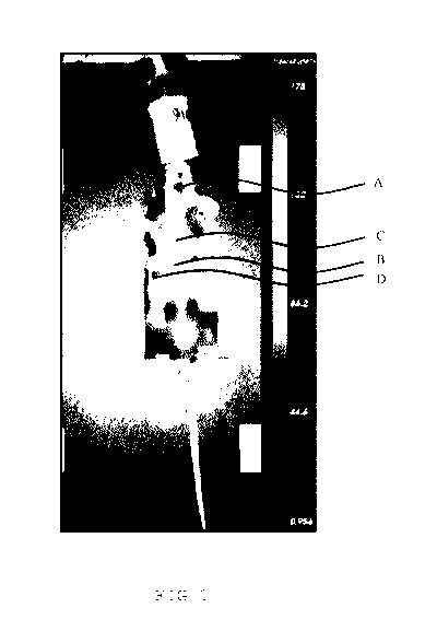
Figure 1 shows the in vivo biodistribution of sphingomyelin lip-
osomes labelled with Indocyaninegreen (ICG). Mice were treated
intravenously with liposomes carrying near-infrared dye and bio-
distribution was analysed 24 hours post-injection. Analysis was
performed with a GE HealthCare eXplore Optix. Signals of the ICG
were found in brain (A) and the spinal cord (B). Total liposome
lipid injection was 45 mg/kg carrying 1:200 weight-to-weight
ICG. Further signals could be found in the clearance organs liv-
er (C) and spleen (D), indicating that after treatment the lipo-
somes can be removed from the body.
Figure 2 shows the in vivo biodistribution of sphingomyelin lip-
osomes labelled with DiR. Different mice A, B, C were treated
intravenously with liposomes carrying near-infrared dye and bio-
distribution was analysed 24 and 48 hours post-injection in a
ventral view and a dorsal view. Analysis was performed with an
optical imaging system, IVIS Spectrum of Perkin Elmer. Signals
of the DiR were found in the brain (circle) and spinal cord
(rectangle). Total liposome lipid injection was 15 mg/kg carry-
ing 50 g/ml DiR. Further signals could be found in the clear-
ance organs liver (plain arrow) and spleen (doted arrow), indi-
cating that after treatment the liposomes can be removed from
the body. The fluorescence scale is termed in the following
unit: total Radiant efficiency [p/s]/[11W/cm2].
Figure 3 shows a comparative example of the in vivo biodistribu-
tion of a liposome with sphingomyelin and GM1, labelled with
DiR. Mice were treated intravenously with liposomes carrying
near-infrared dye and biodistribution was analysed 24 and 48
hours post-injection. Analysis was performed with an optical im-
aging system, IVIS Spectrum of Perkin Elmer. Signals of the DiR
were found in brain (circle) and the spinal cord (rectangle).
Total liposome lipid injection was 25 mg/kg carrying 50 g/m1
CA 03086279 2020-06-18
WO 2019/122220
PCT/EP2018/086352
22
DiR. Further signals could be found in the clearance organs liv-
er (plain arrow) and spleen (doted arrow), indicating that after
treatment the liposomes can be removed from the body. The fluo-
rescence scale is termed in the following unit: total Radiant
efficiency [p/s]/[11W/cm2]. Even though the liposomes are found
in the same organs as the liposomes presented in figure 2, the
biodistribution is less distinct compared to the essentially
GM1-free liposomes in figure 2.
Figure 4 shows a graphic representation of the biodistribution
analysis in the brain, spinal cord, liver and spleen from the in
vivo biodistribution images of Figure 2 and Figure 3. Figure 4A
shows the normalised fluorescence of the biodistribution of the
liposome without ganglioside in four different tissues: brain,
spinal cord, liver and spleen. The biodistribution is displayed
for two different time points: 24 and 48 hours. The bars repre-
sent the standard deviation to the mean. Figure 4B shows the
normalised fluorescence of the biodistribution of the liposome
with ganglioside.
It was surprisingly found, that the in vivo biodistribution of
the liposome essentially lacking ganglioside (Fig. 4A) is higher
than the in vivo biodistribution of the liposome comprising gan-
glioside (Fig. 4B).
Figure 5 shows the characterization of the liposomes without
surface modification. liposomes were visualized using Cryo
Transmission Electron Microscope JEOL JEM-2100F and a TVIPS Tem-
Cam camera (JEOL Ltd., Japan). Figure 5A shows an image of the
liposomes at low magnification (20000x). Figure 5B shows the
liposomes at high magnification (80000x). Figure 5C shows a
qualitative assessment done by ocular/visual observation of the
liposomal distribution. Figure 5D shows the size distribution of
the liposomes of this invention. In order to quantify the mean
CA 03086279 213236-18
WO 2019/122220
PCT/EP2018/086352
23
diameter of the liposomes N(liposomes) = 5128 were analysed (Vi-
ronova Analyzer Software, Vironova, Sweden). The mean diameter
of the liposomes is 30.46 nm with a standard deviation of 10.10
nm.
Figure 6 comprises two further tests for a quantitative charac-
terisation of the liposomes without surface modification by cry-
oTEM (Vironova Analyzer Software, Vironova, Sweden). Figure 6A
shows the circularity distribution of 5128 liposomes. Figure 6B
shows the lamellarity grade of the liposomal distribution. 98%
of 5128 liposomes have been characterised as unilamellar.
Figures 7 and 8 show the size and polydispersity stability of
liposomes according to the invention over time, measured by dy-
namic light scattering. The liposomal formulations were obtained
according to the method described above, by using sphingomyelin
and cholesterol in a 1:1 molar ratio. The liposomes were com-
pletely free of gangliosides, surface modifications, and did not
comprise or encapsulate an active component. The liposomal for-
mulations were stored in PBS at a pH-value of 6.8 and a tempera-
ture of 4 C. Size and polydispersity were determined by DLS
standard methods. It shall be noted that the values measured by
dynamic light scattering are slightly higher than the values ob-
tainable by cryoTEM due to the impact of the hydrodynamic radius
of liposomes on DLS measurements. A diameter of 60 nm as indi-
cated in Figure 7 corresponds to a mean diameter in the range of
10 and 50 nm when measured by Cryo Transmission Electron Micros-
copy.
The dotted curve shows the results of a small scale production
batch of liposomal formulation as described above, while the
dashed curve shows the results of an upscale production, i.e. a
batch size of 2 litres. As can be seen from Figures 7 and 8,
CA 03086279 213236-18
WO 2019/122220
PCT/EP2018/086352
24
both the size and polydispersity of the liposomal formulations
from Q3 2017 to Q3 2018, i.e. during storage time of one year,
remained essentially unchanged.
Figures 9 and 10 show relative in-vivo fluorescence of different
liposomal formulations in the spinal cord and brain of mice. In
both charts, the liposomes of groups 1 to 4 were obtained ac-
cording to the method described above, by using only sphingomye-
lin and cholesterol in a 1:1 molar ratio. Gr. 5 is a control
group of free DiR in PBS. In Gr.1, synthetic sphingomyelin was
used and GM1 was comprised in the liposomes. In Gr. 2, synthetic
sphingomyelin was used and the liposome was completely free from
surface modifications, in particular free from GM1. In Gr. 3,
sphingomyelin of animal origin was used and GM1 was comprised in
the liposomes. In Gr. 4 sphingomyelin of animal origin was used
and the liposome was completely free from surface modifications,
in particular free from GM1. For all four test groups, DiR was
added as a labelling agent. The measurements were performed by
NIR imaging technique.
Figures 9 and 10 show the accumulation of the four different
kinds of liposomes in the spinal cord and brain respectively in
0.1h, 4h, 24h and 48 h post-injection. It can be seen that the
presence of GM1 does not significantly affect the ability of the
liposomes to target the central nervous system. The same holds
true for the use of synthetic sphingomyelin compared to the use
of sphingomyelin of animal origin.
Figure 11 shows the relative in-vivo fluorescence of liposomal
formulations with different lipid compositions in the brain of
mice. The liposomes of groups 1 to 3 were obtained according to
the method described above by using lipids and cholesterol in a
1:1 molar ratio. In group 1, phosphatidylcholine and sphingomye-
CA 03086279 2020-06-18
WO 2019/122220
PCT/EP2018/086352
lin in combination were used as lipids. In group 2, phosphati-
dylcholine alone was used as a lipid. In group 3, sphingomyelin
alone was used as a lipid. In all three test groups, DiR was
added to the formulations as a labelling agent. The measurements
5 were performed by NIR imaging technique.
Figure 11 shows the accumulation of the three different kinds of
liposomes in the brain of mice after 24h and 48 h post-
injection. It can be seen that the composition consisting of
10 sphingomyelin and cholesterol alone results in superior longevi-
ty of circulation and CNS bioavailability of the liposome com-
pared to the other variants (Grps 1 and 2).