Note : Les descriptions sont présentées dans la langue officielle dans laquelle elles ont été soumises.
CA 03088816 2020-07-17
EXTRACORPOREAL FOCUSED ULTRASOUND TREATMENT DEVICE FOR
PELVIC DISEASE
TECHNICAL FIELD
The present disclosure belongs to the field of high intensity focused
ultrasound
treatment technology, and particularly relates to an extracorporeal focused
ultrasound
treatment device for a pelvic disease.
BACKGROUND
High Intensity Focused Ultrasound (HIFU) technology has been widely used to
treat
benign and malignant tumors such as liver cancer, breast cancer, kidney
cancer, bone
tumor, uterine fibroid, etc. By using the focusability and penetrability of
ultrasound,
ultrasound is focused at a lesion site in a human body, and high energy
density
mechanical energy in the focal region is converted into heat energy to cause
coagulative
necrosis (also called ultrasound thermal ablation) of diseased tissues;
meanwhile,
because the ultrasonic energy density on the beam path is low, it can be
guaranteed that
influence on normal tissues around the diseased tissues and on the beam path
is little or
acceptable.
Most of existing focused ultrasonic transducers for extracorporeal high
intensity
focused ultrasound treatment have a sound emitting surface in the shape of a
spherical
cap, and ultrasound emitted from the existing focused ultrasonic transducer is
a traveling
wave. The focal region formed by the existing ultrasound transducer has a
shape similar
to a cigar or a spindle, its length in the direction of the sound axis is
relatively large and
generally exceeds 10 mm, and its dimensions in the other two short axes range
from 2
mm to 3 mm (taking the ultrasound frequency of 1MHz as an example), so that
the focal
region has a relatively large size, which affects the focusing of energy, and
is
unfavorable for ensuring the safety of treatment. In addition, ultrasound
emitted by the
existing ultrasonic transducer may be scattered or reflected by non-uniform
tissues such
as bones, organs containing air, and the like, making the ultrasound propagate
in a
seriously nonlinear manner, which in turn damages tissues in the beam path,
causes an
Date Recue/Date Received 2020-07-17
CA 03088816 2020-07-17
unpredictable deviation and distortion of the focal region, and influences the
positioning
of the focal region.
Due to the disadvantages of the existing ultrasonic transducers, their
application in
therapy is limited. For example, prostate hyperplasia and prostate cancer are
common
diseases for adult men, and the incidence of prostate hyperplasia among men
aged 40
years to 79 years in China is about 50%, and the incidence of prostate
hyperplasia among
men aged over 80 years is 80%. However, the prostate is located in the pelvic
cavity, and
there are a lot of non-uniform tissues such as bones, organs containing air,
and the like
around the prostate, so that ultrasound emitted from the outside of a body can
hardly be
focused at the prostate accurately through the non-uniform tissues. Therefore,
for the
existing focused ultrasound treatment for prostate diseases, an ultrasonic
transducer
needs to be introduced into a body through the urethra or rectum, which causes
discomfort to patients and easily causes damage to the urethra or rectum, and
because the
ultrasonic transducer has a limited size, low energy and difficulty in
movement, the
effect, efficiency and integrity of the treatment are poor. Meanwhile, because
the
existing focal region of ultrasound is cigar-shaped, it is difficult to
accurately limit the
focal region to a required position, and when one part of the focal region is
positioned at
a diseased tissue, other part of the focal region is very likely to exceed the
diseased
tissue and positioned at a normal tissue and may cause damage to the normal
tissue, so
that the treatment safety is reduced.
SUMMARY
The present disclosure at least partially solves the problems of poor
treatment effect,
efficiency and safety of the existing focused ultrasound treatment device for
prostate
diseases, and provides an extracorporeal focused ultrasound treatment device
for pelvic
diseases, which has high treatment efficiency, good effect and good safety.
As a technical solution adopted to solve the technical problem of the present
disclosure, there is provided an extracorporeal focused ultrasound treatment
device for
pelvic diseases, which includes an ultrasonic transducer and a treatment
couch, wherein
the ultrasonic transducer includes a sound emitting surface and a sound
generation
2
Date Recue/Date Received 2020-07-17
CA 03088816 2020-07-17
unit that is configured to generate an ultrasonic wave; the sound emitting
surface is a
spherical surface having a first notch, a second notch and a third notch, a
sphere
corresponding to the spherical surface has a diameter in a range of 400 mm to
800 mm,
one great circle of the sphere is a main great circle, the first notch and the
second notch
are respectively positioned at two intersections of the spherical surface and
a diameter
perpendicular to the main great circle, and the third notch connects the first
notch with
the second notch; within distances of 100 mm to 200mm from the main great
circle
respectively at both sides of the main great circle, a cross-section of the
sound emitting
surface parallel to the main great circle is in a shape of an arc, an opening
of the arc
corresponds to the third notch, and a central angle corresponding to the arc
is larger than
180 degrees and smaller than 300 degrees; and the sound emitting surface is
capable of
reflecting ultrasound, and an ultrasonic wave generated by the sound
generation unit is
focused at a center of the sphere corresponding to the sound emitting surface.
The treatment couch is configured for a human body to lie in a lithotomy
position,
and when the human body lies in the lithotomy position on the treatment couch,
a pelvic
cavity of the human body is positioned at the center of the sphere
corresponding to the
sound emitting surface with two legs of the human body respectively sticking
out of the
sound emitting surface through the first notch and the second notch, and an
upper part of
the human body sticking out of the sound emitting surface through the third
notch.
Optionally, an edge of the first notch and an edge of the second notch are in
a first
plane and a second plane, respectively.
Optionally, the first plane and the second plane are both parallel to the main
great
circle.
Optionally, a distance between the first plane and the second plane is in a
range of
200 mm to 400 mm.
Optionally, a distance between the first plane and the main great circle is
equal to a
distance between the second plane and the main great circle.
Optionally, the diameter of the sphere corresponding to the sound emitting
surface
is in a range of 420 mm to 600 mm; and
within distances of 100 mm to 150 mm from the main great circle respectively
at
3
Date Recue/Date Received 2020-07-17
CA 03088816 2020-07-17
both sides of the main great circle, the central angle corresponding to the
arc in the
cross-section of the sound emitting surface parallel to the main great circle
is larger than
180 degrees and smaller than 300 degrees.
Optionally, each cross-section of the sound emitting surface parallel to the
main
great circle is in a shape of an arc, and the central angle corresponding to
the arc is larger
than 200 degrees and smaller than 260 degrees.
Optionally, the opening of the arc in each cross-section of the sound emitting
surface parallel to the main great circle is oriented in a same direction, and
the central
angle corresponding to the arc is equal.
Optionally, the sound emitting surface is symmetric with respect to the main
great
circle.
Optionally, when the human body lies on the treatment couch in the lithotomy
position, ultrasound emitted from a first region of the sound emitting surface
enters the
pelvic cavity through abdomen of the human body.
When the human body lies on the treatment couch in the lithotomy position,
ultrasound emitted from a second region of the sound emitting surface enters
the pelvic
cavity through an area between coccyx and pubic symphysis of the human body.
Optionally, the extracorporeal focused ultrasound treatment device for pelvic
diseases further includes:
a first B-mode ultrasonic probe configured to emit imaging ultrasound from the
first
region of the sound emitting surface to the pelvic cavity through the abdomen
of the
human body to form an image of the pelvic cavity; and/or
a second B-mode ultrasonic probe configured to emit imaging ultrasound from
the
second region of the sound emitting surface to the pelvic cavity through
perineum of the
human body to form an image of the pelvic cavity.
Optionally, the treatment couch and the ultrasonic transducer are separated
structures; and
the extracorporeal focused ultrasound treatment device for pelvic diseases
further
includes a movement unit configured to cause the treatment couch to be close
to or far
away from the ultrasonic transducer.
4
Date Recue/Date Received 2020-07-17
CA 03088816 2020-07-17
Optionally, the extracorporeal focused ultrasound treatment device for pelvic
diseases further includes:
a medium containing unit configured to keep a sound transmission medium
between
a surface of the human body and the sound emitting surface.
Optionally, the extracorporeal focused ultrasound treatment device for pelvic
diseases further includes:
a driving unit configured to drive the ultrasonic transducer to move relative
to the
treatment couch.
Optionally, the extracorporeal focused ultrasound treatment device for pelvic
diseases further includes:
an imaging unit configured to form an image of the pelvic cavity.
Optionally, the ultrasound generated by the sound generation unit has a
frequency
in a range of 0.4 MHz to 1.5 MHz.
Optionally, an acoustical power of the ultrasound generated by the sound
generation
unit is in a range of OW to 1200W.
Optionally, the acoustical power of the ultrasound generated by the sound
generating unit is in a range of OW to 800W.
The extracorporeal focused ultrasound treatment device for pelvic diseases
adopts a
specific C-shaped ultrasonic transducer, and the focal region of the
ultrasonic transducer
has a shape close to a sphere, a small size and high energy density, so that
the device has
good treatment effect, high efficiency, little influence on normal tissues and
good safety;
moreover, non-uniform tissues such as bones and the like have little influence
on the
focusing effect of the ultrasound generated by the ultrasonic transducer, and
in the
meanwhile, the human body lies on his/her back on the treatment couch in a
specific
position such that the pelvic cavity is positioned near the focal region of
the ultrasonic
transducer, so as to allow the ultrasound to enter the human body with
maximized beam
path. Therefore, the extracorporeal focused ultrasound treatment device for
pelvic
diseases can treat diseases of organs in the pelvic cavity by way of
externally focusing
ultrasonic waves, so that the size of the ultrasonic emitting surface (i.e.,
the sound
emitting surface) of the ultrasonic transducer can be larger, and under the
condition that
5
Date Recue/Date Received 2020-07-17
CA 03088816 2020-07-17
the ultrasonic energy emitted per unit area is the same, the area of the
acoustic window
for ultrasound to enter the human body can be larger, and the energy density
obtained at
the focal region is higher. As a result, the treatment effect is improved, the
treatment
efficiency is improved, the treatment comfort is improved, the operation
convenience is
improved, the harm to the human body is reduced, and the treatment safety is
improved.
The extracorporeal focused ultrasound treatment device for pelvic diseases is
suitable for treating diseases of organs in a pelvic cavity, such as prostate
cancer,
prostate hyperplasia, hysteromyoma, adenomyosis, cervical cancer, ovarian
cancer,
rectal cancer, colon cancer and the like, and is particularly suitable for
treating prostate
diseases.
BRIEF DESCRIPTION OF THE DRAWINGS
Fig. 1 is a schematic structural diagram of an ultrasonic transducer according
to an
embodiment of the present disclosure;
Fig. 2 is a schematic structural diagram of a sound emitting surface in an
ultrasonic
transducer according to an embodiment of the present disclosure;
Fig. 3 is a schematic diagram of a structure, in a direction parallel to a
main great
circle, of a sound emitting surface of an ultrasonic transducer according to
an
embodiment of the present disclosure;
Fig. 4 is a schematic diagram of a structure, in a direction perpendicular to
a main
great circle, of a sound emitting surface of an ultrasonic transducer
according to an
embodiment of the present disclosure;
Fig. 5 is a schematic structural diagram of a cross-section of a sound
emitting
surface parallel to a main great circle in an ultrasonic transducer according
to an
embodiment of the present disclosure;
Fig. 6 is a schematic side view of a structure of an extracorporeal focused
ultrasound treatment device for pelvic diseases in a split state according to
an
embodiment of the present disclosure;
Fig. 7 is a schematic top view of a structure of an extracorporeal focused
ultrasound
treatment device for pelvic diseases in a split state according to an
embodiment of the
6
Date Recue/Date Received 2020-07-17
present disclosure;
Fig. 8 is a schematic side view of a structure of an extracorporeal focused
ultrasound treatment device for pelvic diseases in a combination state
according to an
embodiment of the present disclosure;
Fig. 9 is a schematic top view of a structure of an extracorporeal focused
ultrasound
treatment device for pelvic diseases in a combination state according to an
embodiment
of the present disclosure;
Fig. 10 is a schematic side view of a structure of an extracorporeal focused
ultrasound treatment device for pelvic diseases in a combination state and
with a human
body according to an embodiment of the present disclosure;
Fig. 11 is a schematic top view of a structure of an extracorporeal focused
ultrasound treatment device for pelvic diseases in a combination state and
with a human
body according to an embodiment of the present disclosure.
Reference numerals: 1. ultrasonic transducer; 11. housing; 12. upper cover;
13.
piezoelectric array element; 14. end cover; 2. treatment couch; 3. sound
emitting surface;
31. first notch; 32. second notch; 33. third notch; 35. first region; 36.
second region; 41.
first B-mode ultrasonic probe; 42. second B-mode ultrasonic probe; 91. first
plane; 92.
second plane; 99. main great circle.
DETAILED DESCRIPTION
In order that those skilled in the art can better understand the technical
solutions of
the present disclosure, the present disclosure will be further described in
detail below
with reference to the accompanying drawings and specific implementations.
First embodiment:
As shown in Figs. 1 to 16, the present embodiment provides an extracorporeal
focused ultrasound treatment device for pelvic diseases.
The extracorporeal focused ultrasound treatment device for pelvic diseases
adopts
an ultrasonic transducer 1 in a specific form, and when a human body lies in a
lithotomy
position such that a pelvic cavity enters the ultrasonic transducer 1,
ultrasound emitted
7
Date Recue/Date Received 2021-12-08
by the ultrasonic transducer 1 can be focused at a specific position in the
pelvic cavity of
the human body to treat a disease of an organ in the pelvic cavity, such as
prostate cancer,
prostate hyperplasia, hysteromyoma, adenomyosis, cervical cancer, ovarian
cancer,
rectal cancer, colon cancer or the like, and the extracorporeal focused
ultrasound
treatment device for pelvic diseases is particularly suitable for treating
prostate diseases.
The extracorporeal focused ultrasound treatment device for pelvic diseases of
the
embodiment includes an ultrasonic transducer 1 and a treatment couch 2.
The ultrasonic transducer 1 includes a sound emitting surface 3 and a sound
generation unit that is configured to generate an ultrasonic wave; the sound
emitting
surface 3 is a spherical surface having a first notch 31, a second notch 32
and a third
notch 33, a sphere corresponding to the spherical surface has a diameter in a
range of
400 mm to 800 mm, one great circle of the sphere is taken as a main great
circle 99, the
first notch 31 and the second notch 32 are respectively positioned at two
intersections of
the spherical surface and a diameter perpendicular to the main great circle
99, and the
third notch 33 connects the first notch 31 with the second notch 32; within
distances of
100 mm to 200 mm from the main great circle 99 respectively at both sides of
the main
great circle 99, a cross-section of the sound emitting surface 3 parallel to
the main great
circle 99 is in a shape of an arc, an opening of the arc corresponds to the
third notch 33,
and a central angle corresponding to the arc is larger than 180 degrees and
smaller than
300 degrees; and the sound emitting surface 3 is capable of reflecting
ultrasound, and an
ultrasonic wave generated by the sound generation unit is focused on a center
of the
sphere corresponding to the sound emitting surface 3.
The treatment couch 2 is configured for a human body to lie in a lithotomy
position,
and when the human body lies in the lithotomy position on the treatment couch
2, the
center of the sphere corresponding to the sound emitting surface 3 is
positioned in a
pelvic cavity of the human body, two legs respectively stick out of the sound
emitting
surface 3 through the first notch 31 and the second notch 32, and an upper
part of the
body sticks out of the sound emitting surface 3 through the third notch 33.
The extracorporeal focused ultrasound treatment device for pelvic diseases of
the
embodiment has an ultrasonic transducer 1, and the ultrasonic transducer 1 has
a sound
8
Date Recue/Date Received 2021-12-08
generation unit, which is a device capable of generating ultrasound. For
example, the
material of the sound generation unit may include piezoelectric ceramics, 1-3
type
piezoelectric composite material, or the like. The shape, number, position,
and other
parameters of the sound generation unit may be designed such that the sound
generation
unit can emit ultrasound from all positions of the sound emitting surface 3,
and the
ultrasound emitted at each position propagates along the normal direction of
the sound
emitting surface 3 at the position, and the ultrasound can be finally focused
(including
directly focused or focused after being reflected) at a required position.
In an embodiment, as shown in Fig. 1, the sound emitting surface 3 may be an
acoustically transparent surface with a predetermined shape, and the sound
generation
unit (e.g., a piezoelectric array element 13) may be disposed behind the sound
emitting
surface 3; alternatively, the sound emitting surface 3 may be the emitting
surface of the
sound generation unit itself.
In an embodiment, the sound generation unit may also take different forms. For
example, the sound generation unit may be a plurality of piezoelectric array
elements 13
(e.g., rectangular piezoelectric ceramic plates) disposed at different
positions of the
sound emitting surface 3, that is, the plurality of piezoelectric array
elements 13 are
spliced together to form the sound emitting surface 3; alternatively, the
sound generation
unit may also have the same shape as the sound emitting surface 3 (e.g., the
sound
generation unit is a specially shaped piezoelectric ceramic plate).
Needless to say, as shown in Fig. 1, the ultrasonic transducer 1 may further
include,
in addition to the sound emitting surface 3 and the sound generation unit, a
driving
circuit for the sound generation unit, a casing (e.g., the casing of the sound
generation
unit may include a housing 11, an upper cover 12, a lower cover, an end cover
14, etc.)
.. for enclosing the driving circuit and the sound generation unit, and other
components,
which will not be described in detail herein.
Unlike the conventional sound emitting surface in the shape of a spherical
cap, the
sound emitting surface 3 of the ultrasonic transducer 1 of the present
embodiment is
equivalent to a spherical surface lacking three portions, and the spherical
surface may
.. have a diameter in the range of 400 mm to 800 mm, preferably in the range
of 420 mm to
9
Date Recue/Date Received 2021-12-08
600 mm.
As shown in Figs. 2 to 4, two portions (the first notch 31 and the second
notch 32)
missing from the sound emitting surface 3 are portions of the spherical
surface at both
ends of one diameter, and a great circle (i.e., a plane passing through the
spherical center)
perpendicular to the diameter is the main great circle 99. The third portion
(third notch
33) missing from the sound emitting surface 3 is a portion laterally
connecting the first
notch 31 with the second notch 32.
That is, if the plane in which the main great circle 99 is located is in a
horizontal
direction, and the diameter perpendicular to the main great circle 99 is in a
vertical
direction, parts of the top end and the bottom end of a spherical surface in
the vertical
direction may be cut off, respectively, then a part of one side of the
spherical surface
may be cut off, and the cut-off part of the side should connect the cuts of
the top end and
the bottom end, so that the remaining spherical surface is the sound emitting
surface 3.
Within the distances of 100 mm to 200 mm (preferably 100 mm to 150 mm, and the
distances at two sides may be different) from the main great circle 99
respectively at the
two sides of the main great circle 99, a cross-section of the sound emitting
surface 3
parallel to the main great circle 99 is in the shape of an arc, the central
angle
corresponding to the arc is greater than 180 degrees and less than 300
degrees, and
preferably, greater than 200 degrees and less than 260 degrees, and the
opening of the
arc corresponds to the third notch 33. That is, at least within a certain
distance from the
main great circle 99, the portion of the spherical surface cut off by the
third notch 33 has
a limited range and the central angle corresponding to the remaining portion
is within the
above range.
Furthermore, the sound emitting surface 3 has the capability of reflecting
ultrasound, and at at least part of positions, the third notch 33 only cuts
off a spherical
surface smaller than half spherical surface. Therefore, as shown in Fig. 5,
ultrasound
emitted from a part of the arc at an angle exceeding the central angle of 180
degrees is
reflected by an opposite part of the sound emitting surface 3, and the part of
the arc at
the angle exceeding the central angle of 180 degrees may also reflect
ultrasound emitted
from the opposite part of the sound emitting surface 3, so that ultrasound can
return in
Date Recue/Date Received 2021-12-08
partial region (the region filled with oblique lines in Fig. 5), so as to form
a standing
wave, thereby changing the focusing condition and the focal region form of
ultrasound;
meanwhile, the ultrasound emitted from the part of the arc corresponding to
the opening
is not reflected, so that ultrasound emitted from this part of the arc is
still a traveling
wave.
That is, the ultrasound generated by the ultrasound transducer 1 of the
present
embodiment is actually in the form of a combination of a traveling wave with a
standing
wave, and thus its propagation and focusing will change. Specifically, the
ultrasonic
transducer 1 can compress the major axis of the original cigar-shaped focal
region, so
that the focal region has a shape closer to a spherical shape and has a
smaller size, the
energy density is improved, the treatment effect and efficiency are improved,
the damage
to normal tissues is reduced, and the safety is improved. Meanwhile, the
ultrasonic
transducer 1 can also reduce the adverse effects of non-uniformity of tissues
and bone
tissues and the like on the focusing of ultrasound when the ultrasound
propagates in a
human body, and reduce deviation and distortion of the focal region, which
facilitates
accurate positioning of the focal region.
In an embodiment, edges of the first notch 31 and the second notch 32 are
located in
a first plane 91 and a second plane 92, respectively. In an embodiment, the
first plane 91
and the second plane 92 are both parallel to the main great circle 99.
As shown in Fig. 3, in an embodiment, the first notch 31 and the second notch
32
are spherical caps cut off by planes. In an embodiment, the first notch 31 and
the second
notch 32 are spherical caps cut off by two parallel planes, that is, the
bottom surfaces of
the two cut-off spherical caps are parallel to each other. As such, the
spherical surface
excluding the first notch 31 and the second notch 32 is equivalent to a
structure formed
by butting the bottom surfaces of two spherical segments. Needless to say, the
bottom
surfaces of the two spherical segments are the main great circle 99, and the
two spherical
segments may have different heights. The sound emitting surface 3 in this form
has a
shape similar to a spherical segment, and is regular and simple in structure.
Needless to say, it is also feasible that the first notch 31 and second notch
32 are cut
off by planes that are not parallel to each other, or by curved surfaces that
are not planar.
11
Date Recue/Date Received 2021-12-08
In an embodiment, the distance between the first plane 91 and the second plane
92
ranges from 200 mm to 400 mm. In an embodiment, the distance between the first
plane
91 and the second plane 92 ranges from 200 mm to 300 mm.
That is, the distance between the first notch 31 and the second notch 32
(i.e., the
dimension of the sound emitting surface 3 in the vertical direction) is
preferably in the
above range (of course, the diameter of the sphere corresponding to the sound
emitting
surface 3 should be larger than the distance). Such sound emitting surface 3
has a
sufficient area to generate ultrasound suitable for treatment and a size that
is not too
large, and can allow legs of the human body to stick out.
In an embodiment, the distance between the first plane 91 and the main great
circle
99 is equal to the distance between the second plane 92 and the main great
circle 99.
That is, the first notch 31 and the second notch 32 are preferably obtained by
cutting with two planes that have a same distance to the center of the sphere,
so that the
two notches have a same size and are symmetrically distributed, which
facilitates
symmetry of the focal region and placement of the legs of the human body.
Needless to say, it is also possible that the first notch 31 and the second
notch 32
have different distances to the center of the sphere, or have different
shapes.
In an embodiment, any cross-section of the sound emitting surface 3 parallel
to the
main great circle 99 is in the shape of an arc, and the central angle
corresponding to the
arc is greater than 180 degrees and less than 300 degrees.
It is defined above that the sound emitting surface 3 is arc-shaped in a cross-
section
parallel to the main great circle 99 at least in the vicinity of the main
great circle 99. In
an embodiment, any cross-section of the sound emitting surface 3 parallel to
the main
great circle 99 may be in the shape of the arc, thereby ensuring that the
sound emitting
surface 3 can generate a standing wave at each position in the vertical
direction.
Needless to say, it is also possible that the cross-section of the sound
emitting
surface 3 parallel to the main great circle 99 is not in the shape of an arc
(e.g., is two
separate arcs) at some positions.
In an embodiment, the arcs of the sound emitting surface 3 in any cross-
sections
thereof parallel to the main great circle 99 have openings orientated in a
same direction,
12
Date Recue/Date Received 2021-12-08
and correspond to central angles that are equal.
That is, at different positions in the vertical direction, the third notch 33
is
orientated in the same direction, and corresponds to a same central angle.
That is, the
third notch 33 is preferably obtained by cutting with a plane perpendicular to
the main
great circle 99.
As shown in Fig. 4, the sound emitting surface 3 is shaped like the letter "C"
as
viewed in a direction perpendicular to the main great circle 99.
In an embodiment, the sound emitting surface 3 is symmetrical with respect to
the
main great circle 99.
As shown in Fig. 3. the sound emitting surface 3 is preferably symmetrical
with
respect to the main great circle 99, that is, parts of the sound emitting
surface 3
respectively on both sides of the main great circle 99 are preferably of the
same form, so
that the sound field and focal region formed by the sound emitting surface are
also
symmetrical with respect to the main great circle 99, and are more regular and
easy to
.. control.
As shown in Figs. 6 to 11, the treatment couch 2 is configured to support a
human
body during treatment. In an embodiment, a person lies on his/her back on the
treatment
couch 2, with the legs raised and spread to the sides, i.e., in a lithotomy
position.
Thus, as shown in Figs. 10 and 11, in a case where the third notch 33 of the
ultrasonic transducer 1 is directed toward the treatment couch 2 and the first
notch 31
and the second notch 32 are respectively directed toward both sides, when the
center of
the sphere is located in the pelvic cavity of a human body, the upper part of
the human
body can stick out of the sound emitting surface 3 through the third notch 33,
and at the
same time, both legs of the human body can also stick out of the sound
emitting surface
3 through the first notch 31 and the second notch 32, respectively.
It can be seen that if such a posture is required between the human body and
the
ultrasonic transducer 1, the size and the central angle of the ultrasonic
transducer 1 (the
sound emitting surface 3) need to meet certain requirements, and the above
limitation on
the parameters of the sound emitting surface 3 just enables the ultrasonic
transducer 1 to
be adapted to the human body.
13
Date Recue/Date Received 2021-12-08
Needless to say, in order to allow a human body to lie on the treatment couch
2 in a
lithotomy position, the treatment couch 2 should have a chair, a leg support,
etc., which
will not be described in detail herein.
Needless to say, in an actual extracorporeal focused ultrasound treatment
device for
pelvic diseases, the ultrasound transducer 1 cannot be suspended, and a
corresponding
housing, a supporting structure, a driving circuit, etc. should be provided,
but for
simplicity, these structures are not shown in the drawings.
The extracorporeal focused ultrasound treatment device for pelvic diseases of
the
embodiment adopts a specific C-shaped ultrasonic transducer 1, and the focal
region of
the ultrasonic transducer 1 has a shape close to a sphere, a small size and
high energy
density, so that the extracorporeal focused ultrasound treatment device for
pelvic
diseases has good treatment effect, high efficiency, small influence on normal
tissues
and good safety.
Moreover, non-uniform tissues such as bones and the like have little influence
on
propagation of the ultrasound generated by the ultrasonic transducer 1, in the
meanwhile,
a human body lies on the back on the treatment couch 2 in a specific body
position and a
specific tissue organ in the pelvic cavity is positioned near the focal region
of the
ultrasonic transducer 1, so that the ultrasound is allowed to enter the pelvic
cavity of the
human body with maximized beam path, and the treatment of a specific lesion on
a
specific tissue organ in the pelvic cavity is facilitated.
Thus, the extracorporeal focused ultrasound treatment device for pelvic
diseases
can treat diseases of organs in the pelvic cavity by way of externally
focusing ultrasonic
waves, so that the size of the ultrasonic emitting surface (i.e., the sound
emitting surface
3) of the ultrasonic transducer can be larger, and under the condition that
the ultrasonic
energy emitted per unit area is the same, the area of the acoustic window for
ultrasound
to enter the human body can be larger, and the energy density obtained at the
focal
region is higher. As a result, the treatment effect is improved, the treatment
efficiency is
improved, the treatment comfort is improved, the operation convenience is
improved, the
harm to the human body is reduced, and the treatment safety is improved.
In an embodiment, the treatment couch 2 and the ultrasonic transducer 1 may be
14
Date Recue/Date Received 2021-12-08
separated structures; the extracorporeal focused ultrasound treatment device
for pelvic
diseases also includes a movement unit configured to make the treatment couch
2 and the
ultrasonic transducer 1 closer to or farther away from each other.
As shown in Figs. 10 and 11, according to the above parameters of the sound
emitting surface 3, when the center of the sphere corresponding to the sound
emitting
surface 3 of the transducer is in the pelvic cavity of a human body, the
distances from the
abdomen and the back of the human body to the ends of the sound emitting
surface 3 are
small, and therefore, if the treatment couch 2 is positioned near the
ultrasonic transducer
1, it is difficult for the perineum to enter through the gap between the
treatment couch 2
and the ultrasonic transducer 1. Therefore, as shown in Figs. 6 and 7, the
treatment
couch 2 and the ultrasonic transducer 1 are preferably separated, and the
treatment couch
2 may come closer to or farther away from the ultrasonic transducer 1 through
the
movement unit (e.g., a wheel, a rail, etc.). In this way, the human body can
lie on the
treatment couch 2 in a lithotomy position when the treatment couch 2 is away
from the
ultrasonic transducer 1, and then the treatment couch 2 is caused to come
close to the
ultrasonic transducer 1 to make the perineum enter the sound emitting surface
3 through
the third notch 33, thereby obtaining the structure shown in Figs. 10 and 11.
In an embodiment, when the human body lies on the treatment couch 2 in a
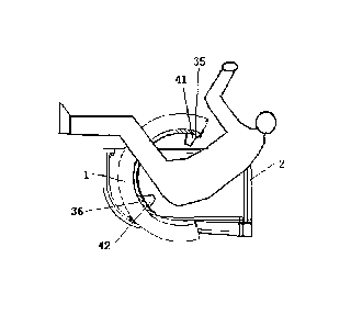
lithotomy position, ultrasound emitted from a first region 35 of the sound
emitting
surface 3 enters the pelvic cavity through the abdomen of the human body; when
the
human body lies on the treatment couch 2 in the lithotomy position, ultrasound
emitted
from a second region 36 of the sound emitting surface 3 enters the pelvic
cavity through
area between the coccyx and pubic symphysis of the human body.
Most part of the pelvic cavity of a human body is surrounded by pelvic bones,
and
the bones have a strong blocking effect on the ultrasound; in contrast, there
is no bone in
the abdomen, and less bones in the area (including the perineum, anus, etc.)
between the
coccyx and pubic symphysis, so ultrasound is less blocked when entering into
the pelvic
cavity through these two portions. Therefore, as shown in Fig. 10, the sound
emitting
surface 3 of the ultrasonic transducer 1 preferably has at least a first
region 35 and a
second region 36, and ultrasonic waves emitted from the two regions may
respectively
Date Recue/Date Received 2021-12-08
pass through the abdomen and the area between the coccyx and pubic symphysis
to enter
the pelvic cavity, so as to maximize the beam path.
Needless to say, the sound emitting surface 3 should also have a region
between the
first region 35 and the second region 36, and since the central angle between
the first
region 35 and the second region 36 is usually less than 150 degrees, as shown
in Fig. 10,
the sound emitting surface 3 actually should have a portion exceeding the
first region 35
and the second region 36, such as a portion corresponding to the sacrum. Thus,
ultrasonic waves emitted from all positions of the sound emitting surface 3
can form a
better sound field together.
In an embodiment, the extracorporeal focused ultrasound treatment device for
pelvic diseases further includes an imaging unit configured to form an image
of the
pelvic cavity.
That is, the extracorporeal focused ultrasound treatment device for pelvic
diseases
may also include an imaging unit (e.g., B-mode ultrasound, CT, MRI or the
combination
thereof) for forming an image of the pelvic cavity, so that a lesion is
positioned before
treatment and an image of an area around the treated part is formed in real
time during
treatment, so as to evaluate the treatment effect at any time and adjust the
treatment plan.
In an embodiment, the extracorporeal focused ultrasound treatment device for
pelvic diseases may include:
a first B-mode ultrasonic probe 41 configured to emit an imaging ultrasonic
wave
from the first region 35 of the sound emitting surface 3 to the pelvic cavity
through the
abdomen of the human body to form an image of the pelvic cavity;
and/or
a second B-mode ultrasonic probe 42 configured to emit an imaging ultrasonic
wave from the second region 36 of the sound emitting surface 3 to the pelvic
cavity
through the perineum of the human body to form an image of the pelvic cavity.
That is, B-mode ultrasound can be used to form an image of the pelvic cavity
for
monitoring, and since the B-mode ultrasound also achieves imaging by using
ultrasound,
it is also blocked by bones, so that the B-mode ultrasonic probes should also
be disposed
in the first region 35 and the second region 36 as shown in Fig. 10, so as to
avoid bones
16
Date Recue/Date Received 2021-12-08
to obtain images at these positions, to ensure clarity of the images, and to
minimize the
influence of the B-mode ultrasonic probe on the therapeutic ultrasound. In an
embodiment, a first B-mode ultrasonic probe 41 is disposed in the first region
35 and
emits, through the abdomen, ultrasound for imaging, while a second B-mode
ultrasonic
probe 42 is disposed at a specific position in the second region 36, i.e.,
emits, through
the perineum (rather than the anus, etc.,) ultrasound for imaging.
Since the human body lies on his/her back in a lithotomy position, the angle
between the first B-mode ultrasonic probe 41 and the vertical direction is
usually about
30 degrees, and the angle between the second B-mode ultrasonic probe 42 and
the
vertical direction is about 80 degrees.
In an embodiment, the B-mode ultrasonic probes may be arranged at
corresponding
positions of the sound emitting surface 3 and perform imaging in a non-contact
manner;
alternatively, as shown in Fig. 10, the B-mode ultrasonic probes may protrude
from the
sound emitting surface 3 and may be retractable, so that one or two of the B-
mode
ultrasonic probes may be selected to extend out and contact with the human
body as
required for imaging.
It can be seen that, for the extracorporeal focused ultrasound treatment
device for
pelvic diseases of the embodiments, by providing the B-mode ultrasonic probes
at
specific positions, an ultrasonic image with the best quality can be obtained
from the
optimal position under the condition of reducing influence on the therapeutic
ultrasound
as much as possible; moreover, the B-mode ultrasonic probes are disposed on
the sound
emitting surface 3 (i.e., on the ultrasonic transducer 1), so that when the
ultrasonic
transducer 1 moves, the B-mode ultrasonic probes will move together with the
ultrasonic
transducer 1, and thus the B-mode ultrasonic probes aim at the optimal imaging
positions
at any time.
In an embodiment, the extracorporeal focused ultrasound treatment device for
pelvic diseases further includes a driving unit for driving the ultrasonic
transducer 1 to
move relative to the treatment couch 2.
It is clear that the focal region of ultrasound needs to be located at the
lesion
position during treatment, and the accurate lesion positions are different
according to the
17
Date Recue/Date Received 2021-12-08
differences in body type, disease type, treatment condition and the like, and
therefore,
the position of the focal region needs to be adjusted in real time during the
treatment.
Therefore, a driving unit may be provided to drive the ultrasonic transducer 1
to move,
and then to drive the focal region to move.
The movement driven by the driving unit may include translations in three
axial
directions perpendicular to one another, and such movement may also cause the
focal
region to translate; alternatively, the movement may include rotating the
ultrasound
transducer 1 around different axial directions, so as to cause the ultrasound
to enter the
human body from different directions.
In an embodiment, the extracorporeal focused ultrasound treatment device for
pelvic diseases further includes a medium containing unit for keeping a sound
transmission medium between a surface of the human body and the sound emitting
surface 3.
In order to reduce attenuation of ultrasound during its propagation in air, a
sound
transmission medium such as deaerated water may be provided between the sound
emitting surface 3 of the ultrasound transducer 1 and the human body, and for
this reason,
a medium containing unit capable of holding a sound transmission medium (e.g.,
deaerated water) is preferably provided to cause the space between the sound
emitting
surface 3 of the ultrasound transducer 1 of the present embodiments and the
surface of
the human body through which ultrasound is to pass to be filled with the sound
transmission medium, and the medium containing unit may be in the form of a
water
basin or the like, and will not be described in detail herein.
In an embodiment, the ultrasound generated by the sound generation unit has a
frequency in the range of 0.4 MHz to 1.5 MHz.
In the embodiment, the ultrasound generated by the sound generation unit has
an
acoustical power in the range of OW to 1200W. In an embodiment, the acoustical
power
of the ultrasound generated by the sound generation unit ranges from OW to
800W.
For the ultrasonic transducer 1 in any one of the above forms, when it is used
for
treating a disease of an organ in the pelvic cavity, the parameters of the
ultrasound
emitted by the ultrasonic transducer 1 are preferably in the above ranges to
achieve good
18
Date Recue/Date Received 2021-12-08
treatment effect.
The extracorporeal focused ultrasound treatment device for pelvic diseases of
the
embodiments emits ultrasonic waves at an acoustical power of 200W toward
deaerated
water, so as to cavitate water in the focal region. The cavitated region
(i.e., the focal
region) in the photograph has a shape close to a circle, a size of 1.8 mm *
1.2 mm, and a
length-width ratio of 3: 2, which indicates that, compared with the
conventional focused
ultrasound transducer with only traveling waves, the extracorporeal focused
ultrasound
treatment device for pelvic diseases of the embodiments has a focused
ultrasound
transducer having a focal region whose major axis is significantly compressed,
whose
shape changes from a cigar shape to an approximately spherical shape, and
which has a
reduced size, an increased energy density, and a more regular shape.
When the extracorporeal focused ultrasound treatment device for pelvic
diseases of
the embodiments is used to treat an exvivo bovine liver with ultrasound
irradiation at an
acoustical power of 400W for 2 seconds, a target area with a depth of 80 mm is
obviously damaged in a short time, and the damaged part is in a fusiform
shape, and has
a clear boundary, a size of 4.4 mm * 1.5 mm, and a length-width ratio of less
than 3: 1,
which is lower than the length-width ratio (generally greater than 5: 1) of
the damaged
part caused by a conventional focused ultrasound transducer. This also
indicates that the
focal region of the extracorporeal focused ultrasound treatment device for
pelvic
diseases of the present embodiments has a more regular shape.
In an embodiment, a pelvic bone is placed at a preset position at an inner
side of the
ultrasonic transducer 1 of the extracorporeal focused ultrasound treatment
device for
pelvic diseases so as to simulate the position of the pelvic bone of a human
body, and
then an exvivo bovine muscle tissue is placed in the pelvic bone. The focal
region is
positioned at a position equivalent to the position having a distance of 10 mm
from the
rectum of the human body, and a treatment process for the prostate is
simulated, the
ultrasonic power is 400 W, the target area has a depth of 55 mm, and the
ultrasonic
irradiation time is 2 seconds * 5 times. The bovine muscle tissue subjected to
ultrasonic
irradiation has an obviously damaged target area with a clear boundary, no
damage is
caused to the envelope, and no damage is caused to the interface between the
simulated
19
Date Recue/Date Received 2021-12-08
rectum and prostate. This shows that the ultrasound emitted by extracorporeal
focused
ultrasound treatment device for pelvic diseases of the embodiments is less
affected by
non-uniform tissues such as bones in a human body, can still maximize a beam
path
when being applied in an actual human body environment, forms a focal region
with
.. small size, excellent shape and accurate position, achieves good treatment
effect and
efficiency, and avoids damage to normal tissues.
It could be understood that the above implementations are merely exemplary
implementations for illustrating the principle of the present disclosure, but
the present
disclosure is not limited thereto. Various modifications and improvements can
be made
by those skilled in the art without departing from the scope of the present
disclosure.
Date Recue/Date Received 2021-12-08