Note : Les descriptions sont présentées dans la langue officielle dans laquelle elles ont été soumises.
CA 03089853 2020-07-28
WO 2019/152663 PCT/US2019/016076
METHOD OF PRODUCING NATURAL KILLER CELLS AND COMPOSITION
FOR TREATING CANCER
INCORPORATION BY REFERENCE TO ANY PRIORITY APPLICATIONS
100011 This application claims the benefit of Korean Patent Application
No. KR-
10-2018-0012938, filed February 1, 2018, Korean Patent Application No. KR-10-
2018-
0012942, filed February 1, 2018, Korean Patent Application No. KR-10-2019-
0001981, filed
January 7, 2019, and Korean Patent Application No. KR-10-2019-0001983, filed
January 7,
2019, the disclosures of which are hereby incorporated by reference in their
entireties.
BACKGROUND
Field
100021 The present disclosure relates to a manufacturing method for
high-purity
natural killer cells.
Description of the Related Art
[00031 The human body is protected from pathogens by an immune
response,
coordinated by the immune system, which is composed of many immune-related
cells,
chemical mediators, such as cytokines, and the like. Leukocytes, especially
lymphocytes,
play an important role in such an immune system. Lymphocytes are involved in
both innate
and acquired immunity.
100041 Natural killer cells (NK cells) are one type of innate immune
cells, which
are known to non-specifically kill cancer, recognize and kill viruses,
bacteria, and the like,
and kill pathogens with enzymes such as perforin and granzyme or by Fas-FasL
interaction.
In the case of cancer patients, it has been reported that a decrease in cancer
cell cytotoxicity
of these NK cells is associated with the onset of various types of cancer,
such as lung cancer
(Carrega P, et al., Cancer, 2008: 112: 863-875), liver cancer (Jinushi M, et
al., J Hepatol.,
2005: 43; 1013-1020), breast cancer (Bauernhofer T, et al., Fur J Immunol.,
2003: 33: 119-
124), uterine cancer (Mocchegiani E., et al., Br j Cancer., 1999: 79: 244-
250), blood cancer
-1-
CA 03089853 2020-07-28
WO 2019/152663 PCT/US2019/016076
(Tajima F., et al, Lekemia 1996: 10: 478-482), and the like. Accordingly, for
cancer therapy,
it is desirable to increase the cancer cell cytotoxicity of the NK cells.
100051 In order to obtain the therapeutic effect of NK-mediated killing
of the
cancer cells, a large amount of NK cells having high purity is required, but
it is not easy to
obtain a large amount of blood from the cancer patient, and of the proportion
of NK cells in
the blood is small, only about 5 to 20%. Thus, it has been difficult for using
the NEC cells as
an immunotherapeutic agent.
[0006] As a result, it is desirable to effectively expand and
proliferate only the NK
cells, but in a conventional method of proliferating NK cells, various
expensive cytokines
need to be used at a high concentration, thus the corresponding therapy is
only available to
some financially stable patients. Further, according to conventional methods
of proliferating
NK cells, other types (e.g., T cells, B cells, etc.) of immune cells may be
present together
with the NK cells, and allogenic administration of the NK cells containing T
cells may cause
a graft versus host disease (GVHD) and allogenic administration of the NK
cells containing
B cells to blood-type incompatible subjects may cause a passenger B-lymphocyte
syndrome,
and thus, the anti-cancer effect is not maximized.
[0007] Further, in addition to expanding and proliferating NK cells, it
is desirable
to highly maintain the functions of NK cells until the expanded and
proliferated NK cells are
actually used. As a result, the development of a composition capable of
promoting the
proliferation of the NK cells, increasing production of cytokines such as TNF-
, INF- and
GM-CSF derived from the NK cells, and increasing cancer cell cytotoxicity of
the NEC cells is
sought.
SUMMARY
100081 This application is related to methods of producing high-purity
natural
killer cells, and a cell therapeutic composition for treating cancer
comprising high-purity
natural killer cells and cytokines. Any features, structures, or steps
disclosed herein can be
replaced with or combined with any other features, structures, or steps
disclosed herein, or
omitted. Further, for purposes of summarizing the disclosure, certain aspects,
advantages,
and features of the inventions have been described herein. It is to be
understood that not
-2-
CA 03089853 2020-07-28
WO 2019/152663 PCT/US2019/016076
necessarily any or all such advantages are achieved in accordance with any
particular
embodiment of the inventions disclosed herein. No individual aspects of this
disclosure are
essential or indispensable.
100091 In an embodiment, a method of producing natural killer cells is
disclosed.
The method includes: isolating peripheral blood mononuclear cells (PBMCs) from
a blood
sample; isolating at least one of CD56+ cells and/or CD3-/CD56+ cells from the
PBMCs;
and co-culturing the at least one of CD56+ cells and/or CD3-/CD56+ cells with
a
combination of feeder cells in the presence of a cytokine.
100101 In certain embodiments, isolating at least one of CD56+ cells
and/or CD3-
/CD56+ cells from the PBMCs is conducted by using at least one of CD56
microbeads and
CD3 microbeads. In certain embodiments, the cytokine is selected from a group
consisting of
IL-2, IL-21, IL-15, Flt3-L, SCF, IL-7, IL-18, IL-4, type I interferons, GM-
CSF, IGF 1, and
combinations thereof. In certain embodiments, the cytokine may be added at a
concentration
of 50-1000 113/mL.
100111 In certain embodiments, the combination of feeder cells includes
irradiated
Jurkat cells and irradiated Epstein-Barr virus transformed lymphocyte
continuous line (EBV-
LCL) cells. In a variation, the ratio of the irradiated Jurkat cells and the
irradiated EBV-LCL
cells may be about 1:0.1-5. Each of the irradiated Jurkat cells and the
irradiated EBV-LCL
cells may be obtained by irradiation of 50-500Gy.
100121 In certain embodiments, the co-culturing may include co-
culturing for 1-50
days.
[0013] In certain embodiments, the method may further include co-
culturing the
at least one of CD56+ cells and/or CD3-/CD56+ cells with a combination of
feeder cells, in
the presence of a first cytokine for a first period; and subsequently co-
culturing the at least
one of CD56+ cells and/or CD3-/CD56+ cells with the combination of feeder
cells, in the
presence of a second cytokine for a second period. In a variation, the second
cytokine may be
added once or more during Day 0-6 of the second period. The second cytokine
may be added
once or more during the first six days of every fourteen-day cycle during the
second period.
The first cytokine may be IL-2. The second cytokine may be IL-21. The second
cytokine
may be added at a concentration of 10-100 ng/mL.
-3-
CA 03089853 2020-07-28
WO 2019/152663 PCT/US2019/016076
100141 In certain embodiments, the at least one of CD56+ cells and/or
CD3-
/CD56+ cells and the combination of feeder cells is co-cultured with a ratio
of about 1:1-100
of CD56+ cells and/or CD3-/CD56+ cells to feeder cells.
(00151 In certain embodiments, a composition made by the method is
disclosed.
100161 In an embodiment, a composition for treating cancer in a patient
in need
thereof is disclosed. The composition includes: an effective amount of CD56+
natural killer
cells derived from peripheral blood, wherein the effective amount is in a
range of about 1 x
106 to 5 x 10⁸ cells per kg of the patient's body weight, and wherein the
CD56+ natural killer
cells are at least about 90% pure; IL-2 having a concentration of 50-50,000
IU/mL; and a
pharmaceutically acceptable carrier.
100171 In certain embodiments, the cytokine may be selected from a
group
consisting of IL-2, IL-21, IL-15, Flt3-L, SCF, IL-7, IL-18, IL-4, type I
interferons, GM-CSF,
IGF 1, and combinations thereof. In a variation, the cytokine may be IL-2. The
cytokine may
have a concentration of 50-50,000 IU/mL.
100181 In certain embodiments, the cancer is selected from a group
consisting of:
blood cancer, stomach cancer, pancreatic cancer, cholangiocarcinoma, colon
cancer, breast
cancer, liver cancer, ovarian cancer, lung cancer, kidney cancer, prostate
cancer and
neuroblastoma.
100191 In certain embodiments, the composition includes less than about
1% T
cells.
BRIEF DESCRIPTION OF THE DRAWINGS
100201 Various embodiments are depicted in the accompanying drawings
for
illustrative purposes, and should in no way be interpreted as limiting the
scope of the
embodiments. Furthermore, various features of different disclosed embodiments
can be
combined to form additional embodiments, which are part of this disclosure.
10021j FIG. IA illustrates a graph showing cell growth rates of NK
cells produced
from PBMCs, CD56+ cells, and CD3-/CD56+ cells.
100221 FIG. 1B illustrates graphs showing cell growth rates of NK cells
produced
from PBMCs and CD56+ cells with or without treating with IL-21.
-4-
CA 03089853 2020-07-28
WO 2019/152663 PCT/US2019/016076
[00231 FIG. 2 illustrates a graph showing the purity of CD3-/CD56+ NK
cells
produced from PBMCs or CD56+ cells with or without treating with IL-21.
100241 FIG. 3 illustrates a plate design for analyzing anticancer
activity of NK
cells.
100251 FIG. 4A illustrates graphs showing the short-term cytotoxicity
of NK cells
produced from PBMCs and CD56+ cells for various effector cell : target cell
(E:T) ratios.
[00261 FIG. 4B illustrates graphs showing the long-term cytotoxicity of
NK cells
produced from PBMCs and CD56+ cells against AGS, A549, and MDA-MB-231 cells.
[00271 FIGS. 5A-5B illustrate graphs showing cell growth rates of NK
cells
produced by treating with IL-21 during various periods.
100281 FIG. 6 illustrates graphs showing cell growth rates of NK cells
produced
by treating with IL-21 with various concentrations.
100291 FIG. 7A illustrates a graph showing the short-term cytotoxicity
of NK cells
produced by treating with IL-21 during various periods for various E:T ratios.
100301 FIGS. 7B-7C illustrate graphs showing the long-term cytotoxicity
of NK
cells produced by treating with IL-21 during various periods against AGS,
A549, and MDA-
MB-231 cells.
[00311 FIG. 8A illustrates graphs showing the short-term cytotoxicity
of NK cells
produced by treating with IL-21 with various concentrations for various E:T
ratios.
[00321 FIG. 8B illustrates graphs showing the long-term cytotoxicity of
NK cells
produced by treating with IL-21 with various concentrations against AGS, A549,
and MDA-
MB-231 cells.
[00331 FIG. 9 illustrates graphs showing cell growth rates of NK cells
with feeder
cell stimulations.
[00341 FIG. 10A illustrates graphs showing cell growth rates of NK
cells
produced from PBMCs of a cancer patient with or without treatment of 1L-21.
[00351 FIG. 10B illustrates a graph showing purity of CD3-/CD56+ NK
cells
produced from PBMCs of a cancer patient with or without treatment of IL-21.
[00361 FIG. 10C illustrates a graph showing the short-term cytotoxicity
of NK
cells produced from PBMCs with or without treatment of IL-21 against K562
cells.
-5-
CA 03089853 2020-07-28
WO 2019/152663 PCT/US2019/016076
100371 FIG. 10D illustrates graphs showing the long-term cytotoxicity
of NK cells
produced from PBMCs with or without treatment of IL-21 against AGS, A549, and
MDA-
MB-231 cells.
100381 FIG. 11 illustrates graphs showing survival rate of NK cells
treated with or
without IL-2.
100391 FIG. 12 illustrates graphs showing the cytotoxicity of NK cells
treated
with or without IL-2 against various cancer cells at various E:T ratios.
[0040] FIG. 13 illustrates photographs of remaining NIH:OVCAR-3 cells
treated
with NK cells treated with or without IL-2.
100411 FIG. 14 illustrates photographs of remaining AGS cells treated
with NK
cells treated with or without IL-2.
DETAILED DESCRIPTION
[0042] A method for producing high-purity NK cells without using
expensive
cytokines has been developed by the inventors. The inventors found that, after
CD56+ cells
are isolated from peripheral blood mononuclear cells, when the CD56+ cells
isolated from
peripheral blood mononuclear cells are co-cultured with feeder cells in the
presence of
cytokines, high-purity CD56+ NK cells could be produced. Also, the present
inventors have
developed a cell therapeutic composition for treating cancer comprising NK
cells which are
effectively usable for allogenic therapy. As a result, the inventors found
that when a specific
cytokine was added to CD56+ NK cells isolated from peripheral blood
mononuclear cells,
high survival rate and high anti-cancer activity were exhibited. Therefore,
the inventors
sought to develop a method for expanding NK cells and to provide a cell
therapeutic
composition for the treatment of cancer comprising expanded peripheral blood-
derived
CD56+ NK cells together with cytokines.
[0043] According to some embodiments, a method for producing high-
purity NK
cells may include: isolating peripheral blood mononuclear cells (PBMCs) from a
blood
sample ("First Isolation Step"); isolating cells selected from a group
consisting of CD56+
cells and CD3-/CD56+ cells from the peripheral blood mononuclear cells
("Second Isolation
Step"); and co-culturing the cells selected from a group consisting of CD56+
cells and CD3-
-6-
CA 03089853 2020-07-28
WO 2019/152663 PCT/US2019/016076
/CD56+ cells together with feeder cells in the presence of cytokine
("Culturing Step"). Each
step is described in greater detail herein. The CD3-/CD56+ cells produced
according to the
disclosed method may exhibit not only higher purity and higher anti-cancer
activity, but also
other distinguished characteristics, such as having different surface markers
or activated
receptors, for example, one or more from CD 16, CD25, CD27, CD28, CD69,
CD94/NKG2C,
CD94/NKG2E, CD266, CD244, NKG2D, KIR2S, KlR3S, Ly94D, NCRs, IFN-a, IFN-
b,CXCR3, CXCR4, CX3CR I, CD62L and CD57, as compared with MC cells produced
from
peripheral blood mononuclear cells without isolating CD56+ cells.
First Isolation Step
100441 In the present specification, the "blood sample" may be, but not
limited to,
whole blood of the peripheral blood or leukocytes isolated from the peripheral
blood using
leukapheresis. Further, the peripheral blood may be obtained from a normal
person, a patient
having a risk of cancer, or a cancer patient, but the source of the peripheral
blood is not
limited thereto.
100451 In the present specification, the term "leukapheresis" may refer
to a
method of selectively removing (isolating) leukocytes from the collected blood
and then
giving the blood to a patient again, and in some embodiments, the leukocytes
isolated by the
method may be used without additional methods such as a Ficoll-Hypaque density
gradient
method.
100461 In the present specification, the term "peripheral blood
mononuclear cell"
may be used interchangeably with "PBMC", "mononuclear cell" or "monocyte", and
may
refer to a mononuclear cell isolated from the peripheral blood which is
generally used for
anti-cancer immunotherapy. The peripheral blood mononuclear cells may be
obtained from
the collected human blood using known methods such as a Ficoll-Hypaque density
gradient
method.
100471 In some embodiments, the peripheral blood mononuclear cells may
be
autologous, but allogenic peripheral blood mononuclear cells may also be used
for producing
high-purity NK cells for anti-cancer immunotherapy according to methods
described herein.
Further, in some embodiments, the peripheral blood mononuclear cells may be
obtained from
-7-
CA 03089853 2020-07-28
WO 2019/152663 PCT/US2019/016076
a normal person, but the peripheral blood mononuclear cells may be also
obtained from a
patient having a risk of cancer and/ or a cancer patient.
100481 In the present specification, the term "CD56+ cells" may be used
interchangeably with "CD56+ NK cells", or "CD56+ natural killer cells", and
the term "CD3-
/CD56+ cells" may be used interchangeably with "CD3-/CD56+ NK cells." The
CD56+ cells
or CD3-!CD56+ cells may include cells in which CD56 glycoprotein on the cell
surface is
expressed, or further, cells in which CD3 glycoprotein is not expressed while
the CD56
glycoprotein is expressed. Even the same type of immune cells may have
differences in CD
type attached to the cell surface and expression rate and thus, the functions
thereof may be
different.
Second Isolation Step
100491 In some embodiments, the isolating of the CD56+ natural killer
cells from
the blood sample may be performed by an isolating method using at least one
selected from
the group consisting of CD56 microbeads and CD3 microbeads, or an isolating
method using
equipment such as CliniMACSs, a flow cytometry cell sorter, etc.
100501 For example, the isolating method using the CD56 microbeads
and/or the
CD3 microbeads may be performed by adding the CD56 microbeads to PBMCs and
then
removing non-specific binding, or performed by adding the CD3 microbeads to
the PBMCs
to remove specific binding and then adding the CD56 microbeads again to remove
non-
specific binding. In some instances, through isolating CD56+ cells and/or CD3-
/CD56+ cells
from PBMCs, T cells or other non-natural killer cells may be removed.
Culturing Step
100511 In the present specification, the term "cytokine" may refer to
an
immunoactive compound that is usable to induce the peripheral blood
mononuclear cells to
differentiate into NK cells.
100521 In some embodiments, the cytokine may be interleukin-2 (IL-2),
IL-15, IL-
21, FMS-like tyrosine kinase 3 ligand (F1t3-L), a stem cell factor (SCF), IL-
7, IL-18, IL-4,
-8-
CA 03089853 2020-07-28
WO 2019/152663 PCT/US2019/016076
type I interferons, a granulocyte-macrophage colony-stimulating factor (GM-
CSF), and an
insulin-like growth factor 1 (IGF 1), but not limited thereto.
100531 In some embodiments, the cytokine may be used at a concentration
of 50-
1,000, 50-900, 50-800, 50-700, 50-600, 50-550, 100-550, 150-550, 200-550, 250-
550, 300-
550, 350-550, 400-550, 450-550 IU/mL. Conventional methods of proliferating NK
cells
utilize high concentrations of various cytokines. Conversely, in some
embodiments of the
method of proliferating NK cells described herein, since two types of feeder
cells may be
used with the high-purity CD56+ cells, NK cells with high yield and high
purity may be
proliferated using only low concentrations of one cytokine.
100541 In the present specification, the term "feeder cell" may refer
to a cell that
does not divide and proliferate, but has metabolic activity to produce various
metabolites and
thus, helps the proliferation of target cells.
100551 In some embodiments, the feeder cells may be at least one
selected from
the group consisting of irradiated Jurkat cells, irradiated Epstein-Barr virus
transformed
lymphocyte continuous line (EBV-LCL) cells, and PBMC, HFWT, RPM! 1866, Daudi,
MM-
170, K562 or cells genetically modified by targeting 1(562 (for example, K562-
mbIL-15-
41BB ligand). For example, in one embodiment, the feeder cells may be the
irradiated Jurkat
cells and the EBV-LCL cells.
100561 In the present specification, the term "Jurkat cell" or "Jurkat
cell line" may
refer to a blood cancer (immortalized acute T cell leukemia) cell line, which
has been
developed by Dr. Arthur Weiss of the University of California at San
Francisco. Jurkat cells,
in which various chemokine receptors are expressed and capable of producing IL-
2, have not
generally been considered as a possible candidate of the feeder cells for anti-
cancer
immunotherapy because MHC class 1, which is a natural killer cell activation
inhibitor, is
highly expressed on the cell surface thereof. The Jurkat cells may be obtained
from the
ATCC (ATCC T1B-152).
100571 In the present specification, the term "EBV-LCL cell" or "EBV-
LCL cell
line" refers to an Epstein-Barr virus transformed lymphocyte continuous line
(EBV-LCL)
(D.M.Koelle et al., J Clin Invest, 1993: 91: 961-968), which is a B cell line
that is made by
infecting human B cells with Epstein-Barr virus in a test tube. The EBV-LCL
cells may be
-9-
CA 03089853 2020-07-28
WO 2019/152663 PCT/US2019/016076
directly prepared and used in a general laboratory by a method of adding
cyclosporine A in a
process of infecting EBV in the PBMC. In some embodiments, the EBV-LCL cell
may be
prepared by following steps. 30 x 106 PBMCs are added in 9 mL of a culture
medium, the
mixture is added in a T 25 culture flask, and then 9 mL of an EBV supernatant
is added. 80
uL of cyclosporine A (50 ug/mL) is added and then cultured at 37 C. After 7
days of culture,
a half of supernatant is removed, a fresh culture medium is added, and then 40
Lit of
cyclosporine A is added. The same process may be repeated once every 7 days
until 28 days
of culture. The cell line may be usable after 28 days of culture, and from
this time, the cell
line may be cultured in the culture medium without adding cyclosporine A.
100581 The Jurkat cells and the EBV-LCL cells may be used as the feeder
cells
after irradiation.
[0059] In some embodiments, the irradiated Jurkat cells and the
irradiated EBV-
LCL cells may be included at a content ratio of 1:0.1-5, 1:0.1-4, 1:0.1-3,
1:0.1-2, 1:0.1-1.5,
1:0.5-1.5, 1:0.75-1.25, 0.1-5:1, 0.1-4:1, 0.1-3:1, 0.1-2:1, 0.1-1.5:1, 0.5-
1.5:1 or 0.75-1.25:1.
For example, the irradiated Jurkat cells and the irradiated EBV-LCL cells may
be included at
a content ratio of 1:1.
[0060] In some embodiments, the irradiated Jurkat cells and the
irradiated EBV-
LCL cells may be obtained by treating with irradiation of 50-500, 50-400, 50-
300, 50-200,
50-150, 70-130, 80-120 or 90-110 Gy. For example, the irradiated Jurkat cells
and/or the
irradiated EBV-LCL cells may be obtained by treating Jurkat cells and/or EBV-
LCL cells
with irradiation of 100 Gy.
100611 In some embodiments, the culturing may be performed for 1-50, 1-
42, 1-
40, 1-35, 1-20, 1-19, 1-18, 1-17, 1-16, 1-15 or 1-14 days.
100621 In some embodiments, the culturing step may further include
following
steps: co-culturing with the feeder cells and a first cytokine ("first
culturing step"); and
further co-culturing after addition of a second cytokine ("second culturing
step")
100631 The second culturing step may include adding the second cytokine
once or
more between day 0-6 of culturing. For example, the second culturing step may
include
adding the second cytokine once on each of day 0 and day 3 of culturing.
-10-
CA 03089853 2020-07-28
WO 2019/152663 PCT/US2019/016076
100641 The second culturing step may include adding the second cytokine
and the
feeder cells during the first 6 days of the cycle of 14 days of culturing. For
example, the
second culturing step may include adding the feeder cells during a 14 days
cycle, and adding
the second cytokine on day 3 and 6 of each cycle once each.
[0065] In some embodiments, the first cytokine may be 1L-2. In some
embodiments, the second cytokine may be IL-21. In some embodiments, the second
cytokine
may be used at the concentration of 10-1000, 10-500, 10-100, 20-100, 30-100,
40-100, 50-
100 or 10-50 ng/mL. In some embodiments, culturing with the addition of the
second
cytokine once or more during day 0-6 may exhibit superior proliferation and/or
anti-cancer
activity. In some embodiments, culturing with the addition of the feeder cells
and the second
cytokine for six days in the cycle of 14 days may exhibit superior
proliferation and/or anti-
cancer activity.
[0066] In some embodiments, the co-culturing may be performed by
including the
peripheral blood mononuclear cells and the feeder cells (for example, the
Jurkat cells and the
EBV-LCL cells) at a mixing ratio of 1:1-100, 1:1-90, 1:1-80, 1:1-70, 1:10-65,
1:20-65, 1:30-
65, 1:40-65, 1:50-65 or 1:55-65.
[0067] The co-culturing may be performed in a medium and any suitable
media
generally used for induction and proliferation of the peripheral blood
mononuclear cells to
the NK cells in the art may be used without a limitation as such a medium. For
example, an
RPMI-1640, DMEM, x-vivo10, x-vivo20, or cellgro SCGM medium may be used as
such a
medium. In addition, the culture conditions such as a temperature may follow
any suitable
culture conditions of the peripheral blood mononuclear cells known in the art.
100681 In some embodiments, within the produced NK cells, a ratio or
purity of
the CD56+ NEC cells may be 85% or more, 90% or more, or 95% or more, or 98% or
more
with respect to the whole cells. In some embodiments, within the produced NK
cells, a ratio
of T cells to whole cells may be 15% or less, 10% or less, 5% or less, 2% or
less, 1% or less.
Cell Therapeutic Composition for Treating Cancer
100691 According to some embodiments, a cell therapeutic composition
for the
treatment of cancer may include peripheral blood derived CD56+ NK cells and a
cytokine.
-11-
CA 03089853 2020-07-28
WO 2019/152663 PCT/US2019/016076
100701 In the present specification, the term "peripheral blood-
derived" may mean
that the cells are derived from "whole blood of the peripheral blood" or
"leukocytes isolated
from the peripheral blood using leukapheresis." The peripheral blood derived
CD56+ NK
cells may be used interchangeably with peripheral blood mononuclear cell
(PBMC) derived
CD56+ MC cells.
100711 In some embodiments, the cytokine may be used at a concentration
of 18-
180,000, 20-100,000, 50-50,000, 50-1,000, 50-900, 50-800, 50-700, 50-600, 50-
550, 100-
550, 150-550, 200-550, 250-550, 300-550, 350-550, 400-550, 450-550 IU/mL. When
the
cytokine is used in these ranges, it may suppress apoptosis of the NK cells
included in the
cancer treatment composition and increase anti-cancer activity of the MC
cells.
100721 In some embodiments, the composition may include 1L-2 as the
cytokine.
100731 In some embodiments, the CD56+ NK cells may be obtained as
described
elsewhere herein. For example, the CD56+ NK cells may be obtained by
coculturing with
feeder cells (e.g. irradiated Jurkat cells and irradiated EBV-LCL cells). In
some
embodiments, the ratio of CD56+ NK cells to whole cells (purity) may be 85% or
more, 90%
or more, 95% or more, or 98% or more.
100741 In some embodiments, the cancer may be blood cancer, stomach
cancer,
pancreatic cancer, cholangiocarcinoma, colon cancer, breast cancer, liver
cancer, ovarian
cancer, lung cancer, kidney cancer, prostate cancer or neuroblastoma, but not
limited thereto.
100751 In some embodiments, the composition may not include T cells, or
may
include only trace amount of T cells. For example, the ratio of T cells to
whole cells in the
composition may be less than 15%, less than 10%, less than 5%, less than 2%,
less than 1%
or less.
100761 In the present specification, the term "T cell" refers to a
lymphocyte
derived from thymus, which can "memorize" previously encountered antigens and
provide
information to B cells, thereby facilitates production of antibody and plays
an important role
in cell immune system. Since these T cells may distinguish very small
differences among
different antigens to induce an immune response to allogenic antigens,
autologous therapy is
possible, but there may be a limit to be used for allogenic therapy.
Accordingly, the cell
therapeutic composition without T cells may be suitable for
allotransplantation.
-12-
CA 03089853 2020-07-28
WO 2019/152663 PCT/US2019/016076
100771 In the present specification, the term "cell therapeutic agent"
refers to a
medicine which is used for treatment, diagnosis, and prevention through a
series of actions,
such as proliferating and screening autologous, allogenic, and xenogenic
living cells in vitro
for restoring functions of cells and tissues or changing biological
characteristics of the cells
by other methods. The cell therapeutic agents have been regulated as medical
products from
1993 in USA and 2002 in Korea. These cell therapeutic agents may be largely
classified into
two fields, that are, first, stem cell therapeutic agents for tissue
regeneration or recovery of
organ functions, and second, immune cell therapeutic agents for regulation of
immune
responses, such as inhibition of the immune response or enhancement of the
immune
response in vivo.
100781 An administration route of cell therapeutic compositions
described herein
may be any suitable route as long as the composition reaches a target tissue.
The
administration may be parenteral administration, for example, intraperitoneal
administration,
intravenous administration, intramuscular administration, subcutaneous
administration, or
intradermal administration, but not limited thereto.
100791 The cell therapeutic composition described herein may be
formulated in a
suitable form together with a pharmaceutically acceptable carrier suitable or
generally used
for cell therapy. The "pharmaceutically acceptable" refers to a composition
which is
physiologically acceptable and does not generally cause an allergic reaction
such as
gastrointestinal disorders, dizziness, or the like, or similar reactions
thereto, when being
administered to the human body. The pharmaceutically acceptable carrier may
include, for
example, parenteral administration carries such as water, suitable oils,
saline, aqueous
glucose and glycol, and the like, and further include stabilizers and
preservatives. The
suitable stabilizer includes an antioxidant such as sodium hydrogen sulfite,
sodium sulfite, or
ascorbic acid, sucrose, albumin, or the like. The suitable preservative
includes DMSO,
glycerol, ethylene glycol, sucrose, trehalose, dextrose, polyvinylpyrrolidone,
or the like.
100801 The cell therapeutic composition may also be administered by any
device
in which the cell therapeutic agent may move to the target cell.
100811 The cell therapeutic composition may include a therapeutically
effective
amount of cell therapeutic agent for treatment of diseases. The term
"therapeutically
-13-
CA 03089853 2020-07-28
WO 2019/152663 PCT/US2019/016076
effective amount" means an amount of an active ingredient or a cell
therapeutic composition
which induces biological or medical responses in tissue systems, animals, or
humans which
are considered by researchers, veterinarians, physicians, or other clinicians,
and includes an
amount of inducing alleviation of symptoms of diseases or disorders to be
treated. It will be
apparent to those skilled in the art that the cell therapeutic agent included
in the cell
therapeutic composition may be changed according to a desired effect.
Therefore, the
optimal content of the cell therapeutic agent may be easily determined by
those skilled in the
art, and may be adjusted according to various factors including a type of
disease, severity of
the disease, contents of other ingredients contained in the composition, a
type of formulation,
and an age, a weight, a general health condition, a gender, and a diet of a
patient, an
administration time, an administration route, a secretion ratio of the
composition, a treatment
period, and simultaneously used drugs. It is important to include an amount
capable of
obtaining a maximum effect by a minimum amount without side effects by
considering all of
the factors. For example, the cell therapeutic composition may include a cell
therapeutic
agent of 1 x 10⁶ to 5 x 10⁸ cells per kg of body weight.
Method for preventing or treating cancer
100821 Further, according to another aspect of the invention, a method
for
preventing or treating cancer is provided, the method comprising administering
a cell
therapeutic composition for anti-cancer including peripheral blood-derived
CD56+ natural
killer cells and cytokines to a subject. The term "subject" refers to a mammal
which is a
subject for treatment, observation, or testing, and preferably, a human. The
subject may be a
patient of blood cancer, stomach cancer, pancreatic cancer,
cholangiocarcinoma, colon
cancer, breast cancer, liver cancer, ovarian cancer, lung cancer, kidney
cancer, prostate cancer
or neuroblastoma, but not limited thereto.
100831 In some embodiments, in the case of an adult, the cell
therapeutic
composition may be administered once to several times a day. The cell
therapeutic
composition may be administered every day or in a 2-180 day interval, the cell
therapeutic
agent included in the composition may include 1 x 10⁶ to 1 x 10¹¹ peripheral
blood-derived
CD56+ natural killer cells, for example, about 1 x 10⁶ to 1 x 10⁸ NK cells per
kg of body
-14-
CA 03089853 2020-07-28
WO 2019/152663 PCT/US2019/016076
weight. In some embodiments, the peripheral blood-derived CD564 natural killer
cells in the
cell therapeutic composition are at least about 90% pure. In some embodiments,
the cytolcine
is IL-2 at a concentration ranging from about 50 ¨ 50,000 IU/ml.
100841 In some embodiments, the cell therapeutic composition of the
present
invention may be administered by any suitable method, such as administration
through a
rectal, intravenous, intraarterial, intraperitoneal, intramuscular,
intrastemal, percutaneous,
topical, intraocular, or intradermal route. In some embodiments, the NK cells
included in the
composition may be allogenic, i.e. obtained from a person other than the
subject being
treated. In some embodiments, the person may be a normal person or a cancer
patient. In
some embodiments, the NK cells included in the composition may be autologous,
i.e.
obtained from the subject being treated.
100851 In some embodiments, the NK cells disclosed herein and the cell
therapeutic composition including the NK cells disclosed herein may be used
for treating
disease or condition other than cancer. It has been reported that NK cells
plays an important
role in the regulation of immune system, for example, by regulating of T-
cells, thus the cell
therapeutic composition having the NK cells may be administered to treat
conditions
associated with the immune system. For example, the cell therapeutic
composition may be
administered to treat neurodegenerative disorders (e.g. Alzheimer's disease
and Parkinson's
disease) or autoimmune diseases (e.g. rheumatoid arthritis, multiple
sclerosis, psoriasis,
spondyloarthropathies, SLE, Sjogren's syndrome, systemic sclerosis).
Advantageous Effects
100861 Features and advantages of the present invention are summarized
as
follows:
100871 (a) The present invention relates to a method of producing
natural killer
cells.
100881 (b) According to the method of producing natural killer cells,
since the
high-purity natural killer cells in which the T cells and the like are removed
can be produced
without using various expensive cytokines, it is possible to enhance an effect
of prevention
and treatment of cancer, particularly, allogenic therapy using the natural
killer cells.
-15-
CA 03089853 2020-07-28
WO 2019/152663 PCT/US2019/016076
100891 (c) The present invention relates to a cell therapeutic
composition for anti-
cancer comprising peripheral blood-derived CD56+ NK cells and cytoldnes.
100901 (d) The composition of the present invention includes high-
purity natural
killer cells with minimal (e.g., less than about 1%) T cells, and thus the
composition may be
effectively used for allogenic therapy as well as autologous therapy.
EXAMPLES
100911 The following examples are provided to illustrate certain
particular
features and/or embodiments. These examples should not be construed to limit
the disclosure
to the particular features or embodiments described.
Example 1. Production of CD56+ natural killer (NK) cells
100921 CD56+ cells and CD3-/CD56+ cells were isolated from PBMCs by the
following method. First, the PBMCs were isolated from the blood using a Ficoll-
Flypaque
density gradient method and then the cells were counted.
Example 1-1. Preparation for producing CD56+ cells
100931 The counted PBMCs were added with a MACS buffer (lx PBS+0.5%
HSA) and suspended, and added with CD56 microbeads (Miltenyi Biotec) to be 1
to 20 pL
per 1.0 x 10⁷ PBMCs, and then incubated at 2 to 8 C for 5 to 30 minutes. After
incubation,
the MACS buffer was added and mixed, and then the mixture was centrifuged (600
x g) to
precipitate the cells. After centrifugation, a supernatant was removed, and
the cells were
suspended by adding the MACS buffer and added in a column connected to a MACS
separator. The MACS buffer passed through the column to remove non-specific
binding.
The column was separated from the MACS separator and transferred to a 15 mL
conical tube,
and then added with the MACS buffer to isolate CD56+ cells attached to the
column.
Example 1-2. Preparation for producing CD3-/CD56+ cells
100941 The counted PBMCs were added with a MACS buffer (lx PBS 0.5%
HSA) and suspended, and added with CD3 microbeads (Miltenyi Biotec) to be 1 to
204 per
-16-
CA 03089853 2020-07-28
WO 2019/152663 PCT/US2019/016076
1.0 x 10⁷ PBMCs, and then incubated at 2 to 8 C for 5 to 30 minutes. After
incubation, the
MACS buffer was added and mixed, and then the mixture was centrifuged (600 x
g) to
precipitate the cells. After centrifugation, a supernatant was removed, and
the cells were
suspended by adding the MACS buffer and added in a column connected to a MACS
separator. The MACS buffer passed through the column to collect CD3- cells.
The collected
CD3- cells were added with a MACS buffer (lx PBS+0.5% HSA) and suspended, and
added
with CD56 microbeads (Miltenyi Biotec) to be 1 to 20 IAL per 1.0 x 10⁷ CD3-
cells, and then
incubated at 2 to 8 C for 5 to 30 minutes. After incubation, the MACS buffer
was added and
mixed, and then the mixture was centrifuged (600 x g) to precipitate the
cells. After
centrifugation, a supernatant was removed, and the cells were suspended by
adding the
MACS buffer and added in a column connected to a MACS separator. The MACS
buffer
passed through the column to remove non-specific binding. The column was
separated from
the MACS separator and transferred to a 15 mL conical tube, and then added
with the MACS
buffer to isolate CD3-/CD56+ cells attached to the column.
Example 1-3. Production of NK cells using the CD56+ cells and CD3-/CD56+ cells
100951 The CD56+ cells or the CD3-/CD56+ cells isolated from the PBMCs
as in
Examples 1-1 and 1-2 were added in a RPM1-1640 medium containing FBS 10% added
with
IL-2 at a concentration of 500 IU/mL together with prepared combination of
feeder cells
(Jurkat cells and EBV-LCL cells) irradiated with 100 Gy radiation and then co-
cultured in an
incubator at 37 C and 5% CO2. The ratio of (CD56+ cells and/or CD3-/CD56+
cells):(Jurkat
cells):(EBV-LCL cells) was about 1:30:30.
100961 Meanwhile, the Jurkat cells may be obtained from ATCC (ATCC T1B-
152), and the EBV-LCL cells were prepared by the following method: 30 x 106
PBMCs were
added in 9 mL of a culture medium, the mixture was added in a T 25 culture
flask, and then 9
m of an EBV supernatant was added. 80 IAL of cyclosporine A was added and then
cultured
at 37 C. After 7 days of culture, a half of supernatant was removed, a fresh
culture medium
was added, and then 40 1AL of cyclosporine A was added. The same process as
the 7th day
was repeated once every 7 days until 28 days of culture. The cell line was
usable after 28
-17-
CA 03089853 2020-07-28
WO 2019/152663 PCT/US2019/016076
days of culture, and from this time, the cell line was cultured in the culture
medium without
adding cyclosporine A.
Example 2. Production of CD56+ natural killer (NK) cells (IL-2/IL-21 treated)
100971 NK cells were produced using same method of Example 1 (1-1 to 1-
3),
except for adding IL-2 (500 IU/mL) and IL-21 (50ng/mL) instead of IL-2 (500
IU/mL).
Comparative Example 1. Production of natural killer (NK) cells without the
CD56+ cells
isolation step (1L-2 treated)
100981 PBMCs were isolated from the blood using a Ficoll-Hypaque
density
gradient method. The PBMCs were added in a RPMI-1 640 medium containing FBS
10%
added with IL-2 at a concentration of 500 IU/mL together with prepared feeder
cells (Jurkat
cells and EBV-LCL cells) irradiated with 100 Gy radiation and then co-cultured
in an
incubator at 37 C and 5% CO2.
Comparative Example 2. Production of natural killer (NK) cells without the
CD56+ cells
isolation step (11.,-2/11,21 treated)
100991 NK cells were produced using same method of Comparative Example
1,
except for adding IL-2 (500 IU/mL) and IL-21 (50ng/mL) instead of IL-2 (500
IU/mL).
Comparative Examples 3&4. Production of natural killer (NK) cells without the
CD56+ cells
isolation step
101001 NK cells were produced using similar methods of Comparative
Examples
1&2, respectively, except for that a ratio of PBMC: (Jurkat cells): (EBV-LCL
cells) was
1:0.5:0.5.
Experimental Example 1. Confirmation of proliferation ability of NK cells
101011 With respect to each of the NK cells cultured in a CO2 incubator
according
to Examples 1,2 and Comparative Examples 1,2, on Day 6 of culture in a T 25
culture flask,
-18-
CA 03089853 2020-07-28
WO 2019/152663 PCT/US2019/016076
cells were inoculated into a 350 mL bag on the basis of the cell number of 1.0
x 105 to 2.0 x
106 /mL and further cultured for 4 days. On Day 10 of culture, the cells were
inoculated into
a 1 L bag on the basis of the cell number of 1.0 x 10⁵ to 2.0 x 10⁶ /mL and
then further
cultured for 4 days. Finally, on Day 14 of culture, the cells were inoculated
into a 1 L bag on
the basis of the cell number of 1.0 x 10⁵ to 2.0 x 10⁶ /mL and then further
cultured for 3 to 6
days.
PM] FIG. 1A illustrates the fold increase of NK cells during the
culture. As
illustrated in FIG. 1A and Table 1 below, the CD56+ NK cells (CD56+ and CD3-
/CD56+) of
Example 1 were proliferated 2675 and 1903 times respectively on Day 17
compared to Day
0, while the PBMC cells of Comparative Example 1 was proliferated 1768 times
on Day 17
compared to Day 0.
Table 1
Expansion Folds
DAY 0 DAY 6 DAY 10 DAY 14 DAY 17
PBMC 1 2 52 608 1768
CD56+ 1 8 188 1311 2675
CD3-/CD56+ 1 6 142 966 1903
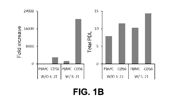
101031 FIG. 1B illustrates the fold increase and the population
doubling level
(PDL) of NK cells. Further, as illustrated in FIG. 1B, the PBMCs of
Comparative Example 1
(PBMC w/o IL-21) and Comparative Example 2 (PBMC w/ IL-21) were proliferated
243 and
1248 times respectively compared to Day 0, while the CD56+ NEC cells of
Example 1 (CD56
w/o IL-21) and Example 2 (CD56 w/ IL-21) were proliferated 2990 and 20434
times
respectively compared to Day 0.
Experimental Example 2. Confirmation of purity of CD56+ NI( cells
101041 The NEC cells of Examples 1, 2 and Comparative Examples 1, 2
were
washed once with a FACS staining buffer and suspended in 100 ttL, and then
stored at 2 to
8 C for 20 to 30 minutes under a dark condition after mixing with a monoclonal
antibody
binding with fluorescence. After one additional washing, the cells were
suspended in 300 to
-19-
CA 03089853 2020-07-28
WO 2019/152663 PCT/US2019/016076
500 111. of the FACS staining buffer and then 10,000 to 100,000 cells per tube
were obtained
and analyzed by using a CD56-FITC/CD3-PE/CD2O-PerCP5/CD14-APC panel of a flow
cytometer. The purity of the CD56+ NK cells was defined as a ratio of cells
introduced in a
CD3-/CD56+ region after FSC/SSC gating, and it was further confirmed that CD20
and
CD14 were not expressed in the cells in the CD3-/CD56+ region.
101051 As illustrated in FIG. 2, the purity of NK cells of Comparative
Example 1
(PBMCs w/o IL-21) and Comparative Example 2 (PBMCs w/ IL-21) were 84.2% and
84.7%
respectively, while the purity of NK cells of Example 1 (CD56 w/o IL-21) and
Example 2
(CD56 w/ IL-21) were 98.6% and 99.2% respectively.
Experimental Example 3. Confirmation of cancer cell eywtoxicity of NK cells
101061 First, the cytotoxicity against 1(562 cells (blood cancer, ATCCS
CCL-
243'), a chronic myelogenous leukemia cell line was confirmed.
101071 Before used in the experiment, IC562 cells were prepared by
subculturing
K562 cells suspended in a RPM' 1640 medium containing FBS 10%, at 37 1 C at an
interval
of three days, for 7 days or more.
101081 The prepared K562 cells were suspended in the RPMI-1640 medium
at a
concentration of 1.0 x 10⁶ cells/mL, and added with a fluorescent material
(Calcein-AM) at a
concentration of 4 1.t1V1. The K562 cells were stained at 37 1 C for 30
minutes, and then
inverted at an interval of ten minutes. The 1(562 cells stained with the
fluorescent material
were centrifuged at 3,300 rpm for 3 minutes, washed three times, and then
suspended in an
SNK medium containing FBS 10%, at a ratio of 1.0 x 10⁶ cells/mL. The K562
cells were
inoculated into a round bottom microwell plate (96-well) in an amount of 1.0 x
104 cells per
well.
101091 The NK cells of the Experimental Example 1 (effector cells) on
the 14 to
20th days of culture were suspended and diluted in a RPMI-1640 medium
containing FBS
10% at ratios of 1.0 x 10⁶ cells/mL, 3.0 x 10⁵ cells/mL, 1.0 x 10⁵ cells/mL
and 0.5 x 10⁵
cells/mL, respectively.
101101 The diluted effector cells were inoculated into the plate
inoculated with the
target cells (the K562 cells) at a concentration of 100 1.1L per well for
three wells each
-20-
CA 03089853 2020-07-28
WO 2019/152663 PCT/US2019/016076
(triplication), respectively. In this case, ratios of the effector cells and
the target cells are
shown in Table 2 below.
Table 2
Effector:Target Effector cells Target cells
: 1 1.0 x 10⁵ 1.0x 104
=
3 : 1 3.0 x 10⁴ 1.0 x 10⁴
1:1 1.0 x 10⁴ 1.0x 10⁴
0.5 : 1 0.5 x 10⁴ 1.0 x 10⁴
101111 The plate design used in the present experiment is shown in FIG.
3, in a
negative control group (Spontaneous), fluorescence-stained living IC562 cells
were added,
and in a positive control group (Maximum release), the K562 cells were
completely killed
using TX-100 and exhibited a maximum fluorescence.
10112] The plate inoculated with the target cells and the effector
cells was
centrifuged at 1000 rpm for 5 minutes, cultured at 37 1 C for 3 to 4 hours,
and then
centrifuged again at 1,000 rpm for 5 minutes. After centrifugation, 80 pi, of
a supernatant
was transferred to a black plate (96-well), and then a fluorescence amount was
measured
using a fluorescence microplate reader and the cytotoxicity against cancer
cells was
calculated using Equation 1 below.
Equation 1
Test ReIecise ¨ Spontaneous Release
Cytotexicity = X 100
Maximum Release ¨ Sponteneous Reiease
101131 FIG. 4A and Table 3 below show % of lysis of K562 cells at
various E:T
ratio. As illustrated in FIG. 4A and Table 3 below, as compared with
Comparative Example
3 (PBMCs w/o 1L-21) and Comparative Example 4 (PBMCs w/ 1L-21), the CD56+
cells
cultured according to Example 1 (CD56+ w/o IL-21) and Example 2 (CD56+ w/ IL-
21)
exhibited higher anti-cancer activity.
-21-
CA 03089853 2020-07-28
WO 2019/152663 PCT/US2019/016076
Table 3
% of lysis
E:T_(10:1) (3:1) LT (1:1) E:T (0.5:1)
PBMC(W/0 1L-21) 97.5 94.6 75.3 60.0
PBMC(W/ 1L-21) 102.3 97.1 84.8 66.9
CD56+(W/0 1L-21) 103.3 99.9 83.8 67.4
CD56+(W/ IL-21) 102.9 100.7 87.7 80.0
101141 Next, the cytotoxicity against solid tumor cells, which are
known to have
greater tolerance against NK cells, is confirmed. AGS (stomach cancer, ATCC
CRL-
1739"), A549 (lung cancer, ATCC CRL-185"), and MDA-MB0231 (breast cancer,
ATCC HTB-26") were used as solid tumor cell lines.
101151 Each solid tumor cells were tagged with green-fluorescent marker
using
CYTO-ID Green long-term tracer kit (Enzo Life Sciences Inc.), inoculated on a
plate, and
cultured for 24 hours. Next day, NK cells and cancer cells were reacted for 48
hours in 0.5:1
ratio. After 48 hours, cytotoxicity was confirmed by measuring the number of
cells
exhibiting green-fluorescence using flow cytometer.
101161 As illustrated in FIG. 4B, as compared with Comparative Example
1
(PBMCs w/o IL-21) and Comparative Example 2 (PBMCs w/ IL-21), the CD56+ cells
cultured according to Example 1 (CD56+ w/o IL-21) and Example 2 (CD56+ w/ IL-
21)
exhibited higher anti-cancer activity.
Experimental Example 4. Comparison of proliferative ability of NK cells
depending on
timing and number of 1L-21 treatment
[01.17] To evaluate the proliferative ability of NK cells according to
the timing of
IL-21 treatment, experiments as outlined below were conducted.
CA 03089853 2020-07-28
WO 2019/152663 PCT/US2019/016076
101181 CD56+ NK cells were produced according to the method of Example
1,
but treated with IL-21 (50ng/mL) during Day 0-6 (D0-6 group), Day 6-10 (D6-10
group),
Day 10-14 (D10-14 group), or Day 14-17 (D-14-17 group), and the proliferative
ability of the
CD56+ NK cells were compared using the method according to Experimental
Example 1.
101191 NK cells were treated with IL-21: for the DO-6 group, twice, on
Day 0 and
3; for the D6-10 group, once, on Day 6; for the D10-14 group, once, on Day 10;
for the D14-
17 group, once, on Day 14. For a control group, NK cells were not treated with
IL-21.
101201 As shown in FIG. 5A and Table 4, the D10-14 group and the D14-17
group did not exhibit significant difference in proliferative ability as
compared with the
control group, while the DO-6 group and the D6-10 group exhibited increased
proliferation
ability as compared with the control group. Especially, the DO-6 group
exhibited the greatest
expansion fold increase.
Table 4
Expansion Folds
control DO-6 1)6-10 D10-14 1)14-17
Donor 1 2996 21859 6388 2894 2330
101211 To evaluate the proliferative ability of NK cells according to
the number
of1L-21 treatments, experiments as outlined below were conducted.
101221 CD56+ NK cells were produced according to the method of Example
1,
but treated with IL-21 (50 ng/mL) during Day 0-3 (D0-3 group), Day 3-6 (D3-6
group), or
Day 0-6 (D0-6 group), and the proliferative ability of the CD56+ NK cells were
compared
using the method according to Experimental Example 1.
101231 NK cells were treated with IL-21: for the DO-3 group, once, on
Day 0; for
the D3-6 group, once, on Day 3; for the DO-6 group, twice, on Day 0 and 3. For
a control
group, NK cells were not treated with IL-21.
101241 As shown in FIG. 5B and Table 5, every group with IL-21
treatment
during earlier stage of the culture exhibited increased expansion fold as
compared with the
control group. Especially, DO-6 exhibited the greatest expansion fold
increase.
-23-
CA 03089853 2020-07-28
WO 2019/152663 PCT/US2019/016076
Table 5
Expansion Fold
control 1)0-3 D3-6 DO-6
Donor 1 2996 16420 4360 21859
Experimental Example 5. Comparison of proliferative ability of NK cells
depending on the
concentration of 1L-21 treatment
101251 CD56+ NK cells were produced according to the method of Example
1,
but treated with IL-21 with a concentration of 0 ng/mL, 10 ng/mL, 30 ng/mL, 50
ng/mL or
100 ng/mL twice, and the proliferative ability of the CD56+ NK cells were
compared using
the method according to Experimental Example 1.
101261 As shown in FIG. 6, even when treated with IL-21 with a
concentration of
ng/mL, the NK cells exhibited greater expansion fold as compared with the NK
cells with
no IL-21 treatment, and the expansion fold of the NK cells increased as the
concentration of
IL-21 increases between 10 ng/mL-50 ng/mL. However, when treated with IL-21
with a
concentration of 100 ng/mL, the NEC cells did not exhibit significant
difference in expansion
from the NK cells treated with IL-21 with a concentration of 50 ng/mL.
Experimental Example 6. Comparison of cytotoxicity of NEC cells depending on
the timing
and number of IL-2 1 treatment
101271 To evaluate the cytotoxicity of NK cells against cancer cells
according to
the timing of IL-21 treatment, experiments as outlined below were conducted.
101281 CD56+ NK cells were produced according to the method of Example
1,
but treated with IL-21 (50ng/mL) during Day 0-6 (D0-6 group), Day 6-10 (D6-10
group),
Day 10-14 (D10-14 group), or Day 14-17 (D-14-17 group), and the cytotoxicity
of the
CD56+ NK cells against blood cancer cells (I(562 cells, CCL-243') were
compared using
the method according to Experimental Example 3.
-24-
CA 03089853 2020-07-28
WO 2019/152663 PCT/US2019/016076
101291 NK cells were treated with IL-21: for the DO-6 group, twice, on
Day 0 and
3; for the D6-10 group, once, on Day 6; for the D10-14 group, once, on Day 10;
for the D14-
17 group, once, on Day 14. For a control group, NK cells were not treated with
IL-21.
101301 As shown in FIG. 7A and Table 6, all groups of NK cells with IL-
21
treatment, except the D14-17 group, exhibited greater anti-cancer activity as
compared with
the control group.
Table 6
ti:T ratio
, 10:1 3:1 1:1 0.5:1
Control (No treat) 98.8 96.9 73.1 55.1
1)0-6 98.6 97.0 77.8 68.4
D6-10 96.78 99.1 80.3 69.8
D10-14 98.4 96.6 68.5 52.4
D14-17 104.5 98.8 79.1 68.8
101311 Further, for each of the produced groups of CD56+ NK cells, the
cytotoxicity of the CD56+ NK cells against solid tumor cells were compared
using the
method according to Experimental Example 3. AGS (stomach cancer, ATCC CRL-
1739"), A549 (lung cancer, ATCC CRL-185"), and MDA-MB0231 (breast cancer,
ATCC HTB-26") were used as solid tumor cell lines.
101321 As shown in FIG. 7B, the NK cells with IL-21 treatment during an
earlier
stage of the culture (the DO-6 group) exhibited the greatest anti-cancer
activity against all
three types of solid tumor cells.
101331 To evaluate the cytotoxicity of the NK cells according to the
number of IL-
21 treatments, experiments as outlined below were conducted.
101341 CD56+ NK cells were produced according to the method of Example
1,
but treated with 1L-21 (50 ng/mL) during Day 0-3 (D0-3 group), Day 3-6 (D3-6
group), or
Day 0-6 (D0-6 group), and the cytotoxicity of the CD56+ NK cells against solid
tumor cells
were compared using the method according to Experimental Example 3. AGS
(stomach
-25-
CA 03089853 2020-07-28
WO 2019/152663 PCT/US2019/016076
cancer, ATCC CRL-1739"), A549 (lung cancer, ATCC CRL-185"), and MDA-
MB0231 (breast cancer, ATCC HTB-26") were used as solid tumor cell lines.
101351 NK cells were treated with 1L-21: for the DO-3 group, once, on
Day 0; for
the D3-6 group, once, on Day 3; for the DO-6 group, twice, on Day 0 and 3. For
a control
group, NK cells were not treated with IL-21.
101361 As shown in FIG. 7C, every group with IL-21 treatment during
earlier
stages of the culture exhibited greater anti-cancer activity as compared with
the control
group.
Experimental Example 7. Comparison of cytotoxicity of NK cells depending on
the
concentration of 1L-21 treatment
101371 CD56+ NK cells were produced according to the method of Example
1,
but treated with IL-21 with a concentration of 0 ng/mL, 10 ng/mL, 30 ng/mL, 50
ng/mL or
100 ng/mL twice, and the cytotoxicity of the CD56+ NK cells against blood
cancer cells
(K562 cells, CCL-243TM) were compared using the method according to
Experimental
Example 3.
101381 As shown in FIG. 8A, most NK cells treated with IL-21 exhibited
greater
cytotoxicity as compared with the NK cells with no IL-21 treatment, when
treated with IL-21
with a concentration of 100 ng/mL, the NK cells did not exhibit significant
difference in
expansion from the NK cells not treated with IL-21.
101391 CD56+ NK cells were produced according to the method of Example
1,
but treated with IL-21 with a concentration of 0 ng/mL, 10 ng/mL, 30 ng/mL, 50
ng/mL or
100 ng/mL twice, and the cytotoxicity of the CD56+ NK cells against solid
tumor cells (K562
cells, CCL-243") were compared using the method according to Experimental
Example 3.
AGS (stomach cancer, ATCC CRL-1739"), A549 (lung cancer, ATCC CRL-185"), and
MDA-MB023 1 (breast cancer, ATCC HTB-26") were used as solid tumor cell
lines.
-26-
CA 03089853 2020-07-28
WO 2019/152663 PCT/US2019/016076
[0140] As shown in FIG. 8B, the NK cells treated with IL-21 with a
concentration
of 50 ng/mL exhibited the greatest anti-cancer activity.
Experimental Example 8. Comparison of proliferative activity of NK cells
depending on the
number of feeder cell treatment
101411 To analyze whether multiple treatments with feeder cells would
sustain
proliferation of NK cells, the NK cells during the culture were treated with
feeder cells in an
interval of 14 days, and the expansion of NK cells were monitored for 42 days.
[0142] To further analyze the increase of NK cells expansion depending
on IL-21
treatment, the NK cells were treated with 1L-21 (50 ng/mL) twice in 3 days
interval, during a
six days period from each treatment with feeder cells (Day 0-6, 14-20, 28-34).
101431 As shown in FIG. 9, when treated with feeder cells twice or more
and IL-
21 together, the NK cells exhibited significantly increased expansion fold,
and the NK cells
treated with IL-21 exhibited greater expansion fold on Day 42, as compared
with the NK
cells not treated with IL-21 (approximately 3.4x101 vs. approximately
5.3x10⁸).
Experimental Example 9. Confirmation of the effect of culturing of NK cells
using blood of
certain cancer patients
101441 CD56+ NK cells were produced according to the method of Example
1 for
17 days, except that PBMCs of colorectal cancer patients was used. The
proliferative ability
and the purity of the produced NK cells was measured using methods according
to
Experimental Examples 1 and 2.
[0145] For some groups, the NK cells were treated with IL-21 with a
concentration of 50 ng/mL twice (Day 0 and Day 3 of culture), to confirm the
effect of IL-21
treatment.
[0146] As illustrated in FIG. 10A, the number of the NK cells not
treated with IL-
21 increased 8 times from Day 0, while when treated with IL-21, the number of
the NK cells
increased 1461 times from Day 0.
-27-
CA 03089853 2020-07-28
WO 2019/152663 PCT/US2019/016076
[0147] Further, as illustrated in FIG. 10B, the purity of the NK cells
not treated
with IL-21 was only 84.2%, while when treated with 1L-21, the purity of the NK
cells was
99.19%.
[0148] Further, the cytotoxicity of the NK cells treated with IL-21,
and NK cells
not treated with 1L-21 against blood cancer cells (K562 cells, CCL-243') were
compared
using the method according to Experimental Example 3. As illustrated in FIG.
10C, the NK
cells treated with IL-21 exhibited greater anti-cancer activity as compared
with the NK cells
not treated with IL-21.
101491 Also, Further, for each of the NK cells treated with 1L-21, and
NK cells
not treated with 1L-21, the cytotoxicity of the NK cells against solid tumor
cells were
compared using the method according to Experimental Example 3. AGS (stomach
cancer,
ATCC CRL-1739'), A549 (lung cancer, ATCC CRL-185'), and MDA-MB-231 (breast
cancer, ATCC HTB-26') were used as solid tumor cell lines. As illustrated in
FIG. 10D,
the NK cells treated with 1L-21 exhibited greater anti-cancer activity as
compared with the
NK cells not treated with IL-21.
[0150] Accordingly, by using methods as set forth herein, it may be
possible to
produce NK cells even for certain cancer patients who do not usually show an
enough growth
of NK cells.
Experimental Example 10. Confirmation of the survival rate of NK cells in
therapeutic
composition
101511 With respect to each of the NK cells cultured in a CO2 incubator
according
to Examples 1, 2 on Day 6 of culture, cells were inoculated into a 350 mL bag
on the basis of
the cell number of 1.0 x 10⁵ to 2.0 x 10⁶ /mL and further cultured for 4 days.
On Day 10 of
culture, the cells were inoculated into a 1 L bag on the basis of the cell
number of 1.0 x 10⁵ to
2.0 x 10⁶ /mL and then further cultured for 4 days. Finally, on Day 14 of
culture, the cells
were inoculated into a 1 L bag on the basis of the cell number of 1.0 x 10⁵ to
2.0 x 10⁶ /mL
and then further cultured for 3 to 6 days.
-28-
CA 03089853 2020-07-28
WO 2019/152663 PCT/US2019/016076
101521 The
CD56+ NK cells on the 14th to 20th days of culture were washed
three times and then suspended in a base compound (physiological saline and
Hartman's
solution) containing 1% albumin to be 2 x I07 /mL. The cells were stored at 4
C for 48
hours and then the cell survival rate was measured.
101531
Further, in order to compare the effect of IL-2, the CD56+ NK cells were
washed and suspended in a base compound containing 1% albumin (physiological
saline, and
Hartmann's solution or phosphate buffered saline), and then added with IL-2 at
a
concentration of 500 IU/mL. After being kept in 4 C for 48 hours, cell
survival rate was
measured.
101541 100
J.LL of each composition was taken to obtain a total of 2 x 10⁶ CD56+
NK cells, washed once with 1 mL of the FACS staining buffer, centrifuged and
suspended in
100 pL of an armexin V binding buffer. 5 pi, of Annexin V-F1TC and 5 pL of 7-
AAD
(Biolegend) were added in the suspension and mixed well, stored in a dark
condition, and
reacted at room temperature for 15 minutes, and then added with 400 of an
Annexin V
binding buffer before flow cytometry and mixed for 5 seconds. Thereafter,
10,000 to
100,000 cells per tube were obtained and analyzed. A cut-off was determined by
setting a
test tube which was not stained with the Annexin V-FITC and the 7-AAD as a
negative
control, and the survival rate was represented by a percentage of fraction of
cells in which the
Annexin V-FITC or the 7-AAD was negative.
101551 As
illustrated in FIG. 11, when treated 11,-2 (WI IL2), the apoptosis of the
CD56+ NK cells was inhibited.
Experimental Example 11. Confirmation of the cytotoxicity of NK cells in
therapeutic
composition
101561 With
respect to each of the NK cells cultured in a CO2 incubator according
to Examples 1, 2 on Day 6 of culture, cells were inoculated into a 350 mL bag
on the basis of
the cell number of 1.0 x 10⁵ to 2.0 x 10⁶ /mL and further cultured for 4 days.
On Day 10 of
culture, the cells were inoculated into a 1 L bag on the basis of the cell
number of 1.0 x 10⁵ to
2.0 x 10⁶ /mL and then further cultured for 4 days. Finally, on Day 14 of
culture, the cells
-29-
CA 03089853 2020-07-28
WO 2019/152663 PCT/US2019/016076
were inoculated into a 1 L bag on the basis of the cell number of 1.0 x 10⁵ to
2.0 x 10⁶ /mL
and then further cultured for 3 to 6 days.
[0157] Before used in the experiment, the cancer cell lines were
prepared by
suspending under the following conditions, and sub-culturing at 37 1 C at an
interval of
three days for 1 week or more:
[0158] CCRF-SB (blood cancer, ATCC CCL-120Tm), AGS (stomach cancer,
ATCC CRL-1739) and MIA-PACA2 (pancreatic cancer, ATCC CRL-1420Tm): RPM!
medium + 10% FBS,
[0159] SNU245 (cholangiocarcinoma, KCLB No. 00245), HCT15 (colon
cancer,
ATCC CCL-225Tm) and NIH:OVCAR-3 (ovarian cancer, ATCC HTB-161'): RPM!
medium + 10% FBS +25 mM HEPES, and
101601 MDA-MB-231 (breast cancer, ATCC HTB-26): DMEM medium +
10% FBS.
101611 The cancer cell lines (except for a blood cancer cell line)
during culturing
were detached from a culture dish using trypsin and suspended in the medium to
be 5 x
104/mL, and then inoculated into a 24-well plate by 1 mL per well and attached
for one day.
To distinguish from the NK cells, the blood cancer cell line, which was a
suspended culture
cell, was labeled with green fluorescence, suspended in the medium to be 5 x
104/mtõ and
inoculated into a 24-well plate by 1 mL per well.
101621 First, in a 24-well plate inoculated with AGS (stomach cancer,
ATCC
CRL-1739'M), M1A-PACA2 (pancreatic cancer, ATCC CRL-1420), SNU245
(cholangiocarcinoma, KCLB No. 00245), HCT15 (colon cancer, ATCC CCL-225) and
MDA-MB-231 (breast cancer, ATCC HTB-261m) among the cancer cell lines, the
CD56+
NK cells were added after one day to observe the cytotoxicity, and ratios of
effector cells
(CD56+ NK cells) and target cells (cancer cell lines) are shown in Table 7
below.
Table 7
Effector : Target Effector cell Target cell
0 : 1 0 5 x 10⁴
1 : 1 5 x 10⁴ 5x10
-30-
CA 03089853 2020-07-28
WO 2019/152663
PCT/US2019/016076
1 : 10 5 x 10³ 5 x 10'
1 : 20 2.5x 10³ 5x10
101631 The plate inoculated with the effector cells and the target
cells was
cultured at 37 1 C for 1 to 3 days, and at this time, in order to observe
whether anti-cancer
activity is increased by IL-2, 500 IU/m1 of IL-2 was further added to an
experimental group.
In a negative control group, the CD56+ NK cells (effector cells) were not
added and there
was no anti-cancer activity reaction.
101641 After 1 to 3 days of culture, the cells were washed with RPMI
three times
to remove the CD56+ NK cells present in the suspended form, and then the
cancer cell lines
remaining in the wells were detached using trypsin, stained with trypan blue
and counted.
Subsequently, the plate inoculated with the target cells and the effector
cells was cultured at
37 1 C for 1 to 3 days, and then the cells labeled with green fluorescence
present in the 24
wells were counted using a flow cytometer.
101651 The cytotoxicity for the cancer cell line was calculated using
Equation 2
below.
Equation 2
aiv,nunther of fluctrescnoce cells >.n weis with NK cells
Cytotoxici* ¨ X100
ars. numlnr of fluorescence +cells well with target cells= only
101661 As a result, as illustrated in FIG. 12, it was confirmed that
the cancer cell
cytotoxicity was increased when IL-2 was treated together (W/ IL2), as
compared to when
only the CD56+ NK cells were treated (W/O IL2).
101671 Next, the CD56+ NK cells were added in the 24-well plate to
which
NIH:OVCAR-3 (ovarian cancer, ATCC HTB-161Tm) cells was attached among the
cancer
cell lines to observe the cytotoxicity, and ratios of target cells (cancer
cell lines) and effector
cells (CD56+ NK cells) were shown in Table 8 below.
Table 8
Effector : Target Effector cells Target cells
-31-
CA 03089853 2020-07-28
WO 2019/152663 PCT/US2019/016076
0 : 1 0 5 x 10⁴
1 : 1 5 x 10⁴ 5 x 10⁴
0.1 : 1 5 x 10³ 5 x 10⁴
0.05 : 1 2.5 x 10-; 5x 10⁴
101681 The plate inoculated with the effector cells and the target
cells was
cultured at 37 1 C for 1 days, and, in order to observe whether anti-cancer
activity is
increased by IL-2, 500 IU/m1 of IL-2 was further added to an experimental
group. In a
negative control group, the CD56+ NK cells were not added and there was no
anti-cancer
activity reaction.
101691 After 1 day of culture, the cells were washed with RPM! three
times to
remove the CD56+ NK cells present in the suspended form, and then the cancer
cell lines
remaining in the wells were photographed using a camera.
101701 As a result, as illustrated in FIG. 13, the cancer cell
cytotoxicity was
increased when cancer cell lines were treated together with 1L-2(+ IL2), as
compared to when
cancer cell lines were treated with only the CD56+ NK cells(-IL2).
101711 Next, the CD56+ NK cells were added in the 24-well plate to
which AGS
(stomach cancer, ATCC CRL-1739) cells was attached among the cancer cell
lines to
observe the cytotoxicity, and ratios of target cells (cancer cell lines) and
effector cells
(CD56+ NK cells) were shown in Table 9 below.
Table 9
Effector : Target Effector cells Target cells
0:1 0 5 x 10⁴
1:1 5 x 10⁴ 5 x 10⁴
0.1:1 5 x 10³ 5 x 10⁴
0.05:1 2.5x 10³ 5 x 10⁴
101721 The plate inoculated with the effector cells and the target
cells was
cultured at 37 1 C for 1 days, and, in order to observe whether anti-cancer
activity is
increased by IL-2, 500 IU/ml of IL-2 was further added to an experimental
group. In a
-32-
CA 03089853 2020-07-28
WO 2019/152663 PCT/US2019/016076
negative control group, the CD56+ NK cells were not added and there was no
anti-cancer
activity reaction.
101731 After 1 day of culture, the cells were washed with RPMI three
times to
remove the CD56+ NK cells present in the suspended form, and then the cancer
cell lines
remaining in the wells were photographed using a camera.
101741 As a result, as illustrated in FIG. 14, the cancer cell
cytotoxicity was
increased when cancer cell lines were treated together with IL-2 (+1L2), as
compared to when
cancer cell lines were treated with only the CD56+ NK cells (-IL2).
Experimental Example 12. Confirmation of anticancer effect of NK cells in
animal models
101751 CD56+ NK cells are produced according to the method of Examples
1, 2
and Comparative Examples 1, 2 for 17 days, except that PBMCs of colorectal
cancer patients
is used. With respect to each of the NK cells cultured in a CO2 incubator
according to
Examples 1, 2 and Comparative Examples 1, 2, on Day 6 of culture in a T 25
culture flask,
cells are inoculated into a 350 mL bag on the basis of the cell number of 1.0
x 105 to 2.0 x 10⁶
/mL and further cultured for 4 days. On Day 10 of culture, the cells are
inoculated into a 1 L
bag on the basis of the cell number of 1.0 x 10⁵ to 2.0 x 10⁶ /mL and then
further cultured for
4 days. Finally, on Day 14 of culture, the cells are inoculated into a 1 L bag
on the basis of
the cell number of 1.0 x 10⁵ to 2.0 x 10⁶ /mL and then further cultured for 3
to 6 days.
101761 Animal models of human cancer are constructed by xenograft of
human
cancer cell line into mice. Following human cancer cell lines are used: AGS
(stomach
cancer), MIA-PACA2 (pancreatic cancer), SNU245 (cholangiocarcinoma), HCT15
(colon
cancer) and NIH:OVCAR-3 (ovarian cancer), and MDA-MB-231 (breast cancer).
After
xenograft of cancer, the mice are grouped randomly and marked. The control
group is
injected 200 ttL of Hartmann's solution into the vein of tail. The NK cell-
treated (+IL-2)
group is injected five times with 1 x10⁷ NK cells/200 1.1L and 500 IU/mL of 1L-
2 at 2-3-day
intervals from 1 week after xenograft of cancers into the vein of tail. The NK
cell-treated (-
IL-2) group is injected five times with 1 x10⁷ NK cells/200 !IL at 2-3-day
intervals from 1
week after xenograft of cancers into the vein of tail.
-33-
CA 03089853 2020-07-28
WO 2019/152663 PCT/US2019/016076
101771 To follow up tumor growth, during the study period, mice are
tested for
body weight and tumor volume three time a week. Length of major axis and minor
axis are
measured using a caliper and tumor volume is determined according to the
following
equation (Equation 3).
Equation 3
Tumor Volume (mm3) = (length of major axis (mm)) x (length of minor axis
(mm))2 x 0.5
101781 The NK cell-treated (-IL-2) group exhibits a reduction in tumor
growth
after 8 weeks treatment of approximately 50% compared to the control group
based on
luciferase images for each cancer type. The NK cell-treated (+IL-2) group
exhibits a further
reduction in tumor growth of approximately 60% compared to the control group
for each
cancer type.
Experimental Example 13. Confirmation of anticancer effect of NK cells in
cancer patients
(01791 CD56+ NK cells are produced according to the method of Examples
1, 2
and Comparative Examples 1, 2 for 18 days, except that PBMCs of colorectal
cancer patients
is used. With respect to each of the NK cells cultured in a CO2 incubator
according to
Examples 1, 2 and Comparative Examples 1, 2, on Day 6 of culture in a T 25
culture flask,
cells are inoculated into a 350 mL bag on the basis of the cell number of 1.0
x 105 to 2.0 x 10⁶
/mL and further cultured for 4 days. On Day 10 of culture, the cells are
inoculated into a 1 L
bag on the basis of the cell number of 1.0 x 10⁵ to 2.0 x 10⁶ /mL and then
further cultured for
4 days. Finally, on Day 14 of culture, the cells are inoculated into a 1 L bag
on the basis of
the cell number of 1.0 x 10⁵ to 2.0 x 10⁶ /mL and then further cultured for 3
to 6 days.
101801 The colorectal cancer patients are grouped randomly and marked.
The
control group is not injected with NK cells. The NK cell-treated (+IL-2) group
is injected
three times with 1-3x10⁷ NK cells/ per kg of body weight and 500 IU/mL of IL-2
at six week
intervals intravenously. The NK cell-treated (-IL-2) group is injected six
times with 1-3x10⁷
NK cells/ per kg of body weight at six week intervals intravenously.
101811 Tumor growth is monitored at 1, 3, 6, 12 months. After 12
months, the
NK cell-treated (-IL-2) group exhibits overall decrease in tumor size, and the
NK cell-treated
(+IL-2) group exhibits further overall decrease in tumor size.
-34-
CA 03089853 2020-07-28
WO 2019/152663 PCT/US2019/016076
Experimental Example 14. Confirmation of anticancer effect of NK cells in
Alzheimer's
Disease patients
101821 CD56+ NK cells are produced according to the method of Examples
1, 2
and Comparative Examples 1, 2 for 18 days, except that PBMCs of colorectal
cancer patients
is used. With respect to each of the NK cells cultured in a CO2 incubator
according to
Examples 1, 2 and Comparative Examples 1, 2, on Day 6 of culture in a T 25
culture flask,
cells are inoculated into a 350 mL bag on the basis of the cell number of 1.0
x 105 to 2.0 x 10⁶
/mL and further cultured for 4 days. On Day 10 of culture, the cells are
inoculated into a 1 L
bag on the basis of the cell number of 1.0 x 10⁵ to 2.0 x 10⁶ /mL and then
further cultured for
4 days. Finally, on Day 14 of culture, the cells are inoculated into a 1 L bag
on the basis of
the cell number of 1.0 x 10⁵ to 2.0 x 10⁶ /mt. and then further cultured for 3
to 6 days.
101831 Alzheimer's Disease patients are grouped randomly and marked.
The
control group is not injected with NK cells. The NK cell-treated group is
injected six times
with 1-3x10⁷ NK cells/ per kg of body weight and 500 IU/mL of IL-2 at weekly
intervals
intravenously.
101841 Cognitive function of the patient are monitored at 1, 3, 6, 12
months.
After 12 months, the NK cell-treated group exhibits improved cognitive
function.
Experimental Example 15. Confirmation of anticancer effect of NK cells in
autoimmune
disease patients
101851 CD56+ NK cells are produced according to the method of Examples
1, 2
and Comparative Examples 1, 2 for 18 days, except that PBMCs of colorectal
cancer patients
is used. With respect to each of the NK cells cultured in a CO2 incubator
according to
Examples 1, 2 and Comparative Examples 1, 2, on Day 6 of culture in a T 25
culture flask,
cells are inoculated into a 350 mL bag on the basis of the cell number of 1.0
x 105 to 2.0 x 10⁶
/mL and further cultured for 4 days. On Day 10 of culture, the cells are
inoculated into a 1 L
bag on the basis of the cell number of 1.0 x 10⁵ to 2.0 x 10⁶ /mL and then
further cultured for
4 days. Finally, on Day 14 of culture, the cells are inoculated into a 1 L bag
on the basis of
the cell number of 1.0 x 10⁵ to 2.0 x 10⁶ /mL and then further cultured for 3
to 6 days.
-35-
CA 03089853 2020-07-28
WO 2019/152663 PCT/US2019/016076
101861 Multiple sclerosis patients are grouped randomly and marked. The
control
group is not injected with NK cells. The NK cell-treated group is injected six
times with 1 -
3x10⁷ NK cells/ per kg of body weight and 500 IU/mL of 11,2 at weekly
intervals
intravenously.
101871 Cognitive function of the patient are monitored at 1, 3, 6, 12
months.
After 12 months, the NK cell-treated group exhibits improved cognitive
function.
Terminology
101881 The foregoing description of the exemplary embodiments has been
presented only for the purposes of illustration and description and is not
intended to be
exhaustive or to limit the invention to the precise forms disclosed. Many
modifications and
variations are possible in light of the above teaching. It is contemplated
that various
combinations or sub combinations of the specific features and aspects of the
embodiments
disclosed above may be made and still fall within one or more of the
inventions. Further, the
disclosure herein of any particular feature, aspect, method, property,
characteristic, quality,
attribute, element, or the like in connection with an embodiment can be used
in all other
embodiments set forth herein. Accordingly, it should be understood that
various features and
aspects of the disclosed embodiments can be combined with or substituted for
one another in
order to form varying modes of the disclosed inventions. Thus, it is intended
that the scope
of the present inventions herein disclosed should not be limited by the
particular disclosed
embodiments described above. Moreover, while the invention is susceptible to
various
modifications, and alternative forms, specific examples thereof have been
shown in the
drawings and are herein described in detail. It should be understood, however,
that the
invention is not to be limited to the particular forms or methods disclosed,
but to the contrary,
the invention is to cover all modifications, equivalents, and alternatives
falling within the
spirit and scope of the various embodiments described and the appended claims.
Any
methods disclosed herein need not be performed in the order recited. The
methods disclosed
herein include certain actions taken by a practitioner; however, they can also
include any
third-party instruction of those actions, either expressly or by implication.
The ranges
disclosed herein also encompass any and all overlap, sub-ranges, and
combinations thereof.
-36-
CA 03089853 2020-07-28
WO 2019/152663 PCT/US2019/016076
101891 The embodiments were chosen and described in order to explain
the
principles of the invention and their practical application so as to activate
others skilled in the
art to utilize the invention and various embodiments and with various
modifications as are
suited to the particular use contemplated. Alternative embodiments will become
apparent to
those skilled in the art to which the invention is defined by the appended
claims rather than
the foregoing description and the exemplary embodiments described therein.
101901 Conditional language, such as "can," "could," "might," or "may,"
unless
specifically stated otherwise, or otherwise understood within the context as
used, is generally
intended to convey that certain embodiments include, while other embodiments
do not
include, certain features, elements, and/or steps. Thus, such conditional
language is not
generally intended to imply that features, elements, and/or steps are in any
way required for
one or more embodiments.
[01911 The terms "comprising," "including," "having," and the like are
synonymous and are used inclusively, in an open-ended fashion, and do not
exclude
additional elements, features, acts, operations, and so forth. Also, the term
"or" is used in its
inclusive sense (and not in its exclusive sense) so that when used, for
example, to connect a
list of elements, the term "or" means one, some, or all of the elements in the
list.
101921 The ranges disclosed herein also encompass any and all overlap,
sub-
ranges, and combinations thereof. Language such as "up to," "at least,"
"greater than," "less
than," "between," and the like includes the number recited.
101931 Numbers preceded by a term such as "approximately", "about", and
"substantially" as used herein include the recited numbers (e.g., about 10%
10%), and also
represent an amount close to the stated amount that still performs a desired
function or
achieves a desired result. For example, the terms "approximately", "about",
and
"substantially" may refer to an amount that is within less than 10% of, within
less than 5% of,
within less than 1% of, within less than 0.1% of, and within less than 0.01%
of the stated
amount.
101941 The term "generally" as used herein represents a value, amount,
or
characteristic that predominantly includes or tends toward a particular value,
amount, or
characteristic. As an example, in certain embodiments, the term "generally
uniform" refers to
-37-
CA 03089853 2020-07-28
WO 2019/152663 PCT/US2019/016076
a value, amount, or characteristic that departs from exactly uniform by less
than 20%, less
than 15%, less than 10%, less than 5%, less than 1%, less than 0.1%, and less
than 0.01%.
101951 The ranges disclosed herein also encompass any and all overlap,
sub-
ranges, and combinations thereof. Language such as "up to," "at least,"
"greater than," "less
than," "between" and the like includes the number recited. Numbers preceded by
a term such
as "about" or "approximately' include the recited numbers. For example, "about
5.0 cm"
includes "5.0 cm."
101961 Some embodiments have been described in connection with
schematic
drawings. However, it should be understood that the schematic drawings are not
drawn to
scale. Distances are merely illustrative and do not necessarily bear an exact
relationship to
actual dimensions and layout of the devices illustrated.
101971 For purposes of this disclosure, certain aspects, advantages,
and novel
features are described herein. It is to be understood that not necessarily all
such advantages
may be achieved in accordance with any particular embodiment. Thus, for
example, those
skilled in the art will recognize that the disclosure may be embodied or
carried out in a
manner that achieves one advantage or a group of advantages as taught herein
without
necessarily achieving other advantages as may be taught or suggested herein.
101981 Moreover, while illustrative embodiments have been described
herein, the
scope of any and all embodiments having equivalent elements, modifications,
omissions,
combinations (e.g., of aspects across various embodiments), adaptations and/or
alterations as
would be appreciated by those in the art based on the present disclosure. The
limitations in
the claims are to be interpreted broadly based on the language employed in the
claims and not
limited to the examples described in the present specification or during the
prosecution of the
application, which examples are to be construed as non-exclusive. Further, the
actions of the
disclosed processes and methods may be modified in any manner, including by
reordering
actions and/or inserting additional actions and/or deleting actions. It is
intended, therefore,
that the specification and examples be considered as illustrative only, with a
true scope and
spirit being indicated by the claims and their full scope of equivalents.
-38-