Note : Les descriptions sont présentées dans la langue officielle dans laquelle elles ont été soumises.
CA 03090687 2020-07-08
WO 2019/140332 PCT/US2019/013386
DEVICES AND METHODS FOR INTRODUCING AN ENDOTRACHEAL TUBE
BACKGROUND
[0001] Many surgical procedures are typically performed while the patient
is under general
anesthesia. During these procedures, the patient is given a combination of
medications to cause a
loss of consciousness and muscle paralysis. The medications that cause loss of
consciousness and
muscle paralysis also interfere with the patient's ability to breath.
Accordingly, patients often
undergo tracheal intubation during these procedures so that the patient may be
connected to an
external ventilator or breathing circuit. Patients may also be intubated for
nonsurgical conditions
in which enhanced oxygen delivery is required. Tracheal intubation may also be
used in other
circumstances.
[0002] During tracheal intubation, an endotracheal tube is placed in the
patient's airway.
Generally, the endotracheal tube is advanced through the patient's nose or
mouth into the
patient's trachea. The endotracheal tube is then connected to an external
ventilator or breathing
circuit. The ventilator is then able to breath for the patient, delivering
oxygen into the patient's
lungs.
[0003] The patient's vocal cords and the space between them form the
entrance to the
trachea, these structures are also known as the glottis. The glottis is
visible from and may be
accessed through the pharynx. The pharynx is the portion of the upper airway
that is located
behind the patient's mouth and below the patient's nasal cavity. The mouth and
the nasal cavity
meet in the pharynx. Additionally, the esophagus and the glottis may be
accessed through the
pharynx. During the intubation process, the endotracheal tube must be
carefully advanced
through the patient's pharynx and placed through the vocal cords into the
trachea. In addition, it
is critical that the endotracheal tube be placed at the proper depth once in
the trachea. If it is
placed to shallow in the trachea, it can fall out. If it is placed too deep,
only one lung may be
ventilated resulting in poor oxygen delivery to the blood or hyperventilation
on the ventilated
lung, and hypoventilation to the non-ventilated lung. All of this can result
in patient injury or
death.
[0004] The intubation process interferes with the patient's ability to
breathe and thus deliver
oxygen to the body independently. If the patient is without oxygen for more
than two or three
minutes, tissue injury may occur, which can lead to death or permanent brain
damage.
Accordingly, the intubation process must be performed quickly and accurately.
1
CA 03090687 2020-07-08
WO 2019/140332 PCT/US2019/013386
SUMMARY
[0005] In general terms, this disclosure is directed to an introducer for
use with a tracheal
intubation system. In one possible configuration and by non-limiting example,
the tracheal
intubation system allows a medical professional to properly position an
endotracheal tube in a
normal or difficult airway quickly, accurately, and safely. In another
configuration and by non-
limiting example, the tracheal intubation system allows a medical professional
to properly
perform an endotracheal tube exchange procedure quickly, accurately, and
safely. One aspect is
an introducer for mounting an endotracheal tube, the introducer comprising: a
shaft comprising:
a proximal shaft portion; and a distal shaft portion comprising a distal tip
portion extending from
the distal shaft portion, the shaft including a plurality of depth assessment
bands, each depth
assessment band having a visually distinct color or pattern from an adjacent
depth assessment
band, and the distal tip portion having a rounded shape and a closed end; and
a handle
removeably connected to the proximal shaft portion..
[0006] Another aspect is a method for inserting an endotracheal tube in a
patient comprising:
inserting a blade of a laryngoscope in a mouth of the patient; viewing a
trachea of the patient
with the laryngoscope; inserting an introducer comprising a handle into a
trachea of the patient;
removing the handle from the introducer; inserting the endotracheal tube over
the introducer and
into the trachea of the patient; and removing the introducer from the
endotracheal tube, while the
endotracheal tube remains in the patient.
[0007] A further aspect is an introducer for mounting an endotracheal tube,
the introducer
comprising: a shaft comprising: a proximal shaft portion; a distal shaft
portion comprising a
distal tip portion extending from the shaft portion, the shaft comprises a
plurality of qualitative
depth assessment band, each depth assessment band having a visually distinct
color or pattern
from an adjacent depth assessment band, and a tip having a round shape and a
closed end; and a
control wire at least partially disposed within both the distal shaft portion
and the proximal shaft
portion and configured to cause the tip portion of the proximal shaft portion
to maintain a curved
configuration.
DESCRIPTION OF THE DRAWINGS
[0008] FIG. 1 is a diagram of an example tracheal intubation system
including a
laryngoscope being used to intubate a patient.
2
CA 03090687 2020-07-08
WO 2019/140332 PCT/US2019/013386
[0009] FIG. 2 is a perspective view of an example laryngoscope.
[0010] FIG. 3 is a perspective view of an example introducer.
[0011] FIG. 4 is a perspective view of an example endotracheal tube.
[0012] FIG. 5 is a flowchart of an example process of placing an
endotracheal tube in a
patient using an example tracheal intubation system including a laryngoscope.
[0013] FIG. 6 is a cross-sectional view of a patient after a laryngoscope
is positioned to view
the glottis during an intubation procedure using an example tracheal
intubation system including
a laryngoscope.
[0014] FIG. 7 is a cross-sectional view of a patient after the tip of the
introducer is advanced
into the field of view of a laryngoscope during an intubation procedure using
an example
tracheal intubation system including a laryngoscope.
[0015] FIG. 8 is a cross-sectional view of a patient after the tip of an
introducer is advanced
into the trachea to a second depth-assessment band during an intubation
procedure using an
example tracheal intubation system including a laryngoscope.
[0016] FIG. 9 is a cross-sectional view of a patient after an endotracheal
tube is advanced
over the introducer into the field of view of the laryngoscope during an
intubation procedure
using an example tracheal intubation system including a laryngoscope.
[0017] FIG. 10 is a cross-sectional view of a patient after an endotracheal
tube is advanced
over the introducer into a final position in the trachea during an intubation
procedure using an
example tracheal intubation system including a laryngoscope.
[0018] FIG. ha is a perspective view of a push-button introducer in a
resting configuration.
[0019] FIG. 1 lb is a perspective view of a push-button introducer in a
straight configuration.
[0020] FIG. 12 is an illustration of an introducer with a handle.
[0021] FIG. 13 is a perspective view of an introducer with a handle.
[0022] FIG. 14 is a side elevation view of a device that includes an
introducer and a handle.
[0023] FIG. 15A is a side view of a device that includes an introducer and
a handle.
[0024] FIG. 15B is a side view of a device that includes an introducer and
a handle.
[0025] FIG. 16A is a top plan view of a handle in a first position.
[0026] FIG. 16B is a side elevation view of the handle of FIG. 16A in the
first position.
[0027] FIG. 16C is a top plan view of the handle of FIG. 16A in a second
position.
[0028] FIG. 16D is a side elevation view of the handle of FIG. 16A in the
second position.
3
CA 03090687 2020-07-08
WO 2019/140332 PCT/US2019/013386
[0029] FIG. 17A is a top plan view of a handle in a first position.
[0030] FIG. 17B is a side elevation view of the handle of FIG. 17A in the
first position.
[0031] FIG. 17C is a top plan view of the handle of FIG. 17A in a second
position.
[0032] FIG. 17D is a side elevation view of the handle of FIG. 17A in the
second position.
[0033] FIG. 18A is a top plan view of a handle in a first position.
[0034] FIG. 18B is a side elevation view of the handle of FIG. 18A in the
first position.
[0035] FIG. 18C is a top plan view of the handle of FIG. 18A in a second
position.
[0036] FIG. 18D is a side elevation view of the handle of FIG. 18A in the
second position.
[0037] FIG. 19A is a top plan view of a handle in a first position.
[0038] FIG. 19B is a side elevation view of the handle of FIG. 19A in the
first position.
[0039] FIG. 19C is a top plan view of the handle of FIG. 19A in a second
position.
[0040] FIG. 19D is a side elevation view of the handle of FIG. 19A in the
second position.
DETAILED DESCRIPTION
[0041] Various embodiments will be described in detail with reference to
the drawings,
wherein like reference numerals represent like parts and assemblies throughout
the several
views. Reference to various embodiments does not limit the scope of the claims
attached hereto.
Additionally, any examples set forth in this specification are not intended to
be limiting and
merely set forth some of the many possible embodiments for the appended
claims.
[0042] The present disclosure relates generally to an introducer that is
usable with a tracheal
intubation system. An introducer is a slender probe that is used to guide
placement of an
endotracheal tube. Introducer are also sometimes referred to a stylet or
catheter. This disclosure
also relates to methods of performing tracheal intubation and endotracheal
tube exchange
procedures.
[0043] Introducers are used to help guide an endotracheal tube into place
in a patient. It can
be difficult to place an endotracheal tube in patients who have abnormal
airways, are overweight,
have undergone trauma, have arthritis, have had cervical fusions, or are
combative. An
introducer helps place an endotracheal tube in a patient when placing the
endotracheal tube
independently it otherwise not possible.
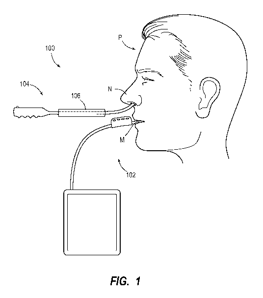
[0044] FIG. 1 is a diagram of an example tracheal intubation system 100
including a
laryngoscope being used to intubate a patient P. The example intubation system
100 includes a
4
CA 03090687 2020-07-08
WO 2019/140332 PCT/US2019/013386
laryngoscope 102, an introducer 104, and an endotracheal tube 106. Also
illustrated are the
mouth M and the nose N of the patient P. In this example, the laryngoscope 102
is inserted into
the mouth M of the patient P, the introducer 104 is inserted into the nose N
of the patient P, and
the endotracheal tube 106 is mounted on the introducer 104. Alternatively, the
introducer 104 is
inserted into the mouth M of the patient P.
[0045] The patient P is a person or animal who is being intubated. Although
the intubation
system 100 is particularly useful to intubate a patient with a difficult
airway, the intubation
system 100 may also be used on a patient with a normal airway. Examples of
patient P include
adults, children, infants, elderly people, obese people, people with tumors
affecting the head or
neck, and people with unstable cervical spines. In some embodiments, the
intubation system 100
may be used to intubate animals with normal or difficult airways. The
intubation system 100 may
be used to intubate other people or animals as well.
[0046] The laryngoscope 102 is a medical instrument configured to permit a
medical
professional to directly or indirectly view, among other things, the glottis
of the patient P. In
some embodiments, the laryngoscope 102 includes a blade with an integrated
optical capture
device and light source. In some embodiments, the blade is configured to be
inserted through the
mouth M of the patient P and positioned so that the glottis is in the field of
view of the optical
capture device. The image captured by the laryngoscope 102 is viewed from a
position that is
external to the patient P. In some embodiments, the image captured by the
laryngoscope 102 is
viewed on an external display device, such as a screen. The laryngoscope 102
is illustrated and
described in more detail with reference to FIG. 2.
[0047] The introducer 104 is a device that is inserted into the patent P's
airway. In this
example, the introducer 104 is used to guide the placement of the endotracheal
tube 106. The
introducer 104 includes a thin, flexible tube that may be directed and
advanced into the airway of
the patient P. The introducer 104 may be configured to be viewed with the
laryngoscope 102
during the intubation procedure. The introducer 104 is illustrated and
described in more detail
throughout the application, including with reference to FIGS. 12-33.
[0048] In some embodiments, the endotracheal tube 106 is a hollow tube that
is configured
to be placed in the airway of the patient P. When the patient P is intubated,
one end of the
endotracheal tube 106 is disposed inside the trachea of the patient P and the
other end is
connected to an external ventilator or breathing circuit. The endotracheal
tube 106 is configured
CA 03090687 2020-07-08
WO 2019/140332 PCT/US2019/013386
to occlude the airway of the patient P. Thus, gases (e.g., room air,
oxygenated gases, anesthetic
gases, expired breath, etc.) may flow into and out of the trachea of the
patient P through the
endotracheal tube 106. In some embodiments, the endotracheal tube 106 may be
connected to a
breathing circuit, including for example a machine-powered ventilator or a
hand-operated
ventilator. In other embodiments, the patient P may breathe through the
endotracheal tube 106
spontaneously. The endotracheal tube 106 is illustrated and described in more
detail with
reference to FIG. 4.
[0049] The endotracheal tube 106 is configured to be mounted on the
introducer 104 by
sliding over the tip and along the shaft of the introducer 104. After a
medical professional has
positioned the tip of the introducer 104 in the trachea of the patient P, the
endotracheal tube 106
is advanced over the shaft of the introducer 104 and into the trachea of the
patient P. In this
manner, the introducer 104 guides the endotracheal tube 106 into the proper
location in the
trachea of the patient P. The process of positioning the endotracheal tube 106
is illustrated and
described in more detail with reference to FIGS. 5-11.
[0050] FIG. 2 is a perspective view of an example of the laryngoscope 102.
In some
embodiments, the laryngoscope 102 includes a blade 110, handle 112, and
display device 114.
[0051] In some embodiments, the blade 110 is curved and has a first end 116
and a second
end 118. The first end 116 is coupled to the handle 112. The second end 118 is
configured to be
inserted through the mouth of the patient and into the pharynx of the patient
as illustrated and
described with reference to FIG. 7. In some embodiments, the blade 110 is
straight. In some
embodiments, the cross section of the blade 110 is trough-like, while in other
embodiments the
cross section of the blade 110 is tubular. Yet other embodiments of the blade
110 are possible.
[0052] In some embodiments, the blade 110 includes an optical capture
device 120 and light
source 122. In some embodiments, the optical capture device 120 and the light
source 122 are
disposed near the second end 118 of the blade 110. Accordingly, when the blade
110 is inserted
into the pharynx of the patient, the light source 122 illuminates the glottis
of the patient and the
optical capture device 120 captures an optical representation of the glottis
of the patient, such as
an image, a video, or light waves. In some embodiments, the blade 110 includes
multiple optical
capture devices 120 and light sources 122.
[0053] The optical capture device 120 is a device for capturing images. In
some
embodiments, the optical capture device 120 is a camera or image capture
sensor, such as a
6
CA 03090687 2020-07-08
WO 2019/140332 PCT/US2019/013386
charge-coupled device or complementary metal-oxide-semiconductor. In some
embodiments, the
optical capture device 120 is a digital video camera. In other embodiments,
the optical capture
device 120 is an optical fiber. In yet other embodiments, the optical capture
device 120 is a
mirror. Yet other embodiments of the optical capture device 120 are possible
as well.
[0054] The light source 122 is a device that is configured to transmit or
direct light towards
the glottis. In some embodiments, the light source 122 is configured to
generate light. In other
embodiments, the light source 122 is configured to reflect light. Examples of
the light source 122
include light emitting diodes, incandescent bulbs, optical fibers, and
mirrors. Other embodiments
include other light sources.
[0055] The handle 112 is coupled to the first end 116 of the blade 110 and
is configured to be
held in a hand of a user. The user may be an autonomous robot, a semi-
autonomous robot, a
robot remotely controlled by a medical professional, or a medical
professional. Throughout the
specification, any user is referred to as a medical profession, which is not
intended to be limiting.
The handle 112 operates to receive inputs from a medical professional and to
adjust the position
and orientation of the blade 110, and accordingly to aim the optical capture
device 120 contained
at the second end 118 thereof
[0056] In some embodiments, the handle 112 has a cylindrical shape. In some
embodiments,
the cross section of the handle 112 is rectangular. In other embodiments, the
cross section of the
handle 112 is rectangular with rounded corners. In some embodiments, the
handle 112 includes
one or more molded finger grips. Other embodiments have other configurations
of handle 112.
[0057] The display device 114 is configured to display, among other things,
videos, images,
or light waves that are captured by the optical capture device 120. In some
embodiments, the
display device 114 includes a screen 126. In some embodiments, the display
device 114 is
coupled to the handle 112 with a cable 124. In other embodiments, the display
device 114 is
formed integrally with the handle 112. In some embodiments, the display device
114 is a mirror.
In some embodiments, a single mirror operates as both the display device 114
and the optical
capture device 120. Yet other embodiments of display device 114 are possible.
[0058] In some embodiments, a cable 124 is disposed inside part or all of
the handle 112, the
blade 110, or both. In some embodiments, the cable 124 is configured to carry
power to the
optical capture device 120 and light source 122 and to carry electrical
signals representing the
video or images generated by the optical capture device 120 to the display
device 114. In other
7
CA 03090687 2020-07-08
WO 2019/140332 PCT/US2019/013386
embodiments, cable 124 is a fiber cable and operates to optically transmit
light waves captured
by the optical capture device 120 to the display device 114. Other embodiments
do not include
cable 124. For example, in some embodiments, video or images captured by the
optical capture
device 120 are transmitted wirelessly to the display device 114. In yet other
embodiments,
images captured by the optical capture device 120 are transmitted with one or
more mirrors.
[0059] In some embodiments, the screen 126 is a liquid crystal display. In
other
embodiments, the screen 126 is a light-emitting diode display or cathode ray
tube. In some
embodiments, screen 126 is the surface of a mirror. Still other embodiments of
the screen 126 are
possible as well. The screen 126 operates to receive a signal representing an
image and display
that image.
[0060] Examples of the laryngoscope 102 include the GLIDESCOPE video
laryngoscope,
manufactured by Verathon Inc. of Bothell, WA, the VIVIDTRAC VT-A100 video
intubation
device, manufactured by Vivid Medical Inc. of Palo Alto, CA, and the C-MAC
video
laryngoscope, manufactured by Karl Storz GmbH & Co. KG of Tuttlingen, Germany.
Other
examples of laryngoscope 102 include other video laryngoscopes, fiber optic
bronchoscopes,
fiber optic stylets, mirror laryngoscopes, and prism laryngoscopes. There are
many other
examples of the laryngoscope 102 as well.
[0061] FIG. 3 is a perspective view of an example introducer 104 configured
to guide an
endotracheal tube into the trachea of a patient. The introducer 104 includes a
handle (not shown),
shaft 134, and tip 138. The shaft 134 includes an exterior surface 136 and a
tip 138. The shaft
134 is configured to be inserted into the nose or mouth of a patient and
directed through the
glottis of the patient and into the trachea of the patient.
[0062] At an end opposite the tip 138, the shaft 134 may be coupled to the
handle (not
shown). In some embodiments, the shaft 134 is between two to three feet in
length and has a
diameter of 3/16 of an inch. In other embodiments, especially those directed
towards pediatric
patients, the shaft 134 has a smaller diameter. Other embodiments, with
smaller or greater
lengths or smaller or greater diameters are possible as well.
[0063] In some embodiments, the shaft 134 has a tubular shape and is formed
from a flexible
material that is configured to adapt to the shape of the airway of the
patient. In some
embodiments, the cross-section of the shaft 134 has an oblong shape. Other
embodiments of
shaft 134 with other shapes are possible.
8
CA 03090687 2020-07-08
WO 2019/140332 PCT/US2019/013386
[0064] In some embodiments, the exterior surface 136 comprises a single,
continuous,
uniform material. In some embodiments, the exterior surface 136 has non-stick
properties. For
example, in some embodiments the exterior surface 136 is formed from
polytetrafluoroethylene.
In other embodiments, the exterior surface 136 is configured to receive a
lubricant. Other
embodiments of the exterior surface 136 are possible as well. Because the
exterior surface 136 is
formed from a continuous material, the exterior surface 136 does not have any
seams.
Accordingly, the exterior surface 136 can be quickly and inexpensively
cleaned. For example,
the exterior surface 136 may be sterilized without the use of expensive and
time-consuming
sterilization equipment (e.g., an autoclave).
[0065] In some embodiments, the tip 138 is configured to minimize trauma as
it moves
through the nose or mouth into the upper airway and advances into the trachea
of the patient. In
some embodiments, the tip 138 is contained within the exterior surface 136. In
some
embodiments, the tip 138 has a blunt rounded shape. In some embodiments, the
tip 138 does not
have edges, corners, or crevices that may potentially injure the patient.
Still other embodiments
of the tip 138 are possible.
[0066] In some embodiments the shaft 134 and tip 138 do not contain, and
are free of, a
camera, light source, or other mechanism to illuminate or capture images of
the patient.
Accordingly, in some embodiments the design of the exterior surface 136 of the
shaft 134 and tip
138 is designed to reduce trauma and simplify sterilization. The design of the
exterior surface
136 of the shaft 134 and tip 138 is not constrained by the requirements of a
camera, light source,
or optical fibers, such as lenses, heating elements for defogging, and lumens
for directing water
or suctioning to clear the field of view.
[0067] The orientation mark 140 is an indicator that is on or visible
through the exterior
surface 136 and is configured to be visible when the introducer 104 is viewed
with the
laryngoscope 102. The orientation mark 140 is configured to convey qualitative
information
about the radial orientation of the introducer 104. In some embodiments,
quantitative information
may be conveyed as well. In some embodiments, the orientation mark 140 is a
straight line that
starts at or near the end of tip 138 and continues longitudinally along the
length of shaft 134. In
some embodiments, the orientation mark 140 is present throughout the entire
length of the shaft
134. In other embodiments, the orientation mark 140 is only present along a
portion of the shaft
134. In some embodiments, the orientation mark 140 is radially aligned with
the direction D1, in
9
CA 03090687 2020-07-08
WO 2019/140332 PCT/US2019/013386
which the tip 138 is configured to move. In this manner, a medical
professional is able to view
the orientation mark 140 on the display device of the laryngoscope 102 to
determine the
direction the tip 138 will move if it is pivoted. Thus, a medical professional
is able to quickly
direct the introducer 104 into the trachea of the patient without erroneously
pivoting the tip 138,
which may result in delay or trauma to the patient. In other embodiments, the
orientation mark
140 is absent.
[0068] In some embodiments, the orientation mark 140 is a dashed line or a
series of dots. In
some embodiments, the orientation mark 140 is not radially aligned with the
direction D1 but
still conveys the orientation information necessary for a medical professional
to direct the
introducer 104. In some embodiments, multiple orientation marks are included.
Yet other
embodiments are possible as well.
[0069] In some embodiments, the introducer 104 includes one or more depth-
assessment
bands 142. In the embodiment shown in FIG. 3, the introducer 104 includes a
first depth-
assessment band 142a, second depth-assessment band 142b, and a third depth-
assessment band
142c. The depth-assessment bands 142 are visual indicators that are on or
visible through the
exterior surface 136 and are configured to be visible when the introducer 104
is viewed with the
laryngoscope 102. The depth-assessment bands 142 are configured to convey
qualitative
information about the placement of the introducer 104 relative to the
anatomical landmarks of
the patient, such as the vocal cords, that are also visible through the
laryngoscope 102. In some
embodiments, quantitative information may be conveyed as well. The depth-
assessment bands
142 are also configured to convey both qualitative and/or quantitative
information about the
longitudinal distance to the end of the tip 138.
[0070] Adjacent depth-assessment bands 142 are visually distinct from each
other so that a
medical professional who views a part of one of the depth-assessment bands 142
from the
laryngoscope is able to identify specifically which of the depth-assessment
bands 142 is in the
field of view. Because the depth-assessment bands 142 are continuous regions,
it is not necessary
for a medical professional to advance or retract the introducer 104 to bring
one of the depth-
assessment bands 142 into the field of view of the laryngoscope 102, which
would create a risk
of trauma to the patient or inadvertent removal of the introducer 104 from the
trachea of the
patient. For example, patients with endotracheal tubes may require a chest x-
ray to determine the
tip depth of the endotracheal tube, when depth assessment bands are not
present. This process
CA 03090687 2020-07-08
WO 2019/140332
PCT/US2019/013386
may be time consuming, costly and may require patient movement in order to
obtain the chest x-
ray. Patient movement during performance of a chest x-ray is a leading cause
of accidental
extubation. It may be therefore advantageous to design equipment to confirm
the depth of
endotracheal tubes, introducers or other devices in the patent's trachea that
does not require chest
x-rays. Nor does a medical professional need to remember or count the depth-
assessment bands
142 as they pass through the field of view. In this manner, the depth-
assessment bands 142
minimize trauma to the patient and allow a medical professional to focus on
using the introducer
104 rather than counting depth-assessment bands 142. Further, using the depth-
assessment bands
142 in this manner may reduce the time necessary to complete a tracheal
intubation procedure.
[0071] In
some embodiments, the depth-assessment bands 142 are continuous regions of
color that extend along a portion of the length of the shaft 134. For example,
the first depth-
assessment band 142a is a first color, the second depth-assessment band 142b
is a second color,
and the third depth-assessment band 142c is a third color. In other
embodiments, the depth-
assessment bands 142 are continuous regions of visually distinct patterns
rather than colors. In
some embodiments, the depth-assessment bands 142 include both visually
distinct patterns and
colors. Yet other embodiments are possible as well.
[0072] In
some embodiments, the lengths of the depth-assessment bands 142 are selected
based on the clinical precision required for the intubation procedure in which
the introducer 104
is intended and the distance into the trachea of the patient, a medical
professional wishes to insert
the tip 138. For example, a medical professional may wish to insert the tip
138 two to four
centimeters into the trachea of an adult patient. In some embodiments for
adult patients, the
length of each of the depth-assessment bands 142 is two centimeters. In this
manner, the medical
professional will know that the tip 138 is properly inserted into the trachea
of the patient when
any part of the second depth-assessment band 142b is aligned with the entrance
of the trachea of
an adult patient (i.e., the patient's vocal cords). In another example, the
medical professional
may not know or be able to recall the safe distance of insertion into the
trachea for an adult
patient in numeric or quantitative form. In some embodiments, this safe depth
is embedded in the
design of the visually distinct colors or patterns of the depth-assessment
band. This allows the
medical professional to achieve safe depth of placement using a qualitative
methodology by
aligning a one or more distinctly visible depth assessment bands up with an
anatomic marker.
(i.e., the patient's vocal cords).
11
CA 03090687 2020-07-08
WO 2019/140332 PCT/US2019/013386
[0073] Similarly, in some embodiments for pediatric patients, the lengths
of the depth-
assessment bands 142 are adapted to the shorter tracheas of those pediatric
patients. For example,
a medical professional may wish to insert the tip 138 one to two centimeters
into the trachea of
the pediatric patient. In some embodiments for pediatric patients, the length
of each depth-
assessment band 142 is one centimeter. In this manner, the medical
professional will know that
the tip 138 is properly inserted into the trachea of the patient when any part
of the second depth-
assessment band 142b is aligned with the entrance of the trachea of a
pediatric patient (i.e., the
patient's vocal cords). In another example, the medical professional may not
know or be able to
recall the safe distance of insertion into the trachea for a pediatric patient
in numeric or
quantitative form. In some embodiments, this safe depth is embedded in the
design of the
visually distinct colors or patterns of the depth-assessment band. This allows
the medical
professional to achieve safe depth of placement using a qualitative
methodology by aligning a
one or more distinctly visible depth assessment bands up with an anatomic
marker. (i.e., the
patient's vocal cords).
[0074] In some embodiments, the colors of the depth-assessment bands 142
convey
information about whether the tip 138 is properly positioned. In some example
embodiments, the
first depth-assessment band 142a is yellow, the second depth-assessment band
142b is green, and
the third depth-assessment band 142c is red. The yellow color of the first
depth-assessment band
142a may convey to a medical professional to use caution in advancing the tip
138 because it is
not yet properly positioned. The green color of the second depth-assessment
band 142b may
convey success to a medical professional because the tip 138 appears to be
properly positioned.
The red color of the third depth-assessment band 142c may convey warning to a
medical
professional because the tip 138 may be positioned too deeply in the trachea
of the patient,
potentially causing trauma.
[0075] Although the embodiment shown in FIG. 3 includes three depth-
assessment bands
142, other embodiments that include fewer or more depth-assessment bands 142
are possible as
well. In some embodiments, the depth-assessment bands 142 are uniform in
length. In other
embodiments, one or more of the depth-assessment bands 142 has a different
length than the
other depth-assessment bands 142. For example, in applications requiring great
precision, one of
the depth-assessment bands 142 is shorter in length than the other depth-
assessment bands 142.
Accordingly, when that one of the depth-assessment bands 142 is aligned with
the entrance to the
12
CA 03090687 2020-07-08
WO 2019/140332 PCT/US2019/013386
trachea of a patient (i.e., the vocal cords), a medical professional is able
to determine the depth of
the tip 138 with greater precision.
[0076] Although the embodiment of the depth-assessment bands 142 shown in
FIG. 3 relates
to an introducer 104, the depth-assessment bands 142 can also be used with
other introducers,
stylets, exchange catheters, and/or endotracheal tubes. For example, in some
embodiments, the
depth-assessment bands 142 are used with an introducer that is not malleable.
In these
embodiments, the introducer is similar to the introducer 104 described herein,
except that the tip
articulates and components that control the tip are included. In these
embodiments, the
introducer still includes the depth-assessment bands 142, which can be viewed
with the
laryngoscope 102 to determine the position of the non-articulating tip of the
introducer relative to
various anatomical landmarks. While it may be advantageous to move the tip in
various
directions about a single point on the shaft of the device, this single point
of tip/shaft articulation
does not always allow easy tube delivery as multiple angles varying in degree
and orientation
may be needed at various points along the shaft and tip to allow easy
navigation into an airway
with a tortuous pathway through the upper airway, to the entrance of the
trachea. It may be also
advantageous to be able to dynamically change the shape of the shaft and tip
at multiple points
along the shaft and tip allowing navigation of the airway into the trachea,
allowing it to wind its
way through a tortuous pathway.
[0077] Although the embodiments described herein relate to placement of an
endotracheal
tube, the depth-assessment bands that convey quantitative and/or qualitative
depth information
are not limited to use in airway devices. In some embodiments, the depth-
assessment bands 142
are included on other medical devices to guide the proper placement of those
medical devices as
well. For example, in some embodiments, the depth-assessment bands 142 are
included in central
venous catheters, endoscopic devices, devices placed in the gastrointestinal
tract, devices placed
inside the cardiovascular system, devices placed inside the urinary system,
devices placed inside
of the ears, devices placed inside of the eyes, devices placed in the central
nervous system,
devices placed inside of the abdomen, devices placed inside the chest, or
devices placed inside
the musculoskeletal system. In these embodiments, the depth-assessment bands
142 are
configured to be compared to various tissue structures. In these embodiments,
the depth-
assessment bands 142 are configured to convey quantitative and/or qualitative
information about
the placement of the device relative to various anatomical landmarks compared
to other organ
13
CA 03090687 2020-07-08
WO 2019/140332 PCT/US2019/013386
systems inside the body or even outside of the body. Additionally, in some
embodiments, the
depth-assessment bands 142 are included on non-medical devices in which depth
control is
desired. For example, the depth-assessment bands 142 can be included in
industrial devices, such
as devices for the inspection of machinery or physical structures, and devices
for the proper
placement of fasteners or other industrial or physical parts.
[0078] FIG. 4 is a perspective view of an example endotracheal tube 106.
The endotracheal
tube 106 includes a pipe 170, a cuff 172, and an inflation lumen 174. In some
embodiments, the
endotracheal tube 106 does not include the cuff 172 or the inflation lumen
174.
[0079] In some embodiments, the pipe 170 is hollow and includes a first end
178, a second
end 180, and an exterior surface 182. In some embodiments, the pipe 170 is
formed from a
flexible material and operates to adapt to the anatomy of the patient. For
example, in some
embodiments, the pipe 170 is formed from polyvinyl chloride. In other
embodiments, the pipe
170 is formed from silicone rubber or latex rubber. In some embodiments, the
pipe 170 is formed
from a rigid or semi-rigid material, such as stainless steel.
[0080] The pipe 170 operates as a passage for gases to enter and exit the
trachea of the
patient. The pipe 170 also operates to protect the lungs of the patient from
stomach contents.
Further, in some embodiments, the pipe 170 operates as a passage to suction
the trachea and
lungs of the patient. The first end 178 is configured to be advanced into the
trachea of the patient.
The second end 180 is configured to be connected to a ventilator or breathing
circuit.
[0081] In some embodiments, the cuff 172 is disposed on the exterior
surface 182 of the pipe
170 near the first end 178. The cuff 172 is configured to form a seal between
the exterior surface
182 of the pipe 170 and the trachea of the patient. In this manner, the cuff
172 prevents gases and
liquids from entering or exiting the trachea of the patient without passing
through the pipe 170.
In addition, the cuff 172 secures the position of the endotracheal tube 106 in
the trachea of the
patient. In some embodiments, the cuff 172 is an inflatable chamber. For
example, in some
embodiments, the cuff 172 is a balloon. Yet other embodiments of the cuff 172
are possible as
well.
[0082] The inflation lumen 174 includes an inflation port 176. The
inflation lumen 174 is
connected to the cuff 172 and operates as a channel for the entry of fluid
into the cuff 172. The
inflation port 176 is configured to receive a fluid. In some embodiments, the
inflation port 176 is
14
CA 03090687 2020-07-08
WO 2019/140332 PCT/US2019/013386
configured to receive a syringe that operates to expel fluid through the
inflation lumen 174 and
into the cuff 172. In this manner, the cuff 172 can be inflated to seal the
trachea of the patient.
[0083] In some embodiments, the endotracheal tube 106 is formed from a
transparent or
translucent material that allows the introducer 104 to be seen there through.
In some
embodiments, the endotracheal tube 106 includes one or more depth-assessment
bands 184a-c
(collectively depth-assessment bands 184). In the embodiment shown in FIG. 6,
the example
endotracheal tube 106 includes a first depth-assessment band 184a, second
depth-assessment
band 184b, and a third depth-assessment band 184c. The depth-assessment bands
184 are
indicators that are on or visible through the exterior surface 182 and are
configured to be visible
when the introducer 104 is viewed with the laryngoscope 102. The depth-
assessment bands 184
are configured to convey information about the placement of the endotracheal
tube 106 relative
to the anatomical landmarks of the patient, such as the vocal cords, that are
also visible through
the laryngoscope 102. The depth-assessment bands 184 are also configured to
convey
information about the longitudinal distance to the end of the first end 178.
[0084] Adjacent depth-assessment bands 184 are visually distinct from each
other so that a
medical professional who views a part of one of the depth-assessment bands 184
from the
laryngoscope 102 is able to identify which specific one of the depth-
assessment bands 184 is in
the field of view. Because the depth-assessment bands 184 are continuous
regions, it is not
necessary for a medical professional to advance or retract the endotracheal
tube 106 to bring the
depth-assessment bands 184 into the field of view of the laryngoscope 102,
which would create a
risk of trauma to the patient or inadvertent removal of the endotracheal tube
106 from the trachea
of the patient. Nor does a medical professional need to remember or count the
depth-assessment
bands 184 as they pass through the field of view. In this manner, the depth-
assessment bands
184, minimize trauma to the patient and allow a medical professional to focus
on advancing the
endotracheal tube 106 rather than counting depth-assessment bands 184.
Further, using the
depth-assessment bands 184, in this manner may reduce the time necessary to
complete a
tracheal intubation procedure.
[0085] In some embodiments, the depth-assessment bands 184 are continuous
regions of
color that extend along a portion of the length of the pipe 170. For example,
the first depth-
assessment band 184a is a first color, the second depth-assessment band 184b
is a second color,
and the third depth-assessment band 184c is a third color. In other
embodiments, the depth-
CA 03090687 2020-07-08
WO 2019/140332 PCT/US2019/013386
assessment bands 184 are continuous regions of visually distinct patterns
rather than colors. In
some embodiments, the depth-assessment bands 184 include both visually
distinct patterns and
colors. In addition, in some embodiments, one or more of the depth-assessment
bands 184 may
include part or all of cuff 172. Yet other embodiments of the depth-assessment
bands 184 are
possible as well.
[0086] In some embodiments, the lengths of the depth-assessment bands 184
are selected
based on the clinical precision required for the intubation procedure in which
the endotracheal
tube 106 is intended and the distance into the trachea of the patient, a
medical professional
wishes to insert the first end 178. For example, a medical professional may
wish to insert the first
end 178 two to four centimeters into the trachea of an adult patient. In some
embodiments for
adult patients, the length of each of the depth-assessment bands 184 is two
centimeters. In this
manner, the medical professional will know that the first end 178 is properly
inserted into the
trachea of the patient when any part of the second depth-assessment band 184b
is aligned with
the entrance of the trachea of an adult patient (i.e., the patient's vocal
cords). In another example,
the medical professional may not know or be able to recall the safe distance
of insertion into the
trachea for an adult patient in numeric or quantitative form. In some
embodiments, this safe
depth is embedded in the design of the visually distinct colors or patterns of
the depth-
assessment band. This allows the medical professional to achieve safe depth of
placement using
a qualitative methodology by aligning a one or more distinctly visible depth
assessment bands up
with an anatomic marker. (i.e., the patients vocal cords)
[0087] Similarly, in some embodiments for pediatric patients, the lengths
of the depth-
assessment bands 184 are adapted to the shorter tracheas of those pediatric
patients. For example,
a medical professional may wish to insert the first end 178 one to two
centimeters into the
trachea of the pediatric patient. In some embodiments for pediatric patients,
the length of each of
the depth-assessment bands 184 is one centimeter. In this manner, the medical
professional will
know that the first end 178 is properly inserted into the trachea of the
patient when any part of
the second depth-assessment band 184b is aligned with the entrance of the
trachea of a pediatric
patient (i.e., the patient's vocal cords). In another example, the medical
professional may not
know or be able to recall the safe distance of insertion into the trachea for
a pediatric patient in
numeric or quantitative form. In some embodiments, this safe depth is embedded
in the design of
the visually distinct colors or patterns of the depth-assessment band. This
allows the medical
16
CA 03090687 2020-07-08
WO 2019/140332 PCT/US2019/013386
professional to achieve safe depth of placement using a qualitative
methodology by aligning a
one or more distinctly visible depth assessment bands up with an anatomic
marker. (i.e., the
patient's vocal cords).
[0088] In some embodiments, the colors of the depth-assessment bands 184
convey
information about whether the first end 178 is properly positioned. In some
example
embodiments, the first depth-assessment band 184a is yellow, the second depth-
assessment band
184b is green, and the third depth-assessment band 184c is red. The yellow
color of the first
depth-assessment band 184a may convey to a medical professional to use caution
in advancing
the first end 178 because it is not yet properly positioned. The green color
of the second depth-
assessment band 184b may convey success to a medical professional because the
first end 178
appears to be properly positioned. The red color of the third depth-assessment
band 184c may
convey warning to a medical professional because the first end 178 may be
positioned too deeply
in the trachea of the patient, potentially causing trauma.
[0089] Although the embodiment shown in FIG. 6 includes three depth-
assessment bands
184, other embodiments that include fewer or more depth-assessment bands 184
are possible as
well. In some embodiments, the depth-assessment bands 184 are uniform in
length. In other
embodiments, one or more of the depth-assessment bands 184 has a different
length than the
other depth-assessment bands 184. For example, in applications requiring great
precision, one of
the depth-assessment bands 184 is shorter in length than the other depth-
assessment bands 184.
Accordingly, when that one of the depth-assessment bands 184 is aligned with
the entrance to the
trachea of a patient (i.e., the vocal cords), a medical professional is able
to determine the depth of
the first end 178 with greater precision.
[0090] FIG. 5 is a flowchart of an example method 500 of generally
positioning an
endotracheal tube in a patient using an example tracheal intubation system
including a
laryngoscope and an introducer.
[0091] Initially, at step 502, the laryngoscope is positioned to view the
glottis of the patient.
In some embodiments, the laryngoscope is inserted through the mouth of the
patient. In other
embodiments, the laryngoscope is inserted through the nose of the patient. A
medical
professional, usually a physician or a person assisting a physician, grips the
handle of the
laryngoscope and maneuvers the handle to position the blade so that the
optical capture device of
the laryngoscope has a clear view of the glottis of the patient. In some
embodiments, the medical
17
CA 03090687 2020-07-08
WO 2019/140332 PCT/US2019/013386
professional verifies that the laryngoscope is properly positioned by checking
the screen of the
display device of the laryngoscope.
[0092] At operation 504, the introducer is advanced into the pharynx of the
patient until it is
visible with the laryngoscope. The introducer is advanced until it is
positioned in the trachea. In
an example embodiment, the introducer is advanced individually into the
patient. In an
alternative embodiment, the endotracheal tube is mounted on the introducer
before being
advanced into the patient. The endotracheal tube is mounted by placing the
second end of the
endotracheal tube over the tip of the introducer and sliding the tube up the
shaft of the introducer.
This operation may be performed by the physician, someone assisting the
physician, or someone
preparing the equipment in advance. Alternatively, the endotracheal tube may
be mounted after
the introducer is placed in the trachea.
[0093] The tip of the introducer is positioned in the pharynx of the
patient and is advanced
until the tip is visible on the screen of the laryngoscope. In some
embodiments, the tip of the
introducer is inserted through the nose of the patient. In other embodiments,
depending on the
anatomy of the patient, the tip of the introducer is inserted through the
mouth of the patient.
[0094] The tip of the introducer may be angled towards the entrance to the
trachea of the
patient. That is, the tip is angled so that when the introducer is advanced,
the tip will pass
between the vocal cords of the patient and into the trachea of the patient. In
some embodiments,
a medical professional, usually a physician or person assisting a physician,
angles the tip of the
introducer before being advanced into the patient. The medical professional
angles the tip of the
introducer while viewing the tip on the screen of the laryngoscope. An example
embodiment of
the introducer with the tip angled towards the entrance of the trachea of the
patient is shown in
FIG. 7.
[0095] In some embodiments, the introducer includes one or more depth-
assessment bands.
The medical professional views the shaft of the introducer on the screen of
the laryngoscope to
determine which depth-assessment band is adjacent to the vocal cords of the
patient. Depending
on which depth-assessment band is adjacent to the vocal cords, the medical
professional may
continue to advance the introducer or stop advancing the introducer.
[0096] For example, in an embodiment in which the introducer includes three
depth-
assessment bands and the second depth-assessment band represents the target
insertion depth, a
medical professional will continue to advance the introducer until the second
depth-assessment
18
CA 03090687 2020-07-08
WO 2019/140332 PCT/US2019/013386
band is adjacent to the vocal cords of the patient. Accordingly, if the screen
of the laryngoscope
shows that the first depth-assessment band is adjacent to the vocal cords, the
medical
professional may continue to advance the introducer. Similarly, if the screen
of the laryngoscope
shows that the second depth-assessment band is adjacent to the vocal cords of
the patient, the
medical professional may qualitatively determine that the tip of the
introducer is properly
positioned and, accordingly, will stop advancing the introducer. Finally, if
the screen of the
laryngoscope shows that the third depth-assessment band is adjacent to the
vocal cords of the
patient, the medical professional may determine that the tip of the introducer
has been advanced
too far and will stop advancing the introducer or, in some cases, will retract
the introducer. An
example embodiment of the introducer with three depth-assessment bands being
advanced into
the trachea of the patient is shown in FIGS. 7-10.
[0097] At operation 506, the endotracheal tube is advanced over the shaft
of the introducer.
In some embodiments, a medical professional, usually a physician or person
assisting a physician
grabs the endotracheal tube and slides it along the introducer until the first
end of the
endotracheal tube enters the trachea of the patient. An example embodiment of
the endotracheal
tube being advanced over the shaft of the introducer is shown in FIGS. 9 and
10.
[0098] At operation 508, the cuff of the endotracheal tube is inflated. In
some embodiments,
a medical professional, usually a physician or person assisting a physician
inserts a fluid into the
inflation port of the endotracheal tube. This causes the inflation cuff to
expand and secures the
endotracheal tube in the trachea of the patient. In addition, the inflation
cuff seals the trachea of
the patient so that gases will not flow around the endotracheal tube. Further,
the inflation cuff
seals the trachea of the patient so that liquids, such as the contents of the
stomach of the patient,
will not enter the trachea and the lungs of the patient. An example embodiment
of an
endotracheal tube with an inflated cuff is shown in FIG. 10. In embodiments
where the
endotracheal tube does not include a cuff, this operation 508 is not
performed.
[0099] At operation 510, the introducer and laryngoscope are removed. The
shaft of the
introducer is pulled out of the endotracheal tube, leaving the endotracheal
tube in place. In
addition, the laryngoscope is also removed from the patient. The laryngoscope
is removed by
grabbing the handle and pulling the blade out of the pharynx of the patient.
19
CA 03090687 2020-07-08
WO 2019/140332 PCT/US2019/013386
[0100] At operation 512, the endotracheal tube is connected to a ventilator
or breathing
circuit to provide ventilation for the patient. In some embodiments, the
endotracheal tube is
connected to the ventilator or breathing circuit before the laryngoscope is
removed.
[0101] FIG. 6 is a cross-sectional view of a patient P during an intubation
procedure using an
example tracheal intubation system including a laryngoscope.
[0102] The mouth M and nose N of the patient P are shown. The blade 110 of
the
laryngoscope 102 is disposed in the pharynx of the patient P. The blade 110 is
oriented so that
the field of view 50 of the optical capture device on blade 110 includes the
vocal cords V and
trachea T of the patient P. Screen 126 shows the contents of the field of view
50 of the optical
capture device in the laryngoscope 102.
[0103] The screen 126 displays an image of the trachea T. The entrance to
the trachea T is
defined by the vocal cords V1 and V2 (collectively vocal cords V). The vocal
cords V meet at
the arytenoids A. The esophagus E is below the trachea T and parallel to the
trachea T. It is
important that the blade 110 of the laryngoscope 102 is oriented so that
screen 126 shows a clear
image of the entrance of the trachea T because the articulating stylet will be
directed into the
trachea T.
[0104] FIG. 7 is a cross-sectional view of a patient P during an intubation
procedure using an
example tracheal intubation system including a laryngoscope. The tip 138 of
the introducer 104
is angled up. The screen 126 shows that the tip 138 is now directed towards
the entrance of the
trachea T.
[0105] FIG. 8 is a cross-sectional view of a patient P during an intubation
procedure using an
example tracheal intubation system including a laryngoscope. The tip 138 of
the introducer 104
is advanced further into the trachea T of the patient P as compared to FIG. 7.
The screen 126
shows that the second depth-assessment band 142b is now adjacent to the vocal
cords V.
Accordingly, a medical professional may determine that the tip 138 is properly
positioned and
does not need to be advanced further into the trachea T.
[0106] FIG. 9 is a cross-sectional view of a patient P during an intubation
procedure using an
example tracheal intubation system including a laryngoscope. The tip 138 of
the introducer 104
is properly positioned in the trachea T of the patient P. The endotracheal
tube 106 has been
advanced over the shaft 134 of the introducer 104. The endotracheal tube 106
is guided by the
introducer 104 through the nose N of the patient P and into the pharynx of the
patient P. The first
CA 03090687 2020-07-08
WO 2019/140332 PCT/US2019/013386
end 178 and the first depth-assessment band 184a of endotracheal tube 106 are
visible on the
screen 126.
[0107] The tip 138 of the introducer 104 is properly positioned in the
trachea T of the patient
P when the second depth-assessment band 184b is adjacent to the vocal cords V.
The
endotracheal tube 106 is guided into the trachea T of the patient P by the
introducer 104. The
screen 126 displays that the first end 178 of endotracheal tube 106 has not
yet reached the vocal
cords V. Both the first depth-assessment band 184a and the second depth-
assessment band 184b
are visible on screen 125. However, neither the first depth-assessment band
184a nor the second
depth-assessment band 184b are adjacent to the vocal cords V yet. Accordingly,
the medical
professional may determine that the first end 178 of the endotracheal tube 106
needs to be
advanced further to enter the trachea T of the patient P.
[0108] FIG. 10 is cross-sectional view of a patient P during an intubation
procedure using an
example tracheal intubation system including a laryngoscope. The endotracheal
tube 106 has
been advanced further along shaft 134 of the introducer 104 as compared to
FIG. 9. The screen
126 displays that the endotracheal tube 106 has entered the trachea T.
Additionally, screen 126
displays that the second depth-assessment band 184b is adjacent to the vocal
cords V.
Accordingly, a caretaker may determine that the endotracheal tube 106 has been
guided into the
trachea T of the patient P and has been properly positioned therein. If
instead the first depth-
assessment band 184a were adjacent to the vocal cords V, a medical
professional may determine
that the endotracheal tube 106 needs to be advanced further into the trachea T
of the patient P.
Conversely, if instead the third depth-assessment band 184c were adjacent to
the vocal cords V,
the medical professional might determine that the endotracheal tube 106 was
advanced too far
into the trachea T of the patient P. Once the endotracheal tube 106 is
properly positioned, the
cuff 172 is inflated to seal the trachea T and secure the endotracheal tube
106 in position.
[0109] FIG. 11A is a perspective view of a push-button introducer 1200 in a
resting
configuration. In this example, the push-button introducer 1200 includes a
shaft 1202 and a tip
portion 1204. The shaft 1202 includes a lumen. The lumen may be formed as a
generally round
recess in an extruded plastic structure. The lumen is configured to constrain
the movement of the
stiffening wire 1210 and push rod 1212. Stiffening wire 1210 and push rod 1212
may extend
through the same lumen, or stiffening wire 1210 may extend through a first
lumen and push rod
1212 may extend through a second lumen. In a resting configuration, tip
portion 1204 is curved
21
CA 03090687 2020-07-08
WO 2019/140332 PCT/US2019/013386
away from a midline. As shown in FIG. 11B, when push rod 1212 is pushed
through shaft 1202,
tip portion 1204 straightens, or otherwise moves toward a midline.
[0110] As shown in the example of FIGS. 11A. 11B, the stiffening wire 1210
is connected to
a corner of the tip portion 1204. In some embodiments, the shaft 1202 and the
tip portion 1204
are formed integrally. Alternatively, the shaft 1202 and the tip portion 1204
are formed
separately and joined together (e.g., with a weld such as a butt weld, an
adhesive, or a coupling
device).
[0111] In another embodiment, push-button introducer 1200 does not include
stiffening wire
1210. In this embodiment, the shaft 1202 is more rigid than tip portion 1204.
In other words, the
tip portion 1204 is more flexible than the shaft 1202. For example, the shaft
1202 may be formed
from a more rigid material such as a plastic having a higher durometer and the
tip portion 1204 is
formed from a more flexible material such as a plastic that has a lower
durometer.
[0112] FIG. 12 illustrates an embodiment of an introducer 104 with a handle
1300.
Introducer 104 may be an introducer as described in co-pending U.S. Patent
Application No.
62/616,426, the disclosure of which is hereby incorporated by reference in its
entirety. The
introducer 104 may include an articulating tip. For example, the introducer
104 includes a shaft
134 having an exterior surface 136, and a tip 138. The shaft 134 is configured
to be inserted into
the nose or mouth of a patient and directed through the glottis of the patient
and into the trachea
of the patient. In some embodiments, the shaft 134 is between two to three
feet in length and has
a diameter of 3/16" of an inch. In other embodiments, especially those
directed towards pediatric
patients, the shaft 134 has a smaller diameter. Other embodiments, with
smaller or greater
lengths or smaller or greater diameters are possible as well.
[0113] When passing an endotracheal tube through the glottis, the beveled
tip of the
endotracheal tube may catch on the glottic structures. This may be problematic
for many reasons.
Catching may interfere with the smooth advancement of the endotracheal tube
into the trachea.
Resolving the catching problem can distract the operator's attention, add
mental task load to the
operator, delay proper placement of the endotracheal tube into the trachea
causing a delay in
delivery of oxygen to the patient, and can even cause a failed intubation.
Catching of the tip on
the glottis may irritate or injure the glottic structures. This catching
problem is especially likely
when an endotracheal tube is placed into the trachea using a technique in
which an introducer is
first placed into the trachea and an endotracheal tube is slid over the
introducer into the trachea
22
CA 03090687 2020-07-08
WO 2019/140332 PCT/US2019/013386
as a gap may form between the introducer and the beveled tip of the
endotracheal tube, which
may increase the likelihood of catching a glottic structure in that gap. In
some cases, a catheter
may be used in place of an introducer.
[0114] It may be advantageous to the patient and operator to decrease the
likelihood and
minimize the severity of the catch problem when intubation of the trachea with
any medical
device. It may be advantageous to design equipment that allows an operator to
view the glottic
opening and its relationship to the devices being placed into the trachea
throughout the
procedure.
[0115] Closing the gap between the external surface of the endotracheal
tube and the tip of
the endotracheal tube or other devices sliding over the introducer as it
passes through the glottis
can decrease the chances that the glottic structures might catch on the tip of
the endotracheal tube
tip as it passes through the glottis.
[0116] It may be advantageous that the endotracheal tube slides as smoothly
and easily as
possible over the introducer guiding the endotracheal tube into the trachea
during placement into
the trachea. It may be advantageous to minimize or reduce surface friction
between the
introducer and the endotracheal tube as the endotracheal tube is passed over
the introducer and
into the trachea.
[0117] It may be advantageous to close the gap between the inside of the
endotracheal tube
tip and the outside of the introducer in the area of the glottic opening to
reduce the chances of
glottic catch problem while at the same time allow a gap between the inside of
the endotracheal
tube and the outside of the introducer along the introducer that is not
interacting with the glottic
opening to allow the endotracheal tube to slide as smoothly and easily over
the introducer and
into the trachea.
[0118] In some cases, closing the gap between the endotracheal tube tip and
the introducer
has been solved by either uniformly increasing the cross-sectional diameter of
the entire
introducer so that it fills the lumen of the endotracheal tube or bending the
endotracheal tube tip
inward so that it rides along the surface of the introducer acting to close
the gap as it rides down
the introducer.
[0119] Both of these solutions present other clinical problems that would
be advantageous to
solve. Uniformly increasing the cross-sectional diameter of the introducer to
fill endotracheal
tube lumen increases surface contact between the introducer and endotracheal
tube making it
23
CA 03090687 2020-07-08
WO 2019/140332 PCT/US2019/013386
more difficult to slide over the introducer into the trachea. Specialized
endotracheal tubes with
inwardly bending tips must be immediately available for use, which may not be
the case in all
care settings.
[0120] An introducer with variable cross-sectional diameters may solve this
problem. Where
the introducer is not interacting with the glottis, the cross-sectional
diameter of the introducer
may be smaller to minimize surface friction between the introducer and the
endotracheal tube as
the endotracheal tube is passed over the introducer and into the trachea.
Where the introducer is
interacting with the glottis, the cross-sectional diameter of the introducer
may be larger to close
the gap between the inside of the endotracheal tube tip and the outside of the
introducer in the
area of the glottic opening to reduce the chances of glottic catch problem. An
introducer with
variable cross-sectional diameters can minimize the glottic catch problem
while at the same time
maximize the overall surface contact between the introducer and endotracheal
tube.
[0121] It may be advantageous for the tip of an airway introducer to be
small, as it is easier
to place a small tip into a glottic opening than a large tip into the same
size glottic opening. It
may be advantageous to have an airway introducer taper from a small diameter
at the tip to a
larger diameter so that it is easy to place the introducer into the glottic
opening. Once the tip is
passed into the trachea to a certain proper depth, it may be advantageous to
have the portion of
the shaft in the glottic opening be a larger diameter, or taper to be a larger
diameter. That larger
diameter could be the proper diameter to fill the inside of the endotracheal
tube, thereby closing
the gap between the endotracheal tube tip and the shaft of the introducer
lying at the glottis in
order to decrease the risk of the tip of the tube catching on the glottis.
[0122] Shaft 134 may be coupled to a handle 1300. Handle 1300 may be
located at a
midpoint of shaft 134, or at a location that is far enough from tip 138 so
introducer 104 may be
placed in patent at an adequate depth. Handle 1300 may be permanently affixed
to shaft 134, or
alternatively, shaft 134 may be removable from introducer 104. Handle 1300
includes connection
mechanism 1304 that allows handle 1300 to be connected to shaft 134. The
connection
mechanism 1304 allows the handle 1300 to be easily and quickly removed from
the introducer
104. In use, an endotracheal tube is able to pass over the introducer 104
after the handle 1300 has
been removed from the introducer 104. Handle 1300 is configured to articulate
the tip 138 of the
introducer 104 once positioned in the patient.
24
CA 03090687 2020-07-08
WO 2019/140332 PCT/US2019/013386
[0123] In an embodiment, handle 1300 includes a trigger 1302, which when
actuated, causes
the tip 134 to articulate. Other articulating mechanisms may include a
scissors-type handles, a
ratchet mechanism, tension spring, or other similar articulating mechanisms.
[0124] FIG. 13 illustrates an example embodiment of the inside of the
connection
mechanism 1304 of the handle 1300. Connection mechanism 1304 is configured to
articulated tip
138. A connection mechanism 1304 may be a friction fit, snap-fit, or other
similar types of
locking mechanisms.
[0125] An example method of using an introducer 104 with a handle 1300
includes the
following. First, an introducer 104 is loaded onto the handle 1300. Next, a
laryngoscope is
placed into the mouth of the patient and advanced until a view of the glottis
is obtained. The
introducer is placed into the patient's mouth and is guided through the vocal
cords. The
introducer is advanced until the green zone of the color depth zones lies
adjacent to the glottis.
Once the introducer is at the appropriate location, it is held in place and
the handle is used to
articulate the tip of the introducer. The tip is articulated in an anterior
direction by the handle.
[0126] Once the introducer is at the appropriate location the handle is
removed from the
introducer, and the introducer remains properly placed in the trachea. Next,
an endotracheal tube
is placed on the proximal end of the introducer and is advanced over the
introducer into the
trachea. Once the distal tip of the new endotracheal tube reaches the glottis,
the medical
professional can observe the tip of the endotracheal tube to pass smoothly
through the glottis
over the introducer. Throughout advancing the endotracheal tube, the medical
professional
continually keeps the green zone of the introducer at the glottis.
[0127] The green zone of the introducer should remain adjacent to the
glottis while the
endotracheal tube is advanced. If the yellow zone of the introducer is
adjacent to the glottis, the
medical professional advances the introducer further into the patient's
trachea until the green
zone is adjacent the glottis again. If the red zone of the introducer is
adjacent to the glottis, the
medical professional retracts the introducer from the patient's trachea until
the green zone is
adjacent the glottis again.
[0128] Once the distal tip of the endotracheal tube is properly placed in
the trachea, the
introducer is removed from the endotracheal tube, while the endotracheal tube
remains properly
positioned in the trachea. The endotracheal tube can then be inflated and
connected to the
ventilator. Finally, the laryngoscope can be removed from the patient.
CA 03090687 2020-07-08
WO 2019/140332 PCT/US2019/013386
[0129] In an alternative method, the handle does not need to be removed
from the introducer
in order to pass the endotracheal tube over the introducer. The endotracheal
tube is advanced into
a patient as discussed above, with the exception of removing the handle from
the introducer.
[0130] FIG. 14 is a side elevation view of a device 1400 that includes an
introducer 1405 and
a handle 1410. The device 1400 is configured with a dynamically shaping tip
that is configured
to articulate or move based on receiving an input. The device 1400 may also
include indicators
configured to enable a qualitative assessment of the depth of the introducer
1405 when it is
positioned in a body of a patient. The introducer 1405 may be an example of
the introducers
described herein. The handle 1410 may be an example of the handles described
above.
[0131] The introducer 1405 may be configured to be inserted into a patient
in order to place a
tube inside of a patient. For example, the introducer may be configured to be
inserted into the
nose or mouth of a patient and directed through the glottis of the patient and
into the trachea of
the patient. The introducer 1405 may include a shaft 1415 and a tip 1420. The
shaft 1415 may be
an example of the shaft 134 described herein. The tip 1420 may be an example
of the tip 138
described herein.
[0132] In some examples, the tip 1420 is a dynamically shaping tip that is
configured to
move or articulate based on inputs received at the handle 1410. The tip 1420
may be positionable
at a variety of positions. For example, the tip 1420 may articulate from a
first position 1425 (e.g.,
resting position) to a second position 1430 (e.g., deployed position), or
anywhere in-between
based on the inputs received at the handle 1410. The tip 1420 may be
configured to be in one of
a plurality of intermediate positions 1435 based on the inputs received. The
tip 1420 may include
markings configured to provide qualitative assessment of the depth of the
introducer within a
patient.
[0133] In some cases, the first position 1425 (e.g., the resting position)
may be a j-shaped
curve. In some cases, the first position may be a straight shaft. In yet other
cases, the first
position 1425 may be any straight or curved position.
[0134] The tip 1420 may be configured to have one or more curved sections.
For example,
the first position 1425 may have a single curved section and the second
position 1430 may have
two curved sections, resulting in at least one inflection point of the curves
defined by the tip
1420. The tip 1420 may include any number of curved section or any number of
inflection
points. For example, the tip 1420 may include one, two, three, four, five,
six, seven, or eight
26
CA 03090687 2020-07-08
WO 2019/140332 PCT/US2019/013386
curved sections and may include one, two, three, four, five, six, seven, or
eight inflection points.
In some cases, the tip 1420 may curve along a single plane, as shown in FIG.
14. In some cases,
the tip 1420 may curve along multiple planes such that the tip 1420 curves in
multiple directions.
[0135] The handle 1410 may include a trigger 1440, a connection mechanism
1445, and a
grip 1450. The connection mechanism 1445 is configured to couple with the
introducer 1405
(e.g., the shaft 1415). In some cases, the connection mechanism 1445 may
secure the introducer
1405 in a fixed position relative to the handle 1410. The handle 1410 may also
include an
actuation mechanism (not shown) that is configured to cause the tip 1420 to
articulate, deform,
or change shape in one or more directions. In some cases, the actuation
mechanism is the
connection mechanism 1445 or is a part of the connection mechanism 1445. The
trigger 1440
may be coupled with the actuation mechanism and may be configured to cause the
actuation
mechanism to operate based on being actuated. The trigger 1440 may be any type
of input
devices that causes an action to occur based on receiving an input. Other
examples of the trigger
1440 may include a button, a lever, joystick, touch screen, etc. As shown in
FIGs. 15A and 15B,
the trigger 1440 may be configured to be in a variety of positions based on
the input received.
For example, the trigger 1440 may be configured to be in a first position
(e.g., a resting position)
based on receiving no forces or input, a second position (e.g., a deployed
position) based on
receiving forces, or any position between the first position and the second
position.
[0136] FIGs. 15A and 15B show side elevation views of a device 1500 (e.g.,
device 1500-a
and device 1500-b) in a first position and in a second position. The device
1500 may be an
example of the device 1400 described with reference to FIG. 14. As such,
features that are
named and/or numbered similarly may be embodied similarly.
[0137] FIG. 15A illustrates the device 1500-a in a first position 1505
(e.g., a resting
position). In the first position 1505, the tip 1420 is in the first position
1425 (e.g., resting
position) and the trigger 1440 is in a first position 1510 (e.g., a resting
position). When in the
first position 1505, a user may not be exerting any forces against trigger
1440 of the handle
1410.
[0138] FIG. 15B illustrates the device 1500-b in a second position 1515
(e.g., deployed
position). In the second position 1515, the tip 1420 is in the second position
1430 (e.g., deployed
position) and the trigger 1440 is in a second position 1520 (e.g., deployed
position). When in the
second position 1525, a user may be exerting a force against the trigger 1440
of the handle 1410.
27
CA 03090687 2020-07-08
WO 2019/140332 PCT/US2019/013386
In some cases, the second position 1515 may be defined as the position when
the trigger 1440
has reached its maximum movement position. In such situations, additional
force applied in the
same direction may not move the trigger 1440 any longer. For example, the
second position 1515
may occur when movement of the trigger 1440 is arrested or stopped because the
trigger 1440 is
butting up against the grip 1450 of the handle 1410.
[0139] FIGs. 16A, 16B, 16C, and 16D views of a device 1600 (e.g., device
1600-a and
device 1600-b) in a first position and in a second position. The device 1600
may be an example
of the devices 1400 and 1500 described with reference to FIGs. 14, 15A, and
15B. As such,
features that are named and/or numbered similarly may be embodied similarly.
Devices 1600-a
and 1600-b include an introducer 1405 and a handle 1410. The handle may
include a trigger
1440 and a grip 1450, among other features.
[0140] Devices 1600-a and 1600-b illustrate an example of an actuation
mechanism 1605 for
causing the tip of the introducer 1405 to articulate, move, deform, or change
shape. The
actuation mechanism 1605 may be an example of gear actuation. The actuation
mechanism 1605
may include a gear 1610 coupled with the trigger 1440. The gear 1610 may
include a plurality of
teeth 1615 that engage with a plurality of slots 1620 in the introducer 1405.
[0141] As a force is applied to the trigger 1440, the trigger 1440 may
rotate, thereby causing
the gear 1610 to rotate. When the gear 1610 rotates, the teeth 1615 exert a
force against the slots
1620 of the introducer 1405, thereby causing the tip of the introduce to move
or articulate, move,
deform, or change shape. The device 1600-a illustrates a first position (e.g.,
a resting position).
The device 1600-b illustrates a second position (e.g., a deployed position).
[0142] FIGs. 17A, 17B, 17C, and 17D views of a device 1700 (e.g., device
1700-a and
device 1700-b) in a first position and in a second position. The device 1700
may be an example
of the devices 1400 and 1500 described with reference to FIGs. 14, 15A, and
15B. As such,
features that are named and/or numbered similarly may be embodied similarly.
Devices 1700-a
and 1700-b include an introducer 1405 and a handle 1410. The handle may
include a trigger
1440 and a grip 1450, among other features.
[0143] Devices 1700-a and 1700-b illustrate an example of an actuation
mechanism 1705 for
causing the tip of the introducer 1405 to articulate, move, deform, or change
shape. The
actuation mechanism 1705 may be an example of pin and slot mechanism. The
actuation
mechanism 1705 may include a pin 1710 coupled with the trigger 1440. The pin
1710 may be
28
CA 03090687 2020-07-08
WO 2019/140332 PCT/US2019/013386
inserted into a slot 1715 formed in the introducer 1405. A wall of the pin
1710 may engage with
a wall of the slot 1715.
[0144] As a force is applied to the trigger 1440, the trigger 1440 may
rotate, thereby causing
the pin 1710 to move. When the pin 1710 moves, it may exert a force against
the slot 1715 of the
introducer 1405, thereby causing the tip of the introduce to articulate, move,
deform, or change
shape. The device 1700-a illustrates a first position (e.g., a resting
position). The device 1700-b
illustrates a second position (e.g., a deployed position).
[0145] FIGs. 18A, 18B, 18C, and 18D views of a device 1800 (e.g., device
1800-a and
device 1800-b) in a first position and in a second position. The device 1800
may be an example
of the devices 1400 and 1500 described with reference to FIGs. 14, 15A, and
15B. As such,
features that are named and/or numbered similarly may be embodied similarly.
Devices 1800-a
and 1800-b include an introducer 1405 and a handle 1410. The handle may
include a trigger
1440 and a grip 1450, among other features.
[0146] Devices 1800-a and 1800-b illustrate an example of an actuation
mechanism 1805 for
causing the tip of the introducer 1405 to articulate, move, deform, or change
shape. The
actuation mechanism 1805 may be an example of an exposed wire mechanism. The
actuation
mechanism 1805 may include a pin 1810 coupled with the trigger 1440. The pin
1810 may be
inserted into a slot 1815 formed in the introducer 1405. The slot 1815 of the
introducer 1405 may
expose a wire 1820 that is internal to the introducer 1405. The wire 1820 may
be configured to
cause the tip of the introducer 1405 to articulate, move, deform, or change
shape. The wire 1820
may be couplable to the pin 1810. In some cases, the wire 1820 may include a
loop that wraps
around the pin 1810. In some cases, the wire 1820 may be inserted into the pin
1810.
[0147] As a force is applied to the trigger 1440, the trigger 1440 may
rotate, thereby causing
the pin 1810 to move. When the pin 1810 moves, it may exert a force against
the wire 1820 of
the introducer 1405, thereby causing the tip of the introduce to articulate,
move, deform, or
change shape. The device 1800-a illustrates a first position (e.g., a resting
position). The device
1800-b illustrates a second position (e.g., a deployed position).
[0148] FIGs. 19A, 19B, 19C, and 19D views of a device 1900 (e.g., device
1900-a and
device 1900-b) in a first position and in a second position. The device 1900
may be an example
of the devices 1400 and 1500 described with reference to FIGs. 14, 15A, and
15B. As such,
features that are named and/or numbered similarly may be embodied similarly.
Devices 1900-a
29
CA 03090687 2020-07-08
WO 2019/140332 PCT/US2019/013386
and 1900-b include an introducer 1405 and a handle 1410. The handle may
include a trigger
1440 and a grip 1450, among other features.
[0149] Devices 1900-a and 1900-b illustrate an example of an actuation
mechanism 1905 for
causing the tip of the introducer 1405 to articulate, move, deform, or change
shape. The
actuation mechanism 1905 may be an example of a pushrod mechanism. The
actuation
mechanism 1905 may include a pin 1910 coupled with the trigger 1440. The pin
1910 may be
inserted into a slot 1915 formed in the introducer 1405. The slot 1915 of the
introducer 1405 may
expose a pushrod 1920 that is internal to the introducer 1405. The pushrod
1920 may be
configured to cause the tip of the introducer 1405 to articulate, move,
deform, or change shape.
The pushrod 1920 may be couplable to the pin 1910. In some cases, an end of
the pushrod 1920
may engage with the pin 1910 or be moved by the pin 1910.
[0150] As a force is applied to the trigger 1440, the trigger 1440 may
rotate, thereby causing
the pin 1910 to move. When the pin 1910 moves, it may exert a force against
the pushrod 1920
of the introducer 1405, thereby causing the tip of the introduce to
articulate, move, deform, or
change shape. The device 1900-a illustrates a first position (e.g., a resting
position). The device
1900-b illustrates a second position (e.g., a deployed position).
[0151] The various embodiments described above are provided by way of
illustration only
and should not be construed to limit the claims attached hereto. Those skilled
in the art will
readily recognize various modifications and changes that may be made without
following the
example embodiments and applications illustrated and described herein, and
without departing
from the true spirit and scope of the following claims.