Note : Les descriptions sont présentées dans la langue officielle dans laquelle elles ont été soumises.
CA 03092278 2020-08-25
WO 2019/178478 PCT/US2019/022469
REAGENTS AND METHODS WITH WNT AGONISTS AND BIOACTIVE LIPIDS
FOR GENERATING AND EXPANDING CARDIOMYOCYTES
111 This invention was made with government support under NIH grants
LM012179 and U01
HL099776 awarded by the National Institutes of Health. The US government has
certain rights
in the invention.
CROSS-REFERENCE TO RELATED APPLICATIONS
[2] This application claims a benefit of priority to US provisional patent
application
62/644,091 filed March 16, 2018, the entire disclosure of which is
incorporated herein by
reference.
FIELD OF THE INVENTION
131 The invention relates to reagents, compositions and methods for
expanding beating
cardiomyocytes. The invention also relates to compositions and methods for
producing
cardiomyocytes from various stem cells, including iPS cells. The invention
also relates to
various applications in which these cardiomyocytes may be used, including
treatment of heart
diseases and high-throughput drug screenings, cardia disease modeling,
precision medicine, and
regenerative therapies.
BACKGROUND
[4] After myocardial infarction (MI), the human heart can lose on the order
of a billion
cardiomyocytes (CMs) thereby resulting in acute cardiac dysfunction and
placing the patient at
risk for developing chronic heart failure. It has been demonstrated that the
adult mammalian
heart exhibit limited ability to regenerate itself and current therapeutic
approaches including
injection of hiPSC-CMs or the creation of cardiac patches using engineered
cardiac tissue are
hampered by cell death after transplantation and arrhythmia in the cell
recipient (Beltrami et al.,
2001; Chong et al., 2014; Senyo et al., 2013). Unlike adult CMs, fetal CMs are
proliferative and
undergo extensive mitosis, accounting for the exponential myocardial growth
during embryonic
development (de Boer et al., 2012; Risebro et al., 2015). While the neonatal
heart possesses the
1
CA 03092278 2020-08-25
WO 2019/178478 PCT/US2019/022469
ability to regenerate following various forms of injury, this ability is lost
shortly after birth
resulting in the significant and permanent loss of myocardial mass upon damage
during
adulthood (Foglia and Poss, 2016; Laflamme and Murry, 2011; Porrello et al.,
2011; Uygur and
Lee, 2016).
i5i It is calculated that the annual CM turnover in the adult heart is less
than 2% and
therefore the human heart is generally considered as a post-mitotic organ
(Bergmann et al., 2009;
Goldstein et al., 1974; Senyo et al., 2013). The extensive self-replicative
properties of fetal CMs
gradually decreases concomitant with progressive maturation (Bruneau, 2013;
Srivastava, 2006).
Consistently, this biology is recapitulated in vitro with hiPSC-CMs. While
cardiomyocyte
progenitor cells can expand easily, they quickly lose their proliferative
capacity and increase in
sarcomeric organization once they have committed to the CM lineage (Birket et
al., 2015;
Burridge et al., 2012; Mauritz et al., 2008; Zhang et al., 2009; Zhang et al.,
2016). Multiple
embryonic pathways have been implicated in cardiac differentiation of
pluripotent stem cells
(Kaltman et al., 2011; Lee et al., 2017; Paige et al., 2010; Protze et al.,
2017; Yang et al., 2008).
Wnt signaling, in particular, plays a crucial role during multiple distinct
phases of cardiomyocyte
development and is necessary for cardiac specification, growth and
differentiation. Early on,
inhibition of Wnt signaling is required for the specification of mesoderm into
cardiac progenitors
(Foley and Mercola, 2005; Schneider and Mercola, 2001). Following
specification, multipotent
second heart field progenitors self-replicate upon stimulation of Wnt
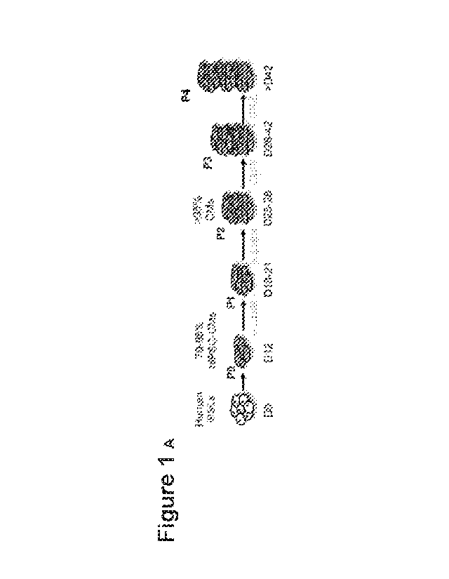
signaling pathway, and later
differentiate into the three cardiovascular linages of the heart upon
cessation of Wnt signaling
(Cohen et al., 2007; Kwon et al., 2009; Lin et al., 2007; Qyang et al., 2007).
Finally, once the
heart has formed, Wnt signaling remains predominantly active in the compact
myocardium to
promote myocardial growth (Buikema et al., 2013; Ye et al., 2015). The
knowledge derived from
these early developmental studies in vivo has been translated into
reproducible methods for the
efficient generation of CMs from pluripotent stem cell sources in vitro.
Current directed
differentiation protocols for hiPSCs have incorporated a simplified form of
Wnt modulation with
an early stage, small molecule-mediated activation of Wnt signaling followed
by later stage Wnt
inhibition in order to subsequently induce a high purity of CMs (Burridge et
al., 2014; Lian et al.,
2012; Lian et al., 2013; Paige et al., 2010). A major limitation in the field,
however, remains the
inability to effectively expand and passage committed CMs to generate the
numbers required for
tissue engineering or true regenerative approaches.
2
CA 03092278 2020-08-25
WO 2019/178478 PCT/US2019/022469
[6] Adult mammalian cardiomyocytes possess limited capacity for cell
division (Sharma et
al. 2015). Radiocarbon dating studies suggest that there is, at baseline, less
than 0.5% yearly cell
turnover in the adult human heart (Bergmann et al. 2009). As such, mammalian
adult heart
regeneration is unable to compensate for the massive loss of cardiomyocytes
following cardiac
injury such as myocardial infarction, leading to adverse cardiac remodeling.
This limited
regenerative capability of the human heart has garnered significant interest
in developing novel
methodologies for both creating cardiomyocytes de novo and inducing
proliferation in terminally
differentiated cardiomyocytes.
171 A major goal in human pluripotent stem cell research is to provide
large quantities of
cardiomyocytes suitable for cellular therapy in regenerative medicine (Chuang
et al. 2011,
Laflamme et al. 2011; Serpooshan et al. 2017; Li et al. 2016). Protocols for
human pluripotent
stem cell cardiac differentiation are vastly improved compared to a decade
ago. Current
protocols can obtain upwards of 90% pure cardiomyocytes during differentiation
followed by
metabolic selection, which can be further augmented by using CRISPR/Cas9 gene
editing to
introduce selectable markers into hiPSCs (Lian et al. 2012; Sharma et al.
2018). The most up-to-
date strategies use biphasic Wnt/I3-catenin modulation for direct cardiac
differentiation from
human induced pluripotent stem cells (hiPSCs) (Burridge et al. 2014; Lian et
al, 2013). To
mimic developmental Wnt signals required for in vivo mesoderm induction,
hiPSCs are initially
treated with CHIR99021 (CHIR), a non-selective glycogen kinase 3 beta (GSK3f3)
inhibitor,
followed by a Wnt/I3-catenin inhibitor to promote cardiac cell
differentiation.
[8] In recent years, growing evidence support lysophospholipids, a
collection of bioactive
lipids harboring multiple functions, as important regulators of stem cell
differentiation in vitro
and cardiovascular development in vivo (Kleger et al. 2011). Among these
bioactive lipids,
sphingosine-l-phosphate (SIP) and lysophosphatidic acid (LPA) are cardinal
members (Kleger
et al. 2011).
191 In vivo studies have demonstrated a necessary role of S113 signaling
via S113 receptor in
cardiomyocytes in normal heart development in mice (Clay et al. 2016). In
vitro studies have
shown that these signaling molecules are capable of regulating pluripotency
and cell cycle
activity in human embryonic stem cells (Avery et al. 2008; Garcia-Gonzalo et
al. 2008).
[10] The bioactive lipids have also been reported to play a role in cell
proliferation in
epithelial cells, fibroblasts, and various cancer cell lines, via their
ability to stimulate important
3
CA 03092278 2020-08-25
WO 2019/178478 PCT/US2019/022469
cellular signaling pathways such as the MAPK/ERK pathway, the Hippo Pathway,
and the
Wnt/f3-catenin signaling pathway (Harvey et al. 2013; Marinissen et al. 2001;
Oskouian et al.
2007; Yang et al. 2005).
[11] US patent 9,074,188 and US patent publication 2013/0244262 disclose
methods for
producing cardiomyocytes in culture.
[12] However, despite recent advances, there remains significant batch-to-
batch variation in
differentiation efficiency, as different hiPSCs lines, even those derived from
the same
individuals, can vary in their abilities to reproducibly generate
cardiomyocytes. Thus, there
remains the need for in vitro hiPSC-CM generation protocols with consistently
high efficiency.
SUMMARY
[13] In one aspect, the present disclosure provides methods for expanding
beating
cardiomyocytes, the methods comprising treating the beating cardiomyocytes
with a WNT
agonist, a bioactive lipid and/or a combination of the WNT agonist and
bioactive lipid. The
beating cardiomyocytes may be human cardiomyocytes. The treatment may be
conducted in
vitro. The methods may further comprise prior to the step of treating the
beating
cardiomyocytes, a step of differentiating pluripotent stem cells into the
beating cardiomyocytes.
The pluripotent stem cells include embryonic stem cells, mesenchymal stem
cells,
cardiomyocyte progenitor cells and/or induced pluripotent stem (iPS) cells.
The WNT agonists
include GSK3P inhibitors. One preferred GSK3P inhibitor is CHIR99021.
Preferred bioactive
lipids include sphingosine-l-phosphate (SIP) and/or lysophosphatidic acid
(LPA). The present
methods for expanding beating cardiomyocytes may comprise treatment of the
beating
cardiomyocytes with CHIR99021, BIO, Wnt3A, Wnt3A plus R-spondin, Wnt surrogate
ScFv-
DKK1c, ScFv-DKK1c plus R-spondin, and/or any combination thereof. The beating
cardiomyocytes may be treated for a period of time from 1 day to 120 days.
[14] In some embodiments, methods for expanding beating cardiomyocytes
comprise treating
the beating cardiomyocytes by adding the WNT agonist and/or the biolipid to
tissue culture
media at a final concentration from 1 to 50 i.tM for each of the bioactive
lipid, 1 to 50 tM for
CHIR99021 or BIO, and from 1 to 500 ng/mL of the recombinant WNT agonist
Wnt3A, Wnt3A
plus R-spondin, Wnt surrogate ScFv-DKK1c, ScFv-DKK1c plus R-spondin, and/or
any
combination thereof.
4
CA 03092278 2020-08-25
WO 2019/178478 PCT/US2019/022469
[15] Further aspects of this disclosure include methods for producing beating
cardiomyocytes
from pluripotent stem cells. These methods comprise treating the pluripotent
stem cells with at
least one bioactive lipid and at least one WNT agonist. The pluripotent stem
cells may be human
cells. The pluripotent stem cells may be embryonic stem cells, mesenchymal
stem cells,
cardiomyocyte progenitor cells and/or induced pluripotent stem (iPS) cells.
The WNT agonist
may be a GSK3P inhibitor. Some of the preferred bioactive lipids are
sphingosine-l-phosphate
(SIP), lysophosphatidic acid (LPA) and their combination. Preferred WNT
agonists include
CHIR99021, B TO, Wnt3A, Wnt3A plus R-spondin, Wnt surrogate ScFv-DKK1c, ScFv-
DKK1c
plus R-spondin, and/or any combination thereof. The pluripotent stem cells may
be treated
during a period of time from 1 hour to 10 days.
[16] In one preferred embodiment of the present method for producing beating
cardiomyocytes from pluripotent stem cells, the pluripotent stem cells are
treated by adding S113
and LPA to tissue culture media at a final concentration 1 to 50 [tM each, at
1 to 50 [tM for
CHIR99021 or BIO, and from 1 to 500 ng/mL of the recombinant WNT agonist
Wnt3A, Wnt3A
plus R-spondin, Wnt surrogate ScFv-DKK1c, ScFv-DKK1c plus R-spondin, and/or
any
combination thereof.
[17] The disclosure also provides a method for obtaining human cardiomyocytes,
the method
comprising:
differentiating hiPS cells into beating cardiomyocytes via the biphasic Wnt
signaling protocol, wherein the hiPS cells are treated with at least one Wnt
agonist
and at least one bioactive lipid during at any time during the first phase of
the
protocol and wherein the cells are further treated with at least on Wnt
antagonist
during the second phase of the protocol, and thereby obtaining the beating
cardiomyocytes; and
expanding the beating cardiomyocytes by treating the beating cardiomyocytes
with at least one bioactive lipid, at least one Wnt agonist, and any
combination
thereof
[18] Other aspects include a human beating cardiomyocyte produced by any of
the present
methods and methods of drug screening in which the human cardiomyocytes are
contacted with a
drug and then monitored for the effect of the drug on the human
cardiomyocytes.
CA 03092278 2020-08-25
WO 2019/178478 PCT/US2019/022469
[19] Further aspects include methods of treating a patient from a heart
disease. The methods
comprise directly administering to the patient's heart the cardiomyocytes
obtained by the present
methods. The heart diseases include heart failure from congenital heart
disease, from myocardial
infarction, from cardiotoxic agents such as anthracyclines, tyrosine kinase
inhibitors, and
immune check-point inhibitors for cancer therapy, from environmental exposure
such as alcohol,
bacteria such as ones causing Chaga's or Lyme disease, myocarditis-causing
viruses, or from
hereditary/genetic cardiomyopathy. In the methods of treatment, the
cardiomyocytes may be
administered to the patient via a patch which is applied to the patient's
heart.
[20] Further methods of treating a patient from a heart disease include
methods comprising
administering to the patient at least one Wnt agonist, Wnt surrogate,
bioactive lipid and/or a
combination thereof.
[21] Other aspects provide method of treating a patient from a heart or
vascular disease, the
methods comprising administering a tissue engineered blood pump comprising the
cardiomyocytes obtained by the present methods.
BRIEF DESCRIPTION OF THE DRAWINGS
[22] Figs. 1A-1I show that Wnt Signaling Stimulates Massive Expansion of
beating hiPSC-
CMs and Long-term Passaging. (A) Schematic timeline of hiPSC-CM expansion and
passaging.
(B) Representative images of hiPSC-CM expansion from initial 10 cm2 confluent
dish at passage
0 (PO) to multiple confluent T-175 cm2 cell culture flasks at subsequent
passages. (C) Total
surface area (cm2) coverage by hiPSC-CMs at each passage. (D) Representative
bright-field
images of confluent hiPSC-CMs in the presence of CHIR or DMSO (CTR) at each
passage.
Same dilution factor was applied to both treatment conditions. (E)
Quantification of total number
of cell from PO to P5. (F) Immunofluorescence analysis for TnT expression at
P3 for hiPSC-CMs
treated with DMSO (CTR) or CHIR. (G) Fold increase in TnT+ cells in CHIR-
treated vs DMSO
treated cells at each passage. (H) Representative flow cytometry plots of TnT
expression in
CHIR-treated cells. (I) Quantification of percentage of TnT+ cells from flow
cytometric analysis
in (H). Scale bars represent 100 m, Data are in mean (n=3-5) error bars
indicate standard
deviation, *p<0,05.
[23] Figs. 2A-2K report extension of hiP SC-CM proliferative window by Wnt
signaling. (A)
Immunofluorescence microscopy images of hiPSC-CMs at each passage starting at
day 12 (PO)
6
CA 03092278 2020-08-25
WO 2019/178478 PCT/US2019/022469
of differentiation. Cardiac troponin T (TnT). (B) Expression of ki67, a cell
cycle index, in TnT+
cells after treatment with CHIR or DMSO (CTR). (C) Expression of pHH3, a
proliferative index,
in CHIR or CTR-treated TnT+ cells. (D) Representative confocal microscopy
images of CHIR-
treated hiPSC-CMs at different phases of mitoses. (E) Quantification of
proliferating hiPSC-
CMs at distinct mitotic phases from (D). (F) Immunofluorescence image of
Aurora B kinase
expression in TnT+ cells undergoing cytokinesis. (G) Quantification of the
percentages of bi-
nucleated hiPSC-CMs (as % total CMs) and (H) ki67 expression for CHIR-treated
P3 cells that
were subsequently treated with either CTR, CHIR, or C59 for 6 additional days.
(I)
Immunofluorescent images of PO hiPSC-CMs treated with CTR, CHIR, or C59 for 24
hours
(24h). (J) Quantification of ki67 expression (i.e. cell cycle index) as a
percentage of total hiPSC-
CM from experiment described in (I). (K) Assessment of canonical Wnt signaling
via TCF/LEF
using the TOPFlash luciferase reporter in hiPSC-CMs from experiment described
in (I). Note the
dramatic increase in TCF/LEF activity in the presence of CHIR. Scale bars
represent 100 m,
Data are in mean (n=3-5) error bars indicating standard deviation, *p<0,05.
[24] Figs. 3A-3M report phenotypic assessment of hiPSC-CMs following Wnt
stimulation.
(A) Confocal microscopy images of P3 hiPSC-CMs on micropatterned surfaces
either treated
with DMSO (CTR), CHIR (2.0 Elm) or CHIR followed by C59 (CHIR>C59) and
immunostained
for the expression of troponin T (TnT) and alpha-sarcomeric actin (a-SA). (B)
Automated
quantification of sarcomere fiber alignment. Vertical axis is defined as zero
degree. (C) The
percentages of the sarcomere area oriented at the indicated degree are
quantified. (D)
Contractility measurements in cells treated in (A). (E) Representative action
potential tracings of
hiPSC-CMs. Data represents changes in membrane potential [Em]) in day 28 (D28)
hiPSC-CMs.
(F) Graphs representing action potential duration (APD) in 90% repolarization
(ADP90) and
maximal diastolic potential (MDP) for each group at day 28. (G) Ca2+
transients (Fluo-4AM)
fluorescence expressed relative to baseline [F/FO]) in hiPSC-CMs at day 28.
(H) Graph
displaying the decay tau for each group. Fold increase in sarcomere gene (I),
electrophysiological gene (J) and metabolic gene (K) expression for expanding
CMs at D28,
D35, D42 and D49. After D28 cells are either continues treated with CHIR,
withdrawn or treated
with C59. (L) Immunohistochemistry for MLC2V and TnT in P3 CMs treated with
CHIR, CTR,
and CHIR>C59. (M) Percent of MLC2V and TnT positive cells. Scale bars
represent 100 m.
7
CA 03092278 2020-08-25
WO 2019/178478 PCT/US2019/022469
Data are in mean (n=3-5) error bars indicating SEM in D, E, G and H and
standard deviation in
J and L, *p<0,05.
[25] Figs. 4A-4K report single cell RNA sequencing analysis of hiPSC-CM
following gain
and loss of Wnt signaling. (A) t-SNE plot of day 12 hiPSC-CM that have been
treated with
DMSO CTR (gray), CHIR (yellow), or C59 (black) for 24 hrs. (B) Heat map of
significantly
differentially expressed genes from the three groups in (A). (C) Expression of
Wnt target genes.
(D and E) Expression of proliferation genes. Note the increase in their
expression in CHIR-
treated cells. (F) t-SNE plot displaying unsupervised clustering of single
hiPSC-CMs from (A).
(G) Heat map of differentially expressed genes in unsupervised clusters. (H)
Genes per cluster
used for pathway enrichment analysis. (I) Expression of ventricular markers in
single hiPSC-
CMs. (J) Expression of atrial markers cells. (K) Expression of ventricular
maturation markers.
For all panels shown, genes represent p<0,01.
[26] Figs. 5A-5K report GSK3P inhibition regulates phosphorylation of AKT
kinases required
for mitosis. (A) Cell count of TnT positive cells represented as fold increase
over CHIR
treatment. (B) TOPFlash luciferase TCF/LEF analysis after 24hrs of treatment
with the indicated
inhibitors. (C) Gene expression for the indicated inhibitors. (D) Schematic of
GSK3 inhibition
with CHIR (CHIR99021) and downstream canonical Wnt signaling inhibition with
PNU74654.
(E) Panel displaying kinases with significantly changed phosphorylation levels
after treatment
with CHIR of 43 screened phosphorylation targets. (F) Western blot analysis
for pAKT T308 in
cells cultured for 100min in the presence or absence of CHIR. (G) Graph
representing pAKT
protein expression. (H) Immunofluorescence analysis for pAKT T308 in day 12
hiPSC-CMs
cultured for 6 days with the indicated treatment. Quantification of the TnT
(I) and pAKT T 308
(J) cell number for each treatment represented as fold over control. (K)
TOPFlash luciferase
TCF/LEF analysis after 24hrs of treatment with the indicated treatment. Scale
bars represent
100 m, Data are in mean (n=3-5) error bars indicating standard deviation,
*p<0,05.
[27] Figs. 6A-6E report Wnt receptor-ligands induce CM cell-cycle
reactivation. (A)
Schematic representation of time points of hiPSC-CMs used for studying cell
cycling (B)
Representative images of day 12 CMs treated with Wnt3A, scFv-DKK1c + RSPO or
H20
control (CTR). (C) Quantification of the TnT positive cell numbers displayed
as fold increase
over the CTR. (D) Immunfluorescence analysis in D66 CMs for the listed
treatments. (E) Fold
8
CA 03092278 2020-08-25
WO 2019/178478 PCT/US2019/022469
increase in mitotic CMs at day 66. Scale bars represent 1001.1m, Data are in
mean (n=3-5) error
bars indicating standard deviation, *p<0,05.
[28] Figs. 7A-7G report Wnt surrogate promotes myocardial growth. (A)
Representative
images of 12-weeks old mice for Wnt Surrogate treatment or CTR. (B) Graph
displaying heart-
weight body-weight ratios. (C) Hematoxylin and Eosin (H&E) staining at 3
levels of the
ventricles. (D) graph displaying LV dimension and (E) wall thickness in 1.1m.
(F)
Immunofluorescence analysis for wheat germ agglutinin (WGA) and dapi (DNA).
(G) Graphs
displaying relative cell number and cell size in 1_1111. Scale bars represent
10001.1m in A and C and
1001.1m in F, Data are in mean (n=3 in A, n=6 in C and D, n=4 in E-H) error
bars indicating
standard deviation, *p<0,05.
[29] Figs. 8A-8C report gene expression analysis of hiPSC cardiac
differentiation. (A)
Pluripotent, cardiac mesoderm, cardiac progenitor and cardiomyocyte
transcription factor
expression during hiPSC-CM differentiation. Dashed lines indicated gene
expression in samples
treated with 2.0 OM CHIR from day 12 to 14. Immunofluorescence images of ISL1
(B) and
Caspase 3 (C) expression in day 14 hiPSC-CMs treated with CHIR or DMSO (CTR)
are shown.
[30] Figs. 9A-9B report phenotypic analysis of hiPSC-CM proliferation upon Wnt
stimulation. (A) Immunofluorescence images for TnT and pHH3 expression in
hiPSC-CMs at
various passages after treatment with CHIR or DMSO (CTR). (B)
Immunofluorescence images
of TnT pHH3 expression in CTR and CHIR-treated hiPSC-CM at passage 3 (P3) on
micropatterned substrate demonstrating the presence of disorganized sarcomeric
structure and
multiple hiPSC-CMs at different phases of mitosis in CHIR-treated but not in
CTR-treated cells.
[31] Fig. 10 reports real-time quantitative PCR analysis of cardiac gene
expression. Three-
months old hiPSC-CMs that were treated with or without CHIR for the first two
months were
harvested for qPCR analysis of cardiac genes expression.
[32] Figs. 11A-11C report generation of 3D cardiac tissue using CHIR-treated
hiPSC-CMs.
(A) Bright field image of CHIR-treated hiPSC-CMs encapsulated in collagen-
based hydrogels
and cultured in vitro for 7 days. (B) Immunofluorescence images showing
organized sarcomeres
and expression of Cx43 gap junction protein in engineered cardiac tissue in
(A). (C)
Quantification of the amount of beating area and contraction velocity in 3D
cardiac constructs
generated with the indicated starting number of CHIR-treated hiPSC-CMs.
9
CA 03092278 2020-08-25
WO 2019/178478 PCT/US2019/022469
[33] Figs. 12A-12E report previously expanded CMs being utilized to create
functional 3-
dimensional cardiac tissue.
[34] Fig. 13 reports immunohistochemistry for pAKT T308 and TnT in hiPSC-CMs
passaged
for 3 times with CHIR.
[35] Figs. 14A-14B report (A) Images of 8-weeks old mice hearts treated with
the Wnt
Surrogate, scFv-DKK1c or the vehicle control (CTR). (B) Confocal images of
Smooth Muscle
Actin, DNA and WGA membrane stain in 8-weeks old mouse hearts.
[36] Figs. 15A-15C report 96-well differentiation which illustrates S1P/LPA-
mediated
enhancement of hiPSC-cardiomyocyte differentiation when added concurrently
with Wnt
activator CHIR99021. A) Illustration of the 'regular' chemically-defined
cardiac differentiation
protocol utilized in this study. S1P/LPA was added at different time points
during hiPSC-CM
differentiation. B) Representative 96-well immunofluorescence images for
cardiac troponin T
(TnT) in green and nuclear DNA in blue of 2D monolayer-based, chemically-
defined
differentiation of two poorly differentiating hiPSC lines into cardiomyocytes.
Staining was
performed in a 96-well plate format on day 8-post differentiation hiPSC-CMs.
SIP, LPA, or both
were added for days 0-2, 4-6 or 6-8 during the hiPSC-CM differentiation
process. C)
Quantification of TnT positive cell numbers of total represented as
percentages TnT positive
cells for each time point when SIP, LPA or both were added. Error bars
represent standard
deviation. * indicates p<0.05 versus control. Experiments were performed in 2
different hiPSC
lines in 3-6 replicates.
[37] Figs. 16A-16E report bioactive lipids S113 and LPA enhance 13-catenin
nuclear
accumulation and activate Wnt signaling during early cardiac differentiation
from hiPSCs. A)
Immunofluorescence for 13-catenin (green), pluripotency marker Nanog (red),
and DAPI (DNA)
(blue) following 2-hour treatment of hiPSCs with DMSO, small molecule GSK3P
inhibitor/Wnt
activator CHIR99021 (CHIR), bioactive lipids S113 + LPA, or CHIR + bioactive
lipids. Arrows
indicate cells exhibiting characteristic 13-catenin nuclear accumulation. B)
Quantification of f3-
catenin staining represented as nuclear intensity over cytoplasmic intensity
for the treatment
groups normalized to DMSO control. C) Luciferase luminescence intensity after
transfection of
hiPSCs with TOPFlash Wnt pathway activity reporter and 2-hour treatment with
CHIR,
S1P/LPA, or both, represented as fold increase over DMSO control. D) Model
illustrating the
signaling cascade linking bioactive lipids and the Wnt/f3-catenin signaling
pathway in the context
CA 03092278 2020-08-25
WO 2019/178478 PCT/US2019/022469
of hiPSCs. Treatment with S1P/LPA on hiPSCs dissociates 0-catenin from
adherens junctions
and E-cadherin, thus increasing the overall 0-catenin pool that can be
utilized for downstream
signaling and gene transcription. Treatment with GSK30 inhibitor CHIR frees 0-
catenin and
increases the overall intracellular 0-catenin pool for downstream signaling
and gene
transcription. E) Microarray analysis illustrating key alterations in gene
expression following 48-
hour treatment of hiPSCs with small molecule GSK30 inhibitor/Wnt activator
CHIR with or
without bioactive lipids S1P/LPA. A list of up- (red) and down- (blue)
regulated genes after
treatment with bioactive lipid is shown. Experiments were performed in 3-4
biological replicates.
[38] Figs 17A-17E report bioactive lipids S1P/LPA rapidly alter hiPSC
morphology and
enhance vimentin expression during early cardiac differentiation. A)
Immunofluorescence and
phase contrast images of hiPSCs treated with S113 and LPA for 24 hours.
Calcein AM dye
staining membranes the entirety of cell bodies. B) Quantification of cell
diameter displayed in
1.tm for hiPSCs treated with DMSO or the combination of S113 and LPA. C)
Normalized cell
count of 3 separate hiPSC lines following treatment with DMSO or S1P/LPA for
24 hours. N=3
biological replicate experiments. Error bars represent SEM. D)
Immunofluorescence staining
following 48-hour treatment of hiPSCs with DMSO, GSK3P inhibitor CHIR99021
(CHIR),
bioactive lipids S1P+LPA or combination. Intermediate filament protein
vimentin (green) marks
epithelial-to-mesenchymal transition and brachyury (red) marks early mesoderm.
E)
Quantification of vimentin (VIM) and brachyury T (BRY) positive cells
represented as
percentages of total cells for control, CHIR, S1P/LPA and combined treatments.
Error bars
represent standard deviations. Error bars represent standard deviation.
Experiment performed in
3 biological replicates. * indicates P<0.05. Cells quantified in N=9 images
per condition.
[39] Figs. 18A-18G report LPA and S113 exhibit cell cycle-inducing effects on
hiPSC-CMs.
A) Schematic overview of replating hiPSC-CMs at different time-points of
differentiation into
96-well format for downstream assays. B) Representative images showing cardiac
troponin T
(TnT) in green, cell cycle activity marker ki67 in red and nuclear dye (DNA)
in blue after 48-
hour culture of day 30 hiPSC-CMs with DMSO, S1P/LPA alone, CHIR alone, or CHIR
with
S1P/LPA. C) Percentage of ki67 positive cardiomyocytes after 48 hours of each
treatment. D)
Normalized cell count for total number of CMs after 48 hours of treatment for
each group. E)
Immunofluorescence staining for cardiac troponin T (TnT) in green, mitosis
marker phospho
Histone H3 (pHH3) in red and nuclear dye (DNA) in blue after 48-hour culture
of day 30 hiPSC-
11
CA 03092278 2020-08-25
WO 2019/178478 PCT/US2019/022469
CMs with DMSO, S1P/LPA alone, CHIR alone, or CHIR plus S1P/LPA. F) Percentages
of
mitotic (pHH3) CMs between various treatment groups. G) Percentages of bi- and
multinucleated CMs within the indicated treatment groups. *indicates P<0.05 in
comparison to
control. N=3 biological replicates. Error bars represent standard deviation.
[40] Figs. 19A-1911 report reactivation of cell cycle in hiPSC-CMs with S113
and LPA is
dependent on ERK signaling. A) Luciferase luminescence intensity after
transfection of day 30
hiPSC-CMs with TOPFlash Wnt signaling pathway activity reporter and 2-hour
treatment with
CHIR, S1P/LPA or both. The data shown represents fold increase over DMSO
control. B)
Quantification of kinase assays illustrating alterations in hiPSC-CM kinome
phosphorylation in
response to 0, 5, 10, and 30-minute S1P/LPA treatment. Data expressed as means
SEM.
*indicates P<0.05. C) Representative kinase assay conducted in day 30 hiPSC-
CMs treated with
and without small molecule MEK inhibitor trametinib, with and without S1P/LPA
(10 [NI each).
Spots corresponding to ERK phosphorylation and antibody control are labeled.
D)
Immunofluorescence for cardiac troponin T (TnT) (green), ki67 (red) and
nuclear DNA (blue) in
day 30 hiPSC-CMs treated with S113 and LPA in the presence or absence of MEK
inhibitor
trametinib. E) Quantification of the percentages of ki67 positive
cardiomyocytes (CMs) in (D).
*indicates P<0.001. F) Immunofluorescence for cardiac troponin T (TnT)
(green), ki67 (red) and
nuclear DNA (blue) in day 30 hiPSC-CMs treated with bioactive lipid S113 in
the presence or
absence of 51.1M SIP receptor antagonist VPC23019. G) Quantification of the
percentages of
ki67 positive CMs after S113 treatments with or without 51.1M VPC23019. H)
Model illustrating
the link between bioactive lipids and the canonical MAPK/MEK/ERK signaling
pathway in
differentiated hiPSC-CMs. N=4.
[41] Figs. 20A-20B report bioactive lipids S113 and LPA increase nuclear beta-
catenin but do
not induce early mesodermal differentiation. A) Time course study on nuclear
beta-catenin
accumulation after 0, 0.5, 1, 2, 4, 8, 16 and 24 hour (hr). Arrows indicate
cells expressing
profound nuclear beta-catenin. B) Immunofluorescence for pluripotency marker
Nanog (green),
early mesoderm marker Brachyury (red) and DAPI (blue) in hiPSCs cultured for
24 hr with
GSK3 inhibitor CHIR99021, bioactive lipids S113 and LPA or DMSO control. Graph
represents
quantification of percentages Brachyury (Bry) positive cells for the listed
treatments. Error bars
represent SEM. *indicates P<0.05. N.S. = not significant. N=3.
12
CA 03092278 2020-08-25
WO 2019/178478 PCT/US2019/022469
[42] Figs. 21A-21B report hiPSC-CMs express relevant S113 and LPA receptors
upon terminal
differentiation and respond to S1P/PA treatment. A) Transcriptome profiling of
day 30 hiPSC-
CMs from 5 different hiPSC-CM lines. High TNNT2 and low PECAM1 indicates high
cardiomyocyte purity and low endothelial cell contamination, respectively.
Expression profiling
conducted using IonTorrent Ampliseq transcriptome profile. Error bars
represent SEM. B)
Bioactive lipids LPA and S113 induce ki67 expression at different time points
of terminal
differentiation. Immunofluorescence for alpha-actinin (green), ki67 (red) and
dapi (blue) after 48
hour treatment with S113 and LPA, IGF or DMSO in day ¨20 and ¨50 hiPSC-CMs.
[43] Figs. 22A-22D report motion-derived contractility parameters in day 30
hiPSC-CMs. A)
Representative heatmaps of day 30 hiPSC-CMs treated with DMSO, CHIR99021 or
S1P/LPA
generated from high resolution and frequency movies, red = high motion, blue =
low motion.
Graphs displaying contraction frequency (beats/minute) B), contraction
deformation distance C),
and contraction velocity D). Data represented as means. Error bars indicate
SEM.
[44] Figs. 23A-23B report LPA is unable to enhance YAP nuclear accumulation in
hiPSC-
CMs. A) Immunofluorescence staining in untreated hiPSCs, hiPSC-CMs, and non-CM
mesodermal derivatives (indicated by yellow boxes) for DAPI (blue), total YAP
(green), and
cell-type specific markers (red): Tra-1-81 for hiPSCs and alpha-actinin for
hiPSC-CMs. YAP is
localized to the nuclei in all three cell types at baseline, as indicated by
representative cells
marked by yellow arrows. B) YAP immunofluorescence for purified day 10 or day
30 hiPSC-
CMs with and without treatment with LPA, stained for downstream Hippo pathway
transcriptional effector YAP (green). No significant (N.S.) increase in YAP
nuclear
accumulation or 0-catenin translocation seen after LPA treatment on day 10 or
day 30 hiPSC-
CMs. Cells quantified in N=9 images per condition. Data are expressed as means
STD.
[45] Fig. 24 reports bioactive lipids do not change ERK phosphorylation in
undifferentiated
hiPSC. ERK1/2 phosphorylation conducted in undifferentiated hiPSCs with and
without 5
minute S113 and LPA (S+L) treatment. Spots corresponding to ERK1/2
phosphorylation and
antibody control are labeled.
[46] Fig. 25 reports S113 and LPA activate MAPK/MEK/ERK signaling in hiPSC-
CMs. Full
version of Figure 19. Kinase assay conducted in day 30 hiPSC-CMs treated with
and without
small molecule MEK inhibitor trametinib, with and without S1P/LPA (1011M
each). Spots
corresponding to ERK phosphorylation and antibody control are labeled.
13
CA 03092278 2020-08-25
WO 2019/178478 PCT/US2019/022469
[47] Fig. 26 reports S113 and LPA do not alter maturation or subtype
specification in hiPSC-
CMs. QPCR gene expression analysis conducted in day 30 hiPSC-CMs treated with
and without
S1P/LPA (1011M each). Genes corresponding to atrial, ventricular, and nodal
subtypes, as well
as maturation markers, are labeled. * indicates p<0.05.
[48] Fig. 27 is Table Si showing panel of different phosphorylated kinases as
screened in
hiPSCs and hiPSC-CMs treated with bioactive lipids S113 and LPA. In table,
proteins with an
asterisk (*) indicate significant alterations (P<0.05) in phosphorylation
following S1P/LPA
treatment (see also Figures 19A-1911).
DETAILED DESCRIPTION
[49] In one aspect, this disclosure provides a method for expanding beating
cardiomyocytes,
including human beating cardiomyocytes. The method comprises treating the
beating
cardiomyocytes with one or more WNT agonists, one or more bioactive lipids or
a combination
of one or more WNT agonists and one or more bioactive lipids.
[50] In this disclosure, the term "beating cardiomyocytes" are used
interchangeably with other
terms such as cardiomyocytes, cardiac muscle cells, heart muscle cells,
myocardiocytes and/or
cardiac myocytes. Cardiomyocytes (CMs) are cells that make up the heart
muscle. In this
disclosure, "cardiomyocytes" refer to primary cardiomyocytes that have been
isolated from the
heart tissue and also to cardiomyocytes that have been obtained by recombinant
technologies,
such as for example, by differentiating stem cells.
[51] In this disclosure, cardiomyocytes include those derived by
differentiating pluripotent
stem cells such as embryonic stem cells, mesenchymal stem cells, cardiomyocyte
progenitor
cells and/or induced pluripotent stem (iPS) cells, or any other cardiomyocyte
progenitor cells. In
this disclosure, iPS cells, including human iPS (hiPS) cells are particularly
preferred for
obtaining cardiomyocytes. Cardiomyocytes derived from iPS cells may be
referred in this
disclosure as iPS-derived cardiomyocytes or cardiomyocytes interchangeably.
[52] Any of beating cardiomyocytes, primary or derived from pluripotent stem
cells, may be
expanded by the present methods in which the beating cardiomyocytes are
treated with one or
more WNT agonists, one or more bioactive lipids or a combination of one or
more WNT
agonists and one or more bioactive lipids. The present expansion method
efficiently increases a
number of beating cardiomyocytes by stimulating their proliferation. This
result is highly
14
CA 03092278 2020-08-25
WO 2019/178478 PCT/US2019/022469
unexpected because beating cardiomyocytes are typically non-dividing cells
which do not
proliferate efficiently even in tissue culture.
[53] Beating cardiomyocytes retain their ability to undergo the
contraction/relaxation cycle in
vitro. The contractility of beating cardiomyocytes may be detected and
recorded with a high-
resolution movie. Additional immunohistochemistry tests can be conducted to
quantify the
sarcomere alignment. Other tests may include electrophysiological studies in
which potentials of
beating cardiomyocytes are recorded in a sharp current clamp mode.
[54] The term "Wnt agonists" means any reagent which either alone or in
combination with
other reagents activates the canonical Wnt signaling pathway in a cell.
Activation of the
canonical Wnt signaling pathway means activation of 13-catenin signaling by
which 13-catenin is
translocated into the cell nucleus. For the purposes of this disclosure, any
reagent either alone or
in combination with other reagents that produces beating cardiomyocytes with
nuclear 13-catenin
is referred to as a Wnt agonist.
[55] Wnt agonists include small compounds, peptides, proteins, antibodies and
their
fragments, siRNA, and surrogate polypeptides. WNT agonists include GSK3P
inhibitors.
Compound CHIR99021 (aminopyrimidine derivative, available from Selleckchem) is
one
preferred GSK3P inhibitor. CHIR99021 may be referred in this disclosure
interchangeably as
CHIR. Another preferred GSK3P inhibitor is BIO (6-bromoindirubin-3'-oxime,
available from
Sigma-Eldridge).
[56] Suitable Wnt agonists also include surrogate polypeptides which
dimerize a Frizzled
(Fzd) receptor with Lrp5/6, as disclosed in W02016040895. These surrogate
polypeptides
comprise a binding domain having a specific affinity with a KD of at least
1X10⁷M for one or
more Fzd proteins and a binding domain having a specific affinity with a KD at
least 1X10⁷M of
at least for one or both Lrp5 and Lrp6 protein.
[57] The Fzd binding domain may be a norrin protein or binding fragment
thereof. The Fzd
binding domain may be scFv comprising the six CDR regions of an anti-Fzd
antibody. A
particularly preferred Fzd-binding domain includes the six CDR regions of pan-
specific frizzled
antibody OMP-18R5. The Fzd binding domain may be a de novo designed Fzd
binding domain.
[58] The Lrp5/6 binding domain may comprise a binding portion of a DKK
protein. In
particular, the Lrp5/6 binding domain may comprise the C-terminal domain of
human DKK1.
CA 03092278 2020-08-25
WO 2019/178478 PCT/US2019/022469
[59] A particularly preferred surrogate Wnt agonist is ScFv-DKK1c which is
disclosed in
W02016040895 and comprises the scFv fragment of the OMP-18R5 antibody
(available from
Oncomed) linked via a linker to the C-terminal domain of human DKK-1.
[60] Suitable Wnt agonists also include protein ligands of the RSPO family,
including RSP01,
RSP02, RSPO3 and RSP04, also referred to as R-spondin 1, 2, 3 or 4. Suitable
Wnt agonists
further include Wnt3 (Wnt family member 3) protein and any derivatives thereof
which can
function as a Wnt-agonist. Suitable agonists further include Wnt3A and any of
its derivatives
that can function as a Wnt agonist.
[61] Any one of the WNT agonists may be used in combination with any other Wnt
agonists.
Some of the preferred combinations may include Wnt3A and at least one of R-
spondin proteins.
Other combinations may include ScFv-DKK1c in combination with at least one of
R-spondin
protein.
[62] Bioactive lipids are lipids which are either alone or as co-stimulators
with Wnt agonists
regulate cell signaling pathways. Bioactive lipids include poly- and
monounsaturated fatty acids,
phospholipid derivatives and lysophospholipids which are a subgroup of the
glycophospholipid
family with one of the hydroxyl groups on the three-carbon glycerol backbone
remaining
unesterified. Lysophospholipids contain only one fatty acid. Suitable
bioactive lipids include
sphingosine-l-phosphate (SIP), lysophosphatidic acid (LPA) and a combination
of the two
compounds. Any other suitable bioactive lipids include any of biolipids which
stimulate or co-
stimulate with one or more Wnt agonists differentiation and/or expansion of
cardiomyocytes.
[63] The term "treating" and/or "treatment" of beating cardiomyocytes is
understood broadly
and may include any of in vitro and/or in vivo procedures by which beating
cardiomyocytes are
exposed to at least some of the Wnt agonists and/or bioactive lipids. The
treatment may include
incubating beating cardiomyocytes with a formulation comprising at least one
of the Wnt
agonists and/or bioactive lipids. In some embodiments, at least one of the Wnt
agonists and/or
bioactive lipids may be dissolved in a solvent and added to a tissue culture
media if beating
cardiomyocytes are incubated in vitro. Suitable solvents may include water, a
buffer, tissue
culture media, an organic solvent, and any combination thereof.
[64] In some embodiments, the treatment may comprise administering at least
some of the
Wnt agonists and/or bioactive lipids to an organ and/or tissue of a patient.
The administration
16
CA 03092278 2020-08-25
WO 2019/178478 PCT/US2019/022469
may include an injection, including an intravenous injection or direct
injection into the heart
tissue, a patch and/or oral administration.
[65] In the present methods for expanding cardiomyocytes, beating
cardiomyocytes may be
treated in vitro with one or more Wnt agonists, one or more bioactive lipids,
or a combination of
one or more Wnt agonists and one or more bioactive lipids. The treatment may
be administered
for a period of time from one day to 120 days.
[66] In one preferred embodiment, beating cardiomyocytes are treated by adding
a
combination of at least one Wnt agonist and at least one bioactive lipid to
tissue culture media
for a period from 1 to 120 days. Bioactive lipids may be added to a final
concentration in the
range from 1 to 50 M. A GSK3P inhibitor may be added to a final concentration
in the range
from 1 to 50 M. A recombinant Wnt agonist, such as a surrogate polypeptide
comprising a Fzd
ligand and Lrp5/6 ligand, may be added to the final concentration in the range
from 1 to 500
ng/mL.
[67] In one preferred embodiment, beating cardiomyocytes are treated by adding
to a tissue
culture media S113 and LPA at a final concentration from 1 to 50 i.tM each. In
another preferred
embodiment, beating cardiomyocytes are treated with S113 and LPA at a final
concentration from
1 to 50 i.tM and at least one from CHIR99021 and BIO also at a final
concentration 1 to 50 M.
In further embodiments, beating cardiomyocytes are treated with S113 and LPA
at a final
concentration from 1 to 50 i.tM and at least one from of the recombinant WNT
agonist Wnt3A,
Wnt3A plus R-spondin, Wnt surrogate ScFv-DKK1c, or ScFv-DKK1c plus R-spondin
at a final
concentration from 1 to 500 ng/mL. In further embodiments, beating
cardiomyocytes are treated
with S113 and LPA at a final concentration from 1 to 50 tM, at least one from
CHIR99021 and
BIO also at a final concentration 1 to 50 and at least one from of the
recombinant WNT
agonist Wnt3A, Wnt3A plus R-spondin, Wnt surrogate ScFv-DKK1c, or ScFv-DKK1c
plus R-
spondin at a final concentration from 1 to 500 ng/mL. The beating
cardiomyocytes may also be
produced by treating cardiomyocytes with 1 to 50 i.tM and at least one from
CHIR99021 and
BIO and/or treating them with one or more from the recombinant WNT agonist
Wnt3A, Wnt3A
plus R-spondin, Wnt surrogate ScFv-DKK1c, or ScFv-DKK1c plus R-spondin at a
final
concentration from 1 to 500 ng/mL.
17
CA 03092278 2020-08-25
WO 2019/178478 PCT/US2019/022469
[68] Further aspects provide cardiomyocytes that have being obtained by
treating beating
cardiomyocytes with one or more Wnt agonists, one or more bioactive lipids or
a combination of
one or more Wnt agonists and one or more bioactive lipids.
[69] In further aspect, this disclosure provides methods for producing
cardiomyocytes from
pluripotent stem cells.
[70] Pluripotent stem cells include embryonic stem cells, mesenchymal stem
cells,
cardiomyocyte progenitor cells, and induced pluripotent stem (iPS) cells.
Other types of
pluripotent cells may be used as well. In this disclosure, iPS cells,
including human iPS (hiPS)
cells are particularly preferred for obtaining cardiomyocytes. iPS cells,
including human iPS
cells, are pluripotent stem cells generated from adult cells by reprogramming.
iPS may be
obtained from keratinocytes or blood cells or some other suitable cells by
reprogramming the
cells to express OCT4, KLF4, SOX2, and MYC.
[71] This disclosure provides methods for generating beating cardiomyocytes by
treating
pluripotent stem cells with a combination of one or more bioactive lipids and
one or more WNT
agonists and differentiating the stem cells into beating cardiomyocytes.
Unexpectedly, there is a
synergism between bioactive lipids and WNT agonists in the present methods for
producing
beating cardiomyocytes from pluripotent stem cells. iPS cells, including hiPS
cells, are
particularly preferred in these methods. Particularly preferred bioactive
lipids in the methods are
sphingosine-l-phosphate (SIP) and lysophosphatidic acid (LPA) which may be
used individually
or in combination. The preferred methods include a combination of S113 and
LPA. Other
bioactive lipids may be added as well.
[72] In the present methods for differentiating pluripotent stem cells into
beating
cardiomyocytes, any of the WNT agonists may be used in combination with one or
more
bioactive lipids. Such WNT agonists include a GSK3P inhibitor. CHIR99021
and/or BIO may
be used in the methods in combination with one or more bioactive lipids.
Methods may also be
performed with one or more bioactive lipids and one or more WNT agonists from
CHIR99021,
BIO, Wnt3A, Wnt3A plus R-spondin, Wnt surrogate ScFv-DKK1c, ScFv-DKK1c plus R-
spondin, Wnt surrogate comprising Frd ligand and Lpr5/6 ligand.
[73] In some of the present methods for differentiating pluripotent stem cells
into beating
cardiomyocytes, the pluripotent stem cells are treated in tissue culture with
one or more bioactive
lipid and one or more WNT agonists for a period of time from 1 hour to 10
days. Bioactive
18
CA 03092278 2020-08-25
WO 2019/178478
PCT/US2019/022469
lipids are added to tissue culture media at a final concentration 1 to 50
each. CHIR99021
and/or BIO are added to tissue culture media at a final concentration 1 to 50
each. WNT
agonist Wnt3A, Wnt3A plus R-spondin, Wnt surrogate ScFv-DKK1c, ScFv-DKK1c plus
R-
spondin or any combination thereof are added to tissue culture media at a
final concentration 1 to
500 ng/mL each.
[74] A person of skill will appreciate that a combination of one or more
bioactive lipids with
one or more Wnt agonists may be used in any protocol for differentiation of
any pluripotent stem
cells into beating cardiomyocytes. These differentiation protocols include the
biphasic Wnt
signaling protocol in which a Wnt agonist is added during the first phase of
differentiation, and
then a Wnt antagonist is added during the second phase of differentiation. In
the biphasic Wnt
signaling protocol, pluripotent stem cells, i.e. iPS cells, are treated with a
combination of one or
more bioactive lipids and one or more Wnt agonists during the first phase of
the protocol.
[75] Further aspects of this disclosure include methods for obtaining human
cardiomyocytes
comprising:
1) differentiating hiPS cells into beating cardiomyocytes via the biphasic Wnt
signaling
protocol, wherein the hiPS cells are treated with one or more Wnt agonists and
one or
more bioactive lipids at any time during the first phase of the protocol
followed by a
treatment with one or more Wnt antagonist during the second phase of the
protocol;
and
2) expanding the beating cardiomyocytes by treating the beating cardiomyocytes
with
one or more bioactive lipids, one or more Wnt agonists, or a combination of
one or
more bioactive lipids and one or more Wnt agonists.
[76] Further aspects include human beating cardiomyocytes obtained by
differentiating
pluripotent stem cells, preferably hiPS cells, via the biphasic Wnt protocol
in which a
combination of one or more bioactive lipids with one or more Wnt agonists is
used during the
first phase of the protocol. The hiPS cell-differentiated beating
cardiomyocytes (hiPSC-CMs)
may be then further expanded by treating the hiPS cell-differentiated beating
cardiomyocytes
with one or more bioactive lipids, one or more Wnt agonists or a combination
of one or more
bioactive lipids and one or more Wnt agonists.
[77] One of the technical advantages provided by the present methods is
production of beating
cardiomyocytes in large numbers, which was previously difficult to accomplish
because beating
19
CA 03092278 2020-08-25
WO 2019/178478 PCT/US2019/022469
cardiomyocytes are typically non-proliferating cells. The beating
cardiomyocytes produced by
the present methods may be used in a number of applications, including a high-
throughput drug
screening in which the beating cardiomyocytes are contacted with a drug and
then monitored for
an effect of the drug on the cardiomyocytes. Many drugs are cytotoxic for
heart muscle. The
high-throughput drug screening methods with the beating cardiomyocytes may
allow for
identification of a potentially cytotoxic drug expeditiously as the screening
can be conducted in
tissue culture.
[78] Particularly preferred embodiments include methods in which bioactive
lipids S113 and
LPA are used to promote cardiomyocyte differentiation from hiPS cells.
Together with
CHIR99021, S113 and LPA treatment in undifferentiated hiPS cells
synergistically increases
nuclear 13-catenin accumulation and mesodermal phenotype. At a later stage of
hiPSC-CM
differentiation, the S113 and LPA treatment stimulates cell cycle activity in
hiPSC-CMs via
ERK/MAPK signaling and enhances cell proliferation.
[79] Further aspects include methods of treating a human patient in need of
treatment for a
heart disease. The patient may be treated by administering to the patient's
heart the beating
cardiomyocytes obtained by the present differentiation and/or expansion
methods. The treatment
methods may be beneficial for any heart disease associated with degeneration
of cardiomyocytes,
including, but not limited to, congenital heart disease, degeneration from
myocardial infarction,
degeneration from cardiotoxic agents such as anthracyclines, tyrosine kinase
inhibitors, and/or
immune check-point inhibitors for cancer therapy, degeneration from
environmental exposure
such as alcohol, bacteria such as ones causing Chaga's or Lyme disease,
myocarditis-causing
viruses, or from hereditary/genetic cardiomyopathy.
[80] The treatment methods may include administering the beating
cardiomyocytes to the
patient via a patch which is applied to the patient's heart. The beating
cardiomyocytes may be
administered in combination with one or more bioactive lipids and one or more
Wnt agonists
which may be included in the patch as well or they may be administered to the
patient separately,
for example via an intravenous injection or orally.
[81] Further aspects provide a method for treating a patient from a heart
disease, the method
comprising administering to the patient one or more Wnt agonists, Wnt
surrogates, bioactive
lipids and/or any combination thereof.
CA 03092278 2020-08-25
WO 2019/178478 PCT/US2019/022469
[82] In further aspect, the present disclosure provides a kit for treating
a heart disease. The kit
comprises beating cardiomyocytes obtained by one of the present
differentiation and/or
expansion methods. The kit may further comprise one or more bioactive lipids
and/or one or
more Wnt agonists and may further include a media for delivery of cells such
as for example
matrix gel designed as a patch.
[83] In further aspect, the present disclosure provides a tissue engineered
blood pump
produced from beating cardiomyocytes which were obtained by the present
differentiation and/or
expansion methods. The disclosure also provides methods for treating a patient
from a heart or
vascular disease. The methods comprise administering the tissue-engineered
blood pump to the
patient.
[84] The present disclosure provides methods in which the canonical Wnt
signaling
stimulation allows for massive expansion and multiple passaging of beating
cardiomyocytes
(CMs). Withdrawal of the Wnt agonist results in rapid cell-cycle exit and
restoration of normal
contractile, electrophysiological and cellular characteristics of CMs.
[85] In one aspect, this system may be used to create functional cardiac
tissue from expanded
CMs in vitro and stimulate in vivo myocardial growth within adult heart
tissue, which may be
used for regeneration of patient-specific cardiac muscle.
[86] The 'holy-grail' of cardiac regenerative medicine remains the restoration
of function
cardiac tissue following myocardial infarct. A major hurdle to this goal,
however, remained the
inability to generate robust numbers of CMs in order to allow for the
generation of patient-
specific, engineered heart tissue or alternatively to boost cell-division of
pre-existing CMs. To
date, expansion and multiple passaging of CMs from hiPS cells has been an
extremely
challenging and largely unsuccessful task. This disclosure aims to address
this need and
provides a method by which immature hiPSC-CMs massively expand for multiple
passages
when continuously subjected to stage-specific small-molecular GSK3 inhibitor
treatment. This
results in increased CM purification (Figure 1).
[87] Following withdrawal of the GSK3 inhibitor, the CMs stop proliferating
and retain the
capacity for normal in vitro maturation spontaneously (Figure 2). As a
demonstration of a pre-
clinical application, previously expanded CMs were utilized to create
functional 3-dimensional
cardiac tissue (Figure 12). Furthermore, stimulation of the canonical Wnt
signaling pathway
21
CA 03092278 2020-08-25
WO 2019/178478 PCT/US2019/022469
with a Wnt surrogate receptor agonist in quiescent hiPSC-CMs and adult mouse
heart, induces
CM replication and promotes myocardial growth (Figure 6 and 7).
[88] Several previous studies have shown the transient expansion of beating
cardiomyocytes
with various GSK3 inhibitors, however, without the ability for multiple
passaging (Buikema et
al., 2013; Titmarsh et al., 2016; Uosaki et al., 2013). Most likely, because
these studies were
using pluripotent stem cell-derived CMs from other species, initiating
treatment at a later time
point of differentiation, culture media containing fetal bovine serum and/or
losing too many cells
when dissociation and passaging. In contrast, the present disclosure provides
that when day 12
hiPSC-CMs are kept in a chemically defined media and Matrigel coated monolayer
culture, they
could be passaged up to 3-5 times with an estimate of >90% survival after
passaging and thereby
acquire a massive increase in CM numbers (Figure 1).
[89] Two recent studies focused on the expansion of cardiovascular progenitors
and showed a
10' and 1010 -fold expansion with a combination of purified proteins, small
molecules and/or
overexpression of oncogenes (Birket et al., 2015; Zhang et al., 2016). Albeit,
the proliferative
capacity of cardiovascular progenitors is higher when compared to hiPSC-CMs
expansion, the
differentiation of multipotent cardiovascular progenitors still remain an
uncontrolled process
resulting in mixed cell populations upon terminal differentiation. The present
methods expand
beating hiPSC-CMs and not progenitors (Figure 9A-B) and robustly yield >95%
TnT purity
during expansion, as will be beneficial for reproducible downstream
applications (Figure 111-I
and Figure 13). After withdrawal of CHIR these cardiomyocytes normally mature
and display
equal sarcomere organization, electrophysiology and force generation to the
controls (Figure 2).
[90] A distinct feature of terminally differentiated CMs is their limited
potential to proliferate
coupled with a cytoskeleton containing highly organized sarcomere structures
to propagate the
billions of contractions over a life-time. A recent report showed that CM cell
division mainly
occurs within the fraction mononuclear diploid cells, but not in the
tetraploid or multinucleated
CMs and that the sarcomere regulator TNNI3k is correlated to this discrepancy
between those
CM populations of the heart (Patterson et al., 2017).
[91] In Figure 2, the present disclosure shows that the proliferating cells
are mono-nucleated.
In Figure 3, the present disclosure shows that after withdrawal of CHIR, there
was an increase in
multinucleated cells and decrease in proliferating cells. Provided in this
disclosure, single cell
RNA sequencing data reveal that activation of the canonical Wnt signaling
pathway
22
CA 03092278 2020-08-25
WO 2019/178478 PCT/US2019/022469
predominantly maintains the relative immature state of CMs and thereby extends
their
proliferative window (Figure 3). This data is in line with the previous in
vivo findings that
immature CM populations proliferate and regenerate the myocardium (Kikuchi et
al., 2010;
Patterson et al., 2017).
[92] Various studies have linked Wnt signaling to heart growth and CM
division, however,
this disclosure provides a finding that Wnt/f3-catenin signaling instead
retains CM immaturity in
order to extend the proliferative window for this subset of proliferative CMs
(Figure 3-4)
(Buikema et al., 2013; Heallen et al., 2011; Kerkela et al., 2008; Titmarsh et
al., 2016; Tseng et
al., 2006; Uosaki et al., 2013). This is conceptually novel and forms a
mechanistic explanation
for the rare CM proliferative response to Wnt signaling in young but not adult
mice (Figure 6).
[93] The biology of the mammalian heart appears to be quite distinct from the
relatively
immature zebrafish heart that possesses regenerative capacities, in which Wnt
signaling is active
upon the response to cardiac injury (Kikuchi et al., 2010; Stoick-Cooper et
al., 2007). A recent
mammalian study demonstrated that the rare population of preexisting CMs which
drive
regeneration are mononuclear diploid cells (Patterson et al., 2017).
[94] It is not clear whether the expanding CMs obtained by the present methods
have made
fate decisions yet towards the atrial of ventricular lineage, but the
sequencing data provides
evidence for an atrial-like populations characterized by SLN and HEY1
expression as well as
ventricular-like populations enriched for MYL2 and the immature ventricular
markers MYL3
and MYL4 (Josowitz et al., 2014; Kurabayashi et al., 1988; Protze et al.,
2017). Interestingly,
both atrial and ventricular-like populations responded to Wnt stimulation and
all CHIR treated
cells together formed a distinct cluster with again atrial-like and
ventricular-like sub-clusters
(Figure 3). Independent from 13-catenin signaling, this disclosure provides
that CHIR/GSK3
regulates the turnover of AKT within the cytoplasm (Figure 5). Importantly,
this disclosure
provides that this component accounted for some of the maturational changes
and roughly 50%
of the proliferative response observed by CHIR exposure seen in Figure 1.
[95] This disclosure provides a robust, long-term in vitro expansion of
functionally immature
CMs that ultimately retain their capacity for further maturation and thus
utility in translational
applications. It presents a conceptually novel principle that Wnt signaling
plays a key role in
maintaining CM immaturity and, by consequence, enhancing the stage-specific
proliferation of
hiPSC-CMs. Furthermore, it also provides an in vivo approach to regulate adult
myocardial
23
CA 03092278 2020-08-25
WO 2019/178478 PCT/US2019/022469
growth. These methods have important implications for scaling up patient-
specific CM
production for various individualized therapies as well as novel in vivo
regenerative approaches
for cardiac repair.
[96] Unexpectedly, this disclosure also provides that S113 and LPA act
synergistically with
GSK3P inhibitor CHIR to regulate early hiPSC mesodermal differentiation
through nuclear f3-
catenin accumulation. At later stages, the combined treatment of S113 and LPA
results in cell-
cycle activation in differentiated hiPSC-CMs, an effect mediated through
ERK/MAPK signaling,
and synergized with I3-catenin signaling to increase cardiomyocyte
proliferation. Bioactive
lipids exhibit stage-specific effects on cardiac differentiation from hiPS
cells.
[97] This disclosure reports highly stage-specific roles for S1P/LPA during
hiPSC-CM
differentiation. When administered to undifferentiated hiPS cells, either
alone or in combination
with CHIR, S1P/LPA increases nuclear I3-catenin level and enhances mesodermal
induction.
After the completion of cardiomyocyte differentiation, the addition of S1P/LPA
initiates a cell
cycle re-entry in hiPSC-CM by activating MAPK/MEK/ERK signaling and enhanced
CHIR-
induced cardiomyocyte proliferation. These findings illustrate the versatility
of the hiPS cell
differentiation platform for studying the effects of signaling pathways on
human cardiomyocyte
development. In addition, the ability to mass-produce differentiated human
cardiomyocytes by
bioactive lipid treatment can be used in development of high throughput assays
for cardiac
disease modeling and discovery of new molecules for future regenerative
applications.
[98] The ability of S113 and LPA to rapidly induce morphological and gene
expression
changes in undifferentiated hiPS cells is an unexpected finding. A rapid
change is observed in
hiPS cell morphology within 12-24 hours from the initiation of S1P/LPA
treatment. These
changes are also accompanied by an increase in the expression of the
intermediate filament
protein vimentin in hiPS cells, a finding that suggests the induction of
epithelial-to-mesenchymal
transition (EMT).
[99] However, the S1P/LPA treatment did not lead to a decreased expression of
Nanog at 24
hrs after treatment, suggesting a more mesodermal specific, rather than
global, effect of
S1P/LPA on hiPSC cardiac differentiation (Figure 22B). A hallmark of EMT is
the loss of cell-
cell contact normally mediated by adherens junction complexes (Lamouille et
al. 2014).
[100] Lysophospholipids are well-established for their ability to dissociate
these adherens
junctions, dramatically loosen cell-cell contact, and release adherens
junction-bound 13-catenin
24
CA 03092278 2020-08-25
WO 2019/178478 PCT/US2019/022469
into the cytoplasm (Kam et al. 2009; Burkhalter et al. 40). Importantly, 13-
catenin also functions
as a downstream nuclear transcriptional effector for activating Wnt signaling
(Lian et al. 2012).
[101] Treatment with S1P/LPA rapidly induces 13-catenin cytoplasmic and
nuclear
accumulation in hiPSCs. Thus, S1P/LPA treatment synergizes with CHIR-mediated
GSK3I3
inhibition to enhance the overall pool of cytoplasmic I3-catenin and promotes
its nuclear entry
(Figure 16D). Beyond promoting an increase in the cytoplasmic pool of I3-
catenin, S1P/LPA
treatment appears to induce an increase in vimentin expression via a different
mechanism, since
the presence of Wnt inhibitor fails to abrogate the ability of S1P/LPA to
simulate vimentin
expression.
[102] The increase in 13-catenin nuclear localization could be due to
stabilization of 13-catenin
(i.e. prevention of GSK313-mediated degradation) or greater release of
13¨catenin from E-cadherin
at the plasma membrane. However, the inability of S1P/LPA to directly induce
early mesoderm
markers such as Bry T (Figure 17D-E) supports their independent effects on
hiPS cell
differentiation besides facilitating I3-catenin nuclear translocation. This is
further supported by
the absence of a strong effect of S1P/LPA on LEF/TCF reporter expression (Fig.
16C, 19A)
suggesting the involvement of Wnt/I3-catenin independent mechanisms on hiPSCs
cardiomyocyte differentiation. Identification of additional signaling pathways
involved in
mesodermal induction by S1P/LPA may help to further improve hiPSC cardiac
differentiation.
[103] A finding that S1P/LPA treatment induced a strong and rapid up-
regulation of
MAPK/MEK/ERK signaling in well-differentiated hiPSC-CMs is unexpected. The
ability of
S1P/LPA to induce ERK signaling, a known regulator of cell proliferation, has
been described in
other cell types (Hannun et al. 2008). The MAPK/MEK/ERK pathway is required
for S1P/LPA-
induced ERK phosphorylation and cell cycle reentry by showing that treatment
with trametinib, a
MEK inhibitor, effectively abolished these effects (Fig. 19D-E). The
involvement of MEK
signaling is further supported by the S1P/LPA-induced down-regulation of HSP27
phosphorylation, a previously reported target of MEK that opposes ERK
phosphorylation
(McMullen et al. 2005).
[104] Interestingly, the P13-Akt pathway, a known pathway involved in
cardiomyocyte
proliferation (Lin et al. 2015), was not activated at baseline or by S1P/LPA
treatment. This may
be because these hiPSC-CMs are phenotypically immature or lack an optimal
culture condition
for stimulating PI3K-Akt signaling. These findings are also consistent with a
recent study
CA 03092278 2020-08-25
WO 2019/178478 PCT/US2019/022469
demonstrating involvement of ERK and YAP signaling in adult cardiomyocyte
division (Bassat
et al. 2017) and suggest that in vivo delivery of S1P/LPA may also enhance
cardiomyocyte
division.
[105] The present methods with bioactive lipids and/or Wnt agonists may also
apply to fetal or
neonatal cardiomyocytes in vivo.
[106] The stage specific effects of bioactive lipids on hiPSC differentiation
and hiPSC-CM
proliferation demonstrate a role for bioactive lipids in enhancing human iPSC
differentiation into
cardiomyocytes. While the efficiency of hiPS cell differentiation into CMs has
increased
remarkably in recent years, there remain significant variations among human
iPSC lines and
between different differentiation batches from the same line. This disclosure
provides a greater
understanding of the role of bioactive lipids in cardiovascular biology and a
novel means of
enhancing the production of hiPSC-CMs that can be used for downstream
applications in
cardiovascular disease modeling, drug screening, and regenerative medicine.
Materials and Methods for Examples 1 ¨ 7
[107] Cell culture. Four hiPS cell lines (LMNA, 273, 202 and HSP8) were
maintained in
DMEM/F12 (Thermo Fisher) supplemented with the essential eight (E8) (Thermo
Fisher) growth
factors in a Matrigel (Corning) coated (1:400 for 24h) polystyrene 2D culture
system. Upon 80-
90% confluency, cells were dissociated in PBS with 0.5% EDTA for 5-10 minutes
at 37 degrees.
Dissociation was performed with gentle pipetting to obtain little clumps of
hiPS cells. Passaging
was performed in 1:15-20 split ratios to reach total confluency within 4-5
days. For the first 24h
1004 of ROCK inhibitor Y-27632 (Selleckchem) was included in the hiPSC
maintenance
media.
[108] CM production was done with the previously described canonical Wnt
stimulation and
inhibition in RPMI 1640 (Thermo Fisher) differentiation media with B27 minus
insulin
(Invitrogen). Between day 0-2 a gradient of CHIR99021 (Seleckchem)
concentrations (3.0, 4.0,
5.0, 6.0, 7.0, 8.0 M) was used. Between day 3-5 Wnt-059 (Selleckchem) was
added to the
differentiation media. At day 7 B27 with insulin was added to the
differentiation media. At day
11, the wells containing by eyeballing more than 80% beating cells were
incubated with TrypLE
Select Enzyme 10X (Invitrogen) at 37 degrees for 20-40 minutes. Gentle rocking
was performed
every 10 minutes. Cells were dissociated very with gentle pipetting and
transferred to a 15mL
26
CA 03092278 2020-08-25
WO 2019/178478 PCT/US2019/022469
conical tube containing a wash buffer (PBS with 20% FBS). Cells were spun down
at 1000 RPM
for 3 minutes and were replated in 1:10-15 split ratios in RPMI 1640 + B27
with 10% Knock
Out Serum Replacement (Gibco) and Thiazovivin 1.0 M (Selleckchem). At day 12
hiPSC-CMs
were further cultured for downstream assays in differentiation media
supplemented with 2.0 to
4.0 M CHIR99021 (Selleckchem). For the first 24h after passaging 10% Knock
Out
Replacement Serum and Thiozovivin 1.0 M were added to the differentiation
media.
[109] Small molecules / growth factors. PNU74654, MK2206, CHIR99021 and Wnt-
059
were obtained from Selleckchem. ScFV-DKK1c and RSPO were produced in
recombinant cells
lines in the Garcia lab (Stanford University). Purified Wnt3A protein was
bought from (R&D
systems).
[110] Protein expression analysis. Immunohistochemistry was performed with the
incubation
of primary antibodies overnight followed by 2 hours of incubation with various
Alexa
fluorescence conjugated secondary antibodies. Images were made with confocal
(Zeiss LSM
710) or regular immunofluorescence microscopy (Leica DM IL LED). Primaries
used in this
study were cardiac troponin T (MS-295, Fisher), Ki67 (ab15580, Abcam), pHH3
(#9701, Cell
Signaling), aurora B (ab2254, Abcam), alpha sarcomeric actinin (A7811, Sigma-
Aldrich),
MLC2V (ab48003, Abcam), phospo AKT T318 (#9275, Cell Signaling).
11111 Kinase phosphorylation levels were screened with a Proteome Profiler
Human Phospho-
Kinase Array Kit (R&D Systems) containing 43 human kinases and total amounts
of 2 proteins.
Validation was performed with regular western blotting. Total protein
expression was measured
with a gel imager (Biorad) and processed with pixel intensity software
(Biorad).
[112] Luciferase assays. Day 12 hiPSC-CMs were transfected for 48h with a TCF
reporter
plasmid (TOPflash M50, Addgene) or the mutant reporter plasmid (TOPflash M51,
Addgene)
and the use of Lipofectamine 3000 (Invitrogen). 72h after transfection cells
were treated with
various small molecules for 24h. Cells were lysed and mixed with luciferase
substrate (Promega)
and the firefly luciferase expression was measured with a 96-well micro plate
reader (Fisher).
[113] Real-time PCR expression analysis. For quantitative analysis of gene
expression, RNA
was extracted with a lysis buffer (Qiagen) from hiPSC-CMs and stored at -80 C.
Total RNA
from each sample was purified from cell lysate using a RNA purification kit
(Qiagen). cDNA
was made using iScript cDNA synthesis kit (BioRad). Quantitative PCR was
performed using a
96-well thermocycler system (Biorad) with SYBR Green substrate (Affymetrix)
for 40 cycles.
27
CA 03092278 2020-08-25
WO 2019/178478 PCT/US2019/022469
All primers sequences were obtained from the PrimerBank (Massachusetts General
Hospital /
Harvard Medical School) online database. Oligos were synthesized at Stanford
University.
[114] Single cell gene expression analysis. For the single cell gene
expression replated
hiPSC-CMs were treated at day 12 with DMSO, CHIR99021 or C59 for 24 hours. At
day 13
cells were digested with TrypLE Select Enzyme for about 5 minutes and
dissociated with gentle
pipetting. Samples were washed and suspended in ice cold RPMI 1640 with B27
supplement and
submitted for 10x Genomics single cell capture platform. cDNA libraries were
made and used
for Illumina HiSeq sequencing resulting in a mean of 93.522 reads per cell.
Single cell analysis
was performed with the Loupe Cell Browser software.
[115] Flow cytometry. Freshly isolated hiPSC-CMs were fixed in PFA 4% for 5
minutes and
stained for TnT (MS-295, Fisher) for 1 hour at room temperature. After
multiple washing an
Alexa488-mouse secondary antibody was applied for 30 minutes. Samples were
analyzed by
flow cytometry using FACSCaliburg (BD Biosciences). Flow cytometry and cell
cycle data
were acquired on a FACSCaliburg (BD Biosciences) flow cytometer and processed
by FlowJo
software (Tree star).
[116] Electrophysiological studies of expanding hiPSC-CMs. Prior to
electrophysiological
studies, expanding HiPSC-CMs passaged for 3 times in RPMI 1640 + B27
Supplement +
CHIR99021 and controls kept for an equivalent number of days RPMI 1640 + B27
Supplement
were seeded sparsely on Matrigel coated 8mm coverslips. Cells on coverslips
were bathed in
extracellular solution containing 140 mM NaCl, 2.8 mM KC1, 2 mM CaCl2, 2 mM
MgCl2, 10
mM HEPES, and 10 mM glucose, at pH 7.4. Patch electrodes were filled with an
intracellular
solution containing 140 mM potassium gluconate, 10 mM NaCl, 2 mM MgCl2, 10 mM
HEPES,
1 mM EGTA, 4 mM Mg-ATP, and 0.3 mM Na-GTP, at pH 7.3, hence, giving
resistances of ¨2-5
MI2. Spontaneous CM action potentials were recorded at room temperature in a
sharp current
clamp mode.
[117] Single cell patterning and contractility. Cells were plated on soft
substrates with
micropatterned proteins on top. After 3-6 days of culture high resolution
movies (Sony
Microscopy) were recorded for at least 10 individual cells per group and
motion velocity analysis
was processed digitally. After recordings, cells were fixed and used for
immunohistochemistry
and confocal imaging. Sarcomere alignment was quantified with Image J
software.
28
CA 03092278 2020-08-25
WO 2019/178478 PCT/US2019/022469
[118] In vivo studies in mice. C57/BL6 mice (Jackson Laboratory, Barharbor,
ME) were
injected twice a week with soluble scFv-DKK1c Wnt protein week 4 and 8 after
birth.
Euthanasia was performed by first sedating the mice via isoflurane (inhalant,
2% in 100%
oxygen, neonate placed on a warm pad), followed by a secondary cervical
dislocation. Death
was verified after euthanasia and prior to disposal. All animal experiments
were approved by the
animal care and use committee (APLAC) at Stanford University. All experiments
were
performed in accordance with relevant guidelines and regulations of Stanford
University. Body
and heart weights were measured by a blinded observer. Freshly isolated adult
hearts were
dissected from mouse chest cavity and washed in PBS to remove excess blood.
The postnatal
hearts were incubated in 30% sucrose in phosphate buffered solution (PBS)
overnight followed
by step-wise incubation with a graded concentration of OCT in PBS for
cryosectioning.
Following cryopreservation, hearts were cut into 101.tm sections and lightly
fixed in 4%
paraformaldehyde in PBS prior to immunostaining. Hematoxylin and eosin
staining of
histological sections was performed according to manufacturer suggested
protocol. All
quantitative analyses of the histological sections were performed on
numerically- coded animals
in an observer-blinded fashion to prevent subjective bias in data analysis.
[119] Data analysis. Numerical data are presented as mean standard deviation
or SEM.
Statistical significance was performed using a two-tailed paired t-test with
equal variance.
Values of p<0.05 were considered statistically significant.
Example 1. Wnt Stimulation Results in Massive Expansion of beating hiPSC-CMs
Upon
Serial Passaging
[120] During cardiac differentiation of hiPS cells, the capacity of beating
hiPSC-CM to divide
declines rapidly and is accompanied by increases in sarcomeric organization
and contractile
force generation, mimicking the maturation process of neonatal cardiomyocytes
in vivo. While
Wnt activation during late embryonic development appears to enhance CM
proliferation in mice
(Buikema et al., 2013; Titmarsh et al., 2016; Uosaki et al., 2013), it is
unclear whether beating
hiPSC-CM can respond similarly in vitro and the degree of expansion that can
be achieved
following serial passaging.
[121] To address this, four previously validated wild type hiPS cell lines
were differentiated
using the established biphasic Wnt signaling protocol to generate a CM purity
of ¨85% by day
29
CA 03092278 2020-08-25
WO 2019/178478 PCT/US2019/022469
11 of differentiation (Burridge etal., 2014; Lian etal., 2013). These cells
expressed stage-
specific gene expression for pluripotent stem cells (NANOG), mesoderm (MESP1)
and cardiac
progenitor cells (Isll) before committing to the cardiomyogenic lineage
(Figure 8A-B).
[122] These hiPSC-CMs initiate beating on day 7 of differentiation and were
treated on day 12
with or without 2.0 M CHIR99021 (CHIR), a GSK3I3 inhibitor that activates the
canonical Wnt
signaling, and serially passaged. Remarkably, in the presence of CHIR, the
hiPSC-CMs
continue to divide without notable cell death (Figure 8C) and can be passaged
up to 5 times
while control dimethyl-sulfoxide (DMS0)-treated cells cease to proliferate
after the first passage
(Figure 1A-C). Interestingly, CHIR-treated early passage hiPSC-CMs continued
to beat
(Supplementary Movie 1) while actively dividing, reaching confluence within 5-
7 days after
passaging (Figure 1D), resulting in greater than 100-fold increase in the
total cell number
(Figure 1E) and CM number (Figure 1F-G).
[123] Typically, a preparation of 2 million day 12 hiPSC-CMs will generate 300-
900 million
hiPSC-CMs. Furthermore, CHIR-treatment appears to maintain, if not slightly
enrich, for CM
purity as opposed control DMSO-treated hiPSC-CMs which decreased CM purity
with each
passage (Figure 111-I), most likely due to over-growth of non-myocytes.
Collectively, these data
support the ability of Wnt signaling activation, via GSK3I3 inhibition, and
serial passaging to
achieve massive expansion of beating hiPSC-CM in vitro.
Example 2. Extension of hiPSC-CM proliferative window by Wnt signaling
[124] The massive expansion of beating hiPSC-CMs by CHIR-treatment raises
intriguing
questions regarding the mechanism responsible for the increased iPSC-CM
division. To
examine this, the percentage was determined of troponin T (TnT) positive hiPSC-
CMs that
express the cell cycle marker ki67 or the mitosis marker phospho-histone H3
(pHH3) at each
passage after treatment with CHIR or DMSO (CTR) (Figure 2A-C).
[125] Interestingly, while CTR hiPSC-CMs showed a rapid decline in their
expression of ki67
and pHH3 with each passage where only a small number of mitotic cells can be
observed after
passage 2 (P2), CHIR-treatment resulted in significant extension (up to 5
passages) of the
window of proliferation (Figure 2C and Figure 9A). To further elucidate the
characteristics of
proliferating CMs, confocal imaging was performed of mitotic CMs on single
cell micropatterns
(Ribeiro etal., 2015). In the presence of CHIR, mitotic CMs were abundant,
especially in the
CA 03092278 2020-08-25
WO 2019/178478 PCT/US2019/022469
anaphase and telophase where they are accompanied by disassembled sarcomeric
structures
(Figure 2D-E and Figure 9B). Aurora B kinase staining of hiPSC-CMs treated
with CHIR
showed that hiPSC-CMs can undergo cytokinesis to complete cell division
(Figure 2F).
Furthermore, CHIR-treated hiPSC-CMs exhibit less binucleation when compared
with CTR or
C59 treated cells. (Figure 2G-H).
[126] The ability of CHIR to increase hiPSC-CM proliferation when compared
with control
DMSO treatment may be accomplished by directly increasing the rate of CM
proliferation/
division, by prevention of CM maturation and cell cycle arrest, or both. The
short-term (24 hrs)
changes were assessed in proliferation rate of day 12 hiPSC-CMs (PO) after
treatment with
CHIR-, CTR-, or C59, a specific inhibitor of Wnt signaling (Figure 21-.1).
[127] A significant difference in ki67 expression in hiPSC-CMs between CHIR
and CTR/C59
treatment was found. However, it was noted that the frequency of proliferating
hiPSC-CMs was
unchanged between CHIR-treated hiPSC-CMs from the starting hiPSC-CM population
(CTR at
0 hr) (Figure 21-.1). This relatively modest effect of CHIR on directly
stimulating hiPSC-CM
proliferation raises the possibility that CHIR-treatment may also prevent
hiPSC-CM maturation
and cell cycle arrest in order to achieve the massive expansion in cell number
over time (Figure
1). Furthermore, the negligible baseline level of Wnt signaling activity in
these hiPSC-CMs
(Figure 2K) explains why treatment with C59 did not result in a decreased rate
of hiPSC-CM
proliferation when compared with CTR treatment (Figure 21-.1).
[128] These results demonstrate the ability of CHIR to significantly extend
the proliferative
window of hiPSC-CMs by maintaining cell cycle activity and preventing
binucleation.
Furthermore, the present immunostaining data demonstrate that hiPSC-CMs
disassemble their
aligned sarcomeres during active mitosis, a finding that suggests the
suppression of hiPSC-CM
maturation by Wnt signaling.
Example 3. Phenotypic assessment of hiPSC-CMs following Wnt stimulation
[129] CM maturation is accompanied by the formation of highly structured
sarcomere, bi- or
multinucleation, and cell cycle exit (Bassat et al., 2017; Bersell et al.,
2009; Senyo et al., 2014;
Uygur and Lee, 2016). The observed massive expansion of beating hiPSC-CMs
following Wnt
stimulation raises the possibility that maturation arrest may accompany their
retained capacity to
proliferate. To address this, age-matched hiPSC-CMs exposed to media
containing DMSO
31
CA 03092278 2020-08-25
WO 2019/178478 PCT/US2019/022469
(CTR) were compared to ones that have undergone 3 passages in media containing
2.0 M
CHIR using various phenotypic assays of CM function. To assess sarcomere
alignment, CHIR
and CTR hiPSC-CMs on 7:1 aspect ratio micropatterns (Ribeiro et al., 2015)
were cultured and
immunostained with TnT and sarcomeric actinin antibodies.
[130] The CTR-treated hiPSC-CMs demonstrated highly organized and aligned
sarcomeres
while CHIR-treated hiPSC-CMs divided actively while on the micropatterns and
exhibit
markedly reduced alignment and organization of sarcomeres (Figure 3A). This
difference in
sarcomeric fiber alignment was quantified by automated imaging assessment of
the angle of
deviation of the sarcomeric fibers from vertical (defined as 0 degrees)
(Figure 3B-C),
confirming the observed sarcomere disorganization in CHIR-treated hiPSC-CMs.
[131] Interestingly, when C59 was added to cells that have previously been
treated with CHIR
(CHIR>C59), these cells divided first followed by reorganization of their
sarcomeres to a state
similar to the age-matched control hiPSC-CMs that have not undergone CHIR-
treatment (Figure
3A-C).
[132] Assessment of the contractile properties of CTR and CHIR-treated cells
demonstrate the
decreased force generation in CHIR-treated cells (Figure 3D and Supplementary
Movie 2-4).
Single cell electrophysiological studies of age-matched CHIR- and CTR treated
hiPSC-CMs
showed similar spontaneous action potentials (Figure 3E-F) and calcium imaging
(Figure 3G-
H). Overall, these data strongly support the inhibition of CM maturation by
Wnt signaling
activation, a phenomenon that has also been observed in second heart field-
derived cardiac
progenitor cells and other cell types upon Wnt stimulation (Qyang et al.,
2009; Sato et al., 2004;
Yin et al., 2014).
[133] To confirm the maturation arrest phenotype at the transcriptional level,
iPSC-CMs treated
with CHIR-containing media from differentiation day 12 to 28 were cultured for
3 additional
weeks in the presence of either CHIR (2.0 M), DMSO (CTR), or C59 and
harvested for RNA
purification and real-time quantitative PCR analysis. When compared with hiPSC-
CMs treated
with CHIR, hiPSC-CMs treated with DMSO or C59 showed an upregulation of
markers
associated with cardiomyocyte maturation (MYL2, TNNI3, MYOM2), excitation
(GJA1),
contractility (RYR2) and metabolism (COX6A2, CKMT) (Figure 31-K). These
findings were
supported at the protein level by the increase in immunofluorescence staining
for MYL2 in
DMSO- and C59-treated hiPSC-CMs compared with CHIR-treated cells (Figure 3L-
M).
32
CA 03092278 2020-08-25
WO 2019/178478 PCT/US2019/022469
[134] Given the diverse role for Wnt signaling to regulate cell lineage
commitment and
differentiation, a prolonged exposure to CHIR may lead to hiPSC-CM phenotype
conversion or
oncogenic transformation that prevent them from appropriate maturation, a
finding that would
significantly hamper their use in cell therapy or drug discovery applications.
Day 12 hiPSC-
CMs were cultured in CHIR continuously for 2 months with serial passaging
followed by CHIR
withdrawl for an additional month.
[135] At the end of 3 months, CM gene expression was measured and compared
with their age-
matched control hiPSC-CMs not exposed to CHIR at day 12 and after (Figure 10).
It was found
that the expression of maturation markers (e.g. MYL2, TNNI3) was similar if
not slightly
increased in cells treated for two months with CHIR, supporting the absence of
aberrant
phenotype conversion in hiPSC-CMs following prolonged Wnt stimulation. This
finding was
also confirmed from a functional perspective, where CHIR-treated hiPSC-CMs was
able to
generate beating 3D cardiac tissue (Figure 11). Altogether, these data
demonstrate that Wnt
stimulation, by GSK3 f3 inhibition, prevents maturation of hiPSC-CMs but upon
withdrawal of
Wnt stimulation, these cells remain capable of undergoing maturation.
Example 4. Single-cell RNA sequencing analysis of hiPSC-CMs following gain and
loss of
Wnt signaling
[136] To investigate the global transcriptional changes of hiPSC-CMs in
response to gain and
loss of Wnt signaling, single cell RNA sequencing was performed in day 12
hiPSC-CMs treated
for 24 hours with either DMSO (CTR), CHIR or C59. A total of 8,381 cells were
captured and
93,552 mean reads per cell were performed resulting in a median of 1,297 genes
read per cell
(Figure 4).
[137] The single-cell gene expression data was first analyzed by using T-
Distributed Stochastic
Neighbor Embedding (tSNE) algorithm to perform dimensionality reduction in our
3 treatment
samples (Figure 4A). It was found that mature cardiac genes were dramatically
downregulated
while Wnt signaling targets genes and cardiogenic transcription factors were
highly upregulated
in hiPSC-CMs treated with CHIR versus hiPSC-CM treated with CTR or C59 (Figure
4B).
Specifically, the Wnt target genes AXIN2, LEF1, BMP4, DOK4 were elevated in
almost all cells
treated with CHIR (Figure 4C and D), whereas the genes associated with the
cell cycle
activation such as CCND2, MKI67, and KIAA0101 were upregulated only in a
subset of hiPSC-
33
CA 03092278 2020-08-25
WO 2019/178478 PCT/US2019/022469
CMs after CHIR treatment (Figure 4D-E). Importantly, stimulating Wnt signaling
did not result
in increased expression of the cardiac progenitor markers Isll and MESP1/2,
suggesting that
expansion of hiP SC-CM with CHIR does not lead to expansion of a rare
population of residual
cardiac mesoderm or second heart field progenitor cells (Figure 12C) (Wu et
al., 2008).
[138] To further investigate the distinct sub-populations of hiPSC-CMs in the
presence or
absence of Wnt signaling, an unsupervised clustering of all cells was
performed, resulting in a
total of 5 clusters (Figure 4F). Based on their gene expression signature for
atrial vs ventricular
CMs (Figure 4G-H), these clusters were re-assigned as V1-3 for ventricular CM
1-3 and A1-2
for atrial CM 1-2. These assignments were confirmed by the V1-3 cluster-
specific expression of
ventricular genes such as MYL2 and early fetal ventricular markers MYL3 and
MYL4 (Figure
41) and A1-2 cluster-specific expression of atrial markers such as HEY1 and
SLN (Figure 4J).
[139] The expression of mature ventricular genes such as MYH7, TNNI3, TNNC1,
ACTN2
and MYOM1 between ventricular clusters V3 (i.e. CHIR-treated ventricular iPSC-
CMs) and
V1-2 (CTR and C59-treated ventricular iPSC-CMs) was compared, and a dramatic
reduction in
the expression of maturation markers in V3 cluster cells was detected as
compared with V1-2
cluster cells (Figure 4K). These data further demonstrate the suppression of
iPSC-CM
maturation by Wnt signaling resulting in an extended window of proliferation
and massive
hiPSC-CM expansion.
Example 5. Requirement of AKT kinases phosphorylation in Wnt/fl-catenin
signaling-
independent hiPSC-CM proliferation
[140] While CHIR-mediated Wnt/I3-catenin signaling occurs via GSK3P
inhibition, GSK3P is
known to be involved in multiple cellular process beyond its inhibition of
canonical Wnt
signaling. To examine whether a non-f3-catenin-mediated event is involved in
hiPSC-CM
proliferation, we treated cells with CHIR in the presence of PNU74654, a
specific TCF/LEF f3-
catenin signaling blocker (Trosset et al., 2006) and assess the changes in hiP
SC-CM proliferation
when compared with cells treated with CHIR alone.
[141] Interestingly, it was found that while CHIR-mediated hiPSC-CM
proliferation decreased
by ¨50% (Figure 5A), the CHIR-induced activation of the TCF/LEF luciferase
reporter was
abolished (Figure 5B). This suggests that non-I3-catenin mediated signaling
contributes to half
of the proliferative activity observed with CHIR treatment.
34
CA 03092278 2020-08-25
WO 2019/178478 PCT/US2019/022469
[142] It was confirmed, by real time quantitative PCR, the induction of Wnt
signaling target
gene expression (e.g. Axin2, LEF, and CCND2) following CHIR treatment in hiPSC-
CMs
(Figure 5C) and the ability of PNU74654 treatment to abolish this increase.
The expression of
markers of hiPSC-CM maturation (MYL2, MYH7, MYOM1) were down-regulated by CHIR
treatment and the recovered fully upon the co-treatment with PNU746554 (Figure
5C). These
results demonstrate that P-catenin-dependent signaling potently regulates
hiPSC-CM maturation
while GSK3I3-mediated but13-catenin-independent signaling drives cell cycling
by hiPSC-CM.
[143] To identify the downstream kinase(s) involved in CHIR-mediated hiPSC-CM
proliferation, a library of 43 kinases was screened with known functions
(Figure 5E and Figure
12) and a significant upregulation of AKT1/2/3 phosphorylation at the residues
T308 was
discovered (Figure 5E) which is a residue required for AKT activation and cell
division (Liu et
al., 2014).
[144] Furthermore, increased phosphorylation of an AKT binding protein HSP27
was also
discovered, a downstream AKT target p70S6K, as well as a subtle increase in
the
phosphorylation of growth suppressor P27 (Figure 5E) (Conejo et al., 2002;
Song et al., 2005).
Using an antibody directed against the T308 residues of phosphorylated AKT
(pAKT), it was
confirmed that the active form of AKT was rapidly increased following the
addition of CHIR to
the cell culture vs DMSO-treated controls (Figure 5F-G).
[145] To investigate the localization of pAKT within CMs, day 12 hiPSC-CMs
were cultured
for 6 days in DMSO (CTR) or with CHIR and found that T308 pAKT was abundantly
expressed
in the cytoplasm of mitotic CMs (Figure 511, left two panels and Figure 12A).
Consistently,
actively proliferating CMs within the fetal murine heart similarly
demonstrated increased
cytoplasmic pAKT (Figure 13).
[146] The role of T308 pAKT in mitotic CMs was determined by using a
previously described
highly selective AKT phosphorylation inhibitor MK2206 (Lindsley et al., 2007).
When cells
were treated with CHIR and MK2206, CM replication decreased when compared to
CHIR-
treatment alone and far fewer mitotic cells were observed expressing pAKT
(Figure 511-4
Investigating the TCF/LEF luciferase activity for all treatment conditions
demonstrated that the
observed decrease in hiPSC-CM proliferation with MK2206 was not due to the
downregulation
of downstream 13-catenin signaling but rather, it was due to the inhibition of
pAkt that led to
decreased proliferation (Figure 5K). These data support the role of CHIR in
the activation of
CA 03092278 2020-08-25
WO 2019/178478 PCT/US2019/022469
the AKT pathway and confirms the role of Wnt signaling in regulating
maturational and
proliferative changes.
Example 6. Wnt receptor-ligands induce CM cell-cycle reactivation
[147] Reactivating Wnt signaling may promote proliferation in aged, non-
proliferative CMs
rather than simply maintain pre-existing proliferation (Figure 6A). Firstly,
it was tested the
efficacy of established Wnt receptor ligands in day 12 proliferative hiPSC-CMs
and found
indeed that Wnt3A and the Wnt surrogate ScFv-DKK1c promoted CM expansion
(Figure 6B-
C). Next, hiPSC-CMs were differentiated for 60 days and treated with the Wnt
surrogate ScFv-
DKK1c. It was observed that a small but significantly increased fraction of
CMs became mitotic
(Figure 6D-E). These results demonstrate that Wnt-activation is sufficient to
reactivate the cell
cycle in a small subset of CMs resulting in cell proliferation.
Example 7. Wnt Surrogate Promotes Adult Myocardial Growth
[148] The Wnt signaling may be sufficient to promote CM proliferation in the
adult
myocardium in vivo. Multiple Wnts are known to be present in the murine fetal
heart (Mazzotta
et al., 2016), including Wnt3A, however, due to its solubility it is less
suitable for in vivo
applications. A recently developed Wnt surrogate ScFv-DKK1c is fully water
soluble and was
therefore used in these in vivo studies (Janda et al., 2017). Eight-week-old
adult mice were
treated with the Wnt surrogate or carrier control (H20) for 4 consecutive
weeks. After 4 weeks
of treatment we observed that the hearts from Wnt surrogate treated mice
appeared bigger than
the mice treated with the carrier control (Figure 7A and Figure 13A). This
observation was
supported by increased heart-weight body-weight ratios (Figure 7B) and LV
diameter (Figure
7D). H&E staining showed globally normal hearts with increased dimensions
(Figure 7C-E).
Cellular analysis revealed that the relative CM size were comparable in mice
treated with or
without the Wnt surrogate (Figure 7F-G), indicating that Wnt surrogate
treatment promoted
adult myocardial growth.
EXPERIMENTAL PROCEDURES for Examples 8-12
[149] Chemically-defined differentiation of hiPSC-CMs. To produce human
cardiomyocytes from pluripotent stem cells, hiPSCs were differentiated into
hiPSC-CMs with a
36
CA 03092278 2020-08-25
WO 2019/178478 PCT/US2019/022469
chemically-defined cardiomyocyte differentiation protocol. These hiPSC-CMs
were maintained
in RPMI 1640 media supplemented with recombinant human albumin and ascorbic
acid (CDM3)
10. Briefly, hiPSCs were first treated with a small molecule inhibitor of GSK3
(3 signaling,
CHIR99021, to activate the Wnt signaling pathway. Two days later, cells were
treated with a
Wnt signaling inhibitor, Wnt-059, until day 4. Afterwards, CDM3 media without
any small
molecules was changed every two days. To purify cardiomyocytes, the cell
population was
glucose-starved and supplemented with 5mM sodium DL-lactate for 2 to 4 days to
metabolically
select hiPSC-CMs 44. When replating hiPSC-CMs, cells were dissociated with
TrypLE Express
(Life Technologies) and reseeded on Matrigel-coated plates.
[150] 96-well differentiation, imaging, and quantitative viability assays. For
96-well,
hiPSC-CM differentiation assays, hiPSCs were plated in Matrigel-coated 96-well
plates at 1000
cells per well and allowed to adhere for 4 days. The hiPSCs were subsequently
treated with
bioactive lipids at the indicated concentrations and durations and assessed
following day 8 of the
chemically-defined hiP SC-CM differentiation protocol. Immunostaining using
previously-
published protocols was conducted to qualitatively assess cell viability and
cardiomyocyte
differentiation efficiency (Sharma et al. 2014). Fluorescence intensity and
cell number was
quantified using ImageJ software. For quantitative viability measurements,
cells were treated
with CellTiter-Glo 2.0 Viability Assay (Promega) or PrestoBlue reagent (Life
Technologies) per
manufacturer-recommended procedures. 96-well imaging and viability assays were
conducted
using a Cytation 5 plate reader/imager (BioTek Instruments). Prism (GraphPad)
was utilized for
graph generation and statistical analysis. Confocal imaging was performed
using a Zeiss LSM
510Meta microscope (Carl Zeiss) using Zen software.
[151] Small molecules. S113 and LPA were obtained from Sigma Aldrich and
dissolved in
water at 1 mM and 10 mM stock solutions. S113 and LPA were applied in 1011M
final
concentrations unless otherwise specified. C59 and CHIR99021 were obtained
from Tocris
Bioscience and dissolved in DMSO at 10 mM stock concentrations. S113
antagonist VPC 23019
was obtained from Tocris Bioscience and dissolved in acidified DMSO.
[152] Kinase phosphorylation profiling. Phosphorylation of human kinases and
other
phosphoproteins (Table 51) was determined using a Human Phospho-Receptor
Tyrosine Kinase
(RTK) Array or Human Phospho-Kinase Antibody Array (R&D Systems). Cells were
treated
with bioactive lipids at indicated concentrations and durations. An RTK or
phospho-kinase
37
CA 03092278 2020-08-25
WO 2019/178478 PCT/US2019/022469
panel was incubated overnight with 10 mg cell protein lysate and subsequently
with an anti-
phospho-tyrosine-horseradish peroxidase antibody to assess phosphorylation.
Blots were
developed using a Gel Doc XR (BioRad). Phosphorylation intensity was
determined using
ImageJ software.
[153] Luciferase luminescence measurements. HiPSCs and day 30 hiPSC-CMs were
replated
in 96-well plates and cultured for 2-3 days before transfection with
Lipofectamine (Invitrogen)
and TOPFlash (TCF/LEF) luciferase Wnt signaling reporter plasmid (M50,
Addgene) at 100
ng/well. After 48 hours, media was changed, and cells were subjected to
different treatments for
2 hours before lysis and luciferase (Promega) luminescence was measured with a
standard
luminescence plate reader.
[154] Gene expression. Quantitative real-time PCR was used to assess the gene
expression
level of specific gene of interest following bioactive lipids treatment. RNA
was isolated using an
RNeasy Plus kit (QIAGEN), and cDNA was produced using the High-Capacity RNA-to-
cDNA
kit (Applied Biosystems). Real-time PCR was performed with CFXTM Connect Real-
Time
System (BIO-RAD) using the USB HotStart-IT SYBR Green qPCR Master Mix (2X)
(Affymetrix). qPCR reactions were performed in duplicate, normalized to the
reference gene
GAPDH, and assessed using the comparative Ct method (Schmittgen et al. 2008).
For more
comprehensive transcriptome analysis of hiPSCs following bioactive lipids
treatment, a
GeneChip Human Gene 1.0 ST DNA Microarray was used (Affymetrix).
[155] Statistical Methods. Data presented as mean standard deviation unless
otherwise
specified. Comparisons were conducted via Student's t-test with significant
differences (*)
defined by P < 0.05, unless otherwise specified. For microarray, multiple P-
value comparisons
were made using a one-way between-subject ANOVA (P < 0.05) using Affymetrix
Transcriptome Analysis Console 2.0 software.
[156] Movie 51: hiPSC-CMs after purification via glucose deprivation.
Following
differentiation, hiPSC-CMs begin to spontaneously contract at approximately
day 8-10 after
cardiac differentiation is initiated. Following glucose deprivation, cell
sheets contained a purer
population of hiPSC-CMs. Movie at 10x magnification.
[157] Derivation of human induced pluripotent stem cells (hiPSCs). All the
protocols for
this study were approved by the Stanford University Institutional Review
Board.
Reprogramming was conducted according to a previously-published protocol
(Churko et al
38
CA 03092278 2020-08-25
WO 2019/178478 PCT/US2019/022469
2013). In summary, peripheral blood mononuclear cells (PBMCs) were obtained by
conducting a
standard blood draw from consenting individuals and isolated using a Ficoll
gradient separation.
PBMCs were reprogrammed using a Sendai virus vector expressing OCT4, KLF4,
SOX2, and
MYC (OKSM) (Life Technologies) following the protocol supplied by the
manufacturer.
Approximately one month after reprogramming, hiPSC clones were isolated and
cultivated on
growth factor-reduced Matrigel (Corning)-coated 6-well tissue culture dishes
(Greiner) in E8
pluripotent stem cell culture medium (Life Technologies).
[158] Gene expression. Expression of bioactive lipid receptors in hiPSC-CMs
was determined
using Ion AmpliSeq (Life Technologies). RNA was extracted with the RNeasy
Micro kit
(Qiagen). cDNA libraries were synthesized using the Ion Ampli Seq
Transcriptome Human Gene
Expression kit. Libraries were added to Ion PI chips and added to the Ion Chef
instrument for
template preparation. Transcriptome sequencing was conducted on an Ion Proton
sequencing
system (Life Technologies). For expression analysis of hiPSC-CMs following
lipid treatment, a
GeneChip Human Gene 1.0 ST DNA Microarray was used (Affymetrix), or qPCR
expression
analysis was conducted (BioRad).
Example 8. Bioactive lipids augment cardiac differentiation from hiPS Cs in a
stage-specific
manner
[159] Five hiPSC lines were generated through reprogramming somatic tissues
from five
individuals by introducing viral vectors expressing the Yamanaka factors
(OCT4, SOX2, KLF4,
and c-MYC). Subsequently, all hiPS cell lines were differentiated using
chemically-defined
protocols to generate cardiomyocytes (Burridge et al. 2014; Churko et al.
2013). Since hiPSC
lines 3, 4, and 5 differentiated well into beating cardiomyocytes without the
addition of bioactive
lipids, we investigated the effects of S1P/LPA on hiPS cell lines 1 and 2 that
exhibited impaired
capacity to differentiate into cardiomyocytes.
[160] It was examined whether S113 and LPA treatment could improve
cardiomyocyte
differentiation in these two hard-to-differentiate hiPS cell lines. It was
established a 96-well
differentiation platform to assess the efficiency of CM differentiation upon
treatment with
bioactive lipids (Fig. 15A). Using this platform, it was determined that the
addition of S113
and/or LPA concurrently with CHIR between days 0-2 in the chemically-defined
differentiation
protocol enhanced hiPSC-CM generation by 2-3 fold in comparison to control, as
assessed by
39
CA 03092278 2020-08-25
WO 2019/178478 PCT/US2019/022469
cardiac troponin T (TnT) expression at day 8 (Burridge et al. 2014;Churko et
al. 2013) (Fig.
15B-C). No significant enhancement of hiPSC-CM differentiation was detected
when S1P/LPA
were added between days 4-6 or 6-8 of differentiation (Fig. 15C). These
results indicate that
bioactive lipids have an early role in augmenting cardiac differentiation and
cardiomyocyte
generation in these otherwise poorly differentiating hiPS cell lines.
Example 9. Bioactive lipids synergize with Wnt/13-catenin in hiPS cells to
induce mesodermal
differentiation
[161] The remainder of these studies was conducted on three independent hiPS
cell lines that
exhibited normal capacity of cardiac differentiation. The treatment of
undifferentiated hiPSCs
with inducers of Wnt/I3-catenin signaling has previously been shown to
effectively enhance
mesodermal differentiation (Burridge et al. 2014). Since it was found that
hiPSC-CM
differentiation is enhanced when bioactive lipids were administered early, it
was hypothesized
that the observed augmentation of hiPSC-CM formation could be attributed to an
increase in the
level of nuclear 13-catenin by S1P/LPA. Previous work in other cell lines
showed that bioactive
lipids enhance 13-catenin dissociation from E-cadherin at adherens junctions,
increasing the
cytoplasmic 13-catenin available for downstream signaling in the nucleus (Kam
et al. 2009). It
was studied whether S1P/LPA treatment of undifferentiated hiPS cells, either
alone or in
combination with CHIR, could increase the level of nuclear 13-catenin. First,
it was noted that
S1P/LPA treatment alone induced significant nuclear accumulation of 13-catenin
in hiPS cells
(Fig. 16A-B). Additionally, serial immunostaining for 13-catenin at different
time points revealed
that this effect could be observed as early as within 2 hours after treatment
(Fig 20A). When
combined with CHIR, S113 and LPA further promoted 13-catenin nuclear
accumulation,
suggesting a synergy between these compounds (Fig. 16A-B). To examine whether
the increase
in nuclear I3-catenin level results in an activation of Wnt signaling at the
transcriptional level,
hiPSCs were transfected with the previously described TOPFlash luciferase
reporter plasmid'
(Veeman et al. 2003). This system provides a proportionate visualization of
Wnt transcriptional
activity using bioluminescence. Subsequently, the cells were treated with
dimethyl sulfoxide
(DMSO), CHIR, S1P/LPA, or a combination of S1P/LPA and CHIR. Interestingly,
when
compared to the DMSO control group, treatment with S1P/LPA alone minimally
increased
CA 03092278 2020-08-25
WO 2019/178478 PCT/US2019/022469
TCF/LEF-luciferase activity (-1.25-fold) (Fig. 16C) whereas treatment with
CHIR alone led to a
highly significant ¨40-fold increase in TCF/LEF-luciferase activity. When
S1P/LPA was
combined with CHIR, the TCF/LEF-luciferase activity was further increased to
more than 60-
fold compared with DMSO control (Fig. 16C). Hence, while S1P/LPA treatment
alone
minimally activated the LEF/TCF reporter, the bioactive lipids synergized with
CHIR to further
increase 13-catenin signaling (Fig. 16D).
[162] To uncover the key changes in gene expression after S1P/LPA treatment, a
genome-wide
microarray expression analysis was performed on hiPS cells at day 2 of
differentiation.
Interestingly, it was found a significant downregulation of DKK4 and DKK1
(Fig. 16E), Wnt
inhibitors that are well known to regulate stem cell development and
differentiation (Paige et al.
2010). In addition, our microarray data also uncovered an increase in the
expression of
cytoskeletal and extracellular matrix genes such as COL12A1 and Vimentin
(VIM), the latter of
which is associated with an epithelial-to-mesenchymal transition during
development.
[163] Taken together, these results suggest that bioactive lipids S113 and LPA
act
synergistically with GSK3P inhibitor CHIR to increase Wnt signaling/f3-catenin
nuclear
accumulation during hiPSC differentiation, potentially through enhancing 13-
catenin release from
membrane-associated E-cadherin (Fig. 16D). S113 and LPA treatment also
suppresses the
expression of Wnt inhibitors such as DKK1/4 and increases the expression of
cytoskeletal/extracellular matrix genes.
Example 10. Bioactive lipids induce changes in cell morphology and gene
expression during
hiPSC differentiation
[164] This microarray data showed an upregulation of the cytoskeletal and
extracellular matrix
genes (e.g. COL12A1 and VIM) induced by S1P/LPA treatment, suggesting a change
in their
biological phenotype (Fig. 16E). Furthermore, a dramatic change was noticed in
cell
morphology and a two-fold increase in cell size within 24 hours of S1P/LPA
treatment (Fig.
17A-B). Interestingly, this was accompanied by little to no increase in cell
number (Fig. 17C).
[165] The changes in gene expression was validated after treatment with
bioactive lipids or
CHIR from the microarray studies with immunostaining and quantitative PCR
analysis (Fig.
17D-E). Interestingly, it was found that only CHIR treatment can increase both
VIM and
Brachyury T (Bry T) expression (Menez et al. 2010) while S1P/LPA treatment
increased only
41
CA 03092278 2020-08-25
WO 2019/178478 PCT/US2019/022469
the expression of VIM without affecting the expression of Bry T (Fig. 17D-E).
In conclusion,
these results demonstrate a synergistic but mechanistically independent effect
of bioactive lipids
on Wnt signaling-mediated mesodermal differentiation of hiPSCs to enhance
cardiac
differentiation.
Example 11. Synergistic effects of bioactive lipids and Wnt/fl-catenin
activation to induce cell
cycle activity in terminally-differentiated hiPSC-CMs
[166] Previous studies showed that 0-catenin activation is required for
ventricular
cardiomyocyte proliferation in the mouse fetal heart and in human embryonic
stem cell-derived
cardiomyocytes, and that metabolic regulation of hiPSC-CM cell cycle arrest
can be reversed
with activation of 0-catenin signaling (Buikema et al. 2013; Mills et al.
2017). Given the
synergy between bioactive lipids and Wnt/I3-catenin signaling to enhance
mesodermal
differentiation (Fig. 16, 17), it was examined whether bioactive lipids could
also synergize with
Wnt/I3-catenin signaling to induce proliferation of hiPSC-CMs.
[167] To address this, well-differentiated hiPSC-CMs were generated (e.g. day
30 or later)
using chemically-defined protocols (Burridge et al. 2014; Churko et al. 2013;
Sharma et al.
2015) (Movie Si) and treated them with S1P/LPA or CHIR or both (Fig. 18A). The
presence of
S1P/LPA receptors in these cells was first validated (Fig. 21).
Immunofluorescence staining was
then performed for ki67, a marker of cell cycle activity, and phospho Histone
H3 (pHH3), a
mitosis marker, in cardiac troponin T (TnT) positive cells. It was found that
treatment with
S1P/LPA alone could induce ki67 expression in immature (¨day 20-30) as well as
well-
differentiated (¨day 50) hiPSC-CMs to a similar level as treatment with CHIR
alone (Fig. 18B-
C, 21), a well-known mitogen (Titmarsh et al. 2016). However, despite the
increase in ki67
expression, S1P/LPA treatment did not lead to an increase in the number of
hiPSC-CMs,
suggesting that cell (Fig. 18D) and nuclear (Fig. 18E-F) division was
incomplete. Nonetheless,
treatment with CHIR alone resulted in an increase in cell number and the
addition of S1P/LPA to
CHIR further enhanced the proliferation of hiPSC-CMs mediated by CHIR, as
demonstrated by
both the increase in cell number and pHH3 expression (Fig. 18D-F). Two-day
treatment with
S1P/LPA or CHIR or both did not increase the number of bi- or multinucleated
cardiomyocytes
(Fig 18G).
42
CA 03092278 2020-08-25
WO 2019/178478 PCT/US2019/022469
[168] Additional functional analyses of day 30 hiPSC-CMs treated with S1P/LPA
or CHIR
show no significant difference in their beating frequency or contractile force
generation when
compared with control well-differentiated hiPSC-CMs (Fig. 22). These studies
illustrate an
additional role for S1P/LPA to regulate cell cycle activity in well-
differentiated hiPSC-CMs and
synergize with I3-catenin signaling to increase hiPSC-CM proliferation.
Example 12. Bioactive lipids activate ERK signaling in hiPSC-CMs
[169] To determine the mechanism through which S1P/LPA regulate cell cycle
activity in
hiPSC-CMs, it was evaluated whether S113 and LPA treatment can directly
stimulate Wnt/f3-
catenin signaling in day 30 cardiomyocytes using the TOPFlash (TCF/LEF ¨
luciferase) reporter.
This disclosure reports that while the canonical Wnt signaling, as measured by
the TOPFlash
reporter, was activated in the presence of CHIR alone, the treatment with
S1P/LPA was unable
to increase TCF/LEF-luciferase activity (Fig. 19A), suggesting the effect of
bioactive lipids is
not mediated directly by Wnt signaling in hiPSC-CMs.
[170] Given the well-known role of Hippo/Yap signaling in regulating
cardiomyocyte and non-
myocyte proliferation and regeneration (Yusuf et al. 2012; Heallen et al.
2011; Morikawa et al.
2017; Leach et al. 2017; von Gise et al. 2012), the ability of bioactive
lipids was assessed to
promote nuclear accumulation of Yap (Fig. 23). Surprisingly, it was found a
strong nuclear YAP
localization (>85-90% of all nuclei counted) at baseline in hiPSC-CMs and many
other cell types
including undifferentiated hiPSCs and hiPSC-derived non-myocytes (Fig. 23A).
Consequently,
no increase in nuclear YAP accumulation was observed following bioactive lipid
treatment (Fig.
23B).
[171] To further elucidate the mechanism through which S1P/LPA regulate cell
cycle activity
in hiPSC-CMs, a broad kinase phosphorylation panel screening was performed in
day 30 hiPSC-
CMs after treatment with S1P/LPA (Table Si).
[172] Notably, a rapid upregulation was observed in phosphorylation of ERK1/2,
a known
regulator of cell cycle activity, after S1P/LPA treatment (Fig. 19B) (Zang et
al. 2002). A down-
regulation in phosphorylation of H5P27 was also observed within 5 minutes of
treatment with
S1P/LPA and subtle alterations in G5K313 and JNK phosphorylation (Fig. 19B).
Consistent with
the lack of a direct effect of S1P/LPA on canonical Wnt/I3-catenin signaling,
the level of f3-
catenin remained unchanged following treatment with S1P/LPA. Analysis of
S1P/LPA
43
CA 03092278 2020-08-25
WO 2019/178478 PCT/US2019/022469
treatment in hiPSCs undergoing cardiac differentiation revealed that ERK1/2
phosphorylation
was not active in day 0 undifferentiated hiPSCs (Fig. 24) despite the effect
of S1P/LPA on
CHIR-induced mesodermal induction (Fig. 16).
[173] To address whether S1P/LPA promotes ERK1/2 phosphorylation in hiPSC-CMs
through
stimulation of the MAPK/MEK/ERK signaling cascade, hiPSC-CMs were treated with
S1P/LPA
in the presence or absence of trametinib, a small molecule inhibitor of MEK
signaling upstream
of ERK (Kim et al. 2013). This disclosure reports that the ability of S1P/LPA
to activate ERK
phosphorylation in hiPSC-CMs was abolished in the presence of trametinib,
confirming that
S1P/LPA-induced ERK phosphorylation is mediated by MAPK/MEK/ERK signaling
(Fig. 19C,
Fig. 25).
[174] The requirement of MAPK/MEK/ERK signaling on S1P/LPA-induced cell cycle
activity
was examined by treating hiPSC-CMs with S1P/LPA in the presence and absence of
trametinib
and assessed the expression of ki67 in hiPSC-CMs (Fig. 19D-E). This disclosure
reports that
trametinib treatment abolished the S1P/LPA-induced upregulation of ki67. This
confirms the role
of MAP/MEK/ERK signaling in S1P/LPA-mediated cell cycle activation in hiPSC-
CMs.
[175] To assess whether S113 receptor signaling is involved S1P-mediated hiPSC-
CM cell cycle
activation, hiPSC-CMs were cultured with S113 alone or together with its
receptor antagonist,
VPC23019 (Davis et al. 2005). It was found that the expression of ki67 in S1P-
treated hiPSC-
CMs was reduced in the presence of VPC23019 (Fig. 19F-G). This supports a role
for the S113
receptor to mediate bioactive lipid-induced hiPSC-CM cell cycle activation
(Fig. 1911). Finally,
although an increase in cell cycle activity was observed in hiPSC-CMs in
response to S1P/LPA
treatment, a shift in maturation status or subtype identity of hiPSC-CMs after
S1P/LPA treatment
was not observed (Fig. 26).
References
Bassat, E., Mutlak, Y.E., Genzelinakh, A., Shadrin, I.Y., Baruch Umansky, K.,
Yifa, 0., Kain, D.,
Rajchman, D., Leach, J., Riabov Bassat, D., et at. (2017). The extracellular
matrix protein agrin
promotes heart regeneration in mice. Nature 547, 179-184.
Beltrami, A.P., Urbanek, K., Kajstura, J., Yan, S.M., Finato, N., Bussani, R.,
Nadal-Ginard, B.,
Silvestri, F., Len, A., Beltrami, C.A., et at. (2001). Evidence that human
cardiac myocytes divide
after myocardial infarction. N Engl J Med 344, 1750-1757.
44
CA 03092278 2020-08-25
WO 2019/178478 PCT/US2019/022469
Bergmann, O., Bhardwaj, R.D., Bernard, S., Zdunek, S., Barnabe-Heider, F.,
Walsh, S., Zupicich,
J., Alkass, K., Buchholz, B.A., Druid, H., et at. (2009). Evidence for
cardiomyocyte renewal in
humans. Science 324, 98-102.
Bersell, K., Arab, S., Haring, B., and Kuhn, B. (2009). Neuregulinl/ErbB4
signaling induces
cardiomyocyte proliferation and repair of heart injury. Cell 138, 257-270.
Birket, M.J., Ribeiro, M.C., Verkerk, A.O., Ward, D., Leitoguinho, A.R., den
Hartogh, S.C.,
Orlova, V.V., Devalla, H.D., Schwach, V., Bellin, M., et at. (2015). Expansion
and patterning of
cardiovascular progenitors derived from human pluripotent stem cells. Nat
Biotechnol 33, 970-
979.
Bruneau, B.G. (2013). Signaling and transcriptional networks in heart
development and
regeneration. Cold Spring Harb Perspect Biol 5, a008292.
Buikema, J.W., Mady, AS., Mittal, N.V., Atmanli, A., Caron, L., Doevendans,
P.A., Sluijter, J.P.,
and Domian, I.J. (2013). Wnt/beta-catenin signaling directs the regional
expansion of first and
second heart field-derived ventricular cardiomyocytes. Development 140, 4165-
4176.
Burridge, P.W., Keller, G., Gold, J.D., and Wu, J.C. (2012). Production of de
novo
cardiomyocytes: human pluripotent stem cell differentiation and direct
reprogramming. Cell stem
cell 10, 16-28.
Burridge, P.W., Matsa, E., Shukla, P., Lin, Z.C., Churko, J.M., Ebert, A.D.,
Lan, F., Diecke, S.,
Huber, B., Mordwinkin, N.M., et at. (2014). Chemically defined generation of
human
cardiomyocytes. Nat Methods 11, 855-860.
Chong, J.J., Yang, X., Don, C.W., Minami, E., Liu, Y.W., Weyers, J.J.,
Mahoney, W.M., Van
Biber, B., Cook, S.M., Palpant, N.J., et at. (2014). Human embryonic-stem-cell-
derived
cardiomyocytes regenerate non-human primate hearts. Nature 510, 273-277.
Cohen, ED., Wang, Z., Lepore, J.J., Lu, MM., Taketo, MM., Epstein, D.J., and
Morrisey, E.E.
(2007). Wnt/beta-catenin signaling promotes expansion of Is1-1-positive
cardiac progenitor cells
through regulation of FGF signaling. J Clin Invest 117, 1794-1804.
Conejo, R., de Alvaro, C., Benito, M., Cuadrado, A., and Lorenzo, M. (2002).
Insulin restores
differentiation of Ras-transformed C2C12 myoblasts by inducing NF-kappaB
through an
AKT/P7056K/p38-MAPK pathway. Oncogene 21, 3739-3753.
de Boer, B.A., van den Berg, G., de Boer, P.A., Moorman, A.F., and Ruijter,
J.M. (2012). Growth
of the developing mouse heart: an interactive qualitative and quantitative 3D
atlas. Dev Biol 368,
203-213.
Foglia, M.J., and Poss, K.D. (2016). Building and re-building the heart by
cardiomyocyte
proliferation. Development 143, 729-740.
CA 03092278 2020-08-25
WO 2019/178478 PCT/US2019/022469
Foley, A.C., and Mercola, M. (2005). Heart induction by Wnt antagonists
depends on the
homeodomain transcription factor Hex. Genes & development 19, 387-396.
Goldstein, M.A., Claycomb, W.C., and Schwartz, A. (1974). DNA synthesis and
mitosis in well-
differentiated mammalian cardiocytes. Science 183, 212-213.
Heallen, T., Zhang, M., Wang, J., Bonilla-Claudio, M., Klysik, E., Johnson,
R.L., and Martin, J.F.
(2011). Hippo pathway inhibits Wnt signaling to restrain cardiomyocyte
proliferation and heart
size. Science 332, 458-461.
Janda, C.Y., Dang, L.T., You, C., Chang, J., de Lau, W., Zhong, Z.A., Yan,
KS., Marecic, 0.,
Siepe, D., Li, X., et at. (2017). Surrogate Wnt agonists that phenocopy
canonical Wnt and beta-
catenin signalling. Nature 545, 234-237.
Josowitz, R., Lu, J., Falce, C., D'Souza, S.L., Wu, M., Cohen, N., Dubois,
N.C., Zhao, Y., Sobie,
E.A., Fishman, G.I., et at. (2014). Identification and purification of human
induced pluripotent
stem cell-derived atrial-like cardiomyocytes based on sarcolipin expression.
PloS one 9, e101316.
Kattman, S.J., Witty, A.D., Gagliardi, M., Dubois, N.C., Niapour, M., Hotta,
A., Ellis, J., and
Keller, G. (2011). Stage-specific optimization of activin/nodal and BMP
signaling promotes
cardiac differentiation of mouse and human pluripotent stem cell lines. Cell
stem cell 8, 228-240.
Kerkela, R., Kockeritz, L., Macaulay, K., Zhou, J., Doble, B.W., Beahm, C.,
Greytak, S., Woulfe,
K., Trivedi, CM., Woodgett, J.R., et at. (2008). Deletion of GSK-3beta in mice
leads to
hypertrophic cardiomyopathy secondary to cardiomyoblast hyperproliferation. J
Clin Invest 118,
3609-3618.
Kikuchi, K., Holdway, J.E., Werdich, A.A., Anderson, R.M., Fang, Y., Egnaczyk,
G.F., Evans, T.,
Macrae, C.A., Stainier, D.Y., and Poss, K.D. (2010). Primary contribution to
zebrafish heart
regeneration by gata4(+) cardiomyocytes. Nature 464, 601-605.
Kurabayashi, M., Komuro, I., Tsuchimochi, H., Takaku, F., and Yazaki, Y.
(1988). Molecular
cloning and characterization of human atrial and ventricular myosin alkali
light chain cDNA
clones. The Journal of biological chemistry 263, 13930-13936.
Kwon, C., Qian, L., Cheng, P., Nigam, V., Arnold, J., and Srivastava, D.
(2009). A regulatory
pathway involving Notchl/beta-catenin/Isll determines cardiac progenitor cell
fate. Nat Cell Biol
11, 951-957.
Laflamme, M.A., and Murry, C.E. (2011). Heart regeneration. Nature 473, 326-
335.
Lee, J.H., Protze, S.I., Laksman, Z., Backx, PH., and Keller, G.M. (2017).
Human Pluripotent
Stem Cell-Derived Atrial and Ventricular Cardiomyocytes Develop from Distinct
Mesoderm
Populations. Cell stem cell 21, 179-194 e174.
Lian, X., Hsiao, C., Wilson, G., Zhu, K., Hazeltine, LB., Azarin, S.M., Raval,
K.K., Zhang, J.,
Kamp, T.J., and Palecek, S.P. (2012). Robust cardiomyocyte differentiation
from human
46
CA 03092278 2020-08-25
WO 2019/178478 PCT/US2019/022469
pluripotent stem cells via temporal modulation of canonical Wnt signaling.
Proceedings of the
National Academy of Sciences of the United States of America 109, E1848-1857.
Lian, X., Zhang, J., Azarin, S.M., Zhu, K., Hazeltine, LB., Bao, X., Hsiao,
C., Kamp, T.J., and
Palecek, S.P. (2013). Directed cardiomyocyte differentiation from human
pluripotent stem cells
by modulating Wnt/beta-catenin signaling under fully defined conditions. Nat
Protoc 8, 162-175.
Lin, L., Cui, L., Zhou, W., Dufort, D., Zhang, X., Cai, C.L., Bu, L., Yang,
L., Martin, J., Kemler,
R., et at. (2007). Beta-catenin directly regulates Isletl expression in
cardiovascular progenitors
and is required for multiple aspects of cardiogenesis. Proceedings of the
National Academy of
Sciences of the United States of America 104, 9313-9318.
Lindsley, C.W., Barnett, S.F., Yaroschak, M., Bilodeau, MT., and Layton, M.E.
(2007). Recent
progress in the development of ATP-competitive and allosteric Akt kinase
inhibitors. Curr Top
Med Chem 7, 1349-1363.
Liu, P., Begley, M., Michowski, W., Inuzuka, H., Ginzberg, M., Gao, D., Tsou,
P., Gan, W., Papa,
A., Kim, B.M., et al. (2014). Cell-cycle-regulated activation of Akt kinase by
phosphorylation at
its carboxyl terminus. Nature 508, 541-545.
Mauritz, C., Schwanke, K., Reppel, M., Neef, S., Katsirntaki, K., Maier, L.
S., Nguemo, F., Menke,
S., Haustein, M., Hescheler, J., et at. (2008). Generation of functional
murine cardiac myocytes
from induced pluripotent stem cells. Circulation 118, 507-517.
Mazzotta, S., Neves, C., Bonner, R.J., Bernardo, AS., Docherty, K., and
Hoppler, S. (2016).
Distinctive Roles of Canonical and Noncanonical Wnt Signaling in Human
Embryonic
Cardiomyocyte Development. Stem Cell Reports 7, 764-776.
Paige, S.L., Osugi, T., Afanasiev, O.K., Pabon, L., Reinecke, H., and Murry,
C.E. (2010).
Endogenous Wnt/beta-catenin signaling is required for cardiac differentiation
in human embryonic
stem cells. PloS one 5, el1134.
Patterson, M., Barske, L., Van Handel, B., Rau, C.D., Gan, P., Sharma, A.,
Parikh, S., Denholtz,
M., Huang, Y., Yamaguchi, Y., et at. (2017). Frequency of mononuclear diploid
cardiomyocytes
underlies natural variation in heart regeneration. Nat Genet 49, 1346-1353.
Porrello, ER., Mahmoud, AT., Simpson, E., Hill, J.A., Richardson, J.A., Olson,
EN., and Sadek,
H.A. (2011). Transient regenerative potential of the neonatal mouse heart.
Science 33/, 1078-
1080.
Protze, S.I., Liu, J., Nussinovitch, U., Ohana, L., Backx, PH., Gepstein, L.,
and Keller, G.M.
(2017). Sinoatrial node cardiomyocytes derived from human pluripotent cells
function as a
biological pacemaker. Nat Biotechnol 35, 56-68.
47
CA 03092278 2020-08-25
WO 2019/178478 PCT/US2019/022469
Qyang, Y., Martin-Puig, S., Chiravuri, M., Chen, S., Xu, H., Bu, L., Jiang,
X., Lin, L., Granger,
A., Moretti, A., et at. (2007). The renewal and differentiation of Isll+
cardiovascular progenitors
are controlled by a Wnt/beta-catenin pathway. Cell stem cell /, 165-179.
Ribeiro, A.J., Ang, Y.S., Fu, J.D., Rivas, R.N., Mohamed, T.M., Higgs, G.C.,
Srivastava, D., and
Pruitt, B.L. (2015). Contractility of single cardiomyocytes differentiated
from pluripotent stem
cells depends on physiological shape and substrate stiffness. Proceedings of
the National Academy
of Sciences of the United States of America 112, 12705-12710.
Risebro, C.A., Vieira, J.M., Klotz, L., and Riley, P.R. (2015).
Characterisation of the human
embryonic and foetal epicardium during heart development. Development 142,
3630-3636.
Sato, N., Meijer, L., Skaltsounis, L., Greengard, P., and Brivanlou, A.H.
(2004). Maintenance of
pluripotency in human and mouse embryonic stem cells through activation of Wnt
signaling by a
pharmacological GSK-3-specific inhibitor. Nat Med 10, 55-63.
Schneider, V.A., and Mercola, M. (2001). Wnt antagonism initiates
cardiogenesis in Xenopus
laevis. Genes & development 15, 304-315.
Senyo, SE., Lee, R.T., and Kuhn, B. (2014). Cardiac regeneration based on
mechanisms of
cardiomyocyte proliferation and differentiation. Stem Cell Res /3, 532-541.
Senyo, SE., Steinhauser, ML., Pizzimenti, CL., Yang, V.K., Cai, L., Wang, M.,
Wu, T.D.,
Guerquin-Kern, J.L., Lechene, C.P., and Lee, R.T. (2013). Mammalian heart
renewal by pre-
existing cardiomyocytes. Nature 493, 433-436.
Song, G., Ouyang, G., and Bao, S. (2005). The activation of Akt/PKB signaling
pathway and cell
survival. J Cell Mol Med 9, 59-71.
Srivastava, D. (2006). Making or breaking the heart: from lineage
determination to morphogenesis.
Cell 126, 1037-1048.
Stoick-Cooper, CL., Weidinger, G., Riehle, K.J., Hubbert, C., Major, MB.,
Fausto, N., and Moon,
R.T. (2007). Distinct Wnt signaling pathways have opposing roles in appendage
regeneration.
Development 134, 479-489.
Titmarsh, D.M., Glass, N.R., Mills, R.J., Hidalgo, A., Wolvetang, E.J.,
Porrello, ER., Hudson,
J.E., and Cooper-White, J.J. (2016). Induction of Human iPSC-Derived
Cardiomyocyte
Proliferation Revealed by Combinatorial Screening in High Density
Microbioreactor Arrays. Sci
Rep 6, 24637.
Trosset, J.Y., Dalvit, C., Knapp, S., Fasolini, M., Veronesi, M., Mantegani,
S., Gianellini, L.M.,
Catana, C., Sundstrom, M., Stouten, P.F., et at. (2006). Inhibition of protein-
protein interactions:
the discovery of druglike beta-catenin inhibitors by combining virtual and
biophysical screening.
Proteins 64, 60-67.
48
CA 03092278 2020-08-25
WO 2019/178478 PCT/US2019/022469
Tseng, A.S., Engel, F.B., and Keating, M.T. (2006). The GSK-3 inhibitor BIO
promotes
proliferation in mammalian cardiomyocytes. Chemistry & biology /3, 957-963.
Uosaki, H., Magadum, A., Seo, K., Fukushima, H., Takeuchi, A., Nakagawa, Y.,
Moyes, K.W.,
Narazaki, G., Kuwahara, K., Laflamme, M., et at. (2013). Identification of
chemicals inducing
cardiomyocyte proliferation in developmental stage-specific manner with
pluripotent stem cells.
Circ Cardiovasc Genet 6, 624-633.
Uygur, A., and Lee, R.T. (2016). Mechanisms of Cardiac Regeneration. Dev Cell
36, 362-374.
Wu, S.M., Chien, KR., and Mummery, C. (2008). Origins and fates of
cardiovascular progenitor
cells. Cell 132, 537-543.
Yang, L., Soonpaa, M.H., Adler, ED., Roepke, T.K., Kaltman, S.J., Kennedy, M.,
Henckaerts, E.,
Bonham, K., Abbott, G.W., Linden, R.M., et at. (2008). Human cardiovascular
progenitor cells
develop from a KDR+ embryonic-stem-cell-derived population. Nature 453, 524-
528.
Ye, B., Hou, N., Xiao, L., Xu, Y., Boyer, J., Xu, H., and Li, F. (2015). APC
controls asymmetric
Wnt/beta-catenin signaling and cardiomyocyte proliferation gradient in the
heart. Journal of
molecular and cellular cardiology 89, 287-296.
Yin, X., Farin, H.F., van Es, J.H., Clevers, H., Langer, R., and Karp, J.M.
(2014). Niche-
independent high-purity cultures of Lgr5+ intestinal stem cells and their
progeny. Nat Methods
11, 106-112.
Zhang, J., Wilson, G.F., Soerens, A.G., Koonce, C.H., Yu, J., Palecek, S.P.,
Thomson, J.A., and
Kamp, T.J. (2009). Functional cardiomyocytes derived from human induced
pluripotent stem cells.
Circulation research 104, e30-41.
Zhang, Y., Cao, N., Huang, Y., Spencer, CI., Fu, J.D., Yu, C., Liu, K., Nie,
B., Xu, T., Li, K., et
at. (2016). Expandable Cardiovascular Progenitor Cells Reprogrammed from
Fibroblasts. Cell
stem cell 18, 368-381.
Sharma, A., Zhang, Y. & Wu, S. M. Harnessing the Induction of Cardiomyocyte
Proliferation for
Cardiac Regenerative Medicine. Current treatment options in cardiovascular
medicine 17, 404,
doi:10.1007/s11936-015-0404-z (2015).
Bergmann, 0. et al. Evidence for cardiomyocyte renewal in humans. Science 324,
98-102,
doi:10.1126/science.1164680 (2009).
Chuang, W. et al. Partial Reprogramming of Pluripotent Stem Cell-Derived
Cardiomyocytes into
Neurons. Scientific reports 7, 44840, doi:10.1038/srep44840 (2017).
Laflamme, M. A. & Murry, C. E. Heart regeneration. Nature 473, 326-335,
doi : 10. 1038/nature10147 (2011).
Serpooshan, V. et al. Nkx2.5+ Cardiomyoblasts Contribute to Cardiomyogenesis
in the Neonatal
Heart. Sci Rep 7, 12590, doi:10.1038/s41598-017-12869-4 (2017).
49
CA 03092278 2020-08-25
WO 2019/178478 PCT/US2019/022469
Li, G. et at. Transcriptomic Profiling Maps Anatomically Patterned
Subpopulations among Single
Embryonic Cardiac Cells. Dev Cell 39, 491-507,
doi:10.1016/j.devce1.2016.10.014 (2016).
Lian, X. et at. Robust cardiomyocyte differentiation from human pluripotent
stem cells via
temporal modulation of canonical Wnt signaling. Proc Natl Acad Sci US A 109,
E1848-1857,
doi:10.1073/pnas.1200250109 (2012).
Sharma, A. et at. CRISPR/Cas9-Mediated Fluorescent Tagging of Endogenous
Proteins in Human
Pluripotent Stem Cells. Current protocols in human genetics 96, 21 11 21-21 11
20,
doi:10.1002/cphg.52 (2018).
Sharma, A., Toepfer, C. N., Schmid, M., Garfinkel, A. C. & Seidman, C. E.
Differentiation and
Contractile Analysis of GFP-Sarcomere Reporter hiPSC-Cardiomyocytes. Current
protocols in
human genetics 96, 21 12 21-21 12 12, doi:10.1002/cphg.53 (2018).
Burridge, P. W. et at. Chemically defined generation of human cardiomyocytes.
Nature methods
11, 855-860, doi:10.1038/nmeth.2999 (2014).
Lian, X. et at. Directed cardiomyocyte differentiation from human pluripotent
stem cells by
modulating Wnt/beta-catenin signaling under fully defined conditions. Nat
Protoc 8, 162-175,
doi:10.1038/nprot.2012.150 (2013).
Kleger, A., Liebau, S., Lin, Q., von Wichert, G. & Seufferlein, T. The impact
of bioactive lipids
on cardiovascular development. Stem cells international 2011, 916180,
doi:10.4061/2011/916180
(2011).
Clay, H. et at. Sphingosine 1-phosphate receptor-1 in cardiomyocytes is
required for normal
cardiac development. Dev Biol 418, 157-165, doi:10.1016/j.ydbio.2016.06.024
(2016).
Avery, K., Avery, S., Shepherd, J., Heath, P. R. & Moore, H. Sphingosine-l-
phosphate mediates
transcriptional regulation of key targets associated with survival,
proliferation, and pluripotency
in human embryonic stem cells. Stem cells and development 17, 1195-1205,
doi:10.1089/scd.2008.0063 (2008).
Garcia-Gonzalo, F. R. & Izpisua Belmonte, J. C. Albumin-associated lipids
regulate human
embryonic stem cell self-renewal. PloS one 3, el384,
doi:10.1371/journal.pone.0001384 (2008).
Pebay, A., Bonder, C. S. & Pitson, S. M. Stem cell regulation by
lysophospholipids.
Prostaglandins Other Lipid Mediat 84, 83-97,
doi:10.1016/j.prostaglandins.2007.08.004 (2007).
Harvey, K. F., Zhang, X. & Thomas, D. M. The Hippo pathway and human cancer.
Nat Rev Cancer
13, 246-257, doi:10.1038/nrc3458 (2013).
Marinissen, M. J. & Gutkind, J. S. G-protein-coupled receptors and signaling
networks: emerging
paradigms. Trends Pharmacol Sci 22, 368-376 (2001).
CA 03092278 2020-08-25
WO 2019/178478 PCT/US2019/022469
Oskouian, B. & Saba, J. Sphingosine-l-phosphate metabolism and intestinal
tumorigenesis: lipid
signaling strikes again. Cell Cycle 6,522-527, doi:10.4161/cc.6.5.3903 (2007).
Yang, M. et al. G protein-coupled lysophosphatidic acid receptors stimulate
proliferation of colon
cancer cells through the {beta}-catenin pathway. Proceedings of the National
Academy of Sciences
of the United States of America 102, 6027-6032, doi:10.1073/pnas.0501535102
(2005).
Churko, J. M., Burridge, P. W. & Wu, J. C. Generation of human iPSCs from
human peripheral
blood mononuclear cells using non-integrative Sendai virus in chemically
defined conditions.
Methods in molecular biology 1036, 81-88, doi:10.1007/978-1-62703-511-8 7
(2013).
Kam, Y. & Quaranta, V. Cadherin-bound beta-catenin feeds into the Wnt pathway
upon adherens
junctions dissociation: evidence for an intersection between beta-catenin
pools. PloS one 4, e4580,
doi :10.1371/j ournal.pone.0004580 (2009).
Veeman, M. T., Slusarski, D. C., Kaykas, A., Louie, S. H. & Moon, R. T.
Zebrafish prickle, a
modulator of noncanonical Wnt/Fz signaling, regulates gastrulation movements.
Curr Biol 13,
680-685 (2003).
Paige, S. L. et al. Endogenous Wnt/beta-catenin signaling is required for
cardiac differentiation in
human embryonic stem cells. PloS one 5, e11134,
doi:10.1371/journal.pone.0011134 (2010).
Mendez, M. G., Kojima, S. & Goldman, R. D. Vimentin induces changes in cell
shape, motility,
and adhesion during the epithelial to mesenchymal transition. FASEB journal :
official publication
of the Federation of American Societies for Experimental Biology 24, 1838-
1851,
doi:10.1096/fi .09-151639 (2010).
Buikema, J. W. et al. Wnt/beta-catenin signaling directs the regional
expansion of first and second
heart field-derived ventricular cardiomyocytes. Development 140, 4165-4176,
doi:10.1242/dev.099325 (2013).
Mills, R. J. et al. Functional screening in human cardiac organoids reveals a
metabolic mechanism
for cardiomyocyte cell cycle arrest. Proceedings of the National Academy of
Sciences of the United
States of America, doi:10.1073/pnas.1707316114 (2017).
Sharma, A. et al. Derivation of highly purified cardiomyocytes from human
induced pluripotent
stem cells using small molecule-modulated differentiation and subsequent
glucose starvation.
Journal of visualized experiments : JoVE, doi:10.3791/52628 (2015).
Titmarsh, D. M. et al. Induction of Human iPSC-Derived Cardiomyocyte
Proliferation Revealed
by Combinatorial Screening in High Density Microbioreactor Arrays. Scientific
reports 6, 24637,
doi:10.1038/srep24637 (2016).
Yusuf, S. W., Sharma, J., Durand, J. B. & Banchs, J. Endocarditis and
myocarditis: a brief review.
Expert review of cardiovascular therapy 10, 1153-1164, doi:10.1586/erc.12.107
(2012).
51
CA 03092278 2020-08-25
WO 2019/178478 PCT/US2019/022469
Heallen, T. et at. Hippo pathway inhibits Wnt signaling to restrain
cardiomyocyte proliferation
and heart size. Science 332, 458-461, doi:10.1126/science.1199010 (2011).
Morikawa, Y., Heallen, T., Leach, J., Xiao, Y. & Martin, J. F. Dystrophin-
glycoprotein complex
sequesters Yap to inhibit cardiomyocyte proliferation. Nature 547, 227-231,
doi:10.1038/nature22979 (2017).
Leach, J. P. et at. Hippo pathway deficiency reverses systolic heart failure
after infarction. Nature
550, 260-264, doi:10.1038/nature24045 (2017).
von Gise, A. et at. YAP1, the nuclear target of Hippo signaling, stimulates
heart growth through
cardiomyocyte proliferation but not hypertrophy. Proceedings of the National
Academy of
Sciences of the United States of America 109, 2394-2399,
doi:10.1073/pnas.1116136109 (2012).
Zhang, W. & Liu, H. T. MAPK signal pathways in the regulation of cell
proliferation in
mammalian cells. Cell research 12, 9-18, doi :10.1038/sj . cr. 7290105 (2002).
Kim, K. B. et at. Phase II study of the MEK1/MEK2 inhibitor Trametinib in
patients with
metastatic BRAF-mutant cutaneous melanoma previously treated with or without a
BRAF
inhibitor. Journal of clinical oncology : official journal of the American
Society of Clinical
Oncology 31, 482-489, doi:10.1200/JC0.2012.43.5966 (2013).
Davis, M. D., Clemens, J. J., Macdonald, T. L. & Lynch, K. R. Sphingosine 1-
phosphate analogs
as receptor antagonists. The Journal of biological chemistry 280, 9833-9841,
doi:10.1074/jbc.M412356200 (2005).
Hannun, Y. A. & Obeid, L. M. Principles of bioactive lipid signalling: lessons
from sphingolipids.
Nature reviews. Molecular cell biology 9, 139-150, doi:10.1038/nrm2329 (2008).
Lamouille, S., Xu, J. & Derynck, R. Molecular mechanisms of epithelial-
mesenchymal transition.
Nature reviews. Molecular cell biology 15, 178-196, doi:10.1038/nrm3758
(2014).
Burkhalter, R. J., Westfall, S. D., Liu, Y. & Stack, M. S. Lysophosphatidic
Acid Initiates Epithelial
to Mesenchymal Transition and Induces beta-Catenin-mediated Transcription in
Epithelial
Ovarian Carcinoma. The Journal of biological chemistry 290, 22143-22154,
doi:10.1074/jbc.M115.641092 (2015).
McMullen, M. E., Bryant, P. W., Glembotski, C. C., Vincent, P. A. & Pumiglia,
K. M. Activation
of p38 has opposing effects on the proliferation and migration of endothelial
cells. The Journal of
biological chemistry 280, 20995-21003, doi:10.1074/jbc.M407060200 (2005).
Lin, Z. et at. Pi3kcb links Hippo-YAP and PI3K-AKT signaling pathways to
promote
cardiomyocyte proliferation and survival. Circulation research 116, 35-45,
doi:10.1161/CIRCRESAHA.115.304457 (2015).
52
CA 03092278 2020-08-25
WO 2019/178478 PCT/US2019/022469
Bassat, E. et at. The extracellular matrix protein agrin promotes heart
regeneration in mice. Nature
547, 179-184, doi:10.1038/nature22978 (2017).
Tohyama, S. et at. Distinct metabolic flow enables large-scale purification of
mouse and human
pluripotent stem cell-derived cardiomyocytes. Cell stem cell 12, 127-137,
doi:10.1016/j.stem.2012.09.013 (2013).
Sharma, A. et at. Human induced pluripotent stem cell-derived cardiomyocytes
as an in vitro
model for coxsackievirus B3-induced myocarditis and antiviral drug screening
platform.
Circulation research 115, 556-566, doi:10.1161/CIRCRESAHA.115.303810 (2014).
Schmittgen, T. D. & Livak, K. J. Analyzing real-time PCR data by the
comparative C(T) method.
Nat Protoc 3, 1101-1108 (2008).
53