Note : Les descriptions sont présentées dans la langue officielle dans laquelle elles ont été soumises.
File number: 11457-041
Title of the Invention
LAP ARO S C OP IC SIMULATOR
Cross-Reference to Related Applications
[0001] The present patent application claims the benefits of priority of
commonly assigned
UK Patent Application no. GB1912903.0, entitled "LAPAROSCOPIC SIMULATOR" and
filed at the UK Patent Office on September 6, 2019.
Field of the Invention
[0002] The present invention relates to an augmented and mixed reality
simulator for
laparoscopic surgical training. More specifically, the invention combines the
physical
world of performing surgery on tissue mimicking models, or phantoms, which
have
embedded sensors, with enhanced digital object augmentation to achieve a more
realistic
surgical experience. Through a process of overlaying digital textures onto
real physical
models, the invention allows users to engage with real tissues negating the
need for
artificial instrument feedback.
Background of the Invention
[0003] Laparoscopic surgery is performed through small incisions in the
abdominal wall.
Procedures are carried out using elongated tools or instruments under camera
view only.
Minimally invasive surgeons often find it difficult to adapt to this method,
struggling with
both depth perception and three-dimensional perspective. This is not uncommon
amongst
.. surgeons with some taking years to achieve the desired level of ability.
Certain training
equipment can be used to help speed up the process, but this option is often
unexplored due
to the high cost involved.
[0004] Traditionally surgical training is learned by trainee surgeons through
repeated
practice on patients. This process can be time consuming, costly and have
variable
.. effectiveness. Consequently, the use of virtual reality and simulated
practice have become
an option to supplement standard training. In 2009 a study found evidence that
virtual
reality can improve training against standard surgical training i.e. see one
do one. The
- 1 -
Date Recue/Date Received 2020-09-08
File number: 11457-041
trials included in the review reported decreased time to complete a task,
increased accuracy,
and decreased errors.
[0005] With technological advances, simulation has become a common approach to
substitute clinical experiences. However, simulated educational experiences
often come at
great expense for training providers and clinical specialists. A 2013 report
suggested that
cost is often the missing outcome when evaluating simulation equipment.
[0006] The laparoscopic surgical simulation market can be split into two
sections, those in
the low fidelity category and those in the high fidelity. This is essentially
defined by the
technology that each simulator uses. Low fidelity simulators are typically
described as a
box trainer or more simply put a webcam in a box. This allows a surgeon to
plug the
simulator into a computer or monitor, insert instruments and operate under
camera view as
experienced in theatre. In contrast high fidelity simulators use virtual
reality and haptic
feedback to give the operating surgeon a more gamified experience. In most
cases allowing
the user to work through an entire operation from start to finish. They are in
some cases
also capable of generating objective metric data, enabling assessment of users
over time.
These simulators are often the obvious choice for training centres with much
higher
budgets but completely inaccessible for individual surgeons looking to train
at home.
[0007] The proposed solution combines real physical medical models in a low-
cost box
trainer environment with overlaid digital imagery. Further development of such
an
affordable product should in theory see even more significant improvement in
operative
performance than has been previously observed in studies such as that by 0'
Sulivan et al
2010.
[0008] This advance in technology should allow for a much-reduced upfront
capital
expenditure. Ultimately allowing to deliver this product to all surgeons and
not just the few
fortunate enough to train in centres with larger education budgets.
Democratising access to
surgical training around the globe.
[0009] The inventors have developed an affordable mixed reality (real and
digital)
laparoscopic surgical training platform simulator. The highly realistic and
affordable
system has the potential to democratise access to procedural based surgical
simulation to
- 2 -
Date Recue/Date Received 2020-09-08
File number: 11457-041
be used for pre-operative surgical simulation and warm up. Allowing surgeons
to access
high fidelity realistic simulation for a low fidelity price point.
[0010] The invention contains all the necessary peripheral items to perform
simulated
laparoscopic procedures with access to the mixed reality platform and
performance
tracking.
[0011] The device when used as a simulation training tool provides an improved
learning
experience for surgical trainees that will ultimately improve performance and
speed up the
operative process for the benefit of the patient.
Summary of the Invention
[0012] According to a first aspect of the invention there is provided an
apparatus for
laparoscopic surgical training, comprising:
a physical simulator unit;
a physical tissue model; and
a computing and display unit;
wherein the physical simulator unit comprises at least one side wall and a
removable
internal base plate plate;
wherein the side wall comprises:
a central opening through which a camera is arranged to view the removable
internal base plate plate; and
two or more laparoscopic surgical tools entry openings;
wherein the internal base plate plate is arranged to hold the physical tissue
model
in the camera's field of view and in a position accessible to laparoscopic
surgical tools
when inserted in the two or more laparoscopic surgical tools entry openings;
and
- 3 -
Date Recue/Date Received 2020-09-08
File number 11457-041
wherein the computing and display unit is arranged to acquire video data from
the
camera and signal data from the physical tissue model, and to then utilise the
data sets to
generate and display in real-time a customised mixed reality or augmented
video.
[0013] Preferably, the physical tissue model comprises tissue mimicking
material
embedded with internal wiring and sensors arranged to be connected to
electronic circuitry,
which is further connected to the computing and display unit.
[0014] Preferably, the video data acquired by the camera may be augmented in
real-time
and displayed in the computing and display unit to simulate the video feed of
a real
laparoscopic surgery.
[0015] Preferably, physical manipulation of the internal wiring and sensors in
the physical
tissue model by the laparoscopic surgical tools may be acquired as signal
data, which signal
data may then be used to generate in real-time a customised video augmentation
to the
video feed acquired by the camera, and a merged video is displayed in the
computing and
display unit.
[0016] Preferably, manipulation of the physical tissue model with the
laparoscopic surgical
tools may be augmented in real-time and displayed in the computing and display
unit.
[0017] Preferably, the angle on the camera's principal axis may be arranged to
be
perpendicular to the plane of the internal base plate.
[0018] Preferably, the angle on the camera's principal axis may be
substantially at 30
degrees to the plane of the side wall.
[0019] Preferably, the internal base plate is at an incline, which imaginary
continuation
plane may be substantially at 30 degrees with the side wall.
[0020] Preferably, the internal base plate may comprise a pigmented silicone
background,
which background is then used in the augmented video to project multiple
backgrounds
onto the surface during different procedures that occur in several regions of
the body.
[0021] Preferably, the physical tissue model may be replaceable and
reconnectable to the
circuitry, and wherein the physical tissue model is arranged to represent
various human or
- 4 -
Date Recue/Date Received 2020-09-08
File number 11457-041
animal tissue shapes, sizes and consistency, and the computing and display
unit is
programmable to represent an augmented video compatible with said tissue.
[0022] Preferably, the camera may be arranged to track and extract data of the
displacement of the physical tissue model, which data is then used to generate
visual
representations of said tissue in the augmented video.
[0023] Preferably, the camera may be arranged to track and extract data of
three
dimensional movements of the customised laparoscopic surgical tools inserted
in the
surgical tools entry openings, which data is then used to generate visual
representations of
the laparoscopic tools in the augmented video.
[0024] Preferably, two or more cameras may be arranged to acquire stereoscopic
or other
forms of depth related information from their field of view.
[0025] Preferably, the camera may be in a camera housing, which housing
further
comprises lighting arranged to illuminate the inside of the physical simulator
unit, wherein
the lighting is in the visible spectrum, infrared, a combination of the above,
or a
combination of colours arranged to enhance or discard for the camera elements
of the
physical tissue model, the background or of the laparoscopic tools.
[0026] Preferably, the physical simulator unit may be in the shape of a box
and comprises
a top panel, a bottom panel, two parallel fixed side panels, two further side
panels which
are removable and parallel to each other; and wherein the side wall is the top
panel.
[0027] The features of the present invention which are believed to be novel
are set forth
with particularity in the appended claims.
Brief Description of the Drawings
[0028] The invention will now be described by way of example only with
reference to the
accompanying drawings in which:
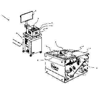
[0029] Figure 1 shows a high fidelity, high cost simulator with Virtual
Reality that allows
highly simulated surgical procedure simulations;
[0030] Figure 2 shows a low fidelity, low cost laparoscopic box trainer;
- 5 -
Date Recue/Date Received 2020-09-08
File number 11457-041
[0031] Figure 3 shows an illustration of high fidelity, low cost simulator in
accordance
with of the invention, showing a physical simulator unit with surgical tools
entry openings
and a display arranged to show live augmented visual representations of the
procedure;
[0032] Figure 4 shows a portable embodiment of high fidelity, low cost
simulator in
accordance with of the invention, showing a physical simulator unit with
surgical tools
entry openings and a laptop arranged to show live augmented visual
representations of the
procedure;
[0033] Figure 5 shows a clearer illustration of the physical simulator unit,
with a side panel
removed, allowing the viewing of a physical tissue model inside the unit,
further showing
surgical tools entry openings and a camera housing, in accordance with of the
invention;
[0034] Figure 6a shows the replaceable physical tissue model and its
electronic and
mechanical connections;
[0035] Figure 6b shows the tissue model with a cover with an image acquisition
trace;
[0036] Figure 7 shows the physical tissue model highlighting the track of an
embedded
sensor, in accordance with an embodiment of the invention;
[0037] Figure 8 shows the physical tissue model highlighting the track of a
more complex
embedded sensor or sensors, in accordance with a second embodiment of the
invention;
[0038] Figure 9 shows a close-up illustration of simulated surgery being
performed on the
physical tissue model, further showing trace marks of the laparoscopic tools;
[0039] Figure 10 shows the surgery result on the physical tissue model
highlighting the cut
track of an embedded sensor;
[0040] Figure 11 a is an illustration of the physical simulator unit, showing
a removable
side panel, in an opened position wherein the replaceable physical tissue
model is inserted;
and
[0041] Figure 1 lb is an illustration of the physical simulator unit, showing
a removable
side panel, in a closed position wherein the replaceable physical tissue model
is inserted.
- 6 -
Date Recue/Date Received 2020-09-08
File number: 11457-041
Detailed Description of the Preferred Embodiment
[0042] The present invention relates to an augmented and mixed reality
simulator for
laparoscopic surgical training. More specifically, the invention combines the
physical
world of performing surgery on tissue mimicking models, or phantoms, which
have
embedded sensors, with enhanced digital object augmentation to achieve a more
realistic
surgical experience. Through a process of overlaying digital textures onto
real physical
models, the invention allows users to engage with real tissues negating the
need for
artificial instrument feedback. The digital textures can also be used to
create complications
such as bleeding, perforations and more on otherwise inanimate objects.
[0043] Figure 1 shows a high fidelity, high cost simulator, such as e.g. the
Lap Mentor
from Simbionix. This is a VR system that allows full surgical procedures to be
performed
with performance metric output generated for each user. These simulators are
often
criticised for their lack of real to life tactile feedback felt through the
instruments due to
the nature of motor driven haptics.
.. [0044] Figure 2 shows a low cost, low fidelity laparoscopic box trainer
into which the user
can place a variety of tasks and operate on them with real laparoscopic
instruments, in this
case the image is streamed onto your laptop giving the training surgeon a
really good
understanding of issues like the fulcrum effect, triangulation and depth
perception. These
simulators are often criticised for their lack of realism and objective
feedback.
[0045] Figures 3 and 4 show the proposed invention. The invention merges real
feel tissue
models with a digital environment to provide real to life haptics, an
immersive environment
and full procedure training on a simple, accessible and affordable
laparoscopic procedure
trainer.
[0046] More specifically Figure 3 shows a laparoscopic surgical training
device 1
comprising a physical simulator unit, also referred to a box trainer 2, and a
computing and
display unit 3.
[0047] Figure 4 shows a more portable embodiment to the invention in Figure 3.
- 7 -
Date Recue/Date Received 2020-09-08
File number: 11457-041
[0048] Referring now to Figure 5, a closeup perspective view of the physical
simulator unit
2 is shown. The physical simulator unit 2 is in the shape of a box and
comprises a top panel,
a bottom panel, two parallel fixed side panels and two further parallel side
panels which
are removable.
[0049] A camera housing 4 is installed on the top panel. The camera housing
comprises a
camera (not visible in the Figures). A central opening in the top panel allows
the camera to
see inside the physical simulator unit. The camera housing 4 may further
comprise lighting
arranged to illuminate the inside of the box trainer 2. The lighting may be at
full visible
spectrum, Infrared, or a combination of the above.
[0050] In this embodiment the camera's line of sight is arranged to be
substantially at a 30-
degree angle with the plane of the top panel. The camera, camera box, the
lighting and any
other optical sensors in the camera housing 4 are connected to the computing
and display
unit 3 via electronic and computing cabling 8.
[0051] It will be appreciated that the computing and display unit 3 may be any
known
computing device such as desktops, laptops, tablets or custom units that are
capable of
acquiring, processing and displaying video and image data, as well as capable
of
controlling electronics such as lighting, power and sleep modes.
[0052] Referring again to Figure 5, two laparoscopic surgical tools entry
openings 5 are
shown close to adjacent corners of the top panel.
.. [0053] During the training procedure the laparoscopic surgical tools are
inserted in the tool
entry openings 5 in a similar manner as shown in Figure 2.
[0054] As with all laparoscopic or image guided procedures the simulator
occludes the
surgeons view of the physical models inside, requiring them to perform each
task via a
monitor or screen. Instruments are inserted via the entry ports located on the
top of the
.. simulator.
[0055] The physical simulator unit 2 further comprises a removable internal
base plate 6.
A removable physical tissue model 7 is installable on the removable internal
base plate.
- 8 -
Date Recue/Date Received 2020-09-08
File number: 11457-041
The camera is arranged to view the removable internal base plate through the
central
opening.
[0056] The camera may be a wired camera, such as USB, or an image data
acquisition and
it is orientated at a 30-degree angle and is perpendicular to the removable
magnetic base
inside of the simulator. The camera observes the image as it is in reality,
then using a
combination of marker based augmented reality and background compositing the
image
that the user sees on the monitor is transformed into a mixed reality
experience.
[0057] The position and angle of the physical tissue model 7 is adjustable.
Likewise the
position and angle of internal base plate 6 is also adjustable.
[0058] Figure 6a shows an illustration of a removable physical tissue model 7.
The
physical tissue model 7 is made of realistic silicone or synthetic tissue
models designed to
mimic the feel of real anatomical structures. One or more retainers 9 made of
stretchy
silicon and part of the physical tissue model 7 connect and retain the model
onto a model
base 12. Screws 11 fasten the model base 12 onto the internal base plate 6.
[0059] The tissue model 7 may be of latex or silicon or other materials or
plastics, which
are constructed in different layers to mimic the resilience of different types
of anatomical
tissues.
[0060] The inventions use of realistic silicone or synthetic tissue models
designed in some
cases to work with electro surgical instruments to mimic the feel of real
anatomical
structures, takes simulated practice to the next level, by allowing surgeons
to practice with
the same instruments used regularly in theatre.
[0061] The physical tissue model 7 may be embedded with wiring and or
electronic
sensors. Electrical connectors 10 connect the tissue model 7 to electric
circuitry and then
to the computing and display unit 3.
[0062] Figure 6b shows a model cover 13 which mates with model base 12 and
covers the
screws and connectors. An image acquisition trace 14 is sketched, embossed or
printed on
the model cover 13. In this embodiment the trace is a stretched pentagon. The
trace is
acquired by the camera. Other shapes may be used to identify to the computing
unit 3 the
- 9 -
Date Recue/Date Received 2020-09-08
File number: 11457-041
tissue model type. The model cover 13 may comprise other identifiable
information, e.g.
barcodes, alphanumeric etc. e.g. to be read by the camera and to tag a
surgical students
name and surgical simulation performance.
[0063] Whilst computer vision could be entirely relied upon across all system
features,
embedded sensors in the physical tissue model 7 act as a more reliable and
robust trigger
for complications that occur during interaction with the physical models
inside of the
simulator.
[0064] Figure 7 shows another embodiment of a physical tissue model 7, showing
electrical connectors 10a and 10b and retainers 9. Figure 7 further shows a
wiring 15
embedded in the body of the tissue model 7.
[0065] In this embodiment copper magnet wire is used. However it will be
appreciated that
various conductive wires with various conductive properties may be used to
simulate and
be programmed and tissue response simulators.
[0066] In this embodiment, one end of the wiring 15 is connected to electrical
connectors
10a and the other end of the wiring is connected to electrical connectors 10b,
thus
electrically looping the two connectors 10a 10b.
[0067] The wiring 15 may be embedded in different shapes and loops inside the
tissue
model 7 to represent e.g. a blood vessel or other ligament. In this embodiment
the wiring
15 is twisted to form one elongated blood vessel track.
[0068] Figure 8 shows another embodiment of the wiring 15, which may be a two-
dimensional mesh and identify the 2D location of the cut.
[0069] The mouldable material of the tissue model 7 may be doped e.g. with
metal or
carbon particles that change the impedance and or magnetic properties of the
structure and
of the tissue model, or phantom.
[0070] The tissue model 7 may also employ force sensitive resistors,
accelerometers or
tensions sensors.
- 10 -
Date Recue/Date Received 2020-09-08
File number: 11457-041
[0071] Referring now to Figure 9, during a laparoscopy simulation, using
laparoscopic
gripper 16 and scissor 17, when cutting through the tissue model, the wiring
15 may
become cut and thus create an open circuit which will be identified by the
signal data
acquisition and the computing and display unit. An augmented representation of
the
bleeding may then be shown on the display unit, in the area of the cut. Thus
simulating a
real-life surgical event.
[0072] When cut with a laparoscopic instrument the magnet wire connection is
lost and the
bleed is triggered through a change in system state that communicates with the
AR
software. The rig markers mentioned above determine the bleeding point in the
digital
environment. This solution can be used across a myriad of procedures and can
be used to
trigger different intraoperative complications (bleeding, bowel perforation,
perforated
common biliary tree). As such, individual solutions for triggering
complications will not
be required and this approach can be used to standardise this aspect of the
simulator across
all procedures.
[0073] The combined reliance on both computer vision and physical sensors
results in a
more stable system, devoid of the usual bugs found in programs reliant solely
on one or the
other.
[0074] In addition to solving issues with tactile feedback and realism, the
system is also
capable of generating accurate objective performance feedback using real
laparoscopic
tools.
[0075] Figure 10 illustrates a tissue model 7 with a cut internal wiring 15,
showing the
wiring cut location 15a.
[0076] In more detail, in the case of the first release, the vascular sensor
is connected to
the bridge during the entire operation. This sensor is formed of a sheathed
copper magnet
wire, designed for low voltage applications. The software platform performs a
search for a
connection upon start-up, once confirmed the procedure can be carried out. If
the vascular
sensor detects an incision, the information is instantly fed back to the
software so that the
relevant complication occurs e.g. bleeding. The user is then prompted to
rectify the bleed
and is guided through the necessary steps to resolve the complication. More
advanced users
- 11 -
Date Recue/Date Received 2020-09-08
File number 11457-041
may decide to use a surgical knots/loops to tie off the vessel before making
an incision. In
this case, the user can confirm that a series of loops have been placed
correctly thus turning
off the bleed trigger during the remainder of the procedure.
[0077] Referring again to Figure 9, gripper tracer 18 and scissor tracer 19
are visible to the
camera, which read and via the computing unit 3 calculates the three-
dimensional position
of the laparoscopic tools, which is then generated and displayed as an
augmented images
on the display.
[0078] Referring to Figure lithe physical simulator unit is shown with a
removable side
panel 20 in an opened position in a) and in a closed position in b). The inner
panel 6 and
replaceable physical tissue model 7 are inserted form the opening created from
the side
panel 20.
[0079] Therefore, the invention describes an apparatus for laparoscopic
surgical training,
which comprises a physical simulator unit; a physical tissue model; and
[0080] a computing and display unit; wherein the physical simulator unit
comprises at least
one side wall and a removable internal base plate; wherein the side wall
comprises: a central
opening through which a camera is arranged to view the removable internal base
plate; and
two or more laparoscopic surgical tools entry openings; wherein the internal
base plate is
arranged to hold the physical tissue model in the camera's field of view and
in a position
accessible to laparoscopic surgical tools when inserted in the two or more
laparoscopic
surgical tools entry openings; and wherein the computing and display unit is
arranged to
acquire video data from the camera and signal data from the physical tissue
model, and to
then utilise the data sets to generate and display in real-time a customised
mixed reality or
augmented video.
[0081] The physical tissue model comprises tissue mimicking material embedded
with
internal wiring and sensors arranged to be connected to electronic circuitry,
which is further
connected to the computing and display unit.
[0082] The video data acquired by the camera is augmented in real-time and
displayed in
the computing and display unit to simulate the video feed of a real
laparoscopic surgery.
- 12 -
Date Recue/Date Received 2020-09-08
File number: 11457-041
[0083] The physical manipulation of the internal wiring and sensors in the
physical tissue
model by the laparoscopic surgical tools is acquired as signal data, which
signal data is
then used to generate in real-time a customised video augmentation to the
video feed
acquired by the camera, and a merged video is displayed in the computing and
display unit.
[0084] The manipulation of the physical tissue model with the laparoscopic
surgical tools
is augmented in real-time and displayed in the computing and display unit.
[0085] The angle on the camera's principal axis is arranged to be
perpendicular to the plane
of the internal base plate.
[0086] The angle on the camera's principal axis is substantially at 30 degrees
to the plane
.. of the side wall.
[0087] The internal base plate is at an incline, which imaginary continuation
plane is
substantially at 30 degrees with the side wall, also known as top panel. The
device has an
angled platform, allowing the camera view to be perpendicular to the plane of
the operative
field. This is extremely important to ensure the instrument tracking aspect of
the software
is accurate. The camera position has been altered for optimal functionality of
the software.
Some optional side skirts have been added to help standardise the light
conditions within
the system and optimise the performance of the software.
[0088] The simulators design plays an integral part in the users mixed realty
experience,
ensuring that quality and realism remains uninterrupted throughout the
procedure. Side
.. panels and plastic surface texture are among some of the modifications
required to prevent
light disturbance inside of the simulator. These changes also optimise the
simulators ability
to track instruments in real time during a procedure.
[0089] A proprietary pigment blend has been used as to form the silicone
background in
the simulator for purpose of stable compositing of the surface. This is used
to project
multiple backgrounds onto the surface during different procedures that occur
in several
regions of the body. The colour was developed for specific use inside of the
simulator so
as not occlude instruments or tools.
- 13 -
Date Recue/Date Received 2020-09-08
File number 11457-041
[0090] The internal base plate may comprise a pigmented silicone background,
which
background is then used in the augmented video to project multiple backgrounds
onto the
surface during different procedures that occur in several regions of the body.
[0091] The physical tissue model may be replaceable and reconnectable to the
circuitry,
and wherein the physical tissue model is arranged to represent various human
or animal
tissue shapes, sizes and consistency, and the computing and display unit is
programmable
to represent an augmented video compatible with said tissue.
[0092] The camera may be arranged to track and extract data of the
displacement of the
physical tissue model, which data is then used to generate visual
representations of said
tissue in the augmented video.
[0093] The camera may be arranged to track and extract data of three
dimensional
movements of the customised laparoscopic surgical tools inserted in the
surgical tools entry
openings, which data is then used to generate visual representations of the
laparoscopic
tools in the augmented video.
[0094] Two or more cameras may be arranged to acquire stereoscopic or other
forms of
depth related information from their field of view.
[0095] The camera may be in a camera housing, which housing further comprises
lighting
arranged to illuminate the inside of the physical simulator unit, wherein the
lighting is in
the visible spectrum, infrared, a combination of the above, or a combination
of colours
arranged to enhance or discard for the camera elements of the physical tissue
model, the
background or of the laparoscopic tools.
[0096] The physical simulator unit may be in the shape of a box and comprises
a top panel,
a bottom panel, two parallel fixed side panels, two further side panels which
are removable
and parallel to each other; and wherein the side wall is the top panel.
[0097] The following table compares the features of the low and high-fidelity
simulators
vs the simulator proposed by the invention.
- 14 -
Date Recue/Date Received 2020-09-08
File number 11457-041
Low Fidelity High Fidelity Inovus LAP AR
Accessible YES NO YES
Affordable YES NO YES
Full Procedure Simulation NO YES YES
Performance Tracking NO YES YES
Validated Curriculum NO NO YES
Realistic Haptics YES NO YES
Immersive and engaging NO YES YES
[0098] The invention employs computer vision to deliver a stable dynamic
environment in
which to operate The invention uses a unique approach to marker-less three-
dimensional
instrument tracking This allows the creation of extremely accurate movement
metrics for
users and provides a platform to assess performance over time in a plethora of
procedures
Reducing the barrier to entry on such products will allow for much larger data
collection
than previously possible using high fidelity systems This is made possible
through the
utilisation of several computer vision-based techniques, including but not
limited to Canny
edge detection and Hough line tracking A proprietary software algorithm is
used to
generate performance data
[0099] The inventions marker-less tracking enables on screen interaction with
virtual
action buttons This allows the operating surgeon to carry out 'in play'
actions and work
through steps of the procedure without having to down tools
[00100] The core features of the simulator include:
= Close to life haptic feedback through synthetic soft tissue models
= Realistic digital anatomy fully integrated with the soft tissue models
= Ability to create and manage intraoperative complications
- 15 -
Date Recue/Date Received 2020-09-08
File number: 11457-041
= Marker-less 3D movement tracking of the instruments within the surgical
field with
objective feedback on the key metrics of surgical performance achieved through
monocular
camera lens
= Online user accounts allow users to store and track progress of their
surgical training
= Online training portals allow trainees to perform and record surgical
procedures specific
to their specialty
= Full procedure simulation across general (including paediatric) surgery,
O&G and
Urology
= Performance tracking and feedback compatible with generic skills tasks
and validated
curricula such as the LapPass programme.
= The invention achieves consistent image registration of the digital
anatomy.
= The invention can be adapted to provide augmented reality procedures for
general,
paediatric, O&G and Urological surgery including but not limited to:
Cholecystectomy,
Bowel anastomosis, Pyloromyotomy, Ectopic pregnancy, Myomectomy, Vaginal Vault
closure and Nephrectomy.
[00101] The lP is obfuscated within codebase and cannot be accessed
without access
to protected source files garnering the necessary level of protection once the
product is
commercialised.
[00102] The technical solution proposed by the application is a major
advance on
the current state of the art. When considering performance tracking of
laparoscopic
simulation, the existing box trainer products are designed to track
performance related to
basic 'generic tasks' only. None of the existing technology has the ability to
track
performance related to 'procedure specific' full surgical tasks.
[00103] The only way to track performance in full surgical procedures
is with
extremely expensive VR simulators, thus making the proposed invention unique
in form
and function.
- 16 -
Date Recue/Date Received 2020-09-08
File number: 11457-041
[00104] While illustrative and presently preferred embodiment(s) of the
invention
have been described in detail hereinabove, it is to be understood that the
inventive concepts
may be otherwise variously embodied and employed and that the appended claims
are
intended to be construed to include such variations except insofar as limited
by the prior
art.
- 17 -
Date Recue/Date Received 2020-09-08