Note : Les descriptions sont présentées dans la langue officielle dans laquelle elles ont été soumises.
CA 03098476 2020-10-26
WO 2019/213054 PCT/US2019/029865
TISSUE OXYGEN SATURATION DETECTION AND RELATED APPARATUS AND
METHODS
CROSS-REFERENCE TO RELATED APPLICATIONS
[0001] The present application claims the benefit under 35 U.S.C. 119(e) of
U.S. Patent
Application Serial No. 62/665,455, filed May 1, 2018 under Attorney Docket No.
D0784.70001U500, and entitled "TISSUE OXYGEN SATURATION DETECTION AND
RELATED APPARATUS AND METHODS," which is hereby incorporated herein by
reference
in its entirety.
BACKGROUND
Field
[0002] The present application relates to monitoring of muscle oxygen
saturation and
related physical performance, as well as to related apparatus and methods.
Related Art
[0003] Oxygen is required for cells to produce energy in a process called
oxidative
phosphorylation. Hemoglobin is the protein in red blood cells that binds
oxygen molecules for
transport from the lungs to all tissues and exists in two states, oxygenated
and deoxygenated.
Oxygen saturation (SO2) denotes the percentage of oxygenated hemoglobin out of
the total
present hemoglobin. Muscle oxygenation (5m02) is the term used here to
indicate the oxygen
saturation in the muscle.
[0004] Muscles at any time, and especially when exercised, require oxygen for
energy
production, and therefore 5m02 is a parameter that encapsulates the metabolic
state of the
muscle. Specifically, it describes how much oxygen is present in the muscle
and when oxygen
consumption exceeds the supply. Lactic acid build-up in the blood is an
indirect measurement
of oxygen deficits after a muscle was in an anaerobic state, as anaerobic
glycolysis results in the
excretion of lactate into the blood stream.
BRIEF SUMMARY
[0005] According to an aspect of the present application an optical device is
provided,
comprising a wearable housing; a plurality of optical sources in the wearable
housing; and a
CA 03098476 2020-10-26
WO 2019/213054 PCT/US2019/029865
- 2 -
plurality of optical detectors in the wearable housing and including at least
three optical
detectors arranged substantially in a linear arrangement.
[0006] According to an aspect of the application, an optical device is
provided,
comprising a plurality of optical sources arranged substantially in a linear
arrangement along a
first direction, and a plurality of optical detectors arranged substantially
in a linear arrangement
along a second direction approximately perpendicular to the first direction.
Methods of
assessing hemoglobin concentration based on signals detected by the optical
device are also
provided.
[0007] According to an aspect of the application, an optical device is
provided,
comprising: a wearable housing; an optical source array in the wearable
housing including a
plurality of optical sources; and a plurality of optical detectors in the
wearable housing including
at least three optical detectors arranged substantially in a linear
arrangement. The at least three
optical detectors include a first optical detector disposed a first distance
from the optical source
array, a second optical detector disposed a second distance greater than the
first distance from
the optical source array, and a third optical detector disposed a third
distance greater than the
second distance from the optical source array.
[0008] According to an aspect of the application, a wearable optical monitor
is provided,
comprising: an item of clothing; an optical detector module embedded in the
item of clothing
and including a linear arrangement of optical detectors; and a control module
coupled to the
optical detector module and configured to receive output signals from the
optical detectors of the
optical detector module.
BRIEF DESCRIPTION OF DRAWINGS
[0009] Various aspects and embodiments of the application will be described
with
reference to the following figures. It should be appreciated that the figures
are not necessarily
drawn to scale. Items appearing in multiple figures are indicated by the same
reference number
in all the figures in which they appear.
[0010] FIG. lA is a schematic view from a first perspective of an optical
device for
detecting optical signals indicative of muscle oxygenation, according to a non-
limiting
embodiment of the present application.
CA 03098476 2020-10-26
WO 2019/213054 PCT/US2019/029865
-3 -
[0011] FIG. 1B is a schematic view of the optical device of FIG. lA from an
opposing
view.
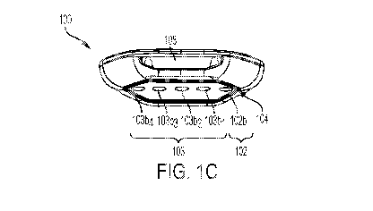
[0012] FIG. 1C is a perspective view of the optical device of FIG. 1A.
[0013] FIG. 1D is an exploded view of the optical device of FIG. 1A.
[0014] FIG. 2A shows a simplified perspective view of an emitter configuration
for an
optical device of the type shown in FIG. 1A, according to a non-limiting
embodiment of the
present application.
[0015] FIGs. 2B and 2C show cross-sectional views of emissions from an emitter
in
accordance with an aspect of the present application.
[0016] FIGs. 3A-3B are block diagrams of the circuitry of an optical device of
the type
shown in FIG. 1A, according to two non-limiting alternative embodiments of the
present
application.
[0017] FIG. 4A shows an illustrative wearable optical device 100 with a strap
105 to
secure the device 100, according to a non-limiting embodiment of the present
application.
[0018] FIG. 4B illustrates the wearable optical device 100 of FIG. 4A secured
to an
individual.
[0019] FIG. 5 is a cross-sectional view of an optical device of the type
illustrated in FIG.
lA in contact with a user, according to a non-limiting embodiment of the
present application.
[0020] FIG. 6 illustrates a timing diagram for one manner of operation of an
optical
device of the types described herein.
[0021] FIG. 7 illustrates a distributed optical device including
emitter/detector strips and
a control module.
[0022] FIGs. 8A-8E illustrate examples of a dashboard as may be implemented on
a
processing device to provide input to a user on physical activity.
[0023] FIG. 9A illustrates a wireless charger for charging optical devices of
the types
described herein.
[0024] FIG. 9B illustrates an optical device coupled to the charger of FIG.
9A.
[0025] FIG. 10a illustrates an alternative arrangement of optical sources and
optical
detectors, accordingly to non-limiting embodiment.
[0026] FIG. 10b shows an exemplary arrangement of sources and detectors from a
top
view, including a region of homogenous tissue.
[0027] FIG. 10c shows a side view of the exemplary arrangement in FIG. 10b.
CA 03098476 2020-10-26
WO 2019/213054 PCT/US2019/029865
- 4 -
DETAILED DESCRIPTION
[0028] Aspects of the present application relate to a wearable optical device
for detecting
muscle oxygenation, processes for determining muscle oxygenation based on
optical signals
detected by the optical device, and apparatus and methods for monitoring
physical performance
based on data produced by the optical device and providing input to a user,
for instance when
engaged in physical activity or when at rest. Applicant has appreciated that
monitoring muscle
oxygenation during physical activity may provide valuable insight into
physical performance,
particularly for those engaged in high endurance activities such as long
distance running, biking,
and swimming. Muscle oxygenation may provide an indication of whether muscle
is aerobic,
close to an anaerobic threshold, or anaerobic, and thus may be relevant to
whether an athlete is
performing optimally. By contrast, conventional techniques for assessing
physical performance,
including heart rate monitoring, provide different information and are
inadequate to assess
performance of muscle.
[0029] Applicant has further appreciated that conventional techniques for
assessing
oxygenation, such as pulse oximetry, are inadequate for assessing muscle
oxygenation.
Accordingly, an aspect of the present application provides an optical device
suitable for
collecting data representative of muscle oxygenation and/or hemoglobin
concentrations during
physical activity. The optical device is wearable in at least some
embodiments. For example, it
can be being positioned on a user's leg or arm. Thus, aspects of the present
application provide
a wearable fitness sensor which may assess the user's muscle oxygenation
and/or hemoglobin
concentrations and therefore provide an indication of the user's physical
state and performance.
[0030] Aspects of the present application relate to a wearable optical device
configured
to emit optical signals into the muscle of a user and collect return signals
in response to such
emission. The wearable optical device may serve as a fitness device in at
least some
embodiments, being configured to provide information about hemoglobin and/or
oxygenation
level within tissue. The information relating to hemoglobin and/or oxygenation
level may be, in
some embodiments, related to lactic acid levels, which may serve as a
performance metric.
[0031] Aspects of the present application provide a processor and processing
techniques
for converting optical signals detected by an optical sensor into an
indication of any one or more
of 5m02, oxygenated hemoglobin concentration (Hb02), deoxygenated hemoglobin
CA 03098476 2020-10-26
WO 2019/213054 PCT/US2019/029865
- 5 -
concentration (Hb), or total hemoglobin concentration (HbT). Sm02 represents a
ratio of
Hb02/HbT. The optical sensor may be of the type(s) described herein, although
alternatives are
possible. In some embodiments, the processor may be part of the optical
sensor, although in
other embodiments the data collected by the optical sensor may be transferred
to an external
processor, such as a computer, smartphone, tablet, sports watch, or other
processing device. The
processing may take into account structural features of the optical sensor,
including the
positioning of optical sources (also referred to herein as "emitters") and
optical detectors of the
optical sensor.
[0032] Aspects of the present application provide apparatus and methods for
providing
input on physical performance to an individual. In some embodiments, a plan is
provided to the
individual based on the assessment of physical performance. A wearable optical
sensor of the
types described herein may be used to collect data indicative of any one or
more of Sm02,
Hb02, Hb, or HbT, although sensors of alternative designs may be employed in
some
embodiments. In response to receiving the data, an assessment of physical
performance may be
made and a plan created for future activity. The assessment of performance and
the plan may be
presented to the individual on a smartphone, tablet, computer, or other
suitable device, and in at
least some embodiments may be done during exercise. The assessment may be
presented
visually, audibly, using a combination of the two, or in any other suitable
manner. In this
manner, athletes may appropriately tailor their training and other activities
for optimal
efficiency.
[0033] The aspects and embodiments described above, as well as additional
aspects and
embodiments, are described further below. These aspects and/or embodiments may
be used
individually, all together, or in any combination of two or more, as the
application is not limited
in this respect.
[0034] As described, according to an aspect of the present application, a
wearable optical
sensor is provided for detecting optical signals indicative of any one or more
of Sm02, Hb,
Hb02, or HbT. FIGs. lA and 1B illustrate views of opposing sides of a wearable
optical device
100 according to a non-limiting embodiment of the present application, with
FIG. 1C providing
a perspective view and FIG. 1D providing an exploded view. The wearable
optical device 100
includes a casing 107 having such a size that a human user can wear the casing
107 on a portion
of the user's body, such as the leg. For example, the casing 107 may have a
maximum
dimension less than 20 cm, less than 15 cm, less than 10 cm, less than 80 mm,
less than 60 mm
CA 03098476 2020-10-26
WO 2019/213054 PCT/US2019/029865
- 6 -
or any value or range of values within such ranges. An example of the
positioning of the sensor
is illustrated in connection with FIG. 4B, described further below. The casing
107 has a back
side 101 proximate the user's body and a front side 111 distal the user's body
during use.
[0035] FIG. 1B shows a back side 101 of the wearable optical device 100. The
optical
device includes multiple optical sources and multiple optical detectors. In
the illustrated non-
limiting embodiment, the wearable optical device 100 includes a light source
array 102 and a
detector array 103 disposed on the back side 101. The back side 101 may be in
contact with the
surface of a user's skin during operation, allowing the wearable optical
device to transmit optical
signals into muscle tissue underneath the skin surface and to collect optical
signals from the
muscle tissue.
[0036] In the illustrated embodiment, the optical emitters may be recessed
relative to the
surface of the back side 101 which contacts the user. With respect to the non-
limiting example
of FIG. 1B, the light source array 102 includes one or more emitters forming
an emitter array
102b disposed within an emitter recess 102a. In alternative implementations,
emitters may have
respective recesses in which they are disposed. The emitter recess 102a may
optically isolate the
emitters from the detectors, such that the optical signals transmitted from
the emitter array 102b
do not directly enter the detectors of detector array 103 without passing
through the tissue of the
user. In some embodiments, the walls of the emitter recess 102a may include
light isolation
material to provide an improved light isolation between components on the back
side 101. In
some embodiments, the light isolation material may comprise compressive foam
material,
although other materials are possible.
[0037] According to an embodiment, a filter may be disposed covering the
emitters, such
as covering emitter array 102b. For example, the filter may cover the emitter
recess 102a. The
filter may be considered a window or cover and may perform a desired optical
function, such as
filtering undesired emissions or diffusing emitted light. A non-limiting
example is described in
connection with FIG. 2A.
[0038] FIG. 2A shows a simplified perspective view of the illustrative emitter
recess
102a of FIG. 1A. In this example a window filter 201 is attached to the
opening of the emitter
recess and is configured to selectively modify the optical signals 203 emitted
from the emitter
array 102b in any suitable way to facilitate a spectrum measurement. In one
embodiment, the
window filter 201 may comprise a tinted filter to modify the spectrum of the
optical signals
emitted into the user. In another embodiment, the window filter 201 may
comprise a roughened
CA 03098476 2020-10-26
WO 2019/213054 PCT/US2019/029865
-7 -
texture to diffuse optical signal 203 by scattering so that light signals 204
exit the window filter
along a plurality of angles Aexit relative to an axis 205 normal to the window
filter. The plurality
of exit angles Aexit may comprise a maximum angle of at least 30 degrees, at
least 45 degrees, at
least 60 degrees, at least 90 degrees, between 20 and 90 degrees, or any value
or range of values
within such ranges.
[0039] As previously described, the optical device 100 contacts a user during
use in at
least some embodiments. For example, the optical device 100 may be configured
such that the
back side 101 contacts a user's skin in use. FIGs. 2B and 2C show a cross-
sectional view of the
optical signals 204 emitted into a medium 206, such as a human tissue (e.g.,
muscle), from the
light source 102 both without window filter 201 (FIG. 2B) and with window
filter 201 (FIG.
2C). In the illustrated examples, the window filter 201 is assumed to be a
diffuser, although not
all embodiments are limited in this respect.
[0040] As can be seen from FIGs. 2B and 2C, in the absence of window filter
201 the
emitted light signal 204 may be focused over a relatively narrow exit cone of
maximum exit
angle Aexit (FIG. 2B). In comparison, the use of window filter 201 diffuses
the light signal 204,
resulting in the light signal 204 exhibiting an exit angle Aexit of greater
maximum extent than in
the absence of the window filter 201 (FIG. 2C). For example, in the context of
FIG. 2C, Aexit
may extend up to approximately 90 degrees and may simulate uniform semi-
spherical light
scattering from a point light source in a homogenous medium. The more diffused
light signal
204 in FIG. 2C comprises more light components with emission directions along
the surface 207
of the medium 206 toward the direction of a detector array 103 along the same
surface 207 and
requires less scattering distance to reach the detector array 103.
[0041] In addition to performing an optical function, window filter 201 may
physically
seal the cavity containing the emitter array, and thus may protect the emitter
array from damage
from environmental factors such as moisture. In one embodiment, the window
filter may be
attached to the emitter recess 102a using optical glue or any other suitable
optically transparent
adhesive or fastener. In an alternative embodiment, the window filter may be
molded directly
into the housing, achieving a mechanical bond by appropriate design of the
window filter and
housing, as well as a chemical bond achieved through adhesion of the plastics
during the
molding process.
[0042] The emitter array 102b may include a plurality of individual light
emitters
arranged to fit within the recess 102a. The emitters may be any suitable type
of emitters, such as
CA 03098476 2020-10-26
WO 2019/213054 PCT/US2019/029865
- 8 -
light emitting diodes (LEDs). In some embodiments, the emitters of the emitter
array 102b may
be narrow-band light sources. As used herein, "narrow-band" means that
substantially all of the
optical signal energy is concentrated at one narrow wavelength band centered
around one peak
wavelength. For example, greater than 90% of the signal intensity is within +/-
10% of a nominal
peak wavelength, or less (e.g., +/-6%). In one embodiment, the emitter array
102b may be
narrow-band light sources that emit light signals with a plurality of peak
wavelengths to provide
multi-colored illumination. In contrast to using a broadband light source with
a spectrometer to
get spectral information, embodiments of the present application use multiple
different colored
narrow-band light sources such as narrow-band LEDs. The use of multiple narrow-
band light
sources allows for collection of spectral information without using large and
expensive
conventional spectrometers with a broadband light source.
[0043] As a non-limiting example, the emitter array 102b may include a
plurality of
narrow-band LEDs that emit light signals with two peak wavelengths at 660 nm
and 855 nm in
the red/infrared spectrum. Those wavelengths may be selected based on the
hemoglobin
absorption spectra with respect to oxygenated and deoxygenated hemoglobin. In
other
embodiments, a greater number of peak wavelengths may be used to obtain a
higher quality
spectrum with less noise. For example, the emitter array 102b may be narrow-
band LEDs that
emit light signals comprising two, three, four, or five different peak
wavelengths.
[0044] While 660 nm and 855 nm are two non-limiting example, it should be
appreciated that other wavelengths may be used. For example, one wavelength in
the range of
650 nm to 710 nm may be used and another wavelength in the range of 820 nm to
860 nm.
More than one wavelength in each of those ranges may be used in some
embodiments. In some
embodiments, an additional wavelength in the range of 950 nm to 1000 nm may be
used. Still
other combinations of wavelengths are possible.
[0045] When narrow-band light sources are used, a single wavelength may be
emitted at
a single time, as an example. A detector may be used to measure the intensity
of a reflected
light from the test subject for that wavelength. Then, a different wavelength
may be emitted and
the light reflected from the subject measured. In this manner, the optical
device may provide
measurements of portions of the reflection spectrum (which may be referred to
as "slices" in
some embodiments). Also, using the optical device 100, in one embodiment, a
spectrum of
detected light dependent on distance may be collected. By shining light
signals from narrow-
band light sources into a subject and measuring the intensity of the light
signals reflected from
CA 03098476 2020-10-26
WO 2019/213054 PCT/US2019/029865
- 9 -
the muscle tissue underneath the skin at several different locations with
varying distances from
the light sources along the skin (e.g., distances D1-D4 in FIG. 1B), the
spectral intensity
corresponding to various portions of the spectrum, is obtained at each
measurement location,
which when combined with the distance from the light source allows for
determination of
hemoglobin and/or muscle oxygenation levels.
[0046] In some embodiments, the emitters of emitter array 102b may be attached
to a
printed circuit board (PCB), such as PCB 202 shown in FIG. 1D and FIG. 2A. In
one
embodiment, the emitter array 102b may be narrow-band LEDs powered by a low
noise analog
LED driver on the PCB 202 as adjustable current sources to set the emitted
light intensity level.
Other manners of housing the emitters within the optical device 100 are also
possible.
[0047] The emitters of the emitter array 102b may be arranged suitably to
provide
desired distances between the emitters and the detectors of the optical device
100. For example,
the emitters of the emitter array 102b may be arranged close together to serve
effectively as a
point source in some embodiments. That is, the emitter(s) may occupy a single
position of the
optical device, with the detectors spread over varying distances. The emitters
array may occupy
less than 20 mm in some embodiments, less than 10 mm in some embodiment, or
other sizes
serving effectively as a point source. For example an emitter array of two
emitters may have a
lateral extent of 10 mm or less in some embodiments. In other embodiments, the
emitters of the
emitter array 102b may be arranged linearly, for example occupying a total
lateral extent of less
than 20 mm or any value within that range. As a non-limiting example, the
emitter array 102b
may include four emitters arranged linearly, with the linear arrangement of
the emitters being
angled with respect to a linear arrangement of optical detectors. For example,
the emitters may
be arranged in a line substantially perpendicular to a line along which the
detectors are arranged.
In some embodiments, the emitters may be positioned toward an edge of the
optical device 100
and the detectors located centrally, such that the path from the emitters to
the detectors points
inward. Such a configuration may reduce the impact of stray light.
[0048] The detectors of the optical device 100 may be arranged suitably to
provide
desired distances relative to the optical emitters. In the examples in FIGs.1B
and 1C, a detector
array 103 is provided including a plurality of detectors 103b1, 103b2, 103b3,
and 103b4 arranged
substantially linearly on a path that originates from the location of the
emitter array 102b. In the
illustrated example, the linear arrangement of detectors is substantially
perpendicular to the
linear arrangement of optical emitters. Although four detectors 103b1-103b4
are shown, any
CA 03098476 2020-10-26
WO 2019/213054 PCT/US2019/029865
- 10 -
other suitable number may be included. In some embodiments, the number of
detectors
included is selected to provide at least three distinct emitter-detector
distances. For example,
detector 103b2 is a different distance D2 from the emitter array 102b than is
detector 103b1
(which is displaced from the emitter by a distance D1) and also a different
distance than is
detector 103b3 (which is spaced from the emitter by a distance D3) and
detector 103b4 (which is
spaced from the emitter by a distance D4). The three or more emitter-detector
distances may
facilitate determination of Sm02, Hb02, Hb, and/or HbT. The distances D1-D4
may assume any
suitable values to facilitate determination of the desired characteristics
(e.g., Sm02) while in at
least some embodiments providing a compact size suitable for implementation in
a wearable
housing. For example, D1 may be greater than 5 mm and D4 may be less than 50
mm in some
embodiments, with D2 and D3 falling within those ranges at any suitable
spacing. That is, in
some embodiments, each of D1-D4 may be between 5 mm and 50 mm in some
embodiments,
although other values are possible. In some embodiments, the optical device
100 includes five
or fewer detectors, and in some embodiments only four detectors.
[0049] In an alternative arrangement, detectors may be provided with different
spacing
on both sides of the LEDs. According to a further alternative, multiple linear
detector arrays
may be provided in different spatial directions. This may allow for mapping of
the spatial
distribution of the muscle oxygenation and/or hemoglobin concentration over
the muscle to
examine a larger portion of the tissue. This may also provide better SNR, for
example by
averaging out scattering from varicose veins, provide robust measurements in
the presence of
superficial skin lesions, and examine the heterogeneity of the Sm02 (or Hb or
Hb02 or HbT)
distribution in the tissue.
[0050] In this example, each detector is disposed inside a detector recess
such as 103ai
to expose the detector for detection of a light signal. Respective recesses
may be provided, such
as 103ai-103a4. The detector recesses such as 103ai also provide light
isolation of the optical
signals entering the detectors from other components exposed on the back side
101. In some
embodiments, it may be preferred that the detector array 103 collects
substantially light signals
from the skin surface (or, more generally, tissue) while minimizing light
transmitted directly
from the emitter array 102b to the detector array 103, to minimize stray
background signals.
Further according to some embodiments, it may be preferred that all light
signals entering a
particular detector substantially correspond to a light signal reflected from
a test subject surface
immediately adjacent the detector recess in which the detector is disposed, to
provide a more
CA 03098476 2020-10-26
WO 2019/213054 PCT/US2019/029865
- 11 -
accurate correlation of detector signal versus location of the detector.
Therefore it may be
preferred that the amount of stray light between different detector recesses
be minimized.
Although not shown, the walls of the detector recesses such as 103ai may
include light
insulation material to provide an improved light isolation between each
detector and between the
detectors and the emitter array. In some embodiments, the light insulation
material may
comprise compressive foam material. In some embodiments, as shown in FIG. 1B,
an isolation
wall 110 may optionally surround the emitter array 102b and detectors of the
detector array 103
to provide light isolation. The wall 110 is shown in dashed lining because of
its optional nature.
The wall 110 may be formed from the casing material or from any other suitable
material for
blocking light.
[0051] In the example in FIG. 1B, each detector recess such as 103ai may
include a
window filter covering the opening area of the detector recess and forming a
cavity containing
the detector such as 103b1. The window filter may be, and in some embodiments
is, configured
to modify the optical signals entering the detector such as detector 103b1 in
any suitable way to
facilitate a spectrum measurement. In one embodiment, the window filter may
comprise a tinted
filter to modify the spectrum of the optical signals transmitted through. In
some embodiments,
the window filter may optionally include one or more additional functions from
focusing (e.g., a
Fresnel lens) and beam steering/spatial filtering. When included, the window
filter may also
physically seal the cavity containing the detector to protect the detector
from damage from
environmental factors such as moisture. In one embodiment, each window filter
may be attached
to the detector recess using optical glue or other suitable adhesive or
fastener. In an alternative
embodiment, the window filter may be molded into the housing, achieving a
mechanical bond
by appropriate design of the window filter and housing, as well as a chemical
bond achieved
through adhesion of the plastics during the molding process.
[0052] The optical detectors 103b1-103b4 measure the intensity of a light
signal. The
detectors may be optical receivers with onboard analog-to-digital conversion
that converts a
light signal that enters a semiconductor junction into electrical energy and
then outputs the
detected light signal intensity as "counts". In some embodiments, the
detectors may convert a
total light signal intensity across all wavelengths into counts. As an
example, the detectors may
be photodiodes. The detectors may be integrating photodetectors with onboard
analog-to-digital
conversion, although alternatives are possible. In another non-limiting
example, the detectors
may be TSL2591 light-to-digital converters. The detectors may be sampled at a
frequency
CA 03098476 2020-10-26
WO 2019/213054 PCT/US2019/029865
- 12 -
suitable to mitigate the effects of muscle movement. For example, the
detectors may be sampled
at a frequency less than 10 Hz, less than 5 Hz, less than 3 Hz, less than 2
Hz, or at any sampling
rate within such ranges. In an alternative embodiment, the detectors may be
silicon photodiodes,
with analog to digital conversion accomplished using a photometric front end
with analog-to-
digital converter to convert current measured from the photodiode into counts.
In one non-
limiting example, the photometric front end may be Analog Devices ADPD103,
available from
Analog Devices, Inc. of Norwood, Massachusetts. In this example the
photodetectors may be
sampled at 4000 Hz, 1000 Hz, 100 Hz, 0.1 Hz, less than 4000 Hz, less than 100
Hz or any
sampling rate within such ranges. In another non-limiting example, the
photodiodes may be
Everlight PD15-22C/TR8, available from Everlight America, Inc. of Carrollton,
Texas.
[0053] In the example in FIG.1B, a plurality of detectors such as 103b1-103b4
are
arranged substantially linearly on a path that originates from the location of
the emitter array
102b for measurement of light signals at varying locations from the emitter
array 102b.
Although four detectors 103b1-103b4 are shown, any other suitable number may
be included. In
a preferred embodiment, at least three detectors are used to measure the
intensity of light signals
for at least three different distances from the emitter array, to provide an
improved fitting of
measured signals as a function of distance with a model used to provide any
one or more of
Sm02, Hb, Hb02, or HbT based on the measured intensities. In the non-limiting
example in FIG.
1B, the detector-emitter distances D1-D4 are such that D4>D3>D2>D1. Providing
more than
three detectors may further improve the quality of measurement data.
[0054] In some embodiments, two or more of the detectors may be of different
sizes than
each other. Because light intensity decreases with distance from the source,
detector 103b1 is
likely to receive a greater light intensity than detector 103b2, while
detector 103b2 is likely to
receive a greater light intensity than detector 103b3, and detector 103b3 is
likely to receive a
greater light intensity than detector 103b4. Thus, using detectors of equal
sizes and sensitivities
in a configuration like that shown in FIG. 1B is likely to result in the
detectors 103b1-103b4
producing different output signal magnitudes, with the detector 103b1 likely
to produce an
output signal of the greatest magnitude and detector 103b4 likely to produce
an output signal of
the smallest magnitude among the detectors. The difference in output signal
magnitudes may be
substantial in some embodiments, for example amounting to an order of
magnitude or more.
Applicant has appreciated that such differences in magnitude can lead to
difficulty in processing
CA 03098476 2020-10-26
WO 2019/213054 PCT/US2019/029865
- 13 -
the received signals, for example due to constraints on the capability of the
processing circuitry
to handle signals of substantially different magnitudes.
[0055] Thus, according to an aspect of the present application, the detectors
may be
sized to produce output signals of magnitudes that are within an acceptable
range of each other.
For example, the detectors may increase in size the farther they are from the
emitter array 102b.
That is, detector 103b2 may be larger than detector 103b1, detector 103b3 may
be larger than
detector 103b2, and detector 103b4 may be larger than detector 103b3. In some
embodiments, at
least one detector is smaller in size than another detector positioned farther
from the emitter
array. For example, detector 103b1 may be smaller than one or more of detector
103b2, detector
103b3, or detector 103b4. In some embodiments, detector 103b2, detector 103b3,
and detector
103b4 are equally sized, and are all larger than detector 103b1. As an
alternative to using
detectors of different sizes, the detectors may be sized equally but have
increasing sensitivities
the farther they are from the emitter array 102b. In this manner, the
detectors 103b1-103b4 may
produce output signals that are relatively close in magnitude to each other,
which may simplify
processing by the processing circuitry. The differences in size and/or
sensitivity of the detectors
may be accounted for by scaling the detector output signals based on the known
differences in
size/sensitivity. For example, the detector output signals may be normalized
in some
embodiments.
[0056] In some embodiments, the optical detectors are arranged and configured
electronically to be operated synchronously with the emitters. An example of a
device which
may be used for such operation is the ADPD103 photometric front end, listed
above. In some
embodiments, synchronously operating the emitters and detectors of a
photometric front end or
other optical device involves aligning the integration (time) windows with the
emitter (time)
windows for accurate measurement of the optical signal. In some embodiments,
emitters and
detectors may be operated synchronously with the emitter windows and
integration windows not
precisely aligned, and the measured values may be mapped to the true values.
Misalignment of
the integration windows and emitter windows of a photometric front end or
other optical device
may occur, for example, if the windows of the measurement channels are not
individually
controllable, but rather are set as a group. In such circumstances, the
alignment of integration
and emitter windows may be imprecise for one or more measurement channels,
resulting in
measurement error, the degree of which may depend on the degree of
misalignment between the
integration and emitter window for a particular channel. Mapping the measured
optical signal
CA 03098476 2020-10-26
WO 2019/213054 PCT/US2019/029865
- 14 -
intensity values when the integration window and emitter window are misaligned
to the true
values may provide improved performance, and represents a calibration of the
system. The
functional form of this mapping may be a polynomial, where the order, and
coefficients of the
polynomial that accomplish the mapping can be determined through calibrating
the sensor
against samples with known optical properties. If the difference in measured
and true values is
due primarily to hardware configuration, such as routing of a circuit board,
then calibrating
(mapping) as described above for a single unit may apply equally well for all
other units of the
same kind.
[0057] Any suitable spacing, or spacing combination between centers of each
detector
on the substantially linear path and between the first detector of the
plurality of detectors and the
emitter array may be provided to arrange the detectors and emitter array on
the back side 101 of
the wearable optical device 100. In a non-limiting example, the detectors may
be spaced 10 mm
apart between centers of each adjacent detector and between the center of the
first detector of the
plurality of detectors and the emitter array. In other embodiments, smaller
spacing may be used
to allow for inclusion of a greater number of detectors, such as a spacing of
8 mm. For example,
the detectors of FIG. 1B may be spaced from the emitters by 8mm, 16mm, 24mm,
and 32mm,
respectively. In some embodiments, the spacing between neighboring detectors
may be between
5mm and 20mm, less than 5mm, less than lmm or any distance or range of
distances within
such ranges. In some embodiments, the detectors may be pixels of an imaging
device, such as a
charge-coupled device (CCD) imager.
[0058] One or more of the detectors may serve to provide a reference signal.
For
example, in the context of optical device 100, the detector closest to the
emitter 103b1 may
provide a reference intensity against which the intensities measured by
detectors 103b2-103b4
are measured. In this manner, control over and knowledge of the variations in
intensity of the
signals emitted by the emitters may be provided, simplifying the device design
and operation. In
such configurations, the detector serving as the reference may not contribute
to the unique
emitter-detector distances, although in other embodiments it may. That is, in
the example of
FIG. 1A, three unique emitter-detector distances are provided by detectors
103b2-103b4, while
detector 103b1 provides a reference (or baseline) for the other detectors.
[0059] Applicant has appreciated that stray light may undesirably impact the
performance of an optical device such as optical device 100. For example, the
optical device 100
may be used in situations in which sunlight or other environmental light is
present. Detection of
CA 03098476 2020-10-26
WO 2019/213054 PCT/US2019/029865
- 15 -
such environmental light could negatively impact device performance.
Accordingly, as
illustrated in FIGs. 1B and 1C, the optical device may include a seal ring 104
configured to
prevent environmental light or other stray light from unintentionally being
detected by the
detectors of the optical device 100. The seal ring 104 may be a raised portion
of the casing 107,
formed by a continuously raised portion around the periphery of the back side
101 as shown.
The height of the seal ring may be less than lmm, less than 2mm, or any other
suitable height.
The seal ring may be constructed from substantially the same material as the
casing material on
the back side 101, or it may be constructed from any other suitable material
for providing a seal
when in contact with a surface of the user. In some embodiments, the seal ring
is constructed
with a dimension that maintains substantially the overall small footprint of
the wearable optical
device. In some embodiments, when the wearable optical device is used on a
user, the back side
101 is placed facing the skin of the user and the seal ring 104 is in contact
with the skin of the
user forming a complete seal with no gaps such that no ambient light reaches
any of the
detectors such as 103b1 to reduce stray background signal and improve SNR of
the detected
signals. The seal ring may serve additional functions, such as helping to
retain the optical device
in position on the user, prevent moisture (e.g., sweat or rain) from
interfering with the optical
operation, or other functions.
[0060] Other features of the optical device 100 include optional buttons,
lights, and
openings for a strap or other fastening mechanism. Referring to FIG. 1A, the
optical device 100
may include buttons such as a "record" button 112 to allow for recording of
data and a "power"
button 113 to allow for controlling the ON/OFF state of the optical device
100. Other buttons,
switches, knobs, or user interface elements may optionally be included.
[0061] The optical device 100 may optionally include an output indicator, such
as a
light. FIG. 1D illustrates an LED status indicator light 114.
[0062] As will be described further below in connection with FIGs. 4A and 4B,
the
optical device 100 may be wearable and may include features allowing it to be
fastened to a
user. An example of a fastening mechanism is a strap, and thus the casing 107
may include
suitable features for holding the strap, such as one or more slots 108. In
some embodiments, a
strap 105 is used to attach the wearable optical device 100 to the user's skin
and to provide
compression force to ensure a tight seal from the seal ring 104.
[0063] FIG. 5 illustrates a cross-sectional view of the optical device 100 in
contact with
a user. A suitable fastening mechanism such as a strap 105 may be used in
combination with
CA 03098476 2020-10-26
WO 2019/213054 PCT/US2019/029865
- 16 -
slots 108 to secure the optical device 100 such that the seal ring 104 on the
back side 101 is
pressed into and forms a ring of indentation in the surface 207 (e.g., the
user's skin) to prevent
stray light from entering the components on the back side 101 of the optical
device 100. During
operation of the optical device in the example in FIG. 5, light signals
emitted from emitter array
102b enter the medium 206 (e.g., the user's muscle tissue), scatter through
the tissue via light
scattering path(s) 208, and then are detected at various locations by
detectors 103b1-103b4. The
detectors 103b1-103b4 and emitter array 102b are close to the surface 207
during operation, with
optical windows 201 between the detectors/emitter array and the skin. The
detectors and emitter
array are physically and electrically connected to a PCB 202 inside the casing
107 of the optical
device.
[0064] FIG. 1D illustrates an exploded view of the optical device 100,
including a front
and back sides of the casing 107, with a circuit board 202 or other substrate
in between. The
circuit board (e.g., a printed circuit board) may support the electronics of
the optical device. An
example of the circuitry is described in connection with FIGs. 3A-3B.
[0065] FIG. 3A is a block diagram showing an internal configuration of the
wearable
optical device 100. A digital board 300 is provided inside the casing 107 to
provide physical
support and electrical connections for various components on the board.
[0066] In one embodiment, the digital board 300 may include an analog emitter
source
driver 301 such as an LED driver, to selectively provide power to the emitter
array 102b based
on communications with a microcontroller 303. In one non-limiting example, the
analog emitter
source driver 301 may include a low noise analog LED driver as adjustable
current sources to
selectively set the emitted light intensity level in narrow-band LEDs.
[0067] In the example in FIG. 3A, the digital board 300 includes a
switch/multiplexer
302 to communicate to each of the detectors in 103 and selectively transmit
counts data from
each detector 103b to the microcontroller 303. In one embodiment, the
multiplexer may be a Bus
Multiplexer that communicates with the microcontroller 303 to send detector
data to the
microcontroller 303. In one non-limiting example, the multiplexer may be a Bus
Multiplexer
based on the I2C communication protocol. In some embodiments, the multiplexer
302 may be a
switch.
[0068] In the example in FIG. 3A, the microcontroller 303 is configured to
control the
output of the light source array 102 by communicating with the emitter source
driver 301. The
microcontroller 303 reads and processes the detector counts by communicating
with the
CA 03098476 2020-10-26
WO 2019/213054 PCT/US2019/029865
- 17 -
switch/multiplexer 302. The microcontroller 303 also communicates with a
memory 304, or
other onboard storage device, for storing and reading data.
[0069] In the example of FIG. 3A, there may be provided at least one
temperature sensor
307 for measuring temperature data and for communicating temperature data with
the
microcontroller 303. The temperature sensor(s) may sense skin temperature,
device temperature,
and/or ambient temperature. The temperature data may be used to account for
temperature
induced variations in operation of the device or optical behavior of the
tissue in question. For
example, temperature influences the emitter (e.g., LED) emissivity in terms of
output power,
may change the spectrum emitted, and/or the battery charge state. Accurate
battery monitors
may benefit from temperature data to predict how long the battery will last.
Device temperature
could be related to skin temperature, which might part of a parameter set used
to extract body
functions (e.g. blood flow). Blood flow would allow to calculate further body
parameters like
calorie consumption. The temperature sensor may include an analog temperature
sensor probe
and an analog-to-digital conversion device for processing the temperature
sensor probe data into
digital data suitable for communication with the microcontroller 303.
[0070] Additional sensors may optionally be included. For example, an
accelerometer
320, heart rate sensor 322, or other sensor may be included. Data from such
sensors may be
used in combination with the optical data to assess physical activity and
provide input to a user,
as described further below. While the accelerometer 320 and heart rate sensor
322 are shown on
the digital board 300, in alternative embodiments they may be discrete
components, and the data
from such sensors may be combined with the optical data by the microcontroller
or an external
processor.
[0071] In one embodiment, the microcontroller 303 transmits data via a
wireless network
interface 305 to an external device. The wireless network interface may be a
Bluetooth
connection, an antenna, or other suitable interface. In some embodiments, the
transmitted data
may be raw detector data received from the switch/multiplexer 302 or any other
sensors such as
the temperature sensor 307. In other embodiments the transmitted data may be
processed by the
microcontroller 303 in any suitable way prior to transmission. The external
device may be a data
storage device to store the transmitted data from the microcontroller, or a
device with a
processor and a user interface for interactively displaying and/or further
processing the
transmitted data. In one embodiment, the wireless network interface 305 is a
Bluetooth Low
Energy (BLE) module. In one non-limiting example, the wireless network
interface 305 and the
CA 03098476 2020-10-26
WO 2019/213054 PCT/US2019/029865
- 18 -
microcontroller 303 are integrated in one unitary component, such as a RFduino
microcontroller
with built-in BLE module, a Nordic Semiconductor microcontroller, or a Cypress
microcontroller with BLE module.
[0072] The digital board 300 also includes at least one antenna 309 for
wirelessly
transmitting and receiving power and/or data. For example, the antenna 309 may
transmit
and/or receive data via the wireless network interface 305. In some
embodiments, wirelessly
transmitting and receiving data via the wireless network interface 305
includes encrypting and
decrypting the data such that unauthorized access to the device or data on the
device is
prevented. In some embodiments, data may be transmitted to the microcontroller
303 via the
wireless network interface 305. The data transmitted to the microcontroller
may include
firmware for reconfiguring the microcontroller.
[0073] In one embodiment, the memory 304 is an onboard storage chip with any
suitable
storage capacity for storing data received from the microcontroller 303 and/or
received via the
wireless network interface 305.
[0074] In the example in FIG. 3A, the digital board 300 further includes a
power source
308. In one embodiment, the power source 308 is a battery. In one non-limiting
example, the
power source 308 is a polymer lithium-ion rechargeable battery with a voltage
of approximately
3.7V.
[0075] In some embodiments, the at least one antenna 309 includes a wireless
charging
coil coupled to the power source 308 via a wireless power receiver 312 to
charge the power
source 308 from a suitable external wireless charging source. The wireless
power receiver 312
may conform to the Qi standard. Voltage regulator 310 is provided in some
embodiments to
regulate and condition the power output of the power source 308. Wireless
charging of the
device may eliminate the need to provide an opening on the casing 107 to allow
a power
charging cable to engage in a receptacle on the digital board 300 therefore
minimizing exposure
to damaging environmental factors such as moisture. The capability for
wireless charging also
eliminates the hassle of plugging in and unplugging a power charging cable for
the user, reduces
metal contacts that can cause skin irritation and experience corrosion, and
thus generally may
render the device more robust.
[0076] In some embodiments, there may be provided buttons 315a, 315b, status
LEDs
319 and a battery fuel gauge 317 on the PCB 202 that are accessible outside
the casing 107 to
provide interactive control and feedback for the user to operate the device.
CA 03098476 2020-10-26
WO 2019/213054 PCT/US2019/029865
- 19 -
[0077] Various alternatives to the digital board 300 are possible. FIG. 3B
illustrates one
non-limiting alternative. The digital board 350 differs from digital board 300
in several ways.
Only a single button 315a is provided on the digital board 350. The power
source 308 is
configured to provide an input to the battery fuel gauge 317. The temperature
sensor 307,
accelerometer 320, and heart rate sensor 322 are omitted. The LED driver 301
and
switch/multiplexer 302 of digital board 300 are replaced by a photometric
front end 352 in
digital board 350. Also, the status LED 319 is driven directly by the
microcontroller 303.
Further alternatives are provided.
[0078] FIG. 4A shows an illustrative wearable optical device 100 with a strap
105 to
secure the device 100 for wearing on a user's body, such as a thigh as shown
in the example in
FIG. 4B. The strap fixes the relative position of the wearable optical device
100 to the attached
body portion regardless of the motion of the body portion such as during
walking, running or
any activity requiring motion of the attached body portion so that the
detectors on the device
continuously measure signals substantially corresponding to the fixed location
on the body
portion. In the example in FIG. 4B, the strap 105 includes two ends, each
attached to one of the
two opposite sides of the casing 107 forming a loop that may wrap around a
body portion. The
strap 105 may include one or more mechanisms to quickly close the loop for
securement to the
body portion and to quickly open the loop for removal from the body portion,
such as a hook
and loop fastener. The quick open/close mechanism provides the user the
convenience to attach
and secure the wearable optical device quickly to a body portion with exposed
skin, without the
need to remove any piece of apparel or body covering in other portions of the
body. The length
of the strap 105 may be adjustable to fit around different portions of the
body depending on the
activity and muscle group usage, without the need to purchase additional
holstering or
securement components. For example, while an athlete may wish to use a
wearable optical
device to monitor oxygenation levels in a thigh muscle group during running or
cycling, the
same athlete may wish to wish to use the device on an arm during swimming.
[0079] The strap may be constructed of a flexible material to provide
compression
tension when securely attached to the body portion on the user. The strap may
further include a
mechanism to provide adjustable levels of compression to allow both a suitable
level of
securement to the body portion and a suitable degree of sealing between the
seal ring 104 on the
back side 101 of the casing 107 and the user's body. Also, the adjustable
nature of the strap may
facilitate achieving a comfortable fit.
CA 03098476 2020-10-26
WO 2019/213054 PCT/US2019/029865
- 20 -
[0080] While a strap is illustrated as being used to secure optical device 100
to a user,
other mechanisms for securing the optical device to a user may be implemented
in different
embodiments.
[0081] In some embodiments, the casing 107 and the strap 105 may include
additional
material and/or mechanisms to allow the wearable optical device 100 to operate
in harsh
environmental conditions. For example, protective covers, seals, or other
materials may be used
to mitigate potentially negative consequences of operation in water, smoky,
dusty, or high
humidity environments, or environments experience high G-forces. Additional
covers, seals,
and protective parts may be used to further shield out ambient light and
maintain a suitable
temperature of the device in hot or cold environments.
[0082] An example of the operation of optical device 100 is now described,
although it
should be appreciated that alternative manners of operation are possible. In
some embodiments,
a calibration procedure is performed prior to normal operation of the device.
The intensity of
emitted light may decay as measured when reflected from the tissue as the
distance along the
surface of the tissue is increased away from the emitter. This decay can for
example be
exponential. Thus, in the case that the photodetectors at each measurement
location are the
same, the intensity measured at the closer photodiodes will be larger than
that measured at the
further photodiodes. Different photodetector active areas and spectral
sensitivities can be used to
help offset this effect, for example by using smaller photodetectors at the
closer distances and
larger ones further away, as described previously. However, regardless of
photodetector
selection, a problem may arise when applying the sensor to different users in
terms of ensuring
sufficient signal is measured at all detectors, but without saturating them.
For a fixed
photodetector configuration, the signal can be adjusted as the sensor is
placed on different users
by changing the output of the emitter. A calibration algorithm may be employed
to automate the
adjustment of the emitter intensity by starting with the emitter in a low-
power configuration, so
as to ensure no photodetectors are saturating. Measurements at all
photodetectors are recorded,
and then the emitter intensity adjusted as follows: While the maximum measured
value across
all of the photodetectors is below a particular threshold, increase the
intensity of the emitter by a
fixed amount, re-measure at each photodetector, and repeat until any
photodetector exceeds the
threshold, at which point reduce the intensity to the previous increment. This
threshold can be
set at a certain percentage of the saturation limit, for example 80 percent
the saturation limit,
between 70% and 85%, or any other percentage.
CA 03098476 2020-10-26
WO 2019/213054 PCT/US2019/029865
- 21 -
[0083] An example of operation of the device 100 after calibration is now
described.
According to an embodiment, the light source array 102 of the wearable optical
device 100
includes narrow-band LEDs that emit light signals with two peak wavelengths on
either side of
approximately 800nm. As an example, the LEDs may include an LED with a peak
between 650
nm and 710 nm (e.g., approximately 660nm) and an LED with a peak between 820
nm and 860
nm (e.g., approximately 855nm). However, those wavelength ranges are examples
and other
wavelengths may be used. Deoxygenated blood is a stronger absorber of red
light than is
oxygenated blood. By contrast, oxygenated blood is a stronger absorber of near
infrared (NIR)
light than is deoxygenated blood. The absorption of the two is approximately
the same at
around 800nm. Muscle includes a mixture of Hb as well as Hb02 in the blood
stream. With
exercise the percentages of Hb and Hb02 may change, resulting in changes in
absorption of
light, and therefore changes in the color of the blood. This change in the
color of blood, when
measured within a muscle by a suitable device, such as the types described
herein, can be used
to determine oxygenation levels in the muscle tissue. Thus, by analyzing the
absorption of light
of wavelengths below and above approximately 800nm, determination of the
percentage of
oxygenated and deoxygenated blood may be made.
[0084] In operation, the optical device 100 may cycle on and off the LEDs (or
other
optical emitters) of different wavelengths while detecting simultaneously with
all detectors. For
instance, during a first cycle, one or more LEDs of a first wavelength may be
activated and all
detectors of the optical device may detect the emitted signals. This period
may be followed by a
first pause period when all LEDs are turned off. Next, one or more LEDs of a
second
wavelength may be turned on and all the detectors may detect the emitted
signals. This period
may be followed by a second pause period when all LEDs are off. The detectors
may detect
during any such pause periods. An example is described in connection with FIG.
6.
[0085] FIG. 6 illustrates a timing diagram of one non-limiting embodiment for
the
operation of optical devices of the types described herein. The horizontal
axis represents time.
The vertical axis illustrates the ON/OFF states of LEDs of a first wavelength
(LED),1), LEDs of
a second wavelength (LED),2), and the detectors. In this non-limiting example,
all LEDs and
detectors may be OFF initially at time TO. This time may represent a time
prior to a user
initiating monitoring of the optical device. At time Ti, the detectors may be
turned ON. At
time T2, LEDs (or other emitters) of a first wavelength may be activated, for
a duration from T2
CA 03098476 2020-10-26
WO 2019/213054 PCT/US2019/029865
- 22 -
to T3. A pause period may ensue from T3 to T4 when all LEDs are OFF. As shown,
the
detectors may optionally remain on during this pause period.
[0086] Next, at time T4, LEDs (or other emitters) of a second wavelength may
be
activated, and may remain on until a time T5. At time T5, another pause period
may ensue in
which all LEDs are OFF. The detectors may remain ON until a time T6, ensuring
that they
capture signals from the entire ON duration of the LEDs.
[0087] Alternatives to the manner of operation shown in FIG. 6 are possible.
Moreover,
the illustrated operation may proceed for additional wavelengths of the
emitters if there are more
than two wavelengths, although in some embodiments the duration of the cycles
and the
sequence of the cycles may vary. For example, in one embodiment the sequence
may be ordered
as "LED 1 ON", "OFF", "LED2 ON", "OFF", etc. Each step might have a different
integration
time and detector gain setting, such as 300ms, 100ms, 200ms,100ms, etc. In
some
embodiments, another wavelength, which may be a third or subsequent wavelength
(e.g., in the
range from 950 nm to 1000 nm as a non-limiting example), may be added to
measure water
content in the skin/tissue. In some embodiments, a separate photodetector may
be included for
the single purpose of continuously measuring the background. Other variations
are also
possible.
[0088] When any group of LEDs is turned on, the detectors in 103 record the
intensity of
the light signals as measured at the different distances of each of the
detectors 103b. In some
embodiments, the detected light signals at each location of the respective
detectors represent
measured reflectance intensity as a function of distances from the light
source.
[0089] The detected intensities for the different wavelengths may be processed
using any
suitable algorithm, such as an exponential algorithm, an example of such
processing being
described below. In some embodiments, processing of the detected signal
intensities includes
applying a thresholding algorithm to ensure the data from any given detector
is considered to be
good data, by falling within a prescribed range. For example, an acceptable
minimum threshold
may be applied to signals detected by the detectors. Signals falling below the
acceptable
minimum may be discarded or otherwise omitted from subsequent processing. In
such
embodiments, the minimum threshold may be selected as any suitable value
considered to
represent an acceptable minimum for subsequent processing. The result of
processing the
received intensities may provide an indication of deoxygenated and oxygenated
hemoglobin
present in the muscle, and therefore provide an indication of 5m02.
CA 03098476 2020-10-26
WO 2019/213054 PCT/US2019/029865
- 23 -
[0090] In some embodiments, it is preferred that detector data are recorded at
each of the
detectors at substantially the same time such that the muscle tissue condition
remains
substantially unchanged in order to provide an accurate calculation of the Hb
and Hb02 levels in
the same muscle tissue at a point of time and to reduce artifacts caused by
drift in a non-limiting
example. Software interpolation between data points is also used in some
embodiments to
improve the signal-to-noise ratio, for example to improve the dark measurement
data (acquired
during the pause).
[0091] In some embodiments, detector data are sampled in a predefined
frequency. At
each sampling, detector data are recorded for a set short period of time and
the recorded small
group of data are processed to produce a single sampled data point to improve
SNR. In some
embodiments, the processing of the data may be averaging or integration. In
some embodiments,
sampling frequency is chosen to be fast enough to reflect time variation in a
user's blood
oxygenation level during the course of exercise. In some embodiments, sampling
frequency is
chosen to be slow enough to average out fluctuations due to user motion. In
some embodiments,
the sampling frequency and level of data processing may be chosen to reduce
workload of the
microcontroller in order to extend battery life of the wearable optical
device. In a non-limiting
example, detector data are sampled at frequency of 2 Hz, although other
frequencies may
alternatively be used, including at a frequency less than 10 Hz, less than 5
Hz, less than 3 Hz,
less than 2 Hz, or at any sampling rate within such ranges. In some
embodiments, the
photodetectors operate autonomously, and during the data acquisition time
(e.g., 100ms) the
BLE controller enters a power saving mode. After the acquisition, the
controller reads out the
value. Such operation can extend the battery life significantly.
[0092] A curve based on a known functional form is fit through the measured
reflectance
intensity versus distance data at each of the two wavelengths to obtain
information about how
the light through the muscle tissue is attenuated with distance at each of the
two wavelengths.
In some embodiments, the curve is an exponential function with the distance as
part of the
exponent, multiplied by a diffusion coefficient. Use of three or more
detectors with three or
more different distances to the light source may be preferred to fit the
reflectance intensity
versus distance data with the curve. The greater the number of emitter-
detector distances used
the better the curve fitting, including a reduction in noise. When only two
detector-light source
distances are provided, the resulting intensity versus distance data represent
only two data
points. Using two data points to fit a curve such as an exponential curve with
distance in the
CA 03098476 2020-10-26
WO 2019/213054 PCT/US2019/029865
- 24 -
exponent may introduce significant fitting uncertainty affecting the accuracy
of the fitted data
and require a number of additional assumptions about the physical
configuration which may be
inaccurate. Two different peak wavelengths may be used as light sources to
obtain an effective
attenuation coefficient at each of the wavelengths. Although in other
embodiments, a greater
number of peak wavelengths may be used to obtain a higher quality spectrum
with less noise.
Also, the use of a greater number of wavelengths would permit fitting the
water content of the
skin/tissue, which may be done in the alternative to assuming water content as
a constant.
[0093] The curve fitting process may be repeated during each time period at
which a
different group of LEDs with a peak wavelength is turned on to determine an
attenuation
coefficient at each peak wavelength. The measurements taken during the period
when all LEDs
are off may be subtracted from the measurements taken while the LEDs are on,
prior to
performing the curve fitting.
[0094] In some embodiments, different combinations of data from the different
detectors
of the optical device may be used in curve fitting to assess properties at
different depths within
the tissue (e.g., muscle) of interest. In practice, tissue is sometimes
inhomogeneous, for
example varying with depth from the skin surface. As shown in FIG. 5, light
detected by the
various optical detectors of an optical device such as that shown in FIGs. 1B-
1C and 5 may have
traveled to differing depths within the tissue. Thus, the detected signals may
be impacted by
inhomogeneous tissue. In some embodiments, curve fitting of the detected data
may be
performed with different combinations of the detectors to provide an
assessment of different
depth-dependent tissue characteristics. Any combination of two or more of the
detectors may be
used. For example, in one embodiments data from detectors 103b1 and 103b2 may
be used for
one first curve fitting procedure. Detectors 103b2 and 103b3 may be used for
another curve
fitting procedure. Detectors 103b3 and 103b4 may be used for another curve
fitting procedure.
Alternative combinations of two or more of the detectors may be used in other
embodiments.
These different curve fitting procedures may yield depth-dependent information
about
oxygenation and/or hemoglobin concentrations within the tissue of interest.
These various
detector combinations may also be beneficial or even necessary in some
embodiments if
measurements of the adipose tissue layer located superficially to the muscle
are performed.
Information gathered from depth dependent measurements may provide additional
health
tracking metrics such as monitoring superficial fat content.
CA 03098476 2020-10-26
WO 2019/213054 PCT/US2019/029865
- 25 -
[0095] In some embodiments, using the attenuation curves and knowledge about
optical
properties of the muscle tissue, the absorption coefficients due to oxygenated
and deoxygenated
hemoglobin within the tissue may be determined. Such data, when combined with
the known
extinction coefficients for oxygenated and deoxygenated hemoglobin, may lead
to determination
of the percentage of hemoglobin that are oxygenated. In some embodiments,
constant tissue
scattering properties may be assumed, irrespective of the user. This means
that determinations
of Hb and Hb02 may be approximations in some embodiments.
[0096] Aspects of the present application relate to methods of processing
detected
optical signals, such as those detected by optical device 100, to assess
oxygenation level and/or
lactic acid level within the user's muscle. A correlation between lactic acid
threshold and
oxygenation may be derived in some embodiments. The transmissivity of light
through the
muscle may be modeled as exponentially decaying quantity, decaying over
distance. According
to an aspect of the present application, optical intensity is detected at
three or more distances
from an optical emitter, and at two or more wavelengths for each such
distance. Two or more
wavelengths are used because of the two unknowns, Hb and Hb02. Using that
detected data, the
percentage of oxygenated and deoxygenated hemoglobin in the muscle may be
determined, thus
providing an indication of 5m02.
[0097] The processing of the data may be done on the optical device 100, for
example by
the microcontroller 303. In alternative embodiments, raw data detected by the
optical detectors
of the optical device may be transmitted to an external processor, such as a
smartphone, tablet,
computer, or other processing device which may calculate the percentage of
oxygenated and
deoxygenated hemoglobin in the muscle. The calculations may be performed
substantially in
real time during use of the optical device 100, at periodic intervals during
use, or subsequent to
use.
[0098] The processing described above may be sufficiently simple to be capable
of being
performed quickly, and on the optical device itself. In this manner, the
optical device 100 may
be of increased value to a user, such as an athlete, in getting timely
feedback on physical
performance. The calculations may avoid costly computations such as Monte
Carlo simulations,
the use of look-up tables requiring a large amount of stored data, or
processor-intensive
techniques.
[0099] According to an aspect of the present application, an optical device
configured to
detect signals which may be used to assess any one or more of Hb, Hb02, HbT,
or 5m02 may be
CA 03098476 2020-10-26
WO 2019/213054 PCT/US2019/029865
- 26 -
configured to be implemented in a garment, such as a shirt. FIG. 7 illustrates
an example of a
system 700.
[0100] As shown, the optical detection system 700 may include one or more
optical
emitter/detector strips 702 and a control module 704. The optical
emitter/detector strips 702
may include an emitter array and plurality of optical detectors of the types
described herein with
respect to FIGs. 1B-1C. For example, each of the illustrated emitter/detector
strips 702 may
include an emitter array 706 including a plurality of linearly arranged
emitters, and a plurality of
detectors 708 arranged linearly with respect to each other. The
emitter/detector strip 702 may
include a flexible substrate 703 supporting the emitter array 706 and detector
708. The use of a
flexible substrate may facilitate employing the emitter/detector strip 702 in
a shirt 710 or other
garment. In some embodiments, the emitters and/or detectors may be embedded in
the garment,
such as in a fitted athletic shirt.
[0101] The control module 704 may control operation of the emitter/detector
strips 702
and the processing of signals produced by the optical detectors of the
emitter/detector strips 702.
For example, the control module 704 may include the circuitry components shown
in FIG. 3A
other than the emitters and detectors. The control module may be housed in any
suitable
housing. In some embodiments, the control module 704 is coupled to the user,
for example
being affixed to the garment, held in a pocket of the garment, or otherwise
held. The control
module 704 may be coupled to the one or more emitter/detector strips 702 by
suitable wires 712
and/or by a wireless connection. If the control module 704 and
emitter/detector strips 702 are
coupled wirelessly then each may include suitable wireless communication
circuitry.
[0102] It should be appreciated from the foregoing description of FIG. 7 that
some
embodiments of the present application provide a distributed optical device.
The distributed
optical device may include multiple emitter and/or detector modules separated
from and
movable relative to a control module.
[0103] Optical systems such as system 700 of FIG. 7 may find use in a variety
of
settings. In some embodiments, the optical system 700 may be used to assess
any of the
physical quantities described herein for the purpose of providing input to a
user on physical
activity, including short term physical activity such as strength training.
The optical emitters
and detectors may be positioned to monitor specific muscle groups, such as
biceps or the
abdomen.
CA 03098476 2020-10-26
WO 2019/213054 PCT/US2019/029865
- 27 -
[0104] Aspects of the present application relate to apparatus and methods for
providing
input to a user as to how to alter user activity in light of determined
oxygenation levels. As has
been described, apparatus and methods are provided for determining any one or
more of Sm02,
Hb, Hb02, or HbT. This information may be valuable to a user, such as an
athlete, in terms of
assessing physical performance. According to an aspect of the present
application, feedback is
provided to the user in the form of an assessment, for example via a dashboard-
type interface,
and/or a recommendation. For example, a user may be notified about how the
Sm02, Hb, and/or
Hb02 compares to a target threshold. As an example, depending on whether Sm02,
Hb, Hb02,
or HbT is increasing, plateauing, or decreasing, and the magnitude of any
changes, a
recommendation may be provided to the user as to whether the use should
increase or decrease
physical activity.
[0105] The feedback may be provided to the user via smartphone, tablet,
computer,
sports watch, or other suitable device, and may be provided continuously, in
real time,
periodically, or subsequent to activity. For example, FIGs. 8A-8E illustrate
examples of
feedback which may be provided, with oxygenation percentage shown on the y-
axes and time on
the x-axes. Specifically, in the non-limiting examples of FIGs. 8A-8C, an
interface 800
displayed on a phone 801 may include a time block 802 indicating a time
duration of physical
activity, a distance indicator 804 providing an indication of distance
traveled during the activity,
a chart 806 illustrating oxygenation percentage during the physical activity,
an assessment block
808 providing an assessment of the physical state of the user, and a sliding
graph 810 providing
a sliding indication of the oxygenation percentage.
[0106] FIG. 8D illustrates a graphical user interface 820 which displays in
its bottom
portion a map 822 of a path taken by the user. FIG. 8E expands on the bottom
portion of FIG.
8D by additionally displaying the percentage of time 824 the user spends in
each state, and the
time splits 826. That is, FIG. 8E is the bottom portion of the screen shown in
FIG. 8D. These
two images show an example of a summary that can be provided to a user upon
completion of
an activity. This summary can provide various information about the activity,
such as classifying
the activity as more of an "endurance" or "interval" activity, quantifying the
user's measured
level of effort, for instance by displaying the percentage of time of the
activity that was spent in
a given state, and many other metrics derived from the activity itself, as
well as comparing the
activity to past activities. For instance, if the user's performance appears
to be increasing,
decreasing, or remaining constant with each consecutive activity. The
information may
CA 03098476 2020-10-26
WO 2019/213054 PCT/US2019/029865
- 28 -
optionally be used to generate a recommendation to the user, as to how future
activities could be
tailored to achieved improved results.
[0107] While aspects of the present application have been described in the
context of
monitoring muscle oxygenation, other aspects may be used to assess muscle
metabolism or
performance more generally. Using Hb, Hb02, and HbT to assess performance
level can also be
done. In addition, the magnitude and trends of these optically measured
parameters can be
analyzed over given time windows. The time window selected may provide
important
information in regards to the muscle hemodynamics, which may be indicative of
performance.
Beyond just Hb, Hb02, HbT, and Sm02, other parameters may be used to quantify
an athlete's
workout. For example cadence could be monitored from the optical signals
sensitive to motion,
or an accelerometer within the sensor. Other parameters may be measured using
other
peripherals that can communicate with the sensor, for example, external heart
rate monitors, or
GPS devices. The variety of parameters recorded could be used to evaluate
athletic performance.
Having more information about the user during activity can help provide a more
complete
picture, allowing for more accurate feedback. For instance, with GPS data
during an outdoor
activity, the hemoglobin parameters may be examined with GPS track and speed
data, to better
understand if the user has more trouble running uphill, or downhill, and how
future training
could be tailored to address these deficiencies. Also, limitations in certain
physiological systems
may be identified by, for example, monitoring heart rate and comparing this to
hemoglobin
parameters, to again inform training recommendations.
[0108] Also, while the users of the technology may be humans in some
embodiments, in
other embodiments the aspects described herein may be used to assess muscle
performance of
animals, such as horses.
[0109] Aspects of the present application provide a charger for charging an
optical
device of the types described herein. An example is described in connection
with FIGs. 9A-9B.
[0110] As shown in FIG. 9A, a charger 900 may be provided. The charger 900 may
be
couplable to a power source via a wire or cable 902. The charger 900 may
include suitable
circuitry for charging an optical device, such as pins, coils, ports, or other
components. In some
embodiments, the charger 900 is configured to wirelessly charge an optical
device of the types
described herein, and therefore may include charging coils configured to
transmit a suitable
charging signal (or power signal) to the optical device.
CA 03098476 2020-10-26
WO 2019/213054 PCT/US2019/029865
- 29 -
[0111] The charger 900 may have any suitable shape to mate with or otherwise
charge
the optical device. For example, as shown in FIG. 9A, the charger 900 may have
a depression or
recess 904 shaped to match the optical device. As shown in FIG. 9B, the
optical device 906 may
fit into the charger 900 and the charging signal may be transmitted wireles
sly to the optical
device.
[0112] Various aspects and embodiments have been described with respect to the
foregoing. However, alternatives may be implemented within the scope of the
present
application.
[0113] Various numbers of source-detector pairs may be implemented in an
optical
sensor of the types described herein. Some non-limiting embodiments have been
described as
having three or more source-detector pairs, with each source-detector pair
having a unique
distance. In some alternative embodiments, only two unique source-detector
pairs are
implemented for each of two wavelengths. The same algorithms described
previously herein
may be implemented with such a configuration to provide desired measured
optical
characteristics. In a further alternative, different algorithms than those
described previously may
be used, allowing for use of only a single source-detector distance for each
of two wavelengths.
Thus, it should be appreciated that the number of source-detector pairs used
may be selected in
connection with the processing algorithms used for processing the resulting
optical data.
[0114] Alternatives are also possible to the layout of sources and detectors
described
previously herein. In some embodiments, an optical device may be provided with
sources and
detectors arranged to provide unique source-detector distances, but having a
non-linear or
arbitrary arrangement. If the tissue to be examined is sufficiently homogenous
in the plane of
the tissue surface (e.g., in the x-y plane, assuming x-y represents the tissue
surface and z
represents the direction into the tissue), then the placement of sources and
detectors may be
made so as to provide unique source-detector distances, but not subject to any
other particular
placement constraints. For example, detectors may be arbitrarily positioned
with respect to the
source(s), so long as they are at different distances from the source. In such
an arrangement, the
sources may not be arranged substantially in a linear arrangement and the
detectors also may not
be arranged substantially in a linear arrangement. Moreover, as a further
alternative, in
arrangements for which the sources are arranged substantially in a linear
arrangement and the
detectors also are arranged substantially in a linear arrangement, the linear
arrangement of the
sources may be angled at an angle other than perpendicular with respect to the
linear
CA 03098476 2020-10-26
WO 2019/213054 PCT/US2019/029865
- 30 -
arrangement of the detectors. For example, the arrangement of sources and the
arrangement of
detectors may be angled at 80 degrees relative to each other, 70 degrees
relative to each other,
between 50 and 90 degrees relative to each other, or at any other suitable
angles.
[0115] In some embodiments, sources and detectors may be configured according
to
other arrangements. Arrangements may be chosen so that light of each source-
detector pair
probes substantially the same volume of tissue. A non-limiting arrangement is
described in
relation to FIGs. 10a-10c. In such an arrangement, the sources and detectors
are disposed within
some zone of a point on the line. For example, in FIG. 10a, ps represents
points about which
sources are arranged and pa represents points about which detectors are
arranged. In the non-
limiting example illustrated, psi is a first point around which two sources
Si,n and Si,i are
arranged, and ps2 is a second point, around which sources S2,1 and S2,n are
arranged. There are
three points around which detectors are arranged, namely pal, 1)(12, and pa3
in this non-limiting
example. The points psi and ps2 may generally be referred to as ps, and the
points pal, 1)(12, and
pa3 may generally be referred to as pa. The points pa and ps are disposed in a
substantially linear
arrangement. The arrangement is configured such that each source-detector pair
probes
substantially the same volume of tissue. The arrangement is not limited to any
particular
number of sources or detectors, and arrangements other than substantially
linear arrangements
may be used. For a given pa or ps, a zone within which a source or detector
may be disposed
may be chosen based on the properties of the medium, the number of unique
source-detector-
wavelength pairs implemented (and in some embodiments, required), and/or the
arrangement of
the other zones within which sources or detectors are disposed. FIG. 10b shows
an exemplary
arrangement of sources and detectors from a top view, including a region of
homogenous tissue.
Each source and detector is disposed within a zone configured around the
points pa and ps. In
FIG. 10b, light propagates from a source si,i to two detectors d3,1 and d3,2
where each detector is
an equal distance away from the source. Such an arrangement may be configured
within a
region of sufficiently homogeneous tissue. FIG. 10c shows a side view of the
exemplary
arrangement in FIG. 10b.
[0116] Another alternative implementation of optical sensors of the types
described
herein relates to the sizes of the photodetectors. As described previously, in
some embodiments
two of more photodetectors may have different sizes, for instance having there
sizes differ in a
manner inversely proportion to the amount of light received from the optical
source array. As an
alternative, the detectors may be of arbitrary sizes. In some exemplary
embodiments, electrical
CA 03098476 2020-10-26
WO 2019/213054 PCT/US2019/029865
- 31 -
signals from a plurality of detectors may be scaled so that the signals have
substantially the same
magnitude. The scaling of electrical signals may be accomplished by electrical
circuitry, for
example in the optical sensor itself. In some exemplary embodiments, the
amount of light
received by a given detector may be managed or controlled such that electrical
signals from a
plurality of detectors are substantially the same magnitude. Managing the
light received by a
given detector may include reducing the amount of light received by the
detector using an
optical filter. In some exemplary embodiments, detectors of the same size but
different
properties may be used. For example, two detectors of the same size but
different sensitivity
may be used. Thus, some embodiments of the present application may employ
optical detectors
of the same, or substantially same, size as each other.
[0117] Some embodiments may include a plurality of source-detector pairs with
the
same source-detector distance. In the non-limiting example in FIG. 1B, the
source-detector
distances D1-D4 are such that D4D3D2D1. However, in other embodiments there
may be
redundant source-detector distances. Such a configuration may improve signal
integrity. In a
configuration where two source-detector pairs have equal source-detector
distances, two
redundant measurements may be made. Redundant measurements may allow improved
signal-
to-noise ratio, and/or the combination of measurements to which may reduce the
effect of noise.
Redundant measurements may improve fault tolerance, and/or readings from
redundant
measurements may be compared to determine if any measurements should be
rejected. Sources
may be arranged so that the arrangement of sources is symmetric with respect
to a detector
array. For example, sources may be arranged such that source-detector
distances are the same
for each source.
[0118] Having thus described several aspects and embodiments of the technology
of this
application, it is to be appreciated that various alterations, modifications,
and improvements will
readily occur to those of ordinary skill in the art. Such alterations,
modifications, and
improvements are intended to be within the spirit and scope of the technology
described in the
application. It is, therefore, to be understood that the foregoing embodiments
are presented by
way of example only and that, within the scope of the appended claims and
equivalents thereto,
inventive embodiments may be practiced otherwise than as specifically
described. In addition,
any combination of two or more features, systems, articles, materials, kits,
and/or methods
described herein, if such features, systems, articles, materials, kits, and/or
methods are not
mutually inconsistent, is included within the scope of the present disclosure.
CA 03098476 2020-10-26
WO 2019/213054 PCT/US2019/029865
- 32 -
[0119] One or more aspects and embodiments of the present application
involving the
performance of methods may utilize program instructions executable by a device
(e.g., a
computer, a processor, or other device) to perform, or control performance of,
the methods. In
this respect, various inventive concepts may be embodied as a computer
readable storage
medium (or multiple computer readable storage media) (e.g., a computer memory,
one or more
floppy discs, compact discs, optical discs, magnetic tapes, flash memories,
circuit configurations
in Field Programmable Gate Arrays or other semiconductor devices, or other
tangible computer
storage medium) encoded with one or more programs that, when executed on one
or more
computers or other processors, perform methods that implement one or more of
the various
embodiments discussed above. The computer readable medium or media can be
transportable,
such that the program or programs stored thereon can be loaded onto one or
more different
computers or other processors to implement various ones of the aspects
discussed above. In
some embodiments, computer readable media may be non-transitory media.
[0120] All definitions, as defined and used herein, should be understood to
control over
dictionary definitions, definitions in documents incorporated by reference,
and/or ordinary
meanings of the defined terms.
[0121] The indefinite articles "a" and "an," as used herein in the
specification and in the
claims, unless clearly indicated to the contrary, should be understood to mean
"at least one."
[0122] The phrase "and/or," as used herein in the specification and in the
claims, should
be understood to mean "either or both" of the elements so conjoined, i.e.,
elements that are
conjunctively present in some cases and disjunctively present in other cases.
[0123] As used herein in the specification and in the claims, the phrase "at
least one," in
reference to a list of one or more elements, should be understood to mean at
least one element
selected from any one or more of the elements in the list of elements, but not
necessarily
including at least one of each and every element specifically listed within
the list of elements
and not excluding any combinations of elements in the list of elements. This
definition also
allows that elements may optionally be present other than the elements
specifically identified
within the list of elements to which the phrase "at least one" refers, whether
related or unrelated
to those elements specifically identified.
[0124] The terms "approximately" and "about" may be used to mean within 20%
of a
target value in some embodiments, within 10% of a target value in some
embodiments, within
CA 03098476 2020-10-26
WO 2019/213054 PCT/US2019/029865
- 33 -
5% of a target value in some embodiments, and yet within 2% of a target value
in some
embodiments. The terms "approximately" and "about" may include the target
value.
[0125] In the claims, as well as in the specification above, all transitional
phrases such as
"comprising," "including," "carrying," "having," "containing," "involving,"
"holding,"
"composed of," and the like are to be understood to be open-ended, i.e., to
mean including but
not limited to. The transitional phrases "consisting of' and "consisting
essentially of' shall be
closed or semi-closed transitional phrases, respectively.