Note : Les descriptions sont présentées dans la langue officielle dans laquelle elles ont été soumises.
CA 03101074 2020-11-20
WO 2019/228927
PCT/EP2019/063488
1
ELECTROSURGICAL INSTRUMENT
FIELD OF THE INVENTION
The invention relates to an electrosurgical instrument
for delivering microwave energy and/or radiofrequency energy
to biological tissue in order to ablate the target tissue. The
probe may be inserted through a channel of an endoscope or
catheter, or may be used in laparoscopic surgery or open
surgery. The instrument may be used in pulmonary or
gastrointestinal applications, but is not limited to such.
BACKGROUND TO THE INVENTION
Electromagnetic (EM) energy, and in particular microwave
and radiofrequency (RF) energy, has been found to be useful in
electrosurgical operations, for its ability to cut, coagulate,
and ablate body tissue. Typically, apparatus for delivering EM
energy to body tissue includes a generator comprising a source
of EM energy, and an electrosurgical instrument connected to
the generator, for delivering the energy to tissue.
Conventional electrosurgical instruments are often designed to
be inserted percutaneously into the patient's body. However,
it can be difficult to locate the instrument percutaneously in
the body, for example if the target site is in a moving lung
or a thin walled section of the gastrointestinal (GI) tract.
Other electrosurgical instruments can be delivered to a target
site by a surgical scoping device (e.g. an endoscope) which
can be run through channels in the body such as airways or the
lumen of the oesophagus or colon. This allows for minimally
invasive treatments, which can reduce the mortality rate of
patients and reduce intraoperative and postoperative
complication rates.
Tissue ablation using microwave EM energy is based on the
fact that biological tissue is largely composed of water.
Human soft organ tissue is typically between 70% and 80% water
content. Water molecules have a permanent electric dipole
moment, meaning that a charge imbalance exists across the
molecule. This charge imbalance causes the molecules to move
in response to the forces generated by application of a time
varying electric field as the molecules rotate to align their
CA 03101074 2020-11-20
WO 2019/228927
PCT/EP2019/063488
2
electric dipole moment with the polarity of the applied field.
At microwave frequencies, rapid molecular oscillations result
in frictional heating and consequential dissipation of the
field energy in the form of heat. This is known as dielectric
heating.
This principle is harnessed in microwave ablation
therapies, where water molecules in target tissue are rapidly
heated by application of a localised electromagnetic field at
microwave frequencies, resulting in tissue coagulation and
cell death. It is known to use microwave emitting probes to
treat various conditions in the lungs and other organs. For
example, in the lungs, microwave radiation can be used to
treat asthma and ablate tumours or lesions.
RF EM energy can be used for cutting and/or coagulation
of biological tissue. The method of cutting using RF energy
operates based on the principle that as an electric current
passes through a tissue matrix (aided by the ionic contents of
the cells, i.e. sodium and potassium), the impedance to the
flow of electrons across the tissue generates heat. When a
pure sine wave is applied to the tissue matrix, enough heat is
generated within the cells to vaporise the water content of
the tissue. There is thus a large rise in the internal
pressure of the cell that cannot be controlled by the cell
membrane, resulting in the cell rupturing. When this occurs
over a wide area it can be seen that tissue has been
transected.
RF coagulation operates by applying a less efficient
waveform to the tissue, whereby instead of being vaporised,
the cell contents are heated to around 65 C. This dries out
the tissue by desiccation and also denatures the proteins in
the walls of vessels and the collagen that makes up the cell
wall. Denaturing the proteins acts as a stimulus to a
coagulation cascade, so clotting is enhanced. At the same
time, collagen in the cell wall is denatured from a rod like
molecule to a coil, which causes the vessel to contract and
reduce in size, giving the clot an anchor point, and a smaller
area to plug. Known systems for cutting or coagulating tissue
using RF energy often involve inserting a needle electrode
into target tissue in the patient, and placing a return
electrode on a skin surface of the patient. The first
electrode and the return electrode are both connected to an RF
CA 03101074 2020-11-20
WO 2019/228927
PCT/EP2019/063488
3
signal generator. RF energy may then be applied to the first
electrode, which may cause heating and ablation/coagulation of
the target tissue. The return electrode provides a return path
for the RF energy to remove stray RF energy from the patient's
body.
SUMMARY OF THE INVENTION
At its most general, the invention provides an
electrosurgical instrument for delivering both microwave and
radiofrequency (RF) energy in which a pair of longitudinally
spaced electrodes are combined with an intermediate tuning
element to enable both effective bipolar RF ablation and/or
coagulation and microwave ablation with a field shape that is
constrained around the instrument tip.
The electrosurgical instrument may be used to cut and/or
ablate biological tissue using both RF and microwave energy.
The RF energy and microwave energy may be applied separately
(e.g. sequentially) or in combination. An advantage of the
electrosurgical instrument of the invention is that less time
may be spent on interchanging instruments during a surgical
procedure, as RF and microwave energy may be applied using the
same instrument, separately or simultaneously. In particular,
the present invention enables a rapid change in functionality
or effective treatment volume of the instrument by switching
between or varying the application of RF and microwave energy.
According to a first aspect of the invention, there is
provided electrosurgical instrument comprising: a coaxial feed
cable for conveying microwave energy and radiofrequency
energy, the coaxial feed cable having an inner conductor, an
outer conductor, and a dielectric material separating the
inner conductor and the outer conductor; and a radiating tip
disposed at a distal end of the coaxial cable to receive the
microwave energy and the radiofrequency energy, the radiating
tip comprising: a longitudinally extending dielectric body; a
distal electrode and a proximal electrode disposed on a
surface of the dielectric body, wherein the distal electrode
and the proximal electrode are physically separated from each
other by an intermediate portion of the longitudinally
extending dielectric body; and a tuning element mounted in the
intermediate portion of the longitudinally extending
CA 03101074 2020-11-20
WO 2019/228927
PCT/EP2019/063488
4
dielectric body, wherein the distal electrode is electrically
connected to the inner conductor, wherein the proximal
electrode being electrically connected to the outer conductor,
wherein the distal electrode and proximal electrode are
configured as an active electrode and a return electrode for
delivering the radiofrequency energy, and wherein the
radiating tip is operable as an antenna (e.g. a dipole
antenna) for emitting the microwave energy.
The instrument may operate to ablate target tissue in the
body. The device is particularly suited to the ablation of
tissue in the lungs or uterus, however it may be used to
ablate tissue in other organs. In order to efficiently ablate
target tissue, the radiating tip should be located as close as
possible (and in many cases inside) the target tissue. In
order to reach the target tissue (e.g. in the lungs), the
device may need to be guided through passageways (e.g.
airways) and around obstacles. This means that the instrument
will ideally be as flexible as possible and have a small cross
section. Particularly, the device should be very flexible near
its tip, where it may need to be steered along narrow
passageways such as bronchioles which can be narrow and
winding.
As the proximal and distal electrodes are electrically
connected to the outer and inner conductors, respectively, the
proximal and distal electrodes may receive RF energy conveyed
along the coaxial feed cable to serve as bipolar RF
electrodes. In this manner, by conveying radiofrequency energy
to the proximal and distal electrodes, biological tissue that
is located between or around the electrodes may be ablated
and/or coagulated. Furthermore, the longitudinal spacing
between the proximal and distal electrodes enables the
proximal and distal electrodes to behave as poles of a dipole
antenna when microwave energy is conveyed along the coaxial
feed cable. Thus, the radiating tip may behave as a microwave
dipole antenna when microwave energy is conveyed along the
coaxial feed cable. The spacing of the proximal and distal
electrodes may depend on the microwave frequency used, and the
loading caused by the target tissue.
The configuration of the radiating tip therefore enables
treatment of tissue using both RF and microwave energy. In
particular, the electrosurgical instrument of the invention
CA 03101074 2020-11-20
WO 2019/228927
PCT/EP2019/063488
enables emission of microwave energy from the radiating tip
whilst maintaining electrical connection to the second
electrode, to enable RF coagulation/ablation between the first
and second electrodes. Several advantages are associated with
5 the ability to cut and ablate tissue using both RF and
microwave energy. First, time may be saved during surgical
procedures, as it is not necessary to swap instruments in
order to ablate tissue using RF or microwave energy. The
ability to switch between RF and microwave ablation may also
enable improved thermal management of the electrosurgical
instrument. This is because attenuation EM energy at microwave
frequencies within the coaxial feed cable may be greater than
at RF frequencies. As a result, switching from microwave
energy to RF energy may cause less energy to be dissipated in
the coaxial feed cable, and reduce the temperature of the
coaxial feed cable.
During RF tissue ablation/coagulation, a local current
path may be formed between the proximal and distal electrodes
(e.g. via target tissue). This may avoid the risk of skin
burns that could occur at the return pad in conventional RF
monopolar electrosurgical systems (e.g. due to heating at the
return pad). Additionally, by creating a local current path
(as opposed to using a remote return pad), the risk of injury
due to stray currents in the patient's body may be reduced.
The bipolar RF arrangement also reduces the risk of no or
reduced energy due to a poor or high impedance contact being
made to the return pad. An effect that may occur during RF
tissue ablation is an increase in impedance of the target
tissue due to heating in the tissue. This may reduce the
effectiveness of RF ablation over time, and is known as the
"drop-off" effect. By switching from RF energy delivery to
microwave energy delivery, it may therefore be possible to
avoid the drop-off effect, as microwave ablation may be less
sensitive to temperature increases in the target tissue. The
effectiveness of RF ablation may also be affected by the flow
of blood or other fluids in the target tissue (perfusion),
which may counteract the heating effect of the RF energy.
Microwave ablation may be less susceptible to perfusion
effects, such that switching from RF energy to microwave
energy may enhance ablation performance where perfusion
effects are a concern.
CA 03101074 2020-11-20
WO 2019/228927
PCT/EP2019/063488
6
Furthermore, the inventors have found that by switching
between RF energy and microwave energy, it is possible to
change the radiation profile (also referred to as an "ablation
profile") of the instrument. In other words, the size and
shape of the volume of tissue ablated by the electrosurgical
instrument may be adjusted by switching between RF energy and
microwave energy. This may enable the ablation profile to be
changed in situ, without having to swap instruments during a
surgical procedure. This is a form of energy delivery profile
control. Moreover, the combination of the physical and
electrical arrangement of the proximal electrode, tuning
element and distal electrode can serve to enhance the shape of
the radiation profile of the microwave energy, compared to an
electrosurgical instrument without the proximal and distal
electrodes. In particular, the proximal and distal electrodes
may act to concentrate radiated energy around the radiating
tip, and reduce a radiation tail that extends along back down
the coaxial feed cable.
The coaxial feed cable may be a conventional low loss
coaxial cable that is connectable at one end to an
electrosurgical generator. In particular, the inner conductor
may be an elongate conductor extending along a longitudinal
axis of the coaxial feed cable. The dielectric material may be
disposed around the inner conductor, e.g. the first dielectric
material may have a channel through which the inner conductor
extends. The outer conductor may be a sleeve made of
conductive material that is disposed on the surface of the
dielectric material. The coaxial feed cable may further
include an outer protective sheath for insulating and
protecting the cable. In some examples, the protective sheath
may be made of or coated with a non-stick material to prevent
tissue from sticking to the cable. The radiating tip is
located at the distal end of the coaxial feed cable, and
serves to deliver EM energy conveyed along the coaxial feed
cable into target tissue. The radiating tip may be permanently
attached to the coaxial feed cable, or it may be removably
attached to the coaxial feed cable. For example, a connector
may be provided at the distal end of the coaxial feed cable,
which is arranged to receive the radiating tip and form the
required electrical connections.
CA 03101074 2020-11-20
WO 2019/228927
PCT/EP2019/063488
7
The dielectric body may be generally cylindrical. The
distal electrode and the proximal electrode may be disposed on
a circumferential outer surface of the body, i.e. they are
exposed on the surface of the radiating tip. The distal
electrode may include a pad made of conductive material which
is disposed on the surface of the radiating tip. Similarly,
the proximal electrode may include a pad of conductive
material which is disposed on the surface of the radiating
tip. The proximal and distal electrodes may have any suitable
shape, and their shape may be chosen in order to obtain a
desired radiation profile of the radiating tip. The distal
electrode may be directly or indirectly connected to the inner
conductor. For example, the distal electrode may be connected
to the inner conductor via an intermediate conductor that
extends between the inner conductor and the distal electrode.
Similarly, the proximal electrode may be directly or
indirectly connected to the outer conductor. The outer
conductor may terminate at the proximal electrode.
In some embodiments, the radiating tip may be formed by
removing a portion of the outer conductor from a distal end of
the coaxial feed cable. Where the proximal electrode includes
a conductive ring, the conductive ring may be formed at the
distal end of the outer conductor. In some examples, the
conductive ring may be formed by an exposed portion of the
outer conductor at its distal end.
In one example, the distal electrode may include a first
conductive ring on the surface of the dielectric body. The
first conductive ring may, for example, be a loop of
conductive material disposed around the surface of the
radiating tip. The first conductive ring may be arranged such
that it is approximately centred on the longitudinal axis of
the electrosurgical instrument. This may improve the symmetry
of the radiation profile of the radiating tip about the
longitudinal axis of the instrument. In some examples, the
first conductive ring may have a cylindrical shape, e.g. it
may be formed by a hollow cylindrical conductor. The
cylindrical shape of the distal electrode may serve to produce
a radiation profile that is symmetrical about the longitudinal
axis of the instrument.
Similarly, the proximal electrode may include a second
conductive ring on the surface of the dielectric body, and
CA 03101074 2020-11-20
WO 2019/228927
PCT/EP2019/063488
8
wherein the inner conductor is connected to the distal
electrode via a conductor that passes through the second
conductive ring. The second conductive ring may, for example,
be a loop of conductive material disposed around the surface
of the radiating tip. The second conductive ring may be
arranged such that it is approximately centred on the
longitudinal axis of the electrosurgical instrument. This may
improve the symmetry of the radiation profile of the radiating
tip about the longitudinal axis of the instrument. The second
conductive ring may define a passageway through which the
conductor passes to connect the inner conductor to the distal
conductor.
The proximal electrode and the distal electrode may have
the same dimensions. Using proximal and distal electrodes of
the same length may serve to ensure that the two electrodes
remain at approximately the same temperature during ablation
with RF energy. This may also serve to ensure that ablation
does not preferentially occur closer to one of the electrodes,
so that a more uniform ablation profile may be obtained.
The longitudinal separation of the distal electrode and
the proximal electrode may comprise a length of intermediate
portion. Thus, the distal electrode and the proximal
electrode may be electrically isolated from one another across
this length. The distal electrode may be closer to a distal
end of the radiating portion (e.g. closer to a distal tip of
the instrument), whilst the proximal electrode may be closer
to a proximal end of the radiating tip (e.g. closer to the
distal end of the coaxial feed cable).
The dielectric body may comprise a protruding portion of
the dielectric material of the coaxial cable that extends
beyond a distal end of the outer conductor. This may simplify
construction of the radiating tip, and avoid reflections of EM
energy at the boundary between the radiating tip and the
coaxial feed cable. In another example, a second dielectric
material, different from the dielectric material of the
coaxial feed cable may be used to form the dielectric body of
the radiating tip. The second dielectric material may be
selected to improve impedance matching with target tissue in
order to improve the efficiency with which the microwave
energy is delivered into target tissue. In other examples, the
radiating tip may include multiple different pieces of
CA 03101074 2020-11-20
WO 2019/228927
PCT/EP2019/063488
9
dielectric material, which are selected and arranged to shape
the radiation profile in a desired manner.
The inner conductor of the coaxial cable may extend
beyond a distal end of the outer conductor through the
dielectric body in order to provide an electrical connection
for the distal electrode. The inner conductor may be
electrically connected to the distal electrode by a conductive
connection element that extends radially from the inner
conductor. The conductive connection element may be a piece
of conductive material that is connected (e.g. welded or
soldered) between the inner conductor and the distal
electrode. The conductive connection element extends laterally
from the inner conductor, meaning that it extends in a
direction that is angled relative to the longitudinal
direction of the inner conductor (which corresponds to the
longitudinal direction of the instrument). For example, the
conductive connection element may be angled at 90 relative to
the inner conductor. The conductive connection element may
include several "branches" (e.g. wires) extending between the
inner conductor and the distal electrode. The branches may be
arranged symmetrically about the longitudinal axis of the
instrument, to improve the axial symmetry of the instrument.
In some examples, the conductive connection element may
include a ring arranged around the inner conductor and
connected between the inner conductor and the distal
electrode, to further improve axial symmetry of the
connection.
The tuning element may comprise an electrically
conductive body mounted within the intermediate portion of the
dielectric body, the electrically conductive body being
electrically connected to the inner conductor. The tuning
element may have dimensions selected to introduce a
capacitance for improving the coupling efficiency of the
antenna. Where the inner conductor extends into the radiating
tip, the conductive tuning element may be located on the
portion of the inner conductor that extends into the radiating
tip. Where the inner conductor is connected to the distal
electrode by an intermediate conductor, the conductive tuning
element may be located on the intermediate conductor. The
conductive tuning element may serve to improve the coupling
efficiency of EM energy into target tissue by reducing the
CA 03101074 2020-11-20
WO 2019/228927
PCT/EP2019/063488
amount of energy reflected from the tissue. The electrically
conductive body may be a sleeve mounted around a portion of
the inner conductor that extends into the dielectric body.
The tuning element may have a longitudinal length less
5 that a longitudinal separation of the distal electrode and the
proximal electrode. The tuning element may be mounted within
the protruding portion of the dielectric material.
The intermediate portion of the longitudinally extending
dielectric body may comprise a electrically insulating collar
10 mounted over the protruding portion of the dielectric
material. The collar may be configured such that the outer
surfaces of the distal electrode, intermediate portion and
proximal electrode are flush along the radiating tip.
In some embodiments, the radiating tip may further
include a dielectric choke. The dielectric choke may be a
piece of electrically insulating material mounted with respect
to the outer conductor (e.g. between the outer conductor and
the proximal electrode) to reduce propagation of EM energy
reflected at the radiating tip back down the coaxial feed
cable. This may reduce an amount by which the radiation
profile of the radiating tip extends along the coaxial feed
cable, and provide an enhanced radiation profile.
The electrosurgical instrument discussed above may form
part of a complete electrosurgical system. For example, the
system may include an electrosurgical generator arranged to
supply microwave energy and radiofrequency energy; and the
electrosurgical instrument of the invention connected to
receive the microwave energy and radiofrequency energy from
the electrosurgical generator. The electrosurgical apparatus
may further include a surgical scoping device (e.g. an
endoscope) having a flexible insertion cord for insertion into
a patient's body, wherein the flexible insertion cord has an
instrument channel running along its length, and wherein the
electrosurgical instrument is dimensioned to fit within the
instrument channel.
In this specification "microwave" may be used broadly to
indicate a frequency range of 400 MHz to 100 GHz, but
preferably the range 1 GHz to 60 GHz. Preferred spot
frequencies for microwave EM energy include: 915 MHz, 2.45
GHz, 3.3 GHz, 5.8 GHz, 10 GHz, 14.5 GHz and 24 GHz. 5.8 GHz
may be preferred. In contrast, this specification uses
CA 03101074 2020-11-20
WO 2019/228927
PCT/EP2019/063488
11
"radiofrequency" or "RF" to indicate a frequency range that is
at least three orders of magnitude lower, e.g. up to 300 MHz.
Preferably, RF energy has a frequency that is high enough to
prevent nerve stimulation (e.g. greater than 10kHz), and low
enough to prevent tissue blanching or thermal spread (e.g.
lower than 10 MHz). A preferred frequency range for RF energy
may be between 100 kHz and 1 MHz.
Herein, the terms "proximal" and "distal" refer to the
ends of the electrosurgical instrument further from and closer
to the treatment site, respectively. Thus, in use, the
proximal end of the electrosurgical instrument is closer to a
generator for providing the RF and/or microwave energy,
whereas the distal end is closer to the treatment site, i.e.
target tissue in the patient.
The term "conductive" is used herein to mean electrically
conductive, unless the context dictates otherwise.
The term "longitudinal" used below refers to the
direction along the length of the electrosurgical instrument,
parallel to the axis of the coaxial transmission line. The
term "inner" means radially closer to the centre (e.g. axis)
of the instrument. The term "outer" means radially further
from the centre (axis) of the instrument.
The term "electrosurgical" is used in relation an
instrument, apparatus or tool which is used during surgery and
which utilises microwave and/or radiofrequency electromagnetic
(EM) energy.
BRIEF DESCRIPTION OF THE DRAWINGS
Examples of the invention are discussed below with
reference to the accompanying drawings, in which:
Fig. 1 is a schematic diagram of an electrosurgical
system for tissue ablation that is an embodiment of the
invention;
Fig. 2 is a schematic side view of an electrosurgical
instrument that is an embodiment of the invention;
Fig. 3 is a schematic cross-sectional side view of the
electrosurgical instrument of Fig. 2;
Fig. 4 is a diagram showing simulated radiation profiles
for an electrosurgical instrument that is an embodiment of the
invention;
CA 03101074 2020-11-20
WO 2019/228927
PCT/EP2019/063488
12
Fig. 5 is a diagram comparing simulated radiation
profiles for an electrosurgical instrument that is not an
embodiment of the invention and for and electrosurgical
instrument that is an embodiment of the invention;
Fig. 6 is a schematic cross-section side view of an
electrosurgical instrument that is not an embodiment of the
invention;
Fig. 7 is a plot of the simulated return loss for an
electrosurgical instrument that is an embodiment of the
invention.
It should be noted that the embodiments shown in the
figures are not drawn to scale.
DETAILED DESCRIPTION; FURTHER OPTIONS AND PREFERENCES
Fig. 1 is a schematic diagram of a complete
electrosurgical system 100 that is capable of supplying
microwave energy and radiofrequency energy to the distal end
of an invasive electrosurgical instrument. The system 100
comprises a generator 102 for controllably supplying microwave
and radiofrequency energy. A suitable generator for this
purpose is described in WO 2012/076844, which is incorporated
herein by reference. The generator may be arranged to monitor
reflected signals received back from the instrument in order
to determine an appropriate power level for delivery. For
example, the generator may be arranged to calculate an
impedance seen at the distal end of the instrument in order to
determine an optimal delivery power level. The generator may
be arranged to deliver power in a series of pulses which are
modulated to match a patient's breathing cycle. This will
allow for power delivery to occur when the lungs are deflated.
The generator 102 is connected to an interface joint 106
by an interface cable 104. If needed, the interface joint 106
can house an instrument control mechanism that is operable by
sliding a trigger 110, e.g. to control longitudinal (back and
forth) movement of one or more control wires or push rods (not
shown). If there is a plurality of control wires, there may be
multiple sliding triggers on the interface joint to provide
full control. The function of the interface joint 106 is to
combine the inputs from the generator 102 and instrument
control mechanism into a single flexible shaft 112, which
CA 03101074 2020-11-20
WO 2019/228927
PCT/EP2019/063488
13
extends from the distal end of the interface joint 106. In
other embodiments, other types of input may also be connected
to the interface joint 106. For example, in some embodiments a
fluid supply may be connected to the interface joint 106, so
that fluid may be delivered to the instrument.
The flexible shaft 112 is insertable through the entire
length of an instrument (working) channel of an endoscope 114.
The flexible shaft 112 has a distal assembly 118 (not
drawn to scale in Fig. 1) that is shaped to pass through the
instrument channel of the endoscope 114 and protrude (e.g.
inside the patient) at the distal end of the endoscope's tube.
The distal end assembly includes an active tip for delivering
microwave energy and radiofrequency energy into biological
tissue. The tip configuration is discussed in more detail
below.
The structure of the distal assembly 118 may be arranged
to have a maximum outer diameter suitable for passing through
the working channel. Typically, the diameter of a working
channel in a surgical scoping device such as an endoscope is
less than 4.0 mm, e.g. any one of 2.8 mm, 3.2 mm, 3.7 mm,
3.8mm. The length of the flexible shaft 112 can be equal to or
greater than 0.3 m, e.g. 2 m or more. In other examples, the
distal assembly 118 may be mounted at the distal end of the
flexible shaft 112 after the shaft has been inserted through
the working channel (and before the instrument cord is
introduced into the patient). Alternatively, the flexible
shaft 112 can be inserted into the working channel from the
distal end before making its proximal connections. In these
arrangements, the distal end assembly 118 can be permitted to
have dimensions greater than the working channel of the
surgical scoping device 114.
The system described above is one way of introducing the
instrument into a patient's body. Other techniques are
possible. For example, the instrument may also be inserted
using a catheter.
Fig. 2 is a perspective view of a distal end of an
electrosurgical instrument 200 that is an embodiment of the
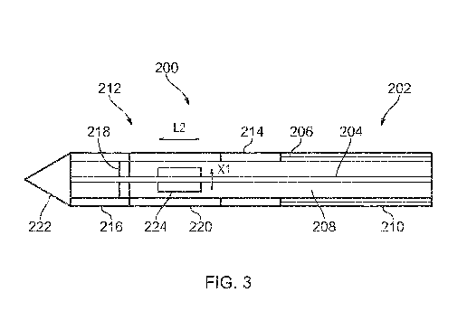
invention. Fig. 3 shows a cross-sectional side view of the
same electrosurgical instrument 200. The distal end of the
electrosurgical instrument 200 may correspond, for example, to
the distal assembly 118 discussed above. The electrosurgical
CA 03101074 2020-11-20
WO 2019/228927
PCT/EP2019/063488
14
instrument 200 includes a coaxial feed cable 202 that is
connectable at its proximal end to a generator (such as
generator 102) in order to convey microwave energy and RF
energy. The coaxial feed cable 202 comprises an inner
conductor 204 and an outer conductor 206 which are separated
by a dielectric material 208. The coaxial feed cable 202 is
preferably low loss for microwave energy. A choke (not shown)
may be provided on the coaxial feed cable 204 to inhibit back
propagation of microwave energy reflected from the distal end
and therefore limit backward heating along the device. The
coaxial cable further includes a flexible outer sheath 210
disposed around the outer conductor 206 to protect the coaxial
cable. The outer sheath 210 may be made of an insulating
material to electrically isolate the outer conductor 206 from
its surroundings. The outer sheath 210 may be made of, or
coated with, a non-stick material such as PTFE to prevent
tissue from sticking to the instrument.
A radiating tip 212 is formed at the distal end of the
coaxial feed cable 202. The radiating tip 212 is arranged to
receive microwave energy and RF energy conveyed by the coaxial
feed cable 202, and deliver the energy into biological tissue.
The radiating tip 212 includes a proximal electrode 214
located near a proximal end of the radiating tip 212. The
proximal electrode 214 is a hollow cylindrical conductor that
forms an exposed ring around an outer surface of the radiating
tip 212. The proximal electrode 214 is electrically connected
to the outer conductor 206 of the coaxial feed cable 202. For
example, the proximal electrode 214 may be welded or soldered
to the outer conductor 206. The proximal electrode 214 may be
electrically connected to the outer conductor 206 by a region
of physical contact that extends around the whole
circumference of the outer conductor 206, in order to ensure
axial symmetry of the connection. The proximal electrode 214
is arranged coaxially with the coaxial feed cable 202 (i.e.
the longitudinal axis of the cylindrical proximal electrode
214 is aligned with the longitudinal axis of the coaxial feed
cable 202), and has an outer diameter that matches that of the
coaxial feed cable 202. In this manner, the proximal electrode
lies flush with the outer surface of the coaxial feed cable
202. This may prevent tissue from catching on the proximal
electrode 214. The outer conductor 206 terminates at the
CA 03101074 2020-11-20
WO 2019/228927
PCT/EP2019/063488
proximal electrode 214, i.e. it does not extend beyond the
proximal electrode 214 in a distal direction. In some
embodiments (not shown), the proximal electrode may be an
exposed distal portion of the outer conductor 206.
5 The radiating tip 212 also includes a distal electrode
216 located at or near a distal end of the radiating tip 212.
The distal electrode 216 is a hollow cylindrical conductor
that forms an exposed ring around an outer surface of the
radiating tip 212. Like the proximal electrode 214, the distal
10 electrode 216 is arranged coaxially with the coaxial feed
cable 202. The proximal and distal electrodes 214, 216 may
have substantially the same shape and size. As illustrated in
Fig. 2, the proximal and distal electrodes 214, 216 have a
length Li in the longitudinal direction of the electrosurgical
15 instrument 200. The distal electrode 216 is spaced apart from
the proximal electrode 214 in the longitudinal direction of
the electrosurgical instrument 200 by a distance G (see Fig.
2). In other words, the distal electrode 216 is further along
the length of the electrosurgical instrument 200 by a distance
G. The proximal and distal electrodes 214, 216 have an outer
diameter which is the same as an outer diameter of the coaxial
feed cable 202, so that the electrosurgical instrument 200 has
a smooth outer surface.
The proximal electrode 214 (which is formed by a hollow
cylindrical conductor) defines a passageway through which a
distally protruding portion of the inner conductor 204 passes.
In this manner, the inner conductor 204 extends into the
radiating tip 212, where it is electrically connected to the
distal electrode 216. The inner conductor 204 is electrically
connected to the distal electrode 216 via a conductor 218 that
extends radially (i.e. outwards) from the inner conductor 206.
The conductor 218 may comprise one or more branches (e.g.
wires or other flexible conductive elements) that are arranged
symmetrically about the axis of the inner conductor 204.
Alternatively, the conductor 218 may comprises a conductive
disc or ring mounted around the inner conductor 204 and
connected between the inner conductor 204 and the distal
electrode 216. The connection between the inner conductor 204
and the distal electrode 216 is preferably symmetric around
the axis defined by the inner conductor 204. This can
CA 03101074 2020-11-20
WO 2019/228927
PCT/EP2019/063488
16
facilitate formation of a symmetrical field shape around the
radiating tip 212.
A portion of the dielectric material 208 of the coaxial
feed cable 202 also extends beyond a distal end of the outer
conductor 206 into the radiating tip 212 through the
passageway formed by the proximal electrode 214. In this
manner, the inner conductor 204 and the proximal electrode 214
are isolated by the dielectric material 208. A collar 220 is
provided around the radiating tip 212 between the proximal
electrode 214 and the distal electrode 216. The collar 220 may
operate to protect the dielectric material 208 and ensure that
the outer surface of the radiating tip is smooth. The collar
220 may be made of the same material, and serve the same
function, as the outer sheath 210.
The radiating tip 212 further includes a pointed distal
tip 222 located at a distal end of the instrument. The distal
tip 222 may be pointed in order to facilitate insertion of the
radiating tip 212 into target tissue. However, in other
embodiments (not shown), the distal tip may be rounded or
flat. The distal tip 222 may be made of a dielectric material,
e.g. the same as dielectric material 208. In some embodiments,
the material of the distal tip 222 may be selected to improve
impedance matching with target tissue, in order to improve the
efficiency with which the EM energy is delivered to the target
tissue. The distal tip 222 may be made of, or covered with a
non-stick material (e.g. PTFE) to prevent tissue from sticking
to it.
The radiating tip 212 further includes a tuning element
224. The tuning element 224 is an electrically conductive
element connected to the inner conductor 204 between the
proximal electrode 214 and the distal electrode 216 to
introduce a capacitive reactance. In this example, the
conductive tuning element is cylindrically shaped, and is
arranged coaxially with the inner conductor 204. The tuning
element 224 has a length L2 in the longitudinal direction, and
an outer diameter X1 (see Fig. 3). These parameters can be
selected to introduce a capacitance that improves the coupling
efficiency (i.e. reduces the reflected signal) of the
instrument when operating as a microwave antenna as discussed
below.
CA 03101074 2020-11-20
WO 2019/228927
PCT/EP2019/063488
17
As the proximal electrode 214 and the distal electrode
216 are electrically connected to the outer conductor 206 and
the inner conductor 204, respectively, they may be used as
bipolar RF cutting electrodes. For example, the distal
electrode 216 may act as an active electrode and the proximal
electrode 214 may act as a return electrode for RF energy
conveyed along the coaxial feed cable 202. In this manner,
target tissue disposed around the radiating tip 212 may be cut
and/or coagulated using RF energy, via the mechanisms
discussed above.
Additionally, the radiating tip 212 may behave as a
microwave dipole antenna, when microwave energy is conveyed
along the coaxial feed cable 202. In particular, the proximal
electrode 214 and the distal electrode 216 may act as
radiating elements of the dipole antenna at microwave
frequencies. Thus, the radiating tip structure enables both
radiofrequency energy and microwave energy to be delivered
into target tissue. This enables target tissue to be ablated
and/or coagulated using radiofrequency and microwave energy,
depending on the type of EM energy conveyed to the radiating
tip. The cylindrical shapes of the proximal and distal
electrodes 214, 216 may serve to produce a radiation profile
that is symmetric about a longitudinal axis of the instrument
200.
The configuration of the electrodes 214, 216 determined
by the parameters Li and G can be selected in advance to
provide a desired ablation diameter (for a given energy
waveform and local tissue properties). Cylindrical electrodes
are used to produce a symmetrical (about the longitudinal
device axis) ablation profile. The following are example
dimensions that can be used for an electrosurgical instrument
that is an embodiment of the invention: Li and L2 may be 3 mm;
G may be 5 mm; X1 may be 1.2 mm; the outer diameter of the
instrument may be approximately 1.9 mm; the inner diameter of
the proximal and distal electrodes may be 1.5 mm.
Fig. 4 shows calculated radiation profiles in target
tissue for an electrosurgical instrument according to an
embodiment of the invention. Panel A of Fig. 4 shows a
simulated radiation profile at 400 kHz (i.e. for
radiofrequency energy) and panel B of Fig. 4 shows a simulated
radiation profile at 5.8 GHz (i.e. for microwave energy). As
CA 03101074 2020-11-20
WO 2019/228927
PCT/EP2019/063488
18
can be seen, at both frequencies, the radiation profile
extends between and around the proximal and distal electrodes.
The radiation profile for the microwave energy (panel B) is
more spherical than for the radiofrequency energy (panel A).
In contrast, the radiation profile for the radiofrequency has
a more elongate shape, and is more concentrated around the
proximal and distal electrodes. Therefore, the radiation
profile changes depending on whether microwave energy or
radiofrequency energy is conveyed to the radiating tip. This
may result in a different ablation volume (i.e. a volume of
target tissue that is ablated by the EM energy), depending on
the type of EM energy conveyed to the radiating tip. Thus, for
example, the ablation volume may be controlled by switching
between microwave energy and radiofrequency energy.
Fig. 5 illustrates how the microwave radiation profile of
the electrosurgical instrument is affected by the presence of
the proximal and distal electrodes. Panel A of Fig. 5 shows a
calculated radiation profile for an electrosurgical instrument
which does not have proximal and distal electrodes. The
structure of the electrosurgical instrument of Panel A of Fig.
5 is illustrated in Fig. 6. The electrosurgical instrument 600
illustrated in Fig. 6 has a similar structure to that shown in
Figs. 2 and 3, except that it does not include proximal and
distal electrodes. Like the electrosurgical instrument 200 of
the embodiment, electrosurgical instrument 600 includes a
coaxial feed cable 602 having an inner conductor 604 and an
outer conductor 606 which are separated by a dielectric
material 608. A radiating tip 610 is formed at the end of the
coaxial feed cable 602. The inner conductor 604 and the
dielectric material extend into the radiating tip 610, however
the outer conductor 606 terminates at the radiating tip 610. A
conductive tuning element 612 is provided on the inner
conductor in the radiating tip 610. Panel B of Fig. 5 shows a
calculated radiation profile for an electrosurgical instrument
having a structure according to an embodiment of the invention
(e.g. similar to that shown in Figs. 2 and 3). Both radiation
profiles are simulations at a microwave energy frequency of
5.8 GHz. Except for the lack of proximal and distal electrodes
in electrosurgical instrument 600, the dimensions of the
electrosurgical instruments used in both simulations are the
same.
CA 03101074 2020-11-20
WO 2019/228927
PCT/EP2019/063488
19
As can be seen from Fig. 5, the shape of the calculated
radiation profiles differs between the electrosurgical
instruments. In particular, the radiation profile of the
electrosurgical instrument according to the embodiment of the
invention (panel B) is more spherical in shape compared to the
radiation profile of electrosurgical instrument 600 (panel A).
As indicated by the lines in Fig. 5, the radiation profile of
the electrosurgical instrument according to the embodiment of
the invention is more concentrated around the radiating tip.
In contrast, the radiation profile of electrosurgical
instrument 600 has a longer tail which extends along a portion
of the coaxial feed cable. This extending of the radiation
profile down the coaxial feed cable may be referred to as the
"teardrop effect". Thus, the use of proximal and distal
electrodes in the electrosurgical instrument serves to reduce
the teardrop effect. The radiation profile of the
electrosurgical instrument of the embodiment may be
advantageous in that it may avoid ablating tissue that is
located away from the radiating tip. The teardrop effect may
further be reduced by including a dielectric choke in the
radiating tip of the electrosurgical instrument of the
embodiment. For example, the dielectric choke may be a piece
of dielectric material that is located in the radiating tip,
between the proximal electrode and the outer conductor (i.e.
in the passageway defined by the proximal electrode).
Fig. 7 shows a simulated plot of the S-parameter (also
known as the "return loss") against frequency of the microwave
energy for the electrosurgical instrument 200. As well known
in the technical field, the S-parameter is a measure of the
return loss of microwave energy due to impedance mismatch, and
as such the S-parameter is indicative of the degree of
impedance mismatch between the target tissue and the radiating
tip. The S-parameter can be defined by the equation PI = SPR,
where PI is the outgoing power in the instrument towards the
tissue, PR is the power reflected back from the tissue, and S
is the S-parameter. As shown in Fig. 6, the S-parameter is -
17.09 dB at 5.8 GHz, meaning that very little microwave energy
was reflected back from the tissue at this frequency. This
indicates a good impedance match at the operating frequency of
5.8 GHz, and that microwave energy is efficiently delivered
from the radiating tip into the tissue at this frequency.
CA 03101074 2020-11-20
WO 2019/228927 PCT/EP2019/063488
The inventors carried out ex-vivo testing of an
electrosurgical instrument having a structure similar to that
illustrated in Figs. 2 and 3. The tests were carried out using
morbid porcine tissue (liver destined for the food chain). The
5 samples were sealed in a bag and placed in a water bath at
37 C prior to testing. The distal end of the electrosurgical
instrument was then inserted into the prepared tissue samples.
RF and microwave energy was then delivered to the samples.
The RF energy had a frequency of 400 kHz and a 18 W
10 coagulation waveform, applied for 66 s with a 91% duty cycle.
The microwave energy had a frequency of 5.8 GHz and a power
level of 25 W, applied as a continuous wave for 120 s.
Measurements of the resulting ablation zones were then
carried out, the results of which are shown in Table 1. The
15 length of the ablation zone corresponds to its measured length
in the longitudinal direction of the electrosurgical
instrument. The width of the ablation zone corresponds to its
width in a direction normal to the longitudinal direction. It
was found that the shapes and sizes of the ablation zones
20 correlate well with the simulated radiation profiles discussed
above.
Ablation Sample 1 Sample 2 Sample 3 Sample 4 Sample 5
RF 14mm x 4mm 13mm x 4mm 14mm x 4mm 13mm x 4mm 14mm x
4mm
Microwave 21mm x 16mm 21mm x 16mm 21mm x 15mm 21mm x 14mm 21mm x 15mm
Table 1: Size of ablation zone