Note : Les descriptions sont présentées dans la langue officielle dans laquelle elles ont été soumises.
BIOLOGICAL FLUID SEPARATION DEVICE
BACKGROUND OF THE INVENTION
1. Field of the Disclosure
[0001] The present disclosure relates generally to devices adapted for use
with biological fluids.
More particularly, the present disclosure relates to devices adapted for
separating components of
biological fluids.
2. Description of the Related Art
[0002] Blood sampling is a common health care procedure involving the
withdrawal of at least
a drop of blood from a patient. Blood samples are commonly taken from
hospitalized, homecare,
and emergency room patients either by finger stick, heel stick, or
venipuncture. Blood samples
may also be taken from patients by venous or arterial lines. Once collected,
blood samples may
be analyzed to obtain medically useful information including chemical
composition, hematology,
or coagulation, for example.
[0003] Blood tests determine the physiological and biochemical states of the
patient, such as
disease, mineral content, drug effectiveness, and organ function. Blood tests
may be performed in
a clinical laboratory or at the point-of-care near the patient. One example of
point-of-care blood
testing is the routine testing of a patient's blood glucose levels which
involves the extraction of
blood via a finger stick and the mechanical collection of blood into a
diagnostic cartridge.
Thereafter, the diagnostic cartridge analyzes the blood sample and provides
the clinician a reading
of the patient's blood glucose level. Other devices are available which
analyze blood gas
electrolyte levels, lithium levels, and ionized calcium levels. Some other
point-of-care devices
identify markers for acute coronary syndrome (ACS) and deep vein
thrombosis/pulmonary
embolism (DVT/PE).
[0004] Blood samples contain a whole blood or cellular portion and a plasma
portion. Plasma
separation from whole blood has been traditionally achieved by centrifugation
which typically
takes 15 to 20 minutes and involves heavy labor or complex work flow. Recently
there are other
technologies that have been used or tried to separate plasma such as
sedimentation, fibrous or non-
fibrous membrane filtration, lateral flow separation, microfluidics cross flow
filtration and other
microfluidics hydrodynamic separation techniques. However many of those
technologies have
various challenges arranging from poor plasma purity, analyte bias or
requiring specific coating to
1
Date Recue/Date Received 2022-02-23
prevent analyte bias, high hemolysis, requiring dilution, long separation
time, and/or difficult to
recover the plasma. For example, most membrane based separation technologies
suffer from an
analyte bias problem, and often require specific coating treatments for the
target analytes.
Additionally, conventional separation technologies that occur while the device
is directly
connected to a patient thru a needle cause patient discomfort.
SUMMARY OF THE INVENTION
[0005] The present disclosure provides a blood separation device that
decouples and separates
the blood collection process from the plasma separation process. The blood
separation device
includes a sample collection module, an activation module, and a separation
module. Because the
plasma separation happens after the blood separation device is disconnected
from a patient, the
device performance is no longer affected by patient blood pressure and needle
gauge, and patient
discomfort is greatly reduced.
[0006] The present disclosure provides a blood separation device and a
separation process that
is fully compatible with a venous blood collection workflow without the need
of centrifugation
and power. Advantageously, the blood separation device of the present
disclosure allows for the
immediate separation of plasma during clinical blood draws and the ability for
collection of the
separated plasma sample in a self-contained plasma container for downstream
diagnostics.
[0007] Furthermore, the blood separation device of the present disclosure
provides for a
separation device that only needs a short on-patient collection time that is
no different than a
conventional blood collection device using vacuum tubes, such as a BD
Vacutainer0 blood
collection tube commercially available from Becton, Dickinson and Company, and
corresponding
venous access sets. Additionally, since the plasma separation happens after
the device is
disconnected from the patient, the device performance is no longer affected by
patient blood
pressure and needle gauge, and patient discomfort is greatly reduced.
[0008] Because the blood separation device of the present disclosure decouples
and separates
the blood collection process from the plasma separation process, the volume of
the plasma
generated is no longer limited by the allowable blood collection time on-
patient. This enables the
potential use of the blood separation device of the present disclosure for
other high volume plasma
applications beyond point of care.
2
Date Recue/Date Received 2022-02-23
[0009] Furthermore, another benefit of decoupling the separation from the
collection process is
that the separation time, plasma quality, and yield is no longer affected by
the needle gauge and
patient blood pressure. If the separation happens while a device is directly
connected to a patient
thru a needle, lower needle gauge and higher patient blood pressure reduce the
separation time,
yield and increases the hemolysis, whereas higher needle gauge and lower
patient blood pressure
increases the separation time, yield and decreases the hemolysis. By isolating
the plasma
separation process from the blood collection workflow using a blood separation
device of the
present disclosure, the blood collection sets and patient blood pressure will
only affect the blood
collection time while not varying the separation time, yield and hemolysis
level.
[0010] In accordance with an embodiment of the present invention, a blood
separation device
adapted to receive a blood sample having a first phase and a second phase
includes a sample
collection module having a housing defining a collection chamber; an
activation module connected
to the sample collection module, the activation module having a first seal and
a second seal for
sealing the housing, the first seal transitionable from a closed position in
which the collection
chamber has a first pressure to an open position, by actuation of a portion of
the activation module,
in which the collection chamber is in fluid communication with a second
pressure greater than the
first pressure; and a separation module in fluid communication with the
collection chamber of the
sample collection module, the separation module defining a first chamber
having a first volume
and a second chamber having a second volume and including a separation member
disposed
between the first chamber and the second chamber, wherein the first volume and
the second
volume are different.
[0011] In one configuration, the activation module includes a switch, wherein
actuation of the
switch transitions the first seal to the open position. In another
configuration, the switch comprises
a push button defining a vent hole therethrough and a piercing portion,
wherein actuation of the
switch moves the piercing portion to break the first seal thereby
transitioning the first seal to the
open position. In yet another configuration, with the first seal in the open
position, the collection
chamber of the sample collection module is in fluid communication with the
second pressure via
the vent hole of the switch. In one configuration, the second seal comprises a
cap having a
pierceable self-sealing stopper within a portion of the cap. In another
configuration, the blood
separation device is connectable to a blood collection device via the cap. In
yet another
configuration, the activation module defines an inlet channel, and wherein
with the blood
3
Date Recue/Date Received 2022-02-23
collection device connected to the blood separation device via the cap, the
collection chamber
receives the blood sample via the inlet channel. In one configuration, the
collection chamber
includes an inlet end and an exit end and defines a plurality of sequential
flow direction alternating
collection channels. In another configuration, the collection chamber includes
an inlet end and an
exit end and defines a first collection channel extending from the inlet end
to the exit end, a second
collection channel in communication with a portion of the first collection
channel and extending
from the exit end to the inlet end, and a third collection channel in
communication with a portion
of the second collection channel and extending from the inlet end to the exit
end. In yet another
configuration, the inlet end of the collection channels is in fluid
communication with the inlet
channel of the activation module. In one configuration, the blood sample
travels through the first
collection channel in a first direction, the blood sample travels through the
second collection
channel in a second direction opposite the first direction, and the blood
sample travels through the
third collection channel in a third direction opposite the second direction.
In another configuration,
the first collection channel is spaced from the second collection channel
which is spaced from the
third collection channel. In yet another configuration, the first chamber
includes a first chamber
inlet and a first chamber outlet, and the second chamber includes a second
chamber outlet. In one
configuration, the first chamber inlet is in fluid communication with the exit
end of the collection
channels. In another configuration, with the first seal in the open position,
a first pressure
difference between the second pressure defined by atmospheric pressure and the
first pressure
defined within the collection chamber draws the blood sample into the first
chamber. In yet
another configuration, with the first seal in the open position, the first
volume and the second
volume being different provides a second pressure difference between the first
chamber and the
second chamber to drive the second phase of the blood sample through the
separation member into
the second chamber. In one configuration, the separation member traps the
first phase in the first
chamber and allows the second phase to pass through the separation member into
the second
chamber. In another configuration, the blood separation device includes a
second phase collection
container in communication with the second chamber outlet, wherein the second
phase collection
container receives the second phase. In yet another configuration, the blood
separation device
includes a blood sample discard chamber in communication with the first
chamber outlet, wherein
the blood sample discard chamber receives the first phase. In one
configuration, the separation
member comprises a track-etched membrane. In another configuration, with the
blood collection
4
Date Recue/Date Received 2022-02-23
device connected to the blood separation device via the cap, the collection
chamber receives the
blood sample via the inlet channel. In yet another configuration, with the
blood collection device
disconnected from the blood separation device, and wherein upon actuation of
the switch to
transition the first seal to the open position, the first pressure difference
between the second
pressure defined by atmospheric pressure and the first pressure defined within
the collection
chamber draws the blood sample into the first chamber. In one configuration,
with the first seal in
the open position, the first volume and the second volume being different
provides the second
pressure difference between the first chamber and the second chamber to drive
the second phase
of the blood sample through the separation member into the second chamber. In
another
configuration, with the second phase contained within the second phase
collection container, the
second phase collection container is removable from the blood separation
device. In yet another
configuration, the first phase is a cellular portion and the second phase is a
plasma portion.
BRIEF DESCRIPTION OF THE DRAWINGS
[0012] The above-mentioned and other features and advantages of this
disclosure, and the
manner of attaining them, will become more apparent and the disclosure itself
will be better
understood by reference to the following descriptions of embodiments of the
disclosure taken in
conjunction with the accompanying drawings, wherein:
[0013] Fig. 1 is a perspective view of a blood separation device in accordance
with an
embodiment of the present invention.
[0014] Fig. 2 is an exploded, perspective view of a blood separation device in
accordance with
an embodiment of the present invention.
[0015] Fig. 3 is a perspective view of a first step of using a system of the
present disclosure in
accordance with an embodiment of the present invention.
[0016] Fig. 4 is a perspective view of a second step of using a system of the
present disclosure
in accordance with an embodiment of the present invention.
[0017] Fig. 5 is a perspective view of a third step of using a system of the
present disclosure
illustrating the device of the present disclosure separates plasma independent
of the device
orientation in accordance with an embodiment of the present invention.
Date Recue/Date Received 2022-02-23
[0018] Fig. 6 is a perspective view of a fourth step of using a system of the
present disclosure
in accordance with an embodiment of the present invention.
[0019] Fig. 7A is a perspective view of an activation module of a blood
separation device in a
closed position in accordance with an embodiment of the present invention.
[0020] Fig. 7B is a cross-sectional view of the activation module of Fig. 7A
in accordance with
an embodiment of the present invention.
[0021] Fig. 8A is a perspective view of an activation module of a blood
separation device in an
open position in accordance with an embodiment of the present invention.
[0022] Fig. 8B is a cross-sectional view of the activation module of Fig. 8A
in accordance with
an embodiment of the present invention.
[0023] Fig. 9 is a perspective view of a collection chamber of a blood
separation device in
accordance with an embodiment of the present invention.
[0024] Fig. 10 is a perspective view of a collection chamber of a blood
separation device in
accordance with another embodiment of the present invention.
[0025] Fig. 11 is a perspective view of a blood separation device in
accordance with an
embodiment of the present invention.
[0026] Fig. 12 is a perspective view of a portion of a separation module of a
blood separation
device in accordance with an embodiment of the present invention.
[0027] Fig. 13 is a perspective view of a blood separation device in
accordance with an
embodiment of the present invention.
[0028] Corresponding reference characters indicate corresponding parts
throughout the several
views. The exemplifications set out herein illustrate exemplary embodiments of
the disclosure,
and such exemplifications are not to be construed as limiting the scope of the
disclosure in any
manner.
DETAILED DESCRIPTION
[0029] The following description is provided to enable those skilled in the
art to make and use
the described embodiments contemplated for carrying out the invention. Various
modifications,
equivalents, variations, and alternatives, however, will remain readily
apparent to those skilled in
6
Date Recue/Date Received 2022-02-23
the art. Any and all such modifications, variations, equivalents, and
alternatives are intended to
fall within the spirit and scope of the present invention.
[0030] For purposes of the description hereinafter, the terms "upper",
"lower", "right", "left",
"vertical", "horizontal", "top", "bottom", "lateral", "longitudinal", and
derivatives thereof shall
relate to the invention as it is oriented in the drawing figures. However, it
is to be understood that
the invention may assume alternative variations and step sequences, except
where expressly
specified to the contrary. It is also to be understood that the specific
devices and processes
illustrated in the attached drawings, and described in the following
specification, are simply
exemplary embodiments of the invention. Hence, specific dimensions and other
physical
characteristics related to the embodiments disclosed herein are not to be
considered as limiting.
[0031] Figs. 1 and 2 illustrate an exemplary embodiment of a blood separation
device of the
present disclosure. Referring to Figs. 1 and 2, a blood separation device 10
of the present
disclosure is adapted to receive a biological fluid, such as a blood sample 12
(Figs. 3-6) having a
first phase 14 and a second phase 16. The first phase 14 of the blood sample
12 is a cellular portion
and the second phase 16 of the blood sample 12 is a plasma portion.
[0032] A blood separation device 10 of the present disclosure decouples and
separates the blood
collection process from the plasma separation process. Because the plasma
separation happens
after the blood separation device 10 is disconnected from a patient, the
device performance is no
longer affected by patient blood pressure and needle gauge, and patient
discomfort is greatly
reduced.
[0033] Because the blood separation device 10 of the present disclosure
decouples and separates
the blood collection process from the plasma separation process, the volume of
the plasma
generated is no longer limited by the allowable blood collection time on-
patient. This enables the
potential use of the blood separation device 10 of the present disclosure for
other high volume
plasma applications beyond point of care.
[0034] The present disclosure provides a blood separation device 10 and a
separation process
that is fully compatible with a venous blood collection workflow without the
need of centrifugation
and power. Advantageously, the blood separation device 10 of the present
disclosure allows for
the immediate separation of plasma during clinical blood draws, with the
device 10 off-patient,
and the ability for collection of the separated plasma 16 sample in a self-
contained plasma
container, e.g., a second phase or plasma collection container 80, for
downstream diagnostics.
7
Date Recue/Date Received 2022-02-23
[0035] Furthermore, the blood separation device 10 of the present disclosure
provides for a
separation device that only needs a short on-patient collection time that is
no different than a
conventional blood collection device using vacuum tubes, such as a BD
Vacutainer0 blood
collection tube commercially available from Becton, Dickinson and Company, and
corresponding
venous access sets. Additionally, since the plasma separation happens after
the device 10 is
disconnected from the patient, the device performance is no longer affected by
patient blood
pressure and needle gauge, and patient discomfort is greatly reduced.
[0036] Furthermore, another benefit of decoupling the plasma separation
process from the
collection process is that the separation time, plasma quality, and yield is
no longer affected by the
needle gauge and patient blood pressure. If the plasma separation process
occurs while a device
is directly connected to a patient thru a needle, lower needle gauge and
higher patient blood
pressure reduce the separation time, yield and increases the hemolysis,
whereas higher needle
gauge and lower patient blood pressure increases the separation time, yield
and decreases the
hemolysis. By isolating the plasma separation process from the blood
collection process using a
blood separation device 10 of the present disclosure, the blood collection
sets and patient blood
pressure will only affect the blood collection time while not varying the
separation time, yield and
hemolysis level.
[0037] Referring to Figs. 1-13, in an exemplary embodiment, a blood separation
device 10
generally includes a sample collection module 20, an activation module 22, and
a separation
module 24. In one embodiment, after collecting a blood sample 12, the blood
separation device
is able to separate a second phase 16 of the blood sample 12 from a first
phase 14 of the blood
sample 12 as described in more detail below. Advantageously, the blood
separation device 10
decouples and separates the blood collection process from the plasma
separation process. In one
embodiment, after plasma separation, a portion that is removable, e.g., a
second phase collection
container 80, from the blood separation device 10 is able to transfer the
second phase 16 of the
blood sample 12 to a point-of-care testing device.
[0038] Referring to Figs. 1-6 and 9-11, in an exemplary embodiment, the sample
collection
module 20 includes a housing 30 defining a collection chamber 32. In one
embodiment, the
collection chamber 32 includes an inlet end or inlet 34 and an exit end or
exit 36 and defines a
plurality of sequential flow direction alternating collection channels 38.
8
Date Recue/Date Received 2022-02-23
[0039] The collection chamber 32 utilizes multiple interconnected parallel
channels 38 to
maximize collection and storage space within the constrained diameter of a
blood collection set
and also to ensure that the capillary force dominates over gravity during
filling. A blood sample
12 fills the interconnected channels 38 of the sample collection module 20 in
a back-and-forth
motion as shown in Figs. 9-11.
[0040] For example, referring to Fig. 9, in a first exemplary embodiment, the
collection chamber
32 of the sample collection module 20 defines a first collection channel 40
extending from the
inlet end 34 to the exit end 36, a second collection channel 42 in
communication with a portion of
the first collection channel 40 and extending from the exit end 36 to the
inlet end 34, and a third
collection channel 44 in communication with a portion of the second collection
channel 42 and
extending from the inlet end 34 to the exit end 36. Referring to Fig. 9, the
first collection channel
40 is spaced from the second collection channel 42 which is spaced from the
third collection
channel 44.
[0041] In this manner, referring to the arrow in Fig. 9 indicating a flow path
100 of the blood
sample 12 through the channels 38 of the collection chamber 32, a blood sample
12 collected into
the collection chamber 32 travels through the first collection channel 40 in a
first direction, the
blood sample 12 travels through the second collection channel 42 in a second
direction opposite
the first direction, and the blood sample 12 travels through the third
collection channel 44 in a third
direction opposite the second direction. Referring to Fig. 9, the collection
chamber 32 utilizes
multiple interconnected parallel channels 38 to maximize collection and
storage space within the
constrained diameter of a blood collection set and also to ensure that the
capillary force dominates
over gravity during filling.
[0042] In one embodiment, the entrance into the collection chamber 32 is the
inlet 34 of the first
collection channel 40 and the exit out of the collection chamber 32 is the
exit 36 of the third
collection channel 44. The inlet 34 of the first collection channel 40 is in
fluid communication
with an inlet channel 66 (Figs. 7B and 8B) of the activation module 22, as
described in more detail
below.
[0043] Referring to Fig. 10, in a second exemplary embodiment, the collection
chamber 32 of
the sample collection module 20 defines a first collection channel 40
extending from the inlet end
34 to the exit end 36, a second collection channel 42 in communication with a
portion of the first
collection channel 40 and extending from the exit end 36 to the inlet end 34,
a third collection
9
Date Recue/Date Received 2022-02-23
channel 44 in communication with a portion of the second collection channel 42
and extending
from the inlet end 34 to the exit end 36, a fourth collection channel 46 in
communication with a
portion of the third collection channel 44 and extending from the exit end 36
to the inlet end 34,
and a fifth collection channel 48 in communication with a portion of the
fourth collection channel
46 and extending from the inlet end 34 to the exit end 36. Referring to Fig.
10, the first collection
channel 40 is spaced from the second collection channel 42 which is spaced
from the third
collection channel 44 which is spaced from the fourth collection channel 46
which is spaced from
the fifth collection channel 48.
[0044] In this manner, a blood sample 12 collected into the collection chamber
32 travels
through the first collection channel 40 in a first direction, the blood sample
12 travels through the
second collection channel 42 in a second direction opposite the first
direction, the blood sample
12 travels through the third collection channel 44 in a third direction
opposite the second direction,
the blood sample 12 travels through the fourth collection channel 46 in a
fourth direction opposite
the third direction, and the blood sample 12 travels through the fifth
collection channel 48 in a fifth
direction opposite the fourth direction. Referring to Fig. 10, the collection
chamber 32 utilizes
multiple interconnected parallel channels 38 to maximize collection and
storage space within the
constrained diameter of a blood collection set and also to ensure that the
capillary force dominates
over gravity during filling.
[0045] In one embodiment, the entrance into the collection chamber 32 is the
inlet 34 of the first
collection channel 40 and the exit out of the collection chamber 32 is the
exit 36 of the fifth
collection channel 48. The inlet 34 of the first collection channel 40 is in
fluid communication
with an inlet channel 66 (Figs. 7B and 8B) of the activation module 22, as
described in more detail
below.
[0046] In other exemplary embodiments, the collection chamber 32 of the sample
collection
module 20 may define any odd number of channels 38 based on a specific volume
requirement.
Importantly, the collection chamber 32 of the sample collection module 20
utilizes multiple
interconnected parallel channels 38 to maximize collection and storage space
within the
constrained diameter of a blood collection set and also to ensure that the
capillary force dominates
over gravity during filling. A blood sample 12 fills the interconnected
channels 38 of the sample
collection module 20 in a back-and-forth motion as described above.
Date Recue/Date Received 2022-02-23
[0047] In one exemplary embodiment, the plurality of sequential flow direction
alternating
collection channels 38 are configured in a parallel configuration as shown in
Figs. 9 and 10. In
other exemplary embodiments, the collection channels 38 are configured in a
spiral or meandering
channel configuration or in other configurations that maximize collection and
storage space within
the constrained diameter of a blood collection set and also to ensure that the
capillary force
dominates over gravity during filling.
[0048] In an exemplary embodiment, the collection chamber 32 is designed to
ensure that the
blood 12 fills the channels 38 of the collection chamber 32 continuously
without trapping air
bubbles regardless of device orientation and blood flow rate. This is
accomplished by controlling
the diameter of the channels 38 for desired applications. For example, in an
exemplary
embodiment, to prevent the blood stream from breaking up and trapping air
bubbles, the diameter
of the channels 38 needs to simultaneously meet two requirements. First, the
static pressure
difference at the flow front at any orientation needs to be smaller than the
Laplace pressure so that
the meniscus will hold its shape. Second, the selected diameter needs to make
sure that the inertia
force is smaller than the surface tension at the highest flow rate.
[0049] Referring to Figs. 1, 2, and 7A-8B, in an exemplary embodiment, the
activation module
22 is connected or connectable to the sample collection module 20 and includes
a housing 49, a
first seal 50, and a second seal 52 for sealing the blood separation device
10, e.g., the housing 30
of the sample collection module 20, the housing 49 of the activation module
22, and a housing 68
of the separation module 24. In this manner, the seals 50, 52 of the
activation module 22 control
the pressure within the blood separation device 10 as described in more detail
below. The first
seal 50 is transitionable from a closed position (Figs. 7A and 7B) in which
the collection chamber
32 has a first pressure P1 (Fig. 13) to an open position (Figs. 8A and 8B), by
actuation of a portion
of the activation module 22, in which the collection chamber 32 is in fluid
communication with a
second pressure P2 (Fig. 13) greater than the first pressure Pl.
[0050] In an exemplary embodiment, referring to Figs. 7A-8B, the activation
module 22
includes a switch 54. In such an embodiment, actuation of the switch 54
transitions the first seal
50 from the closed position (Figs. 7A and 7B) to the open position (Figs. 8A
and 8B). Referring
to Figs. 7A-8B, the switch 54 comprises a push button 56 defining a vent hole
58 therethrough and
a piercing portion 60. In this manner, actuation of the switch, e.g.,
depressing or pushing the push
11
Date Recue/Date Received 2022-02-23
button 56 into the position shown in Figs. 8A and 8B, moves the piercing
portion 60 to break the
first seal 50 thereby transitioning the first seal 50 to the open position.
[0051] With the first seal 50 in the open position, the collection chamber 32
of the sample
collection module 20 is in fluid communication with a second pressure P2 via
the vent hole 58 of
the switch 54. The vent hole 58 provides a venting mechanism for the blood
separation device 10.
For example, in one embodiment, the piercing portion 60 breaks the first seal
50, e.g., an aluminum
foil seal, to create a vent to power the plasma separation process.
[0052] The second pressure P2 defined by atmospheric pressure is greater than
the first pressure
P1 defined within the blood separation device 10, e.g., the collection chamber
32 of the sample
collection module 20. In this manner, the pressure difference between the
second pressure P2
defined by atmosphere pressure and the residual vacuum in the blood separation
device 10, i.e.,
the first pressure P1 defined within the blood separation device 10,
continuously drive the plasma
separation process as described in more detail below. Advantageously, using
the activation
module 22 of the present disclosure, a user can precisely control when the
plasma separation
process begins.
[0053] In an exemplary embodiment, referring to Figs. 7A-8B, the second seal
52 of the
activation module 22 includes a cap 62 having a pierceable self-sealing
stopper 64 within a portion
of the cap 62. The cap 62 provides a mechanism for allowing the blood
separation device 10 to
be connectable to a blood collection device 200 (Fig. 3) as described in more
detail below.
[0054] In one exemplary embodiment, the cap 62 of the present disclosure may
be formed
substantially similar to a closure described in U.S. Published Patent
Application No. 20210161448.
[0055] Referring to Figs. 7A-8B, in one embodiment, the activation module 22
defines an inlet
channel 66. Referring to Fig. 3, with a blood collection device 200 connected
to the blood
separation device 10 via the cap 62, the collection chamber 32 of the sample
collection module 20
receives a blood sample 12 via the inlet channel 66. A blood sample 12 flows
from the inlet
channel 66 of the activation module 22 to the plurality of channels 38 of the
collection chamber
32 via the inlet 34.
[0056] Referring to Figs. 1-6 and 11-13, in an exemplary embodiment, the
separation module
24 is in fluid communication with the collection chamber 32 of the sample
collection module 20
and includes a housing 68 and defines a first chamber 70 having a first volume
V1 (Fig. 13) and a
second chamber 72 having a second volume V2 (Fig. 13) and including a
separation member 74
12
Date Recue/Date Received 2022-02-23
disposed between the first chamber 70 and the second chamber 72. The first
volume V1 of the
first chamber 70 and the second volume V2 of the second chamber 72 are
different to create a
second pressure difference between the first chamber 70 and the second chamber
72 to drive the
second phase 16 of a blood sample 12 through the separation member 74 into the
second chamber
72 as described in more detail below. In one embodiment, a portion of the
separation module 24
forms a microfluidic chip.
[0057] Referring to Figs. 11 and 12, in an exemplary embodiment, the
separation member 74
traps the first phase 14 in the first chamber 70 and allows the second phase
16 to pass through the
separation member 74 into the second chamber 72. In one embodiment, the
separation member
74 comprises a track-etched membrane. In certain configurations, the membrane
may be less than
100 microns in thickness, such as from 5 to 25 microns in thickness. The
membrane may have
submicron pores or holes, such as from 0.2 to 0.8 microns in diameter. This
dimensionality allows
for continuous filtering of a plasma portion of a blood sample flowing
parallel to the membrane
surface, which prevents clogging of the membrane pores or holes. In other
embodiments, the
separation member 74 may comprise any filter, and/or any other separation
device, that is able to
trap the first phase 14 in the first chamber 70 and allow the second phase 16
to pass through the
separation member 74 into the second chamber 72.
[0058] Referring to Figs. 11 and 12, the first chamber 70 includes a first
chamber inlet 75 and a
first chamber outlet 76, and the second chamber 72 includes a second chamber
outlet 78. The first
chamber inlet 75 is in fluid communication with the exit 36 of the collection
channels 38. In this
manner, upon actuation of a portion of the activation module 22, a blood
sample 12 can flow from
the collection chamber 32 of the sample collection module 20 to the first
chamber 70 of the
separation module 24 for plasma separation.
[0059] Referring to Figs. 1-6, 11, and 13, the separation module 24 of the
blood separation
device 10 includes a second phase collection container 80 that is in
communication with the second
chamber outlet 78. The second phase collection container 80 receives the
second phase 16 of the
blood sample 12. The second phase collection container 80 is able to collect
and store the separated
second phase 16. Advantageously, referring to Fig. 6, with the second phase 16
contained within
the second phase collection container 80, the second phase collection
container 80 is removable
from the blood separation device 10. In this manner, the second phase 16 of a
blood sample 12
can be collected or stored in a secondary second phase container, e.g., a
second phase collection
13
Date Recue/Date Received 2022-02-23
container 80, for further diagnostic tests. For example, after separation,
with the second phase
collection container 80 removed from the blood separation device 10, the
second phase collection
container 80 is able to transfer the second phase 16 of the blood sample 12 to
a point-of-care testing
device or other testing device. In an exemplary embodiment, the second phase
collection container
80 includes structure allowing the second phase collection container 80 to
dispense a portion of
the plasma 16, when desired. In one embodiment, the second phase collection
container 80 is
sealed via a cap or septum 81 to protectively seal the plasma portion 16
within the second phase
collection container 80.
[0060] Referring to Fig. 11, in an exemplary embodiment, a portion of the
second chamber 72
of the separation module 24 is in fluid communication with an interior of the
second phase
collection container 80 to allow the plasma portion 16 to flow through the
separation member 74
and the second chamber 72 into the interior of the second phase collection
container 80 for
collection.
[0061] Referring to Figs. 11-13, the separation module 24 of the blood
separation device 10 also
includes a blood sample discard chamber 82 that is in communication with the
first chamber outlet
76. The blood sample discard chamber 82 receives the remaining first phase 14
of the blood
sample 12 after a blood sample 12 flows over the separation member 74 in the
first chamber 70.
In this manner, the remaining first phase 14 of the blood sample 12 can be
collected and stored in
the blood sample discard chamber 82. Also, the blood sample discard chamber 82
ensures that the
remaining first phase 14 of the blood sample 12 can be safely stored when the
rest of the blood
separation device 10 is discarded after use.
[0062] Referring to Figs. 3-6, use of a blood separation device 10 of the
present disclosure will
now be described.
[0063] Referring to Fig. 3, a first step of using a blood separation device 10
of the present
disclosure involves collecting a blood sample 12 from a patient, e.g., the
blood collection process.
For example, first, a given volume of a blood sample 12 from a patient is
pulled into the collection
chamber 32 of the blood separation device 10 under a vacuum force, immediately
following the
connection of the blood separation device 10 to a blood collection device 200,
such as a tube holder
202. In one embodiment, such a connection consists of a non-patient needle
(not shown) of the
tube holder 202 piercing the stopper 64 of the cap (Fig. 7B). The opposite end
of a line 204 of the
tube holder 202 consists of a patient needle of a venous access set in
communication with a patient.
14
Date Recue/Date Received 2022-02-23
[0064] Referring to Fig. 3, with the tube holder 202 of the blood collection
device 200 connected
to the blood separation device 10 via the cap 62 (Fig. 7B), the collection
chamber 32 of the sample
collection module 20 receives the blood sample 12 via the inlet channel 66
(Fig. 7B) of the
activation module 22. The blood separation device 10 of the present disclosure
collects and stores
a fixed amount of the patient's blood. In one exemplary embodiment, a blood
separation device
of the present disclosure collects and stores 3 mL of a patient's blood in
less than 30 seconds.
[0065] The blood sample 12 flows through the inlet channel 66 of the
activation module 22 to
the collection chamber 32 of the sample collection module 20. Advantageously,
during blood
collection, the plurality of sequential flow direction alternating collection
channels 38 of the
collection chamber 32 maximize collection and storage space within the
constrained diameter of a
blood collection set and also to ensure that the capillary force dominates
over gravity during filling.
[0066] A user can select one of the ways, sources, or methods that the blood
separation device
10 is able to receive a blood sample 12. For example, referring to Fig. 3, the
blood separation
device 10 of the present disclosure is able to receive a blood sample 12 from
a conventional blood
collection device 200. For example, the blood collection device 200 may
include a tube holder
202 and corresponding venous access set, such as a BD Vacutainer0 blood
collection tube
commercially available from Becton, Dickinson and Company. In other
alternative embodiments,
blood is collected in a conventional blood collection tube or any other
intermediate blood sample
container. The blood sample container is then connected to the off-patient
separation device to
generate plasma.
[0067] Once a desired amount of a blood sample 12 is collected into the
collection chamber 32
and the blood collection process is complete, the blood separation device 10
is disconnected from
the blood collection device 200. In this manner, a blood separation device 10
of the present
disclosure decouples and separates the blood collection process from the
plasma separation
process. Because the plasma separation happens after the blood separation
device 10 is
disconnected from the patient, the device performance is no longer affected by
patient blood
pressure and needle gauge, and patient discomfort is greatly reduced.
[0068] Upon disconnection of the blood separation device 10 of the present
disclosure from the
blood collection device 200 and the patient, the collected blood remains
stationary in the channels
38 until the plasma separation is activated. The blood separation device 10
accomplishes this by
utilizing the second seal 52, e.g., the stopper 64 of the cap 62. The stopper
64 of the cap 62 ensures
Date Recue/Date Received 2022-02-23
that the second seal 52 is properly resealed after a needle of the blood
collection device 200 is
retracted out from the stopper 64 so that there is no pressure difference
between the front and back
end of the stored blood within the blood separation device 10.
[0069] Referring to Fig. 4, after the blood separation device 10 is
disconnected from the blood
collection device 200, the plasma separation process can be started.
Advantageously, the blood
separation device 10 of the present disclosure does not require being
connected to a patient to
perform plasma separation. The plasma separation process is completely
controllable and can be
started at a convenient and desired time.
[0070] Referring to Fig. 4, the plasma separation process is started with the
blood separation
device 10 off-patient by simply actuating the switch 54 (Figs. 8A and 8B),
e.g., pushing the push
button 56, on the blood separation device 10. Actuation of the switch 54
allows the blood
separation device 10 to automatically generate plasma 16 from the blood sample
12 stored within
the blood separation device 10.
[0071] Actuation of the switch 54 transitions the first seal 50 to the open
position (Fig. 8B), in
which the collection chamber 32 is in fluid communication with a second
pressure P2 defined by
atmospheric pressure that is greater than the first pressure P1 defined within
the collection chamber
32. In this manner, the first pressure difference, e.g., the difference in
pressure between the second
pressure P2 defined by atmospheric pressure and the first pressure P1 defined
within the collection
chamber 32, draws the blood sample 12 into the first chamber 70 of the
separation module 24. In
other words, the first pressure difference between the atmosphere pressure and
the residual vacuum
in the blood separation device 10 continuously drives the plasma separation
within the blood
separation device 10. In an exemplary embodiment, the separation module 24
allows for
continuous plasma separation as a blood sample 12 flows through the first
chamber 70 and over
the separation member 74 by utilizing a cross-flow filtration flow pattern in
a microfluidic chip,
e.g., the separation module 24 as shown in Fig. 12. In one configuration, the
pressure in the
collection chamber 32 is limited by the maximum allowable pressure difference
across the
membrane such that the end point pressure within the collection chamber 32
after blood collection
and before filtration should be smaller than 5.5 psi.
[0072] Advantageously, the activation module 22 starts the plasma separation
process after
blood collection and with the blood separation device 10 disconnected from a
blood collection
device 200 and a patient. To start the plasma separation process after blood
collection, it is
16
Date Recue/Date Received 2022-02-23
essential to re-establish a pressure gradient on the stored blood within the
collection chamber 32.
This is accomplished via the activation module 22 controlling the pressures
within the blood
separation device 10. Before activation, the first seal 50 and the second seal
52 of the activation
module 22 seal the housing 30 of the blood separation device 10 and with the
first seal 50 in the
closed position (Fig. 7B), the activation module 22 seals the collection
chamber 32 at a first
pressure Pl. After activation of the activation module 22, the first seal 50
is transitioned to the
open position (Fig. 8B), in which the collection chamber 32 is in fluid
communication with a
second pressure P2 defined by atmospheric pressure that is greater than the
first pressure P1
defined within the collection chamber 32.
[0073] Importantly, a second pressure difference is used within the blood
separation device 10
to drive the plasma 16 to pass through the separation member 74 into the
second chamber 72 and
be collected within the second phase collection container 80. With the first
seal 50 in the open
position (Fig. 8B), the first volume V1 of the first chamber 70 of the
separation module 24 and the
second volume V2 of the second chamber 72 of the separation module 24 being
different provides
the second pressure difference between the first chamber 70 and the second
chamber 72 to drive
the second phase 16 of the blood sample 12 through the separation member 74
into the second
chamber 72 and to be collected within the second phase collection container
80. In other words,
the second pressure difference across the blood flow in the first chamber 70
and the plasma flow
path in the second chamber 72 and their dynamic profiles during the separation
provides a power
source that further drives the plasma separation process. In an exemplary
embodiment, controlling
the second pressure difference across the blood flow in the first chamber 70
and the plasma flow
path in the second chamber 72 and their dynamic profiles for a given plasma
separation chip, e.g.,
separation module 24, is achieved via setting the appropriate initial vacuum
level and balancing
the volume ratio of the blood sample discard chamber 82 and the second phase
collection container
80. In an exemplary embodiment, a volume of the blood sample discard chamber
82 is designed
to ensure that the volume is big enough to have sufficient residual vacuum in
the end to drive the
blood flow without clogging the separation member 74. In an exemplary
embodiment, the volume
also needs to be small enough so that at the end of the separation, the
pressure in the blood sample
discard chamber 82 is higher than a pressure in the second phase collection
container 80 to keep
the separation member 74 from collapsing. In one configuration, the volume of
the blood sample
discard chamber 82 is at least twice as large as the volume of the collection
chamber 32, and
17
Date Recue/Date Received 2022-02-23
smaller than the volume of the second phase collection container 80 multiplied
by the factor (1-
yield)/yield. The pressure difference across the membrane may need to be
smaller than 5.5 psi at
all times during filtration.
[0074] Utilizing the first pressure difference and the second pressure
difference within the blood
separation device 10 forces the blood 12 to flow through the first chamber 70
and over the
separation member 74. As the blood 12 flows thru the separation module 24,
plasma 16 is
continuously separated from the first phase 14 of the blood sample 12.
[0075] During plasma separation, the separation member 74 allows the second
phase or plasma
16 to pass through the separation member 74 into the second chamber 72 which
can be collected
or stored in a secondary plasma container, e.g., a second phase collection
container 80, for further
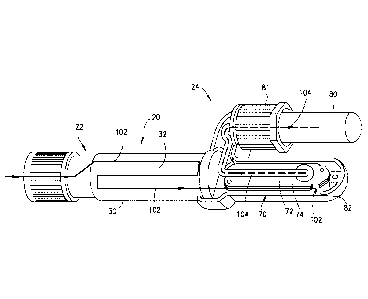
diagnostic tests. Referring to Fig. 11, the arrow comprising a broken line
indicates the second
phase flow path 104 that the plasma 16 takes after passing through the
separation member 74. In
one embodiment, after plasma separation, with the second phase or plasma 16
contained within
the second phase collection container 80, the second phase collection
container 80 is removable
from the blood separation device 10. The second phase collection container 80
can then be used
to transfer the plasma portion 16 to a point-of-care testing device or other
diagnostic testing system.
[0076] During plasma separation, the separation member 74 traps the first
phase 14 of the blood
sample 12 within the first chamber 70, e.g., the first phase 14 of the blood
sample 12 is not allowed
to pass through the separation member 74 into the second chamber 72. Referring
to Fig. 11, the
arrow comprising a straight line indicates the flow path 102 that the blood
sample 12 takes through
the collection chamber 32 and the flow path 102 that the first phase 14 of the
blood sample 12
takes after passing over the separation member 74 and to the blood sample
discard chamber 82.
Referring to Figs. 11 and 12, the first phase 14 of the blood sample 12 flows
into the first chamber
70 through the first chamber inlet 75 and over the separation member 74
surface, and then exits
the first chamber 70 via the first chamber outlet 76 into the blood sample
discard chamber 82.
[0077] In one exemplary embodiment, a blood separation device 10 of the
present disclosure is
able to generate 350 to 600 uL of plasma 16 from the stored 3 mL of blood in
less than 7 minutes.
[0078] Referring to Fig. 5, the blood separation device 10 of the present
disclosure allows for
plasma separation to occur independent of an orientation of the blood
separation device 10. In
other words, the blood separation device 10 separates plasma regardless of
whether the blood
18
Date Recue/Date Received 2022-02-23
separation device 10 is in an upright orientation, e.g., the blood separation
device 10 is contained
in a tube rack, or if the blood separation device 10 is lying in a flat
orientation on a table or tray.
[0079] Referring to Fig. 6, with the second phase or plasma 16 contained
within the second
phase collection container 80, the second phase collection container 80 is
removable from the
blood separation device 10. The second phase collection container 80 can then
be used to transfer
the plasma portion 16 to a point-of-care testing device or other diagnostic
testing system. In one
embodiment, the second phase collection container 80 is removably connectable
to the blood
separation device 10 via a luer lock septum seal.
[0080] In other words, after plasma separation is completed, the plasma 16
within the second
phase collection container 80 is removed from the blood separation device 10
for use in clinical
tests. The rest of the blood separation device 10 can then be discarded.
[0081] As described herein, the present disclosure provides a blood separation
device that
decouples and separates the blood collection process from the plasma
separation process. The
blood separation device includes a sample collection module, an activation
module, and a
separation module. Because the plasma separation happens after the blood
separation device is
disconnected from the patient, the device performance is no longer affected by
patient blood
pressure and needle gauge, and patient discomfort is greatly reduced.
[0082] While this disclosure has been described as having exemplary designs,
the present
disclosure can be further modified within the spirit and scope of this
disclosure. This application
is therefore intended to cover any variations, uses, or adaptations of the
disclosure using its general
principles. Further, this application is intended to cover such departures
from the present
disclosure as come within known or customary practice in the art to which this
disclosure pertains
and which fall within the limits of the appended claims.
19
Date Recue/Date Received 2022-02-23