Note : Les descriptions sont présentées dans la langue officielle dans laquelle elles ont été soumises.
CA 03102965 2020-12-07
WO 2019/237102
PCT/US2019/036290
CALIBRATION OF MULTISPECTRAL ANALYSIS
SYSTEMS
CROSS-REFERENCE TO RELATED APPLICATIONS
This application claims priority to U.S. Provisional Patent Application No.
62/682,819, filed on June 8, 2018, the entire contents of which are
incorporated by reference
herein.
BACKGROUND
Multispectral analysis systems can be used in a variety of assays to determine
information about fluorophore and chromophore binding and expression in
biological
samples. Typically, where multiple spectral contributors in a sample emit or
absorb
radiation, contributions from each of the spectral contributors are separated
to individually
assess attributes such as the spatial location and concentration of each
contributor. Such
assessments can provide important information about a sample, including
disease status,
immunological response, protein expression, and the efficacy of pharmaceutical
treatment.
Multispectral analysis systems are typically calibrated prior to undertaking
assessment
of samples. Calibration generally involves undertaking various steps to ensure
that such
systems can resolve individual contributions from different spectral
contributors.
SUMMARY
The methods and devices disclosed herein use mixtures of calibration dyes in
single
calibration plate wells to calibrate multispectral imaging systems. In
particular, mixtures of
calibration dyes are used to reduce or eliminate cross-talk among spectral
channels that are
relatively closely spaced. Cross-talk impairs recovery of accurate
quantitative information
from a variety of assays by allowing contributions from one spectral
contributor to
contaminate or obscure contributions from another spectral contributor.
In methods and devices disclosed herein, by calibrating with mixtures of
calibration
dyes, the "background" contributions of an interfering spectral contributor to
measured
emission or absorption from a spectral contributor of interest can be reduced
or eliminated.
As a result, spectral cross-talk is reduced and spectral multiplexing is
enhanced. In other
words, by using mixtures of calibration dyes at particular calibration
wavelengths or bands,
the number of different fluorophores in a sample that can be analyzed can be
increased, and
1
CA 03102965 2020-12-07
WO 2019/237102
PCT/US2019/036290
contributions from fluorophores with emission spectra that at least partially
overlap can be
distinguished and quantitatively analyzed with accuracy and precision.
In general, in a first aspect, the disclosure features methods for calibrating
a
multispectral analysis system that include calibrating the system to detect
fluorescence
emission from a first fluorescent entity in a biological sample that includes
the first
fluorescent entity and a second fluorescent entity using a calibration sample,
where the
calibration sample features a first concentration of the first fluorescent
entity and a second
concentration of the second fluorescent entity, and where the first
concentration is larger than
the second concentration.
Embodiments of the methods can include any one or more of the following
features.
The first fluorescent entity can be Cy5 and the second fluorescent entity can
be Cy5.5.
The calibration sample can be a calibration plate that includes a plurality of
sample wells.
A fluorescence emission spectrum of the first fluorescent entity can least
partially
overlap with a fluorescence emission spectrum of the second fluorescent
entity. The first and
second fluorescent entities can each be associated with spectral emission
channels in the
multispectral analysis system, and fluorescence emission from the first
fluorescent entity can
be detected by the multispectral analysis system in the spectral emission
channel associated
with the second fluorescent entity.
The first and second fluorescent entities can each be fluorescent dyes. The
first
fluorescent entity can be an endogenous fluorescent moiety, and the second
fluorescent entity
can be a fluorescent dye. A fraction of the second fluorescent entity in the
calibration sample
relative to a total amount of the first and second fluorescent entities in the
calibration sample
can be between 0.02 and 0.08 (e.g., between 0.03 and 0.07, between 0.04 and
0.06).
The methods can include using the calibrated multispectral analysis system to
identify
one or more gene targets in the biological sample.
Embodiments of the methods can also include any of the other features
disclosed
herein, including any combinations of individual features disclosed in
connection with
different embodiments, except as expressly stated otherwise.
In another aspect, the disclosure features calibration samples that include a
calibration
.. plate featuring a plurality of sample wells, where the calibration plate is
dimensioned to be
received in a multispectral analysis system, and a calibration composition
positioned in one
or more of the wells, the composition including a first fluorescent entity and
a second
fluorescent entity, where a fluorescence emission spectrum of the first
fluorescent entity
2
CA 03102965 2020-12-07
WO 2019/237102
PCT/US2019/036290
overlaps at least partially with a fluorescence emission spectrum of the
second fluorescent
entity.
Embodiments of the calibration samples can include any one or more of the
following
features.
A fraction of the second fluorescent entity in the composition relative to a
total
amount of the first and second fluorescent entities in the composition can be
between 0.02
and 0.08 (e.g., between 0.03 and 0.07, between 0.04 and 0.06). The first
fluorescent entity
can be Cy5 and the second fluorescent entity can be Cy5.5.
Embodiments of the calibration samples can also include any of the other
features
disclosed herein, including any combinations of individual features disclosed
in connection
with different embodiments, except as expressly stated otherwise.
In a further aspect, the disclosure features methods that include calibrating
a
multispectral analysis system to detect fluorescence emission from multiple
fluorescent dyes
in a biological sample, where fluorescence emission is detected in a different
spectral channel
of the system for each of the different fluorescent dyes, and where, for a
first fluorescent dye
in the biological sample, the calibrating includes introducing into the system
a calibration
sample corresponding to the first fluorescent dye and featuring a first amount
of the first
fluorescent dye and a second amount of a second fluorescent dye in the
biological sample.
Embodiments of the methods can include any one or more of the following
features.
The first and second fluorescent dyes can have respective first and second
fluorescence emission spectra that at least partially overlap. Fluorescence
emission from the
first fluorescent dye can be detected in a first spectral channel of the
detection system and
fluorescence emission from the second fluorescent dye can be detected in a
second spectral
channel of the detection system, and at least a portion of fluorescence
emission from the first
fluorescent dye can also be detected in the second spectral channel. The first
fluorescent dye
can be Cy5 and the second fluorescent dye can be Cy5.5.
Embodiments of the methods can also include any of the other features
disclosed
herein, including any combinations of individual features disclosed in
connection with
different embodiments, except as expressly stated otherwise.
The details of one or more embodiments are set forth in the accompanying
drawings
and the description below. Other features and advantages will be apparent from
the
description, drawings, and claims.
3
CA 03102965 2020-12-07
WO 2019/237102
PCT/US2019/036290
DESCRIPTION OF DRAWINGS
FIG. 1 is a graph showing polymerase chain reaction (PCR) amplification curves
for
gene targets in a biological sample.
FIG. 2 is a graph showing PCR amplification curves for gene targets in a
biological
sample, with Cy5 fluorescence emission cross-talk into a spectral emission
channel
corresponding to Cy5.5.
FIG. 3 is a graph showing PCR amplification curves for gene targets in a
biological
sample, measured using a multispectral analysis system calibrated with a
calibration
formulation that included both Cy5 and Cy5.5.
FIG. 4 is a graph showing PCR amplification curves for gene targets in a
biological
sample, measured using a multispectral analysis system calibrated with a
calibration
formulation that included only Cy5.
FIG. 5 is a graph showing PCR amplification curves for gene targets in a
biological
sample, measured using a multispectral analysis system calibrated with a
calibration
formulation that included a dye mixture of 5% Cy5.5 and 95% Cy5.
FIG. 6 is a graph showing PCR amplification curves for gene targets in a
biological
sample, measured using a multispectral analysis system calibrated with a
calibration
formulation that included a dye mixture of 10% Cy5.5 and 90% Cy5.
FIG. 7 is a graph showing the deviation of a multicomponent PCR amplification
curve
from a constant fluorescence intensity, measured using a multispectral
analysis system
calibrated with a calibration formulation that include a dye mixture of Cy5.5
and Cy5, as a
function of the percentage of Cy5.5 in the dye mixture.
FIG. 8 is a graph showing PCR amplification curves for gene targets in a
biological
sample, measured using a multispectral analysis system calibrated with a
calibration
formulation that included a dye mixture of 4% Cy5.5 and 96% Cy5.
FIG. 9 is a schematic diagram of an example of a calibration sample.
FIG. 10 is a schematic diagram of an example of a multispectral analysis
system.
Like reference symbols in the drawings indicate like elements.
DETAILED DESCRIPTION
Multispectral analysis systems typically include different filters for use in
quantitatively measuring contributions from specific fluorophores in a
biological sample.
When a sample is prepared with (or expresses) multiple fluorophores, each of
which has a
4
CA 03102965 2020-12-07
WO 2019/237102
PCT/US2019/036290
different emission spectrum, emission from each of the fluorophores can be
isolated for
analysis by selecting a corresponding filter with a central wavelength and
bandpass filter that
effectively excludes emission from all fluorophores in the sample but the
fluorophore of
interest. As the number of fluorophores increases, and for fluorophores with
relatively wide
spectral emission bandwidths, it is increasingly challenging to implement
spectral filters that
isolate emission from each fluorophore by sufficiently excluding emission from
the others,
and at the same time transmit sufficient emission radiation from the
fluorophore of interest to
generate a detectable signal.
As an example, the QuantStudioi'm Dx imaging system (available from
ThermoFisher
Scientific, Waltham, MA) uses a filter designated m5 to quantify spectral
emission from Cy5
dye in samples (excited at a wavelength of 648 nm, and fluoresces at a nominal
wavelength
of 668 nm), and a filter designated m6 to quantify spectral emission from
Cy5.5 dye in
samples (excited at 685 nm, and fluoresces at a nominal wavelength of 706 nm).
Both Cy5
and Cy5.5 dyes have significant fluorescence emission bandwidth and spectral
overlap. Note
that in this disclosure, "Cyanine" is abbreviated as "Cy".
Various assays and other quantitative analysis techniques rely on accurate
quantification of multiple fluorophores in a single sample for imaging and
diagnostic
purposes. One such assay is the NeoMDx qPCR assay (available from Perkin
Elmer,
Waltham, MA), which is a multiplex polymerase chain reaction (PCR) assay that
uses dyes
ROX114, FAMTm, HEX, Cy5, and Cy5.5 in a single PCR reaction.
A multispectral analysis system (such as the QuantStudio Dx) is used in this
assay
to measure, separate, and quantify fluorescence emission from each of these
dyes during PCR
reaction cycling. These dyes are used to target the following genes in
samples:
Table 1
Dye Sample Target Gene
ROX None (reference dye)
FAM TREC
HEX RPP30
Cy5 SMN1
Cy5.5 KREC
5
CA 03102965 2020-12-07
WO 2019/237102
PCT/US2019/036290
As shown in Table 1, during sample preparation prior to performing the assay,
the
SMN1 target gene is labeled with the Cy5 fluorophore, and the KREC target gene
is labeled
with the Cy5.5 fluorophore. Using the dyes listed in Table 1, the NeoMDxTm
qPCR
multiplex assay can detect the presence or absence of gene targets TREC, SMN1,
and KREC
in a sample, along with the presence or absence of the RPP30 reference gene.
FIG. 1 is a graph showing example qPCR amplification curves obtained by
performing the NeoMDXTm qPCR assay on a sample taken from a "normal" newborn
infant.
In FIG. 1, each of the target genes was amplified, along with the RPP30
control gene, and
each amplified gene was separately detected based on fluorescence emission
from its
corresponding conjugated dye.
However, it has been discovered while performing the NeoMDX qPCR assay on a
variety of samples that, in some trials, cross-talk in the Cy5.5 spectral
channel due to
fluorescence emission from Cy5 can occur, which impairs accurate
quantification of both
SMN1 and KREC gene targets. For example, in certain samples that were KREC
negative
and also included relatively high concentrations of SMN1 (yielding relatively
high intensity
fluorescence emission from Cy5), an amplification curve corresponding to
emission from
Cy5.5 was also measured, even though the KREC signal was relatively low (but
above the
predefined detection threshold). Since the samples were known to be KREC
negative, such
results amounted to false positive tests for the KREC target.
Without wishing to be bound by theory, it is believed that the false KREC
amplification curve derived from measured Cy5.5 fluorescence emission resulted
from cross-
talk due to Cy5 into the Cy5.5 spectral emission channel. Specifically, due to
the inability of
the filter used to spectrally isolate Cy5.5 fluorescence emission from Cy5
fluorescence
emission in the QuantStudiolm Dx system used to perform the analysis, some
fluorescence
emission from Cy5 was falsely detected as Cy5.5 fluorescence emission in the
Cy5.5 spectral
channel.
FIG. 2 is a graph showing example qPCR amplification curves for a KREC
negative
sample. In FIG. 2, the sample contained 1x10⁵ copies of the SMN1 target gene,
but none of
the other gene targets from Table 1 (and specifically, no KREC target genes).
Nonetheless, a
.. KREC amplification curve was detected based on measured fluorescence
emission in the
Cy5.5 spectral emission channel (i.e., with the m6 filter in place in the
QuantStudiolm Dx
system). The fluorescence emission in the Cy5.5 spectral emission channel was
due to Cy5
fluorescence emission that was not sufficiently extinguished by the filter
associated with the
Cy5.5 spectral emission channel to escape detection.
6
CA 03102965 2020-12-07
WO 2019/237102
PCT/US2019/036290
Based on the amplification curves shown in FIG. 2, the sample might be
diagnosed as
belonging to a patient that is KREC positive, i.e., a "normal" baby. However,
a baby with
XLA (X-linked Agammaglobulinemia) would display no KREC amplification. Thus,
the
result shown in FIG. 2 represents a false negative assessment of the patient
for XLA.
Effectively, the portion of the Cy5 fluorescence emission signal in FIG. 2
that is
detected in the Cy5.5 spectral emission channel functions as "background"
against which any
"true" Cy5.5 fluorescence emission should be detected to obtain accurate
quantitative results
for each of the gene targets. For many multispectral analysis systems
(including the
QuantStudioi'm Dx system), before samples are analyzed, the systems are
calibrated with
"reference" samples to provide the systems with reference spectral information
for each of
the dyes that are being measured. The reference samples are typically
implemented as
calibration plates in which some (or all) of the wells in the plate contain
reference samples of
one of the dyes for which fluorescence emission will be measured. Thus, to
calibrate the
QuantStudio Dx system for measurement of fluorescence emission from each of
the dyes
in Table 1, a series of multi-well calibration plates is used, where each one
of the calibration
plates in the series includes wells filled with a different one of the dyes in
Table 1. By using
only one of the dyes in the wells of each calibration plate, the system
measures "pure"
reference spectra for that dye at each well location. To complete the
calibration, calibration
plates filled with a different one of the dyes that will be measured are
processed through the
system in succession, so that the system is provided with pure reference
spectra for each of
the dyes.
However, the inventors have discovered that by mixing multiple dyes together
in the
wells of a single calibration plate, reference spectra are provided to the
system that contain
contributions from each of the dyes in the plate. These reference spectra are
not pure spectra
of any one of the dyes. Instead, they contain contributions from each dye in
the plate, and are
effectively "mixed" reference spectra. Moreover, it has been observed that
such mixed
reference spectra significantly improve the quantitative analysis of
fluorescence emission
from the dyes, even when substantial fluorescence emission from one dye is
detected in the
spectral emission channel of another dye.
As an example, a number of different calibration formulations were tested for
the Cy5
calibration plate used in the analysis described above and shown in FIG. 2. In
particular,
each formulation contained different ratios of Cy5- and Cy5.5-labeled
oligonucleotides, with
the same calibration buffer. The QuantStudioi'm Dx system was calibrated with
each of the
different formulations, and then the same data file that was generated from
the sample
7
CA 03102965 2020-12-07
WO 2019/237102
PCT/US2019/036290
containing 1x10⁵ copies of the SMN1 target gene (and none of the other target
genes shown
in Table 1) was analyzed with according to calibrations defined by each of the
different
formulations..
By calibrating the system for Cy5 fluorescence emission using a calibration
formulation that included both Cy5 and Cy5.5, it was generally observed that
fluorescence
emission cross-talk from Cy5 fluorescence emission into the Cy5.5 spectral
emission channel
was reduced. During testing, a formulation that included about 95% Cy5-labeled
oligonucleotide and about 5% Cy5.5-labeled oligonucleotide was found to
provide a
significant reduction in fluorescence emission cross-talk into the Cy5.5
spectral emission
channel. The Cy5 and Cy5.5 fluorophores were conjugated to the 5' position on
the
oligonucleotides (sequence AGGGTTT for the Cy5 conjugate, and TCTGCAC for the
Cy5.5
conjugate).
The complete formulation of this calibrator was as follows:
Table 2
Amount/Concentration Component
285 nM Cy5-labeled oligonucleotide
15 nM Cy5.5-labeled olignonucleotide
lx Phosphate buffered saline, pH 7.3-7.5
0.1% Tween 20 detergent
0.01% Antifoam B
Molecular grade water
FIG. 3 is a schematic diagram showing qPCR amplification curves for a sample
analyzed following calibration of the QuantStudioi'm Dx system with the
calibrator
formulation shown in Table 2. As described above, the sample contained 1x10⁵
copies of the
SMN1 target gene, and none of the other target genes shown in Table 1. In
particular, in FIG.
3, KREC amplification is not observed, consistent with the absence of KREC in
the sample
and, effectively, the elimination of Cy5 fluorescence emission cross-talk into
the Cy5.5
spectral emission channel.
8
CA 03102965 2020-12-07
WO 2019/237102
PCT/US2019/036290
Experiments with several different calibrator formulations resulted in a
reduction of
Cy5 fluorescence emission cross-talk into the Cy5.5 spectral emission channel.
In general, it
was observed that cross-talk was reduced when the concentration of Cy5.5
relative to the
total concentration of Cy5 and Cy5.5 in the calibrator formulation was greater
than zero (e.g.,
0.001 or more, 0.002 or more, 0.005 or more, 0.01 or more, 0.02 or more, 0.03
or more, 0.04
or more, 0.05 or more, 0.06 or more, 0.07 or more, 0.08 or more, 0.09 or more,
0.10 or more,
0.12 or more, 0.14 or more, 0.16 or more, 0.18 or more, 0.20 or more, 0.25 or
more). In some
embodiments, it was observed that an over-correction for Cy5 fluorescence
emission cross-
talk into the Cy5.5 spectral emission channel occurred when the concentration
of Cy5.5
relative to the total concentration of Cy5 and Cy5.5 in the calibrator
formulation was greater
than 0.10.
To determine which relative concentration of Cy5.5 in the prepared calibrators
yielded the optimum correction for Cy5 fluorescence emission cross-talk, a set
of calibrators
were prepared with different relative concentrations of Cy5- and Cy5.5-
conjugated
oligonucleotides, and the other components listed in Table 2. Each of these
calibrators was
used to calibrate the QuantStudioTM Dx system, after which the same sample
described above
was analyzed by the system. FIGS. 4, 5, and 6 show qPCR amplification curves
for the
sample after calibration of the system with calibrators that contained 0%
Cy5.5-labeled
oligonucleotide, 5% Cy5.5-labeled oligonucleotide, and 10% Cy5.5-labeled
oligonucleotide,
respectively. The amplification curves corresponding to the Cy5.5-labeled
oligonucleotides
are labeled "Other" in the figure legends.
To assess whether each calibrator yielded amplification curves that were
undercorrected (e.g., still showed some cross-talk), overcorrected, or ideally
corrected, the
flatness of the multicomponent amplification curve over 40 PCR amplification
cycles was
used as a metric. In general, it was assumed that the flatter the
multicomponent amplification
curve, the closer that correction was to being ideal.
FIG. 7 is a graph showing the change in the multicomponent amplification curve
fluorescence intensity as a function of the percentage of Cy5.5-labeled
oligonucleotide in the
calibrator used to calibrate the system prior to measuring each multicomponent
amplification
curve. In FIG. 7, results fall generally along a straight line, which is
confirmed by the
regression analysis shown in the figure. The regression analysis predicts that
a calibrator
prepared with approximately 4% Cy5.5-labeled oligonucleotide (and
approximately 96%
Cy5-labeled oligonucleotide) yields the best compensation for Cy5 fluorescence
emission
into the Cy5.5 spectral emission channel. qPCR amplification curves (including
the
9
CA 03102965 2020-12-07
WO 2019/237102
PCT/US2019/036290
multicomponent curve) obtained after calibrating the system with the
calibrator prepared with
approximately 4% Cy5.5-labeled oligonucleotide and approximately 96% Cy5-
labeled
oligonucleotide are shown in the graph in FIG. 8.
While the foregoing examples of calibrator formulations specifically involve
mixtures
of Cy5 and Cy5.5 to reduce or eliminate fluorescence emission cross-talk,
similar principles
apply to mixtures of any two fluorophores used in calibration formulations to
reduce cross-
talk in spectral emission channels. For example, consider the more general
situation where
two spectral contributors (fluorescent dyes, endogenous fluorophores,
expressed fluorescent
moieties) A and B each exhibit fluorescence emission such that at least some
fluorescence
emission from A is detected in the spectral emission channel of B, due to the
inability of the
detection system to fully separate the emission from A and B (e.g., using
filters).
To calibrate the multispectral analysis system to properly correct for this
cross-talk, a
calibrator can be prepared that includes a relatively large fraction of A and
a relatively
smaller, but non-zero, fraction of B, as a mixture. The mixture is then used
to calibrate the
system for detection of fluorescence emission from A. In general, the fraction
of B in the
calibration formulation relative to the total amount of A and B in the
formulation can be
0.001 or more (e.g., 0.002 or more, 0.003 or more, 0.004 or more, 0.005 or
more, 0.01 or
more, 0.02 or more, 0.025 or more, 0.03 or more, 0.035 or more, 0.04 or more,
0.045 or
more, 0.05 or more, 0.055 or more, 0.06 or more, 0.065 or more, 0.07 or more,
0.08 or more,
0.09 or more, 0.10 or more, 0.12 or more, 0.14 or more, 0.16 or more, 0.18 or
more, 0.20 or
more, 0.25 or more).
A variety of different calibrators (corresponding to different pairs of
fluorophores A
and B can be prepared according to the methods disclosed herein. In general,
calibrators can
include one or more dyes. Suitable dyes for use in calibrators can include
(but are not limited
to) one or more rhodamine-based dyes such as ROXim,TAMRATm, and Texas Red
(rhodamines, carboxyrhodamines, methylrhodamines, and derivatives thereof),
one or more
fluorescein-based dyes such as FAWN', VIC , SIMAim, TETTm, and HEX Tm
(fluoresceins,
carboxyfluoresceins, chlorofluoresceins, and derivatives thereof), one or more
cyanine-based
dyes (e.g., cyanine, and derivatives thereof) such as Cy3, Cy3.5, Cy5, and
Cy5.5, and one or
more xanthene-based dyes such as JOE'
Examples of pairs of fluorophores A and B (in addition to Cy5 and Cy5.5)
include,
but are not limited to: TAMRATm and HEX; FAMTm and HEX, FAMTm and SIMATm,
HEX Tm and Cy3, SIMA and Cy3, Cy3 and Cy3.5; Cy3 and ROXim; Cy3.5 and Cy5; and
ROXTm and Cy5.
CA 03102965 2020-12-07
WO 2019/237102
PCT/US2019/036290
In some embodiments, mutual cross-talk can occur between the spectral emission
channels of fluorophores A and B. In other words, some fluorescence emission
from A can
be observed in the spectral emission channel of B, and some fluorescence
emission from B
can be observed in the spectral emission channel of A. To correct for such
spectral cross-
talk, the system can be calibrated to detect fluorescence emission from A with
a first
calibrator that includes concentrations of both A and B, with the
concentration of A being
larger than the concentration of B in the first calibrator. The system can
also be calibrated to
detect fluorescence emission from B with a second calibrator that includes
concentrations of
both A and B, with the concentration of B being larger than the concentration
of A in the
second calibrator.
In general, the relative concentrations of A and B in the first and second
calibrators
can be selected as desired to provide sufficient correction for spectral cross-
talk. In some
embodiments, for example, the first calibrator can have a composition similar
to the
compositions discussed above. Further, in certain embodiments for example, the
second
calibrator can have a composition in which the fraction of A in the
calibration formulation
relative to the total amount of A and B in the formulation can be 0.001 or
more (e.g., 0.002 or
more, 0.003 or more, 0.004 or more, 0.005 or more, 0.01 or more, 0.02 or more,
0.025 or
more, 0.03 or more, 0.035 or more, 0.04 or more, 0.045 or more, 0.05 or more,
0.055 or
more, 0.06 or more, 0.065 or more, 0.07 or more, 0.08 or more, 0.09 or more,
0.10 or more,
0.12 or more, 0.14 or more, 0.16 or more, 0.18 or more, 0.20 or more, 0.25 or
more).
While the foregoing discussion has focused on calibrators that provide for
correction
when two different spectral contributors A and B exhibit fluorescence emission
cross-talk,
more complex calibrators can also be prepared for situations when cross-talk
occurs among
more than two spectral contributions. For example, in situations where
fluorescence emission
from spectral contributors A and B is detected in the spectral emission
channel for spectral
contributor C, the multispectral analysis system can be calibrated with
calibration
formulations that include mixtures of A, B, and C. For example, the system can
be calibrated
to detect fluorescence emission from A with a calibration formulation that
includes a
relatively high fraction of A and a relatively low fraction of C (similar to
the low fractions of
A and B discussed above), and calibrated to detect fluorescence emission from
B with a
calibration formulation that includes a relatively high fraction of B and
relatively low fraction
of C (similar to the low fractions of A and B discussed above). The fractions
of A, B, and C
in the various calibration formulations can vary as desired to achieve
suitable compensation
for fluorescence emission cross-talk.
11
CA 03102965 2020-12-07
WO 2019/237102
PCT/US2019/036290
Similarly, where fluorescence emission from a spectral contributor A is
observed in
spectral emission channels for spectral contributors B and C, the
multispectral analysis
system can be calibrated to detect fluorescence emission from A with a
calibration
formulation that includes a relatively high fraction of A and relatively low
fractions of B and
C (similar to the low fractions of A and B discussed above).
More generally, the calibration formulations disclosed herein can include
combinations of two, three, four, five, or even more than five spectral
contributors
(fluorophores such as dyes, endogenous fluorescent entities, and expressed
fluorescent
moieties), used to calibrate multispectral analysis systems to compensate for
fluorescence
emission cross-talk by one or more of the spectral contributors into spectral
emission
channels corresponding to one or more of the other spectral contributors.
While specific examples of fluorophores (or spectral contributors) A and B
have been
described above, more generally, the methods and calibrators described herein
can be used in
any circumstance in which fluorescence from one spectral contributor (e.g., B)
is detected in
.. a spectral wavelength band dedicated to the measurement of fluorescence
emission from
another spectral contributor (e.g., A). For example, where a sample includes
spectral
contributors A and B, and fluorescence from contributor A is measured in a
wavelength band
centered at wavelength XA, then mixtures of A and B can be used in calibrators
if the total
fluorescence intensity of spectral contributor B in the wavelength band
centered at XA is, for
example, 1% or more (e.g., 2% or more, 3% or more, 5% or more, 7% or more, 10%
or more,
15% or more, 20% or more, 25% or more, 30% or more, 40% or more, 50% or more)
of the
fluorescence intensity of spectral contributor A at wavelength XA. Similar
considerations
apply for spectral interference (i.e., cross-talk) among more than two
spectral contributors.
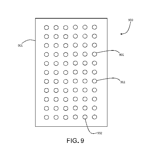
FIG. 9 is a schematic diagram showing an example of a calibration sample 900.
Sample 900 includes a substrate 901 (e.g., a plate, a slide, a wafer, a block)
with a plurality of
calibration regions 902. Calibration regions 902 can be formed on substrate
901 in various
ways. In some embodiments, for example, calibration regions 902 are simply
defined areas
on the surface of substrate 901. In certain embodiments, calibration regions
902 correspond
to recesses (i.e., wells) formed in the surface of substrate 901. In some
embodiments,
calibration regions 902 can correspond to protrusions, extensions, or other
raised structures
that extend outward from the surface of substrate 901.
In general, calibration sample 900 can include any number of calibration
regions 902.
For example, the number of calibration regions 902 can be 10 or more (e.g., 20
or more, 50 or
12
CA 03102965 2020-12-07
WO 2019/237102
PCT/US2019/036290
more, 100 or more, 200 or more, 300 or more, 400 or more, 500 or more, 700 or
more, 1000
or more, or even more).
Calibration sample 900 includes a calibrator ¨ which corresponds to a mixture
of
reagents, including one or more fluorophores ¨ in one or more of the
calibration regions 902.
As described above, the calibrator in one or more of the calibration regions
can include
mixtures of flurorophores (e.g., fluorophores A and B) with emission spectra
that are
relatively close spectrally, such that emission from one of the fluorophores
is detected in a
spectral band dedicated to the other fluorophore.
In some embodiments, a set of calibration samples can be provided to accompany
an
assay that features multiple dyes, each of which functions as a spectral
reporter for a specific
molecular target in a sample. For example, in an assay that includes dyes A,
B, C, D, and E,
a set of 5 calibration samples can be provided, one or for each of the 5 dyes
in the assay. If,
for example, dyes C, D, and E are measured in distinct spectral bands with no
cross-talk from
any of the other dyes, then the calibration samples corresponding to dyes C,
D, and E can
each include only one dye (i.e., dyes C, D, and E, respectively) at each of
the calibration
regions.
If a non-negligible amount of fluorescence from dye A is measured in the
spectral
band dedicated to the measurement of dye B, then the calibration sample for
dye B can
include a mixture of mostly dye B and a relatively small amount of dye A at
each of the
calibration regions 902, as described above.
If a non-negligible amount of fluorescence from dye B is measured in the
spectral
band dedicated to the measurement of dye A, then the calibration sample for
dye A can
include a mixture of mostly dye A and a relatively small amount of dye B at
each of the
calibration regions 902, as described above. Alternatively, if no appreciable
amount of
fluorescence from dye B is measured in the spectral band dedicated to the
measurement of
dye A, then the calibration sample for dye A can include only dye A at each of
the calibration
regions 902.
In general, for an assay with N fluorescent dyes that are measured
independently, N
calibration samples can be provided. For each particular calibration sample
(corresponding
to a dye M) in the set of N samples, the calibration regions 902 can each
include a relatively
large proportion of the dye M that nominally corresponds to that sample, and
relatively small
proportions of any other dyes that exhibit measurable fluorescence in the
spectral detection
band that is dedicated to the measurement of dye M, as discussed above.
13
CA 03102965 2020-12-07
WO 2019/237102
PCT/US2019/036290
The calibration samples described can be used to calibrate a variety of
different
multispectral analysis systems. An example of one such system is the
QuantStudioi'm Dx
system. More generally, FIG. 10 shows an example of a multispectral analysis
system 1000
that can be calibrated with the calibration samples described. System 1000
includes a
radiation source 1002, an emission filter 1004, a beam splitter 906, beam
optics 1008, a
sample stage 1010, an emission filter 1012, and a detector 1014. The source,
filters, and
detector are connected to controller 1016 which transmits control signals to
these
components, and receives reporting signals and measurement information from
the
components.
To calibrate system 1000, a calibration sample 900 ¨ which is dimensioned to
be
received by system 1000 (and specifically, on stage 1010) is positioned on the
stage.
Calibration sample 900 corresponds to a particular dye M, and controller 1016
adjusts
emission filter 1012 to select a spectral band (i.e., a "spectral channel") in
filter 1012 that is
dedicated to the measurement of fluorescence emission from dye M.
Radiation source 1002 then directs incident radiation through filter 1004,
beam
splitter 1006, and beam optics 1008, and onto calibration sample 900. The
incident radiation
excites the calibrator in calibration regions 902, causing the calibrator to
emit fluorescence.
The emitted fluorescence passes through beam optics 1008, is reflected by beam
splitter
1006, passes through emission filter 1012, and is detected by detector 1014.
Detector 1014 measured fluorescence emission from each of the calibration
regions
902, and transmits the fluorescence emission measurements to controller 1016.
Controller
1016 then uses the measured fluorescence emission information to calibrate
each of the
calibration regions 902 for fluorescence measurements of dye M. The specific
manner in
which each of the calibration regions 902 are calibrated can vary widely. In
some
embodiments, for example, the spectrally-resolved measured fluorescence
emission
information forms a "baseline" measurement signal against which fluorescence
emission
measurements from samples are normalized, or which is subtracted from
fluorescence
emission measurements from samples. In certain embodiments, the spectrally-
resolved
measured fluorescence emission information from the calibrators forms an
effective
eigenspectrum or pure spectrum corresponding to dye M, which is then used to
quantify
measured fluorescence information from the dye M in samples. The measured
fluorescence
information from the calibrators of calibration sample 900 can also be used in
a wide variety
of other calibration techniques.
14
CA 03102965 2020-12-07
WO 2019/237102
PCT/US2019/036290
To complete the calibration of system 1000, controller 1016 cycles through
each of
the dyes used in a corresponding assay in turn, for each dye adjusting
emission filter 1012 to
select a spectral band dedicated to measurement of fluorescence emission from
the dye, and
then measuring fluorescence emission from each calibration region 902 on a
calibration
sample 900 positioned on stage 1010 and corresponding to the dye. The measured
fluorescence information from the calibration sample 900 corresponding to each
dye is then
used to calibrate system 1000 to measure fluorescence emission from that dye
in samples.
The calibration information can be stored in a storage unit, for example,
connected to
controller 1016.
When system 1000 has been calibrated, the system can be used to measure
fluorescence emission from each of the dyes in an assay that is performed on a
sample. For
example, if a sample is prepared with an assay that includes N dyes, then
fluorescence
emission measurements corresponding to each of the N dyes are performed
sequentially, with
controller 1016 adjusting emission filter 1012 to select a spectral
measurement band
corresponding to each dye in turn, as in the calibration procedure described
above.
Raw fluorescence emission measurements for each dye in the assay can be
corrected
using the stored calibration information, which permits quantitative
information to be
obtained. For example, in an assay with multiple dyes, each of which functions
as a spectral
reporter for a particular gene target in a sample, each of the gene targets
can be identified
based on the corrected measured fluorescence emission information for the
corresponding
dyes, and expression of each of the gene targets in the sample can be
quantified.
Hardware and Software Implementation
In general, controller 1016 can be configured to perform any of the control,
calibration, or data analysis functions described herein. These functions can
be performed
entirely by controller 1016 (e.g., autonomously), or a set of functions or
steps can be
performed by part by controller 1016 and in part by a user of system 1000.
When performed
fully or partly by controller 1016, the functions can be implemented in
computer programs
using standard programming techniques. Such programs are designed to execute
on
programmable computers, dedicated controllers, or specifically designed
integrated circuits,
each comprising an electronic processor (e.g., an electronic processor in
controller 1016), a
data storage system (including memory and/or storage elements), at least one
input device,
and least one output device, such as a display. Each computer program can be
implemented
in a high-level procedural or object-oriented programming language, or an
assembly or
CA 03102965 2020-12-07
WO 2019/237102
PCT/US2019/036290
machine language. Furthermore, the language can be a compiled or interpreted
language.
Each such computer program can be stored on a computer readable storage medium
(e.g., a
magnetic storage medium such as a hard drive, an optical storage medium such
as a CD-
ROM or DVD, a persistent solid state storage medium such as a solid state hard
drive) that,
when read by a device with a processor, can cause the processor in the device
to perform the
control and analysis functions described herein.
EXAMPLES
To investigate the performance of calibration samples with mixtures of two
dyes from
a single assay, studies were performed to determine three quantities relating
to measurement
limits for assay targets. The Limit of Blank (LoB) is the highest measurement
result that is
likely to be observed (with a stated probability [a]) for a blank sample, that
contains no
analyte. It defines the variation of the background or the zero sample. The
Limit of Detection
is the measured quantity value, obtained by a given measurement procedure, for
which the
probability of falsely claiming the absence of a measurand in a material is
13, given a
probability a of falsely claiming its presence. For molecular measurement
procedures which
differ from typical measurement procedures because all blank or negative
sample results
normally are reported as negative, the LoD is calculated from a probit
regression model as the
measurand concentration at which, with a predefined probability (usually 95%),
measurement
results yield a positive classification. The Limit of Quantification (LoQ) is
the lowest
amount of a measurand in a material that can be quantitatively determined with
state accuracy
(as total error or as independent requirements for bias and precision) under
stated
experimental conditions. LoQ may be defined based on functional sensitivity or
based upon
total error.
Studies were performed to deterimine: (1) the LoB for all the analytes
targeted by an
assay (the "LoB study"); (2) the LoD for two semi-quantitative analytes,
sjTREC and KREC,
targeted by an assay (the "LoD" study); and (3) the LoQ for the same two semi-
quantitative
analytes, sjTREC and KREC, targeted by the assay (the "LoQ" study). The assay
was the
NeoMDx assay, and the LoD and LoQ for two germline targets (SMN1 and RPP30) of
the
assay were not evaluated.
The LoB study was performed with contrived analyte-negative samples which were
created by spiking SMN1-negative cells (obtained from Coriell Institute for
Medical
Research, Camden, NJ) into leukocyte-depleted human blood. The Coriell cell
line, which is
also sjTREC- and KREC-negative naturally, was added at a target of 30,000
genome-
16
CA 03102965 2020-12-07
WO 2019/237102
PCT/US2019/036290
copies/pL, a concentration typical for a neonate. Five individual contrived
analyte-negative
samples (CANS1-5) were prepared by spiking the Coriell cell line into five
different lots of
leukocyte-depleted human blood.
The five lots of leukocyte-depleted human blood (obtained from Zen-Bio,
Research
Triangle Park, NC) were washed three times with saline solution and the
hematocrit adjusted
to 40 ¨ 55% (Table 3). The Coriell SMA cells were cultured in a RPMI medium
supplemented with FBS and penicillin/streptomycin. The cells were counted
using a Cell
Countess instrument (Thermo Fisher, Waltham, MA). Approximately 15000 cells
were
spiked per microliter of leukocyte-depleted human blood to obtain the
contrived analyte-
negative samples. After dispensing the prepared blood on filter paper, the DBS
samples were
dried overnight and then stored at -30 C to -16 C in a sealed bag with
desiccant until use.
Table 3 describes the samples used to determine LoB.
Table 3
Hematocrit
Sample name (%) Lot# of Leukocyte Depleted Blood
CANS1 49.2 W36981900271000
CANS2 49.8 W36981900281900
CANS3 49.5 W36981900270800
CANS4 50.0 W36981900281700
CANS5 49.5 W36981900270500
As the cell line contained a normal level of RPP30, the contrived analyte-
negative
samples were not suitable to establish the LoB of the RPP30 target, but
suitable for the other
three analytes, sjTREC, KREC and SMN1.
For the LoD and LoQ studies, due to the rarity of research specimens for the
SCID
and XLA disorders, it was challenging to find representative newborn DBS
samples that
happen to have sjTREC and KREC at desired levels. Therefore, the studies were
conducted
using contrived samples. The contrived samples were prepared by diluting cord
blood with
adult whole blood or with leucocyte-depleted human red blood that has sjTREC
and/or
KREC levels undetectable by the assay. HL-60 cells were spiked into the adult
whole blood
or leucocyte-depleted blood to adjust the RPP30 Ct values to match the RPP30
Ct value in
cord blood (within 1 Ct difference).
17
CA 03102965 2020-12-07
WO 2019/237102
PCT/US2019/036290
First, the initial values of sjTREC and KREC (copies/pL blood) in each cord
blood
was assigned by testing against a 5-level (250, 500, 1000, 3000, 10000
copies/pL blood)
standard curve generated using DBS samples containing serial dilutions of
ddPCR-quantified
linearized TREC, KREC, SMN1 and RPP30 plasmids in leucocyte-depleted human
blood.
The results are summarized in Table 4.
Table 4
Hematocrit sjTREC KREC
Blood Lot# (%) (copies/1AL (copies/4
blood)
blood)
Cord blood ZEN00403 (Lot 48.2 368 1132
AB+ 1)
Cord blood A+ ZEN00410 (Lot 47.2 492 1174
2)
Cord blood 0- ZEN00409 (Lot 51.9 344 910
3)
Three lots of adult whole blood were then tested to determine their endogenous
levels
of each analyte to check if they were suitable to be used as the diluent. The
mean Ct values
of 6 replicates of each adult whole blood are summarized in Table 5.
Table 5
Blood Lot #
TREC Ct KREC Ct SMN1 Ct RPP30 Ct
Adult whole No Ct 32.48 24.47 25.17
blood W36981900485900
Adult whole 37.35 33.35 24.47 24.39
blood W36981900296700
Adult whole 34.84 34.41 25.77 27.35
blood W36981900297300
Although sjTREC are usually low in adult whole blood, two out of the three
lots had
detectable levels of sjTREC and thus they were replaced by leucocyte-depleted
human blood
as diluent in the preparation of samples for TREC LoD/LoQ. All three lots of
adult whole
blood also had detectable levels for KREC so for the preparation of KREC
LoD/LoQ
samples, leucocyte-depleted human blood was used as a diluent. The diluents
used for each
cord blood are listed in Table 6. The RPP30 Ct values of the diluents were
adjusted to match
the cord bloods RPP30 Ct values (within 1 Ct difference) by spiking HL-60
cells into the
diluents according to the study design.
18
CA 03102965 2020-12-07
WO 2019/237102
PCT/US2019/036290
Table 6
Analyte Cord blood Lot# Diluent Name
Diluent lot#
name
ZEN00403 (Lot 1) Adult whole blood
W36981900296700
Leukocyte Depleted
W36981900281700
sjTREC ZEN00410 (Lot 2) Blood
Leukocyte Depleted
W36981900270500
ZEN00409 (Lot 3) Blood
Leukocyte Depleted
W36981900270800
ZEN00403 (Lot 1) Blood
KREC Leukocyte Depleted
W36981900281700
ZEN00410 (Lot 2) Blood
Leukocyte Depleted
W36981900270500
ZEN00409 (Lot 3) Blood
Each cord blood sample was diluted into different levels in which at least the
lowest
three levels shall yield hit rates within the range of 0.10 to 0.90 using the
corresponding
diluents (Table 6). The samples were then spotted onto Ahlstrom 226 filter
paper and dried
overnight. Then they were stored at -30 C to -16 C in a sealed bag with
desiccant until use.
The final dilutions used in the study and their corresponding hit rates (10
replicates) observed
in a pre-test are summarized in Table 7.
Table 7
Concentration
Analyte CordBlood Sample Dilution
(copies/pt Hit
rates
Name Sample Level factors
blood)
Li 100%
CordBlood 368 1.00
(ZEN00403)
Li 100%
Level 1 184 2.00
(ZEN00403)
Li 100%
Level 2 110 3.33
(ZEN00403)
Li 100%
Level 3 73.6 5.00
(ZEN00403)
Li 50%
sjTREC Level 4 36.8 10.0
(ZEN00403)
Li 50%
Level 5 29.4 12.5
(ZEN00403)
Li 10%
Level 6 22.1 16.7
(ZEN00403)
Li 0%
Level 7 14.7 25.0
(ZEN00403)
Li 0%
Diluent 0 N/A
(ZEN00403)
19
CA 03102965 2020-12-07
WO 2019/237102
PCT/US2019/036290
L2 (ZEN00410) 100%
CordBlood 492 1.00
L2 (ZEN00410) 100%
Level 1 55.2 6.67
L2 (ZEN00410) 70%
Level 2 36.8 10.0
L2 (ZEN00410) 100%
Level 3 29.4 12.5
L2 (ZEN00410) 60%
Level 4 14.7 25.0
L2 (ZEN00410) 30%
Level 5 11.0 33.3
L2 (ZEN00410) 30%
Level 6 7.36 50.0
L2 (ZEN00410) 0%
Level 7 3.68 100
L2 (ZEN00410) 0%
Diluent 0 N/A
L3 (ZEN00409) 100%
CordBlood 344 1.00
L3 (ZEN00409) 100%
Level 1 110 3.33
L3 (ZEN00409) 100%
Level 2 55.2 6.67
L3 (ZEN00409) 70%
Level 3 29.4 12.5
L3 (ZEN00409) 90%
Level 4 22.1 16.7
L3 (ZEN00409) 40%
Level 5 14.7 25.0
L3 (ZEN00409) 20%
Level 6 11.0 33.3
L3 (ZEN00409) 30%
Level 7 7.36 50.0
L3 (ZEN00409) 10%
Level 8 3.68 100
L3 (ZEN00409) 0%
Diluent 0 N/A
Li 100%
(ZEN00403) CordBlood 1132 1.00
Li 100%
(ZEN00403) Level 1 792.4 1.43
Li 100%
KREC (ZEN00403) Level 2 679.2 1.67
Li 100%
(ZEN00403) Level 3 566.0 2.00
Li 100%
(ZEN00403) Level 4 226.4 5.00
Li Level 5 90.56 12.5 100%
CA 03102965 2020-12-07
WO 2019/237102
PCT/US2019/036290
(ZEN00403)
Li 50%
(ZEN00403) Level 6 33.96 33.3
Li 40%
(ZEN00403) Level 7 11.32 100
Li 0%
(ZEN00403) Diluent 0 N/A
L2 100%
(ZEN00410) CordBlood 1174 1.00
L2 100%
(ZEN00410) Level 1 176.1 6.67
L2 100%
(ZEN00410) Level 2 117.4 10.0
L2 100%
(ZEN00410) Level 3 93.92 12.5
L2 90%
(ZEN00410) Level 4 46.96 25.0
L2 80%
(ZEN00410) Level 5 35.22 33.3
L2 50%
(ZEN00410) Level 6 23.48 50.0
L2 60%
(ZEN00410) Level 7 11.74 100
L2 0%
(ZEN00410) Diluent 0 N/A
L3 100%
(ZEN00409) CordBlood 910.0 1.00
L3 100%
(ZEN00409) Level 1 273.0 3.33
L3 100%
(ZEN00409) Level 2 136.5 6.67
L3 100%
(ZEN00409) Level 3 72.80 12.5
L3 100%
(ZEN00409) Level 4 54.60 16.7
L3 80%
(ZEN00409) Level 5 36.40 25.0
L3 50%
(ZEN00409) Level 6 27.30 33.3
L3 50%
(ZEN00409) Level 7 18.20 50.0
L3 30%
(ZEN00409) Level 8 9.10 100
L3 0%
(ZEN00409) Diluent 0 N/A
The LoB study was performed using two sets of NeoMDxTm assay systems and run
in
days totalling 10 runs. The DNA extraction and PCR setup were done on JANUSI'm
21
CA 03102965 2020-12-07
WO 2019/237102 PCT/US2019/036290
automated liquid handlers (Perkin Elmer, Waltham, MA. For the TREC/KREC/SMN1
LoB
determination, five contrived analyte-negative samples (CANS) were used and 6
replicates
were tested in each run, totalling 300 results. For the RPP30 LoB
determination, blank
samples were used (no template DNA), 20 replicates were tested in each run,
totalling 200
results. In every plate, kit controls NTC, Cl, C2, and C3, assayed in
duplicate, were used for
run acceptance.
A summary of the overall procedure for the LoB study is shown in Tables 8
(TREC/KREC/SMN1) and 9 (RPP30).
Table 8
Instrument Sample Results
Day/Run Kit Lot
Set (6 replicates/sample x number of
samples)
1 / 1 1 1 6x530
1 / 2 2 2 6x530
2 / 3 2 1 6x530
2 / 4 1 2 6x530
3 / 5 1 1 6x5 =30
3 / 6 2 2 6x5 =30
4 / 7 2 1 6x5 =30
4 / 8 1 2 6x5 =30
5 / 9 1 1 6x530
5 / 10 2 2 6x530
Results / kit lot 150
Results / Instrument set 150
Results / sample 60
Results / sample / kit lot 30
Total number of results 300
Table 9
Instrument
Day/Run Set Kit lot Replicates
1 / 1 1 1 20
1 / 2 2 2 20
2 / 3 2 1 20
2 / 4 1 2 20
3 / 5 1 1 20
3 / 6 2 2 20
4 / 7 2 1 20
4 / 8 1 2 20
5 / 9 1 1 20
5 / 10 2 2 20
Results / kit lot 100
Results / Instrument set 100
22
CA 03102965 2020-12-07
WO 2019/237102
PCT/US2019/036290
Total number of results 200
For the LoD and LoQ studies, for each analyte sjTREC or KREC, DNA extraction
and PCR setup were done on JANUS automated liquid handlers. For each sample
and each
dilution, including negative samples (diluents), five replicates were tested
in each run,
totalling 960 results with 20 replicates per dilution per lot. Each run
consisted of a full 96-
well plate plus a partial 96-well plate, consolidated into a 384-well plate. A
summary of the
overall procedure for LoD and LoQ studies is shown in Table 10.
Table 10
Sample Results
Instrument
Day/Run Kit Lot (5 replicates/sample x number of samples
x dilutions
per sample)
1 / 1 1 1 5 x 3 x 8 = 120
1 / 2 2 2 5 x 3 x 8 = 120
2 / 3 2 1 5 x 3 x 8 = 120
2 / 4 1 2 5 x 3 x 8 = 120
3 / 5 1 1 5 x 3 x 8 = 120
3 / 6 2 2 5 x 3 x 8 = 120
4 / 7 2 1 5 x 3 x 8 = 120
4 / 8 1 2 5 x 3 x 8 = 120
Results / kit lot 480
Results / Instrument set 480
Results / dilution / sample / kit lot 20
Results / sample / kit lot 160
Total number of results 960
For the LoB study, the percentage of false-positive results of each measurand
was
calculated for each reagent lot. If the percentage of false-positive results
for a given reagent
lot does not exceed 5%, LoB = zero is confirmed for that lot and for that
measurand. Each
reagent lot was confirmed separately.
For TREC, KREC and SMN1, all the replicates had no Ct value reported for
either of
the three analytes. Therefore, the percentage of false-positive results,
defined as the
percentage of replicates of the contrived analyte-negative sample that had a
valid Ct value
reported for the corresponding measurand (excluding the replicates that were
reported as
"Invalid" due to RPP30 > 28.4) was zero for both kit lots.
For RPP30, there were only two replicates having Ct values reported at 34.59
and
36.23. Therefore, the percentage of false-positive results, defined as the
percentage of
23
CA 03102965 2020-12-07
WO 2019/237102 PCT/US2019/036290
replicates of the blank sample that has a valid RPP30 Ct value < 28.4, was
also zero for both
kit lots.
A summary of the measurement results of all the LoB study samples for KREC,
SMN1 and TREC is shown in Table 11. There were no false-positive results with
any of the
analytes and thus false positive rates were 0% with both kit lots.
Table 11
0 kit
Analyte N False Positive Negative False Positive %ii
0 Lot
KREC 150 0 150 0.00
Lotl SMN1 150 0 150 0.00
TREC 150 0 150 0.00
KREC 150 0 150 0.00
Lot2 SMN1 150 0 150 0.00
TREC 150 0 150 0.00
A summary of the measurement results of all the blank samples for RPP30 is
shown
in Table 12. There were no false-positive results with RPP30 and thus false
positive rates
were 0% with both kit lots.
Table 12
Analyte N False Positive Negative False Positive %
Lot =
Lotl RPP30 100 0 100 0.00
Lot2 RPP30 100 0 100 0.00
For the LoD and LoQ studies, both LoD and LoQ were calculated in two units:
copies/4 blood and copies/105 cells. To calculate LoD and LoQ in the unit of
copies/105
cells, the initial sjTREC and KREC concentrations (copies/105 cells) in each
cord blood
sample were calculated based on the ACt values between the mean sjTREC Ct
value of the
cord blood sample and the mean RPP30 Ct value of all the dilutions derived
from the same
cord blood sample and the ACt values between the mean KREC Ct value of the
cord blood
sample and the mean RPP30 Ct value of all the dilutions derived from the same
cord blood
sample using the following two formulas:
24
CA 03102965 2020-12-07
WO 2019/237102 PCT/US2019/036290
(FREC Cr-RPP30 ct)
TREC: 2 x 2- x 117000
KREC: 2 x 2-(KREC Ct-RPP30 Ct) x 254000
The sjTREC and KREC concentrations (copies/105 cells) in their dilutions were
then
calculated based on their corresponding dilution factors.
LoD and LoQ were evaluated separately for each analyte, sjTREC or KREC. The
data collected from all three cord blood samples and their dilutions were
pooled together for
the calculation. The LoD were calculated using probit analysis at 95%
probability for each
reagent kit lot. The LoQ was evaluated as the functional sensitivity which
represented the
measurand concentration associated with a desired within-laboratory precision.
Only the
.. dilutions that yielded 100% hit rates were included in the LoQ calculation.
For each qualified
dilution, the mean and the SD of the concentrations in Ln (copies/105 cells)
were calculated.
A power function model (SD vs the mean concentration) was then used to fit the
datasets for
each reagent kit lot. The LoQ estimate for each reagent kit lot was determined
as the
predicted lowest concentration that has within-laboratory precision equal to
90% SD of
precision requirements (sjTREC, 0.9 Ln (copies/105 cells), and KREC, 1.49 Ln
(copies/105
cells)).
The higher value of LoD/LoQ obtained separately from two reagent kit lots was
accepted as the assay LoD/LoQ. However, if LoQ happened to be smaller than
LoD, the
LoD was reported as both assay LoD and assay LoQ, as in theory it is
impossible to have
LoQ smaller than LoD.
For TREC determined in the LoD study, the number of positive results observed,
total
number of measurements and calculated hit rate percentages are shown in Table
13 along
with sample concentrations in both units: copies/4 blood and copies/105 cells.
Table 13
Conc. in Dilut.
Cord copies/ Conc. in
Hit
Blood Sample pt copies/ Obs'd Obs'd Rate
Kit Lot Samp. Level blood 105 cells Pos.
Neg. %
1 Li CordBlood 368 1.00 1436 0 20 100%
1 Li Level 1 184 2.00 718 0 20
100%
1 Li Level 2 110 3.33 431 0 20
100%
1 Li Level 3 73.6 5.00 287 0 20
100%
1 Li Level 4 36.8 10.0 144 5 15
75%
1 Li Level 5 29.4 12.5 115 6 14
70%
CA 03102965 2020-12-07
WO 2019/237102 PCT/US2019/036290
Conc. in Dilut.
Cord copies/ Conc. in Hit
Blood Sample pt copies/ Obs'd Obs'd Rate
Kit Lot Samp. Level blood 105 cells Pos. Neg.
%
1 Li Level 6 22.1 16.7 86 9 11
55%
1 Li Level 7 14.7 25.0 57 12 8
40%
1 Li Diluent 0 N/A 0 18 2
10%
1 L2 CordBlood 492 1.00 4207 0 20 100%
1 L2 Level 1 55.2 6.67 631 0 20
100%
1 L2 Level 2 36.8 10.0 421 2 18
90%
1 L2 Level 3 29.4 12.5 337 2 18
90%
1 L2 Level 4 14.7 25.0 168 11 9
45%
1 L2 Level 5 11.0 33.3 126 10 10
50%
1 L2 Level 6 7.36 50.0 84 13 7
35%
1 L2 Level 7 3.68 100 42 17 3
15%
1 L2 Diluent 0 N/A 0 20 0
0%
1 L3 CordBlood 344 1.00 2232 0 20 100%
1 L3 Level 1 110 3.33 669 0 20
100%
1 L3 Level 2 55.2 6.67 335 2 18
90%
1 L3 Level 3 29.4 12.5 179 3 17
85%
1 L3 Level 4 22.1 16.7 134 7 13
65%
1 L3 Level 5 14.7 25.0 89 5 15
75%
1 L3 Level 6 11.0 33.3 67 9 11
55%
1 L3 Level 7 7.36 50.0 45 14 6
30%
1 L3 Level 8 3.68 100 22 16 4
20%
1 L3 Diluent 0 N/A 0 20 0
0%
2 Li CordBlood 368 1.00 1175 0 20 100%
2 Li Level 1 184 2.00 588 0 20
100%
2 Li Level 2 110 3.33 353 0 20
100%
2 Li Level 3 73.6 5.00 235 1 19
95%
2 Li Level 4 36.8 10.0 118 7 13
65%
2 Li Level 5 29.4 12.5 94 16 4
20%
2 Li Level 6 22.1 16.7 71 9 11
55%
2 Li Level 7 14.7 25.0 47 17 3
15%
2 Li Diluent 0 N/A 0 19 1
5%
2 L2 CordBlood 492 1.00 2796 0 20 100%
2 L2 Level 1 55.2 6.67 419 0 20
100%
2 L2 Level 2 36.8 10.0 280 3 17
85%
2 L2 Level 3 29.4 12.5 224 2 18
90%
2 L2 Level 4 14.7 25.0 112 8 12
60%
2 L2 Level 5 11.0 33.3 84 6 14
70%
2 L2 Level 6 7.36 50.0 56 11 9
45%
2 L2 Level 7 3.68 100 28 17 3
15%
2 L2 Diluent 0 N/A 0 20 0
0%
2 L3 CordBlood 344 1.00 1754 0 20 100%
26
CA 03102965 2020-12-07
WO 2019/237102 PCT/US2019/036290
Conc. in Dilut.
Cord copies/ Conc. in Hit
Blood Sample luL copies/ Obs'd Obs'd Rate
Kit Lot Samp. Level blood 105 cells Pos. Neg.
%
2 L3 Level 1 110 3.33 526 2 18
90%
2 L3 Level 2 55.2 6.67 263 0 20
100%
2 L3 Level 3 29.4 12.5 140 1 19
95%
2 L3 Level 4 22.1 16.7 105 6 14
70%
2 L3 Level 5 14.7 25.0 70 11 9
45%
2 L3 Level 6 11.0 33.3 53 13 7
35%
2 L3 Level 7 7.36 50.0 35 15 5
25%
2 L3 Level 8 3.68 100 18 17 3
15%
2 L3 Diluent 0 N/A 0 20 0
.. 0%
Probit analysis in copies/4 blood unit was performed and the results for both
kit lots
with 95% confidence intervals are summarized in Table 14. The LoD estimate
from the
concentration value probit analysis gave an estimated hit rate of 95%.
Table 14
LoD Estimate 95% Confidence
Kit Lot
(copies/105 cells) Interval
438 (RPP30 Ct =
1 341 620
24.6) -
342 (RPP30 Ct =
2 271 ¨ 467
24.1)
To estimate the clinical significance of LoD concentration level in copies/105
cells
unit, the RPP30 level of newborn sample distributions was adjusted to the same
level as in
the LoD samples. Table 15 shows TREC LoD newborn distribution lower percentile
values
in copies/105 cells with RPP30 values fixed to LoD kit lot average values.
Based on the
newborn distribution percentile values, kit lot 1 LoD result was at 0.7%
percentile of NBS
distribution and kit lot 2 LoD result was at 0.8% percentile of NBS
distribution.
Table 15
LoD Sample
Kit RPP30 0.5% 0.6% 0.7% 0.8%
Lot Average Ct percentile percentile percentile percentile
Value
1 24.6 372 390 440 470
2 24.1 263 276 311 332
27
CA 03102965 2020-12-07
WO 2019/237102 PCT/US2019/036290
For KREC determined in the LoD study, the number of positive results observed,
total
number of measurements and calculated hit rate percentages are shown in Table
16 along
with sample concentrations in both units: copies/4 blood unit and copies/105
cells.
Table 16
Cord Conc. in Dilut. Conc. in Hit
Kit Blood copies/pt copies/ Obs'd
Obs'd Rate
Lot Sample Sample Level blood 105 cells Pos. Neg.
%
1 Li CordBlood 1132 1.00 5128 0 20
100%
1 Li Level 1 792.4 1.43 3590 0 20
100%
1 Li Level 2 679.2 1.67 3077 0 20
100%
1 Li Level 3 566.0 2.00 2564 0 20
100%
1 Li Level 4 226.4 5.00 1026 1 19
95%
1 Li Level 5 90.56 12.5 410 4 16
80%
1 Li Level 6 33.96 33.3 154 7 13
65%
1 Li Level 7 11.32 100 Si 17 3
15%
1 Li Diluent 0 N/A 0 20 0
0%
1 L2 CordBlood 1174 1.00 10102 0 20
100%
1 L2 Level 1 176.1 6.67 1515 0 20
100%
1 L2 Level 2 117.4 10.0 1010 1 19
95%
1 L2 Level 3 93.92 12.5 808 0 20
100%
1 L2 Level 4 46.96 25.0 404 6 14
70%
1 L2 Level 5 35.22 33.3 303 7 13
65%
1 L2 Level 6 23.48 50.0 202 7 13
65%
1 L2 Level 7 11.74 100 101 11 9
45%
1 L2 Diluent 0 N/A 0 20 0
0%
1 L3 CordBlood 910.0 1.00 7438 0 20
100%
1 L3 Level 1 273.0 3.33 2231 0 20
100%
1 L3 Level 2 136.5 6.67 1116 0 20
100%
1 L3 Level 3 72.80 12.5 595 2 18
90%
1 L3 Level 4 54.60 16.7 446 2 18
90%
1 L3 Level 5 36.40 25.0 298 3 17
85%
1 L3 Level 6 27.30 33.3 223 5 14
74%
1 L3 Level 7 18.20 50.0 149 16 4
20%
1 L3 Level 8 9.10 100 74 11 9
45%
1 L3 Diluent 0 N/A 0 20 0
0%
2 Li CordBlood 1132 1.00 4065 0 20
100%
2 Li Level 1 792.4 1.43 2846 0 20
100%
2 Li Level 2 679.2 1.67 2439 0 20
100%
2 Li Level 3 566.0 2.00 2033 0 20
100%
2 Li Level 4 226.4 5.00 813 0 20
100%
2 Li Level 5 90.56 12.5 325 2 18
90%
2 Li Level 6 33.96 33.3 122 6 14
70%
28
CA 03102965 2020-12-07
WO 2019/237102 PCT/US2019/036290
Cord Conc. in Dilut. Conc. in Hit
Kit Blood copies/AL copies/ Obs'd
Obs'd Rate
Lot Sample Sample Level blood 105 cells Pos. Neg.
%
2 Li Level 7 11.32 100 41 15 5
25%
2 Li Diluent 0 N/A 0 20 0
0%
2 L2 CordBlood 1174 1.00 6548 0 20
100%
2 L2 Level 1 176.1 6.67 982 0 20
100%
2 L2 Level 2 117.4 10.0 655 0 20
100%
2 L2 Level 3 93.92 12.5 524 0 20
100%
2 L2 Level 4 46.96 25.0 262 1 19
95%
2 L2 Level 5 35.22 33.3 196 3 17
85%
2 L2 Level 6 23.48 50.0 131 7 13
65%
2 L2 Level 7 11.74 100 65 10 10
50%
2 L2 Diluent 0 N/A 0 20 0
0%
2 L3 CordBlood 910.0 1.00 5721 0 20
100%
2 L3 Level 1 273.0 3.33 1716 0 20
100%
2 L3 Level 2 136.5 6.67 858 0 20
100%
2 L3 Level 3 72.80 12.5 458 2 18
90%
2 L3 Level 4 54.60 16.7 343 3 17
85%
2 L3 Level 5 36.40 25.0 229 3 17
85%
2 L3 Level 6 27.30 33.3 172 5 15
75%
2 L3 Level 7 18.20 50.0 114 9 11
55%
2 L3 Level 8 9.10 100 57 15 5
25%
2 L3 Diluent 0 N/A 0 20 0
0%
Probit analysis was performed and the results for both kit lots with 95%
confidence
intervals are summarized in Tables 17 and 18. The LoD estimate is the
concentration value
for which probit analysis gave an estimated hit rate of 95%.
Table 17
LoD Estimate
Kit Lot 95% Confidence Interval
(copies/AL blood)
1 119 90 - 176
2 75 59 - 105
Table 18
LoD Estimate
Kit Lot 95% Confidence Interval
(copies/105 cells)
1 839 (RPP30 Ct = 24.6) 646 - 1208
2 409 (RPP30 Ct = 24.1) 323 - 572
29
CA 03102965 2020-12-07
WO 2019/237102
PCT/US2019/036290
To estimate the clinical significance of LoD concentration level in copies/105
cells
unit, the RPP30 level of newborn sample distributions was adjusted the same
level as in the
LoD samples. Table 19 shows KREC newborn distribution lower percentile values
in
copies/105 cells when RPP30 values have been fixed to LoD kit lot average
values.
Table 19
LoD Sample
Kit RPP30 0.3% 0.4% 0.5% 0.6%
Lot Average Ct percentile percentile percentile percentile
Value
1 24.6 491 580 759 910
2 24.1 347 410 536 643
Based on the newborn distribution percentile values, kit lot 1 LoD result was
at 0.6%
percentile of NBS distribution, and kit lot 2 LoD was at 0.4% percentile of
NBS distribution.
For TREC determined in the LoQ study, the number of TREC samples with 100% hit
rate was 8 with kit lot 1 and 7 with kit lot 2. Therefore, a precision
profiling approach was
difficult to perform, and a more conservative approach was chosen by comparing
each
individual sample to within lot variation requirements. Table 20 shows TREC
sample
precision results compared to within lot specifications.
Table 20
Conc. in Mean Ln SD Ln Spec.
Kit Sample copies/ft Mean copies/cell
Copies within Kit
Lot Level L Blood copies/cells s per Cells Lot
1 Li T Lvl 3 73.6 201 5.30 0.56 0.89
1 L2 TK Lvl 1 55.2 293 5.68 0.75 0.86
1 Ll T Lvl 2 110 471 6.15 0.57 0.86
1 L3 TK Lvl 1 110 474 6.16 0.60 0.86
1 Ll T Lvl 1 184 630 6.45 0.50 0.86
1 CB Li 368 1200 7.09 0.44 0.86
1 CB L3 344 1590 7.37 0.67 0.86
1 CB L2 492 1970 7.58 0.41 0.86
2 L3 TK Lvl 2 55.2 156 5.05 0.79 0.92
2 L2 TK Lvl 1 55.2 234 5.45 0.87 0.87
2 Ll T Lvl 1 184 310 5.74 0.75 0.86
2 Ll T Lvl 2 110 396 5.98 0.60 0.86
2 CB L1 368 1140 7.04 0.39 0.86
2 CB L3 344 1480 7.30 0.59 0.86
CA 03102965 2020-12-07
WO 2019/237102
PCT/US2019/036290
2 CB L2 492 1780 7.49 0.37 0.86
All samples fulfilled precision requirements with both kit lots. Therefore,
the lot
specific LoQ value was equal to the mean value of the lowest sample or the LoD
determined
in the LoD study, whichever is higher, as when the value is below LoD, the hit
rate is
expected to be below 95%, and the LoQ value will not fulfill the precision
requirement.
Tables 21 and 22 show TREC LoQ estimates in units of copies/uL blood and
copies/105 cells
units, respectively.
Table 21
LoD LoD/LoQ
Kit Lot LoQ Estimate (copies/4 blood)
(copies/4 blood)
(copies/4 blood)
1 68 55.2
2 95 55.2
Table 22
L LoD LoQ Estimate LoQ
ot Kit
(copies/105 cells) (copies/105 cells)
(copies/105 cells)
1 438 (RPP30 Ct = 24.6) 201
342 (RPP30 Ct = 24.1)
2 342 (RPP30 Ct = 24.1) 156
For KREC determined in the LoQ study, the number of KREC samples with 100% hit
15 rate was 10 with kit lot 1 and 12 with kit lot 2. The same conservative
approach as with
TREC was chosen by comparing each individual sample to a within lot variation
requirement.
Table 23 shows KREC sample precision results compared to the within lot
specification. All
samples fulfilled the precision requirement with both kit lots. Therefore, the
lot specific LoQ
value was be equal to the mean value of the lowest sample, or the LoD value
determined
20 previously, whichever is higher, as when the value is below the LoD
value, the hit rate is
expected to be below 95% and the LoQ value will not fulfill the precision
requirement.
Table 23
Conc. in SD Ln Spec.
Kit Sample copies/pt Mean Mean Ln Copies within Kit
Lot Level blood copies/cells copies/cells per Cells
Lot
1 L2 TK Lvl 93.92 576 6.36 0.94 1.35
31
CA 03102965 2020-12-07
WO 2019/237102 PCT/US2019/036290
3
L2 TK Lvl 176.1
1 1080 6.99 0.65 1.35
1
L3 TK Lvl 136.5
1 1120 7.02 0.64 1.35
2
L3 TK Lvl 273.0
1 2070 7.64 0.65 1.35
1
1 Ll K Lvl 3 566.0 2460 7.81 0.39 1.35
1 Li K Lvl 1 1132.0 3580 8.18 0.62 1.35
1 Li K Lvl 2 792.4 3630 8.20 0.45 1.35
1 CB L2 1174 4720 8.46 0.40 1.35
1 CB Li 1132 4780 8.47 0.41 1.35
1 CB L3 910.0 5300 8.58 0.45 1.35
L2 TK Lvl 93.92
2 356 5.87 0.79 1.35
3
L2 TK Lvl 117.4
2 596 6.39 0.69 1.35
2
L3 TK Lvl 136.5
2 757 6.63 0.70 1.35
2
2 Li K Lvl 4 226.4 835 6.73 0.68 1.35
L2 TK Lvl 176.1
2 1040 6.95 0.71 1.35
1
L3 TK Lvl 273.0
2 1650 7.41 0.51 1.35
1
2 Ll K Lvl 3 566.0 1960 7.58 0.50 1.35
2 Ll K Lvl 1 792.4 3140 8.05 0.52 1.35
2 Ll K Lvl 2 679.2 3370 8.12 0.34 1.35
2 CB Li 1132 4070 8.31 0.34 1.35
2 CB L2 1174 4180 8.34 0.36 1.35
2 CB L3 910.0 4840 8.48 0.50 1.35
Tables 24 and 25 show KREC LoQ estimates in units of copies/uL blood and
copies/105 cells, respectively.
Table 24
LoD LoQ Estimate LoD/LoQ
Kit Lot
(copies/4 blood) (copies/4 blood) (copies/4 blood)
1 119 93.92
119
2 75 93.92
Table 25
LoD LoQ Estimate LoD/LoQ
Kit Lot
(copies/105 cells) (copies/105 cells)
(copies/105 cells)
839 (RPP30 Ct = 839
(RPP30 Ct = 24.6)
1 576
24.6) or 593 (RPP30 Ct =
32
CA 03102965 2020-12-07
WO 2019/237102
PCT/US2019/036290
2
409 (RPP30 Ct = 376 24.1)
24.1)
Based on the results of the foregoing LoB, LoD, and LoQ studies, the LoB of
the
NeoMDxTm assay was 0 copies/pL blood and 0 copies/105 cells for all analytes.
The TREC
LoD/LoQ of the NeoMDxTm assay was 95 copies/pL blood and 342 copies/105 cells
(RPP30
Ct = 24.1) and the KREC LoD/LoQ of the NeoMDxTm assay was 119 copies/pL blood
and
593 copies/105 cells (RPP30 Ct = 24.1).
OTHER EMBODIMENTS
A number of embodiments have been described. Nevertheless, it will be
understood
that various modifications may be made without departing from the spirit and
scope of the
disclosure. Accordingly, other embodiments are within the scope of the
following claims.
33