Note : Les descriptions sont présentées dans la langue officielle dans laquelle elles ont été soumises.
CA 03111562 2021-03-03
WO 2020/068398
PCT/US2019/050003
COMPOSITIONS AND METHODS FOR IMMUNE
CHECKPOINT INHIBITION
CLAIM OF PRIORITY
This application claims the benefit of U.S. Patent Application Serial No.
62/728,459, filed September 7, 2018, and 62/790,953, filed on January 10,
2019, the
entire contents of which are hereby incorporated by reference.
FEDERALLY SPONSORED RESEARCH OR DEVELOPMENT
This invention was made with Government support under Grant No.
CA163461 awarded by the National Institutes of Health. The Government has
certain
rights in the invention.
TECHNICAL FIELD
Described herein are therapeutic compositions and methods for treating
cancer, e.g., pancreatic cancer, that use nanoparticles linked to inhibitory
nucleic
acids, e.g., siRNAs, targeting an immune checkpoint molecule, e.g., programmed
cell
death 1 ligand 1 (PD-L1).
BACKGROUND
The recent past has seen impressive progress in the treatment of various
malignancies using immunotherapy. One of the most promising approaches
involves
immune checkpoint inhibitors.
Despite the promise of checkpoint inhibition for cancer immunotherapy, the
response is generally variable, with a large number of patients failing to
respond.
Notable examples of FDA approved PD-Li inhibitors include atezolizumab for
metastatic non-small cell lung cancer (NSCLC) 21, and durvalumab for locally
advanced or metastatic urothelial carcinoma 22. However, despite initial
encouraging
results and fast-track approval of atezolizumab for bladder cancer 23,24, the
confirmatory trial failed to achieve its primary endpoint of overall survival
25.
Similarly, a phase III trial of durvalumab with tremelimumab as a first-line
treatment
of non-small cell lung cancer failed to meet its primary endpoint of
progression-free
survival 26.
1
CA 03111562 2021-03-03
WO 2020/068398
PCT/US2019/050003
SUMMARY
Described herein are strategies for treating cancer that include combining
chemotherapeutics such as gemcitabine and an immune checkpoint molecule
inhibitor, e.g., a programmed death-ligand 1 (PD-L1) inhibitor, termed MN-
siPDLl.
As an example, MN-siPDL1 incorporates small interfering RNA against PD-Li
(siPDL1) conjugated to a magnetic nanocarrier (MN). As shown herein, delivery
of
MN-siPDL1 to tumors can be monitored semi-quantitatively by noninvasive
magnetic
resonance imaging (MRI), because the MN carrier is superparamagnetic.
Combination therapy consisting of chemotherapeutics such as gemcitabine and MN-
lo siPDL1 in a syngeneic murine pancreatic cancer model resulted in a
significant
reduction in tumor growth and an increase in survival. Following dose
optimization, a
90% reduction in tumor volume was achieved 3 weeks after the beginning of
treatment. Whereas 100% of the control animals had succumbed to their tumors
by
week 6 after the beginning of treatment, there was no mortality in the
experimental
group by week 5, and 67% of the experimental animals survived for 12 weeks.
These
methods can be used to therapeutic benefit is an intractable disease for which
there are
no effective treatments and which is characterized by a mere 1% 5-year
survival.
Thus provided herein are therapeutic nanoparticles, wherein said nanoparticles
have a diameter of between 10 nm to 30 nm; and preferably comprise an iron
oxide
core, a polymer coating, and an inhibitory nucleic acid targeting an immune
checkpoint molecule, e.g., programmed cell death 1 ligand 1 (PD-L1), that is
covalently linked to the nanoparticle.
In some embodiments, the nucleic acid comprises a sequence of at least 10
contiguous nucleotides complementary to SEQ ID NO: l.
In some embodiments, the nucleic acid comprises at least one modified
nucleotide, e.g., a locked nucleotide.
In some embodiments, the polymer coating comprises dextran.
In some embodiments, the nucleic acid is a small interfering RNA (siRNA)
molecule.
In some embodiments, the nucleic acid is covalently-linked to the nanoparticle
through a chemical moiety comprising a disulfide bond or a thioether bond.
In some embodiments, the nanoparticle is magnetic.
2
CA 03111562 2021-03-03
WO 2020/068398
PCT/US2019/050003
Also provided are pharmaceutical compositions comprising the therapeutic
nanoparticles described herein, and optionally a chemotherapeutic agent.
Further, provided here are methods for treating a subject having a cancer. The
methods include administering a therapeutically effective amount of a
therapeutic
nanoparticle as described herein, preferably in combination with a
chemotherapeutic
agent, to a subject having a cancer. Also provided are the therapeutic
nanoparticles
described herein, preferably in combination with a chemotherapeutic agent, for
use in
treating cancer in a subject.
In some embodiments, the cancer is selected from the group consisting of:
breast cancer, colon cancer, kidney cancer, lung cancer, skin cancer, ovarian
cancer,
pancreatic cancer, prostate cancer, rectal cancer, stomach cancer, thyroid
cancer, and
uterine cancer.
In some embodiments, the methods include imaging a tissue of the subject to
determine a location or number of cancer cells in the subject, or a location
of the
therapeutic nanoparticles in the subject.
In some embodiments, the therapeutic nanoparticle is administered in two or
more doses to the subject. In some embodiments, the therapeutic nanoparticle
is
administered to the subject at least once a week.
In some embodiments, the therapeutic nanoparticle is administered to the
subject by intravenous, subcutaneous, intraarterial, intramuscular, or
intraperitoneal
administration.
In some embodiments, the subject has pancreatic cancer.
In some embodiments, the chemotherapeutic agent is gemcitabine.
Also provided are pharmaceutical compositions containing any of the
magnetic particles described herein.
Also provided are methods for decreasing tumor growth in a subject having a
cancer (e.g., pancreatic cancer) that include administering a therapeutic
nanoparticle
(any of the therapeutic nanoparticles described herein) to a subject having a
cancer,
where the therapeutic nanoparticle is administered in an amount sufficient to
tumor
growth in the subject. In some embodiments, the cancer cell is selected from
the
group of: a breast cancer cell, a colon cancer cell, a kidney cancer cell, a
lung cancer
cell, a skin cancer cell, an ovarian cancer cell, a pancreatic cancer cell, a
prostate
cancer cell, a rectal cancer cell, a stomach cancer cell, a thyroid cancer
cell, and a
3
CA 03111562 2021-03-03
WO 2020/068398
PCT/US2019/050003
uterine cancer cell. Some embodiments of these methods further include imaging
a
tissue of the subject to determine the location or number of cancer cells in
the subject,
or the location of the therapeutic nanoparticles (e.g., the location of
therapeutic
magnetic nanoparticles or therapeutic nanoparticles containing a covalently-
linked
fluorophore) in the subject.
In another aspect, the disclosure describes methods of treating a metastatic
cancer in a subject. These methods include administering a therapeutic
nanoparticle
(any of the therapeutic nanoparticles described herein) to a subject having a
metastatic
cancer, where the therapeutic nanoparticle is administered in an amount
sufficient to
lo inhibit metastastic progression in the subject. In some embodiments, the
metastatic
cancer results from a primary pancreatic cancer. In some embodiments, the
administering results in a decrease (e.g., a significant, detectable, or
observable
decrease) or stabilization of primary or metastatic tumor size or a decrease
(e.g., a
significant, detectable, or observable decrease) in the rate of primary or
metastatic
tumor growth in the subj ect.
In any of the methods described herein, the therapeutic nanoparticles can be
administered in multiple doses to the subject. In some embodiments of the
methods
described herein, the therapeutic nanoparticles are administered to the
subject at least
once a week. In some embodiments of the methods described herein, the
therapeutic
nanoparticles are administered to the subject by intravenous, subcutaneous,
intraarterial, intramuscular, or intraperitoneal administration. In some
embodiments
of the methods described herein, the subject is further administered a
chemotherapeutic agent.
The term "magnetic" is used to describe a composition that is responsive to a
magnetic field. Non-limiting examples of magnetic compositions (e.g., any of
the
therapeutic nanoparticles described herein) can contain a material that is
paramagnetic, superparamagnetic, ferromagnetic, or diamagnetic. Non-limiting
examples of magnetic compositions contain a metal oxide selected from the
group of:
magnetite; ferrites (e.g., ferrites of manganese, cobalt, and nickel); Fe(II)
oxides; and
hematite, and metal alloys thereof. Additional magnetic materials are
described
herein and are known in the art.
4
CA 03111562 2021-03-03
WO 2020/068398
PCT/US2019/050003
The term "diamagnetic" is used to describe a composition that has a relative
magnetic permeability that is less than or equal to 1 and that is repelled by
a magnetic
field.
The term "paramagnetic" is used to describe a composition that develops a
magnetic moment only in the presence of an externally-applied magnetic field.
The term "ferromagnetic" or "ferromagnetic" is used to describe a
composition that is strongly susceptible to magnetic fields and is capable of
retaining
magnetic properties (a magnetic moment) after an externally-applied magnetic
field
has been removed.
By the term "nanoparticle" is meant an object that has a diameter between
about 2 nm to about 200 nm (e.g., between 10 nm and 200 nm, between 2 nm and
100
nm, between 2 nm and 40 nm, between 2 nm and 30 nm, between 2 nm and 20 nm,
between 2 nm and 15 nm, between 100 nm and 200 nm, and between 150 nm and 200
nm). Non-limiting examples of nanoparticles include the therapeutic
nanoparticles
described herein.
By the term "magnetic nanoparticle" is meant a nanoparticle (e.g., any of the
therapeutic nanoparticles described herein) that is magnetic (as defined
herein). Non-
limiting examples of magnetic nanoparticles are described herein. Additional
magnetic nanoparticles are known in the art.
By the term "polymer coating" is meant at least one molecular layer (e.g.,
homogenous or non-homogenous) of at least one polymer (e.g., dextran) applied
to a
surface of a three-dimensional object (e.g., a three-dimensional object
containing a
magnetic material, such as a metal oxide). Non-limiting examples of polymers
that
can be used to generate a polymer coating are described herein. Additional
examples
of polymers that can be used to generate a polymer coating are known in the
art.
Methods for applying a polymer coating to an object (e.g., a three-dimensional
object
containing a magnetic material) are described herein and are also known in the
art.
By the term "nucleic acid" is meant any single- or double-stranded
polynucleotide (e.g., DNA or RNA, cDNA, semi-synthetic, or synthetic origin).
The
term nucleic acid includes oligonucleotides containing at least one modified
nucleotide (e.g., containing a modification in the base and/or a modification
in the
sugar) and/or a modification in the phosphodiester bond linking two
nucleotides. In
some embodiments, the nucleic acid can contain at least one locked nucleotide
5
CA 03111562 2021-03-03
WO 2020/068398
PCT/US2019/050003
(LNA). Non-limiting examples of nucleic acids are described herein. Additional
examples of nucleic acids are known in the art.
By the term "modified nucleotide" is meant a DNA or RNA nucleotide that
contains at least one modification in its base and/or at least one
modification in its
sugar (ribose or deoxyribose). A modified nucleotide can also contain
modification in
an atom that forms a phosphodiester bond between two adjoining nucleotides in
a
nucleic acid sequence.
By the term "fluorophore" is meant a molecule that absorbs light at a first
wavelength and emits light at a second wavelength, where the first wavelength
is
shorter (higher energy) than the second wavelength. In some embodiments, the
first
wavelength absorbed by the fluorophore can be in the near-infrared range. Non-
limiting examples of fluorophores are described herein. Additional examples of
fluorophores are known in the art.
By the term "near-infrared light" is meant light with a wavelength of between
about 600 nm to about 3,000 nm.
By the term "targeting peptide" is meant a peptide that is bound by a molecule
(e.g., protein, sugar, or lipid, or combination thereof) present in or on the
plasma
membrane of a target cell (e.g., a cancer cell). As described herein, a
targeting
peptide can be covalently linked to a secondary molecule or composition (e.g.,
any of
the therapeutic nanoparticles described herein) to target the secondary
molecule or
composition to a target cell (e.g., a cancer cell). In some embodiments, a
targeting
peptide that is covalently linked to a secondary molecule or composition
(e.g., any of
the therapeutic nanoparticles described herein) results in the uptake of the
secondary
molecule or composition by the targeted cell (e.g., cellular uptake by
endocytosis or
pinocytosis). Non-limiting examples of targeting peptides are described
herein.
Additional examples of targeting peptides are known in the art.
By the term "small interfering RNA" or "siRNA" is meant a double-stranded
nucleic acid molecule that is capable of mediating RNA interference in a cell.
The
process of RNA interference is described in Ebalshir et al. (Nature 411:494-
498,
2001). Each strand of a siRNA can be between 19 and 23 nucleotides in length.
As
used herein, siRNA molecules need not be limited to those molecules containing
only
native or endogenous RNA nucleotides, but can further encompass chemically-
6
CA 03111562 2021-03-03
WO 2020/068398
PCT/US2019/050003
modified nucleotides. Non-limiting examples of siRNA are described herein.
Additional examples of siRNA are known in the art.
By the phrase "tumor growth" is meant the expansion of tumor mass in a
subject. Non-limiting examples of tumor growth include: the formation of new
tumor
cells, the proliferation of existing tumor cells, the resistance to apoptosis
in existing
tumor cells. Exemplary methods for detecting and determining tumor growth are
described herein. Additional methods for detecting and determining tumor
growth are
known in the art.
By the term "metastasis" is meant the migration of a cancer cell present in a
primary tumor to a secondary, non-adjacent tissue in a subject. Non-limiting
examples of metastasis include: metastasis from a primary tumor to a lymph
node
(e.g., a regional lymph node), bone tissue, lung tissue, liver tissue, and/or
brain tissue.
The term metastasis also includes the migration of a metastatic cancer cell
found in a
lymph node to a secondary tissue (e.g., bone tissue, liver tissue, or brain
tissue). In
some non-limiting embodiments, the cancer cell present in a primary tumor is a
breast
cancer cell, a colon cancer cell, a kidney cancer cell, a lung cancer cell, a
skin cancer
cell, an ovarian cancer cell, a pancreatic cancer cell, a prostate cancer
cell, a rectal
cancer cell, a stomach cancer cell, a thyroid cancer cell, or a uterine cancer
cell.
Additional aspects and examples of metastasis are known in the art or
described
herein.
By the term "primary tumor" is meant a tumor present at the anatomical site
where tumor progression began and proceeded to yield a cancerous mass. In some
embodiments, a physician may not be able to clearly identify the site of the
primary
tumor in a subject.
By the term "metastatic tumor" is meant a tumor in a subject that originated
from a tumor cell that metastasized from a primary tumor in the subject. In
some
embodiments, a physician may not be able to clearly identify the site of the
primary
tumor in a subject.
By the term "lymph node" is meant a small spherical or oval-shaped organ of
the immune system that contains a variety of cells including B-lymphocytes, T-
lymphocytes, and macrophages, which is connected to the lymphatic system by
lymph
vessels. A variety of lymph nodes are present in a mammal including, but not
limited
to: axillary lymph nodes (e.g., lateral glands, anterior or pectoral glands,
posterior or
7
CA 03111562 2021-03-03
WO 2020/068398
PCT/US2019/050003
subscapular glands, central or intermediate glands, or medial or subclavicular
glands),
sentinel lymph nodes, sub-mandibular lymph nodes, anterior cervical lymph
nodes,
posterior cervical lymph nodes, supraclavicular lymph nodes, sub-mental lymph
nodes, femoral lymph nodes, mesenteric lymph nodes, mediastinal lymph nodes,
inguinal lymph nodes, subsegmental lymph nodes, segmental lymph nodes, lobar
lymph nodes, interlobar lymph nodes, hilar lymph nodes, supratrochlear glands,
deltoideopectoral glands, superficial inguinal lymph nodes, deep inguinal
lymph
nodes, brachial lymph nodes, and popliteal lymph nodes.
By the term "imaging" is meant the visualization of at least one tissue of a
lo -- subject using a biophysical technique (e.g., electromagentic energy
absorption and/or
emission). Non-limiting embodiments of imaging include: magnetic resonance
imaging (MRI), X-ray computed tomography, and optical imaging.
By the phrase "stabilization of tumor size" is meant that a tumor has reached
a
stage in which there is only an insignificant or non-detectable change in the
total or
-- approximate volume of a tumor in a subject over time.
By the phrase "rate of tumor growth" is meant a change in the total or
approximate volume of a tumor or a change in the total or approximate number
of
cells present in a tumor over time in a subject. The rate of tumor growth can
be
determined using the exemplary methods described herein. Additional methods
for
-- determining the rate of tumor growth are known in the art.
Unless otherwise defined, all technical and scientific terms used herein have
the same meaning as commonly understood by one of ordinary skill in the art to
which this invention belongs. Methods and materials are described herein for
use in
the present invention; other, suitable methods and materials known in the art
can also
-- be used. The materials, methods, and examples are illustrative only and not
intended
to be limiting. All publications, patent applications, patents, sequences,
database
entries, and other references mentioned herein are incorporated by reference
in their
entirety. In case of conflict, the present specification, including
definitions, will
control.
Other features and advantages of the invention will be apparent from the
following detailed description and figures, and from the claims.
8
CA 03111562 2021-03-03
WO 2020/068398
PCT/US2019/050003
DESCRIPTION OF DRAWINGS
FIGs. 1A-B. Structure, synthesis, and characterization of exemplary MN-
siPDLl. A. MN-siPDL1 was synthesized by sequential conjugation of 20-nm
aminated dextran-coated superparamagnetic iron oxide nanoparticles to the
heterobifunctional labile linker SPDP and siRNA against PD-Li. B. MN-siPDL1
characterization.
FIGs. 2A-C. Image-guided delivery of MN-siRNA to tumors. A. T2 maps of
tumor bearing animals. The localization of MN-siPDL1 in tumor tissue caused
shortening of the T2 relaxation time and resulted in negative contrast as
compared to
lo the pre-contrast image. B. MN-siPDL1 concentration measurements over the
tumor
region-of-interest (ROI) in experimental and control animals during the first
3 weeks
of treatment. The accumulation rate of MN-siPDL1 was 1.5-fold faster than that
of
MN-siSCR during the first three weeks of treatment. C. MN-siPDL1 concentration
measurements over the tumor region-of-interest (ROT) in experimental and
control
animals during weeks 3-12 of treatment. The concentration of the agent in the
control
group treated with MN-siSCR decreased 5.1-fold faster than in the experimental
group treated with MN-siPDLl.
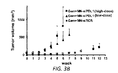
FIGs. 3A-D. Combination treatment with gemcitabine and MN-siPDLl. A.
Representative color-coded T2-weighted MR images during the course of
treatment.
B. Change in tumor volume during treatment. The response was significantly
different
between the high-dose active MN-siPDL1 and inactive MN-siSCR therapeutic in
combination with gemcitabine beginning as early as week 2. In the low-dose MN-
siPDL1 group, this difference was evident after week 6. C. Kaplan-Meier
survival
analysis demonstrating improvement in survival in animals treated with MN-
siPDLl+gemcitabine vs. MN-siSCR+gemcitabine. D. Photographs of tumor-bearing
mice at week 6 demonstrating necrosis and ulceration in the control tumors.
FIGs. 4A-B. Immunofluorescence of tumors from mice treated with MN-
siPDL1 and gemcitabine. A. Representative micrographs and B. Quantitative
analysis
of fluorescence signal intensity demonstrating efficient PD-Li inhibition, T1L
recruitment and activation, Treg attenuation, and inhibition of tumor cell
proliferation.
TL: T lymphocytes; CTL: cytotoxic T lymphocytes; Treg: regulatory T cells; Ki-
67:
proliferation.
9
CA 03111562 2021-03-03
WO 2020/068398
PCT/US2019/050003
FIGs. 5A-D. Combination treatment with gemcitabine and MN-siPDLl.
Change in tumor volume during treatment for each of the treatment groups. A,
MN-
siPDL1 high dose responders; B, MN-siPDL1 high dose non-responders; C, MN-
siPDL1 low dose; D, MN-siSCR (scrambled control). The sample size for the
study
was 6. In the high dose group, there were 2 mice that did not respond, i.e.,
their tumor
volumes did not regress; their results are shown in Fig. 5B.
FIG. 6. Schematic with additional details of preparation of exemplary
nanodrug (MN-siPDL1) for the inhibition of PD-Li mRNA in cancer cells.
DETAILED DESCRIPTION
lo While they hold great promise, clinical results with checkpoint
inhibitors have
demonstrated variability in the response. Pancreatic cancer, in particular,
has proven
resistant to initial immunotherapy approaches.
Pancreatic cancer is the fourth-leading cause of cancer-related death in the
United States with an overall 5-year survival rate of only 8%.1 Surgical
resection
remains the treatment of choice for patients with resectable disease. However,
less
than 20% of the diagnosed patients qualify for curative resections,' 30% of
patients
present with regional disease, and 50% present with distal metastases' with
survival
rates of 11% and 2%, respectively.' The reasons behind such poor prognosis
have
been postulated to involve the advanced stage at the time of diagnosis,' and
resistance
to standard chemotherapies.' There are multiple factors that are conceived to
confer
chemo-resistance: the formation of desmoplastic stroma limiting drug delivery,
the
activation of pancreatic stellate cells by reactive oxygen species, cytokines,
and/or
growth factors, and activated stellate cell secretion of immunosuppressive
signaling
molecules.4'5 Due to the complex tumor biology of pancreatic cancer, multiple
combination chemotherapies have emerged. As such, FOLFIRINOX (a combination
consisting of 5-fluorouracil, leucovorin, irinotecan, and oxaliplatin), and
gemcitabine/nab-paclitaxel have shown improvements in overall survival
compared to
standard gemcitabine monotherapy treatment.6'7 However, these combination
therapies are heavily dependent on the patient's overall health, and the
overall
.. survival benefit for the latest cytotoxic combination therapies is only ¨ 2-
5 months.
In pancreatic cancer, advances in checkpoint inhibitor-based therapies have
shown disappointing clinical results. In a Phase II trial anti-CTLA-4,
Ipilimumab,
monotherapy was ineffective with no responders resulting from the trial."'
Similarly,
CA 03111562 2021-03-03
WO 2020/068398
PCT/US2019/050003
in a multicenter Phase I trial an anti-PDL-1 antibody was administered
intravenously
in a variety of advanced cancer patients. Out of the 14 pancreatic cancer
patients
recruited there were no objective responses reported.'
In light of the tremendous suffering caused by this disease and the modest
progress achieved thus far with cytotoxic treatments, it is clear that we need
to
explore radical, transformative approaches for therapy that attack the disease
from
multiple angles.
The last decade has seen tremendous progress in the field of cancer
immunotherapy. In fact, immunotherapy represents the most promising new cancer
treatment approach since the development of the first chemotherapies in the
1940s.
Checkpoint inhibitors have worked against lethal cancers such as melanoma and
some
lung cancers ¨ sometimes with dramatic success ¨ and are being tested in
dozens of
other cancer types.8'9 But pancreatic cancer has proven difficult to treat
with
conventional drugs and has been resistant to initial immunotherapy
approaches. Partly, the reason for this is the complex tumor microenvironment
that
characterizes pancreatic adenocarcinoma. Chiefly, the presence of desmoplastic
tumor
stroma that is both immunosuppressive in nature and a physical barrier for
antibody
and T lymphocytes infiltration.' Consequently, it is important to design
alternative
approaches that combine:
innovative checkpoint inhibitors that can be delivered efficiently to tumor
cells
and tumor resident macrophages, and strategies that enhance the permeation of
the
tumor by T lymphocytes.
Presented herein is an alternative strategy that relies on combining
chemotherapeutics, e.g., gemcitabine (Gem), 5-FLOROURACIL, FOLFIRINOX, and
a novel immune checkpoint inhibitors, e.g., PD-L1 inhibitor (termed MN-
siPDL1),
that incorporate a nanoparticle carrier that is delivered with high efficiency
to tumor
cells in vivo 11-19, where it post-transcriptionally inhibits immune
checkpoint
molecules, e.g., PD-L1, expression on tumor cells via the RNA interference
mechanism. The approach is advantageous over small molecules or antibodies
because the siRNA component inhibits the target antigen at the post-
transcriptional
level and not at the protein level. Also, the RNAi mechanism is catalytic and
necessitates the delivery of only picomolar amounts of siRNA to the tumor cell
for the
abolition of the target antigen. By contrast, small molecules or antibodies
require the
11
CA 03111562 2021-03-03
WO 2020/068398
PCT/US2019/050003
achievement of at least a 1:1 molar ratio of antigen to therapeutic molecule
and could
be ineffective in the presence of a compensatory increase in the expression of
the
target antigen by the tumor cell.
In the current study, 7 weeks of combination therapy consisting of gemcitabine
and MN-siPDL1 were administered in a syngeneic murine pancreatic cancer model.
This approach resulted in significantly lower morbidity and toxicity, leading
to tumor
regression and a dramatic improvement in survival. In particular, following
dose
optimization, a 90% reduction in tumor volume was achieved 3 weeks after the
beginning of treatment. Whereas 100% of the control animals had succumbed to
their
tumors by week 6 after the beginning of treatment, there was no mortality in
the
experimental group by week 5, and 67% of the experimental animals survived for
12
weeks.
The described methodology represents an integrated tool for drug delivery,
image guidance of the delivery process, and a synchronous biomarker of
therapeutic
response.
Compositions
Provided herein are therapeutic nanoparticles that have a diameter of between
about 2 nm to about 200 nm (e.g., between about 10 nm to about 30 nm, between
about 5 nm to about 25 nm, between about 10 nm to about 25 nm, between about
15
nm to about 25 nm, between about 20 nm and about 25 nm, between about 25 nm to
about 50 nm, between about 50 nm and about 200 nm, between about 70 nm and
about 200 nm, between about 80 nm and about 200 nm, between about 100 nm and
about 200 nm, between about 140 nm to about 200 nm, and between about 150 nm
to
about 200 nm), and contain a polymer coating, and a nucleic acid containing at
least
10 (e.g., at least 11, 12, 13, 14, 15, 16, 17, 18, 19, 20, or 21) contiguous
nucleotides
within a sequence that is complementary to a human immune checkpoint molecule,
e.g., PD-L1, PD-1, CTLA-4 (Cytotoxic T-Lymphocyte-Associated Protein-4;
CD152);
LAG-3 (Lymphocyte Activation Gene 3; CD223); TIM-3 (T-cell Immunoglobulin
domain and Mucin domain 3, HAVCR2); TIGIT (T-cell Immunoreceptor with Ig and
ITIM domains); B7-H3 (CD276); VSIR (V-set immunoregulatory receptor, aka
VISTA, B7H5, Cl0orf54); BTLA (B- and T-Lymphocyte Attenuator, CD272); GARP
(Glycoprotein A Repetitions Predominant; PVRIG (PVR related immunoglobulin
12
CA 03111562 2021-03-03
WO 2020/068398
PCT/US2019/050003
domain containing); or VTCN1 (V-set domain containing T cell activation
inhibitor 1,
aka B7-H4).
PD-Li
PD-Li is also known as CD274, B7-H; B7H1; PDL1; PDCD1L1; and
PDCD1LG1. Exemplary sequences for human PD-Li are provided at the NCBI
GenBank Acc. Nos. shown in Table A.
Table A ¨ Exemplary sequences for human PD-Li
Nucleic Acid Notes Protein Notes
NM 014143.4 Variant 1 NP 054862.1 isoform a
precursor
NM 001267706.1 Variant 2 NP 001254635.1 isoform b
precursor
NM 001314029.1 Variant 4 NP 001300958.1 isoform c
precursor
According to the NCBI reference notes, Variant 1 is the longest transcript and
encodes the longest isoform (isoform a precursor). Variant 2 lacks an
alternate in-
frame exon in the 5' coding region, compared to variant 1, resulting in the
shorter
protein isoform b. Variant 4 lacks several exons and its 3' terminal exon
extends past a
splice site that is used in variant 1, resulting in a novel 3' coding region
and novel 3'
UTR compared to variant 1, and encodes isoform c (which is shorter than and
has a
distinct C-terminus compared to isoform a).
In the present methods and compositions, siRNA targeting any or all of the
above (e.g., targeting a region that is common to all three of the above) can
be used.
The sequence of the human variant 1 mRNA is as follows:
1 agttctgcgc agcttcccga ggctccgcac cagccgcgct tctgtccgcc tgcagggcat
61 tccagaaaga tgaggatatt tgctgtcttt atattcatga cctactggca tttgctgaac
121 gcatttactg tcacggttcc caaggaccta tatgtggtag agtatggtag caatatgaca
181 attgaatgca aattcccagt agaaaaacaa ttagacctgg ctgcactaat tgtctattgg
241 gaaatggagg ataagaacat tattcaattt gtgcatggag aggaagacct gaaggttcag
301 catagtagct acagacagag ggcccggctg ttgaaggacc agctctccct gggaaatgct
361 gcacttcaga tcacagatgt gaaattgcag gatgcagggg tgtaccgctg catgatcagc
421 tatggtggtg ccgactacaa gcgaattact gtgaaagtca atgccccata caacaaaatc
481 aaccaaagaa ttttggttgt ggatccagtc acctctgaac atgaactgac atgtcaggct
541 gagggctacc ccaaggccga agtcatctgg acaagcagtg accatcaagt cctgagtggt
601 aagaccacca ccaccaattc caagagagag gagaagcttt tcaatgtgac cagcacactg
661 agaatcaaca caacaactaa tgagattttc tactgcactt ttaggagatt agatcctgag
721 gaaaaccata cagctgaatt ggtcatccca gaactacctc tggcacatcc tccaaatgaa
781 aggactcact tggtaattct gggagccatc ttattatgcc ttggtgtagc actgacattc
841 atcttccgtt taagaaaagg gagaatgatg gatgtgaaaa aatgtggcat ccaagataca
901 aactcaaaga agcaaagtga tacacatttg gaggagacgt aatccagcat tggaacttct
961 gatcttcaag cagggattct caacctgtgg tttaggggtt catcggggct gagcgtgaca
1021 agaggaagga atgggcccgt gggatgcagg caatgtggga cttaaaaggc ccaagcactg
13
CA 03111562 2021-03-03
WO 2020/068398
PCT/US2019/050003
1081 aaaatggaac ctggcgaaag cagaggagga gaatgaagaa agatggagtc aaacagggag
1141 cctggaggga gaccttgata ctttcaaatg cctgaggggc tcatcgacgc ctgtgacagg
1201 gagaaaggat acttctgaac aaggagcctc caagcaaatc atccattgct catcctagga
1261 agacgggttg agaatcccta atttgagggt cagttcctgc agaagtgccc tttgcctcca
1321 ctcaatgcct caatttgttt tctgcatgac tgagagtctc agtgttggaa cgggacagta
1381 tttatgtatg agtttttcct atttattttg agtctgtgag gtcttcttgt catgtgagtg
1441 tggttgtgaa tgatttcttt tgaagatata ttgtagtaga tgttacaatt ttgtcgccaa
1501 actaaacttg ctgcttaatg atttgctcac atctagtaaa acatggagta tttgtaaggt
1561 gcttggtctc ctctataact acaagtatac attggaagca taaagatcaa accgttggtt
1621 gcataggatg tcacctttat ttaacccatt aatactctgg ttgacctaat cttattctca
1681 gacctcaagt gtctgtgcag tatctgttcc atttaaatat cagctttaca attatgtggt
1741 agcctacaca cataatctca tttcatcgct gtaaccaccc tgttgtgata accactatta
1801 ttttacccat cgtacagctg aggaagcaaa cagattaagt aacttgccca aaccagtaaa
1861 tagcagacct cagactgcca cccactgtcc ttttataata caatttacag ctatatttta
1921 ctttaagcaa ttcttttatt caaaaaccat ttattaagtg cccttgcaat atcaatcgct
1981 gtgccaggca ttgaatctac agatgtgagc aagacaaagt acctgtcctc aaggagctca
2041 tagtataatg aggagattaa caagaaaatg tattattaca atttagtcca gtgtcatagc
2101 ataaggatga tgcgagggga aaacccgagc agtgttgcca agaggaggaa ataggccaat
2161 gtggtctggg acggttggat atacttaaac atcttaataa tcagagtaat tttcatttac
2221 aaagagaggt cggtacttaa aataaccctg aaaaataaca ctggaattcc ttttctagca
2281 ttatatttat tcctgatttg cctttgccat ataatctaat gcttgtttat atagtgtctg
2341 gtattgttta acagttctgt cttttctatt taaatgccac taaattttaa attcatacct
2401 ttccatgatt caaaattcaa aagatcccat gggagatggt tggaaaatct ccacttcatc
2461 ctccaagcca ttcaagtttc ctttccagaa gcaactgcta ctgcctttca ttcatatgtt
2521 cttctaaaga tagtctacat ttggaaatgt atgttaaaag cacgtatttt taaaattttt
2581 ttcctaaata gtaacacatt gtatgtctgc tgtgtacttt gctattttta tttattttag
2641 tgtttcttat atagcagatg gaatgaattt gaagttccca gggctgagga tccatgcctt
2701 ctttgtttct aagttatctt tcccatagct tttcattatc tttcatatga tccagtatat
2761 gttaaatatg tcctacatat acatttagac aaccaccatt tgttaagtat ttgctctagg
2821 acagagtttg gatttgttta tgtttgctca aaaggagacc catgggctct ccagggtgca
2881 ctgagtcaat ctagtcctaa aaagcaatct tattattaac tctgtatgac agaatcatgt
2941 ctggaacttt tgttttctgc tttctgtcaa gtataaactt cactttgatg ctgtacttgc
3001 aaaatcacat tttctttctg gaaattccgg cagtgtacct tgactgctag ctaccctgtg
3061 ccagaaaagc ctcattcgtt gtgcttgaac ccttgaatgc caccagctgt catcactaca
3121 cagccctcct aagaggcttc ctggaggttt cgagattcag atgccctggg agatcccaga
3181 gtttcctttc cctcttggcc atattctggt gtcaatgaca aggagtacct tggctttgcc
3241 acatgtcaag gctgaagaaa cagtgtctcc aacagagctc cttgtgttat ctgtttgtac
3301 atgtgcattt gtacagtaat tggtgtgaca gtgttctttg tgtgaattac aggcaagaat
3361 tgtggctgag caaggcacat agtctactca gtctattcct aagtcctaac tcctccttgt
3421 ggtgttggat ttgtaaggca ctttatccct tttgtctcat gtttcatcgt aaatggcata
3481 ggcagagatg atacctaatt ctgcatttga ttgtcacttt ttgtacctgc attaatttaa
3541 taaaatattc ttatttattt tgttacttgg tacaccagca tgtccatttt cttgtttatt
3601 ttgtgtttaa taaaatgttc agtttaacat ccca (SEQ ID
NO: 1)
Table B ¨ Exemplary sequences for other human immune checkpoint molecules
immune Nucleic Acid NCBI Notes Protein NCBI Notes
checkpoint RefSeq ID RefSeq ID
molecule
PD-1 NM 005018.3 NP 005009.2
CD40 NM 001250.5 Variant 1 NP 001241.1 Isoform 1
NM 152854.3 Variant 2 NP 690593.1 Isoform 2
NM 001322422.1 Variant 5 NP 001309351.1 Isoform 5
NM 001322421.1 Variant 4 NP 001309350.1 Isoform 4
NM 001302753.1 Variant 3 NP 001289682.1 Isoform 3
NM 001362758.1 Variant 6 NP 001349687.1 Isoform 6
14
CA 03111562 2021-03-03
WO 2020/068398
PCT/US2019/050003
CTLA-4 NM 005214.5 NP 005205.2
Tim3 NM 032782.5 NP 116171.3
Lag3 NM 002286.6 NP 002277.4
TIGIT NM 173799.4 NP 776160.2
B7-H3 NM 001024736.2 Variant 1 NP 001019907.1 Isoform a
NM 001329628.1 Variant 3 NP 001316557.1 Isoform b
NM 001329629.1 Variant 4 NP 001316558.1 Isoform c
NM 025240.2 Variant 2 NP 079516.1 Isoform b
VSIR/VISTA NM 022153.2 NP 071436.1
VTCN1/ NM 024626.4 Variant 1 NP 078902.2 Isoform 1
B7-H4 NM 001253849.1 Variant 2 NP 001240778.1 Isoform 2
NM 001253850.1 Variant 3 NP 001240779.1 Isoform 3
PVRIG XM 011516575.2 XP 011514877.1
GARP NM 005512.2 Variant 1 NP 005503.1 Isoform 1
NM 001128922.2 Variant 2 NP 001122394.1 Isoform 2
BTLA NM 181780.4 Variant 1 NP 861445.4 Isoform 1
NM 001085357.1 Variant 2 NP 001078826.1 Isoform 2
Although the present methods exemplify human subjects, other mammalian
subjects,
e.g., veterinary subjects such as cats, dogs, horses, pigs and sheep, can also
be treated
using the present methods. In preferred embodiments, the nucleic acid targets
a
sequence from the same species as the subject to be treated.
In some embodiments, the therapeutic nanoparticles provided herein can be
spherical or ellipsoidal, or can have an amorphous shape. In some embodiments,
the
therapeutic nanoparticles provided herein can have a diameter (between any two
points on the exterior surface of the therapeutic nanoparticle) of between
about 2 nm
to about 200 nm (e.g., between about 10 nm to about 200 nm, between about 2 nm
to
about 30 nm, between about 5 nm to about 25 nm, between about 10 nm to about
25
nm, between about 15 nm to about 25 nm, between about 20 nm to about 25 nm,
between about 50 nm to about 200 nm, between about 70 nm to about 200 nm,
between about 80 nm to about 200 nm, between about 100 nm to about 200 nm,
between about 140 nm to about 200 nm, and between about 150 nm to about 200
nm).
In some embodiments, therapeutic nanoparticles having a diameter of between
about
2 nm to about 30 nm localize to the lymph nodes in a subject. In some
embodiments,
therapeutic nanoparticles having a diameter of between about 40 nm to about
200 nm
localize to the liver.
In some embodiments, the compositions can contain a mixture of two or more
of the different therapeutic nanoparticles described herein. In some
embodiments, the
compositions contain at least one therapeutic nanoparticle containing at least
10
CA 03111562 2021-03-03
WO 2020/068398
PCT/US2019/050003
contiguous nucleotides within the target sequence covalently linked to the
nanoparticle (a nanoparticle for decrease miR-10b levels in a target cell),
and at least
one therapeutic nanoparticle containing a sequence that is complementary to a
sequence of at least 10 other contiguous nucleotides present within a
sequence,
In some embodiments, the therapeutic nanoparticles can be magnetic (e.g.,
contain a core of a magnetic material).
Nanoparticles
In some embodiments, any of the therapeutic nanoparticles described herein
can contain a core of a magnetic material (e.g., a therapeutic magnetic
nanoparticle).
In some embodiments, the magnetic material or particle can contain a
diamagnetic,
paramagnetic, superparamagnetic, or ferromagnetic material that is responsive
to a
magnetic field. Non-limiting examples of therapeutic magnetic nanoparticles
contain
a core of a magnetic material containing a metal oxide selected from the group
of:
magnetite; ferrites (e.g., ferrites of manganese, cobalt, and nickel); Fe(II)
oxides, and
hematite, and metal alloys thereof. The core of magnetic material can be
formed by
converting metal salts to metal oxides using methods known in the art (e.g.,
Kieslich
et al., Inorg. Chem. 2011). In some embodiments, the nanoparticles contain
cyclodextrin gold or quantum dots. Non-limiting examples of methods that can
be
used to generate therapeutic magnetic nanoparticles are described in Medarova
et al.,
Methods Mol. Biol. 555:1-13, 2009; and Medarova et al., Nature Protocols 1:429-
431,
2006. Additional magnetic materials and methods of making magnetic materials
are
known in the art. In some embodiments of the methods described herein, the
position
or localization of therapeutic magnetic nanoparticles can be imaged in a
subject (e.g.,
imaged in a subject following the administration of one or more doses of a
therapeutic
magnetic nanoparticle).
In some embodiments, the therapeutic nanoparticles described herein do not
contain a magnetic material. In some embodiments, a therapeutic nanoparticle
can
contain, in part, a core of containing a polymer (e.g., poly(lactic-co-
glycolic acid)).
Skilled practitioners will appreciated that any number of art known materials
can be
used to prepare nanoparticles, including, but are not limited to, gums (e.g.,
Acacia,
Guar), chitosan, gelatin, sodium alginate, and albumin. Additional polymers
that can
be used to generate the therapeutic nanoparticles described herein are known
in the
16
CA 03111562 2021-03-03
WO 2020/068398
PCT/US2019/050003
art. For example, polymers that can be used to generate the therapeutic
nanoparticles
include, but are not limited to, cellulosics, poly(2-hydroxy ethyl
methacrylate),
poly(N-vinyl pyrrolidone), poly(methyl methacrylate), poly(vinyl alcohol),
poly(acrylic acid), polyacrylamide, poly(ethylene-co-vinyl acetate),
poly(ethylene
glycol), poly(methacrylic acid), polylactides (PLA), polyglycolides (PGA),
poly(lactide-co-glycolides) (PLGA), polyanhydrides, polyorthoesters,
polycyanoacrylate and polycaprolactone.
Skilled practitioners will appreciate that the material used in the
composition
of the nanoparticles, the methods for preparing, coating, and methods for
controlling
lo the size of the nanoparticles can vary substantially. However, these
methods are well
known to those in the art. Key issues include the biodegradability, toxicity
profile, and
pharmacokinetics/pharmacodynamics of the nanoparticles. The composition and/or
size of the nanoparticles are key determinants of their biological fate. For
example,
larger nanoparticles are typically taken up and degraded by the liver, whereas
smaller
nanoparticles (<30 nm in diameter) typically circulate for a long time
(sometimes over
24-hr blood half-life in humans) and accumulate in lymph nodes and the
interstitium
of organs with hyperpermeable vasculature, such as tumors.
Polymer Coatings
The therapeutic nanoparticles described herein contain a polymer coating over
the core magnetic material (e.g., over the surface of a magnetic material).
The
polymer material can be suitable for attaching or coupling one or more
biological
agents (e.g., such as any of the nucleic acids, fluorophores, or targeting
peptides
described herein). One of more biological agents (e.g., a nucleic acid,
fluorophore, or
targeting peptide) can be fixed to the polymer coating by chemical coupling
(covalent
bonds).
In some embodiments, the therapeutic nanoparticles are formed by a method
that includes coating the core of magnetic material with a polymer that is
relatively
stable in water. In some embodiments, the therapeutic nanoparticles are formed
by a
method that includes coating a magnetic material with a polymer or absorbing
the
magnetic material into a thermoplastic polymer resin having reducing groups
thereon.
A coating can also be applied to a magnetic material using the methods
described in
U.S. Pat. Nos. 5,834,121, 5,395,688, 5,356,713, 5,318,797, 5,283,079,
5,232,789,
17
CA 03111562 2021-03-03
WO 2020/068398
PCT/US2019/050003
5,091,206, 4,965,007, 4,774,265, 4,770,183, 4,654,267, 4,554,088, 4,490,436,
4,336,173, and 4,421,660; and WO 10/111066 (each disclosure of which is
incorporated herein by reference).
Method for the synthesis of iron oxide nanoparticles include, for example,
physical and chemical methods. For example, iron oxides can be prepared by co-
precipitation of Fe2+ and Fe3+ salts in an aqueous solution. The resulting
core
consists of magnetite (Fe304), maghemite (-y-Fe2O3) or a mixture of the two.
The
anionic salt content (chlorides, nitrates, sulphates etc), the Fe2+ and Fe3+
ratio, pH
and the ionic strength in the aqueous solution all play a role in controlling
the size. It
is important to prevent the oxidation of the synthesized nanoparticles and
protect their
magnetic properties by carrying out the reaction in an oxygen free environment
under
inert gas such as nitrogen or argon. The coating materials can be added during
the co-
precipitation process in order to prevent the agglomeration of the iron oxide
nanoparticles into microparticles. Skilled practitioners will appreciated that
any
number of art known surface coating materials can be used for stabilizing iron
oxide
nanoparticles, among which are synthetic and natural polymers, such as, for
example,
polyethylene glycol (PEG), dextran, polyvinylpyrrolidone (PVP), fatty acids,
polypeptides, chitosin, gelatin.
For example, U.S. Pat. No. 4,421,660 note that polymer coated particles of an
inorganic material are conventionally prepared by (1) treating the inorganic
solid with
acid, a combination of acid and base, alcohol or a polymer solution; (2)
dispersing an
addition polymerizable monomer in an aqueous dispersion of a treated inorganic
solid
and (3) subjecting the resulting dispersion to emulsion polymerization
conditions.
(col. 1, lines 21-27) U.S. Pat. No. 4,421,660 also discloses a method for
coating an
inorganic nanoparticles with a polymer, which comprises the steps of (1)
emulsifying
a hydrophobic, emulsion polymerizable monomer in an aqueous colloidal
dispersion
of discrete particles of an inorganic solid and (2) subjecting the resulting
emulsion to
emulsion polymerization conditions to form a stable, fluid aqueous colloidal
dispersion of the inorganic solid particles dispersed in a matrix of a water-
insoluble
polymer of the hydrophobic monomer (col. 1, lines 42-50).
Alternatively, polymer-coated magnetic material can be obtained
commercially that meets the starting requirements of size. For example,
commercially
available ultrasmall superparamagnetic iron oxide nanoparticles include
NC100150
18
CA 03111562 2021-03-03
WO 2020/068398
PCT/US2019/050003
Injection (Nycomed Amersham, Amersham Health) and Ferumoxytol (AMAG
Pharmaceuticals, Inc.).
Suitable polymers that can be used to coat the core of magnetic material
include without limitation: polystyrenes, polyacrylamides, polyetherurethanes,
polysulfones, fluorinated or chlorinated polymers such as polyvinyl chloride,
polyethylenes, and polypropylenes, polycarbonates, and polyesters. Additional
examples of polymers that can be used to coat the core of magnetic material
include
polyolefins, such as polybutadiene, polydichlorobutadiene, polyisoprene,
polychloroprene, polyvinylidene halides, polyvinylidene carbonate, and
lo polyfluorinated ethylenes. A number of copolymers, including
styrene/butadiene,
alpha-methyl styrene/dimethyl siloxane, or other polysiloxanes can also be
used to
coat the core of magnetic material (e.g., polydimethyl siloxane,
polyphenylmethyl
siloxane, and polytrifluoropropylmethyl siloxane). Additional polymers that
can be
used to coat the core of magnetic material include polyacrylonitriles or
acrylonitrile-
containing polymers, such as poly alpha-acrylanitrile copolymers, alkyd or
terpenoid
resins, and polyalkylene polysulfonates. In some embodiments, the polymer
coating
is dextran.
Nucleic Acids
The therapeutic nanoparticles provided contain at least one nucleic acid
comprising a sequence that is complementary at least 10 (e.g., at least 11,
12, 13, 14,
15, 16, 17, 18, 19, 20, 21, or 22) contiguous nucleotides within a sequence of
an
immune checkpoint molecule, e.g., a PD-Li sequence, e.g., SEQ ID NO: 1, that
is
covalently-linked to the nanoparticle. In some embodiments, the covalently-
linked
nucleic acid molecule contains a sequence that is complementary to all or part
of an
mRNA encoding an immune checkpoint protein (e.g., any of the immune checkpoint
proteins described herein). For example, the covalently-linked nucleic acid
can be
complementary to all or part of a non-coding region of the coding strand of a
nucleotide sequence encoding an immune checkpoint protein (e.g., any of the
immune
checkpoint proteins described herein). Non-coding regions ("5' and 3'
untranslated
regions") are the 5' and 3' sequences that flank the coding region in a gene
and are not
translated into amino acids. In some embodiments, the nucleic acid covalently-
linked
to the therapeutic nanoparticle is complementary to the translational start
codon or a
19
CA 03111562 2021-03-03
WO 2020/068398
PCT/US2019/050003
sequence encoding amino acids 1 to 5 of an immune checkpoint protein (e.g.,
any of
the immune checkpoint proteins described herein).
The attached nucleic acid can be single-stranded or double-stranded. In some
embodiments, the nucleic acid has a total length of between 23 nucleotides and
50
nucleotides (e.g., between 23-30 nucleotides, between 30-40 nucleotides, and
between
40-50 nucleotides). In some embodiments, the nucleic acid can be an antisense
RNA
or siRNA.
Antisense nucleic acid molecules can be covalently linked to the therapeutic
nanoparticles described herein.
Based upon the sequences provided herein (e.g., the sequences for human
immune checkpoint molecules, e.g., PD-L1, e.g., SEQ ID NO:1 and the other
sequences in Tables A and B), one of skill in the art can easily choose and
synthesize
any of a number of appropriate antisense molecules (e.g., antisense molecules
to
target an immune checkpoint molecule, e.g., PD-L1). For example, an antisense
nucleic acid that targets PD-Li can contain a sequence complementary to at
least 10
(e.g., at least 15 or 20) contiguous nucleotides present in SEQ ID NO: 1 or a
sequence
for PD-L1 known in the art.
An anti sense nucleic acid can be constructed using chemical synthesis and
enzymatic ligation reactions using procedures known in the art. For example,
an
antisense nucleic acid (e.g., an antisense oligonucleotide) can be chemically
synthesized using naturally occurring nucleotides or modified nucleotides
(e.g., any of
the modified oligonucleotides described herein) designed to increase the
biological
stability of the molecules or to increase the physical stability of the duplex
formed
between the antisense and sense nucleic acids, e.g., phosphorothioate
derivatives and
acridine-substituted nucleotides can be used. Alternatively, the anti sense
nucleic acid
can be produced biologically using an expression vector into which a nucleic
acid has
been subcloned in an antisense orientation (i.e., RNA transcribed from the
inserted
nucleic acid will be of an antisense orientation to a target nucleic acid of
interest). In
some embodiments, the antisense nucleic acid molecules described herein can
hybridize to a target nucleic acid by conventional nucleotide
complementarities and
form a stable duplex.
An anti sense nucleic acid molecule can be an a-anomeric nucleic acid
molecule. An a-anomeric nucleic acid molecule forms specific double-stranded
CA 03111562 2021-03-03
WO 2020/068398
PCT/US2019/050003
hybrids with complementary RNA in which, contrary to the usual n-units, the
strands
run parallel to each other (Gaultier et al., Nucleic Acids Res. 15:6625-6641,
1987).
The antisense nucleic acid molecule can also comprise a 2'-0-
methylribonucleotide
(Inoue et al., Nucleic Acids Res., 15:6131-6148, 1987) or a chimeric RNA-DNA
analog (Inoue et al., FEBS Lett. 215:327-330, 1987).
In some embodiments, the nucleic acid is a small interfering RNA (siRNA).
RNAi is a process in which RNA is degraded in host cells. To decrease
expression of
an RNA, double-stranded RNA (dsRNA) containing a sequence corresponding to a
portion of the target RNA (e.g., an immune checkpoint molecule, e.g., human PD-
L1)
is introduced into a cell. The dsRNA is digested into 21-23 nucleotide-long
duplexes
called short interfering RNAs (or siRNAs), which bind to a nuclease complex to
form
what is known as the RNA-induced silencing complex (or RISC). The RISC targets
the endogenous target RNA by base pairing interactions between one of the
siRNA
strands and the endogenous RNA. It then cleaves the endogenous RNA about 12
nucleotides from the 3' terminus of the siRNA (see Sharp et al., Genes Dev.
15:485-
490, 2001, and Hammond et al., Nature Rev. Gen. 2:110-119, 2001).
Standard molecular biology techniques can be used to generate siRNAs. Short
interfering RNAs can be chemically synthesized, recombinantly produced, e.g.,
by
expressing RNA from a template DNA, such as a plasmid, or obtained from
commercial vendors such as Dharmacon. The RNA used to mediate RNAi can
include modified nucleotides (e.g., any of the modified nucleotides described
herein),
such as phosphorothioate nucleotides. The siRNA molecules used to decrease the
levels of mature human miR-10b can vary in a number of ways. For example, they
can include a 3 hydroxyl group and strands of 21, 22, or 23 consecutive
nucleotides.
They can be blunt ended or include an overhanging end at either the 3' end,
the 5' end,
or both ends. For example, at least one strand of the RNA molecule can have a
3'
overhang from about 1 to about 6 nucleotides (e.g., 1-5, 1-3, 2-4 or 3-5
nucleotides
(whether pyrimidine or purine nucleotides) in length. Where both strands
include an
overhang, the length of the overhangs may be the same or different for each
strand.
To further enhance the stability of the RNA duplexes, the 3' overhangs can be
stabilized against degradation (by, e.g., including purine nucleotides, such
as
adenosine or guanosine nucleotides, or replacing pyrimidine nucleotides with
modified nucleotides (e.g., substitution of uridine two-nucleotide 3'
overhangs by 2'-
21
CA 03111562 2021-03-03
WO 2020/068398
PCT/US2019/050003
deoxythymidine is tolerated and does not affect the efficiency of RNAi). Any
siRNA
can be used provided it has sufficient homology to the target of interest.
There is no
upper limit on the length of the siRNA that can be used (e.g., the siRNA can
range
from about 21-50, 50-100, 100-250, 250-500, or 500-1000 base pairs).
In some embodiments, the nucleic acid molecule can contain at least one
modified nucleotide (a nucleotide containing a modified base or sugar). In
some
embodiments, the nucleic acid molecule can contain at least one modification
in the
phosphate (phosphodiester) backbone. The introduction of these modifications
can
increase the stability, or improve the hybridization or solubility of the
nucleic acid
molecule.
The molecules described herein can contain one or more (e.g., two, three,
four, of
five) modified nucleotides. The modified nucleotides can contain a modified
base or
a modified sugar. Non-limiting examples of modified bases include: 8-oxo-N6-
methyladenine, 7-deazaxanthine, 7-deazaguanine, N4, N4-ethanocytosin, N6, N6-
ethano-2,6-diaminopurine, 5-(C3-C6)-alkynyl-cytosine, pseudoisocytosine, 2-
hydroxy-5-methy1-4-triazolopyridin, isocytosine, isoguanine, 5-fluorouracil, 5-
bromouracil, 5-chlorouracil, 5-iodouracil, hypoxanthine, xanthine, 4-
acetylcytosine,
5-(carboxyhydroxylmethyl) uracil, 5-carboxymethylaminomethy1-2-thiouridine, 5-
carboxymethylaminomethyluracil, dihydrouracil, beta-D-galactosylqueosine,
inosine,
N6-isopentenyladenine, 1-methylguanine, 1-methylinosine, 2,2-dimethylguanine,
2-
methyladenine, 2-methylguanine, 3-methylcytosine, 5-methylcytosine, N6-
adenine, 7-
methylguanine, 5-methylaminomethyluracil, 5-methoxyaminomethy1-2-thiouracil,
beta-D-mannosylqueosine, 5'-methoxycarboxymethyluracil, 5-methoxyuracil, 2-
methylthio-N6-isopentenyladenine, uracil-5-oxyacetic acid (v), wybutoxosine,
pseudouracil, queosine, 2-thiocytosine, 5-methyl-2-thiouracil, 2-thiouracil, 4-
thiouracil, 5-methyluracil, uracil-5-oxyacetic acid methylester, uracil-5-
oxyacetic acid
(v), 5-methyl-2-thiouracil, 3-(3-amino-3-N-2-carboxypropyl) uracil, (acp3)w,
and 2,6-
diaminopurine.
Additional non-limiting examples of modified bases include those nucleobases
described in U.S. Pat. Nos. 5,432,272 and 3,687,808 (herein incorporated by
reference), Freier et al., Nucleic Acid Res. 25:4429-4443, 1997; Sanghvi,
Antisense
Research and Application, Chapter 15, Ed. S. T. Crooke and B. Lebleu, CRC
Press,
1993; Englisch, et al., Angewandte Chemie 30:613-722, 1991, Kroschwitz,
Concise
22
CA 03111562 2021-03-03
WO 2020/068398
PCT/US2019/050003
Encyclopedia of Polymer Science and Engineering, John Wiley & Sons, pp. 858-
859,
1990; and Cook, Anti-Cancer Drug Design 6:585-607, 1991. Additional non-
limiting examples of modified bases include universal bases (e.g., 3-
nitropyrole and
5-nitroindole). Other modified bases include pyrene and pyridyloxazole
derivatives,
pyrenyl, pyrenylmethylglycerol derivatives, and the like. Other preferred
universal
bases include pyrrole, diazole, or triazole derivatives, including those
universal bases
known in the art.
In some embodiments, the modified nucleotide can contain a modification in
its sugar moiety. Non-limiting examples of modified nucleotides that contain a
modified sugar are locked nucleotides (LNAs). LNA monomers are described in WO
99/14226 and U.S. Patent Application Publications Nos. 20110076675,
20100286044,
20100279895, 20100267018, 20100261175, 20100035968, 20090286753,
20090023594, 20080096191, 20030092905, 20020128381, and 20020115080 (herein
incorporated by reference). Additional non-limiting examples of LNAs are
disclosed
in U.S. Patent No. 6,043,060, U.S. Patent No. 6,268,490, WO 01/07455, WO
01/00641, WO 98/39352, WO 00/56746, WO 00/56748, and WO 00/66604 (herein
incorporated by reference), as well as in Morita et al., Bioorg. Med. Chem.
Lett.
12(1):73-76, 2002; Hakansson et al., Bioorg. Med. Chem. Lett. 11(7):935-938,
2001;
Koshkin et al., I Org. Chem. 66(25):8504-8512, 2001; Kvaerno et al., J. Org.
Chem.
66(16):5498-5503, 2001; Hakansson et al., J. Org. Chem. 65(17):5161-5166,
2000;
Kvaerno et al., J. Org. Chem. 65(17):5167-5176, 2000; Pfundheller et al.,
Nucleosides
Nucleotides 18(9):2017-2030, 1999; and Kumar et al., Bioorg. Med. Chem. Lett.
8(16):2219-2222, 1998. In some embodiments, the modified nucleotide is an oxy-
LNA monomer, such as those described in WO 03/020739.
Modified nucleotides can also include antagomirs (2'-0-methyl-modified,
cholesterol-conjugated single stranded RNA analogs); ALN (0 -L-LNA); ADA (2'-N-
adamantylmethylcarbony1-2'-amino-LNA); PYR (2'-N-pyreny1-1-methyl-2'-amino-
LNA); OX (oxetane-LNA); ENA (2'-O, 4"-C-ethylene bridged nucleic acid); AENA
(2'-deoxy-2'-N, 4' -C-ethylene-LNA); CLNA (2',4'-carbocyclic-LNA); and CENA
(2',4'-carbocyclic-ENA); HM-modified DNAs (4' -C-hydroxymethyl-DNA); 2' -
substituted RNAs (with 2'-0-methyl, 2' -fluoro, 2'-aminoethoxymethyl, 2'-
aminopropoxymethyl, 2' -aminoethyl, 2'-guanidinoethyl, 2' -cyanoethyl, 2'-
aminopropyl); and RNAs with radical modifications of the ribose sugar ring,
such as
23
CA 03111562 2021-03-03
WO 2020/068398
PCT/US2019/050003
Unlocked Nucleic Acid (UNA), Altritol Nucleic Acid (ANA) and Hexitol Nucleic
Acid (HNA) (see, Bramsen et al., Nucleic Acids Res. 37:2867-81, 2009).
The molecules described herein can also contain a modification in the
phosphodiester backbone. For example, at least one linkage between any two
contiguous (adjoining) nucleotides in the molecule can be connected by a
moiety
containing 2 to 4 groups/atoms selected from the group of: -CH2 , 0 , S ,
-NRH-, >C=0, >C=NRH, >C=S, -Si(R")2-, -SO-, -S(0)2-, -P(0)2-, -
PO(BH3)-, -P(0,S)-, -P(S)2-, -PO(R")-, -PO(OCH3)-, and -
PO(NHRH)-, where RH is selected from hydrogen and C1.4-alkyl, and R" is
selected
from C1-6-alkyl and phenyl. Illustrative examples of such linkages are -CH2-
CH2-CH2-, -CH2-CO-CH2-, -CH2-CHOH-CH2-, -0-CH2-0-,
-0-CH2-CH2-, -0-CH2-CH= (including R5 when used as a linkage to a
succeeding monomer), -CH2-CH2-0-, -NRH-CH2-CH2-, -CH2-CH2-
NRH-, -CH2-NR"-CH2-, -0-CH2-CH2-NR"-, -NRH-00-0-, -
NRH-CO-NRH-, -NRH-C S-NR"-, -NRH-C(=NRH)-NRH-, -NRH-
CO-CH2-NR14-, -0-00-0-, -0-CO-CH2-0-, -0-CH2-00-
0-, -CH2-CO-NR"-, -0-CO-NRH-, -NRH-00-CH2-, -0-
CH2-CO-NRH-, -0-CH2-CH2-NRH-, -CH=N-0-, -CH2-NR1-
0-, -CH2-0-N= (including R5 when used as a linkage to a succeeding
monomer), -CH2-0-NR"-, -CO-NR1-CH2-,
CH2-NRH-00-, -0-NRH-CH2-, -0-NRH-, -0-CH2-S-, -S-
CH2-0-, -CH2-CH2-S-, -0-CH2-CH2-S-, -S-CH2-CH=
(including R5 when used as a linkage to a succeeding monomer), -S-CH2-CH2-,
-S-CH2-CH2-0-, -S-CH2-CH2-S-, -CH2-S-CH2-, -CH2-
SO-CH2-, -CH2-502-CH2-, -0-S0-0-, -0-S(0)2-0-, -0-
S(0)2-CH2-, -0-S(0)2-NRIT-, -NRH-S(0)2-CH2-, -0-S(0)2-
CH2-, -0-P(0)2-0-, -0-P(0, S)-0-, -0-P(S)2-0-, -S-P(0)2-
0-, -S-P(0, S)-0-, -S-P(S)2-0-, -0-P(0, S)-S-, -0-P(S)2-S-
, -S-P(0)2-S-, -S-P(0, S)-S-, -S-P(S)2-S-, -0-PO(R")-0-, -
0-PO(OCH3)-0-, -0-PO-(OCH2CH3)-0-, -0-PO(OCH2S-R)-0-
, -0-PO(BH3)-0-, -0-PO(NHRN)-0-, -0-P(0)2-NRH-, -NRH-
P(0)2-0-, -0-P(O,NRH)2-0-, -CH2-P(0)2-0-, -0-P(0)2-CH2-,
and -0-Si(R")2-0-; among which -CH2-CO-NRH-,
24
CA 03111562 2021-03-03
WO 2020/068398
PCT/US2019/050003
¨S¨CH2-0¨, ¨0¨P(0)2-0¨, ¨0¨P(0, S)-0¨, ¨0¨P(S)2-0¨, ¨
NRH¨P(0)2-0¨, ¨0¨P(O,NRH)-0¨, ¨0¨PO(R")-0¨, ¨0¨
PO(CH3)-0¨, and ¨0-130(1\THRN)-0¨, where RH is selected form hydrogen
and CI-4-alkyl, and R" is selected from CI-6-alkyl and phenyl. Further
illustrative
examples are given in Mesmaeker et. al., Curr. Opin. Struct. Biol. 5:343-355,
1995;
and Freier et al., Nucleic Acids Research 25:4429-43, 1997. The left-hand side
of the
inter-nucleoside linkage is bound to the 5-membered ring as substituent P* at
the 3'-
position, whereas the right-hand side is bound to the 5'-position of a
preceding
monomer.
lo In some embodiments, the deoxyribose phosphate backbone of the nucleic
acid can be modified to generate peptide nucleic acids (see Hyrup et al.,
Bioorganic &
Medicinal Chem. 4(1): 5-23, 1996). Peptide nucleic acids (PNAs) are nucleic
acid
mimics, e.g., DNA mimics, in which the deoxyribose phosphate backbone is
replaced
by a pseudopeptide backbone and only the four natural nucleobases are
retained. The
neutral backbone of PNAs allows for specific hybridization to DNA and RNA
under
conditions of low ionic strength. The synthesis of PNA oligomers can be
performed
using standard solid phase peptide synthesis protocols, e.g., as described in
Hyrup et
al., 1996, supra; Perry-O'Keefe et al., Proc. Natl. Acad. Sci. U.S.A. 93:14670-
675,
1996.
PNAs can be modified, e.g., to enhance their stability or cellular uptake, by
attaching
lipophilic or other helper groups to PNA, by the formation of PNA-DNA
chimeras, or
by the use of liposomes or other techniques of delivery known in the art. For
example, PNA-DNA chimeras can be generated which may combine the
advantageous properties of PNA and DNA. Such chimeras allow DNA recognition
enzymes, e.g., RNAse H, to interact with the DNA portion while the PNA portion
would provide high binding affinity and specificity. PNA-DNA chimeras can be
linked using linkers of appropriate lengths selected in terms of base
stacking, number
of bonds between the nucleobases, and orientation (Hyrup,1996, supra). The
synthesis of PNA-DNA chimeras can be performed as described in Hyrup,1996,
supra, and Finn et al., Nucleic Acids Res. 24:3357-63, 1996. For example, a
DNA
chain can be synthesized on a solid support using standard phosphoramidite
coupling
chemistry and modified nucleoside analogs. Compounds such as 544-
methoxytrityl)amino-5'-deoxy-thymidine phosphoramidite can be used as a link
CA 03111562 2021-03-03
WO 2020/068398
PCT/US2019/050003
between the PNA and the Send of DNA (Mag et al., Nucleic Acids Res., 17:5973-
88,
1989). PNA monomers are then coupled in a stepwise manner to produce a
chimeric
molecule with a 5' PNA segment and a 3' DNA segment (Finn et al., Nucleic
Acids
Res. 24:3357-63, 1996). Alternatively, chimeric molecules can be synthesized
with a
5' DNA segment and a 3' PNA segment (Peterser et al., Bioorganic Med. Chem.
Lett.
5:1119-11124, 1975).
In some embodiments, any of the nucleic acids described herein can be
modified at either the 3' or 5' end (depending on how the nucleic acid is
covalently-
linked to the therapeutic nanoparticle) by any type of modification known in
the art.
For example, either end may be capped with a protecting group, attached to a
flexible
linking group, or attached to a reactive group to aid in attachment to the
substrate
surface (the polymer coating). Non-limiting examples of 3' or 5' blocking
groups
include: 2-amino-2-oxyethyl, 2-aminobenzoyl, 4-aminobenzoyl, acetyl,
acetyloxy,
(acetylamino)methyl, 3-(9-acridinyl), tricyclo[3.3.1.1(3,7)]dec-1-yloxy, 2-
aminoethyl,
propenyl, (9-anthracenylmethoxy)carbonyl, (1,1-dmimethylpropoxy)carbonyl, (1,1-
dimethylpropoxy)carbonyl, [1-methyl-1 -[4-(phenylazo)phenyl]ethoxy] carbonyl,
bromoacetyl, (benzoylamino)methyl, (2-bromoethoxy)carbonyl,
(diphenylmethoxy)carbonyl, 1-methyl-3-oxo-3-pheny1-1-propenyl, (3-bromo-2-
nitrophenyl)thio, (1,1-dimethylethoxy)carbonyl, [[(1,1-
dimethylethoxy)carbonyl]
amino]ethyl, 2-(phenylmethoxy)phenoxy, (1=[1,1'-bipheny1]-4-y1-1-methylethoxy)
carbonyl, bromo, (4-bromophenyl)sulfonyl, 1H-benzotriazol-1-yl,
[(phenylmethyl)
thio]carbonyl, [(phenylmetyl)thio]methyl, 2-methylpropyl, 1,1-dimethylethyl,
benzoyl, diphenylmethyl, phenylmethyl, carboxyacetyl, aminocarbonyl,
chlorodifluoroacetyl, trifluoromethyl, cyclohexylcarbonyl, cycloheptyl,
cyclohexyl,
cyclohexylacetyl, chloro, carboxymethyl, cyclopentylcarbonyl, cyclopentyl,
cyclopropylmethyl, ethoxycarbonyl, ethyl, fluoro, formyl, 1-oxohexyl, iodo,
methyl,
2-methoxy-2-oxoethyl, nitro, azido, phenyl, 2-carboxybenzoyl, 4-
pyridinylmethyl, 2-
piperidinyl, propyl, 1-methylethyl, sulfo, and ethenyl. Additional examples of
5' and
3' blocking groups are known in the art. In some embodiments, the 5' or 3'
blocking
groups prevent nuclease degradation of the molecule.
The nucleic acids described herein can be synthesized using any methods
known in the art for synthesizing nucleic acids (see, e.g., Usman et al., J.
Am. Chem.
Soc. 109:7845, 1987; Scaringe et al., Nucleic Acid Res. 18:5433, 1990; Wincott
et al.,
26
CA 03111562 2021-03-03
WO 2020/068398
PCT/US2019/050003
Methods Mol. Biol. 74:59, 1997; and Milligan, Nucleic Acid Res. 21:8783,
1987).
These typically make use of common nucleic acid protecting and coupling
groups.
Synthesis can be performed on commercial equipment designed for this purpose,
e.g.,
a 394 Applied Biosystems, Inc. synthesizer, using protocols supplied by the
manufacturer. Additional methods for synthesizing the molecules described
herein
are known in the art. Alternatively, the nucleic acids can be specially
ordered from
commercial vendors that synthesize oligonucleotides.
In some embodiments, the nucleic acid is attached to the therapeutic
nanoparticle at its 5' end. In some embodiments, the nucleic acid is attached
to the
lo therapeutic nanoparticle at its 3' end. In some embodiments, the nucleic
acid is
attached to the therapeutic nanoparticle through a base present in the nucleic
acid.
In some embodiments, the nucleic acid (e.g., any of the nucleic acids
described herein) is attached to the therapeutic nanoparticle (e.g., to the
polymer
coating of the therapeutic nanoparticle) through a chemical moiety that
contains a
thioether bond or a disulfide bond. In some embodiments, the nucleic acid is
attached
to the therapeutic nanoparticle through a chemical moiety that contains an
amide
bond. Additional chemical moieties that can be used to covalently link a
nucleic acid
to a therapeutic nanoparticle are known in the art.
A variety of different methods can be used to covalently link a nucleic acid
to
a therapeutic nanoparticle. Non-limiting examples of methods that can be used
to link
a nucleic acid to a magnetic particle are described in EP 0937097; US RE41005;
Lund
et al., Nucleic Acid Res. 16:10861, 1998; Todt et al., Methods Mol. Biol.
529:81-100,
2009; Brody et al., J. Biotechnol. 74:5-13, 2000; Ghosh et al., Nucleic Acids
Res.
15:5353-5372, 1987; U.S. Patent No. 5,900,481; U.S. Patent No. 7,569,341; U.S.
Patent No. 6,995,248; U.S. Patent No. 6,818,394; U.S. Patent No. 6,811,980;
U.S.
Patent No. 5,900,481; and U.S. Patent No. 4,818,681 (each of which is
incorporated
by reference in its entirety). In some embodiments, carboiimide is used for
the end-
attachment of a nucleic acid to a therapeutic nanoparticle. In some
embodiments, the
nucleic acid is attached to the therapeutic nanoparticle through the reaction
of one of
its bases with an activated moiety present on the surface of the therapeutic
nanoparticle (e.g., the reaction of an electrophilic base with a nucleophilic
moiety on
the surface of the therapeutic nanoparticle, or the reaction of a nucleophilic
base with
a electrophilic residue on the surface of the therapeutic nanoparticle). In
some
27
CA 03111562 2021-03-03
WO 2020/068398
PCT/US2019/050003
embodiments, a 5'-NH2 modified nucleic acid is attached to a therapeutic
nanoparticle
containing CNBr-activated hydroxyl groups (see, e.g., Lund et al., supra).
Additional
methods for attaching an amino-modified nucleic acid to a therapeutic
nanoparticle
are described below. In some embodiments, a 5'-phosphate nucleic acid is
attached to
a therapeutic nanoparticle containing hydroxyl groups in the presence of a
carbodiimide (see, e.g., Lund et al., supra). Other methods of attaching a
nucleic acid
to a therapeutic nanoparticle include carboiimide-mediated attachment of a 5'-
phosphate nucleic acid to a NH2 group on a therapeutic nanoparticle, and
carboiimide-
mediated attachment of a 5'-NH2 nucleic acid to a therapeutic nanoparticle
having
carboxyl groups (see, e.g., Lund et al., supra).
In exemplary methods, a nucleic acid can be produced that contains a reactive
amine or a reactive thiol group. The amine or thiol in the nucleic acid can be
linked
to another reactive group. The two common strategies to perform this reaction
are to
link the nucleic acid to a similar reactive moiety (amine to amine or thiol to
thiol),
which is called homobifunctional linkage, or to link to the nucleic acid to an
opposite
group (amine to thiol or thiol to amine), known as heterobifunctional linkage.
Both
techniques can be used to attach a nucleic acid to a therapeutic nanoparticle
(see, for
example, Misra et al., Bioorg. Med. Chem. Lett. 18:5217-5221, 2008; Mirsa et
al.,
Anal. Biochem. 369:248-255, 2007; Mirsa et al., Bioorg. Med. Chem. Lett.
17:3749-
3753, 2007; and Choithani et al., Methods Mol Biol. 381:133-163, 2007).
Traditional attachment techniques, especially for amine groups, have relied
upon homobifunctional linkages. One of the most common techniques has been the
use of bisaldehydes such as glutaraldehyde. Disuccinimydyl suberate (DSS),
commercialized by Syngene (Frederick, MD) as synthetic nucleic acid probe
(SNAP)
technology, or the reagent p-phenylene diisothiocyanate can also be used to
generate a
covalent linkage between the nucleic acid and the therapeutic nanoparticle.
N,N'-o-
phenylenedimaleimide can be used to cross-link thiol groups. With all of the
homobifunctional cross-linking agents, the nucleic acid is initially activated
and then
added to the therapeutic nanoparticle (see, for example, Swami et al., Int. J.
Pharm.
374:125-138, 2009, Todt et al., Methods Mol. Biol. 529:81-100, 2009; and
LimanskiI,
Biofizika 51:225-235, 2006).
Heterobifunctional linkers can also be used to attach a nucleic acid to a
therapeutic nanoparticle. For example, N-succinidimidy1-3-(2-
28
CA 03111562 2021-03-03
WO 2020/068398
PCT/US2019/050003
pyridyldithio)proprionate (SPDP) initially links to a primary amine to give a
dithiol-
modified compound. This can then react with a thiol to exchange the
pyridylthiol
with the incoming thiol (see, for example, Nostrum et al., J. Control Release
15;153(1):93-102, 2011, and Berthold et al., Bioconjug. Chem. 21:1933-1938,
2010).
An alternative approach for thiol use has been a thiol-exchange reaction. If a
thiolated nucleic acid is introduced onto a disulfide therapeutic
nanoparticle, a
disulfide-exchange reaction can occur that leads to the nucleic acid being
covalently
bonded to the therapeutic nanoparticle by a disulfide bond. A multitude of
potential
cross-linking chemistries are available for the heterobifunctional cross-
linking of
amines and thiols. Generally, these procedures have been used with a thiolated
nucleotide. Reagents typically employed have been NHS (N-hydroxysuccinimide
ester), MBS (m-maleimidobenzoyl-N-succinimide ester), and SPDP (a
pyridyldisulfide-based system). The heterobifunctional linkers commonly used
rely
upon an aminated nucleic acid. Additional methods for covalently linking a
nucleic
acid to a therapeutic nanoparticle are known in the art.
Targeting peptide
In some embodiments, the therapeutic nanoparticle further contains a
covalently-linked targeting peptide, e.g., as described in W02013/016126. By
the
term "targeting peptide" is meant a peptide that is bound by a molecule (e.g.,
protein,
sugar, or lipid, or combination thereof) present in or on the plasma membrane
of a
target cell (e.g., a cancer cell). As described herein, a targeting peptide
can be
covalently linked to a secondary molecule or composition (e.g., any of the
therapeutic
nanoparticles described herein) to target the secondary molecule or
composition to a
target cell (e.g., a cancer cell). In some embodiments, a targeting peptide
that is
covalently linked to a secondary molecule or composition (e.g., any of the
therapeutic
nanoparticles described herein) results in the uptake of the secondary
molecule or
composition by the targeted cell (e.g., cellular uptake by endocytosis or
pinocytosis).
Non-limiting examples of targeting peptides are described herein. In some
embodiments, the targeting peptide contains an RGD peptide, an EPPT peptide,
NYLHNHPYGTVG (SEQ ID NO: 2), SNPFSKPYGLTV (SEQ ID NO: 3),
GLHESTFTQRRL (SEQ ID NO: 4), YPHYSLPGSSTL (SEQ ID NO: 5),
SSLEPWIIRTTSR (SEQ ID NO: 6), LPLALPRHNASV (SEQ NO: 7), or pAla-
(Arg)7-Cys (SEQ ID NO: 8). In some embodiments, the targeting peptide is
29
CA 03111562 2021-03-03
WO 2020/068398
PCT/US2019/050003
covalently linked to the nanoparticle through a chemical moiety that contains
a
disulfide bond. In some embodiments, the therapeutic nanoparticle is magnetic.
Additional examples of targeting peptides, and methods for attaching them to
the
nanoparticle, are known in the art; see, e.g., W02013/016126.
Pharmaceutical Compositions
Also provided herein are pharmaceutical compositions that contain a
therapeutic nanoparticle as described herein. Two or more (e.g., two, three,
or four)
of any of the types of therapeutic nanoparticles described herein can be
present in a
pharmaceutical composition in any combination. The pharmaceutical compositions
can be formulated in any manner known in the art.
Pharmaceutical compositions are formulated to be compatible with their
intended route of administration (e.g., intravenous, intraarterial,
intramuscular,
intradermal, subcutaneous, or intraperitoneal). The compositions can include a
sterile
diluent (e.g., sterile water or saline), a fixed oil, polyethylene glycol,
glycerine,
propylene glycol or other synthetic solvents, antibacterial or antifungal
agents such as
benzyl alcohol or methyl parabens, chlorobutanol, phenol, ascorbic acid,
thimerosal,
and the like, antioxidants such as ascorbic acid or sodium bisulfite,
chelating agents
such as ethylenediaminetetraacetic acid, buffers such as acetates, citrates,
or
phosphates, and isotonic agents such as sugars (e.g., dextrose), polyalcohols
(e.g.,
manitol or sorbitol), or salts (e.g., sodium chloride), or any combination
thereof
Liposomal suspensions can also be used as pharmaceutically acceptable carriers
(see,
e.g., U.S. Patent No. 4,522,811). Preparations of the compositions can be
formulated
and enclosed in ampules, disposable syringes, or multiple dose vials. Where
required
(as in, for example, injectable formulations), proper fluidity can be
maintained by, for
example, the use of a coating such as lecithin, or a surfactant. Absorption of
the
therapeutic nanoparticles can be prolonged by including an agent that delays
absorption (e.g., aluminum monostearate and gelatin). Alternatively,
controlled
release can be achieved by implants and microencapsulated delivery systems,
which
can include biodegradable, biocompatible polymers (e.g., ethylene vinyl
acetate,
polyanhydrides, polyglycolic acid, collagen, polyorthoesters, and polylactic
acid; Alza
Corporation and Nova Pharmaceutical, Inc.).
Compositions containing one or more of any of the therapeutic nanoparticles
described herein can be formulated for parenteral (e.g., intravenous,
intraarterial,
CA 03111562 2021-03-03
WO 2020/068398
PCT/US2019/050003
intramuscular, intradermal, subcutaneous, or intraperitoneal) administration
in dosage
unit form (i.e., physically discrete units containing a predetermined quantity
of active
compound for ease of administration and uniformity of dosage).
Toxicity and therapeutic efficacy of compositions can be determined by
standard pharmaceutical procedures in cell cultures or experimental animals
(e.g.,
monkeys). One can, for example, determine the LD50 (the dose lethal to 50% of
the
population) and the ED50 (the dose therapeutically effective in 50% of the
population): the therapeutic index being the ratio of LD50:ED50. Agents that
exhibit
high therapeutic indices are preferred. Where an agent exhibits an undesirable
side
effect, care should be taken to minimize potential damage (i.e., reduce
unwanted side
effects). Toxicity and therapeutic efficacy can be determined by other
standard
pharmaceutical procedures.
Data obtained from cell culture assays and animal studies can be used in
formulating an appropriate dosage of any given agent for use in a subject
(e.g., a
human). A therapeutically effective amount of the one or more (e.g., one, two,
three,
or four) therapeutic nanoparticles (e.g., any of the therapeutic nanoparticles
described
herein) will be an amount that treats decreases cancer cell invasion or
metastasis in a
subject having cancer (e.g., breast cancer) in a subject (e.g., a human),
treats a
metastatic cancer in a lymph node in a subject, decreases or stabilizes
metastatic
tumor size in a lymph node in a subject, decreases the rate of metastatic
tumor growth
in a lymph node in a subject, decreases the severity, frequency, and/or
duration of one
or more symptoms of a metastatic cancer in a lymph node in a subject in a
subject
(e.g., a human), or decreases the number of symptoms of a metastatic cancer in
a
lymph node in a subject (e.g., as compared to a control subject having the
same
disease but not receiving treatment or a different treatment, or the same
subject prior
to treatment).
The effectiveness and dosing of any of the therapeutic nanoparticles described
herein can be determined by a health care professional using methods known in
the
art, as well as by the observation of one or more symptoms of a metastatic
cancer in a
lymph node in a subject (e.g., a human). Certain factors may influence the
dosage
and timing required to effectively treat a subject (e.g., the severity of the
disease or
disorder, previous treatments, the general health and/or age of the subject,
and the
presence of other diseases).
31
CA 03111562 2021-03-03
WO 2020/068398
PCT/US2019/050003
Exemplary doses include milligram or microgram amounts of any of the
therapeutic nanoparticles described herein per kilogram of the subject's
weight.
While these doses cover a broad range, one of ordinary skill in the art will
understand
that therapeutic agents, including the therapeutic nanoparticles described
herein, vary
in their potency, and effective amounts can be determined by methods known in
the
art. Typically, relatively low doses are administered at first, and the
attending health
care professional (in the case of therapeutic application) or a researcher
(when still
working at the development stage) can subsequently and gradually increase the
dose
until an appropriate response is obtained. In addition, it is understood that
the specific
lo dose level for any particular subject will depend upon a variety of
factors including
the activity of the specific compound employed, the age, body weight, general
health,
gender, and diet of the subject, the time of administration, the route of
administration,
the rate of excretion, and the half-life of the therapeutic nanoparticles in
vivo.
The pharmaceutical compositions can be included in a container, pack,
or dispenser together with instructions for administration.
Methods of Treatment
The therapeutic nanoparticles described herein were discovered to decrease
cancer growth. In view of this discovery, provided herein are methods of
treating a
cancer in a subject. Specific embodiments and various aspects of these methods
are
described below.
Methods of Treating Cancer
The methods generally include identifying a subject who has a tumor, e.g., a
cancer. As used herein, the term "cancer" refers to cells having the capacity
for
autonomous growth, i.e., an abnormal state or condition characterized by
rapidly
proliferating cell growth. Hyperproliferative and neoplastic disease states
may be
categorized as pathologic, i.e., characterizing or constituting a disease
state, or may be
categorized as non-pathologic, i.e., a deviation from normal but not
associated with a
disease state. In general, a cancer will be associated with the presence of
one or more
tumors, i.e., abnormal cell masses. The term "tumor" is meant to include all
types of
cancerous growths or oncogenic processes, metastatic tissues or malignantly
transformed cells, tissues, or organs, irrespective of histopathologic type or
stage of
invasiveness. "Pathologic hyperproliferative" cells occur in disease states
characterized by malignant tumor growth. While the present study focused on
32
CA 03111562 2021-03-03
WO 2020/068398
PCT/US2019/050003
pancreatic cancer because of its dismal prognosis and the lack of progress
against its
metastatic form, the present compositions and methods are broadly applicable
to solid
malignancies. Thus the cancer can be of any type of solid tumor, including but
not
limited to: breast, colon, kidney, lung, skin, ovarian, pancreatic, rectal,
stomach,
thyroid, or uterine cancer.
Tumors include malignancies of the various organ systems, such as affecting
lung, breast, thyroid, lymphoid, gastrointestinal, and genito-urinary tract,
as well as
adenocarcinomas which include malignancies such as most colon cancers, renal-
cell
carcinoma, prostate cancer and/or testicular tumors, non-small cell carcinoma
of the
lung, cancer of the small intestine and cancer of the esophagus. The term
"carcinoma" is art recognized and refers to malignancies of epithelial or
endocrine
tissues including respiratory system carcinomas, gastrointestinal system
carcinomas,
genitourinary system carcinomas, testicular carcinomas, breast carcinomas,
prostatic
carcinomas, endocrine system carcinomas, and melanomas. In some embodiments,
the disease is renal carcinoma or melanoma. Exemplary carcinomas include those
forming from tissue of the cervix, lung, prostate, breast, head and neck,
colon and
ovary. The term also includes carcinosarcomas, e.g., which include malignant
tumors
composed of carcinomatous and sarcomatous tissues. An "adenocarcinoma" refers
to
a carcinoma derived from glandular tissue or in which the tumor cells form
recognizable glandular structures. The term "sarcoma" is art recognized and
refers to
malignant tumors of mesenchymal derivation.
In some embodiments, cancers evaluated or treated by the methods described
herein include epithelial cancers, such as a lung cancer (e.g., non-small-cell
lung
cancer (NSCLC)), breast cancer, colorectal cancer, kidney cancer, head and
neck
cancer, prostate cancer, pancreatic cancer (e.g., Pancreatic ductal
adenocarcinoma
(PDAC)) or ovarian cancer. Epithelial malignancies are cancers that affect
epithelial
tissues.
A cancer can be diagnosed in a subject by a health care professional (e.g., a
physician, a physician's assistant, a nurse, or a laboratory technician) using
methods
known in the art. For example, a metastatic cancer can be diagnosed in a
subject, in
part, by the observation or detection of at least one symptom of a cancer in a
subject
as known in the art. A cancer can also be diagnosed in a subject using a
variety of
imaging techniques (e.g., alone or in combination with the observance of one
or more
33
CA 03111562 2021-03-03
WO 2020/068398
PCT/US2019/050003
symptoms of a cancer in a subject). For example, the presence of a cancer can
be
detected in a subject using computer tomography, magnetic resonance imaging,
positron emission tomography, and X-ray. A cancer can also be diagnosed by
performing a biopsy of tissue from the subject. A cancer can also be diagnosed
from
serum biomarkers, such as CA19.9, CEA, PSA, etc.
In some embodiments, the methods can include determining whether the
cancer expresses or overexpresses an immune checkpoint molecule, e.g., PD-Li.
Methods for detecting expression of an immune checkpoint molecule, e.g., PD-Li
in a
cancer, e.g., in a biopsy or other sample comprising cells from the cancer,
are known
in the art, e.g., including commercially available or laboratory-developed
immunohistochemistry (IHC); see, e.g., Udall et al., Diagn Pathol. 2018; 13:
12. The
level can be compared to a threshold or reference level, and if a level of
expression of
an immune checkpoint molecule, e.g., PD-Li above the threshold or reference
level
are seen, the subject can be selected for a treatment as descried herein. In
some
embodiments, the methods can include determining whether the cancer has high
levels of microsatellite instability (MSI), e.g., as described in Kawakami et
al., Curr
Treat Options Oncol. 2015 Jul;16(7):30; Zeinalian et al., Adv Biomed Res.
2018; 7:
28, and selecting for treatment a cancer that is MSI-high or that has levels
of MSI
above a threshold or reference level..
Any one or more of the therapeutic nanoparticles described herein can be
administered to a subject having cancer. The one or more therapeutic
nanoparticles
can be administered to a subject in a health care facility (e.g., in a
hospital or a clinic)
or in an assisted care facility. In some embodiments, the subject has been
previously
diagnosed as having a cancer. In some embodiments, the subject has already
received
therapeutic treatment for the cancer. In some embodiments, one or more tumors
has
been surgically removed prior to treatment with one of the therapeutic
nanoparticles
described herein.
In some embodiments, the administering of at least one therapeutic
nanoparticle results in a decrease (e.g., a significant or observable
decrease) in the
size of a tumor, a stabilization of the size (e.g., no significant or
observable change in
size) of a tumor, or a decrease (e.g., a detectable or observable decrease) in
the rate of
the growth of a tumor present in a subject. A health care professional can
monitor the
size and/or changes in the size of a tumor in a subject using a variety of
different
34
CA 03111562 2021-03-03
WO 2020/068398
PCT/US2019/050003
imaging techniques, including but not limited to: computer tomography,
magnetic
resonance imaging, positron emission tomography, and X-ray. For example, the
size
of a tumor of a subject can be determined before and after therapy in order to
determine whether there has been a decrease or stabilization in the size of
the tumor in
the subject in response to therapy. The rate of growth of a tumor can be
compared to
the rate of growth of a tumor in another subject or population of subjects not
receiving
treatment or receiving a different treatment. A decrease in the rate of growth
of a
tumor can also be determined by comparing the rate of growth of a tumor both
prior
to and following a therapeutic treatment (e.g., treatment with any of the
therapeutic
lo .. nanoparticles described herein). In some embodiments, the visualization
of a tumor
can be performed using imaging techniques that utilize a labeled probe or
molecule
that binds specifically to the cancer cells in the tumor (e.g., a labeled
antibody that
selectively binds to an epitope present on the surface of the cancer cell).
In some embodiments, administering a therapeutic nanoparticle to the subject
decreases the risk of developing a metastatic cancer (e.g., a metastatic
cancer in a
lymph node) in a subject having (e.g., diagnosed as having) a primary cancer
(e.g., a
primary breast cancer) (e.g., as compared to the rate of developing a
metastatic cancer
in a subject having a similar primary cancer but not receiving treatment or
receiving
an alternative treatment). A decrease in the risk of developing a metastatic
tumor in a
subject having a primary cancer can also be compared to the rate of metastatic
cancer
formation in a population of subjects receiving no therapy or an alternative
form of
cancer therapy.
A health care professional can also assess the effectiveness of therapeutic
treatment of a cancer by observing a decrease in the number of symptoms of
cancer in
the subject or by observing a decrease in the severity, frequency, and/or
duration of
one or more symptoms of a cancer in a subject. A variety of symptoms of a
cancer are
known in the art and are described herein.
In some embodiments, the administering can result in an increase (e.g., a
significant increase) in lifespan or chance of survival or of a cancer in a
subject (e.g.,
.. as compared to a population of subjects having a similar cancer but
receiving a
different therapeutic treatment or no therapeutic treatment). In some
embodiments,
the administering can result in an improved prognosis for a subject having a
cancer
CA 03111562 2021-03-03
WO 2020/068398
PCT/US2019/050003
(e.g., as compared to a population of subjects having a similar cancer r but
receiving a
different therapeutic treatment or no therapeutic treatment).
Dosing, Administration, and Compositions
In any of the methods described herein, the therapeutic nanoparticle can be
administered by a health care professional (e.g., a physician, a physician's
assistant, a
nurse, or a laboratory or clinic worker), the subject (i.e., self-
administration), or a
friend or family member of the subject. The administering can be performed in
a
clinical setting (e.g., at a clinic or a hospital), in an assisted living
facility, or at a
pharmacy.
In some embodiments of any of the methods described herein, the therapeutic
nanoparticle is administered to a subject that has been diagnosed as having a
cancer.
In some embodiments, the subject has been diagnosed with breast cancer or
pancreatic cancer. In some non-limiting embodiments, the subject is a man or a
woman, an adult, an adolescent, or a child. The subject can have experienced
one or
more symptoms of a cancer or metastatic cancer (e.g., a metastatic cancer in a
lymph
node). The subject can also be diagnosed as having a severe or an advanced
stage of
cancer (e.g., a primary or metastatic cancer). In some embodiments, the
subject may
have been identified as having a metastatic tumor present in at least one
lymph node.
In some embodiments, the subject may have already undergone surgical
resection,
e.g., partial or total pancreatectomy, lymphectomy and/or mastectomy.
In some embodiments of any of the methods described herein, the subject is
administered at least one (e.g., at least 2, 3, 4, 5, 6, 7, 8, 9, 10, 15, 20,
25, or 30) dose
of a composition containing at least one (e.g., one, two, three, or four) of
any of the
magnetic particles or pharmaceutical compositions described herein. In any of
the
methods described herein, the at least one magnetic particle or pharmaceutical
composition (e.g., any of the magnetic particles or pharmaceutical
compositions
described herein) can be administered intravenously, intaarterially,
subcutaneously,
intraperitoneally, or intramuscularly to the subject. In some embodiments, the
at least
magnetic particle or pharmaceutical composition is directly administered
(injected)
into a lymph node in a subject.
In some embodiments, the subject is administered at least one therapeutic
nanoparticle or pharmaceutical composition (e.g., any of the therapeutic
nanoparticles
or pharmaceutical compositions described herein) and at least one additional
36
CA 03111562 2021-03-03
WO 2020/068398
PCT/US2019/050003
therapeutic agent. The at least one additional therapeutic agent can be a
chemotherapeutic agent. By the term "chemotherapeutic agent" is meant a
molecule
that can be used to reduce the rate of cancer cell growth or to induce or
mediate the
death (e.g., necrosis or apoptosis) of cancer cells in a subject (e.g., a
human). In non-
limiting examples, a chemotherapeutic agent can be a small molecule, a protein
(e.g.,
an antibody, an antigen-binding fragment of an antibody, or a derivative or
conjugate
thereof), a nucleic acid, or any combination thereof. Non-limiting examples of
chemotherapeutic agents include one or more alkylating agents; anthracyclines;
cytoskeletal disruptors (taxanes); epothilones; histone deacetylase
inhibitors;
lo inhibitors of topoisomerase I; inhibitors of topoisomerase II; kinase
inhibitors;
nucleotide analogs and precursor analogs; peptide antibiotics; platinum-based
agents;
retinoids; and/or vinca alkaloids and derivatives; or any combination thereof.
In some
embodiments, the chemotherapeutic agent is a nucleotide analog or precursor
analog,
e.g., azacitidine; azathioprine; capecitabine; cytarabine; doxifluridine;
fluorouracil;
gemcitabine; hydroxyurea; mercaptopurine; methotrexate; or tioguanine. Other
examples include cyclophosphamide, mechlorethamine, chlorabucil, melphalan,
daunorubicin, doxorubicin, epirubicin, idarubicin, mitoxantrone, valrubicin,
paclitaxel, docetaxel, etoposide, teniposide, tafluposide, bleomycin,
carboplatin,
cisplatin, oxaliplatin, all-trans retinoic acid, vinblastine, vincristine,
vindesine,
vinorelbine, and bevacizumab (or an antigen-binding fragment thereof).
Additional
examples of chemotherapeutic agents are known in the art.
In some embodiments, the chemotherapeutic agent is chosen based on the
cancer type or based on genetic analysis of the cancer; for example, for
pancreatic
cancer, one or more of ABRAXANE (albumin-bound paclitaxel), Gemzar
.. (gemcitabine), capecitabine, 5-FU (fluorouracil) and ONIVYDE (irinotecan
liposome
injection), or combinations thereof, e.g., FOLFIRINOX, a combination of three
chemotherapy drugs (5-FU/leucovorin, irinotecan and oxaliplatin), or modified
FOLFIRINOX (mFOLFIRINOX) can be administered. Further combinations of
targets that may work synergistically by complementary mechanisms could be
used.
For example, combination therapies can be used that physically alter the tumor
microenviroment by enzymatic degradation via recombinant human hyaluronidase
(PEGPH20),30'31 or other alternative chemotherapy agents, and/or alternative
37
CA 03111562 2021-03-03
WO 2020/068398
PCT/US2019/050003
checkpoint inhibitors that may promote a synergistic effect in activating T-
cells (e.g.,
anti-PD-1 and/or anti-CTLA-4).
The methods and compositions can also include administration of an analgesic
(e.g., acetaminophen, diclofenac, diflunisal, etodolac, fenoprofen,
flurbiprofen,
ibuprofen, indomethacin, ketoprofen, ketorolac, meclofenamate, mefenamic acid,
meloxicam, nabumetone, naproxen, oxaprozin, phenylbutazone, piroxicam,
sulindac,
tolmetin, celecoxib, buprenorphine, butorphanol, codeine, hydrocodone,
hydromorphone, levorphanol, meperidine, methadone, morphine, nalbuphine,
oxycodone, oxymorphone, pentazocine, propoxyphene, and tramadol).
lo In some embodiments, at least one additional therapeutic agent and at
least
one therapeutic nanoparticle (e.g., any of the therapeutic nanoparticles
described
herein) are administered in the same composition (e.g., the same
pharmaceutical
composition). In some embodiments, the at least one additional therapeutic
agent and
the at least one therapeutic nanoparticle are administered to the subject
using different
routes of administration (e.g., at least one additional therapeutic agent
delivered by
oral administration and at least one therapeutic nanoparticle delivered by
intravenous
administration).
In any of the methods described herein, the at least one therapeutic
nanoparticle or pharmaceutical composition (e.g., any of the therapeutic
nanoparticles
or pharmaceutical compositions described herein) and, optionally, at least one
additional therapeutic agent can be administered to the subject at least once
a week
(e.g., once a week, twice a week, three times a week, four times a week, once
a day,
twice a day, or three times a day). In some embodiments, at least two
different
therapeutic nanoparticles are administered in the same composition (e.g., a
liquid
composition). In some embodiments, at least one therapeutic nanoparticle and
at least
one additional therapeutic agent are administered in the same composition
(e.g., a
liquid composition). In some embodiments, the at least one therapeutic
nanoparticle
and the at least one additional therapeutic agent are administered in two
different
compositions (e.g., a liquid composition containing at least one therapeutic
nanoparticle and a solid oral composition containing at least one additional
therapeutic agent). In some embodiments, the at least one additional
therapeutic agent
is administered as a pill, tablet, or capsule. In some embodiments, the at
least one
additional therapeutic agent is administered in a sustained-release oral
formulation.
38
CA 03111562 2021-03-03
WO 2020/068398
PCT/US2019/050003
In some embodiments, the one or more additional therapeutic agents can be
administered to the subject prior to administering the at least one
therapeutic
nanoparticle or pharmaceutical composition (e.g., any of the therapeutic
nanoparticles
or pharmaceutical compositions described herein). In some embodiments, the one
or
more additional therapeutic agents can be administered to the subject after
administering the at least one therapeutic nanoparticle or pharmaceutical
composition
(e.g., any of the magnetic particles or pharmaceutical compositions described
herein).
In some embodiments, the one or more additional therapeutic agents and the at
least
one therapeutic nanoparticle or pharmaceutical composition (e.g., any of the
therapeutic nanoparticles or pharmaceutical compositions described herein) are
administered to the subject such that there is an overlap in the bioactive
period of the
one or more additional therapeutic agents and the at least one therapeutic
nanoparticle
(e.g., any of the therapeutic nanoparticles described herein) in the subject.
In some embodiments, the subject can be administered the at least one
therapeutic nanoparticle or pharmaceutical composition (e.g., any of the
therapeutic
nanoparticles or pharmaceutical compositions described herein) over an
extended
period of time (e.g., over a period of at least 1 week, 2 weeks, 3 weeks, 1
month, 2
months, 3 months, 4 months, 5 months, 6 months, 7 months, 8 months, 9 months,
10
months, 11 months, 12 months, 1 year, 2 years, 3 years, 4 years, 5 years, or
10 years).
A skilled medical professional may determine the length of the treatment
period using
any of the methods described herein for diagnosing or following the
effectiveness of
treatment (e.g., using the methods above and those known in the art). As
described
herein, a skilled medical professional can also change the identity and number
(e.g.,
increase or decrease) of therapeutic nanoparticles (and/or one or more
additional
therapeutic agents) administered to the subject and can also adjust (e.g.,
increase or
decrease) the dosage or frequency of administration of at least one
therapeutic
nanoparticle (and/or one or more additional therapeutic agents) to the subject
based on
an assessment of the effectiveness of the treatment (e.g., using any of the
methods
described herein and known in the art). A skilled medical professional can
further
determine when to discontinue treatment (e.g., for example, when the subject's
symptoms are significantly decreased).
39
CA 03111562 2021-03-03
WO 2020/068398
PCT/US2019/050003
Monitoring Therapy
An additional key advantage of our therapeutic approach derives from the fact
that it presents the unique opportunity to develop a clinically-relevant,
image-guided
treatment protocol that combines knowledge about drug bioavailability in the
target
tissue and therapeutic outcome. This capability is made possible by the fact
that MN-
siPDL1 incorporates a 20-nm superparamagnetic nanoparticle carrier, which
ensures
highly efficient delivery to tumor cells and whose accumulation in tissues can
be
monitored by quantitative noninvasive MRI. This capability is unique in the
context
of all other available therapeutic approaches, which do not present the
possibility of
noninvasively measuring drug bioavailability during treatment.
As illustrated in the present study, knowledge about relative concentration of
MN-siPDL1 in tissue through the delta-R2 parameter allowed the development and
optimization of a therapeutic protocol associated with durable tumor
regression.
Concurrently, anatomical MRI allowed the objective measurement of tumor volume
as a morphologic biomarker of response. However, the application of dynamic MR
imaging protocols could readily be used to also measure physiologic variables
related
to tumor blood flow and microvessel permeability.
Thus the present methods can include the use of imaging modalities that detect
the magnetic nanoparticles.
EXAMPLES
The invention is further described in the following examples, which do not
limit the scope of the invention described in the claims.
METHODS
Small interfering RNA (siRNA) oligos
The sequence of the siRNA oligo againstpd-// (siPDL1; MW = 13,788.9
g/mol), consisted of 5'-ThioMC6-D/GGUCAACGCCACAGCGAAUUU-3 (sense
sequence; SEQ ID NO:2) and 5'-PAUUCGCUGUGGCGUUGACCUU-3' (anti-sense
sequence; SEQ ID NO:3). The sequence of the scrambled siRNA oligo (siSCR; MW
= 13,728.8 g/mol) was 5'-ThioMC6-D/UGGUUUACAUGUCGACUAAUU-3' (sense
sequence; SEQ ID NO:4) and 5'-PUUAGUCGACAUGUAAACCAUU-3' (anti-sense
sequence; SEQ ID NO:5). Both siRNAs were designed and synthesized by
Dharmacon (Lafayette, CO). The 51-Thiol-Modifier C6 disulfide (5'-ThioMC6) was
introduced in sense sequences for conjugating to magnetic nanoparticles.
CA 03111562 2021-03-03
WO 2020/068398
PCT/US2019/050003
Synthesis of dextran coated magnetic nanoparticles (MN)
MN was synthesized following a protocol published previously 20. Briefly,
30m1 of Dextan-T10 (0.3 g m14, Pharmacosmos A/S, Holbaek, Denmark) was mixed
with lml of FeC13.6H20 (0.65 g m11, Sigma, Saint Louis, MO) while flushing
Argon
gas for an hour. lml of FeC12.4H20 (0.4 g m1-1, Sigma) was added into the
mixture
and 15m1 of cold NH4OH (28%, Sigma) was added dropwise to the stirred mixture.
The temperature was increased to 85 C for 1 h to start the formation of a
nanoparticulate dispersion and then cooled to room temperature. The magnetic
nanoparticles were concentrated to 20 ml using Amicon ultra centrifugal units
lo (MWCO 30 kDa; Millipore, Darmstadt, Germany). The resulting dextran-
coated
magnetic nanoparticles were cross-linked by epichlorohydrin (14 ml, 8 h,
Sigma) and
aminated with subsequent addition of NH4OH (28%, 60 m1). Aminated magnetic
nanoparticles (MN) were purified by dialysis and concentrated using Amicon
ultra
centrifugal units. The properties of MN were as follows: concentration, 10.94
mg m1-1
as Fe; the number of amine groups per MN, 64; relaxivity (R2), 82.5 mM-Isec-1;
size
of MN, 20.3 0.6 nm (NanoSight LM-10 system and Nanoparticles Tracking Analysis
software (Ver. 3.2), Malvern, UK).
Nanodrug Synthesis and Characterization
Nanodrug synthesis was carried out according to a previously published
protocol 20. See also Fig. 1A and Fig. 6. Briefly, MN was conjugated to the
heterobifunctional linker N-Succinimidyl 3-[2-pyridyldithio]-propionate (SPDP,
Thermoscientific Co., Rockford, IL), which was utilized for the linkage of
activated
siRNA oligos. SPDP was dissolved in anhydrous DMSO and incubated with MN,
which has a pyridyldithio group to form a reduction labile disulfide linkage
with the
siRNA oligos. The 5'-ThioMC6 of the siRNA oligo was activated to release the
thiol
via 3% TCEP (Thermoscientific Co., Rockford, IL) treatment in nuclease free
PBS.
The siRNA oligos were purified using an ammonium acetate/ethanol precipitation
method. After TCEP-activation and purification, each oligo (siPDL1 and siSCR)
was
dissolved in water and incubated with the SPDP modified MN overnight to
prepare
.. nanodrugs (MN-siPDL1 and MN-siSCR). Oligos were added to MN at two
different
ratios to obtain nanodrugs incorporating 2.1 0.4 (low-dose) or 4.8 0.7 (high-
dose)
siRNA oligos per MN, as quantified by the electrophoresis analysis method 20
.
41
CA 03111562 2021-03-03
WO 2020/068398
PCT/US2019/050003
Nanodrug was freshly prepared each week. The size of the final MN-siPDL1/SCR
was 23.2 0.9 nm.
Cell lines
The murine pancreatic ductal adenocarcinoma, PAN 02 cell line (NCI,
Frederick, MID) was cultured in RPMI 1640 culture medium (Sigma), supplemented
with 10% FBS (Thermoscientific, Waltham, MA), 1% antibiotics (Invitrogen,
Carlsbad, CA), and 2 mM L-glutamine, per the supplier's instructions. The
medium
was changed 3 times per week and trypsinized for sub-culturing once per week.
Immunohistological Tissue staining and Fluorescence Microscopy
Primary and secondary antibodies were purchased from Abcam (Cambridge,
MA) and included: anti-CD3 (Cat. #: AB16669), anti-CD8 (Cat. #: AB25478), anti-
FoxP3 (Cat. #: AB75763), Granzyme B (Cat. #: AB4069), anti-Ki67 (Cat. #:
AB16667), and anti-PDL1 (Cat. #: AB80276) as primary antibodies, Goat Anti-Rat
IgG H&L (DyLight 488 pre-adsorbed, AB98420) and Goat Anti-Rabbit IgG H&L
(DyLight 488 pre-adsorbed, AB96899) as secondary antibodies.
The immunohistological tissue staining was performed following the protocol
for each biomarker. Briefly, excised tumor tissues were embedded in Tissue-Tek
OCT
compound (Sakura Finetek, Torrance, CA) and snap frozen in liquid nitrogen.
The
tissues were cut into 7 [tm sections and fixed in 4% formaldehyde for 10 min.
Detergent permeabilization was performed using 0.1% Triton X-100 in PBS, when
needed. After blocking with 5% goat serum in 0.5% bovine serum albumin in PBS,
each slide was incubated with corresponding primary antibody (dilution 1/200)
at 5 C
overnight. Each slide was incubated with secondary antibody (dilution 1/200)
for 60
min and mounted with Vectashield mounting medium with DAPI (Vector
Laboratories, Inc. Burlingame, CA). The slides were analyzed using a Nikon
E400
fluorescence microscope (Nikon, Tokyo, Japan), equipped with the necessary
filter
sets (MVI Inc., Avon, MA). Images were acquired using a charge coupled device
camera with near-IR sensitivity (SPOT 7.4 Slider RTKE; Diagnostic Instruments,
Sterling Heights, MI). The images were analyzed using SPOT 4.0 Advance version
software (Diagnostic Instruments) and ImageJ (Ver. 1.51c, Nil-I).
In vivo MR Imaging
MR imaging was performed before and after each weekly treatment with MN-
siPDL1/SCR using a Bruker 9.4T horizontal bore magnet (Magnex Scientific) with
42
CA 03111562 2021-03-03
WO 2020/068398
PCT/US2019/050003
gradient insert and operated using ParaVision 5.1 software. Mice formed tumors
2
weeks after inoculation and were monitored by MR imaging for the quantitative
measurement of tumor volumes, utilizing a rapid acquisition with relaxation
enhancement (RARE) Ti weighted protocol (TE = 8.5 msec, TR = 2500 msec,
number of average = 4, RARE factor = 4, FOV = 4 x 4 cm2, matrix size = 128 x
128
pixels, number of slices = 50, slice thickness = 0.5mm, and interslice
thickness =
0.5mm) and T2 weighted protocol (TE = 8.5 msec, TR = 7500 msec, number of
average = 4, RARE factor = 16, FOV = 4 x 4 cm2, matrix size = 128 x 128
pixels,
number of slices = 50, slice thickness = 0.5mm, and interslice thickness =
0.5mm, flip
angle = 162 degree). Multi slice multi-echo (MSME) T2-weighted maps were
collected with the following parameters: TE=8, 16, 24, 32, 40, 48, 56, 64, 72,
and 80
msec, TR=4500 msec, number of slices = 5, slice orient = axial, number of
average =
1, RARE factor = 1, field of view = 4 x 4 cm2, matrix size = 128 x 128 pixels,
slice
thickness = 0.5 mm, interslice thickness = 0.5 mm, flip angle = 128 degree).
The
.. measurement of tumor volumes and the reconstructions of the T2 maps were
performed by two independent investigators blinded to sample identity to
account for
variability in region of interest (ROI) selection. T2 maps and T2 relaxation
times in
the tumors were calculated using ImageJ software (Ver. 1.50c, NIH). The
relaxation
rate R2 was obtained as a reciprocal of relaxation time. Consequently, the
change of
relaxation rate (delta R2) is proportional to the concentration of iron oxide
in MN.
Delta R2 was calculated using the following equations: S=So Exp(TE/T2), AR2=
1/T2,1- 1/T2,2=(1/TE)*ln(Si/S2), and correlated to the concentration of MN
following
the equation: AR2= r2* [C] . All delta R2 values were calculated relative to
week 0.
Animal model
Six-week-old female mice (C57B1/6, n = 12) were implanted in the right flank
with a murine pancreatic cancer cell line, Pan02 cells (0.25 x 10⁶ cells). One
week
after cell injection, tumor size was monitored by caliper measurements. Tumor
volume was calculated according to the equation: Volume = 0.5 x L x W2, where
L is
length, and W, width. Treatment was initiated once tumor volumes reached 50
mm3,
as estimated using calipers. Thereafter, tumor volume was measured by MRI once
mice were enrolled in the study and before and after each weekly treatment.
All
animal experiments were performed in compliance with institutional guidelines
and
43
CA 03111562 2021-03-03
WO 2020/068398
PCT/US2019/050003
approved by the Institutional Animal Care and Use Committee at Massachusetts
General Hospital.
Therapy
Mice (n = 6) were randomly assigned to an experimental group (treated with
MN-siPDL1 + gemcitabine) or a control group (treated with MN-siSCR +
gemcitabine). Mice in the experimental group were treated with low-dose MN-
siPDL1 (10 mg kg' as iron; 520 nmoles/kg siRNA) in solution with gemcitabine
(333.3 mg/kg), or high-dose MN-siPDL1 (10 mg kg' as iron; 937 nmoles/kg siRNA)
in solution with gemcitabine (333.3 mg/kg). Mice in the control group received
MN-
siSCR and gemcitabine at the same doses. In both cases, the drugs were
administered
as a mixture of nanodrug and gemcitabine through tail vein (i.v. injection)
weekly
bases. After week 7, co-administration of gemcitabine was discontinued to
avoid
exceeding the maximum tolerated dose, and only nanodrug was administered until
the
end of the study. All mice were monitored weekly by magnetic resonance imaging
to
keep track of changes in tumor volume for a maximum of 12 weeks after the
first
treatment or until animals became moribund.
Statistical Analysis.
Data were expressed as mean s.d. or s.e.m., where indicated. Statistical
comparisons were drawn using a two-tailed t-test (SigmaStat 3.0; Systat
Software,
Richmond, CA). A value of P < 0.05 was considered statistically significant.
Example 1. RNAi-Mediated PD-L1 Inhibition for Pancreatic Cancer
Immunotherapy.
MN-siPDL1 can be delivered in vivo to primary tumors and the delivery can
be monitored by noninvasive MRI.
In order to successfully deliver therapeutic amounts of siRNA to tumor cells
following intravenous injection, we needed to optimize the design of MN-siPDL1
in
terms of hydrodynamic size, conjugation method, and number of siRNA oligos per
nanoparticle. An exemplary synthetic scheme of MN-siPDL1 is illustrated in
Fig.1A.
To ensure long circulation times (>4 hrs.) and efficient diffusion across the
vascular
endothelium and throughout the tumor interstitium, we designed MN-siPDL1 so
that
its final size after sequential conjugation to the SPDP linker and the oligo
was
23.2 0.9 nm. The number of siRNA oligonucleotides per nanoparticle was
adjusted to
no more than 4.8+0.7 with the goal of minimizing steric interference with
44
CA 03111562 2021-03-03
WO 2020/068398
PCT/US2019/050003
bioconjugation. This design is optimized to enhance the uptake of the nanodrug
by
tumor tissue through the Enhanced Permeation-Retention (EPR) effect. In
addition,
the SPDP linker was chosen due to its reducible nature, which ensures
dissociation of
the oligo from the nanoparticle in cancer cells and efficient entry into the
RNA-
induced silencing complex (RISC). Finally, to permit detection of MN-siPDL1 by
magnetic resonance imaging, the relaxivity (R2) of the final preparation was
adjusted
by varying the ratios of [Fe3+]/[Fe2 to achieve an R2 of 82.5 mM-lsec-1 (Fig.
1B).
For the purposes of establishing an effective therapeutic protocol, we needed
to confirm delivery of MN-siPDL1 to pancreatic tumor tissue and to demonstrate
the
capability of MRI to semi-quantitatively measure MN-siPDL1 bioavailability in
tumors. We tested our hypothesis using the PANO2 syngeneic pancreatic cancer
model. These animals formed tumors 2 weeks after inoculation and were
monitored
by MR imaging, using single-echo and multi-echo T2 weighted protocols. The
difference in relaxation rate (Delta R2) was calculated using the following
equations:
S=S0 Exp(TE/T2), AR2= 1/T2,1- 1/T2,2=(1/TE)*Ln(S1/S2) and AR2= r2=A[C]. For
the visualization of MN-siPDL1 distribution in tumor tissue, T2 maps were
reconstructed from the multiecho T2-weighted image set.
As shown in Fig 2A, the localization of MN-siPDL1 in tumor tissue caused
shortening of the T2 relaxation time and resulted in negative contrast as
compared to
the pre-contrast image. The delta R2- derived concentration of MN-siPDL1
showed a
linear increase during the first three weeks. The accumulation rate of MN-
siPDL1 was
1.5-fold faster than that of MN-siSCR during that time period (Fig. 2B). Since
the
concentration of the nanodrug in tumor cells reflects mostly dilution due to
cell
division, the faster growth rate of the control tumors treated with MN-si SCR
likely
led to the observed slower increase in concentration over time in this group.
This
difference was more pronounced at the later stages of tumor growth, further
supporting this hypothesis. The rate of concentration decrease, reflective of
rapid
nanodrug dilution due to tumor cell division in the control group treated with
MN-
siSCR, was 5.1-fold greater than in the experimental group treated with MN-
siPDL1,
indicating a more rapid growth of the tumor in the control animals (Fig. 2C).
Combination treatment with gemcitabine and MN-siPDL1 is effective in a
model of syngeneic pancreatic cancer
CA 03111562 2021-03-03
WO 2020/068398
PCT/US2019/050003
Our therapeutic studies illustrated the potential of the combination treatment
with gemcitabine and MN-siPDL1 in pancreatic cancer. In these studies, a
syngeneic
model of pancreatic cancer was generated by implanting the murine pancreatic
cancer
cell line PANO2 in the right flank of six-week old, female C57BL/6 mice.
To determine whether treatment with gemcitabine in combination of MN-
siPDL1 could inhibit tumor growth, the mice were treated with gemcitabine in
solution with a low dose of MN-siPDL1 or siSCR (10mg/kg Fe; 520nmo1es/kg
siRNA in both groups) or a high dose of MN-siPDL1 or siSCR (10 mg/kg Fe,
937nmo1es/kg siRNA in both groups) delivered intravenously through the tail
vein
lo (i.v.). The combination treatment was initiated when the tumor size
reached >50mm3
as measured by anatomical MR imaging and continued for 12 weeks. In all of the
therapeutic studies, the change in tumor volume was monitored by anatomical MR
imaging before the administration of each weekly treatment (Fig 3A).
The mice co-treated with MN-siPDL1 and gemcitabine demonstrated
significant inhibition of tumor growth, relative to the inactive MN-siSCR
controls (P
<0.05). This difference was evident at week 2 from the beginning of treatment,
when
tumor volume had decreased from 52.8 6.7 mm3 in week 0 to 5.3 0.8 mm3 in week
2
(p = 0.012). The difference persisted for the duration of the study (p <0.05).
Tumor
volumes in the low-dose group were not different from the MN-siSCR control
until
week 6 (Figs. 3A-B and Figs. 5A-D).
In the high-dose MN-siPDL1 group, 67% of the mice showed objective
response to treatment defined as inhibition of tumor growth. 33% of the mice
failed to
respond and died after 6 weeks, indicating variability of the response. The
time
constants of tumor growth stratifying the experimental animals according to
response
are presented in Table I.
46
CA 03111562 2021-03-03
WO 2020/068398
PCT/US2019/050003
Table I. Tumor Growth Rate Constants in Animals treated with MN-siPDL1 or MN-
siSCR and Gemcitabine.
Treatment Group Tumor growth rate constant (week
1)
High-dose MN-siPDL1+gemcitabine 0.0813
(responder)
High-dose MN-siPDL1+gemcitabine 0.5427
(non-responder)
Low-dose MN-siPDL1+gemcitabine 0.3627
MN-siSCR+gemcitabine 0.5562
Finally, the advantage of the combination treatment was clearly seen when
assessing animal survival (Fig 3C). 67% of the mice treated with gemcitabine
and
MN-siPDL1 (high dose) survived until week 12. 67% of the mice treated with
gemcitabine and MN-siPDL1 (low dose) survived until week 8. All of the control
mice treated with MN-siSCR and gemcitabine succumbed by week 6.
Interestingly, all of the mice in the group treated with gemcitabine and MN-
siSCR developed large necrotic tumors, presumably due to the high rate of
tumor
growth. Tumor necrosis and ulceration was not seen in the experimental animals
(Fig.
3D).
Combination treatment prevented the inactivation of cytotoxic T cells
In order to assess the effect of treatment on the anti-tumor immune response,
we analyzed tissue biomarkers of immune cell recruitment and activation in the
tumors of treated mice. After combination treatment with MN-siPDL1 and
gemcitabine, PD-Li expression was significantly reduced. There was evidence of
recruitment of CD8+ tumor infiltrating lymphocytes (TILs) and an increase in
cell-
mediated cytotoxicity, as evidenced by higher levels of Granzyme B. The tumor
infiltration by immunosuppressive Foxp3+ regulatory T (Treg) cells was also
significantly reduced. Finally, tumor cell proliferation was inhibited (Figs.
4A-B).
Interestingly, the expression of these biomarkers in non-responsive animals
treated
with high-dose MN-siPDL1 and gemcitabine, was intermediate between the control
animals and the regressing experimental animals, suggesting that there is a
critical
level of PD-Li inhibition needed in order to observe macroscopic response
(Figs. 4A-
47
CA 03111562 2021-03-03
WO 2020/068398
PCT/US2019/050003
B). These results suggested that the observed therapeutic effect was the
result of
successful induction of an anti-tumor immune response.
REFERENCES
1 Sheahan, A. V., Biankin, A. V., Parish, C. R. & Khachigian,
L. M.
Targeted therapies in the management of locally advanced and metastatic
pancreatic
cancer: a systematic review. Oncotarget 9, 21613-21627 (2018).
2 Oberstein, P. E. & Olive, K. P. Pancreatic cancer: why is it
so hard to
treat? Therap Adv Gastroenterol 6, 321-337 (2013).
3 McAllister, F. et al. Current Status and Future Directions
for Screening
Patients at High Risk for Pancreatic Cancer. Gastroenterol Hepatol (NY) 13,
268-275
(2017).
4 Dauer, P., Nomura, A., Saluja, A. & Banerjee, S.
Microenvironment in
determining chemo-resistance in pancreatic cancer: Neighborhood matters.
Pancreatology 17, 7-12 (2017).
5 Erstad, D. J. et al. Orthotopic and heterotopic murine models of
pancreatic cancer and their different responses to FOLFIRINOX chemotherapy.
Dis
Model Mech 11 (2018).
6 Conroy, T. et al. FOLFIRINOX versus gemcitabine for
metastatic
pancreatic cancer. N Engl J Med 364, 1817-1825 (2011).
7 Vogel, A. et al. Efficacy and safety profile of nab-paclitaxel plus
gemcitabine in patients with metastatic pancreatic cancer treated to disease
progression: a subanalysis from a phase 3 trial (MPACT). BMC Cancer 16, 817
(2016).
8 Wilden, S. M., Lang, B. M., Mohr, P. & Grabbe, S. Immune
checkpoint inhibitors: a milestone in the treatment of melanoma. J Dtsch
Dermatol
Ges 14, 685-695 (2016).
9 Pabani, A. & Butts, C. A. Current landscape of immunotherapy
for the
treatment of metastatic non-small-cell lung cancer. Curr Oncol 25, S94-S102
(2018).
10 Beatty, G. L. & Gladney, W. L. Immune escape mechanisms as a
guide
for cancer immunotherapy. Clin Cancer Res 21, 687-692 (2015).
11 Ghosh, S. K. et al. Targeted imaging of breast tumor
progression and
therapeutic response in a human uMUC-1 expressing transgenic mouse model. Int
J
Cancer 132, 1860-1867 (2013).
48
CA 03111562 2021-03-03
WO 2020/068398
PCT/US2019/050003
12 Ghosh, S. K. et al. Sequence-dependent combination therapy
with
doxorubicin and a survivin-specific small interfering RNA nanodrug
demonstrates
efficacy in models of adenocarcinoma. Int J Cancer 134, 1758-1766 (2014).
13 Kumar, M., Yigit, M., Dai, G., Moore, A. & Medarova, Z. Image-
guided breast tumor therapy using a small interfering RNA nanodrug. Cancer Res
70,
7553-7561 (2010).
14 Medarova, Z., Pham, W., Farrar, C., Petkova, V. & Moore, A.
In vivo
imaging of siRNA delivery and silencing in tumors. Nat Med 13, 372-377 (2007).
Yigit, M. V. et al. Context-dependent differences in miR-10b breast
10 oncogenesis can be targeted for the prevention and arrest of lymph node
metastasis.
Oncogene 32, 1530-1538 (2013).
16 Yigit, M. V., Moore, A. & Medarova, Z. Magnetic nanoparticles
for
cancer diagnosis and therapy. Pharm Res 29, 1180-1188 (2012).
17 Yoo, B. et al. Design of nanodrugs for miRNA targeting in
tumor cells.
15 J Biomed Nanotechnol 10, 1114-1122 (2014).
18 Yoo, B. et al. Combining miR-10b-Targeted Nanotherapy with
Low-
Dose Doxorubicin Elicits Durable Regressions of Metastatic Breast Cancer.
Cancer
Res 75, 4407-4415 (2015).
19 Yoo, B. et al. Therapy targeted to the metastatic niche is
effective in a
model of stage IV breast cancer. Sci Rep 7, 45060 (2017).
20 Medarova, Z., Balcioglu, M. & Yigit, M. V. Controlling RNA
Expression in Cancer Using Iron Oxide Nanoparticles Detectable by MRI and In
Vivo
Optical Imaging. Methods Mol Biol 1372, 163-179 (2016).
21 Yoo, B., Ifediba, M. A., Ghosh, S., Medarova, Z. & Moore, A.
Combination treatment with theranostic nanoparticles for glioblastoma
sensitization to
TMZ. Mol Imaging Biol 16, 680-689 (2014).
22 Powles, T. et al. Efficacy and Safety of Durvalumab in
Locally
Advanced or Metastatic Urothelial Carcinoma: Updated Results From a Phase 1/2
Open-label Study. JAMA Oncol 3, e172411 (2017).
23 Crist, M. & Balar, A. Atezolizumab in invasive and metastatic
urothelial carcinoma. Expert Rev Clin Pharmacol 10, 1295-1301 (2017).
49
CA 03111562 2021-03-03
WO 2020/068398
PCT/US2019/050003
24 Balar, A. V. et al. Atezolizumab as first-line treatment in
cisplatin-
ineligible patients with locally advanced and metastatic urothelial carcinoma:
a single-
arm, multicentre, phase 2 trial. Lancet 389, 67-76 (2017).
25 Powles, T. et al. Atezolizumab versus chemotherapy in patients
with
platinum-treated locally advanced or metastatic urothelial carcinoma
(IMvigor211): a
multicentre, open-label, phase 3 randomised controlled trial. Lancet 391, 748-
757
(2018).
26 AstraZeneca. AstraZeneca reports initial results from the
ongoing
MYSTIC trial in Stage IV lung cancer. . (2016).
lo 27 Royal, R. E. et al. Phase 2 trial of single agent Ipilimumab
(anti-
CTLA-4) for locally advanced or metastatic pancreatic adenocarcinoma. J
Immunother 33, 828-833 (2010).
28 Brahmer, J. R. et al. Safety and activity of anti-PD-Li
antibody in
patients with advanced cancer. N Engl J Med 366, 2455-2465 (2012).
29 Renouf, D. J. D., N.C.; Kavan, P.; Jonker, D.J.; Chia-chi, W. A.; Hsu,
T. The Canadian Cancer Trials Group PA.7 trial: results from the safety run in
of a
randomized phase II study of gemcitabine (GEM) and nab-paclitaxel (Nab-P)
versus
GEM, nab-P, durvalumab (D), and tremelimumab (T) as first-line therapy in
metastatic pancreatic ductal adenocarcinoma (mPDAC). J Clin Oncol 36 (2018).
30 Doherty, G. J., Tempero, M. & Corrie, P. G. HALO-109-301: a Phase
III trial of PEGPH20 (with gemcitabine and nab-paclitaxel) in hyaluronic acid-
high
stage IV pancreatic cancer. Future Oncol 14, 13-22 (2018).
31 Neesse, A., Algul, H., Tuveson, D. A. 8z Gress, T. M. Stromal
biology
and therapy in pancreatic cancer: a changing paradigm. Gut 64, 1476-1484
(2015).
OTHER EMBODIMENTS
It is to be understood that while the invention has been described in
conjunction with the detailed description thereof, the foregoing description
is intended
to illustrate and not limit the scope of the invention, which is defined by
the scope of
the appended claims. Other aspects, advantages, and modifications are within
the
scope of the following claims.