Note : Les descriptions sont présentées dans la langue officielle dans laquelle elles ont été soumises.
CA 03113969 2021-03-23
WO 2020/072713
PCT/US2019/054395
IMMUNOABLATIVE THERAPIES
Field of the Invention
[001] This invention pertains to compositions for use in the treatment of
diseases by
immunoablation. In particular, the compositions of the invention may be for
use in the
treatment of diseases that are mediated by immune cells such as lymphocytes.
Background of the Invention
[002] The present inventors had previously found that high concentrations of
glucocorticoids could be used to condition patients to enhance the efficacy of
cellular
immunotherapies such as adoptive T cell therapy; described in International
patent
application PCT/US2018/025517 (published as W02018/183927). In that
application, the
inventors had noted the toxicities associated with chemotherapy and radiation
mediated
preconditioning, which is believed to non-selectively destroy the cellularity
of the spleen.
The inventors had provided glucocorticoids (a subclass of steroids) and other
non-toxic
lymphodepleting agents, at acute doses, to benefit cancer patients who receive
cellular
immunotherapies.
[003] W02018/183927notes that high dose glucocorticoids can cause ablation of
lymphoid
tissues to reduce the binding of cellular immunotherapies to lymphoid tissue,
in particular to
germinal centers and marginal zones in lymph nodes and germinal centers and
marginal
zones in the spleen. W02018/183927further notes that the high dose
glucocorticoids also
lymphodeplete peripheral blood lymphocytes via a biologic mechanism (in
contrast to the
cytotoxic mechanism underpinning preconditioning with chemotherapeutic agents
or
radiotherapy).
[004] Prior studies into the use of steroids to precondition a patient prior
to ACT had shown
this approach to be ineffective. Hinrichs (J Immunother. 2005 Nov-
Dec;28(6):517-24.) had
evaluated dexamethasone as a preconditioning treatment prior to ACT. In
comparison to
total body irradiation (TBI), Hinrichs demonstrated that an HED of 0.8 mg/kg
administered
on day -6, day -4, and day -2 lymphodepleted equivalently to 5Gy TBI. Hinrichs
demonstrate
that pretreatment with systemic intraperitoneal dexamethasone at 10 mg/kg (HED
0.81
mg/kg) on day -6, -4, and -2 before ACT induced equivalent lymphodepletion
compared to
radiation, but this pretreatment did not enhance ACT tumor killing. In
contrast, Hinrichs
discloses that pretreatment with radiation did enhance ACT tumor killing. In
the Hinrichs
paper, the dexamethasone reportedly caused splenic lymphodepletion as
demonstrated by
1
CA 03113969 2021-03-23
WO 2020/072713
PCT/US2019/054395
99% reduced spleen cellularity. However, while Hinrichs reported 99%
lymphodepletion, no
enhancement of ACT tumor killing was observed. In contrast, Hinrichs observed
that
radiation does enhance ACT tumor killing. Experiments to repeat Hinrichs
reported
lymphodepletion, however, demonstrate that the Hinrichs doses of
intraperitoneal
dexamethasone at 10 mg/kg (HED 0.81 mg/kg) on day -6, day -4, and day -2, do
not
effectively lymphodeplete peripheral blood lymphocytes. With Hinrichs dosing,
only B
lymphocytes in the peripheral blood were significantly lymphodepleted, from
10680 (vehicle
control) to 3733 live events measured by flow cytometry of CD3-CD19+ cells, a
65%
reduction. In contrast, CD3+ T lymphocytes were reduced from 3370 to 2441 live
events,
only a non-significant 33% reduction. CD3+CD4+ T lymphocytes were reduced from
1779
to 902 live events, only a non-significant 50% reduction. CD3+CD8 T
lymphocytes were
reduced from 1318 to 1277 live events, only a non-significant 3% reduction.
CD3+CD4+CD25+FoxP3+ Tregs were reduced from 198 to 70 live events, only a non-
significant 65% reduction. And natural killer (NK) cells were reduced from
1153 to 958 live
.. events, only a non-significant 17% reduction.
***
[005] Autoimmunity is the phenomena of the immune system aberrantly mounting
an attack
on a subject's own constituents. (In healthy subjects, the immune system
avoids damaging
autoimmune reactions by establishing tolerance to the subject's own
constituents.) Diseases
that result from damaging autoimmune reactions are termed autoimmune diseases.
Different
autoimmune diseases affect different parts of the body; these can be
debilitating (e.g. in the
case of rheumatoid arthritis, which affects the joints)
neurodegenerative/neurodestructive
(e.g. in the case of multiple sclerosis) and are in some cases, such as
diabetes mellitus,
associated with substantial mortality rates (Thomas et at., 2010).
[006] The pathogenesis of autoimmune disorders is widely attributed to a
crucial role to T
and B lymphocytes inappropriately recognizing self antigens and initiating a
cell-mediated or
humoral reaction, or both, resulting in inflammatory tissue and vascular
damage (Sullivan et
at,. 2010; Shlomchik et al., 2001).
[007] Autoimmune diseases are very often treated by prolonged administration
of
immunosuppressives such as steroids. For instance, pemphigus patients have
been treated
with 100 mgs dexamethasone by 2 hour IV infusion daily for 3 days (Pasricha et
at., 2008).
This dose is not lymphoablating. The pemphigus patients were treated in this
way every 28
days until cure. It took between 3 and 12 months to cure them. The relapse
rate was 15%
and all patients went in to remission with another Dexa treatment. This dose
of Dexa is
2
CA 03113969 2021-03-23
WO 2020/072713
PCT/US2019/054395
between about 1-2 mg/kg. While helping to manage the autoimmune disease and
reducing
symptoms, such treatment regimens are not curative, involve several long term
side effects
and the increased risk of infection (Patt et al., 2013).
[008] Lymphodepletion therapies are increasingly tested for controlling immune
damage.
One appealing premise for such a therapy is that it may 'reboot' the immune
system and
restore immune tolerance (Lu et at., 2011). However, the tolerogenic potential
of
lymphodepletion therapies remains controversial. The debate is exemplified by
conflicting
evidence from the studies of anti-thymocyte globulin (ATG), a prototype of
immunodepleting
drugs, in particular on whether it induces CD4+ CD25+ Foxp3+ regulatory T
(Treg) cells (Lu
et at., 2011). To understand the impact of ATG on T cells at a clonal level in
vivo, Lu et at
studied the effect of anti-mouse thymocyte globulin (mATG) in a reductionist
model in
which the T-lymphocyte repertoire consists of a single clone of pathogenic T
effector (Teff)
cells specific to a physiological self-antigen. The mATG treatment led to
peripheral
induction of antigen-specific Treg cells from an otherwise monoclonal Teff
repertoire,
independent of thymic involvement. The de novo induction of Treg cells
occurred
consistently in local draining lymph nodes, and persistence of induced Treg
cells in blood
correlated with long-term protection from autoimmune destruction. (Lu et at.,
2011) thus
provides in vivo evidence for clonal conversion from a pathogenic self-antigen-
specific Teff
cell to a Treg cell in the setting of immunodepletion therapies.
***
[009] Type 1 diabetes mellitus (T1D) is an autoimmune disease that
progressively results in
the depletion of insulin-secreting 13-cells that eventually culminates in
clinically significant
hyperglycemia and metabolic instability (Atkinson et at., 2014). Overall, T1D
accounts for
approximately 5% of diabetes and affects about 20 million individuals
worldwide (Menke et
at., 2013). About 1.25 million Americans have T1D and an estimated 40,000
people will be
newly diagnosed each year in the U.S (American Diabetes Association, Diabetes
Care 37,
2014). TD1 is associated with an annual economic burden in U.S. of $14.4
billion,
considering medical expenses and indirect costs such as lost income.
[0010] Therapeutic insulin and other treatment based on external hypoglycemic
agents do not
cure T1D, but simply offer solutions to control glucose level in blood.
Patients remain
susceptible to labile blood glucose levels and the development of
microvascular and
macrovascular diabetic complications (Peng et at., 2018).
[0011] Safe interventions to remove autoimmune substrates from diabetes
patients are
missing. Autoimmunity in TD1 includes many arms of the immune response
(Snarski et al
3
CA 03113969 2021-03-23
WO 2020/072713
PCT/US2019/054395
2016; Cantu-Rodriguez et at., 2016). As a consequence, antigen-specific
immunotherapies
based on the use of antibodies, fusion proteins, cytokines, regulatory T
cells, and small-
molecule inhibitors lead only to some degrees of 13-cells preservation and
reduction of blood
glucose level in patients with T1D (Kim et at., 2013). Even in combinations,
immunotherapies targeting specific components of autoimmunity repertoire
failed to
guarantee restoration of insulin independence (Bone et at., 2017).
[0012] Autologous hematopoietic stem cell transplantation (HSCT) is so far the
only proven
strategy for T1D cure (Voltarelli et at., 2007). Autologous HSCT has been
performed for
twelve years as a therapeutic option for autoimmune diseases (ADs) such as
multiple
sclerosis, systemic sclerosis, rheumatoid arthritis, systemic lupus
erythematous, Crohn's
disease and others (Swart et at., 2017). This more intense and wider
immunologic approach
consists in an "immunologic reset" performed with high-dose immunosuppression
which
comprises non-specific abrogation of autoreactive T- and B-cell responses
followed by
hematopoietic stem cell transplantation for reconstitution of a tolerant
immune system.
Remarkably, in clinical trials, this approach has enabled up to 80% of T1D
patients to
experience periods of insulin independence in parallel with relevant
increments in C-peptide
levels during mixed meal tolerance test (Couri et at., 2018). However, serious
concerns are
preventing the adoption of immunologic reset as a therapeutic approach for
T1D.
[0013] Risks associated with the HSCT procedure exceed the positive effects
offered for
T1D: HSCT is still associated with significant toxicities and up to 3%
mortality (Alexander
et al., 2018; Pallera et al., 2004; Henig et al., 2014). Moreover, current
protocols for
immunologic reset are based on cytotoxic immunosuppressive regimens (e.g.
chemotherapy,
radiotherapy) that expose patients to a series of safety issues including
short-term risks of
infection, acute organ dysfunction and death, and long-term risks of
malignancies and
secondary autoimmune diseases (Daikeler et at., 2012).
[0014] Almost all T1D patients treated with HSCT resumed exogenous insulin
use, with a
subsequent decrease in C peptide levels (Magdalena et al., 2018) as the effect
of incomplete
ablation of autoimmune pathophysiologic substrates after preconditioning (PC)
(Loh et at.,
2007). Increasing the intensity of transplant conditioning regimens or
repeating the procedure
to improve treatment outcomes would expose patients to excessive risks and
toxicities (Couri
et al., 2018).
[0015] HSCT is associated with high costs, which range from approximately
$80,000 to
$300,000, depending on conditioning regimens given before HSCT, transplant
type, and
inpatient costs associated with hospitalization (Broder et at., 2017).
4
CA 03113969 2021-03-23
WO 2020/072713
PCT/US2019/054395
***
[0016] A need exists for further treatments of autoimmune disorders and other
diseases that
are mediated by lymphocytes. Further treatments that are simpler and less
costly than HSCT
would be desired.
Summary of the Invention
[0017] The present invention is based on the surprising finding that high
doses of
glucocorticoids can act to cause lymphodepletion of peripheral blood
lymphocytes without
substantially affecting the cell count of other cells. Further actions such as
the ablation of
germinal centers also underpins certain aspects of this invention. The present
invention
provides medical applications of these actions of high dose glucocorticoid
agonists; for use in
the treatment of lymphocyte mediated diseases.
[0018] Accordingly, in a first aspect, the invention provides a pharmaceutical
composition
comprising a glucocorticoid, for use in the treatment of a lymphocyte mediated
disease in a
subject, wherein the treatment comprises administering a dose of the
pharmaceutical
composition to the patient to deliver the glucocorticoid at a dose equivalent
to about 3 ¨ 26
mg/kg human equivalent dose (HED) of dexamethasone base. The dose of
glucocorticoid
may be termed an 'acute high dose'. In some embodiments, the dose of the
pharmaceutical
composition to the patient to deliver the glucocorticoid at a dose equivalent
to about 10 ¨ 26,
or about 12 ¨ 26 mg/kg human equivalent dose (HED) of dexamethasone base. The
pharmaceutical composition may (or may not) comprise a pharmaceutically
acceptable carrier
as defined herein. The pharmaceutical composition may (or may not) comprise a
pharmaceutically acceptable preservative as defined herein. The pharmaceutical
composition
may (or may not) comprise a pharmaceutically acceptable chelating agent as
defined herein.
However, in all embodiments of this aspect, the pharmaceutical composition
does comprise
one or more ingredients selected from the group consisting of: a
pharmaceutically acceptable
carrier, a preservative, and/or a chelating agent. The pharmaceutical
composition may also
include excipients, in some embodiments. In some embodiments, the
pharmaceutical
composition comprises more than one pharmaceutically acceptable carriers. In
some
embodiments, the pharmaceutical composition comprises more than one
pharmaceutically
acceptable preservatives. In some embodiments, the pharmaceutical composition
comprises
more than one pharmaceutically acceptable chelating agents. Embodiments of
this invention
can be defined as acting to achieve systemic lymphodepletion in the subject.
5
CA 03113969 2021-03-23
WO 2020/072713
PCT/US2019/054395
[0019] In some embodiments, the lymphocyte mediated disease is an autoimmune
disease,
for instance an autoimmune disease selected from the group consisting of Type
1 diabetes,
multiple sclerosis, amyotrophic lateral sclerosis, scleroderma, pemphigus and
lupus. The
lymphodepletive action of the invention underpins the efficacy of these
embodiments.
[0020] Despite glucocorticoids having a well-established use in many
autoimmune
conditions (Flammer et at., 2011) they have never been considered for
immunologic reset. In
addition, studies based on the use of pharmaceutical low doses to precondition
patients prior
to autologous cell transplant showed this approach to be ineffective
(Medicines Agency;
2017). The complex mode of action based on multiple in vivo effects of the
pharmaceutical
composition of the present invention provides the first effective replacement
of chemotherapy
that can be used as safe immunologic reset regimen for treatment of autoimmune
conditions
such as diabetes mellitus.
[0021] Several advantages are associated with the present invention, relating
to the actions of
the pharmaceutical composition, including (i) Non-myeloablative immunologic
reset: The
pharmaceutical composition can deplete all peripheral blood lymphocyte types,
for example,
including islet-specific autoreactive T-cells responsible for diabetes
autoimmunity, but spare
neutrophils, platelets, RBCs and stem cells (both HSCs and MSCs) based on the
specific
receptor-mediated mode of action. The invention therefore reduces risks of
infection and
removes the need of HSCT to recover blood cells after immune-reset. The result
is a non-
myeloablative regimen that can perform a safe immunologic reset with efficacy
comparable
to chemotherapy. (ii) Reduction of germinal centers (GCs) and marginal zones
in
secondary lymphatics. The pharmaceutical composition transiently ablates
germinal centers
in the secondary lymphoid organs that give rise to high-affinity antibodies
and long-lived
plasma cells (DeFranco et at., 2016) for increased efficacy over autoimmune
pathophysiologic substrates. (iii) Simple modes of administration. The
pharmaceutical
composition can be formulated for oral or intravenous administration routes,
making it
effective within hours with lymphocyte and GC recovery within 7-14 days. For
the first time,
complete lymphodepletion will not require hospitalization. (iv) Reduced
chances of
relapse. Unlike chemotherapy or radiation, the pharmaceutical composition of
the invention
can be safely administered at completely lymphoablating doses to remove memory
T and B
cell responsible of relapse. (v) Acceptable re-administration. In case of
relapse of
autoimmune pathophysiologic substrates, the safety profile of high dose
glucocorticoids will
allow repetitive dosing of the pharmaceutical compositions of the invention.
6
CA 03113969 2021-03-23
WO 2020/072713
PCT/US2019/054395
[0022] These actions and advantages associated with the present invention,
disclosed herein,
mean that the skilled person will understand that the invention provides an
effective strategy
for treating autoimmune diseases as well as the other lymphocyte mediated
diseases
discussed herein.
[0023] In some embodiments, the lymphocyte mediated disease is residual HIV
disease. In
these embodiments, as described herein, a reduced number of germinal centers
in the
subject's lymphoid organs can force residual HIV infected T cells, which bind
to niches in
these centers, into the circulation where they can be eliminated by the immune
system or
standard therapies. Within the context of this disclosure, the skilled person
will understand
that HIV is a lymphocyte mediated disease in the sense that the virus infects
T lymphocytes.
The lymphodepletive action of the invention also contributes to the efficacy
of these
embodiments.
[0024] In other embodiments, the lymphocyte mediated disease is a lymphoma,
e.g. a
germinal centre lymphoma (GC lymphoma) or marginal zone lymphoma. In these
embodiments, as described herein, a reduced number of germinal centers in the
subject's
lymphoid organs can force cancer cells (for example germinal center
lymphomas), which
bind to niches in these centers, into the circulation where they can be
eliminated by the
immune system or standard therapies. The skilled person will be aware that
standard cancer
therapies include chemotherapy for instance. Thus, the glucocorticoid based
therapies
described herein may be used in combination with chemotherapy, preferably in
combination
with reduced intensity cytotoxic chemotherapy (where the effective dose of
chemotherapy is
less, when used in combination with the high dose glucocorticoid based
therapies described
herein than an effective dose of the same chemotherapy without high dose
glucocorticoid
described herein). The lymphodepletive action of the invention also
contributes to the
efficacy of these embodiments. In particular embodiments, treatment of
Burkitt's Lymphoma
(BL) is specifically envisaged. In Africa, BL treatment revolves around a
combination of
three chemotherapy drugs, Cyclophosphamide, Vincristine, and Methotrexate
(systemic and
intrathecal). This combination is repeated at 2-week intervals for a total of
six cycles over 12
weeks (Burkitt's Lymphoma National Treatment Guidelines. 2009). Lower doses of
dexamethasone are currently on the WHO List of Essential Medicines, however,
the existing
WHO listed dexamethasone products are not suitable for BL treatment, as the
higher dose of
the present invention would require compounding vials that could lead to
contamination and
serious or fatal infections in patients as well as to excipients such as
benzyl alcohol or
parabens that reach toxic levels with compounding.
7
CA 03113969 2021-03-23
WO 2020/072713
PCT/US2019/054395
[0025] In yet further embodiments, the lymphocyte mediated disease is graft
versus host
disease (GvHD). GvHD is a medical complication following the receipt of
transplanted
tissue from a genetically different person. GvHD can occur even with
autologous transplant,
most likely caused by the processing and storage of the autologous cells such
that the
transplanted cells then recognize the body as foreign. In GvHD, the white
blood cells of the
donor's immune system which remain within the donated tissue (the graft)
recognize the
recipient (the host) as foreign (non-self). The white blood cells present
within the
transplanted tissue then attack the recipient's body's cells, which leads to
this condition.
GvHD is commonly associated with stem cell transplants such as those that
occur with bone
marrow transplants. GvHD also applies to other forms of transplanted tissues
such as solid
organ transplants. The lymphodepletive action of the invention also
contributes to the
efficacy of these embodiments.
[0026] In other embodiments, the lymphocyte mediated disease is an allergic
disorder. This
includes chronic and acute allergies. For instance, the pharmaceutical
composition of the
invention could be used in the treatment of asthma. The lymphodepletive action
of the
invention also contributes to the efficacy of these embodiments.
[0027] In some embodiments, the pharmaceutical composition comprises a
pharmaceutically
acceptable carrier. In some embodiments, the pharmaceutical composition
comprises a
preservative and/or a chelating agent. In some embodiments, the pharmaceutical
composition
comprises a preservative. Preferably, the preservative is a sulfite. In some
embodiments, the
pharmaceutical composition comprises a chelating agent, which may be EDTA.
[0028] In preferred embodiments, the glucocorticoid of the pharmaceutical
composition
comprises dexamethasone. This may be in the form of dexamethasone base,
dexamethasone
sodium phosphate or dexamethasone acetate. Most preferably, the glucocorticoid
is
dexamethasone sodium phosphate.
[0029] As noted above, and as defined by the claims, the pharmaceutical
composition of the
invention is for use in the treatment of a lymphocyte mediated disease. The
treatment may
comprise administering the dose of the pharmaceutical composition as a single
acute dose.
Alternatively, the treatment comprises administering the dose of the
pharmaceutical
composition as a total dose given over about a 72 hour period.
[0030] The treatment of lymphocyte mediated diseases include administration of
the
compositions to patients in need of anti-inflammatory, immunosuppressive,
lymphoablation,
germinal center elimination, IL-2 IL-7 IL-12 and/or IL-15 elevation,
mesenchymal stem cell
8
CA 03113969 2021-03-23
WO 2020/072713
PCT/US2019/054395
elevation, G-CSF increase, or neutrophil increase. Moreover, the treatment of
lymphocyte
mediated disease may result in detectable changes in PD-1 or PD-Li or CTLA-4
expression.
[0031] As noted above, and as defined by the claims, the pharmaceutical
composition of the
invention is for use in the treatment of a lymphocyte mediated disease,
wherein the treatment
comprises administering a dose of the pharmaceutical composition to the
patient. The
pharmaceutical composition may be administered intravenously (IV) or orally.
When
intravenous administration is performed, preferably, the dose is administered
as a single IV
infusion over 0.25-2 hours. The infused composition may be in normal or half-
normal saline
or Lactated Ringer's or 5% Dextrose or another standard IV fluid solution. For
oral
administration, the composition may be given as a single oral dose mixed with
a small
amount of juice or sweetener.
[0032] In preferred embodiments, the pharmaceutical composition is provided as
an aqueous
glucocorticoid solution. The skilled person will appreciate that this means
that water is used
as a solvent in the pharmaceutical compositions of these embodiments.
[0033] The pharmaceutical composition for the use according to the invention
is
administered to deliver the glucocorticoid at a dose equivalent to at least
about 3 mg/kg, at
least about 4 mg/kg, at least about 5 mg/kg, at least about 6 mg/kg, at least
about 7 mg/kg, at
least about 8 mg/kg, at least about 9 mg/kg, at least about 10 mg/kg, at least
about 11 mg/kg,
at least about 12 mg/kg, or at least about 13 mg/kg, or at least about 14
mg/kg, or at least
about 15 mg/kg, or at least about 16 mg/kg, or at least about 17 mg/kg, or at
least about 18
mg/kg, or at least about 19 mg/kg, or at least about 20 mg/kg, or at least
about 21 mg/kg, or at
least about 22 mg/kg, or at least about 23 mg/kg, or at least about 24 mg/kg,
or at least about
mg/kg, or at least about 26 mg/kg of a human equivalent dose (HED) of
dexamethasone
base. The dose of the pharmaceutical composition can be defined as delivering
the
25 glucocorticoid at a dose equivalent to a value taken from a range of
doses equivalent to HED
of dexamethasone base, where the range is defined by endpoints selected from
the above list
of values, e.g. about 10 mg/kg ¨26 mg/kg, or about 15 mg/kg ¨ 25 mg/kg (or any
two values
from the above list). In preferred embodiments, the subject is human, the
glucocorticoid
contains dexamethasone base, and the pharmaceutical composition is
administered to the
human subject at a dose of between about 3.0 and about 18.0 mg/kg of
dexamethasone base.
[0034] The skilled person will understand that conventional methodology can be
employed to
measure the lymphodepletion achieved by the invention. For instance, CD4+,
CD8+, Tregs
and/or B cells populations can be measured after the pharmaceutical
composition has been
9
CA 03113969 2021-03-23
WO 2020/072713
PCT/US2019/054395
administered, for instance 48 hours after its administration. Flow cytometry
is one exemplary
method that may be used to perform the cell counts.
[0035] The skilled person will understand that this invention can be used in
conjunction with
other therapeutic approaches as described herein, for instance chemotherapy
and/or cell based
therapies. In these embodiments, the subject may be administered chemotherapy.
In these
embodiments, the subject may be administered a cell based therapy. However,
most
embodiments of the invention do not involve chemotherapy or cell based
therapies. Thus, in
some embodiments, the subject is not administered chemotherapy. In some
embodiments,
the subject is not administered cell based therapies.
[0036] The mechanism of action of the invention is discussed in detail herein
and these
mechanisms can form part of the distinctive features of the invention in some
instances,
particularly where the mechanism opens up a new clinical situation (e.g. by
allowing patient
subgroups to be selected as the subjects).
Summary of the Figures
[0037] Embodiments and experiments illustrating the principles of the
invention will now be
discussed with reference to the accompanying figures in which:
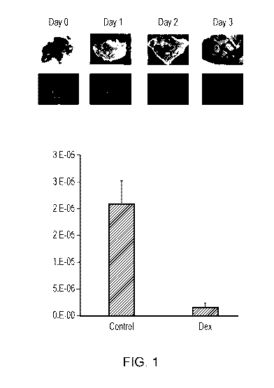
[0038] Figure 1. Acute high dose dexamethasone eliminates binding niches in
the mouse
spleen and secondary lymphatics. Shown are black and white scale bright field
(top) and
immunofluorescent (bottom) images of fresh thick spleen sections stained with
FITC-PNA to
quantitate germinal centers from mice administered IP human equivalent dose
(HED) 9.3 mg
kg dexamethasone base 96 hours before spleen harvest. The graph shows column
plots of
average germinal cell count per spleen area plus standard area of the mean
(SEM) for mice
administered IP placebo control and IP HED 9.3 mg kg dexamethasone base 96
hours before
spleen harvest. Control mice have significant FITC-PNA immunofluorescence,
while mice
who were injected with dexamethasone have almost no immunofluorescent signal.
[0039] Figure 2. Acute high dose dexamethasone dose-dependently eliminates
binding
niches in the mouse spleen. A graph of column plots of average Germinal Center
staining
intensity measured using immunofluorescent staining of fresh thick spleen
sections stained
with FITC-PNA is shown. Immunofluorescent intensity was calculated using
thresholding
and MetaMorph Image Analysis. Columns are average plus SEM. The mice were
administered placebo, 3mg/kg HED, 6 mg/kg HED, 9 mg/kg HED, or 12 mg/kg HED
dexamethasone base 48 hours before spleen harvest. Germinal center reduction
is apparent at
HED 6 mg/kg and is significantly reduced at HED of 9 and 12 mg/kg doses.
CA 03113969 2021-03-23
WO 2020/072713
PCT/US2019/054395
[0040] Figure 3. Acute high dose dexamethasone eliminates binding niches in
the rat spleen
(MZ: marginal zone). Column plots of marginal zone widths measured on 5 micron
spleen
sections from rats treated IV or PO with placebo, 20 mg/kg (HED 3.23 mg/kg),
40 mg/kg
(HED 6.45 mg/kg) or 80 mg/kg (HED 12.9 mg/kg) dexamethasone base 48 hours
before
spleen harvest are shown. Marginal zone area was reduced at all dexamethasone
doses, and
was maximally inhibited at 12.9 mg/kg HED. n = 5 per group. * p<0.05 ANOVA
(Dunnett's
post-hoc) vs. Vehicle IV; t p<0.05 ANOVA (Dunnett's post-hoc) vs. Vehicle PO;
1: p<0.05
Student's t-test vs. Vehicle IV.
[0041] Figure 4. Acute high dose dexamethasone eliminates binding niches in
the rat spleen.
Column plots of the area per spleen of BCL-6 staining of 5 micron fixed spleen
sections as a
measure of germinal center numbers given as average per section are shown.
Rats were
treated IV or PO with placebo, 20 mg/kg (HED 3.23 mg/kg), 40 mg/kg (HED 6.45)
or 80
mg/kg (HED 12.9 mg/kg) dexamethasone base 48 hours before spleen harvest.
Germinal
center area was reduced at all dexamethasone doses, and was maximally
inhibited at 12.9
mg/kg HED. Groups 1-4 IV: 1 =20 mg/kg (HED 3.23 mg/kg), 2 =40 mg/kg (HED 6.45
mg/kg), 3 = 80 mg/kg (HED 12.9 mg/kg), 4 = Placebo. Groups 5-9 PO: 5 =20 mg/kg
(HED
3.23 mg/kg), 6 = 40 mg/kg (HED 6.45 mg/kg), 7 = 80 mg/kg (HED 12.9 mg/kg), 8 =
Placebo.
[0042] Figure 5. Acute high dose dexamethasone reduces thymic mass.
Photographs show
size of thymus from placebo-treated murine subjects (top photograph) and of
thymus of
murine subjects treated with a 6 mg/kg HED dose of the pharmaceutical
composition of the
invention (lower photograph). The lower panel shows the thymus weight to body
weight
percentage of the thymus of placebo-treated subjects (control) and of subjects
treated with the
pharmaceutical composition of the invention at 3 mg/kg HED, 6 mg/kg HED, 9
mg/kg HED
and 12 mg/kg HED.
[0043] Figure 6. Acute high dose dexamethasone reduces rat lymphocyte number.
Graphs of
individual absolute lymphocyte numbers and averages measured by complete blood
count 48
hours after rats were treated IV (right) or PO (left) with placebo, 20 mg/kg
(HED 3.23
mg/kg), 40 mg/kg (HED 6.45) or 80 mg/kg (HED 12.9 mg/kg) dexamethasone base
are
shown. Dexamethasone was administered 48 hours before blood withdrawal.
Significant
lymphodepletion was observed at all doses vs. controls in rats whether IV
(right) or oral
dosing (left). Doses are shown as HED (human equivalent dose).
[0044] Figure 7. Acute high dose dexamethasone does not reduce rat neutrophil
number.
Graphs of individual absolute neutrophil numbers and averages measured by
complete blood
11
CA 03113969 2021-03-23
WO 2020/072713
PCT/US2019/054395
count 48 hours after rats were treated IV (right) or PO (left) with placebo,
20 mg/kg (HED
3.23 mg/kg), 40 mg/kg (HED 6.45) or 80 mg/kg (HED 12.9 mg/kg) dexamethasone
base are
shown. Data in Figures 3, 4, and 6 are from the same rats. Acute high dose
dexamethasone
has a lymphodepletion profile that is neutrophil sparing. Oral (left) and IV
(right) doses were
administered 1 x 48 hours before blood withdrawal. Doses are shown as HED
(human
equivalent dose).
[0045] Figure 8. CD3 and CD4 positive lymphocytes. Graphs of individual CD3+
(left) and
CD4+ (right) lymphocytes and averages measured by flow cytometry as relative
counts and
normalized to relative absolute counts using complete blood counts 48 hours
after mice were
treated PO with placebo, HED 3 mg/kg, HED 6 mg/kg, HED 9 mg/kg or HED 12.mg/kg
dexamethasone base. Relative counts/ul = flow cytometry and complete blood
count
combined. Compared to control, in the 12 mg/kg group: 65% reduction in CD3+
cells; 75%
reduction in CD4+ cells. Doses are shown as HED (human equivalent dose). A one-
way
ANOVA followed by Tukey's test was incorporated to determine statistical
significance
between the treatment groups; * p<0.05, ** p<0.01, *** p<0.001.
[0046] Figure 9. Acute high dose dexamethasone reduces mouse CD8 positive
lymphocytes
and Tregs. Graphs of individual CD8+ (left) and Treg (right) lymphocytes and
averages
measured by flow cytometry as relative counts and normalized to relative
absolute counts
using complete blood counts 48 hours after mice were treated PO with placebo,
HED 3
mg/kg, HED 6 mg/kg, HED 9 mg/kg or HED 12.mg/kg dexamethasone base are shown.
Treg lymphocytes were identified by being CD3+CD4+CD25+FoxP3+. Relative
counts/ul =
flow cytometry and complete blood counts combined. Compared to control, in the
12 mg/kg
group: 56% reduction in CD8+ cells; 78% reduction in mouse Tregs. Doses are
shown as
HED (human equivalent dose). A one-way ANOVA followed by post-hoc Tukey's test
was
.. incorporated to determine statistical significance between the treatment
groups; * p<0.05, **
p<0.01.
[0047] Figure 10. Acute high dose dexamethasone reduces mouse NK cells and B
lymphocytes. Graphs of individual natural killer (NK) cells (left) and B
lymphocytes (right)
and averages measured by flow cytometry as relative counts and normalized to
relative
absolute counts using complete blood counts 48 hours after mice were treated
PO with
placebo, HED 3 mg/kg, HED 6 mg/kg, HED 9 mg/kg or HED 12.mg/kg dexamethasone
base
are shown. NK cells were identified by being CD3-CD49b+. B lymphocytes were
identified
by being CD3-B220+. Relative counts/ul = flow cytometry and complete blood
counts
combined. Compared to control, in the 12 mg/kg group: 87% reduction in NK
cells; 83%
12
CA 03113969 2021-03-23
WO 2020/072713
PCT/US2019/054395
reduction B cells. Doses are shown as HED (human equivalent dose). A one-way
ANOVA
followed by post-hoc Tukey's test was incorporated to determine statistical
significance
between the treatment groups; * p<0.05, ** p<0.01; *** p<0.001.
[0048] Figure 11. Acute high dose dexamethasone reduces mouse absolute
lymphocyte
numbers while sparing neutrophils. Graphs of individual absolute neutrophils
(left) and total
lymphocytes (right) and averages measured by complete blood counts 24 - 48
hours after
mice were treated PO with placebo, HED 3 mg/kg, HED 6 mg/kg, HED 9 mg/kg, HED
12.mg/kg, or HED 17.5 mg/kg dexamethasone base are shown. Cells/ul = absolute
numbers
obtained from complete blood counts (CBC). Acute high dose dexamethasone
causes almost
.. complete lymphoabalation at HED doses greater than 12 mg/kg, but does not
affect
neutrophils. Acute high dose dexamethasone therefore eliminates the need for
transfusion,
and provides a safer, non-toxic alternative to chemotherapeutic regimens.
Doses are shown as
HED (human equivalent dose).
[0049] Figure 12. Acute high dose dexamethasone spares mouse RBCs and
Platelets.
.. Graphs of individual absolute RBC (left) and platelet (right) and averages
measured by
complete blood counts 48 hours after mice were treated PO with placebo, HED 3
mg/kg,
HED 6 mg/kg, HED 9 mg/kg, HED 12.mg/kg, or 17.5 mg/kg dexamethasone base are
shown.
Cells/ul = absolute numbers obtained from CBC. Acute high dose dexamethasone
does not
affect RBCs or platelets, eliminates the need for transfusion, and therefore
provides a safer,
non-toxic alternative to chemotherapeutic regimens. Doses are shown as HED
(human
equivalent dose).
[0050] Figure 13. Number of live hematopoietic stem cells measured 48 hours
after
treatment of naïve mice with placebo (vehicle), or low or high doses of acute
high dose
dexamethasone are shown. Even high doses of acute high dose dexamethasone did
not
significantly alter the number of live hematopoietic stem cells. The non-
myeloablative
regimen represented by acute high dose dexamethasone could, therefore,
eliminate the need
for transfusions of stem cells for hematopoietic recovery after immune-reset.
[0051] Figure 14. Fifty percent (2 of 4) of human patients treated with 3
mg/kg
dexamethasone base depleted CD3, CD4 and CD8 positive lymphocytes. Individual
pre-and
.. post-treatment, 48 hours after oral administration of 3 mg/kg dexamethasone
base to four
human patients, values and line plots of CD3+, CD4+, and CD8+ lymphocytes
measured by
flow cytometry are shown. Each patient's pre-treatment values are connected to
post-
treatment values by a connecting line. CD4+ cells are also CD3+. CD8+ cells
are also
CD3+.
13
CA 03113969 2021-03-23
WO 2020/072713
PCT/US2019/054395
[0052] Figure 15. Twenty-five percent (1 of 4) of human patients treated with
3 mg/kg
dexamethasone base depleted Tregs and B lymphocytes. Line are individual pre-
and post-,
48 hours after oral administration of 3 mg/kg dexamethasone base to four human
patients,
values and line plots of Treg and B lymphocytes measured by flow cytometry.
Each patient's
pre-treatment values are connected to post-treatment values by a connecting
line. Tregs are
identified by being CD3+CD4+CD25+FoxP3+. B lymphocytes are identified by being
CD3-
CD19+.
[0053] Figure 16. Seventy-five percent (3 of 4) of human patients treated with
3 mg/kg
dexamethasone base depleted NK cells while hematopoietic stem cells were
spared. Line are
individual pre-and post-treatment, 48 hours after oral administration of 3
mg/kg
dexamethasone base to four human patients, values and line plots of NK cells
and
Hematopoietic Stem Cells (HSCs) measured by flow cytometry. Each patient's pre-
treatment
values are connected to post-treatment values by a connecting line. NK cells
are identified by
being CD3-CD16/56+. HSCs are identified by being CD34+CD38-.
[0054] Figure 17. 100% of human patients treated with 3 mg/kg dexamethasone
base
showed increased serum IL-2 and/or IL-15 levels, but no elevation in IL-6.
Column plots of
each patients pre- and post-treatment, 48 hours after oral administration of 3
mg/kg
dexamethasone base to four human patients, plasma levels of interleukin 2 and
interleukin 15
measured by ProCartaPlex- 9 plx Luminex assay. Figure 14, Figure 15, Figure 16
and Figure
17 show data from the same four human patients.
[0055] Figure 18. Oral administration of 3 mg/kg dexamethasone base increased
bone
marrow MSC number 48 hours later. Column plots of data from 31 historical
naive control
humans plus standard deviation, and two human patients treated with 3 mg/kg
dexamethasone base 48 hours before aspiration of concentrated bone marrow from
the ileac
crest using a MarrowCellutionTM needle. Plots show bone marrow CFU/ml +/-
stdev. Bone
marrow was added directly to colony forming unit assay fibroblast (CFU-F)
media without
further manipulation 24 hours after harvest and shipment at controlled room
temperature.
CFU-F colony number is a measure of mesenchymal stem cell (MSC) number in the
starting
material. 48 hours after oral administration of 3 mg/kg dexamethasone base,
ileac crest bone
marrow MSC numbers appear about twice as high as 31 historical controls. 3
mg/kg oral
dexamethasone base increases human bone marrow CFU-F per ml 48 hours later
compared to
31 historical controls aspirated using the same MarrowCellutionTM needle as
for patients M
and P.
14
CA 03113969 2021-03-23
WO 2020/072713
PCT/US2019/054395
[0056] Figure 19. Comparison of a 12 mg/kg and 17-18 mg/kg dexamethasone base
oral
dose on day -2 to a single dose of Cyclophosphamide 166 mg/kg (HED 500 mg/m2)
and
Fludarabine 10 mg/kg on day -5 combined with 12 mg/kg or 17-18 mg/kg
dexamethasone
base on day -2, and to 2 days of repeat Cyclophosphamide 166 mg/kg on day -5
and -4 and 4
days of Fludarabine 10 mg/kg (HED 30 mg/m2) on days -5, -4, -3, -2. Shown is a
graph of
individual absolute lymphocytes and averages (left) measured by complete blood
counts 48
hours after mice were treated IP with PBS (Vehicle), or with repeat IP
Cyclophosphamide
166 mg/kg on day -5 and -4 and 4 days of IP Fludarabine 10 mg/kg (HED 30
mg/m2) on days
-5, -4, -3, -2 (Flu+Cy), or with a single IP dose of Cyclophosphamide 166
mg/kg (HED 500
mg/m2) and IP Fludarabine 10 mg/kg both on day -5 and then with oral 12 mg/kg
or 17-18
mg/kg dexamethasone base on day -2 (Flu+Cy + AV1V10703(12 mg/kg); Flu+Cy +
AVM0703(17 mg/kg)), or with oral 12 mg/kg or 17-18 mg/kg dexamethasone base
(AVM0703(12 mg/kg); AVM0703(17 mg/kg)). Also shown (right) is a representation
of the
dosing schedules in mice of these regimens.
[0057] Figure 20. A single dose of Cyclophosphamide 166 mg/kg (HED 500 mg/m2)
and
Fludarabine 10 mg/kg on day -5 combined with 12 mg/kg or 17-18 mg/kg
dexamethasone
base on day -2 equivalently lymphodepleted CD3+ and CD4+ lymphocytes compared
to 2
days of repeat Cyclophosphamide 166 mg/kg on day -5 and -4 and 4 days of
Fludarabine 10
mg/kg (HED 30 mg/m2) on days -5, -4, -3, -2. Shown are graphs of individual
CD3+ (left)
and CD4+ (right) lymphocytes and averages measured by flow cytometry as
relative counts
and normalized to relative absolute counts using complete blood counts 48
hours after mice
were treated IP with PBS (Vehicle), or with repeat IP Cyclophosphamide 166
mg/kg on day -
5 and -4 and 4 days of IP Fludarabine 10 mg/kg (HED 30 mg/m2) on days -5, -4, -
3, -2
(Flu+Cy), or with a single IP dose of Cyclophosphamide 166 mg/kg (HED 500
mg/m2) and
IP Fludarabine 10 mg/kg both on day -5 and then with oral 12 mg/kg or 17-18
mg/kg
dexamethasone base on day -2 (Flu+Cy+AV1V10703(12 mg/kg); Flu+Cy + AVM0703(17
mg/kg)), or with oral 12 mg/kg or 17-18 mg/kg dexamethasone base (AVM0703(12
mg/kg);
AVM0703(17 mg/kg)). On both the CD3+ plot (left) and the CD4+ plot (right),
the 12
mg/kg or 17-18 mg/kg dexamethasone base data are shown in the right-hand
columns of each
(relative counts are '92', '71', '37' and '25').
[0058] Figure 21. A single dose of Cyclophosphamide 166 mg/kg (HED 500 mg/m2)
and
Fludarabine 10 mg/kg on day -5 combined with 12 mg/kg or 17-18 mg/kg
dexamethasone
base on day -2 equivalently lymphodepleted CD8+ lymphocytes and Tregs compared
to 2
days of repeat Cyclophosphamide 166 mg/kg on day -5 and -4 and 4 days of
Fludarabine 10
CA 03113969 2021-03-23
WO 2020/072713
PCT/US2019/054395
mg/kg (HED 30 mg/m2) on days -5, -4, -3, -2. Shown are graphs of individual
Treg (right)
and CD8+ lymphocytes (left) and averages measured by flow cytometry as
relative counts
and normalized to relative absolute counts using complete blood counts 48
hours after mice
were treated IP with PBS (Vehicle), or with repeat IP Cyclophosphamide 166
mg/kg on day -
5 and -4 and 4 days of IP Fludarabine 10 mg/kg (HED 30 mg/m2) on days -5, -4, -
3, -2
(Flu+Cy), or with a single IP dose of Cyclophosphamide 166 mg/kg (HED 500
mg/m2) and
IP Fludarabine 10 mg/kg both on day -5 and then with oral 12 mg/kg or 17-18
mg/kg
dexamethasone base on day -2 (Flu+Cy+AV1V10703(12 mg/kg); Flu+Cy + AVM0703(17
mg/kg)), or with oral 12 mg/kg or 17-18 mg/kg dexamethasone base (AVM0703(12
mg/kg);
AVM0703(17 mg/kg)). On both the CD8+ plot (left) and the CD4+ plot (right),
the 12
mg/kg or 17-18 mg/kg dexamethasone base data are shown in the right-hand
columns of each
(relative counts are '33', '1.4', '0.2' and '0.5').
[0059] Figure 22. A single dose of Cyclophosphamide 166 mg/kg (HED 500 mg/m2)
and
Fludarabine 10 mg/kg on day -5 combined with 12 mg/kg or 17-18 mg/kg
dexamethasone
.. base on day -2 equivalently lymphodepleted NK cells and B lymphocytes
compared to 2 days
of repeat Cyclophosphamide 166 mg/kg on day -5 and -4 and 4 days of
Fludarabine 10 mg/kg
(HED 30 mg/m2) on days -5, -4, -3, -2. Shown are graphs of individual B
lymphocytes (left)
and NK cell lymphocytes (right) and averages measured by flow cytometry as
relative counts
and normalized to relative absolute counts using complete blood counts 48
hours after mice
were treated IP with PBS (Vehicle), or with repeat IP Cyclophosphamide 166
mg/kg on day -
5 and -4 and 4 days of IP Fludarabine 10 mg/kg (HED 30 mg/m2) on days -5, -4, -
3, -2
(Flu+Cy), or with a single IP dose of Cyclophosphamide 166 mg/kg (HED 500
mg/m2) and
IP Fludarabine 10 mg/kg both on day -5 and then with oral 12 mg/kg or 17-18
mg/kg
dexamethasone base on day -2 (Flu+Cy+AV1V10703(12 mg/kg); Flu+Cy + AVM0703(17
mg/kg)), or with oral 12 mg/kg or 17-18 mg/kg dexamethasone base (AVM0703(12
mg/kg);
AVM0703(17 mg/kg)). On both the B cell plot (left) and the NK cell plot
(right), the 12
mg/kg or 17-18 mg/kg dexamethasone base data are shown in the right-hand
columns of each
(relative counts are '111' and '58' for B cells; not shown for NK cells).
[0060] Figure 23. A single dose of Cyclophosphamide 166 mg/kg (HED 500 mg/m2)
and
Fludarabine 10 mg/kg on day -5 combined with 12 mg/kg or 17-18 mg/kg
dexamethasone
base on day -2 equivalently lymphodepleted absolute lymphocytes, but spared
neutrophils,
compared to 2 days of repeat Cyclophosphamide 166 mg/kg on day -5 and -4 and 4
days of
Fludarabine 10 mg/kg (HED 30 mg/m2) on days -5, -4, -3, -2. Shown are graphs
of
individual absolute neutrophils (left) and absolute lymphocytes (right) and
averages measured
16
CA 03113969 2021-03-23
WO 2020/072713
PCT/US2019/054395
by complete blood counts 48 hours after mice were treated IP with PBS
(Vehicle), or with
repeat IP Cyclophosphamide 166 mg/kg on day -5 and -4 and 4 days of IP
Fludarabine 10
mg/kg (HED 30 mg/m2) on days -5, -4, -3, -2 (Flu+Cy), or with a single IP dose
of
Cyclophosphamide 166 mg/kg (HED 500 mg/m2) and IP Fludarabine 10 mg/kg both on
day -
5 and then with oral 12 mg/kg or 17-18 mg/kg dexamethasone base on day -2
(Flu+Cy+AVM0703(12 mg/kg); Flu+Cy + AVM0703(17 mg/kg)), or with oral 12 mg/kg
or
17-18 mg/kg dexamethasone base (AVM0703(12 mg/kg); AVM0703(17 mg/kg)). On both
the neutrophil plot (left) and the lymphocyte plot (right), the 12 mg/kg or 17-
18 mg/kg
dexamethasone base data are shown in the right-hand columns of each (relative
counts are
'321', '605', '521' and '88').
[0061] Figure 24. A single dose of Cyclophosphamide 166 mg/kg (500 mg/m2) and
Fludarabine 10 mg/kg (HED 30 mg/m2) on day -5 combined with 12 mg/kg or 17-18
mg/kg
dexamethasone base on day -2 spared red blood cells (RBCs) and platelets.
Shown are graphs
of individual absolute platelet and absolute RBCs and averages measured by
complete blood
counts 48 hours after mice were treated IP with PBS (Vehicle), or with repeat
IP
Cyclophosphamide 166 mg/kg on day -5 and -4 and 4 days of IP Fludarabine 10
mg/kg (HED
30 mg/m2) on days -5, -4, -3, -2 (Flu+Cy), or with a single IP dose of
Cyclophosphamide
166 mg/kg (HED 500 mg/m2) and IP Fludarabine 10 mg/kg both on day -5 and then
with oral
12 mg/kg or 17-18 mg/kg dexamethasone base on day -2 (Flu+Cy+AVM0703(12
mg/kg);
__ Flu+Cy + AVM0703(17 mg/kg)), or with oral 12 mg/kg or 17-18 mg/kg
dexamethasone
base (AVM0703(12 mg/kg); AVM0703(17 mg/kg)). On both the RBC plot (left) and
the
platelet plot (right), the 12 mg/kg or 17-18 mg/kg dexamethasone base data are
shown in the
right-hand columns of each (relative counts are '10', '10', '348' and '373').
[0062] Figure 25. A single dose of Cyclophosphamide 166 mg/kg (500 mg/m2) and
Fludarabine 10 mg/kg on day -5 combined with 12 mg/kg or 17-18 mg/kg
dexamethasone
base on day -2 spared body weight, a measure of toxicity, compared to 2 days
of repeat
Cyclophosphamide 166 mg/kg on day -5 and -4 and 4 days of Fludarabine 10 mg/kg
on days
-5, -4, -3, -2. Shown (left) are graphs of individual body weight differences
and averages
calculated by subtracting body weight 48 hours after mice were treated IP with
PBS
(Vehicle), or with repeat IP Cyclophosphamide 166 mg/kg on day -5 and -4 and 4
days of IP
Fludarabine 10 mg/kg (HED 30 mg/m2) on days -5, -4, -3, -2 (Flu+Cy), or with a
single IP
dose of Cyclophosphamide 166 mg/kg (HED 500 mg/m2) and IP Fludarabine 10 mg/kg
both
on day -5 and then with oral 12 mg/kg or 17-18 mg/kg dexamethasone base on day
-2
(Flu+Cy+AVM0703(12 mg/kg); Flu+Cy + AVM0703(17 mg/kg)), or with oral 12 mg/kg
or
17
CA 03113969 2021-03-23
WO 2020/072713
PCT/US2019/054395
17-18 mg/kg dexamethasone base (AV1V10703(12 mg/kg); AV1V10703(17 mg/kg)) from
pretreatment body weights. The acute high dose dexamethasone group is not
associated with
body weight loss unlike the chemotherapy groups. Acute high dose dexamethasone
therefore
provides a similar lymphodepletion effect as chemotherapy but with no
associated toxicity.
Also shown (right) is a representation of the dosing schedules in these
regimens.
[0063] Figure 26. Comparison of 15 mg/kg dexamethasone base HED (AVM0703) to
standard chemotherapy regimen: anti-tumor efficacy. A20 B cell lymphoma mice
(8-10
weeks old) were treated with PBS (control), 15 mg/kg dexamethasone base HED
(AVM0703), or either 1 cycle or two cycles of cyclophosphamide 100 mg/kg i.p,
doxorubicin
6 mg/kg i.p, vincristine 0.1 mg/kg i.p and dexamethasone 0.2 mg/kg i.p (CHOP).
Dosing for
1 cycle CHOP was performed on day 0, 2 cycle chop mice were dosed on day 0 and
day 10,
and dexamethasone dosing was performed on days 7, 10, 18, 23, 24, 28, 35, and
42 (indicated
by arrows). Mice were followed for tumor growth with tumor volume (mm3)
measured every
2-3 days. The efficacy of 15 mg/kg dexamethasone base HED is greater than 1
cycle of
CHOP, but not quite as effective as 2 cycles of CHOP in terms of tumor volume
control.
However, 15 mg/kg dexamethasone base HED was associated with a much favourable
toxicity profile compared to 2 cycles of CHOP.
[0064] Figure 27. Comparison of 15 mg/kg dexamethasone base HED (AVM0703) to
standard chemotherapy regimen: toxicity. Panel A shows the percent body weight
change for
.. 15 mg/kg dexamethasone base HED (AVM0703) compared to PBS control. Panel B
shows
the percent body weight change for 1 cycle or two cycles of cyclophosphamide
100 mg/kg
i.p, doxorubicin 6 mg/kg i.p, vincristine 0.1 mg/kg i.p and dexamethasone 0.2
mg/kg i.p
(CHOP) compared to PBS control. The reduction in body weight seen in mice
treated with 2
cycles of CHOP (B) is much greater than that seen for mice treated with 15
mg/kg
dexamethasone base HED (A). Additionally, 18% of mice treated with 2 cycles of
CHOP
died from the CHOP treatment, whereas no mice died from the dexamethasone
treatment.
[0065] Figure 28. Statistical comparison of 15 mg/kg dexamethasone base HED
(AVM0703)
with PBS control. A20 B cell lymphoma mice (8-10 weeks old) were treated with
PBS
(control) or 15 mg/kg dexamethasone base HED (AVM0703). Dexamethasone dosing
was
performed on days 7, 10, 18, 23, and 24 (indicated by arrows). Mice were
followed for tumor
growth with tumor volume (mm3) measured every 2-3 days. The efficacy of 15
mg/kg
dexamethasone base HED (black squares) is greater than PBS control (black
circles), as seen
by reduced tumor growth. Statistically significant differences in tumor volume
were seen at
days 15, 17 and 20.
18
CA 03113969 2021-03-23
WO 2020/072713
PCT/US2019/054395
[0066] Figure 29. Glucocorticoid therapy reduces the required dose for
effective
chemotherapy: Tumor bearing subjects treated with PBS only (Control') or with
the
dexamethasone base (AVM0703') exhibit continued tumor growth, with generally
high
growth rates after 20 days. Tumor bearing subjects treated with AV1V10703 on
day 11
followed by one dose of Cy/Flu chemotherapy on day 14 (Combo') exhibit a
steady and
sustained reduction in tumor volume, in a similar manner as the tumors of
subjects treated
with two doses of Cy/Flu chemotherapy, on day 11 and day 14 (Cy/Flu').
[0067] Figure 30. High dose glucocorticoid therapy reduces tumor density,
without
significantly affecting body weight. After establishment of tumor, the tumor
density of
subjects was measured following administration of weekly doses of the
glucocorticoid
AVM0703, at 6 mg/Kg HED weekly, 15 mg/Kg HED weekly or 21 mg/Kg HED weekly
(left
panel). The body weights of the mice over the course of the study is also
shown (right
panel); dotted line represents 20% loss of the average weight of the mice at
the start of the
study. Shows no significant loss of bodyweight due to toxicity and no mice
were taken down.
Detailed Description
[0068] Cytotoxic chemotherapeutic agents trigger cell death via mechanisms or
means that
are not receptor mediated. Cytotoxic chemotherapeutic agents trigger cell
death by
interfering with functions that are necessary for cell division, metabolism,
or cell survival.
Because of this mechanism of action, cells that are growing rapidly (which
means
proliferating or dividing) or are active metabolically will be killed
preferentially over cells
that are not. The status of the different cells in the body as dividing or as
using energy
(which is metabolic activity to support function of the cell) determines the
dose of the
chemotherapeutic agent that triggers cell death. The skilled person will
appreciate that the
glucocorticoid that is utilized in this invention is not a cytotoxic
chemotherapeutic. Cytotoxic
chemotherapeutic agents non-exclusively relates to alkylating agents, anti-
metabolites, plant
alkaloids, topoisomerase inhibitors, antineoplastics and arsenic trioxide,
carmustine,
fludarabine, IDA ara-C, myalotang, GO, mustargen, cyclophosphamide,
gemcitabine,
bendamustine, total body irradiation, cytarabine, etoposide, melphalan,
pentostatin and
radiation.
[0069] The present invention pertains to pharmaceutical compositions
comprising a
glucocorticoid for use in the treatment of diseases by immunoablation. In
particular, the
compositions of the invention may be for use in the treatment of diseases that
are mediated by
19
CA 03113969 2021-03-23
WO 2020/072713
PCT/US2019/054395
immune cells such as lymphocytes. The treatment comprises administering a dose
of the
pharmaceutical composition to the patient to deliver the glucocorticoid at a
dose equivalent to
about 3 ¨ 26 mg/kg human equivalent dose (HED) of dexamethasone base.
[0070] As used herein, the term glucocorticoid includes glucocorticoid
receptor agonists and
any compound that binds to the glucocorticoid receptor. Such compounds relate
to, but are
not limited to, dexamethasone, dexamethasone containing agents,
hydrocortisone, methyl
predisone, prednisone, corticone, budesonide, betamethasone and
beclomethasone. Other
glucocorticoids include prednisolone, mometasone furoate, Triamcinolone
Acetonide and
methylprednisolone. Glucocorticoids further include glucocorticoid receptor
modulating
agonists. Additionally, selective glucocorticoid receptor agonists may be used
in the
pharmaceutical compositions disclosed herein. Such agonists or modulators
include for
example, selective glucocorticoid receptor modulators (SEGRMs) and selective
glucocorticoid receptor agonists (SEGRAs). Glucocorticoids, glucocorticoid
receptor
modulators and selective glucocorticoid receptor agonists (SEGRAs) that may be
utilized in
the herein disclosed methods and compositions are well know to those skilled
in the art.
[0071] Glucocorticoids and glucocorticoid-receptor (GR) modulating agents
exert their
effects through both membrane glucocorticoid receptors and cytoplasmic GRs
which activate
or repress gene expression. Some of the desirable lymphodepletion effects of
the
glucocorticoids and GR modulating agents appear to be mediated via membrane
GRs or other
non-genomic effects in addition to their genomic effects. Interestingly, co-
treatment with
dexamethasone has been shown to be able to reduce glucocorticoid resistance
(Serafin et at.,
2017).
[0072] The effects of glucocorticoids are complex and depend on each specific
glucocorticoid's affinity for the GR and mineralocorticoid receptor (MR).
Additionally, there
are now 9 known isoforms of the cytosolic GR and additional membrane expressed
GR
receptors that have been identified but which are not fully characterized.
Glucocorticoids
have been reported to have varied effects on lymphocyte levels, depending on
the
concentration of the glucocorticoid administered and the duration of
treatment. In general, at
low doses typically used for chronic therapy, glucocorticoids have been
reported to
redistribute lymphocytes from the peripheral blood into the bone marrow, at
medium doses
glucocorticoids have been reported to cause leukocytosis thought to be a
redistribution of
leukocytes from the bone marrow, spleen and thymus into the peripheral blood,
and at high
doses glucocorticoids have a lymphotoxic action on lymphocytes by triggering
apoptosis and
necroptosis. The duration of effect also depends on the dose level, for
instance Fauci et at
CA 03113969 2021-03-23
WO 2020/072713
PCT/US2019/054395
(1976) reports a single oral 0.24 mg/kg dexamethasone dose suppresses
peripheral blood T
and B lymphocytes 80% with recovery beginning at 12 hours and normal levels by
24 hours.
However, the present invention demonstrates that acute oral doses of 3 mg/kg
or greater are
necessary to reduce peripheral blood T and B cells 24-48 hours after
administration, with
return to baseline levels occurring around 5 to 14 days after dosing.
[0073] The desired in vivo effects of exemplary glucocorticoids would include
reductions in
germinal center and marginal zones in secondary lymphatics, direct tumor
killing of some
cancers particularly; multiple myeloma, renal cell carcinoma, leukemia and
lymphoma, non-
small cell lung cancer (NSCLC), prostate and breast cancer; depletion of all
peripheral blood
lymphocyte types, lack of lymphocyte redistribution to the BM or other organs,
and elevation
of plasma cytokines including IL-2, and/or IL-7, and/or IL-12, and/or IL-15 to
levels
preferably of 20 pg/ml or greater, among others. Exemplary glucocorticoids do
not elevate
plasma levels of IL-6, one of the major contributors to ACT induced cytokine
release
syndrome (CRS). Exemplary glucocorticoids do not elevate plasma levels of GM-
CSF, one
of the major contributors to ACT induced neuroedema. Acute doses of
dexamethasone of
about HED 6 mg/kg and above reduce germinal centers and marginal zones in
secondary
lymphatics; acute doses of dexamethasone of about 1.6 mg/kg HED in a 48 hour
period have
about 50% direct tumor killing against multiple myeloma and other cancer cell
lines which is
maintained but not increased with doses up to about 12 mg/kg HED; acute doses
of
dexamethasone of greater than about HED 3 mg/kg are required for
lymphodepletion
demonstrated by the observation that 50% of patients treated with 3 mg/kg HED
showed
lymphocytosis (Figure 14); plasma IL-2 and IL-15 cytokine elevations are
observed at doses
of dexamethasone base of about HED 3 mg/kg or higher (Figure 17). Based on the
desired in
vivo effects in the indications disclosed in this application, the most
preferred acute
dexamethasone base doses, which can be converted to equivalent doses of other
glucocorticoids based on known calculators or as disclosed in this
description, will be most
likely about HED 9 mg/kg and above.
[0074] A single high dose of glucocorticoid can be given as an oral
administration or about a
one hour IV infusion. A total dose may be given as repetitive IV or oral doses
in any quantity
such that the total dose, e.g. of dexamethasone, is about 3 mg/kg to about 26
mg/kg within
about a 24 to about a 72 hour period.
[0075] Equivalent doses of another glucocorticoid or glucocorticoid receptor
modulating
agent can be readily and easily calculated using publicly available corticoid
conversion
algorithms, preferably http://www.medcalc.com. For instance, 3 to 12 mg/kg
dexamethasone
21
CA 03113969 2021-03-23
WO 2020/072713
PCT/US2019/054395
converts to 19 to 75 mg/kg prednisone. Since prednisone's biologic half-life
is about 20
hours, while dexamethasone's biologic half-life is about 36 to 54 hours.
Therefore,
prednisone would be dosed between 19 to 75 mg/kg every 24 hours for equivalent
biologic
dosing. More specifically, a 12 mg/kg dose of dexamethasone corresponds to 1)
a 75 mg/kg
dose of prednisolone that would require repeat dosing of about two to about
three doses every
24 hours. A 10mg/kg dose of betamethasone is about 12 mg/kg dexamethasone and
has a
pharmacodynamic (biologic) half-life similar to dexamethasone. However,
betamethasone
reduces RBC at doses of about 24 mg/50 kg (Gaur 2017).
[0076] DEX (dexamethasone base) doses in the examples in the present
application are given
as human equivalent doses (HED). AV1V10703 (also referred to as AugmenStemTM
or
PlenaStemTM) in the examples given is Dex (dexamethasone base) as
dexamethasone sodium
phosphate in a proprietary buffer.
[0077] Methods for calculating the human equivalent dose (HED) are known in
the art. For
example the FDA's Centre for Drug Evaluation and Research (CDER) issued a
highly-cited
guidance document in 2005 (U.S Department of Health CDER, 2005), which sets
out the
established algorithm for converting animal doses to HED based on body surface
area (the
generally accepted method for extrapolating doses between species) at Table 1
on page 7 of
that document. For reference, Table 1 is reproduced below. The skilled person
understands
that the animal dose in mg/kg, explained below, the HED is calculated easily
using the
standard conversion factors in the right hand columns of Table 1:
[0078] Table 1: Conversion of Animal Doses to Human Equivalent Doses Based on
Body
Surface Area
To Convert Animal Dose in mg/kg to
HED a in mg/kg, Either:
Species To Convert Animal Dose in Divide Animal Dose
Multiply
mg/kg to Dose in mg/m2, By Animal
Dose
Multiply by km By
Human 37
Child (20 kg)b 25
Mouse 3 12.3 0.08
Hamster 5 7.4 0.13
Rat 6 6.2 0.16
22
CA 03113969 2021-03-23
WO 2020/072713
PCT/US2019/054395
Ferret 7 5.3 0.19
Guinea pig 8 4.6 0.22
Rabbit 12 3.1 0.32
Dog 20 1.8 0.54
Primates:
Monkeys' 12 3.1 0.32
Marmoset 6 6.2 0.16
Squirrel monkey 7 5.3 0.19
Baboon 20 1.8 0.54
Micro-pig 27 1.4 0.73
Mini-pig 35 1.1 0.95
'Assumes 60 kg human. For species not listed or for weights outside the
standard ranges,
HED can be calculated from the following formula:
HED = animal dose in mg/kg x (animal weight in kg/human weight in kg) 33.
b This km value is provided for reference only since healthy children will
rarely be volunteers
for phase 1 trials.
For example, cynomolgus, rhesus, and stumptail.
[0079] Doses described herein can be presented as a "weight based dose" or as
a "body
surface area (B S A) based dose." A weight based dose is a dose that is
administered to a
patient that is calculated based on the weight of the patient, e.g., mg/kg. A
BSA based dose is
a dose that is administered to a patient that is calculated based on the
surface area of the
patient, e.g., mg/m2 . The two forms of dose measurement can be converted
within the
context of human dosing by multiplying the weight based dose by 37 or dividing
the BSA
based dose by 37 as shown in Table 1 above.
[0080] The terms "subject" and "patient" are used interchangeably herein, and
refer to a
human or animal.
[0081] Dexamethasone, like the other glucocorticoid steroids at equivalent
doses, inhibits the
formation and proliferation of germinal centers in the lymph tissues and
lymphodepletes
peripheral blood. The doses of glucocorticoid, particularly dexamethasone,
preferably
achieve greater than 75% lymphodepletion. More preferably, the doses of
glucocorticoid,
particularly dexamethasone, achieve greater than 80% lymphodepletion. Most
preferably,
the dose of glucocorticoid, particularly dexamethasone, achieves greater than
23
CA 03113969 2021-03-23
WO 2020/072713
PCT/US2019/054395
95%lymphodepletion. The skilled person will understand that lymphodepletion
can be
measured readily by measuring complete blood counts (CBCs). .
[0082] Dexamethasone and other preferred glucocorticoids spare neutrophils and
do not
inhibit neutrophil function (Schleimer RP, J Pharmacol Exp Ther 1989;250:598-
605), and
spare red blood cells (RBCs), platelets, mesenchymal stem cells (MSC) and
hematopoietic
stem cells (HSC). Neutrophil sparing in humans is an absolute neutrophil count
(ANC)
greater than 500 per mm3. By sparing neutrophils, RBCs and platelets,
lymphoablating
glucocorticoids would reduce or eliminate the need for transfusions.
Lymphoablating
glucocorticoids also spare bone marrow mesenchymal stem cells (MSCs) and do
not affect
the capacity of bone marrow MSCs to differentiate towards chondrocytes,
osteocytes or
adipocytes. Lymphoablating glucocorticoids also increase the endogenous number
of BM
MSCs or their ex vivo survival in both humans and horses. Lymphoablating
glucocorticoids
increase plasma IL-2, IL-7, IL-12 and IL-15 levels, but not IL-6 or GM-CSF
levels. In some
embodiments of the invention, the subject is selected before treatment and/or
assessed after
treatment, based on measurements of the plasma levels of one or more of these
cytokines.
[0083] Dexamethasone is approved for use with an initial dosage of
dexamethasone sodium
phosphate injection that varies from 0.5 to 9 mg a day depending on the
disease being treated,
which is a daily dose of 0.01 to 0.18 mg/kg based on a 50 kg BW. In less
severe diseases
doses lower than 0.5 mg may suffice, while in severe diseases doses higher
than 9 mg may be
required. There is a tendency in current medical practice to use high
(pharmacologic) doses
of corticosteroids for the treatment of unresponsive shock. For cerebral edema
Dexamethasone sodium phosphate injection is generally administered initially
in a dosage of
10 mg intravenously followed by four mg every six hours intramuscularly until
the symptoms
of cerebral edema subside. This total dose would correspond to a total 24 hour
dose of about
.34 to .48 mg/kg and a total 72 hour dose of 0.8 to 1.12 mg/kg in 72 hours,
which is not an
effective dose according to the present invention, which uses doses between
about 3 mg/kg
and about 26 mg/kg.
[0084] For acute allergic disorders, dexamethasone sodium phosphate injection,
USP 4
mg/mL; is recommended: first day, 1 or 2 mL (4 or 8 mg), intramuscularly, then
Dexamethasone sodium phosphate tablets, 0.75 mg; second and third days, 4
tablets in two
divided doses each day; fourth day, 2 tablets in two divided doses; fifth and
sixth days, 1
tablet each day; seventh day, no treatment; eighth day, follow-up visit.
Dexamethasone has
been used in the emergency room for severe acute pediatric asthma at 2 mg/kg,
a dose which
is below the glucocorticoid doses as defined in this invention.
24
CA 03113969 2021-03-23
WO 2020/072713
PCT/US2019/054395
[0085] Conventional formulations of glucocorticoids such as dexamethasone may
be
unsuitable for use in the therapeutic applications of the present invention.
For instance,
dexamethasone sodium phosphate (DSP) is currently available in low dose (2-4
mg/ml) and
low volume formulations (e.g. APP Pharmaceuticals, Mylan), which contain
antimicrobial
__ preservatives such as benzyl alcohol (BA) and propyl paraben (PP). The
target dose of DSP
required for performing complete lymphoablation would entail the use of
multiple vials
resulting in overdoses of excipients. Exceeding WHO acceptable daily intake
(ADI) of both
benzyl alcohol and propyl paraben has been associated with genotoxicity and
increased risk
of cancer (Darbre et at., 2014), reproductive toxicity (Aker et at., 2016),
increased risks of
allergic disease (Savage et al., 2012; Spanier et al., 2014), and neonatal CNS
dysfunctions
(Medicines Agency, 2017). Moreover, with commercially available DSP package
inserts,
serious neuropsychiatric effects occur in about 6% of patients who receive
steroids
(Malmegrim et at., 2017). As the present invention involves the administration
of high doses
of glucocorticoids, formulations with low levels of potentially toxic
preservatives, or
formulations without toxic preservatives, should be used. Preferably, the
preservative is an
antioxidant.
[0086] The pharmaceutical composition of the invention may include a
preservative (e.g. an
antioxidant) additive such as sodium sulfite to maintain the stability of the
composition.
Sulfites are also widely used as preservative and antioxidant additives in the
pharmaceutical
__ industries. Exposure to such sulfites has been reported to induce a range
of adverse clinical
effects in sensitive individuals, ranging from dermatitis, urticaria,
flushing, hypotension and
abdominal pain to life-threatening anaphylactic and asthmatic reactions.
Sulfite-inducing
symptoms range from mild in some individuals, to severe in others, and in some
individuals
the reactions can be life threatening. In preferred embodiments, where sodium
sulfite is
included as an antioxidant, the concentration is between 0 ¨ 70 ppm Sodium
Sulfite
(Anhydrous).
[0087] Antioxidants may be added in amounts that are reduced from those levels
typically
employed in glucocorticoid containing compositions thereby reducing the
toxicity and
adverse side effects associated with the use of such antioxidants. In some
instances, the
__ formulations of the invention may lack the addition of antioxidants.
[0088] As used herein, antioxidants are those excipients that delay or inhibit
the oxidation
process of molecules thereby increasing the stability of the composition.
Antioxidants that
may be used include, for example, ascorbic acid, acetylcysteine,
butylhydroxyanisol, cysteine
hydrochloride, dithionite sodium, gentisic acid, glutamate monosodium,
glutathione,
CA 03113969 2021-03-23
WO 2020/072713
PCT/US2019/054395
formaldehyde sulfoxylate sodium, methionine, monothioglycerol, propyl gallate,
sulfites,
sodium thioglycolate, a-thioglycerol, tocopherol alpha, alpha tocopherol
hydrogen succinate
and thioglycolate sodium.
[0089] In addition to an active glucocorticoid and antioxidant additional
components well
known to those of skill in the art may be included in the pharmaceutical
compositions
disclosed herein. Pharmaceutical compositions may be prepared using a
pharmaceutically
acceptable "carrier" composed of materials that are considered safe and
effective.
"Pharmaceutically acceptable" refers to molecular entities and compositions
that are "generally
regarded as safe", e.g., that are physiologically tolerable and do not
typically produce an
-- allergic or similar untoward reaction, such as gastric upset and the like,
when administered to a
human. In some embodiments, this term refers to molecular entities and
compositions
approved by a regulatory agency of the US federal or a state government, as
the GRAS list
under section 204(s) and 409 of the Federal Food, Drug and Cosmetic Act, that
is subject to
premarket review and approval by the FDA or similar lists, the U.S.
Pharmacopeia or another
generally recognized pharmacopeia for use in animals, and more particularly in
humans.
[0090] The term "carrier" refers to diluents, binders, lubricants and
disintegrants. Those with
skill in the art are familiar with such pharmaceutical carriers and methods of
compounding
pharmaceutical compositions using such carriers.
[0091] The pharmaceutical compositions provided herein may include one or more
excipients,
-- e.g., solvents, solubility enhancers, suspending agents, buffering agents,
isotonicity agents,
antioxidants or antimicrobial preservatives. When used, the excipi ents of the
compositions
will not adversely affect the stability, bioavailability, safety, and/or
efficacy of the active
ingredients, ie., glucocorticoids, used in the composition. Thus, the skilled
person will
appreciate that compositions are provided wherein there is no incompatibility
between any of
the components of the dosage form. Excipients may be selected from the group
consisting of
buffering agents, solubilizing agents, tonicity agents, chelating agents,
antioxidants,
antimicrobial agents, and preservatives.
[0092] The pharmaceutical composition of the invention may include a chelating
agent which
is used to sequester and decrease the reactivity of metal ions that may be
present in the
-- compositions. Possible chelators are calcium disodium EDTA 0.01-0.1% (EDTA
=
Ethylenediaminetetra acetic acid or Edetate), Disodium EDTA 0.01-0.11%, Sodium
EDTA
0.20%, Calcium Versetamide Sodium 2.84%, Calteridol 0.023%, DTPA 0.04-1.2%
(Diethylenetriaminepenta acetic acid). In a preferred embodiment the
concentration of
Disodium EDTA (Edetate) is between 0 and 500 ppm.
26
CA 03113969 2021-03-23
WO 2020/072713
PCT/US2019/054395
***
[0093] As noted in W02018/183927, glucocorticoids can also be used as a
preconditioning
agent, in conjunction with adoptive cell therapies (ACT). Glucocorticoids,
particularly
dexamethasone dosed between about 3 mg/kg and about 26 mg/kg single acute dose
about 12
to about 72 hours prior to cell immunotherapy administration or total dose of
about 3 mg/kg
to about 26 mg/kg given between about 12 to about 72 hours of cell therapy
administration
increases plasma IL-2 and IL-15 levels.
[0094] Glucocorticoids, particularly dexamethasone dosed between about 3 mg/kg
and about
26 mg/kg single acute dose or total dose of about 3 mg/kg to about 26 mg/kg
given over
about a 72 hour period, either alone, or in combination with reduced intensity
cytotoxic
preconditioning can be useful for the treatment of autoimmune diseases. For
the treatment of
autoimmune disease an ACT could be targeted to the immune cells driving the
disease in an
effort to eradicate the autoimmune recognizing cells. Additionally, for
autoimmune diseases
the ACT could be a Treg targeted by a CAR or TCR or expressed antibody for an
antigen
expressed specifically or selectively by the region or organ of the body where
the
autoimmune attack goes on. The Tregs could non-exclusively relate to CD4+
Tregs,
CD4+CD45RA+ Tregs, CD4+CD25+CD45RA+ Tregs, FoxP3+ Tregs,
CD4+CD25+FoxP3+CD152+ Tregs, CD4+CD25+CD152+ Tregs, CD8+ Tregs, CD8+CD28-
Tregs, CD4+CD25int/high, CD127low, CTLA4+, GITR+, FoxP3+, CD127low, CD4+CD25-
induced Tregs, or Type I T regs.
[0095] "Natural" regulatory T cells originally recognised by their
constitutive expression of
CD4 and CD25 can be further defined by expression of the transcription factor
foxP3 and
surface CD152. Their generation and some of their suppressive activity is
dependent on TGF-
beta, and it has been shown that they can induce IDO in appropriate DCs by
CD152 mediated
ligation of CD80/86. Anergic CD4+ T cells generated by antigen stimulation in
the absence
of costimulation seem to be characterised by an intrinsic raising of their
threshold for antigen
stimulation that may be maintained by expression of E3 ubiquitin ligases such
as GRAIL, c-
cbl and Itch. Anergic cells can act as regulatory T cells by competing at the
sites of antigen
presentation and adsorbing out stimulatory cytokines such as IL-2. Trl cells
represent an
induced subset of CD4 helper T cells that are dependent on IL-10 for their
differentiation and
for some of their regulatory properties. They do not express foxP3 but may
express markers
associated with Th2 cells and repressor of GATA (ROG). Like natural Tregs,
they express
high levels of surface CD152 and can induce IDO and trypophan catabolism in
appropriate
DCs. CD8+CD28- suppressor T (Ts) cells were first characterised in human, but
have
27
CA 03113969 2021-03-23
WO 2020/072713
PCT/US2019/054395
recently also been demonstrated in rodents. Like Trl cells, they are induced
in the presence
of IL-10, and IL-10 may be involved in the downregulation of dendritic cell
costimulation
and the upregulation of ILT-3 and ILT-4 (in human DC) that seem to play an
important role
in presenting antigen to tolerise further cohorts of T cells.
[0096] Regulatory T cells (Tregs) play an important role in maintaining immune
homeostasisl. Tregs suppress the function of other T cells to limit the immune
response.
Alterations in the number and function of Tregs has been implicated in several
autoimmune
diseases including multiple sclerosis, active rheumatoid arthritis, and type 1
diabetes. High
levels of Tregs have been found in many malignant disorders including lung,
pancreas, and
breast cancers. Tregs may also prevent antitumor immune responses, leading to
increased
mortality.
[0097] Two major classes of Tregs have been identified to date: CD4 and CD8
Tregs. CD4
Tregs consist of two types, "natural" Tregs (nTregs) that constitutively
express CD25 and
FoxP3, and so-called adaptive or inducible Tregs (iTregs).
[0098] Natural Tregs (nTregs) originate from the thymus as CD4 + cells
expressing high
levels of CD25 together with the transcription factor (and lineage marker)
FoxP3. nTregs
represent approximately 5-10% of the total CD4 + T cell population, and can
first be seen at
the single-positive stage of T lymphocyte development. They are positively
selected
thymocytes with a relatively high avidity for self-antigens. (Fehervari Z,
Sakaguchi S.
Development and function of CD25+CD4+ regulatory T cells. Curr Opin Immunol.
2004;16:203-208.)
[0099] The signal to develop into Treg cells is thought to come from
interactions between the
T cell receptor and the complex of MHC II with self peptide expressed on the
thymic stroma.
nTregs are essentially cytokine independent.
[00100] Adaptive or inducible Tregs originate from the thymus as single-
positive CD4
cells. They differentiate into CD25 and FoxP3 expressing Tregs (iTregs)
following adequate
antigenic stimulation in the presence of cognate antigen and specialized
immunoregulatory
cytokines such as TGF-f3, IL-10, and IL-4. (Chatenoud L, Bach JF. Adaptive
human
regulatory T cells: myth or reality? J Clin Invest. 2006;116:2325-2327.)
[00101] FoxP3 is currently the most accepted marker for Tregs, although
there have
been reports of small populations of FoxP3" Tregs. The discovery of
transcription factor
FoxP3 as a marker for Tregs has allowed scientists to better define Treg
populations leading
to the discovery of additional Treg markers including CD127.
28
CA 03113969 2021-03-23
WO 2020/072713
PCT/US2019/054395
[00102] Glucocorticoids, particularly dexamethasone dosed between
about 3 mg/kg
and about 26 mg/kg single acute dose or total dose of about 3 mg/kg to about
26 mg/kg given
over about a 72 hour period, either alone or in combination with reduced
intensity cytotoxic
chemotherapy or radiation can be useful for the treatment of residual HIV
disease, and for the
treatment of germinal center lymphomas such as Burkitt's Lymphoma.
[00103] Follicular helper CD4 T cells, TFH, residing in B-cell
follicles within
secondary lymphoid tissues, are readily infected by AIDS viruses and are a
major source of
persistent virus despite relative control of viral replication. This
persistence is due at least in
part to a relative exclusion of effective antiviral CD8 T cells from B-cell
follicles. AIDS virus
persistence in individuals under effective drug therapy or those who
spontaneously control
viremia remains an obstacle to definitive treatment. Infected follicular
helper CD4 T cells,
TFH, present inside B-cell follicles represent a major source of this residual
virus. While
effective CD8 T-cell responses can control viral replication in conjunction
with drug therapy
or in rare cases spontaneously, most antiviral CD8 T cells do not enter B-cell
follicles, and
those that do fail to robustly control viral replication in the TFH
population. Thus, these sites
are a sanctuary and a reservoir for replicating AIDS viruses. Lymphodepletion
and reduction
of germinal centers and marginal zones in the spleen would force residual HIV
infected cells
into the blood stream where they could be killed by existing therapies.
Latently infected
resting CD4 T cells have been detected in the peripheral blood,
gastrointestinal (GI) tract, and
lymph nodes of HIV-1-infected individuals and are also likely to exist in
other organs
containing lymphoid tissue.
[00104] Highly active antiretroviral therapy (HAART) enables long-term
suppression
of plasma HIV-1 loads in infected persons, but low-level virus persists and
rebounds
following cessation of therapy. During HAART, this virus resides in latently
infected cells,
such as resting CD4 T cells, and in other cell types that may support residual
virus
replication. Therapeutic eradication will require elimination of virus from
all reservoirs.
[00105] Burkitt's Lymphoma is a germinal center lymphoma originating
and growing
within the secondary lymphatic system, always associated with a c-Myc
activating
chromosomal translocation. It is one of the fastest growing cancers and can
double in size
every 14-18 hours. BL is an aggressive B-cell lymphoma found in germinal
centers of the
spleen and secondary lymphatics. BL is named after Dr. Denis Parsons Burkitt,
a surgeon
who first described the disease in 1958 while working in equatorial Africa
(Burket, D., 1958).
BL is most commonly found in children living in sub-Saharan Africa, with the
highest
incidence and mortality rates found in East Africa (Orem, J., et al.,). Boys
are more
29
CA 03113969 2021-03-23
WO 2020/072713
PCT/US2019/054395
susceptible to BL than girls. Outside of Africa, BL is most likely to occur in
people who
have a compromised immune system.
[00106] Among B-cell malignancies, CLL is the most responsive to
ibrutinib, and thus
unfortunately ibrutinib is not likely to significantly benefit people
afflicted with Burkitt's
Lymphoma and other germinal center lymphomas. However, the same result to
redistribute
B-cell cancers into the circulation where they are more susceptible to
chemotherapy and less
proliferative can be achieved for germinal center lymphomas such as Burkitt's
lymphoma
with the use of agents that ablate secondary lymphatic germinal centers. Thus,
in some
embodiments, the invention increases the susceptibility of a lymphoma to a
chemotherapy
and/or provides a combination therapy involving reduced intensity cytotoxic
chemotherapy in
addition to the glucocorticoid, e.g. dexamethasone. Various suitable
chemotherapies are
disclosed herein.
[00107] Clinical observations on the ability of a Bruton's Tyrosine
Kinase inhibitor
ibrutinib for treatment of chronic lymphocytic leukemia has demonstrated that
redistribution
of CLL cells from the lymphatics into the bloodstream is a contributing
mechanism of action
to its benefit in CLL. Circulating CLL cells are not proliferative, with
proliferation of the
clone limited to the lymphatic microenvironment. Therefore, redistribution
into the blood
stream reduces cancerous proliferation. Similarly, redistribution of ALL from
the bone
marrow to the bloodstream, has also been reported to enhance sensitivity to
standard
chemotherapy (Chang BY, Blood 2013 122 : 2412-24;).
[00108] Glucocorticoids have been reported to have multiple and
contradictory actions
on lymphocytes, depending on the dose, the duration of dosing and the species
investigated.
Glucocorticoids have been investigated as lymphocytosis inducing agents,
agents which
increase circulating lymphocyte numbers, since 1943 (for review see Burger et
at., 2013),
typically with the use of prednisone between 0.5 and 1 mg/kg, which would be
an equivalent
0.1-0.2 mg/kg dexamethasone dose. High dose methylprednisone (HDMP) used for
refractory CLL, in contrast, does not appear to induce lymphocytosis at the
methylprednisone
equivalent to the 0.5-1.0 mg/kg dose at which prednisone did. Lymphotoxic high-
dose
steroids are typically considered to be approximately 100 mg daily of
prednisone equivalent,
which would be a dexamethasone equivalent dose of 16 mg which is approximately
0.23 to
0.32 mg/kg, and which we have demonstrated is not an effective preconditioning
dose.
Dexamethasone does not reduce germinal centers in mice until an HED of about 3
mg/kg or
greater is administered. Prednisone does not significantly impact spleen
weights or germinal
centers until used at doses in mice over 2.5 mg/kg po daily for 13 weeks (Yan
et at., 2015), a
CA 03113969 2021-03-23
WO 2020/072713
PCT/US2019/054395
human dose which would have unacceptable mineralocorticoid activity as a dose
of 30 mgs
per day (-0.48 - 0.72 mg/kg) is considered a high dose in human lupus
patients.
[00109] For Burkitt's lymphoma (BL) treatment with standard
chemotherapy regimens
such as COPADM, prednisone is included in various cycles typically at 60
mg/m2, which
converts to 1.62 mg/kg prednisone and an equivalent 0.3 mg/kg dexamethasone
dose, which
is not an effective preconditioning dose. Dexamethasone is also used
clinically for the
treatment of B-cell cancers, typically in an oral four-five day 40 mg daily
regimen or 6
mg/m2 for 5 days. In some indications such as ALL, dexamethasone is given
daily for weeks
and can be associated with osteonecrosis, particularly in adolescent boys.
Risk of
osteonecrosis can be substantially eliminated by alternate week dosing of
dexamethasone and
may be particularly present in ALL because of the asparaginase regimen that is
part of the
treatment for ALL (Chang BY, Blood 2013 122 : 2412-24).
[00110] Epstein-Barr virus (EBV) infection is found in nearly all
African BL patients,
and chronic malaria is believed to reduce resistance to EBV, allowing it to
take hold. The
disease characteristically involves the jaw or other facial bone, distal
ileum, cecum, ovaries,
kidney, or breast. Additionally, BL strikes immunocompromised people, such as
those with
HIV.
[00111] BL is classified into three main clinical variants: Endemic,
Sporadic, and the
Immunodeficiency-associated variants, with the Endemic variant (also called
the "African
variant") most commonly occurring in children living in malaria endemic
regions of the
world.
[00112] One effect of the present invention can be to ablate germinal
centers and/or
marginal zones to selectively drive BL and other germinal center cancer cells
or marginal
zone cancer cells from the germinal centers or marginal zones into circulation
where they can
be more easily killed with chemotherapy or other agents. This could
dramatically, safely and
cost-effectively advance BL treatment outcomes.
Asthma is a chronic inflammation characterized by an increased number of CD8
Type-I T-
lymphocytes and macrophages in the lung tissue and neutrophils in the airway
lumen. Lymphocytes, which are markedly different in the two inflammatory
conditions, play
a crucial role in the pathogenesis of asthma and COPD. There is now
overwhelming
evidence to support a major role for T cells in asthma, in particular the
involvement of T
helper type 2 (Th2) cells in atopic allergic asthma as well as nonatopic and
occupational
asthma. There may also be a minor contribution from T cytotoxic type 2 CD8+T
cells.
Several Th2 cytokines have potential to modulate airway inflammation, in
particular
31
CA 03113969 2021-03-23
WO 2020/072713
PCT/US2019/054395
interleukin-13 which induces airway hyperresponsiveness independently of IgE
and
eosinophilia in animal models. Asthma and chronic obstructive pulmonary
disease (COPD)
are two different inflammatory disorders of the lungs which share a common
functional
abnormality, i.e. airflow limitation (Baraldo et at., 2007)..
[00113] In asthma, airflow limitation is largely reversible, either
spontaneously or with
treatment, and does not progress in most cases. On the other hand, airflow
limitation in
COPD is usually progressive and poorly reversible. In asthma, the chronic
inflammation
causes an associated increase in airway responsiveness to a variety of
stimuli, leading to
recurrent episodes of wheezing, breathlessness, chest tightness and cough,
particularly at
night and in the early morning. Many cells are involved in the inflammatory
response in
asthma and, among these, CD4+ Type-2 lymphocytes, mast cells and eosinophils
are thought
to play a crucial role. In COPD, the poorly reversible airflow limitation is
associated with an
abnormal inflammatory response of the lungs to noxious particles or gases.
This chronic
inflammation is characterized by an increased number of CD8+ Type-1 T-
lymphocytes and
macrophages in the lung tissue and neutrophils in the airway lumen.
Lymphocytes, which are
markedly different in the two inflammatory conditions, play a crucial role in
the pathogenesis
of asthma and COPD (Baraldo et al., 2007).
Definitions
[00114] Definitions used to describe the embodiments of the invention:
[00115] Biologic mechanism of lymphodepletion means induction of programmed
cell
death via apoptosis or necroptosis or pyroptosis or autophagy or oncosis.
Various stimuli can
engage a non-apoptotic form of cell death called necroptosis, which occurs
when caspases
required for apoptosis are inhibited. Pyroptosis is a caspase-dependent form
of programmed
cell death that differs in many respects from apoptosis. Unlike apoptosis, it
depends on the
activation of caspase-1 or caspase-11 (caspase-5 in humans). Autophagy is a
lysosome-
dependent process.
[00116] Apoptosis: A form of cell death in which a programmed sequence
of events
leads to the elimination of cells without releasing harmful substances into
the surrounding
area. Apoptosis plays a crucial role in developing and maintaining the health
of the body by
eliminating old cells, unnecessary cells, and unhealthy cells.
[00117] The term "and/or" where used herein is to be taken as specific
disclosure of
each of the two specified features or components with or without the other.
Thus, the term
"and/or" as used in a phrase such as "A and/or B" herein is intended to
include "A and B," "A
or B," "A" (alone), and "B" (alone). Likewise, the term "and/or" as used in a
phrase such as
32
CA 03113969 2021-03-23
WO 2020/072713
PCT/US2019/054395
"A, B, and/or C" is intended to encompass each of the following aspects: A, B,
and C; A, B,
or C; A or C; A or B; B or C; A and C; A and B; Band C; A (alone); B (alone);
and C (alone).
[00118] The term "about' when referring to a measurable value such as
an amount or a
temporal duration and the like refers to variations of +/- 20% or +/- 10%.
[00119] Administering" refers to the physical introduction of an agent to a
subject,
using any of the various methods and delivery systems known to those skilled
in the art.
Exemplary routes of administration for the formulations disclosed herein
include intravenous,
intramuscular, subcutaneous, intraperitoneal, spinal or other parenteral
routes of
administration, for example by injection or infusion. The phrase "parenteral
administration"
.. as used herein means modes of administration other than enteral and topical
administration,
usually by injection, and includes, without limitation, intravenous,
intramuscular,
intraarterial, intrathecal, intralymphatic, intralesional, intracapsular,
intraorbital, intracardiac,
intradermal, intraperitoneal, transtracheal, subcutaneous, subcuticular,
intraarticular,
subcapsular, subarachnoid, intraspinal, epidural and intrasternal injection
and infusion, as
well as in vivo electroporation. In some embodiments, the formulation is
administered via a
non-parenteral route, e.g., orally. Other non-parenteral routes include a
topical, epider- mal or
mucosal route of administration, for example, intranasally, vaginally,
rectally, sublingually or
topically.
[00120] A pharmacologic dose is a dose far in excess of normal levels
in the body.
[00121] An "anti-tumor effect" as used herein, refers to a biological
effect that can
present as a decrease in tumor volume, a decrease in the number of tumor
cells, a decrease in
tumor cell proliferation, a decrease in the number of metastases, an increase
in overall or
progression-free survival, an increase in life expectancy, or amelioration of
various
physiological symptoms associated with the tumor. An anti-tumor effect can
also refer to the
prevention of the occurrence of a tumor, e.g., a vaccine.
[00122] A therapeutic agent is an agent that enhances the efficacy of
cellular
immunotherapies compared to the cellular immunotherapies without said
therapeutic agent.
[00123] The term "autologous" refers to any material derived from the
same individual
to which it is later to be re-introduced, whether the individual is a human or
other animal.
[00124] The term "allogeneic" refers to any material derived from one
individual
which is then introduced to another individual of the same species, whether
the individual is a
human or other animal.
[00125] The term dexamethasone (also referred to as Dex) non-
exclusively relates to
any formulation whether a liquid solution, liquid suspension, oral solution,
tablet form, tablet
33
CA 03113969 2021-03-23
WO 2020/072713
PCT/US2019/054395
form dissolved in a liquid containing the active ingredient of dexamethasone,
injectable form,
gel formulation, patch formulation or any formulation containing the active
ingredient
dexamethasone.
[00126] The term glucocorticoid-receptor modulating agents non-
exclusively relates to
glucocorticoid receptor agonists or glucocorticoid receptor modulators
including but not
limited to: compound A [CpdA; (24(4-acetopheny1)-2-chloro-N-
methyl)ethylammonium-
chloride)] and N-(4-methyl-1-oxo-1H-2,3-benzoxazine-6-y1)-4-(2,3-
dihydrobenzofuran-7-y1)-
2-hydroxy-2-(trifluoromethyl)-4-methylpentanamide (ZK216348), AL-438,
Mapracorat ,
LGD-5552 , RU-24858, Fosdagrocorat, PF-802, Compound 10, MK5932, C108297,
LGD5552, and ORG 214007-0.
[00127] Immunotoxins are proteins that contain a toxin along with an
antibody or
growth factor that binds specifically to target cells. Immunotoxins are
created by chemically
conjugating an antibody to a whole protein toxin, devoid of its natural
binding domain.
Immunologic proteins that are smaller than monoclonal antibodies (MoAbs), like
growth
factors and cytokines, have also been chemically conjugated and genetically
fused to protein
toxins. Toxins used in immunotoxin constructs are derived from bacteria,
fungi, and plants,
and most function by inhibiting protein synthesis. Bacterial toxins commonly
used in
immunotoxins include Diphtheria toxin (DT) and the toxin from Pseudomonas
exotoxin (PE).
Plant toxins utilized in immunotoxins include the A chain of ricin (RTA), and
the ribosome
inactivating proteins (RIPs) gelonin, pokeweed antiviral protein, and
dodecandron. Because it
is an enzyme, one toxin molecule can work on many substrate molecules, having
a
devastating effect on the cell. Toxins such as diphtheria toxin (DT) and
Pseudomonas
exotoxin (PE) prevent protein synthesis by an effect on elongation factor 2
(EF-2).
[00128] The term systemic injection as used herein non-exclusively
relates to a route
of administration that rapidly, within seconds or a few hours, leads to
circulating levels of
cellular immunotherapies, and non-exclusively relates to intravenous,
intraperitoneally,
subcutaneous, via nasal submucosa, lingual, via bronchoscopy, intravenous,
intra-arterial,
intra-muscular, intro-ocular, intra-striatal, subcutaneous, intradermal, by
dermal patch, by
skin patch, by patch, into the cerebrospinal fluid, into the portal vein, into
the brain, into the
lymphatic system, intra-pleural, retro-orbital, intra-dermal, into the spleen,
intra-lymphatic,
among others.
[00129] The term 'site of injection' as used herein non-exclusively
relates to intra-
tumor, or intra-organ such as the kidney or liver or pancreas or heart or lung
or brain or
34
CA 03113969 2021-03-23
WO 2020/072713
PCT/US2019/054395
spleen or eye, intra-muscular, intro-ocular, intra-striatal, intradermal, by
dermal patch, by
skin patch, by patch, into the cerebrospinal fluid, into the brain, among
others.
[00130] The term lymphodepletion as used herein non-exclusively
relates to the
reduction of lymphocyte number in the peripheral blood without causing
redistribution of
lymphocytes to another organ such as the bone marrow, thymus, lymph nodes,
lung or spleen
or another organ.
[00131] The term lymphoablation as used herein non-exclusively relates
to reduction
of lymphocyte number in the peripheral blood to below 200 per microliter,
preferably to
below 100 per microliter, without causing redistribution of lymphocytes to
another organ
such as the bone marrow, thymus, lymph nodes, lung or spleen or another organ.
[00132] The term cytotoxic lymphodepletion as used herein relates to
the reduction of
lymphocyte number in the peripheral blood by a mechanism of ADCC, cell-
mediated
cytotoxicity or direct lysis or cytotoxic elimination of lymphocytes,
chemotherapy or
radiation.
[00133] The antibody-dependent cell-mediated cytotoxicity (ADCC), also
referred to
as antibody-dependent cellular cytotoxicity, is a mechanism of cell-inediated
immune defense
whereby an effector cell of the immune system actively lyses a target cell,
whose membrane
-
surface antigens have been bound by specific antibodies.
[00134] The term 'cellular immunotherapy', 'adoptive cellular
immunotherapy',
'adoptive cellular therapy' (ACT) or cell immunotherapy or cell therapy as
used herein non-
exclusively relates to treatments that contain a cell used to help the immune
system fight
diseases or a cell from the immune lineage which directly fights diseases such
as cancer,
autoimmune diseases and infections with certain viruses. The cellular
immunotherapy can be
from either an autologous or allogeneic source. In preferred embodiments, the
adoptive
immunotherapy used in the methods disclosed herein may be an adoptive T cell
immunotherapy, i.e. 'T cell therapy'.
[00135] The term preconditioning as used herein relates to the
preparation of a patient
with a cytotoxic lymphodepleting agent or a non-toxic lymphodepleting agent
prior to ACT.
[00136] The term immunotherapy, also called biologic therapy, as used
herein non-
exclusively relates to a type of treatment for cancer, autoimmune disease or
infection
treatment designed to boost the body's natural defenses to fight the cancer,
autoimmune
disease or infection. It uses substances either made by the body or in a
laboratory to improve
or restore immune system function. The term "immunotherapy" refers to the
treatment of a
subject afflicted with, or at risk of contracting or suffering a recurrence
of, a disease by a
CA 03113969 2021-03-23
WO 2020/072713
PCT/US2019/054395
method comprising inducing, enhancing, suppressing or otherwise modifying an
immune
response. Examples of immunotherapy include, but are not limited to, T cell
therapies. T cell
therapy can include adoptive T cell therapy, tumor-infiltrating lymphocyte
(TIL)
immunotherapy, autologous cell therapy, engineered autologous cell therapy
(eACT), and
allogeneic T cell transplantation. However, one of skill in the art would
recognize that the
conditioning methods disclosed herein would enhance the effectiveness of any
transplanted T
cell therapy. Examples of T cell therapies are described in U.S. Patent
Publication Nos.
2014/0154228 and 2002/0006409, U.S. Pat. No. 5,728,388, and International
Publication No.
WO 2008/081035.
[00137] The term 'immune modulation' as used herein non-exclusively relates
to, in
cancer, autoimmune disease or infection, a range of treatments aimed at
harnessing a
patient's immune system to achieve tumor, autoimmune causing cell or viral
control,
stabilization, and potential eradication of disease.
[00138] The term immunomodulator as used herein non-exclusively
relates to a
chemical agent (such as dexamethasone) or biologic agent (such as HUMIRA and
rituximab) that modifies the immune response or the functioning of the immune
system (as
by the stimulation of antibody formation or the inhibition of white blood cell
activity).
Traditional immune modulating drugs that are immunesuppressants non-
exclusively relates
to glucocorticoids, calcineurin inhibitors, antimetabolites, and alkylating
agents.
Antimetabolites non-exclusively relates topurine analogues (e.g., azathioprine
and
mycophenolate mofetil), and folate antagonists (e.g., methotrexate and
dapsone).
[00139] Immunesuppressants (also termed immunosuppressants) can be
chemical or
biologic agents that can suppress or prevent the immune response. For
instance, antagonists
to CD26 and dexamethasone are immunesuppressants. The NTLAs used in this
invention
may be NTLA immunesuppressants.
[00140] The terms "conditioning" and "pre-conditioning" are used
interchangeably
herein and indicate preparing a patient or animal in need of a T cell therapy
for a suitable
condition. Conditioning as used herein includes, but is not limited to,
reducing the number of
germinal centers and marginal zones, reducing the number of endogenous
lymphocytes,
removing a cytokine sink, increasing a serum level of one or more homeostatic
cytokines or
pro-inflammatory factors, enhancing an effector function of T cells
administered after the
conditioning, enhancing antigen presenting cell activation and/or
availability, or any
combination thereof prior to a T cell therapy.
36
CA 03113969 2021-03-23
WO 2020/072713
PCT/US2019/054395
[00141] The term 'adoptive immunotherapy' or 'cellular adoptive
immunotherapy' as
used herein non-exclusively relates to immune cells that are collected from a
patient
(autologous or autogenic) or a donor (allogeneic), either related or
unrelated, and grown in
the laboratory. This increases the number of immune cells that are able to
kill cancer cells,
__ autoimmune causing cells or fight infections. These immune cells are given
back to the
patient to help the immune system fight disease. This is also called cellular
adoptive
immunotherapy. The immune cell can be a T cell and/or other cell of the immune
system
non-exclusively relating to macrophages, monocytes, dendritic cells,
neutrophils,
granulocytes, phagocytes, mast cells, basophils, thymocytes, or innate
lymphoid cells, or any
combination thereof
[00142] The term agonist as used herein non-exclusively relates to any
entity that
activates a specific receptor or downstream signaling pathway essential to
mediate the
receptor's effect(s). Agonists may non-exclusively relates tobut are not
limited to
antibodies, antibody fragments, soluble ligands, small molecules, cyclic
peptides, cross-
__ linking agents.
[00143] The term antagonist as used herein non-exclusively relates to
any entity that
interferes with the binding of a receptor's counter structure(s), or with the
activation of a
specific receptor or downstream signaling pathway essential to mediate the
receptor's
effect(s). Antagonists may non-exclusively relates tobut are not limited to
antibodies,
__ antibody fragments,soluble ligands, Fc fusion receptors, chimeric
receptors, small molecules,
cyclic peptides, peptides.
[00144] The term inhibitor as used herein non-exclusively relates to
any entity that
diminishes the target effect of a specific receptor. Inhibitors may be small
molecules,
antisense agents, nucleic acids including siRNA and microRNA.
[00145] The term "lymphocyte" as used herein includes natural killer (NK)
cells, T
cells, or B cells. NK cells are a type of cytotoxic ( cell toxic) lymphocyte
that represent a
major component of the inherent immune system. NK cells reject tumors and
cells infected
by viruses. It works through the process of apoptosis or progranmied cell
death. They were
termed "natural killers" because they do not require activation in order to
kill cells. T-cells
__ play a major role in cell-mediated-immunity (no antibody involvement). Its
T-cell receptors
(TCR) differentiate themselves from other lymphocyte types. The thymus, a
specialized
organ of the immune system, is primarily responsible for the T cell's
maturation. There are
six types of T-cells, namely: Helper T-cells (e.g., CD4+ cells), Cytotoxic T-
cells (also known
as TC, cytotoxic T lymphocyte, CTL, T-killer cell, cytolytic T cell, CDS+ T-
cells or killer T
37
CA 03113969 2021-03-23
WO 2020/072713
PCT/US2019/054395
cell), Memory T-cells ((i) stem memory T scM cells, like naive cells, are
CD45R0-, CCR 7
+, CD45RA+, CD62L+(L-selectin), CD27+, CD28+ and IL-7Ra+, but they also
express large
amounts of CD95, IL-2R, CXCR3, and LFA-1, and show numerous functional
attributes
distinctive of memory cells); (ii) central memory TcM cells express L-selectin
and the CCR7,
they secrete IL-2, but not IFNy or IL-4, and (iii) effector memory T DV cells,
however, do
not express L-selectin or CCR 7 but produce effector cytokines like IFNy and
IL-4),
Regulatory T-cells (Tregs, suppressor T cells, or CD4+CD25+ regulatory T
cells), Natural
Killer T-cells (NKT) and Gamma Delta T-cells. B-cells, on the other hand, play
a principal
role in humoral immunity (with antibody involvement). It makes antibodies and
antigens and
performs the role of antigen presenting cells (APCs) and turns into memory B-
cells after
activation by antigen interaction. In manmials, immatureB-cells are formed in
the bone
marrow, where its name is derived from.
[00146] The term "cancer" refers to a disease characterized by the
uncontrolled growth
of aberrant cells. Cancer cells can spread locally or through the bloodstream
and lymphatic
system to other parts of the body. Examples of various cancers are described
herein and
include but are not limited to, breast cancer, prostate cancer, ovarian
cancer, cervical cancer,
skin cancer, pancreatic cancer, colorectal cancer, renal cancer, liver cancer,
brain cancer,
lymphoma, leukemia, lung cancer and the like. The terms "tumor" and "cancer"
are used
interchangeably herein, e.g., both terms encompass solid and liquid, e.g.,
diffuse or
circulating, tumors. As used herein, the term "cancer" or "tumor" includes
premalignant, as
well as malignant cancers and tumors.
[00147] The particular cancer can be responsive to chemo- or radiation
therapy or the
cancer can be refractory. A refractory cancer refers to a cancer that is not
amendable to
surgical intervention and the cancer is either initially unresponsive to chemo-
or radiation
therapy or the cancer becomes unresponsive over time.
[00148] An "anti-tumor effect" as used herein, refers to a biological
effect that can
present as a decrease in tumor volume, a decrease in the number of tumor
cells, a decrease in
tumor cell proliferation, a decrease in the number of metastases, an increase
in overall or
progression-free survival, an increase in life expectancy, or amelioration of
various
physiological symptoms associated with the tumor. An anti-tumor effect can
also refer to the
prevention of the occurrence of a tumor, e.g., a vaccine.
[00149] The term "progression-free survival," which can be abbreviated
as PFS, as
used herein refers to the time from the treatment date to the date of disease
progression per
the revised IWG Response Criteria for Malignant Lymphoma or death from any
cause.
38
CA 03113969 2021-03-23
WO 2020/072713
PCT/US2019/054395
[00150] "Disease progression" is assessed by measurement of malignant
lesions on
radiographs or other methods should not be reported as adverse events. Death
due to disease
progression in the absence of signs and symptoms should be reported as the
primary tumor
type (e.g., DLBCL).
[00151] The "duration of response," which can be abbreviated as DOR, as
used herein
refers to the period of time between a subject's first objective response to
the date of
confirmed disease progression, per the revised IWG Response Criteria for
Malignant
Lymphoma, or death.
[00152] The term "overall survival," which can be abbreviated as OS,
is defined as the
time from the date of treatment to the date of death.
[00153] The terms "reducing" and "decreasing" are used interchangeably
herein and
indicate any change that is less than the original. "Reducing" and
"decreasing" are relative
terms, requiring a comparison between pre- and post-measurements. "Reducing"
and
"decreasing" include complete depletions.
[00154] "Treatment" or "treating" of a subject refers to any type of
intervention or
process performed on, or the administration of an active agent to, the subject
with the
objective of reversing, alleviating, ameliorating, inhibiting, slowing down or
preventing the
onset, progression, development, severity or recurrence of a symptom,
complication or
condition, or biochemical indicia associated with a disease. In one
embodiment, "treatment"
or "treating" includes a partial remission. In another embodiment, "treatment"
or "treating"
includes a complete remission.
[00155] The use of the alternative (e.g., "or") should be understood
to mean either one,
both, or any combination thereof of the alternatives. As used herein, the
indefinite articles "a"
or "an" should be understood to refer to "one or more" of any recited or
enumerated
component.
[00156] The terms "about" or "comprising essentially of refer to a
value or
composition that is within an acceptable error range for the particular value
or composition as
determined by one of ordinary skill in the art, which will depend in part on
how the value or
composition is measured or determined, i.e., the limitations of the
measurement system. For
example, "about" or "comprising essentially of can mean within 1 or more than
1 standard
deviation per the practice in the art. Alternatively, "about" or "comprising
essentially of can
mean a range of up to 20% (i.e., 20%). For example, about 3 mg can include
any number
between 2.3 mg and 3.6 mg (for 20%). Furthermore, particularly with respect to
biological
systems or processes, the terms can mean up to an order of magnitude or up to
5-fold of a
39
CA 03113969 2021-03-23
WO 2020/072713
PCT/US2019/054395
value. When particular values or compositions are provided in the application
and claims,
unless otherwise stated, the meaning of'about" or "comprising essentially of
should be
assumed to be within an acceptable error range for that particular value or
composition.
[00157] As described herein, any concentration range, percentage
range, ratio range or
__ integer range is to be understood to include the value of any integer
within the recited range
and, when appropriate, fractions thereof (such as one-tenth and one-hundredth
of an integer),
unless otherwise indicated.
[00158] Ranges : Various aspects of the invention are presented in
range format. The
description in range format is for convenience and brevity and should not be
construed as an
inflexible limitation on the scope of the invention. Accordingly, the
description of a range
should be considered to have specifically disclosed all the possible subranges
as well as
individual numerical values within that range. For example, a range from 3 to
12 includes
3.1, 3.2, 3.3 etc.
[00159] Autoimmune disorders and other diseases that are mediated by
lymphocytes
__ and which are in need of treatments that are simpler and less costly than
HSCT are related to,
but not limited by the following list; allergies, asthma, residual HIV,
germinal center
lymphomas such as Burkitts Lymphoma and Diffuse Large B cell Lymphoma,
marginal zone
lymphoma, graft versus host disease (GvHD), steroid-resistant GvHD, Achalasia,
Addison's
disease, Adult Still's disease, Agammaglobulinemia, Alopecia areata,
Amyloidosis,
Ankylosing spondylitis, Anti-GBM/Anti-TBM nephritis, Antiphospholipid
syndrome,
Autoimmune angioedema, Autoimmune dysautonomia, Autoimmune encephalomyelitis,
Autoimmune hepatitis, Autoimmune inner ear disease (AIED), Autoimmune
myocarditis,
Autoimmune oophoritis, Autoimmune orchitis, Autoimmune pancreatitis,
Autoimmune
retinopathy, Autoimmune urticaria, Axonal & neuronal neuropathy (AMAN), Balo
disease,
__ Behcet's disease, Benign mucosal pemphigoid, Bullous pemphigoid, Castleman
disease
(CD), Celiac disease, Chagas disease, Chronic inflammatory demyelinating
polyneuropathy
(CIDP), Chronic recurrent multifocal osteomyelitis (CRMO), Churg-Strauss
Syndrome (CSS)
or Eosinophilic Granulomatosis (EGPA), Cicatricial pemphigoid, Cogan's
syndrome, Cold
agglutinin disease, Congenital heart block, Coxsackie myocarditis, CREST
syndrome,
Crohn's disease, Dermatitis herpetiformis, Dermatomyositis, Devic's disease
(neuromyelitis
optic), Discoid lupus, Dressler's syndrome, Endometriosis, Eosinophilic
esophagitis (EoE),
Eosinophilic fasciitis, Erythema nodosum, Essential mixed cryoglobulinemia,
Evans
syndrome, Fibromyalgia, Fibrosing alveolitis, Giant cell arteritis (temporal
arteritis), Giant
cell myocarditis, Glomerulonephritis, Goodpasture's syndrome, Granulomatosis
with
CA 03113969 2021-03-23
WO 2020/072713
PCT/US2019/054395
Polyangiitis, Graves' disease, Guillain-Barre syndrome, Hashimoto's
thyroiditis, Hemolytic
anemia, Henoch-Schonlein purpura (HSP), Herpes gestationis or pemphigoid
gestationis
(PG), Hidradenitis Suppurativa (HS) (Acne Inversa), Hypogammalglobulinemia,
IgA
Nephropathy, IgG4-related sclerosing disease, Immune thrombocytopenic purpura
(ITP),
Inclusion body myositis (IBM), Interstitial cystitis (IC), Juvenile arthritis,
Juvenile diabetes
(Type 1 diabetes), Juvenile myositis (JM), Kawasaki disease, Lambert-Eaton
syndrome,
Leukocytoclastic vasculitis, Lichen planus, Lichen sclerosus, Ligneous
conjunctivitis, Linear
IgA disease (LAD), Lupus, Lyme disease chronic, Meniere's disease, Microscopic
polyangiitis (MPA), Mixed connective tissue disease (MCTD), Mooren's ulcer,
Mucha-
Habermann disease, Multifocal Motor Neuropathy (MMN) or MMNCB, Multiple
sclerosis,
Myasthenia gravis, Myositis, Narcolepsy, Neonatal Lupus, Neuromyelitis optica,
Neutropenia, Ocular cicatricial pemphigoid, Optic neuritis, Palindromic
rheumatism (PR),
PANDAS, Paraneoplastic cerebellar degeneration (PCD), Paroxysmal nocturnal
hemoglobinuria (PNH), Parry Romberg syndrome, Pars planitis (peripheral
uveitis),
Parsonage-Turner syndrome, Pemphigus, Peripheral neuropathy, Perivenous
encephalomyelitis, Pernicious anemia (PA), POEMS syndrome, Polyarteritis
nodosa,
Polyglandular syndromes type I, II, III, Polymyalgia rheumatica, Polymyositis,
Postmyocardial infarction syndrome, Postpericardiotomy syndrome, Primary
biliary cirrhosis,
Primary sclerosing cholangitis, Progesterone dermatitis, Psoriasis, Psoriatic
arthritis, Pure red
cell aplasia (PRCA), Pyoderma gangrenosum, Raynaud's phenomenon, Reactive
Arthritis,
Reflex sympathetic dystrophy, Relapsing polychondritis, Restless legs syndrome
(RLS),
Retroperitoneal fibrosis, Rheumatic fever, Rheumatoid arthritis, Sarcoidosis,
Schmidt
syndrome, Scleritis, Scleroderma, Sjogren's syndrome, Sperm & testicular
autoimmunity,
Stiff person syndrome (SPS), Subacute bacterial endocarditis (SBE), Susac's
syndrome,
Sympathetic ophthalmia (SO), Takayasu's arteritis, Temporal arteritis/Giant
cell arteritis,
Thrombocytopenic purpura (TTP), Tolosa-Hunt syndrome (THS), Transverse
myelitis, Type
1 diabetes, Ulcerative colitis (UC), Undifferentiated connective tissue
disease (UCTD),
Uveitis, Vasculitis, Vitiligo, Vogt-Koyanagi-Harada Disease
[00160]
In certain embodiments of this invention one might want to exclude diseases
such as allergies, asthma, residual HIV, germinal center lymphomas such as
Burkitts
Lymphoma and Diffuse Large B cell Lymphoma, marginal zone lymphoma, graft
versus host
disease (GvHD), steroid-resistant GvHD, Achalasia, Addison's disease, Adult
Still's disease,
Agammaglobulinemia, Alopecia areata, Amyloidosis, Ankylosing spondylitis, Anti-
GBM/Anti-TBM nephritis, Antiphospholipid syndrome, Autoimmune angioedema,
41
CA 03113969 2021-03-23
WO 2020/072713
PCT/US2019/054395
Autoimmune dysautonomia, Autoimmune encephalomyelitis, Autoimmune hepatitis,
Autoimmune inner ear disease (AIED), Autoimmune myocarditis, Autoimmune
oophoritis,
Autoimmune orchitis, Autoimmune pancreatitis, Autoimmune retinopathy,
Autoimmune
urticaria, Axonal & neuronal neuropathy (AMAN), Balo disease, Behcet's
disease, Benign
__ mucosal pemphigoid, Bullous pemphigoid, Castleman disease (CD), Celiac
disease, Chagas
disease, Chronic inflammatory demyelinating polyneuropathy (CIDP), Chronic
recurrent
multifocal osteomyelitis (CRMO), Churg-Strauss Syndrome (CS S) or Eosinophilic
Granulomatosis (EGPA), Cicatricial pemphigoid, Cogan's syndrome, Cold
agglutinin
disease, Congenital heart block, Coxsackie myocarditis, CREST syndrome,
Crohn's disease,
__ Dermatitis herpetiformis, Dermatomyositis, Devic's disease (neuromyelitis
optic), Discoid
lupus, Dressler's syndrome, Endometriosis, Eosinophilic esophagitis (EoE),
Eosinophilic
fasciitis, Erythema nodosum, Essential mixed cryoglobulinemia, Evans syndrome,
Fibromyalgia, Fibrosing alveolitis, Giant cell arteritis (temporal arteritis),
Giant cell
myocarditis, Glomerulonephritis, Goodpasture's syndrome, Granulomatosis with
__ Polyangiitis, Graves' disease, Guillain-Barre syndrome, Hashimoto's
thyroiditis, Hemolytic
anemia, Henoch-Schonlein purpura (HSP), Herpes gestationis or pemphigoid
gestationis
(PG), Hidradenitis Suppurativa (HS) (Acne Inversa), Hypogammalglobulinemia,
IgA
Nephropathy, IgG4-related sclerosing disease, Immune thrombocytopenic purpura
(ITP),
Inclusion body myositis (IBM), Interstitial cystitis (IC), Juvenile arthritis,
Juvenile diabetes
__ (Type 1 diabetes), Juvenile myositis (JM), Kawasaki disease, Lambert-Eaton
syndrome,
Leukocytoclastic vasculitis, Lichen planus, Lichen sclerosus, Ligneous
conjunctivitis, Linear
IgA disease (LAD), Lupus, Lyme disease chronic, Meniere's disease, Microscopic
polyangiitis (MPA), Mixed connective tissue disease (MCTD), Mooren's ulcer,
Mucha-
Habermann disease, Multifocal Motor Neuropathy (MN/IN) or MN/[NCB, Multiple
sclerosis,
__ Myasthenia gravis, Myositis, Narcolepsy, Neonatal Lupus, Neuromyelitis
optica,
Neutropenia, Ocular cicatricial pemphigoid, Optic neuritis, Palindromic
rheumatism (PR),
PANDAS, Paraneoplastic cerebellar degeneration (PCD), Paroxysmal nocturnal
hemoglobinuria (PNH), Parry Romberg syndrome, Pars planitis (peripheral
uveitis),
Parsonage-Turner syndrome, Pemphigus, Peripheral neuropathy, Perivenous
__ encephalomyelitis, Pernicious anemia (PA), POEMS syndrome, Polyarteritis
nodosa,
Polyglandular syndromes type I, II, III, Polymyalgia rheumatica, Polymyositis,
Postmyocardial infarction syndrome, Postpericardiotomy syndrome, Primary
biliary cirrhosis,
Primary sclerosing cholangitis, Progesterone dermatitis, Psoriasis, Psoriatic
arthritis, Pure red
cell aplasia (PRCA), Pyoderma gangrenosum, Raynaud's phenomenon, Reactive
Arthritis,
42
CA 03113969 2021-03-23
WO 2020/072713
PCT/US2019/054395
Reflex sympathetic dystrophy, Relapsing polychondritis, Restless legs syndrome
(RLS),
Retroperitoneal fibrosis, Rheumatic fever, Rheumatoid arthritis, Sarcoidosis,
Schmidt
syndrome, Scleritis, Scleroderma, Sjogren's syndrome, Sperm & testicular
autoimmunity,
Stiff person syndrome (SPS), Subacute bacterial endocarditis (SBE), Susac's
syndrome,
Sympathetic ophthalmia (SO), Takayasu's arteritis, Temporal arteritis/Giant
cell arteritis,
Thrombocytopenic purpura (TTP), Tolosa-Hunt syndrome (THS), Transverse
myelitis, Type
1 diabetes, Ulcerative colitis (UC), Undifferentiated connective tissue
disease (UCTD),
Uveitis, Vasculitis, Vitiligo, Vogt-Koyanagi-Harada Disease.
Further discussion of the immune setting of the invention
[00161] The spleen contains both a white pulp and a red pulp. The red
pulp of the
spleen holds macrophages that normally filter and remove senescent or
defective red blood
cells (RBCs) and antibody-coated bacteria or red blood cells from the
circulation. The white
pulp of the spleen contains the lymphoid compartments and is crucial for
immune
surveillance and response: it synthesizes antibodies against invading
pathogens and releases
platelets and neutrophils in response to bleeding or infection. During
development the spleen
is believed to have multiple roles including being the first site of
hematopoiesis (at six weeks
of gestation). Preclinical and clinical trials have demonstrated that without
cytotoxic
chemotherapy preconditioning, cellular immunotherapies are cleared from the
circulation,
largely within one hour after administration, and accumulate in the spleen.
The cytotoxic
chemotherapy preconditioning must be immediate to administration of the
cellular
immunotherapies in order to maintain the cellular immunotherapies in the
circulation,
typically 48 hours before administration of the cellular immunotherapies. When
cytotoxic
chemotherapy preconditioning is given 4 weeks before or at a pretreatment time
which allows
bone marrow recovery, it is not effective to keep the cellular immunotherapies
in the
circulation Ritchie DS et at. Mot Ther. Nov;21(11):2122-9 (2013)
[00162] The periarterial lymphoid sheaths (PALS) of the white pulp of
the spleen are
populated mainly by T cells, while the lymphoid portions are populated mainly
by B cells.
Germinal centers (GC) are sites within lymph nodes or lymph nodules in
peripheral lymph
tissues, and in the white pulp of the spleen where intense mature B
lymphocytes, otherwise
known as Centrocytes rapidly proliferate, differentiate, mutate through
somatic
hypermutation and class switch during antibody responses. Germinal centers are
an important
part of the B-cell humoral immune response. They develop dynamically after the
activation
of B-cells by T-dependent antigen. Histologically, the GCs describe
microscopically
43
CA 03113969 2021-03-23
WO 2020/072713
PCT/US2019/054395
distinguishable parts in lymphoid tissues. Activated B-cells migrate from the
primary focus
into the primary follicles follicular system and begin monoclonal expansion in
the
environment of follicular dendritic cells (FDC).
[00163] After several days of expansion the B cells mutate their
antibody-encoding
DNA and thus generate a diversity of clones in the germinal center. This
involves random
substitutions, deletions and insertions due to somatic hypermutation. Upon
some unidentified
stimulus from the FDC, the maturing B cells (Centroblasts) migrate from the
dark zone to the
light zone and start to expose their antibody to their surface and in this
stage are referred to as
Centrocytes. The Centrocytes are in a state of activated apoptosis and compete
for survival
signals from FDCs that present the antigen. This rescue process is believed to
be dependent
on the affinity of the antibody to the antigen. The functional B-cells have
then to interact with
helper T cells to get final differentiation signals. This also involves
isotype switching for
example from IgM to IgG. The interaction with T cells is believed to prevent
the generation
of autoreactive antibodies. The B cells become either a plasma cell spreading
antibodies or a
memory B cell that will be activated in subsequent contacts with the same
antigen. They may
also restart the whole process of proliferation, mutation and selection
according to the
recycling hypothesis.
[00164] The B cells contained within the white pulp region of the
spleen can be further
divided into specific areas, identified by staining with specific molecular
markers. The
marginal zone of the spleen contains noncirculating mature B cells that border
on the white
pulp creating a separation between the white and the red pulp and express high
levels of
CD21 and IgM and CD24 and CD79a, and measurable levels of CD9 and CD22. The
mantle
zone surrounds normal germinal center follicles and expresses CD21, CD23 and
CD38. The
follicular zone is contained within the germinal centers and expresses high
levels of IgD and
CD23, intermediate levels of CD21 and CD24, and can also be identified by PNA
staining.
The germinal center is best distinguished by PNA binding and expresses higher
levels of
CD54 than the follicular zone. Germinal centers have a special population of
helper T cells
that seem to distribute evenly in all germinal centers. Germinal centers are
traditionally
associated with immune responses that require T helper cells, although this is
not absolute.
Germinal centers are where hypervariable gene mutation occurs and high
affinity IgG
producing B cells are generated. Active germinal centers have tangible
macrophages and
CD21 expressing dendritic cells. Follicular centers can also be identified by
the expression
of CD45R (B220) (Cytotoxicologic Pathology, 35:366-375, 2007). CD45R
follicular centers
44
CA 03113969 2021-03-23
WO 2020/072713
PCT/US2019/054395
are found surrounding germinal centers expressing Bc16 and Bc12. BioEssays
29:166-177,
2007; Cytotoxicol Pathol 34(5): 648-655, (2006)]
[00165] The response to pathogens or cancer cells is orchestrated by
the complex
interactions and activities of the large number of diverse cell types involved
in the immune
response. The innate immune response is the first line of defense and occurs
soon after
pathogen exposure. It is carried out by phagocytic cells such as neutrophils
and macrophages,
cytocytotoxic natural killer (NK) cells, and granulocytes. The subsequent
adaptive immune
response elicits antigen-specific defense mechanisms and may take days to
develop. Cell
types with critical roles in adaptive immunity are antigen-presenting cells
including
macrophages and dendritic cells. Antigen-dependent stimulation of various cell
types
including T cell subsets, B cells, and macrophages all play critical roles in
host defense.
Immune cells non-exclusively relates to: B Cells, Dendritic Cells,
Granulocytes, Innate
Lymphoid Cells (ILCs), Megakaryocytes, Monocytes/Macrophages, Myeloid-derived
Suppressor Cells (MDSC), Natural Killer (NK) Cells, Platelets, Red Blood Cells
(RBCs), T
Cells, Thymocytes.
[00166] Zwang et at (2014) have shown that, following lymphodepletion,
lymphocytes
repopulate the immune space both through enhanced thymopoiesis and
proliferation of
residual non-depleted peripheral lymphocytes. The term homeostatic
proliferation
(alternatively homeostatic expansion or lymphopenia-induced proliferation)
refers to the
latter process. Homeostatic proliferation is especially relevant to
reconstitution of the
lymphocyte compartment following immunodepletion therapy in transplantation.
Repopulating lymphocytes can skew toward an effector memory type capable of
inducing
graft rejection, autoimmunity, or, in the case of allogeneic bone marrow
transplantation, graft
versus host disease.
[00167] Two immune-depleting agents, alemtuzumab and rabbit antithymocyte
globulin, have been well-characterized in their abilities to induce an
effector-memory
phenotype in repopulating lymphocytes.
[00168] Early studies of homeostatic proliferation showed that T cells
surviving
lymphodepletion divided, developed memory phenotype and function, and then
acted in a
dominant fashion to render animals resistant to cardiac or renal allograft
tolerance via
costimulatory blockade.1,2 In line with these findings, recent studies have
shown that
lymphopenia itself is enough to break stable costimulatory blockade-based
peripheral
tolerance.3 In a mouse model of MHC-mimatched cardiac transplantation,
lymphopenia
(achieved either by irradiation or anti-CD4+/CD8+ monoclonal antibodies)
induced acute T
CA 03113969 2021-03-23
WO 2020/072713
PCT/US2019/054395
and B cell-mediated rejection, accompanied by a T cell shift toward a CD44hi
effector-
memory (EM) phenotype and the appearance of donor-specific antibodies. The
process of
homeostatic proliferation can be divided into "slow" (one cell division per 24-
36 hours) or
"fast" (one division per 6-8h) kinetics. While slow proliferation occurs in
response to a
"sensing of empty space", rapid proliferation is primarily a gut antigen-
driven process.4 Slow
homeostatic proliferation predominates in homeostatic proliferation following
lymphodepletion in mouse models. Furthermore, both T and B cells can undergo
homeostatic
proliferation.
[00169]
Alemtuzumab (anti-CD52) is a potent lymphocyte depletional agent that has
been used as induction therapy for transplantation and for treatment of
multiple sclerosis.
CD4+ cells and, to a lesser extent, naïve CD8+ cells, are most susceptible to
alemtuzumab-
induced lymphodepletion.5,6,7,8 A larger population of naive T cells may
remain undeleted,
however, as peripheral lymph nodes may be a reservoir for these cells
following
alemtuzumab induction.9 Alemtuzumab therapy leads to skewing toward memory
CD4+ and
CD8+ phenotypes in renal transplant recipients; those with evidence of
rejection (by biopsy,
new or donor-specific antibodies) following alemtuzumab therapy have an
increased
proportion of CD8+ effector memory cells (CD45RO¨CD62L¨).10 These same
patients
further have decreased frequencies of regulatory T cells (Tregs) among CD4+
cells. While
other work, in contrast, has suggested an increased frequency of Foxp3+ cells
following
alemtuzumab induction.11 It is possible that in this instance Foxp3 expression
may be only a
transient marker of T cell activation.12,13, 14 Among patients with multiple
sclerosis,
homeostatic proliferation following alemtuzumab therapy leads to recovery of a
highly
activated, proliferative, oligoclonal, and memory-like population of CD4+ and
CD8+ cells.15
In particular, the CD8 pool is dominated by a terminally-differentiated,
effector memory
CD28¨CD57+CD8 population expressing perforin and Granzyme B. Such as
population is
known to be associated with autoimmunity, and indeed in this study of 87
patients, two thirds
developed (primarily thyroid) autoimmunity.
[00170]
Recent examination of the kinetics of lymphocyte depletion following rATG
given as induction therapy in renal transplantation found that rATG durably
depletes the T
cell compartment to counts below 250 CD3+ cells/uL at six months, compared to
minimal T
cell depletion following basiliximab or no induction therapy.19 In contrast to
prior studies,
this recent investigation found no increase in thymopoiesis (i.e., CD31+ cells
among CD4+
or CD8+ cells) one month following rATG induction. Rather, peripheral cytokine-
mediated
signaling by IL-7 and IL-15 via Stat5 increased in the first month following
rATG therapy,
46
CA 03113969 2021-03-23
WO 2020/072713
PCT/US2019/054395
particularly among memory T cell subsets. These studies indicate that T cell
recovery
following ATG comes from peripheral T cell pools rather than heightened
thymopoiesis.
[00171]
In humans, unlike mice, the majority of proliferating T cells derives from the
periphery rather than the thymus.20 Therefore, peripheral cytokine signaling
is essential to
maintain the lymphoreplete state and repopulate the T cell compartment in
lymphopenia. IL-7
is the primary cytokine responsible for T cell homeostatic proliferation. In
young
thymectomized and elderly adults, circulating IL-7 levels are higher than
those of healthy
controls.21 IL-7 in these patients with low or no thymic function appears to
stimulate T cell
proliferation via STAT5 signaling. IL-7 itself has been described as a
"rheostat" to maintain
the T cell compartment.22 In lymphopenia, excess IL-7 stimulates T cell
proliferation.
Proliferating T cells consume IL-7, and levels fall to the basal state as the
T cell compartment
repopulates. This mechanism prevents excess proliferation and preserves T cell
homeostasis.
A recent study found that IL-7-induced proliferation requires intermittent
(rather than
continuous) signaling and that TCR engagement provides this interruption.23 T
cells with
inadequate affinity for peripheral (self) TCR ligands die following prolonged
IL-7 signaling;
this mechanism maintains a population of T cells with appropriate affinity for
self ligands. In
addition to IL-7, IL-15 signaling is important for CD8+ T cell survival and
proliferation.24,25,26 While IL-15 enhances homeostatic proliferation of
memory CD8+
cells, IL-15 alone is not enough for homeostatic proliferation of naïve CD8 T
cells. 27 In
naïve CD8+ cells, MHC I engagement is also necessary for homeostatic
proliferation.28
Emerging data show that memory CD4+ may also be responsive to IL-15.29,30,31
Finally,
TGB-f3 may attenuate IL-15 signaling and act as a brake on homeostatic
proliferation-driven
autoimmunity.32,33, 34,35,36,37
[00172]
The protein tyrosine phosphatase gene product PTPN2, which dampens TCR
.. signaling in CD4+ and CD8+ cells, is implicated in human autoimmunity.38,39
T cell
knockout of PTPN2 in a mouse model resulted in more rapid lymphopenia-induced
CD8+
proliferation compared to control animals. Adoptive transfer of PTPN2-deleted
CD8+ cells
into congenic hosts resulted in effector/memory differentiation and
autoimmunity compared
to adoptive transfer of control CD8+ cells. .40 This response was IL-7-
independent. miRNA-
181a enhances TCR signaling, in part by suppressing expression of other
protein
phosphatases.41 Thus, miRNA-181 or another miRNA might inhibit PTPN2
expression and
thereby dampen lymphopenia-induced proliferation. It has been suggested that
transcription
factors may regulate the ability of hematopoietic stem cells to repopulate the
lymphocyte
compartment. For example, Hoxb4 signaling may promote a hematopoietic stem
cell CD4+
47
CA 03113969 2021-03-23
WO 2020/072713
PCT/US2019/054395
central memory (CD44hiCD62L+) phenotype in response to lymphopenia.42 In
competitive
adoptive transfer experiments, Hoxb4- overexpressing central memory cells
contributed less
than wild-type central memory cells to reconstitution of lymphoid organs.
Finally, the
integrin CD18 (lymphocyte function-associated antigen-1, or LFA-1) functions
in naïve T
cell trafficking between the gut and secondary lymphoid 0rgan543,44 and is
implicated in gut
autoimmunity.45 Adoptive transfer of CD4+ CD18¨/¨ cells into Rag¨/¨ hosts has
shown the
requirement of CD18 both for fast and slow lymphopenia-induced
proliferation.46 The above
studies have illustrated the importance of non-cytokine regulators of
homeostatic
proliferation that skew toward an effector memory phenotype in homeostatic
proliferation.
[00173] Another potential approach to overcoming homeostatic proliferation
as a
barrier to transplantation is to delete potentially pathologic CD8+ cells
specifically in
transplant recipients. Yamada et al employed this approach with the use of
anti-CD8 mAbs at
the time of lymphodepletion in a mixed chimerism model of MHC mismatched renal
transplantation in nonhuman primates;54 their findings of decreased Tmem
responses in
CD8-depleted antimals are encouarging. The same group subsequently studied
alefacept, a
fusion protein of the extracellular CD2-binding portion of the human leukocyte
function
antigen-3 (LFA-3) adhesion molecule.55 This agent is thought to interrupt
cytotoxic effector
memory T cell proliferation by blocking the interaction between effector-
memory CD2+ cells
and LFA-3. Alefacept therapy for psoriasis preferentially depleted CD4+CD45R0+
effector
memory cells, which correlated with clinical improvement in skin lesions.56
Alefacept
preferentially and reversibly depleted CD8+ effector memory (CD28¨CD95+) cells
in a
nonhuman primate transplantation mode157; CD28¨ cells in this model were
CD2hi, helping
to explain alefacept's ability to preferentially deplete CD8+ cells.
[00174] Post-transplant cyclophosphamide administration is an
attractive approach to
.. prevent GVHD by depleting alloreactive CD8+ cells that might otherwise
survive induction
therapy.58,59 Recent data suggest that post-transplant cyclophosphamide
administration
primarily targets rapidly-dividing allo-specific cells, relatively sparing
naïve cells essential to
maintenance of immunocompetence following HSCT.60 CD4+Foxp3+ Tregs appear
resistant
to cyclophosphamide and recover quickly following cyclophosphamide induction
for
allogeneic bone marrow transplantation.61 Sparing of Tregs may partly underlie
the
mechanism by which cyclophosphamide prevents GVHD.
[00175] Thangavelu et at., (2005) demonstrated prolonged, profound
CD4+ T-
lymphopenia in rheumatoid arthritis (RA) patients following lymphocyte-
depleting therapy.
Poor reconstitution could result either from reduced de novo T-cell production
through the
48
CA 03113969 2021-03-23
WO 2020/072713
PCT/US2019/054395
thymus or from poor peripheral expansion of residual T-cells. Interleukin-7
(IL-7) is known
to stimulate the thymus to produce new T-cells and to allow circulating mature
T-cells to
expand, thereby playing a critical role in T-cell homeostasis. In the present
study we
demonstrated reduced levels of circulating IL-7 in a cross-section of RA
patients. IL-7
production by bone marrow stromal cell cultures was also compromised in RA. To
investigate whether such an IL-7 deficiency could account for the prolonged
lymphopenia
observed in RA following therapeutic lymphodepletion, we compared RA patients
and
patients with solid cancers treated with high-dose chemotherapy and autologous
progenitor
cell rescue. Chemotherapy rendered all patients similarly lymphopenic, but
this was sustained
in RA patients at 12 months, as compared with the reconstitution that occurred
in cancer
patients by 3-4 months. Both cohorts produced naive T-cells containing T-cell
receptor
excision circles. The main distinguishing feature between the groups was a
failure to expand
peripheral T-cells in RA, particularly memory cells during the first 3 months
after treatment.
Most importantly, there was no increase in serum IL-7 levels in RA, as
compared with a
fourfold rise in non-RA control individuals at the time of lymphopenia. Our
data therefore
suggest that RA patients are relatively IL-7 deficient and that this
deficiency is likely to be an
important contributing factor to poor early T-cell reconstitution in RA
following therapeutic
lymphodepletion. Furthermore, in RA patients with stable, well controlled
disease, IL-7
levels were positively correlated with the T-cell receptor excision circle
content of CD4+ T-
.. cells, demonstrating a direct effect of IL-7 on thymic activity in this
cohort.
Examples
[00176] The following examples demonstrate that high dose
glucocorticoid receptor
agonists can cause near complete lymphodepletion of peripheral blood
lymphocytes as well
as reduce the number of germinal centers in lymphoid organs and deplete thymus
lymphocytes. These effects are achieved without substantially affecting cell
counts of
neutrophils, platelets, RBCs and stem cells (both HSCs and MSCs).
[00177] These examples also show that this lymphodepletion profile of
high doses of
glucocorticoid agonists is similar to that of standard chemotherapy regimens
(based on
Cyclophosphamide (Cy) and Fludarabine (Flu)), but does not elicit associated
weight loss (a
general measure of toxicity of such chemotherapeutic regimens).
[00178] High doses of glucocorticoid agonists thus represent a non-
myeloablative
regimen that can produce "immunologic reset" with efficacy comparable to
chemotherapy
49
CA 03113969 2021-03-23
WO 2020/072713
PCT/US2019/054395
but without associated toxicity. Accordingly, high dose glucocorticoid
receptor agonists
represent a promising therapy for use in the treatment of diseases mediated by
immune cells
such as lymphocytes.
EXAMPLE 1 - Immunosuppressant reduction of secondary and primary lymphatic
sites
[00179] Acute high dose dexamethasone may also be referred to herein
as Dex,
AugmenStemTM, PlenaStemTM or AVM0703.
[00180] For mice, male mice were intraperitoneally injected with
dexamethasone
sodium phosphate for 114.6 mg/kg dexamethasone base (HED 9.32 mg/kg) day 0 and
were
sacrificed 96 hours after the dexamethasone injection. The mice were
sacrificed by
exsanguination and then residual blood cells flushed out with 5U heparin/ml
PBS via
retrograde flush into the thoracic jugular vein. The spleens were removed,
weighed wet, and
then fixed in 10% formalin. Subsequently the spleens were sectioned via
proprietary
methods and then incubated with FITC-PNA at 4 degC for 24 hours, washed,
placed on slides
and immunofluorescent images were captured. Metamorph software was used to
quantify the
immunofluorescent signal. Sample images and the results, normalized to spleen
area, are
shown in Figure 1.
[00181] Control mice have significant FITC-PNA immunofluorescence,
while mice
who were injected with dexamethasone sodium phosphate have almost no
immunofluorescent
signal. FITC-PNA labels germinal centers, which non-exclusively relates to the
spleen and
lymph nodes. This example demonstrates the ability of high dose dexamethasone
to reduce
the number of germinal centers (GCs) in lymphoid organs, which could eliminate
autoreactive immunologic memory. Reducing the number of germinal centers in
lymphoid
organs can also force cancer cells (for example germinal center lymphomas) or
residual HIV
infected T cells, which bind to niches in these centers, into the circulation
where they can be
eliminated by the immune system or standard therapies.
[00182] Figure 2 shows the dose response of acute high dose
dexamethasone (in HED)
effect on germinal center number in spleens of mice. Germinal center reduction
is apparent
at HED 6 mg/kg but not significantly reduced until HED of 9 and 12 mg/kg
doses.
[00183] For rat, dexamethasone HED between 3.23, 6.45 and 12.9 mg/kg (rat
doses 20,
and 80 mg/kg) was administered (IV or PO) to determine GC and marginal zone
inhibition
48 hours later. In the rat, the HED Dex dose of 12.9 mg/kg maximally inhibited
both GC and
marginal zone number and area as shown in Figure 3 and Figure 4. Formalin-
fixed spleens
were cross-sectioned in 5 pieces, trimmed and embedded in paraffin, sectioned
and stained
CA 03113969 2021-03-23
WO 2020/072713
PCT/US2019/054395
with hematoxylin and eosin (H&E). Measurements of the periarteriolar lymphoid
sheath
(PAL) diameter and the width of the marginal zone (MZ) in areas of white pulp
that had PAL
with the greatest diameter were measured using an ocular micrometer. BCL-6
immunohistochemical staining in rat spleens was evaluated to determine GC area
using
automated image analysis methods.
[00184] Acute high dose dexamethasone also reduces thymic mass and
volume (Figure
5). For mice, male mice were orally administered vehicle or dexamethasone
sodium
phosphate for 3, 6, 9 and 12 mg/kg HED dexamethasone base day 0 and were
sacrificed 48
hours after the dexamethasone treatment. The mice were sacrificed by
exsanguination and
then residual blood cells flushed out with 5U heparin/ml PBS via retrograde
flush into the
thoracic jugular vein. The thymus from each mouse were removed, weighed wet,
graphed as
thymus weight/body weight.
EXAMPLE 2 - Immunosuppressant lymphodepletion in mice and rats 24-48 hours
after acute
.. administration of dexamethasone, with neutrophil, RBC, platelet and stem
cell sparing
properties
[00185] Preliminary dose escalation studies performed in naïve mouse
and rat models
showed that administration of high-dose dexamethasone results in complete
lymphodepletion
(Figure 11, right side). High-doses of dexamethasone were able to induce ¨98%
reduction in
CD4+, CD8+, Tregs and B cells population measured 48 hours after
administration,
supporting rapid ablation of autoimmune pathophysiologic substrates. Early
stage validation
showed that acute high dose dexamethasone has 2-3 hour half-life by
pharmacokinetic and a
pharmacodynamics half-life of 4-5 days, which exclude prolonged immune
suppression. In
addition, oral dosing of acute high dose dexamethasone has comparable effects
to IV dosing,
.. which supports the use of acute high dose dexamethasone as a single oral
treatment.
[00186] As shown in Figure 6, IV or PO administration of dexamethasone
at 20 (3.2
HED), 40 (6.5 HED) or 80 (12.9 HED) mg/kg to male Lewis rats weighing 250-300
grams
significantly reduced lymphocyte count at all doses compared to Placebo 48
hours after
administration. In contrast, as shown in Figure 7, neutrophils were not
reduced by acute high
dose dexamethasone. Neutrophil number are actually increased by all doses of
dexamethasone, likely via a demargination effect. RBCs, platelets, Hct, HgB
were not
affected by the dexamethasone treatment.
[00187] Oral acute administration of dexamethasone to C57B1 male mice
at HED of 3
mg/kg (n=4), HED 6 mg/kg (n=6), 9 mg/kg (n=4), 12 mg/kg (n=4), 15 mg/kg (n=4)
or 17.5
51
CA 03113969 2021-03-23
WO 2020/072713
PCT/US2019/054395
mg/kg (n=4) compared to placebo (n=7) reduced CD3+ T lymphocytes by 65% and
CD4+ T
lymphocytes by 75% (Figure 8), reduced CD8+ T lymphocytes by 56% and Tregs by
78%
(Figure 9), reduced natural killer cells (NK) by 87% and B lymphocytes by 83%
(Figure 10),
reduced absolute lymphocyte count by 84% but spared neutrophils (Figure 11),
RBCs (Figure
12) and platelets (Figure 13). Blood was drawn for complete blood chemistry
(CBC) and
flow cytometry 24 to 48 hours after dexamethasone administration by oral
gavage. At HED
doses greater than 12 mg/kg almost complete lymphoabalation was observed in
normal mice.
In tumor bearing mice a near complete lymphoablating dose will be HED greater
than 6
mg/kg.
[00188] Acute high dose dexamethasone activates receptor-mediated apoptosis
via the
caspase pathway and lympho-depletes or lympho-ablates all lymphocytes
depending on the
dose used. As expected from its receptor-mediated mode of action,
dexamethasone induces
lymphodepletion sparing neutrophils, platelets, and Red Blood Cells (RBCs) due
to the lack
of or due to different glucocorticoid receptors on these cells. A tendency to
elevate neutrophil
counts above placebo in both peripheral blood and bone marrow was observed
with high
doses of dexamethasone, supporting possible protection against infections
during
lymphodepletion treatments.
[00189] Remarkably, high doses of dexamethasone did not significantly
alter the
amount of hematopoietic stem cells in mice (Figure 13). The non-myeloablative
regimen
represented by acute high dose dexamethasone could, therefore, eliminate the
need for
transfusions of stem cells for hematopoietic recovery after immune-reset.
EXAMPLE 3 - Immunosuppressant lymphodepletion in humans 36-48 hours after
acute
administration of dexamethasone, with neutrophil, RBC, platelet and stem cell
sparing
properties
[00190] Oral acute administration of 3 mg/kg dexamethasone base
equivalent (all
doses given are dexamethasone base equivalent in these examples) to four human
patients,
three with knee osteoarthritis and one with aortic aneurysm, was administered.
Blood was
drawn before drug treatment and 48 hours post-treatment for CBC analysis and
flow
cytometry to determine lymphocyte and other blood cell populations. Serum was
analyzed
for cytokine levels. For one patient, pre-treatment CBCs were not drawn and
thus normalized
flow cytometry data is shown for only 3 patients. By un-normalized flow
cytometry data
only 2 of the 4 patients responded to the dexamethasone with lymphodepletion
(Figures 14,
15, and 16), while 2 of 4 patients showed a lymphocytosis response in CD3 and
CD4
52
CA 03113969 2021-03-23
WO 2020/072713
PCT/US2019/054395
lymphocytes and 1 of 4 patients showed a lymphocytosis response in CD8, B
lymphocytes
and NK cells, to this dose of dexamethasone. 3 of 4 patients showed elevated
levels of IL-2
and 4 of 4 showed elevated levels of IL-15 48 hours after acute oral
dexamethasone base (3
mg/kg) (Figure 17). IL-6, a cytokine known to be the primary driver of
potentially fatal
cytokine release syndrome (CRS) was not elevated in any patient. Based on the
lymphocytosis response observed in 2 of 4 non cancer patients at the 3 mg/kg
dose, preferred
lymphodepleting doses will be 3 mg/kg or higher based on the increased
sensitivity of tumor
bearing mice to dexamethasone where the lowest lethal dose was HED 43 mg/kg in
tumor
bearing mice compared to HED 114 mg/kg in healthy mice (Scorza Barcellona,
1984).
[00191] Bone marrow was drawn 48 hours after dexamethasone administration
and
mesenchymal stem cell (MSC) number was determined by colony-forming assay
fibroblast
(CFU-F). Oral administration of dexamethasone base 3 mg/kg increased ileac
crest bone
marrow (BM) MSC almost two fold (Figure 18). Trilineage differentiation
capacity of the
BM MSC was also determined in a study in horses. A 6 mg/kg HED doubled sternal
BM
MSC stem cell number 48 hours after a one hour IV infusion administration to
horses, but did
not alter trilineage differentiation capacity of the MSC towards osteocytes,
chondrocytes or
adipocytes.
EXAMPLE 4 - Comparison of acute 12 mg/kg and 17.5 mg/kg dexamethasone base HED
to
a standard Cy (cyclophosphamide) Flu (fludarabine) chemotherapy regimen
[00192] Dexamethasone base was administered by oral gavage to adult
male mice at
12 mg/kg or 17.5 mg/kg HED on day -2. To another group of mice Cy was
administered IP
at 166 mg/kg (HED 500 mg/m2) on day -5 and day -4 and Fludarabine 10 mg/kg
(HED 30
mg/m2) on days -5, -4, -3, -2. To a third group of mice Cy was administered IP
at 166 mg/kg
(HED 500 mg/m2) on day -5 and Fludarabine 10 mg/kg (HED 30 mg/m2) on days -5,
then 12
mg/kg or 17.5 mg/kg HED dexamethasone base was administered orally on day -2.
CBC and
flow cytometry results are shown in Figures 19-24, and body weights are shown
in Figure 25.
[00193] Dexamethasone base 12 mg/kg or 17.5 mg/kg HED given between 12-
72
hours before blood draw leads to a comparable lymphodepletion profile compared
to standard
2 day Cy with 4 day Flu, as does the combination of a single Cy on day -5 and
a single Flu on
day -5 with 12 mg/kg dexamethasone HED on day -2 (Figure 23). The single Cy
and single
Flu dose can be administered on day -6, day -4, or day-3 with equal effect.
The
lymphodepletion profile of dexamethasone alone may be preferable because
absolute
lymphocytes are not depleted as dramatically as with CyFlu, and the degree of
53
CA 03113969 2021-03-23
WO 2020/072713
PCT/US2019/054395
lymphodepletion may be related to neuroedema when adoptive cell therapy is
given after
CyFlu.
[00194] The standard repeat CyFlu regimen significantly reduced body
weight as a
general measure of toxicity, while 12 mg/kg or 17.5 mg/kg dexamethasone base
HED did not
impact body weight. The combination of one Cy and one Flu dose on day -5 with
12 mg/kg
dexamethasone HED impacted body weight significantly less than the standard
CyFlu
regimen, while the combination of one Cy and one Flu dose on day -5 with 17.5
mg/kg
dexamethasone HED did not impact body weight (Figure 25). This demonstrates
that acute
high dose dexamethasone has a lymphodepletion profile equivalent to standard
chemotherapy
based on Cyclophosphamide (Cy) and Fludarabine (Flu) but with no associated
weight loss,
confirming the safety of the dexamethasone formulation compared to
chemotherapy.
[00195] Additionally, in a double-blind controlled horse trial with
acute high dose
dexamethasone, no adverse side effects were observed for out to 70 days.
[00196] Data collected to date suggest that acute high dose
dexamethasone presents
with a safety profile consistent with that of approved DSP products. The
proposed doses of
acute high dose dexamethasone (HED 3-18 mg/kg) are equivalent or less than the
cumulative
doses of DSP that are used safely and effectively for pulse therapy daily for
up to 5 days for a
variety of conditions, and DSP has been well tolerated in physician initiated
high-dose pulse
therapy clinical use (Han et al, 2014; Annane et al, 2004; Ayache et al,
2014). A preliminary
study performed on a small number of human osteoarthritis patients revealed
that acute high
dose dexamethasone elevates levels of plasma IL-2 and IL-15 cytokines, without
affecting
the concentration of pro-inflammatory cytokines (e.g. IL-6), as seen after
chemotherapy
regimens (U59855298B2). A full analysis of clinical chemistries in mice
treated with acute
high dose dexamethasone at increasing HED doses (6-12 mg/kg) showed that acute
oral
doses are safe and do not elevate clinical chemistry levels out of normal
range including
cholesterol and total protein. Moreover, while chronic low doses of DSP have
been shown to
cause undesirable side effects, including weight gain and glucose increase
(Ferris & Kahn,
2012), the glucose level after acute high dose dexamethasone has been found
not elevated
over the normal range. Altogether, the lymphodepleting activity of acute high
dose
dexamethasone and its safe profile strongly support its use as immunologic
reset treatment
for autoimmune diseases with efficacy comparable to chemotherapy.
[00197] Other standard chemotherapeutic regimens that can be given as
a single
dose(s) on day -1 or day -2 or day -3 or day -4 or day -5 and be combined with
Dexamethasone between about 3 to about 12 mg/kg on day -2 include: Cy 120
mg/kg and Flu
54
CA 03113969 2021-03-23
WO 2020/072713
PCT/US2019/054395
75 mg/m2; 30 mg/m2 flu and 50 mg/kg Cy and 200 cGy TBI; Cy 1500 mg/m2 and
Bendamustine 120 mg/m2; Cy between about 300 mg/m2 and about 2300 mg/m2; Flu
between about 10 mg/m2 and about 900 mg/m2; Cy 600 mg/m2 and Flu 30 mg/m2;
Busulfan
and Melphalan and Flu; Busulfan (dose adjusted according to weight) and
Thiotepa (10
mg/kg) and Fludarabine (160 mg/m2); Flu 30 mg/m2 andCy 300 mg/m2 and Mensa 300
mg/m2; Flu 30 mg/m2 and Cy 60 mg/m2 and Alemtuzumab 0.2 mg/kg.
EXAMPLE 5 - Treatment of patients with autoimmune diseases
[00198] A patient with an autoimmune disease such as, but not limited
to: SLE,
psoriasis, rheumatoid arthritis, sporiatic arthritis, type I diabetes,
multiple sclerosis, Sjogren's
Syndrome, scleroderma, Grave's Disease, Hashimoto's thyroiditis, Celiac
Disease, Addison's
Disease, Myasthenia Gravis, Autoimmune hepatitis, Antiphospholipid syndrome,
biliary
cholangitis, can be treated with a glucocorticoid immune suppressant, or with
dexamethasone
dose. Acute high dose dexamethasone (as base) doses range from about 3 mg/kg
to about 24
.. mg/kg, with doses between about 9 mg/kg and about 18 mg/kg being preferred.
[00199] B lymphocyte numbers are reduced by greater than 90% with
acute high dose
dexamethasone doses, and as memory B cells make up approximately 50% of the B
cell
compartment in people over age 20, memory B cell populations are also reduced
by greater
than 90%. The patient's autoimmune attacking B cells have apoptosed and the
patient ceases
.. to have active self-immune attacks. The patient's physical symptoms are
improved or
eliminated. Remission from the autoimmune disease lasts indefinitely in most
patients,
however, should the patient relapse then a repeat dose of the glucocorticoid
immune
suppressant, dexamethasone doses, or antagonist to CD26 can be administered.
Repeat
treatments can occur as often as once per month if necessary, but preferably
not more than
one a year, and most preferably not more than once every 5 years.
EXAMPLE 6 - Treatment of residual HIV
[00200] A patient with residual HIV is treated with glucocorticoid
immune
suppressant, or with dexamethasone. Acute high dose dexamethasone (as base)
doses range
from about 3 mg/kg to about 24 mg/kg, with doses between about 9 mg/kg and
about 18
mg/kg being preferred. The treatment eliminates the niches in the spleen where
HIV hides
and sends the infected T cells into the circulation where they can be killed
by standard HIV
therapies that include anti-retroviral drugs, including but not limited to
nucleoside reverse
transcriptase inhibitors (NTRIs), non-nucleoside reverse transcriptase
inhibitors (NNRTIs),
CA 03113969 2021-03-23
WO 2020/072713
PCT/US2019/054395
protease inhibitors (PIs), fusion and entry inhibitors, pharmacokinetic
enhances and integrase
strand transfer inhibitors (INSTIs).
EXAMPLE 7 - Treatment of germinal center lymphomas, for example Burkitts
Lymphoma
[00201] A patient with a germinal center lymphoma such as but not limited
to
Burkitt's Lymphoma or diffuse large B-cell lymphoma (DLBCL) is treated with
glucocorticoid immune suppressant, or with dexamethasone. Acute high dose
dexamethasone (as base) doses range from about 3 mg/kg to about 24 mg/kg, with
doses
between about 9 mg/kg and about 18 mg/kg being preferred. The treatment
eliminates the
niches in the spleen where the germinal center lymphomas bind and sends the
cells into the
circulation where they can be eliminated more completely, or with lower doses,
of standard
chemotherapy such as R-CHOP, or by antibodies to CD20 such as Rituxan, Bexxar,
or
Zevalin, or by antibodies to CD22 or CD70 such as Lymphocide or Vorsetuzumab
mafodotin, or by Bc1-2 inhibitors such as Oblimersen sodium, ABT-737 (oral
form
navitoclax, ABT-263), or Fenretinide, or by Syk inhibitors such as
Fostamatinib or
Tamatinib, or by proteasome inhibitors such as Bortezomib (Velcade), or
COMPADME,
CODOX-M/IVAC. Relapse rates are reduced and disease free survival rates are
increased.
EXAMPLE 8 - Conversion of a dexamethasone dose to an equivalent dose of
another
glucocorticoid
[00202] To calculate the equivalent dosing for another glucocorticoid,
the dose of
dexamethasone is entered into a publicly available glucocorticoid conversion
calculator,
preferably http://www.medcalc.com. Then the total dosing is determined based
on the half-
life of the glucocorticoid. For instance, 3 to 12 mg/kg dexamethasone converts
to 19 to 75
mg/kg prednisone. Since prednisone's biologic half-life is about 20 hours,
while
dexamethasone's biologic half-life is about 36 to 54 hours. Therefore,
prednisone would be
dosed between 19 to 75 mg/kg every 24 hours for equivalent biologic dosing.
EXAMPLE 9 - Treatment of patients with autoimmune diseases with prednisone
[00203] A patient with an autoimmune disease such as, but not limited to:
SLE,
psoriasis, rheumatoid arthritis, sporiatic arthritis, type I diabetes,
multiple sclerosis, Sjogren's
Syndrome, scleroderma, Grave's Disease, Hashimoto's thyroiditis, Celiac
Disease, Addison's
Disease, Myasthenia Gravis, Autoimmune hepatitis, Antiphospholipid syndrome,
biliary
cholangitis, can be treated with acute high dose prednisone. Acute high dose
prednisone
56
CA 03113969 2021-03-23
WO 2020/072713
PCT/US2019/054395
doses range from about 19 mg/kg to about 150 mg/kg, with doses between about
56 mg/kg
and about 112 mg/kg being preferred, with a repeat (second) administration of
this dose 24
hours later and an optional repeat (third) administration of this dose 48-72
hours after the
initial dose.
[00204] B lymphocyte numbers are reduced by greater than 90% with acute
high dose
prednisone doses, and as memory B cells make up approximately 50% of the B
cell
compartment in people over age 20, memory B cell populations are also reduced
by greater
than 90%. The patient's autoimmune attacking B cells have apoptosed and the
patient ceases
to have active self-immune attacks. The patient's physical symptoms are
improved or
eliminated. Remission from the autoimmune disease lasts indefinitely in most
patients,
however, should the patient relapse then a repeat dose of the prednisone can
be administered.
Repeat treatments can occur as often as once per month if necessary, but
preferably not more
than one a year, and most preferably not more than once every 5 years.
EXAMPLE 10 - Comparison of 15 mg/kg dexamethasone base HED to standard
chemotherapy regimen
[00205] Previous studies have shown that the standard chemotherapy
regimen for
aggressive non-Hodgkin lymphoma have significant toxicities in the A20 B cell
lymphoma mouse model (Bascus et at., 2016).
[00206] The standard chemotherapy regimen in patients for aggressive NHL as
well as
the most used regimen for indolent NHL is a combination of cyclophosphamide,
doxorubicin, vincristine and prednisone/steroids (CHOP) given every 21 days
for 6-8
cycles. Bascus et at. (2016) assessed the efficacy and toxicities of CHOP in
the A20 B
cell lymphoma mouse model.
[00207] In the Bascus et at. (2016) study, 8-10 weeks old female BALB/c
mice were
used for in vivo experiments. Animals were housed on a 12:12 h light/dark
cycles in racks
with filtered air where food and water were given ad libitum. The A20 cell
line was
derived from B lymphocytes of a naturally occurring reticulum cell sarcoma
from an old
BALB/cAnN mouse and was obtained from the American Type Culture Collection
(Manassas, VA, USA). In each chemotherapy cycle the following doses were used:
cyclophosphamide 100 mg/kg i.p, doxorubicin 6 mg/kg i.p, vincristine 0.1 mg/kg
i.p and
dexamethasone 0.2 mg/kg i.p. At day 25 post-tumor implantation (p.t.i.),
groups of mice
(n = 9) that were inoculated with A20 cell line were treated either with one
cycle of
chemotherapy (CHOP xl), two cycles of chemotherapy (CHOP x2) or PBS as control
and
57
CA 03113969 2021-03-23
WO 2020/072713
PCT/US2019/054395
were followed for tumor growth. Tumor volume (mm3) was measured every 2-3
days. To
evaluate in vivo toxicity, body weight was measured before and after CHOP
administration.
[00208] In the present study, mice were housed, inoculated and treated
in the same
way as reported in the Bascus et at. (2016) study. Mice were treated either
with 15 mg/kg
dexamethasone base HED or PBS as control and were followed for tumor growth.
Dexamethasone dosing was performed at 15mg/kg HED on days 7, 10, 18, 23, 24,
28, 35,
and 42 after A20 2M tumor cell inoculation. Tumor volume (mm3) was measured
every
2-3 days. To evaluate in vivo toxicity, body weight was measured before and
after
dexamethasone administration.
[00209] The efficacy of 15 mg/kg dexamethasone base HED compared with
CHOP
and PBS control is shown in Figure 26. As can be seen in Figure 26, the
efficacy of 15
mg/kg dexamethasone base HED is greater than 1 cycle of CHOP, but not quite as
effective as 2 cycles of CHOP in terms of tumor volume control.
[00210] However, Figure 27 demonstrates that 15 mg/kg dexamethasone base
HED
has a favourable toxicity profile compared to 2 cycles of CHOP, The reduction
in body
weight seen in mice treated with 2 cycles of CHOP (Figure 27B) is much greater
than that
seen for mice treated with 15 mg/kg dexamethasone base HED (Figure 27A).
Additionally, 18% of mice treated with 2 cycles of CHOP died from the CHOP
treatment,
whereas no mice died from the dexamethasone treatment. Therefore, it can be
concluded
that dexamethasone can be as effective as traditional chemotherapy treatment
without the
associated toxicities.
[00211] Results of the statistical analysis of 15 mg/kg dexamethasone
base HED
compared with PBS control is shown in figure 28A. This demonstrates a
significant
difference in tumor volume at multiple time points during the study, with
dexamethasone
treated mice having a significantly reduced tumor volume.
EXAMPLE 11 ¨ Sensitisation to chemotherapy
[00212] This example shows that glucocorticoid therapy reduces the
required dose for
effective chemotherapy.
[00213] The A20 B cell lymphoma mouse model was used essentially as
described in
Example 10, but with male, not female mice. The mice were inoculated on day 0.
Mice
treated with PBS only (Control') or with HED 15 mg/kg dexamethasone base on
day 11
and day 14 (AVM0703') exhibit continued tumor growth, with high growth rates
after 20
58
CA 03113969 2021-03-23
WO 2020/072713
PCT/US2019/054395
days (see Figure 29). The 'combination therapy' was an administration of HED
15 mg/kg
dexamethasone base on day 11 followed by an administration of Cy/Flu therapy
(13.5
mg/kg HED cyclophosphamide and 0.8 mg/kg HED fludarabine) on day 14 exhibit a
steady reduction in tumor volume, which after 24 and 26 days has reduced to a
similar
extent as the tumors size in mice treated with two administrations of Cy/Flu
chemotherapy (13.5 mg/kg HED cyclophosphamide and 0.8 mg/kg HED fludarabine),
on
day 11 and day 14 (Figure 29). Subjects treated with either the combination
therapy, or
the double dose of Cy/Flu exhibit reducing tumor volumes after day 14.
EXAMPLE 12 - Tumor selectivity of the glucocorticoid in lymphoma
[00214] This example shows that administration with high dose
dexamethasone
preferentially affects cancer cells.
[00215] The A20 B cell lymphoma mouse model was used essentially as
described in
Example 10, but with male, not female mice. The mice were inoculated on day 0
and
treated with either PBS (placebo) or 15 mg/kg HED dexamethasone on day 28.
Cell
counts were measured by using a Complete Blood Count analyzer (CBC analyzer)
and
the results are presented in Table 2, below.
1907100234 1907100232 1907100231 1907100230
Test
7/10/2019 7/10/2019 7/10/2019 7/10/2019
Treatment 15 mg/kg Dexa Placebo
animal id 12 7 4 1
WBC 860 1150 2170 1.7
RBC 9.32 9.76 8.90 8.87
HGB 14.8 15.2 13.8 14.0
HCT 44.3 47.6 42.3 42.4
MCV 47.5 48.8 47.6 47.8
MCH 15.9 15.6 15.5 15.7
MCHC 33.5 31.9 32.7 32.9
pltc 423 402 496 764
NEU% 70 44 27 23
LYM% 30 53 68 76
59
CA 03113969 2021-03-23
WO 2020/072713
PCT/US2019/054395
MON% 0 3 4 0
EOS% 0 0 1 1
BAS% 0 0 0 0
NEUCT 550 280 488 391
LYMCT 134 683 1473 1292
MONCT 10 35 23 0
EOSCT 158 92 86 17
BASCT 38 57 21 0
Meta% 0 0 0 0
NRBC 0* 0*
RETIC N N N N
Comments Cannot get Cannot get Cannot get Cannot get
accurate accurate accurate accurate
platelet platelet count platelet count platelet count
count due to due to due to due to
clumping. clumping. clumping. clumping.
50 Cell Polychromasia Polychromasia Polychromasia
differential. is present, is present, is present,
which is within which is which is within
normal limits within normal normal limits
for this species. limits for this for this species.
species.
PTR N N N N
GLU 155 133 112 132
BUN 29 28 31 24
CRE 0.5 0.5 0.5 0.5
CA 9.9 10.0 8.6 9.1
PHOS 10.7 9.2 7.7 7.7
TP 5.5 5.4 4.3 4.4
ALB 3.6 3.3 2.8 3.0
GLO 1.9 2.1 1.5 1.4
CA 03113969 2021-03-23
WO 2020/072713
PCT/US2019/054395
A/G 1.9 1.6 1.9 2.1
TBIL 0.2 0.1 0.1 0.1
ALP 37 72 78 75
GGT 0 0 0 0
ALT 94 74 41 31
AST 250 267 196 120
CHOL 231 208 112 106
Table 2
The data presented in Table 2 support the finding of increased sensitivity to
dexamethasone
of tumor bearing mice (discussed herein; see Example 3 for instance).
Peripheral
lymphodepletion is not observed in tumor bearing mice because the
dexamethasone appears
to be absorbed by the tumor. In contrast, healthy mice exhibit
lymphodepletion. This
indicates that dexamethasone is having a direct effect on the tumor, raising
the possibility of
profound lymphodepletion in the tumor and thus the better tumor targeting
profile for the
high dose glucocorticoid therapies described herein. Dexamethasone is more
specific to
tumor infiltrating lymphocytes (TILs) than the peripheral lymphocytes when
tested in tumor
bearing mice.
EXAMPLE 13 ¨ Tumor response to increasing dose glucocorticoid treatment
The objective of this study was to evaluate the effect of different doses of
dexamethasone on
tumors. After establishment of tumor in 10 week old BALB/c mice, the mice were
randomized via Excel into four groups with approximately equivalent average
tumor
volumes. Mice were dosed with at 6 mg/Kg HED dexamethasone weekly, 15 mg/Kg
HED
dexamethasone weekly or 21 mg/Kg HED dexamethasone weekly for four cycles (5
mice in
each dosage group). Mice were considered to be at endpoint and taken down once
the tumors
reached a volume of 1500 mm3 using the published formula V = L x W2 x 0.5. As
shown in
Figure 30, increasing doses of dexamethasone reduce average cells per tumor
density.
61
CA 03113969 2021-03-23
WO 2020/072713
PCT/US2019/054395
References
[00216]
A number of publications are cited above in order to more fully describe and
disclose the invention and the state of the art to which the invention
pertains. Full citations
for these references are provided below:
Aker, A. M. et at. Phenols and parabens in relation to reproductive and
thyroid hormones in
pregnant women. Environ. Res. 151, 30-37 (2016)
Alexander, T. et at. Hematopoietic stem cell therapy for autoimmune diseases -
Clinical
experience and mechanisms. I Autoimmun. 92, 35-46 (2018).
American Diabetes Association. Diagnosis and classification of diabetes
mellitus. Diabetes
Care 37 Suppl 1, S81-90 (2014)
American Diabetes Association. Type 1 Diabetes. (2018)
Atkinson, M. A., Eisenbarth, G. S. & Michels, A. W. Type 1 diabetes. Lancet
(London,
England) 383, 69-82 (2014) autoimmune disease. Nat Rev Immunol; 1:147-153
(2001)
Autoimmune disease: Updates from Europe and the United States. Biol Blood
Marrow
Transplant; 16(1 Suppl): S48¨S56. doi:10.1016/j.bbmt.2009.10.034 (2010)
Baraldo, S., Kim Lokar Oliani, Graziella Turato, Renzo Zuin and Marina Saetta,
" The
Role of Lymphocytes in the Pathogenesis of Asthma and COPD", Current Medicinal
Chemistry 14: 2250. (2007)
Bascus T., Moreno M., Monaco A., Reyes L, Paolino A., Oliver P., Kramer M.G.,
Engler
H., Pacheco J.P., Grille S., Chabalgoity J. A novel non-Hodgkin lymphoma
murine
model closer to the standard clinical scenario. J Transl Med. 2016 Nov
22;14(1):323.
Bone, R. N. & Evans-Molina, C. Combination Immunotherapy for Type 1 Diabetes.
Curr.
Diab. Rep. 17, 50 (2017)
Broder, M. S. et at. The Cost of Hematopoietic Stem-Cell Transplantation in
the United
States. Am. Heal. drug benefits 10, 366-374 (2017).
Burger, J. A. & Montserrat, E., Coming full circle: 70 years of chronic
lymphocytic
leukemia cell redistribution, from glucocorticoids to inhibitors of B-cell
receptor
signaling; Blood 2013 vol. 121 no. 9 1501-1509, doi:
https://doi.org/10.1182/blood-
2012-08-452607 (2013).
Burkitt's Lymphoma National Treatment Guidelines. Health, IMA World. 2009
Burkit, D. A sarcoma involving the jaws in African children. The British
Journal of
Surgery., Vols. 46 (197): 218-23 (1958)
62
CA 03113969 2021-03-23
WO 2020/072713
PCT/US2019/054395
Cantu-Rodriguez, 0. G. et al. Long-Term Insulin Independence in Type 1
Diabetes Mellitus
Using a Simplified Autologous Stem Cell Transplant. I Cl/n. Endocrinol. Metab.
101, 2141-2148 (2016)
Couri, C. E. B., Malmegrim, K. C. R. & Oliveira, M. C. New Horizons in the
Treatment of
Type 1 Diabetes: More Intense Immunosuppression and Beta Cell Replacement.
Front. Immunol. 9, 1086 (2018).
Daikeler, T., Tichelli, A. & Passweg, J. Complications of autologous
hematopoietic stem
cell transplantation for patients with autoimmune diseases. Pediatr. Res. 71,
439-444
(2012).
Darbre, P. D. & Harvey, P. W. Parabens can enable hallmarks and
characteristics of cancer
in human breast epithelial cells: a review of the literature with reference to
new
exposure data and regulatory status. I Appl. Toxicol. 34, 925-938 (2014)
DeFranco, A. L. Germinal centers and autoimmune disease in humans and mice.
Immunol.
Cell Biol. 94, 918-924 (2016)
Fauci AS. Mechanisms of corticosteroid action on lymphocyte subpopulations.
II.
Differential effects of in vivo hydrocortisone, prednisone and dexamethasone
on in
vitro expression of lymphocyte function. Clinical and Experimental Immunology;
24(1):54-62 (1976)
Flammer, J. R. & Rogatsky, I. Minireview: Glucocorticoids in autoimmunity:
unexpected
targets and mechanisms. Mol. Endocrinol. 25, 1075-1086 (2011)
Henig, I. & Zuckerman, T. Hematopoietic stem cell transplantation-50 years of
evolution
and future perspectives. Rambam Maimonides Med. 1 5, e0028 (2014).
Kim, J. H., Jin, S.-M., Kim, H. S., Kim, K.-A. & Lee, M.-S. Immunotherapeutic
treatment
of autoimmune diabetes. Crit. Rev. Immunol. 33, 245-281 (2013)
Loh, Y. et at. Development of a secondary autoimmune disorder after
hematopoietic stem
cell transplantation for autoimmune diseases: role of conditioning regimen
used.
Blood 109, 2548-2643 (2007).
Lu, Y., Suzuki, J., Guillioli, M., Umland, 0., & Chen, Z. Induction of self-
antigen-specific
Foxp3+ regulatory T cells in the periphery by lymphodepletion treatment with
anti-
mouse thymocyte globulin in mice. Immunology 134, 50-59 (2011)
Magdalena, W. et al. Lack of persistent remission following initial recovery
in patients with
type 1 diabetes treated with autologous peripheral blood stem cell
transplantation.
Diabetes Res. Cl/n. Pract. (2018). doi:10.1016/j.diabres.2018.07.020
63
CA 03113969 2021-03-23
WO 2020/072713
PCT/US2019/054395
Malmegrim, K. C. R. et at. Immunological Balance Is Associated with Clinical
Outcome
after Autologous Hematopoietic Stem Cell Transplantation in Type 1 Diabetes.
Front.
Immunol. 8, 167 (2017)
Medicines Agency, E. Benzyl alcohol and benzoic acid group used as excipients.
(2017)
Menke, A. et at. The prevalence of type 1 diabetes in the United States.
Epidemiology
(Cambridge, Mass.) 24, 773-774 (2013)
Orem, J., et at. Burkitt's Lymphoma in Africa, a Review of the Epidemiology
and Etiology.
s.l. : African Health Sciences, Vols. 7.3: 166-175 (2007)
Pallera, A. M. & Schwartzberg, L. S. Managing the toxicity of hematopoietic
stem cell
transplant. I Support. Oncol. 2, 223-228-247 (2004).
Pasricha, J. Current regimen of pulse therapy for pemphigus: Minor
modifications,
improved results. Indian J Dermatol Venereol Leprol 74;3, pp217-221 (2008)
Patt H, Bandgar T, Lila A, Shah N. Management issues with exogenous steroid
therapy.
Indian Journal of Endocrinology and Metabolism. 17(Suppl 3):S612-S617.
doi:10.4103/2230-8210.123548 (2013)
Peng, B.-Y. et at. Addressing Stem Cell Therapeutic Approaches in Pathobiology
of
Diabetes and Its Complications. I Diabetes Res. 2018, 7806435 (2018)
Ponchel et at., Interleukin-7 deficiency in rheumatoid arthritis: consequences
for therapy-
induced lymphopenia. Arthritis Res Ther, 7:R80-R92 (DOT 10.1186/ar1452) (2005)
Savage, J. H., Matsui, E. C., Wood, R. A. & Keet, C. A. Urinary levels of
triclosan and
parabens are associated with aeroallergen and food sensitization. I Allergy
Clin.
Immunol. 130, 453-60.e7 (2012)
Serafin, V., Capuzzo, G., Milani, G., Minuzzo, S. A., Pinazza, M., Bortolozzi,
R., Bresolin,
S., Porcn, E., Frasson, C., Indraccolo, S., Basso, G., & Accordi, B.
Glucocorticoid
resistance is reverted by LCK inhibition in pediatric T-cell acute
lymphoblastic
leukemia. Blood, 130(25), 2750-2761 (2017)
Shlomchik MJ, Craft JE, Mamula MJ. From T to B and back again: positive
feedback in
systemic
Snarski, E. et at. Immunoablation and autologous hematopoietic stem cell
transplantation in
the treatment of new-onset type 1 diabetes mellitus: long-term observations.
Bone
Marrow Transplant. 51, 398-402 (2016)
Spanier, A. J., Fausnight, T., Camacho, T. F. & Braun, J. M. The associations
of triclosan
and paraben exposure with allergen sensitization and wheeze in children.
Allergy
asthma Proc. 35, 475-481 (2014)
64
CA 03113969 2021-03-23
WO 2020/072713
PCT/US2019/054395
Sullivan, K., Muraro, P., & Tyndall, A. Hematopoietic cell transplantation for
Swart, J. F. et at. Haematopoietic stem cell transplantation for autoimmune
diseases. Nat.
Rev. Rheumatol. 13, 244-256 (2017).
Thomas SL, Griffiths C, Smeeth L, Rooney C, Hall AJ. Burden of Mortality
Associated
With Autoimmune Diseases Among Females in the United Kingdom. American
Journal of Public Health. 100(11):2279-2287. doi:10.2105/AJPH.2009.180273
(2010)
Thangavelu Gl, Parkman JC, Ewen CL, Uwiera RR, Baldwin TA, Anderson CC.
Programmed death-1 is required for systemic self-tolerance in newly generated
T cells
during the establishment of immune homeostasis. Arthritis Res Ther.7(1):R80-
92.
(2005)
U.S Department of Health and Human Services Food and Drug Administration
Center for
Drug Evaluation and Research (CDER) Guidance for Industry: Estimating the
Maximum Safe Starting Dose in Initial Clinical Trials for Therapeutics in
Adult
Healthy Volunteers. Pharmacology and Toxicology July (2005)
Voltarelli, J. C. et at., Autologous nonmyeloablative hematopoietic stem cell
transplantation
in newly diagnosed type 1 diabetes mellitus. AMA 297, 1568-1576 (2007).
Yan, SX et at., Prednisone treatment inhibits the differentiation of B
lymphocytes into
plasma cells in MRL/MpSlac-lpr mice, Acta Pharmacologica Sin/ca volume 36,
pages 1367-1376 (2015).
Zwang Homeostatic expansion as a barrier to lymphocyte depletion strategies
Curr Opin
Organ Transplant. August; 19(4): 357-362 (2014)
NUMBERED PARAGRAPHS
The following numbered paragraphs, describing aspects of our proposals, are
part of the
description
1. A pharmaceutical composition comprising a glucocorticoid, for
use in the
treatment of a lymphocyte mediated disease in a subject, wherein the treatment
comprises
administering a dose of the pharmaceutical composition to the patient to
deliver the
glucocorticoid at a dose equivalent to about 3 ¨ 26 mg/kg human equivalent
dose (HED) of
dexamethasone base,
wherein the pharmaceutical composition comprises one or more pharmaceutically
acceptable carriers, preservatives, and/or chelating agents.
CA 03113969 2021-03-23
WO 2020/072713
PCT/US2019/054395
2. The pharmaceutical composition for the use according to
paragraph 1, wherein
the lymphocyte mediated disease is an autoimmune disease.
3. The pharmaceutical composition for the use according to paragraph 1,
wherein
the lymphocyte mediated disease is residual HIV disease.
4. The pharmaceutical composition for the use according to paragraph 1,
wherein
the lymphocyte mediated disease is a germinal centre lymphoma.
5. The pharmaceutical composition for the use according to paragraph 1,
wherein
the lymphocyte mediated disease is a graft versus host disease.
6. The pharmaceutical composition for the use according to paragraph 1,
wherein
the lymphocyte mediated disease is an allergic disorder, optionally wherein
the allergic
disorder is asthma.
7. The pharmaceutical composition for the use according to paragraph 2,
wherein
the autoimmune disease is selected from the group consisting of Type 1
diabetes, multiple
sclerosis, amyotrophic lateral sclerosis, scleroderma, pemphigus, and lupus.
8. The pharmaceutical composition for the use according to any one of the
preceding paragraphs, wherein the pharmaceutical composition comprises a
preservative,
wherein the preservative is a sulfite.
9. The pharmaceutical composition for the use according to any one of the
preceding paragraphs, wherein the pharmaceutical composition comprises a
chelating agent,
wherein the chelating agent is EDTA.
10. The pharmaceutical composition for the use according to any one of the
preceding paragraphs, wherein the glucocorticoid comprises dexamethasone,
optionally
wherein the dexamethasone is selected from the group consisting of
dexamethasone base,
dexamethasone sodium phosphate and dexamethasone acetate.
66
CA 03113969 2021-03-23
WO 2020/072713
PCT/US2019/054395
11. The pharmaceutical composition for the use according paragraph 10,
wherein
the dexamethasone is dexamethasone sodium phosphate.
12. The pharmaceutical composition for the use according to any one of the
preceding paragraphs, wherein the dose of the pharmaceutical composition is a
single acute
dose or a total dose given over about a 72 hour period.
13. The pharmaceutical composition for the use according to any one of the
preceding paragraphs, wherein the pharmaceutical composition is administered
as an
intravenous (IV) or oral dose, optionally wherein the IV or oral dose is
administered as a
single IV or oral dose.
14. The pharmaceutical composition for the use according to any one of the
preceding paragraphs, wherein the pharmaceutical composition is an aqueous
glucocorticoid
solution.
15. The pharmaceutical composition for the use according to any one of the
preceding paragraphs, wherein the pharmaceutical composition is administered
at a dose
equivalent to at least about 4 mg/kg, at least about 5 mg/kg, at least about 6
mg/kg, at least
about 7 mg/kg, at least about 8 mg/kg, at least about 9 mg/kg, at least about
10 mg/kg, at least
about 11 mg/kg, at least about 12 mg/kg, at least about 15 mg/kg, at least
about 18 mg/kg, or
at least about 24 mg/kg of a human equivalent dose (HED) of dexamethasone
base.
The present invention is to be construed by reference to the claims, which
follow beneath:
67