Note : Les descriptions sont présentées dans la langue officielle dans laquelle elles ont été soumises.
CA 03116327 2021-04-13
WO 2020/081966
PCT/US2019/056988
NANO-ENGINEERED THERAPEUTIC STEALTH CELLS
CROSS-REFERENCE TO RELATED APPLICATIONS
[0001] This application claims benefit of U.S. Provisional Application No.
62/747,980,
filed October 19, 2018, which is hereby incorporated herein by reference in
its entirety.
BACKGROUND
[0002] Tumor cell dissemination is a major driver of cancer-related deaths
(>90%)
(Gallego-Perez, D. et al. Lab Chip 12:4424-4432 (2012); Fidler, I.J. Nat Rev
Cancer 3:453-
458 (2003); Gupta, G.P. & Massague, J. Cell 127:679-695 (2006)). Glioblastoma
multiforme
(GBM), in particular, is a lethal form of brain cancer with a highly invasive
nature (Bellail,
AC., et al. Int J Biochem Cell Biol 36:1046-1069 (2004)). This aggressive
tumor exhibits
distinct intracranial spreading patterns, effectively disseminating as single
cells along pre-
aligned white matter tracts (Gallego-Perez, D. et al. Lab Chip 12: 4424-4432
(2012); Bellail,
AC., et al. Int J Biochem Cell Biol 36:1046-1069 (2004)). A growing amount of
evidence
suggests that the invasive phenotype of GBMs is modulated by cell motility
(Giese, A., et al.
J Clin Oncol 21:1624-1636 (2003)). Moreover, recurrence seems to be primarily
driven by a
subset of highly motile tumor initiating cells, known as glioma stem cells
(GSCs), which are
resistant to conventional therapies (Calabrese, C. et al. Cancer Cell 11:69-82
(2007);
Ghotra, V.P., et al. Int J Radiat Biol 85, 955-962 (2009)). As GSCs continue
to draw
significant interest from the scientific and medical communities, new
analytical and
engineering tools are needed in order to better understand and counteract the
mechanisms
by which GSCs spread to develop new foci of tumor growth in the brain.
Research on GSC
motility and therapy resistance, however, has been limited compared to ongoing
efforts on
oncogenic transformation. This is due, in part, to the lack of effective tools
to identify, study,
and manipulate specific subsets of GSCs, or other cells of interest from the
GBM niche, for
research, diagnosis and/or therapeutic purposes. Characterizing tumors at the
single-clone
level via in vivo imaging is extremely challenging Orimia, D. & Toner, M.
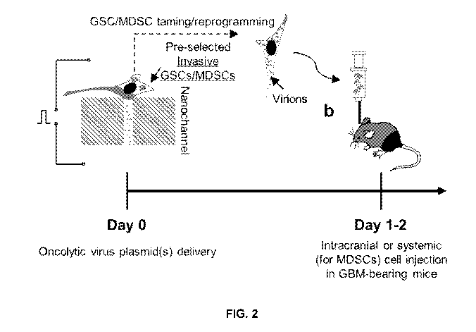
lntegr Biol (Camb)
1:506-512 (2009); Condeelis, J. & Segall, J.E. Nat Rev Cancer 3:921-930
(2003)). Moreover,
current technologies for ex vivo analysis of tissue explants tend to be
laborious and limited
(Johnson, J. et al. Tissue Eng Part C Methods 15:531-540 (2009)). Conventional
in vitro
assays (Boyden, S. J Exp Med 115, 453-466 (1962); Albini, A. & Benelli, R. Nat
Protoc 2,
504-511 (2007); Rao, J.S. Nat Rev Cancer 3, 489-501 (2003); Yamada, K.M. &
Cukierman,
E. Cell 130, 601-610 (2007); Liang, C.C., Park, A.Y. & Guan, J.L. Nat Protoc
2, 329-333
1
CA 03116327 2021-04-13
WO 2020/081966
PCT/US2019/056988
(2007)), on the other hand, are not physiologically-relevant, and/or are end-
point tests that
only focus on the bulk behavior of highly heterogeneous cellular populations.
SUMMARY
[0003] There is a subset of GSCs and MDSCs that exhibit high dissemination and
therapy-resistance capacity. These findings suggest that GSCs and MDSCs are
not
monolithic populations, and that specific clonal subsets exhibit significantly
more
"aggressive" phenotypes, which could presumably be responsible for driving
disease
relapse.
[0004] Disclosed herein is a method of "reprogramming" highly motile cells
found in
lo tumors, such as these highly motile GSC and/or M DSC clones, into "auto-
destructive" cell
"missiles" (referred to herein as therapeutic stealth cells) that can seek and
destroy new foci
of recurrence within the body, such as the brain. Cells with enhanced motility
can be sorted
out from heterogeneous populations and then be rendered "auto-destructive" by
deterministic delivery of an anti-cancer agent, such as an oncolytic virus
plasmid cocktail.
[0005] The disclosed method can involve sorting cells from a subject to create
the
therapeutic stealth cells. In some embodiments, the cells are autologous, such
as a blood
cells or a tumor biopsy from the subject to be treated. However, in some
cases, the cells are
allogenic.
[0006] The disclosed method can involve sorting cells from a subject for a
highly
motile subpopulation and then reprogramming the subpopulation to deliver anti-
cancer
agents. In some embodiments, the subpopulation can be sorted in a migration
assay using a
chemoattractant gradient. In particular, the chemoattractant gradient can
involve a
chemokine produce by the tumor to be treated. For example, in some
embodiments, the
chemoattractant comprises Matrigele. In some embodiments, the chemoattractant
comprises tumor cell conditioned media.
[0007] In some embodiments, the subpopulation is sorted in a migration assay
using
a nanotextured and/or biomimetic surface. For example, MDSCs are responsive
to, and can
be guided along, pre-aligned structural cues in the absence of biochemical
stimulation.
Therefore, in some embodiments, the surface comprise ridges/grooves at the
micro nor
nanoscale. For example, the depth and width of the ridges/grooves can have
dimensions
from 100 nm to 10 pm, including, 100 nm to 1pm, 1pm to 10pm, 500 nm to 5 pm.
The
ridges/grooves can have a variety of shapes and patterns, including straight
grooves.
[0008] In some embodiments, the subpopulation is sorted in a transwell
migration
assay or cell invasion assay. A transwell migration assay measures the number
of cells
2
CA 03116327 2021-04-13
WO 2020/081966
PCT/US2019/056988
passing a porous membrane, whereas a cell invasion assay focuses on invasive
cell
migration via an extracellular matrix.
[0009] Once subpopulation is sorted and optionally expanded, the cells can
then be
reprogrammed to heterologously express a transgene encoding an anti-tumor
protein,
oligonucleotide, or combination thereof.
[0010] The introduction of an efficient "safety switch" can in some cases be
used to
reduce the risk of severe graft-vs-host disease. Therefore, in some
embodiments, the
subpopulation is also reprogrammed with a kill switch system. The most
extensively studied
safety-switch to date is the HSV I-derived thymidine kinase (HSV-TK) gene
product. Non-
lo immunogenic safety switch system have also been developed that involve
fusion proteins
composed of human proapoptotic molecules (e.g. caspase-9) linked to modified
human
FK506-binding proteins (i.e. iCasp9). These safety switches can be activated
by injection of
a chemical inducer of dimerization (CID), consisting of a dimer of two
synthetic variants of
FK506. Other inducible and self-destructive kill switches are in development
and can be
used in the disclosed therapeutic stealth cells.
[0011] Also disclosed is a composition comprising a plurality of therapeutic
stealth
cell produced by the disclosed methods. In particular embodiments, the
composition further
comprises a pharmaceutically acceptable excipient.
[0012] Also disclosed is a method for treating a tumor in a subject,
comprising
administering to the subject an effective amount of the disclosed
pharmaceutical
composition. The disclosed method can be used to treat any solid tumor. In
particular
embodiments, the tumor is matched to the source of cells used to develop the
therapeutic
stealth cells. For example, highly motile MDSCs obtained from a breast tumor
biopsy can be
reprogrammed to treat breast cancer. Likewise, highly motile GSCs/MDSCs
obtained from a
glioblastoma multiforme (GBM) biopsy can be reprogrammed to treat GBM.
[0013] The details of one or more embodiments of the invention are set forth
in the
accompanying drawings and the description below. Other features, objects, and
advantages
of the invention will be apparent from the description and drawings, and from
the claims.
DESCRIPTION OF DRAWINGS
[0014] FIGs. 1A to 1E show results of a migration-based
sorting/"chromatography".
FIG. 1A shows nanotextured surfaces induce guided motility. Clones of with
high motility are
lured into a collection chamber by chemoattraction. FIG. 1B shows studies with
GSCs show
that highly motile clones were resistant to anti-miR363 therapy. FIG. 10 shows
migration-
based sorting of MDSCs uncovered a clonal subset with superior motility
compared to bulk
3
CA 03116327 2021-04-13
WO 2020/081966
PCT/US2019/056988
MDSCs. FIG. 1D shows sorting exhibited a diverse phenotype with granulocytic
(P4) and
monocytic (P5) subtypes. Unclassified subtypes exhibiting either low (P6) or
high (P3) Ly6-
C/G were also present. FIG. 1 E shows MDSC clones with high motility were
either P4 or P3.
* p<0.05.
[0015] FIG. 2 depicts reprogrammed/tamed GSCs and/or MDSCs being
intracranially injected in GBM-bearing mice and tumor progression being
monitored.
[0016] FIG. 3 shows MDSCs exhibit significant motility (i.e., guided) on
textured/
biomimetic surfaces. MDSCs cultured on TOP, on the other hand, exhibit limited
motility.
These results suggest that much like tumor cells, MDSCs may be responsive to
the same
lo structural cues that enhance tumor cell dissemination in vivo. * p<0.05.
[0017] FIGs. 4A and 4B illustrate a migrational chromatography setup, where
MDSCs are selectively seeded on one side of the platform, and induced to
migrate in a
single direction via chemotaxis. The surface nanotexture triggers clone
separation based on
guided motility. FIG. 40 shows velocity for fast- vs. slow-moving cells. FIG.
4D and 4E
contains flow cytometry analysis showing that fast-moving clones have a
distinct phenotype
compared to slow-moving clones (e.g., bulk MDSCs). * p<0.05.
[0018] FIGs. 5A and 5B show single-clone motility assays of circulating MDSCs
from
melanoma patients. Differences in velocity (FIG. 5A) and effective
displacement (FIG. 5B)
for each patient. The results indicate that MDSCs from certain patients
exhibit enhanced
velocities. However, when effective displacement is considered (i.e.,
geometrical distance
from starting to ending location), certain MDSC batches with low velocity
showed significant
displacement, which may be reflective of more directional/persistent motility
(without
chemotaxis). P1: stage IIIC, tx nivolumab+surgery; P2: stage IV, tx nivolumab;
P3: stage IV
V600E/BRAF, tx radiation+pembrolizumab; P4: stage IV, tx nivolumab +
ipilumamab; P5:
stage IV, tx pembrolizumab/ipilumamab/nivolumab; P6: stage IV V600E/BRAF,
treated with
INF-a).
[0019] FIGs. 6A and 6B shows nanotextured surfaces can be used to unmask drug
sensitivities not observed on standard TOP. FIG. 6A show single clone motility
assays of
patient-derived MDSCs show inhibition of a specific clonal subset (average
velocity > 40
pm/h) in response to ibrutinib. FIG. 6B shows single-clone motility on TOP did
not reveal any
effect of ibrutinib on MDSC dissemination.
[0020] FIGs. 7A and 7B illustrates a device for migrational chromatography
with
integrated microfluidics to enable automated detachment of clones of interest.
FIGs. 70 and
7D show that once migration-based separation occurs, the underlying
microfluidic system
4
CA 03116327 2021-04-13
WO 2020/081966
PCT/US2019/056988
can be used to sequentially flow chilled water at given locations, which
facilitates selective
detachment of MDSC clones of interest due to thermal actuation of the PINIPAM
layer.
[0021] FIG. 8A shows co-culturing MDSCs and noncancerous MCF10As led to
enhanced motility in a group of MCF10A clones. FIG. 8B shows
immunofluorescence
analysis indicates that coculture conditions triggered a decrease in the
expression of
epithelial markers such as ECadherin, and an increase in mesenchymal markers
in certain
clones (e.g., Vimentin). FIG. 80 shows coculturing MDSCs with already
aggressive MDAMB-
231 cells did not lead to major changes in motility in the MDA-MB-231
population. =:
monoculture, =: 50:50 co-culture = :90:10 coculture (MDSC:breast cancer/tissue
cells).
lo [0022] FIG. 9. Shows co-culturing MDSCs and breast tissue/cancer cells
led to a
marked increase in velocity for certain MDSC clones, especially when co-
cultured with MDA-
MB-231. These results potentially suggest that MDSC motility is positively
regulated in the
presence of metastatic cells, facilitating co-dissemination outside the tumor,
and continued
immunoprotection during the tumor cell dissemination process.
[0023] FIGs. 10A to 10E show MDSCs are responsive to aligned structural cues
and
exhibit guided dissemination patterns. FIG. 10A is a schematic diagram of the
tumor
microenvironment showing invasive cancer cells and infiltrative MDSCs using
pre-aligned
structural cues (e.g., remodeled ECM, blood vessel walls) to escape and invade
the tumor
stroma, respectively. FIG. 10B is a SEM micrograph (with superimposed MDSC
mock-ups)
of a PDMS-based biomimetic textured surface used to evaluate structurally
guided MDSC
migration at the single-clone level. FIG. 100 shows Actin ¨ Nuclei staining of
MDSCs
cultured on textured vs. control/TOP surfaces. MDSCs assume an aligned/more
migratory
morphology on the textured surfaces compared to TOP. FIGs. 10D and 10E show
single-
clone dissemination tracks (FIG. 10D) and quantification of MDSCs (FIG. 10E)
on textured
vs. control/TOP surfaces confirming enhanced dissemination capabilities (i.e.,
average
single-clone velocity and net track distance) for MDSCs when exposed to pre-
aligned
structural cues. The net track distance is a reflection of the geometrical
distance traveled by
a cell during the tracking period. *p < 0.01 and f p < 0.02 (t-test, n= 4).
[0024] FIGs. 11A to 111 show MDSCs subpopulations exhibit distinct
dissemination
and gene expression patterns. FIGs. 11A and 11B are schematic diagrams of an
experimental design. Here MSC-2 cultures were sorted by flow cytometry into
three distinct
subpopulations, including granulocytic (CD11b+Ly6CI0Ly6G+) and monocytic
(CD11b+Ly6ChiLy6G-) MDSCs, as well as CD11b+Ly6C+Ly6G+ cells. Each population
was
then subjected to single-clone motility assays on textured PDMS and qRT-PCR
analyses of
pro- and anti-inflammatory markers. FIG. 110 shows Actin¨Nucleistaining of
different MSC-2
5
CA 03116327 2021-04-13
WO 2020/081966
PCT/US2019/056988
subtypes cultured on textured surfaces. Granulocytic MDSCs had a tendency to
exhibit a
more aligned and migration-prone morphology compared to their counterparts.
FIG. 11D
shows single-clone dissemination (i.e., average velocities and net track
distances)
quantification for each subtype on textured surfaces. *p=0.006, **p<0.001,
kvp=0.001,
tp=0.09 (2-way ANOVA, n= 4). FIG. 11E shows single-clone tracks for each
population.
FIG. 11F shows fluorescently labeled flow-sorted MDSCs vs. "fresh"/unsorted
MDSCs
injected (i.e., via the tail vein) into tumor-bearing mice (i.e., orthotopic
breast tumor
developed from human cells in nude mice). Photographs to the right depict
tumor
progression/growth from week 1 to week 4. FIG. 11G shows tumors and other
target organs
lo imaged to detect the degree to MDSC infiltration 24 hours post-injectio.
FIGs. 11H and 111
show qRT-PCR analysis of pro-inflammatory (FIG. 11H) and anti-inflammatory
(FIG. 111)
genes for each subtype. *p<0.001, ** p<0.0001, p=0.03 (2-way ANOVA, n= 3-4).
[0025] FIGs. 12A to 12G show single MDSC subpopulations appear to show
phenotypic plasticity that can drive the replenishment the entire phenotypic
spectrum. Fig.
12A is a schematic diagram of the experimental design. FIG. 12B shows single-
clone
dissemination (i.e., average velocities and net track distances) studies did
not show
significant differences between all three populations by day 7. FIG. 120 to
12E show flow
cytometry analyses indicate that while by day 1 post-sorting all
subpopulations remained
relatively pure, by day 7 the entire spectrum of phenotypes had been
replenished regardless
of the phenotype of the starting cell population. *p<0.0001, p=0.01, #p=0.03,
kvp=0.0001 (2-
way ANOVA/Tukey's multiple comparisons, n= 3-4). FIGs. 12F and 12G show qRT-
PCR
analyses of pro-inflammatory (FIG. 12F) and anti-inflammatory (FIG. 12G) genes
at day 7
post-sorting. *p=0.006, **p=0.01 (2-way ANOVA/Tukey's multiple comparisons, n=
3-6).
[0026] FIGs. 13A and 13B show circulating MDSCs derived from melanoma patients
show different dissemination profiles at the single-clone level. FIGs. 13A and
13B show
average single clone velocities (FIG. 13A) and net track distances (FIG. 13B)
had a
tendency to be significantly higher for certain patients compared to the rest
of the patient
population, which could be a reflection of the patient's background.
[0027] FIGs. 14A to 14F show distinct subpopulations of patient-derived MDSCs
show different dissemination capabilities. FIGs. 14A to 140 show melanoma
patient MDSCs
were sorted into granulocytic (CD11b+CD15+CD14-) and monocytic (CD11b+CD15-
CD14+)
subpopulations via flow cytometry. FIGs. 14D to 14F show that similar to
observations in
mouse MDSCs, the granulocytic subpopulation of patient-derived MDSCs also
shows
increased dissemination (i.e., average single-clone velocities and net track
distances)
6
CA 03116327 2021-04-13
WO 2020/081966
PCT/US2019/056988
capabilities compared to the monocytic subtype. *p=0.0005, **p=0.002 (Mann-
Whitney,
n=3).
[0028] FIGs. 15A and 15B show differences in gene expression of pro-
inflammatory
(FIG. 15A) and anti-inflammatory (FIG. 15B) markers as a function of time for
flow
cytometry-sorted subpopulations. *p<0.0001, p=0.06 (2-way ANOVA/Sidak's
multiple
comparisons, n= 3-6).
DETAILED DESCRIPTION
[0029] Before the present disclosure is described in greater detail, it is to
be
understood that this disclosure is not limited to particular embodiments
described, and as
lo such may, of course, vary. It is also to be understood that the
terminology used herein is for
the purpose of describing particular embodiments only, and is not intended to
be limiting,
since the scope of the present disclosure will be limited only by the appended
claims.
[0030] Where a range of values is provided, it is understood that each
intervening
value, to the tenth of the unit of the lower limit unless the context clearly
dictates otherwise,
between the upper and lower limit of that range and any other stated or
intervening value in
that stated range, is encompassed within the disclosure. The upper and lower
limits of these
smaller ranges may independently be included in the smaller ranges and are
also
encompassed within the disclosure, subject to any specifically excluded limit
in the stated
range. Where the stated range includes one or both of the limits, ranges
excluding either or
both of those included limits are also included in the disclosure.
[0031] Unless defined otherwise, all technical and scientific terms used
herein have
the same meaning as commonly understood by one of ordinary skill in the art to
which this
disclosure belongs. Although any methods and materials similar or equivalent
to those
described herein can also be used in the practice or testing of the present
disclosure, the
preferred methods and materials are now described.
[0032] All publications and patents cited in this specification are herein
incorporated
by reference as if each individual publication or patent were specifically and
individually
indicated to be incorporated by reference and are incorporated herein by
reference to
disclose and describe the methods and/or materials in connection with which
the
publications are cited. The citation of any publication is for its disclosure
prior to the filing
date and should not be construed as an admission that the present disclosure
is not entitled
to antedate such publication by virtue of prior disclosure. Further, the dates
of publication
provided could be different from the actual publication dates that may need to
be
independently confirmed.
7
CA 03116327 2021-04-13
WO 2020/081966
PCT/US2019/056988
[0033] As will be apparent to those of skill in the art upon reading this
disclosure,
each of the individual embodiments described and illustrated herein has
discrete
components and features which may be readily separated from or combined with
the
features of any of the other several embodiments without departing from the
scope or spirit
of the present disclosure. Any recited method can be carried out in the order
of events
recited or in any other order that is logically possible.
[0034] Embodiments of the present disclosure will employ, unless otherwise
indicated, techniques of chemistry, biology, and the like, which are within
the skill of the art.
[0035] The following examples are put forth so as to provide those of ordinary
skill in
lo the art with a complete disclosure and description of how to perform the
methods and use
the probes disclosed and claimed herein. Efforts have been made to ensure
accuracy with
respect to numbers (e.g., amounts, temperature, etc.), but some errors and
deviations
should be accounted for. Unless indicated otherwise, parts are parts by
weight, temperature
is in C, and pressure is at or near atmospheric. Standard temperature and
pressure are
defined as 20 C and 1 atmosphere.
[0036] Before the embodiments of the present disclosure are described in
detail, it is
to be understood that, unless otherwise indicated, the present disclosure is
not limited to
particular materials, reagents, reaction materials, manufacturing processes,
or the like, as
such can vary. It is also to be understood that the terminology used herein is
for purposes of
describing particular embodiments only, and is not intended to be limiting. It
is also possible
in the present disclosure that steps can be executed in different sequence
where this is
logically possible.
[0037] It must be noted that, as used in the specification and the appended
claims,
the singular forms "a," "an," and "the" include plural referents unless the
context clearly
dictates otherwise.
[0038] "Suicide gene" as used herein refers to a gene that will cause a cell
to kill
itself through apoptosis. Activation of these genes may be due to many
processes, but the
main cellular "switch" to induce apoptosis is the p53 protein. Stimulation or
introduction
(through gene therapy) of suicide genes may be used to treat cancer or other
proliferative
diseases by making cancer cells more vulnerable, more sensitive to
chemotherapy. Parts of
genes expressed in cancer cells are attached to other genes for enzymes not
found in
mammals that can convert a harmless substance into one that is toxic to the
tumor. The
suicide genes that mediate this sensitivity may encode for viral or bacterial
enzymes that
convert an inactive drug into toxic antimetabolites that inhibit the synthesis
of nucleic acid.
8
CA 03116327 2021-04-13
WO 2020/081966
PCT/US2019/056988
[0039] Highly motile GSC and/or MDSC clones can be sorted from heterogenous
populations by migration-based sorting, such as nano chip-supported single-
clone motility
chromatography. The method of migration-based cell sorting involves
identifying clonal
subsets and/or cell subpopulations that exhibit enhanced dissemination
capabilities
compared to the rest of the population. Such cells are inherently more prone
to
homing/infiltrating to primary tumors and/or metastatic outgrowths, and as
such could serve
as more efficient drug/gene delivery vehicles. Identifying highly
disseminative clonal subsets
could be achieved in many different ways.
[0040] One option is to seed cell mixtures on a micro- or nano-textured
surface with
lo lines, which would induce contact-guided directional migration of the
cells. For example,
MDSCs are responsive to, and can be guided along, pre-aligned structural cues
in the
absence of biochemical stimulation
[0041] Cells could be exposed to a chemoattractant gradient, which would
define a
specific direction in which the cells would migrate, and "fast-moving" clones
could be
progressively collected in a reservoir as they migrate towards the
chemoattractant. Running
this sorting in the absence of a chemoattractant could also be used as a way
to identify
clonal subsets that may be more prone to showing single-direction motility
(i.e., towards the
collection reservoir), even in the absence of a chemoattractant. Even if these
cells are not
necessarily the fastest movers, their ability to exhibit persistent motility
in a single direction
could translate into enhanced ability to disseminate in vivo (e.g., cells with
high migration
velocity but with lack of directionality may not necessarily be the most
effective "infiltrators"),
which would also make these clones desirable for enhanced drug/gene delivery
to the
primary tumor and/or metastatic outgrowths.
[0042] Another way to select cells with enhanced dissemination capabilities
could
be through a translocation assay on a transwell system (e.g. 8 micron pores).
For example,
the cells can be seeded on one the top chamber of the transwell, and cells
with enhanced
dissemination capabilities will gradually translocate across the pores into
the bottom
chamber, where they could be collected for further modification (for gene/drug
delivery
applications).
[0043] As disclosed herein, the disclosed subpopulation of cells are
CD11b+Ly6C10Ly6G+ myeloid-derived suppressor cells. Therefore, in some
embodiments the
highly motile cells are obtained by cell sorting of tumor-derived GSCs and/or
MDSCs using a
combination of antibodies that selectively bind CD11 b, Ly6C, and Ly6G.
[0044] In some embodiments, the cells are derived from primary tumor cells
(e.g.,
isolated from a routine biopsy). In some embodiments, the cells are derived
from myeloid-
9
CA 03116327 2021-04-13
WO 2020/081966
PCT/US2019/056988
derived suppressor cells (e.g., isolated from the circulation). However, the
disclosed
methods could be applied to any other cell type that is prone to infiltrating
into cancerous
tissue (e.g., other monocytes, T cells, etc.).
[0045] Once the pre-selection of highly disseminative clones is complete,
these cells
could be first expanded, and then genetically engineered through various
routes, including
viral or non-viral (e.g., bulk electroporation, tissue nano-transfection)
delivery of transgenes,
and/or CRISPR/CAS9-driven transgene insertion. The goal of this step is to
induce the
production of anti-tumor proteins, oligos, and/or other entities (e.g., glut1,
mir146, oncolytic
viruses, etc.) by these cells. Once genetic engineering of these highly motile
subpopulations
lo is complete, these cells could then be delivered back into the patient,
either systemically
(e.g., in blood, lymphatic system), or locally (into primary tumors or
metastatic ones), with
the intent to eradicate cancerous outgrowths. In some embodiments, the
transgene encodes
tissue inhibitor of metalloproteinase-3 (TIMP-3).
[0046] For cancer applications, these cells can be engineered (through
transfection)
to express pro-inflammatory molecules (ccI4, mir146, glut1 for example) to
promote T cell
infiltration into the tumor, or anti-metastasis components (e.g., timp3) to
prevent cancer
dissemination.
[0047] In some embodiments, MDSCs are used to deliver therapeutics in other
conditions, such as Alzheimer's disease or diabetes, delivering anti-
inflammatory molecules,
or other forms of brain injury (e.g., ischemic stroke), where MDSCs home
naturally, so that
once can deliver therapeutic cargo such as pro-angiogenic and/or pro-neuronal,
or anti-
inflammatory agents.
[0048] These autologous cells could be further engineered (before injecting
them
back into the patient) with a drug-inducible (e.g., doxycycline) "kill switch"
system,
to eradicate the therapeutic cells when their action is no longer needed. Kill-
switch system is
known in the art, and therefore, it is within the purview of one skilled in
the art to select and
employ a suitable kill-switch system,
[0049] A number of embodiments of the invention have been described.
Nevertheless, it will be understood that various modifications may be made
without departing
from the spirit and scope of the invention. Accordingly, other embodiments are
within the
scope of the following claims.
CA 03116327 2021-04-13
WO 2020/081966
PCT/US2019/056988
EXAMPLES
Example 1: Nano-engineering therapeutic stealth cells
[0050] The dissemination capabilities of GSCs, as well as their ability to
evade the
immune system or standard therapies, continue to be major drivers of
lethality. Nanoscale
tools were used to isolate and study a specific subset of GSCs exhibiting high
dissemination
and therapy-resistance capacity (Fig. 1).
[0051] Pilot studies with MDSCs have also revealed a clonal subset with
remarkable
dissemination ability, akin to GSCs (Fig. 1). These findings suggest that GSCs
and MDSCs
are not monolithic populations, and that specific clonal subsets exhibit
significantly more
lo "aggressive" phenotypes, which could presumably be responsible for
driving disease
relapse. Disclosed in this Example is the development of a transformative,
high-risk/high-
reward approach, to minimize GBM recurrence by "reprogramming" highly motile
GSC
and/or MDSC clones into "auto-destructive" cell "missiles" that can seek and
destroy new
foci of recurrence within the brain. GSCs and MDSCs with enhanced motility are
sorted out
from heterogeneous populations via nano chip-supported single-clone motility
"chromatography", as illustrated in Fig. 1. These cells then undergo a limited
clonal
expansion (2-5 passages), and subsequently are rendered "auto-destructive" by
deterministic, nanochannel-based delivery (Gallego-Perez, D. et al.
Nanomedicine 12:399-
409 (2016)) of an oncolytic virus plasmid cocktail. These viruses can kill
cancerous cells
through different mechanisms compared to conventional therapies, and as such
have been
proposed as a potential therapeutic alternative to eradicate GSCs (Cripe,
T.P., et al. Mol
Ther 17:1677-1682 (2009)).
[0052] A series of in vitro studies are conducted to determine the optimum
plasmid
dosage and ratios at which the select subgroup of GSCs or MDSCs are rendered
"auto-
destructive", while retaining superior motility for prolonged periods of time.
These "tamed"
but highly motile GSC/MDSC populations are then intracranially injected
(together and
separately) into GBM-bearing mice, with the intent to have them effectively
disseminate, and
strategically release therapeutic virions throughout the diseased brain (Fig.
2).
[0053] Comparative experiments of systemic delivery of reprogrammed/drugged
MDSCs are also run in GBM-bearing mice in order to verify that these cells are
able home to
the diseased brain and hamper tumor progression. Advanced imaging technologies
(e.g.,
IVIS, PET, MRI) are used to monitor the fate of therapeutic GSCs/MDSCs.
Although cell-
based oncolytic virus therapies have previously shown promising results
compared to direct
treatment with oncolytic virus particles (Power, A.T. & Bell, J.C. Mol Ther
15:660-665
11
CA 03116327 2021-04-13
WO 2020/081966
PCT/US2019/056988
(2007)), a major limitation is that most of the cells that have been studied
so far have
reduced dissemination capabilities, especially when compared to the pace of
intracranial
dissemination of GSCs. Tamed/reprogrammed GSCs or MDSCs, on the other hand,
can
have inherently high intracranial motility capabilities in addition to stealth
ability toward the
immune system, thus allowing them to colonize, surveil and treat the diseased
brain more
effectively.
Example 2: Structurally guided dissemination of mouse MDSCs
[0054] Recent studies indicate that MDSCs are responsive to, and can be guided
along, pre-aligned structural cues (Figs. 3-4), in the absence of biochemical
stimulation. In
lo contrast, single-clone motility assays on tissue culture polystyrene
(TOP) revealed little
dissemination capabilities (Fig. 3), suggesting that much like tumor cells,
MDSC
dissemination/infiltration is presumably favored by structurally guided
migration. Chip-
supported migrational chromatography studies revealed a clonal subset with
enhanced
dissemination capabilities compared to the rest of the population (Fig. 4A-
40), comparable
to highly aggressive cancerous cells. Flow cytometry analyses of fast-moving
clones
revealed that such population was predominately Ly6-Gh1gh/Ly6-Cbw
(granulocytic) and Ly6-
Gh1gh/Ly6-Ch1gh (unidentified). The phenotype of slow-moving clones was more
evenly
distributed between monocytic (Ly6-Gl0w/Ly6-Ch1gh) and granulocytic, as well
as the
unidentified variants Ly6-GiowiLy6_ciow and Ly6-Gh1gh/Ly6-Chigh. Therefore,
MDSCs clearly
have specialized clonal subsets with improved dissemination capabilities,
which presumably
would be more prone to colonizing tumors/ganglia to exert immunosuppression.
Such clonal
subsets could thus represent novel therapeutic targets in the fight against
cancer.
Example 3: Structurally guided dissemination of patient MDSCs
[0055] Next tested was whether patient-derived MDSCs also exhibit structurally
guided migration in the absence of biochemical stimuli. MDSCs isolated from
peripheral
blood of different stage melanoma patients, under different treatment
modalities, were
tracked for -24h. The MDSCs of each patient exhibited unique dissemination
patterns/
signatures, with some patients showing clonal subsets with enhanced mobility
compared to
the bulk population (Fig. 5), which remained clustered below 25pm/h. Of note,
some patients
had MDSCs whose velocity clustered entirely below 25pm/h, possibly indicative
of an
apparently "quiescent" population, presumably reflective of the type of
malignancy, and/or
the modality/stage of the therapy. While more studies are needed to establish
a clear
correlation between these factors and the single-clone dissemination
capabilities/signatures
12
CA 03116327 2021-04-13
WO 2020/081966
PCT/US2019/056988
of MDSCs, which could serve as a proxy for their level of in vivo activity
and/or disease
outcomes, these data further support the notion that MDSCs are not monolithic,
and that
exploring motility-related mechanisms could potentially pave the way for the
development of
not only improved therapies but also new diagnostics/prognosis tools.
Example 4: Structurally guided migration uncovers drug susceptibilities
[0056] Impairing MDSC migration/infiltration into the tumor/ganglia could be a
viable
strategy to reduce the immunosuppressive burden. Inhibitors of Bruton's
tyrosine kinase
(BTK) have been commonly used in the treatment of hematologic cancers. BTK
plays a role
in numerous biological processes, including cell migration. While MDSCs
express BTK,
lo single-clone motility assays on TOP in the presence of ibrutinib (BTK-
inhibitor) did not show
a significant effect on the migration (Fig. 6). In contrast, motility assays
on biomimetic
surfaces appear to show selective targeting of a highly motile subset of MDSCs
(Fig. 5).
Such results indicate that migrationdriven changes (e.g., cytoskeletal
alignment) may partly
modulate drug sensitivity in MDSCs.
Example 5: Biomimetic platforms for migrational chromatography
[0057] In-house nanofabrication expertise (i.e., contact/projection-based
lithography,
and soft-lithography) is leveraged to fabricate pre-aligned structural cues (-
300nm wide)
from polydimethylsiloxane (PDMS). Textured surfaces will then be
functionalized with
thermoresponsive Poly(N-isopropylacrylamide) (PNIPAM) under argon plasma
(30Watts,
-1000microTorr). The PIN IPAM-coated substrates (-100pm thick) will then be
interfaced
with a microfluidic system with arrayed microchannels (50pm wide, 500pm pitch,
independently operated, Fig. 7). These channels are used to selectively flow
chilled water
underneath the textured PDMS and facilitate selective cell detachment via
thermal activation
of the PI NIPAM (i.e., switch from mildly hydrophobic to highly hydrophilic).
Nanotextured
surfaces are characterized by scanning electron (SEM) and atomic force (AFM)
microscopy.
PNIPAM coating will be verified via contact angle measurements at different
temperatures,
X-ray photoelectron spectroscopy (XPS) and Fourier transform infrared
spectroscopy (FTIR).
Example 6: MDSC motility
[0058] MDSCs are isolated from freshly procured tissue (i.e., peripheral
blood, tumor
and lymphoid tissue) of breast cancer tumor patients under protocol OSU-09142
using
standard procedures. Tumor cells/tissue will also be collected using standard
procedures36.
Migrational chromatography will be conducted on the biomimetic surfaces (Figs.
4, 7) using
13
CA 03116327 2021-04-13
WO 2020/081966
PCT/US2019/056988
GM-CSF (200ng/mL) as chemoattractant. Singleclone migration for different
source MDSCs
(i.e., circulating vs. tumor- vs. lymphoid tissue-resident) are recorded via
time lapse
microscopy in a confocal microscope fitted with a culture chamber. Images will
be collected
every 10min for 24-72h, and postprocessed/ analyzed using the manual tracker
plugin in Fiji.
MDSC clones that exhibit different degrees of motility will be isolated by
selectively
"operating" the microchannels of the platform. The cells are partitioned as
high- vs. medium-
vs. low motility depending on the traveled distance from the starting location
(Fig. 7). The
biomechanics (i.e., stiffness and contractility) of these clonal subsets are
then analyzed by
oscillatory AFM, which is a technique developed by co-I Ghadiali to analyze
viscoelastic
lo properties of single cells, and Traction Force Microscopy (TFM), as
described elsewhere.
Moreover, flow cytometry is run for monocytic (CD15+/CD14+) and granulocytic
(CD15+/CD14-) markers, and combinations thereof. To evaluate immunosuppressive
activity
in each clonal subset, they are cultured (at different concentrations) with
CFSE-labeled T
cells, and T cell proliferation are evaluated by flow cytometry. RMPI media
alone, and
SI I NFEKL peptide will be used as negative and positive controls,
respectively. Finally, clonal
subsets with the strongest suppressive activity are further analyzed by single-
cell
sequencing, as described elsewhere.
Example 7: BTK inhibition
[0059] Once highly mobile and/or immunosuppressive clones are identified from
different source MDSCs, the extent to which BTK inhibitors (i.e., ibrutinib)
hamper guided
dissemination is evaluated. First, immunoblotting is used to evaluate the
level of BTK and
phosphorylated BTK (p-BTK) in each clonal subset exposed to 0-10pM ibrutinib.
Each clonal
subset is then plated on the nanotextured surfaces (-103-104cells/cm2), and
guided
migration is monitored via time lapse microscopy while being exposed to 0-10pM
ibrutinib.
Images are processed/analyzed via Fiji. Experiments with ACP-196 (selective
and
irreversible BTK inhibitor) and GDC-0853 (selective and reversible BTK
inhibitor) are run for
comparison purposes. AFM and TFM are used again to evaluate single-cell
stiffness and
contractility, respectively, after exposure to ibrutinib. The effects of BTK
inhibition on MDSC
motility and biomechanics are further evaluated in breast cancer patients
receiving ibrutinib
under the auspices of an OSU CCC-sponsored clinical study that is open and
accruing at
OSU (OSU-18015). Following the acquisition of informed consent, 30cc of
peripheral blood
is drawn pre-treatment and at 2 and 4 weeks after the initiation of therapy.
MDSCs are
isolated and single-clone motility and biomechanics (i.e., AFM and TFM) are
evaluated as
described above.
14
CA 03116327 2021-04-13
WO 2020/081966
PCT/US2019/056988
Example 8: Reciprocal modulation of MDSCs/tumor cell dissemination
[0060] Preliminary studies indicate that MDSCs have specialized clonal subsets
with
improved mobility, which presumably are more prone to colonizing
tumors/lymphoid tissue,
or to co-disseminating along with highly invasive tumor cells to provide
"protective"
immunosuppression early during metastasis. Pilot studies on biomimetic
surfaces (Figs. 8-9)
indicated that the presence of MDSCs triggered enhanced motility and
epithelial to
mesenchymal-like transitions in a clonal subset of non-cancerous breast tissue
cells (Fig.
8A-8B), suggesting that MDSCs may have the ability to induce potentially
cancerous
transformations in healthy tissue. In contrast, MDSCs did not appear to induce
significant
lo changes in the overall motility pattern of highly aggressive breast
cancer cells (Fig. 80).
Interestingly, the most aggressive tumor cells induced the strongest changes
in single-clone
motility in MDSCs (Fig. 9). MDSCs went from single-clone velocities that
clustered
around/below 40pm/h, to velocities that could reach in some cases -100pm/h,
likely due to
enhanced cytokine/chemokine secretions from aggressive tumor cells.
Example 9: Guided Migration Studies at the Single-Clone Level Uncover Possible
Targets of Therapeutic Interest in Tumor-Associated Myeloid-Derived Suppressor
Cell
Populations
[0061] Methods
[0062] Textured PDMS surfaces: microtextured PDMS surfaces were fabricated
from
photolithographically patterned silicon masters via a replica molding process.
A parallel array
of ridges and grooves (2 pm wide, 2 pm tall, spaced by 2 pm) was first
patterned on a silicon
master via standard UV photolithography using S1813 photoresist. A 10:1
mixture of PDMS
with curing agent was then cast on the master and allowed de-gas and cure for
several
hours. The PDMS was then demolded from the master, sterilized and placed on
multi-well
plates for single-cell migration experiments. Scanning electron microscopy
(SEM) was used
to characterize the surface morphology.
[0063] MDSC cultures: the mouse MDSC cell line (MSC-2) was a kind donation
from
Gregoire Mignot. MSC-2 cells were cultured in RPM! 1640 media supplemented
with 25 mM
HEPES, 10% heat-inactivated fetal bovine serum (FBS), 1% antibiotic-
antimycotic, and 1
mM sodium pyruvate. Patient-derived MDSCs were enriched from peripheral blood
using the
RosetteSep HLA-myeloid cell enrichment kit (Stemcell Technologies) followed by
Ficoll-
Paque centrifugation (GE healthcare). MDSC were isolated by subsequent
negative
selection of HLA-DRneg cells using anti-HLA-DR MicroBeads (Miltenyi Biotec)
for 15
CA 03116327 2021-04-13
WO 2020/081966
PCT/US2019/056988
minutes at 4 C and isolated using a MS-MACS column. Samples were acquired with
informed consent under IRB-approved protocols for human subject research.
[0064] Single-cell migration assays: Approximately 1.5x10⁵ MSC-2 cells were
seeded and allowed to adhere on the textured PDMS surfaces or TCP controls in
regular
culture media for several hours. Cells were imaged via time-lapse microscopy
every 10
minutes for over 16 h using a cell culture chamber (Okolab) mounted on an
inverted
microscope. Images were analyzed using the manual tracker plugin in Fiji.
Single-cell
displacement data were then analyzed via MATLAB to determine velocities and
net track
traveled distances.
lo [0065] Flow cytometty-based analysis and sorting: the following
antibodies were
used for the MSC-2 cells: anti-CD11b-FITC, anti-Ly6-C-APC and anti-Ly6-G-PE,
all
purchased from Biolegend. For patient-derived MDSCs, we used anti-CD33-APC,
anti-
CD11b-AP, and anti-HLA-DR-PECy7, purchased from Beckman Coulter. Data were
acquired using an LSRII flow cytometer (BD Biosciences). All colors were
evaluated against
their respective isotype controls and samples with no staining.
[0066] Gene expression analyses: Total RNA was extracted using the TRizol
reagent (ThermoFisher). Reverse transcription reactions were performed using
500-1000 ng
RNA in a 20 .1 reaction with the superscript VI LO cDNA synthesis kit
(ThermoFisher). cDNA
was used as a template to measure the expression levels of pro- and anti-
inflammatory
genes by quantitative real-time PCR using predesigned primers. Real-time PCR
reactions
were performed using the QuantStudio 3 Real-Time PCR System with TaqMan fast
advance
chemistry (Thermo Scientific) with the following conditions: 95 C 10 min, 40
cycles of 95 C
1 min, 60 C 1 min, and 72 C 1 min. Gene expression was normalized against the
house
keeping genes GAPDH and ATP-6.
[0067] Orthotopic tumor xenografts: immunodeficient nude mice (Jackson
Laboratory), 6-8-week-old, were first injected with 1 million human breast
cancer cells (MDA-
MB-231) in the mammary fat pad to generate tumors. After 4 weeks of tumor
development,
sorted MDSC subpopulations were stained using PKH67 green fluorescent cell
linker kit for
general cell membrane labeling (Millipore Sigma) following the instructions
suggested by the
manufacturer. Tumor-bearing mice were then injected with approximately 2.5 x
105 MDSCs
via the tail vein. The mice were then collected 1-day post-injection, and the
tumors, lungs
and spleens were characterized with an IVIS Imaging System (Xenogen Imaging
Technologies). All animal studies were performed in accordance with protocols
approved by
the Laboratory Animal Care and Use Committee of The Ohio State University.
16
CA 03116327 2021-04-13
WO 2020/081966
PCT/US2019/056988
[0068] Statistical analysis: All statistical analyses were run in Sigma Plot
12 or
GraphPad Prism 7. We used n=3-6 replicates per experiment. Specific
information on the
number replicates, statistical tests, and levels of significance can be found
in the figure
legends.
[0069] Results and Discussion
[0070] MDSCs respond to topographical cues and exhibit structurally guided
dissemination patterns. Structurally guided cell dissemination has been known
to play a role
in the escape of cancerous cells from the primary tumor and the establishment
of metastatic
outgrowths in peripheral organs and tissues. Highly aggressive cancer cells
tend to exhibit
lo distinct spreading patterns, disseminating preferentially along pre-
aligned anatomical
microstructures within the tissues, including radially oriented fibrils from
the extracellular
matrix (ECM), white matter tracts, the basal lamina of blood vessels, and the
subpial/subperitoneal spaces, among others (Figure 10A) (Gallego-Perez D, et
al. Lab Chip
2012, 12:4424-4432; Bellail AC, et al. Int J Biochem Cell Biol 2004, 36:1046-
1069; Johnson
J, et al. Tissue Eng Part C Methods 2009, 15:531-540). Micro- and nanoscale
tools have
been used to develop systems that can be utilized to probe cancer cell
motility under these
physiologically relevant conditions (Gallego-Perez D, et al. Lab Chip 2012,
12:4424-4432;
Bellail AC, et al. Int J Biochem Cell Biol 2004, 36:1046-1069; Johnson J, et
al. Tissue Eng
Part C Methods 2009, 15:531-540; lrimia D, et al. lntegr Biol (Camb) 2009,
1:506-512; Doyle
AD, et al. J Cell Biol 2009, 184:481-490; Petrie RJ, et al. Nat Rev Mol Cell
Biol 2009,
10:538-549). While topographical or cell confinement cues have been used to
mimic rapid
and highly directional motility in a wide variety of cancerous cells (Gallego-
Perez D, et al.
Lab Chip 2012, 12:4424-4432; Johnson J, et al. Tissue Eng Part C Methods 2009,
15:531-
540; lrimia D, et al. lntegr Biol (Camb) 2009, 1:506-512; Sidani M, et al. J
Mammary Gland
Biol Neoplasia 2006, 11:151-163; Provenzano PP, et al. BMC Med 2006, 4:38;
Wong IY, et
al. Nat Mater 2014, 13:1063-1071), no studies have looked into the influence
of such cues
on the dissemination/infiltration capabilities of tumor-associated MDSCs. Next
tested was
whether MDSCs respond to topographical cues by exhibiting structurally guided
dissemination patterns similar to invasive cancerous cells. The murine MDSC
cell line, MSC-
2, was used as a model (Stiff A, et al. Cancer Res 2016, 76:2125-2136; Trikha
P, et al.
Oncoimmunology 2016, 5:e1214787). These cells were plated on microtextured
polydimethylsiloxane (PDMS) surfaces (Figure 10B), which were fabricated via
replica
molding from photolithographically fabricated silicon masters, and were
designed as an array
of parallel ridges and grooves with dimensions that have been previously
tested in cancer
cell dissemination studies (approximately 2 pm x 2 pm with 2 pm spacing)
(Gallego-Perez D,
17
CA 03116327 2021-04-13
WO 2020/081966
PCT/US2019/056988
et al. Nano Lett 2016, 16:5326-5332; Gallego-Perez D, et al. Lab Chip 2012,
12:4424-4432;
Kim SH, et al. Cancer Cell 2016, 29:201-213; Gu SQ, et al. Nucleic Acids Res
2016,
44:5811-5819). MDSC motility was monitored at the single-clone level in real
time via time-
lapse microscopy. Cells plated on a standard cell culture surface (i.e.,
tissue culture
polystyrene or TCP) were used for comparison purposes. The results indicate
that MDSCs
show limited motility at the single-clone level on TCP (Figure 10C-10E), with
most cells
exhibiting a rounded morphology (Figure 10C). Textured surfaces, on the other
hand, clearly
induced cytoskeletal and morphological rearrangements (i.e., alignment) in
some of the
MDSCs (Figure 10C), which were conducive to increased motility (Figure 10D,
10E).
lo Average single-clone velocities reached a maximum of approximately 40
p.m h-1 on textured
surfaces compared to approximately 20 p.m h-1 on TCP. Net track distances,
which are a
measure of the effective displacement of a single clone, reached a maximum of
approximately 400 p.m over a period of 16 hours on textured surfaces compared
to <100 p.m
on TCP. Notably, MDSCs migrating on textured surfaces exhibited significant
inter-clonal
variability in the dissemination potential, with cells spanning the whole
spectrum from low to
high motility. In contrast, MDSCs migrating on TCP showed markedly less inter-
clonal
variability. Studies with circulating MDSCs derived from cancer patients
(Figure 13) further
confirmed the existence of highly motile MDSC populations exhibiting marked
inter-clonal
variability, with some clones showing average guided migration velocities of
up to
approximately 200 h-1, and total net displacements that approached 1 mm
over a period
of 16 hours. However, certain populations of patient-derived circulating MDSCs
exhibited
limited overall motility, which could potentially be a direct reflection of
the underlying
malignancy (e.g., type, stage, mutations) and/or concurrent treatment
modalities (Tables 1-
3).
Table 1. Background information for MDSC samples obtained from cancer
patients.
Patient ID Malignancy Stage Mutation Therapy
Nivolumab
P1 Melanoma IIIC +BRAF V600
Surgery
P2 Melanoma IV -BRAF Nivolumab
Radiation
P3 Melanoma IV +BRAF V600
Pembrolizumab
Nivolumab
P4 Melanoma IV -BRAF
1pilimumab
Pembrolizumab
P5 Melanoma IV BRAF unknown
1pilimumab Nivolumab
P6 Melanoma IV +BRAF V600 IFN-Alpha
P7 Melanoma IV NA Leukine
18
CA 03116327 2021-04-13
WO 2020/081966
PCT/US2019/056988
Nivolumab
P8 Melanoma IIB NA Nivolumab
P9 Melanoma IIB NA Nivolumab
Table 2. Single-clone velocity comparisons across patients (One-way
ANOVA/Tukey).
Patient comparison Significantly different? p value
P1 vs. P2 Yes <0.0001
P1 vs. P3 No 0.9993
P1 vs. P4 No 0.9967
P1 vs. P5 No 0.0582
P1 vs. P6 Yes 0.0423
P1 vs. P7 No 0.2149
P1 vs. P8 No 0.6429
P1 vs. P9 No 0.7526
P2 vs. P3 Yes <0.0001
P2 vs. P4 Yes <0.0001
P2 vs. P5 Yes <0.0001
P2 vs. P6 Yes <0.0001
P2 vs. P7 Yes <0.0001
P2 vs. P8 Yes <0.0001
P2 vs. P9 Yes <0.0001
P3 vs. P4 No 0.9104
P3 vs. P5 Yes 0.0181
P3 vs. P6 Yes 0.0121
P3 vs. P7 No 0.0748
P3 vs. P8 No 0.3195
P3 vs. P9 No 0.4792
P4 vs. P5 No 0.3736
P4 vs. P6 No 0.3870
P4 vs. P7 No 0.7764
P4 vs. P8 No 0.9859
P4 vs. P9 No 0.9795
P5 vs. P6 No >0.9999
P5 vs. P7 No 0.9976
P5 vs. P8 No 0.9122
P5 vs. P9 No 0.9972
P6 vs. P7 No 0.9998
P6 vs. P8 No 0.9516
P6 vs. P9 No 0.9996
P7 vs. P8 No 0.9992
P7 vs. P9 No >0.9999
P8 vs. P9 No >0.9999
19
CA 03116327 2021-04-13
WO 2020/081966
PCT/US2019/056988
Table 3. Single-clone net track distance comparisons across patients (One-way
ANOVA/Tukey).
Patient comparison Significantly different? p value
P1 vs. P2 No 0.9996
P1 vs. P3 No 0.2711
P1 vs. P4 Yes 0.0017
P1 vs. P5 Yes 0.0474
P1 vs. P6 Yes 0.0005
P1 vs. P7 Yes 0.0026
P1 vs. P8 Yes 0.0005
P1 vs. P9 No 0.5859
P2 vs. P3 No 0.5493
P2 vs. P4 Yes 0.0071
P2 vs. P5 No 0.1324
P2 vs. P6 Yes 0.0023
P2 vs. P7 Yes 0.0104
P2 vs. P8 Yes 0.0022
P2 vs. P9 No 0.8158
P3 vs. P4 No 0.8720
P3 vs. P5 No 0.9957
P3 vs. P6 No 0.7667
P3 vs. P7 No 0.9140
P3 vs. P8 No 0.7310
P3 vs. P9 No >0.9999
P4 vs. P5 No 0.9999
P4 vs. P6 No >0.9999
P4 vs. P7 No >0.9999
P4 vs. P8 No >0.9999
P4 vs. P9 No 0.97777
P5 vs. P6 No 0.9991
P5 vs. P7 No >0.9999
P5 vs. P8 No 0.9981
P5 vs. P9 No 0.9995
P6 vs. P7 No >0.9999
P6 vs. P8 No >0.9999
P6 vs. P9 No 0.9518
P7 vs. P8 No >0.9999
P7 vs. P9 No 0.9870
P8 vs. P9 No 0.9379
[0071] MDSC subpopulations exhibit different dissemination capabilities. Based
on
the clear inter-clonal variability in motility, we proceeded to further
stratify and probe the
MDSC population via flow cytometry-based sorting into granulocytic
(CD11b+Ly6C1 Ly6G+)
and monocytic (CD11b+Ly6Chty6G-) subpopulations (Figure 11A-11C) based on
standard
MDSC nomenclature (Bronte V, et al. Nat Commun 2016, 7:12150). A subpopulation
of
CD11b+Ly6C+Ly6G+ cells was also identified from the flow cytometry data and
included in
our analyses. Flow-sorted subpopulations were then subjected to structurally
guided motility
CA 03116327 2021-04-13
WO 2020/081966
PCT/US2019/056988
studies on textured surfaces, as described above, in addition to qRT-PCR
analyses of pro-
and anti-inflammatory markers. Single-clone dissemination studies indicate
that when
probed in isolation, granulocytic MDSCs have superior dissemination
capabilities compared
to monocytic MDSCs and the CD11b+Ly6C+Ly6G+ subpopulation (Figure 11D), with
single
clones reaching in some cases average velocities and net displacements of >100
p.m h-1 and
approximately 1.5 mm over a period of 16 hours. And while some clones within
the
monocytic MDSC and CD11b+Ly6C+Ly6G+ subpopulations showed relatively high
average
migration velocities, approximately 50 p.m h-1, net displacements were
considerably limited,
thus suggesting that these cells tend to show very short range and/or
disorganized motility
lo patterns compared to granulocytic MDSCs (Figure 11E). These observations
were further
confirmed via in vivo studies (Figure 11F, 11G), where tumor-bearing mice were
systemically
injected with fluorescently labeled suspensions of sorted vs. "fresh"/unsorted
MDSCs, and
IVIS was used to document MDSC accumulation within the tumor niche vs.
peripheral
organs/tissues. The mice that were injected with granulocytic MDSCs showed
more
pronounced fluorescence signal accumulation within the tumor (Figure 11G).
Parallel single-
clone motility studies with circulating MDSCs derived from cancer patients
(Figure 14) also
suggest that the granulocytic subpopulation (CD11b+CD15+CD14-) exhibits
enhanced
motility compared to the monocytic one (CD11b+CD15-CD14+). MSC-2 cell gene
expression
analysis of pro-inflammatory markers indicate no statistically significant
differences in the
expression of TNF-a, iNOS, and IL-27 between the "fresh" (i.e., unsorted) MDSC
population
and the purified granulocytic, monocytic, and CD11b+Ly6C+Ly6G+ subpopulations.
However,
IL-6 was significantly overexpressed in the fresh population vs. the flow-
sorted
subpopulations. Gene expression analyses of anti-inflammatory markers, on the
other hand,
suggest that the flow-sorted granulocytic subpopulation has a tendency to
overexpress
arginase and IL-10 compared to the fresh and flow-sorted monocytic MDSC and
CD11b+Ly6C+Ly6G+ subpopulations. Altogether, these results suggest that the
granulocytic
MDSC subpopulation appears to be not only more prone to disseminating and
colonizing
cancerous tissue, but also to overexpress anti-inflammatory/suppressive
markers compared
to the monocytic MDSC and the CD11b+Ly6C+Ly6G+ subpopulations.
[0072] MDSC subpopulations show phenotypic plasticity that drives populational
homeostasis under prolonged culture conditions. Following flow-based
purification of the
MSC-2 cells into distinct subpopulations of granulocytic and monocytic MDSCs,
as well as
CD11b+Ly6C+Ly6G+ cells, the cells were maintained in culture for 1-7 days.
Phenotypic
plasticity was evaluated via flow cytometry at days 1 and 7. Single-clone
motility assays and
gene expression analyses were run at day 7 (Figure 12A). Surprisingly, and in
contrast to
21
CA 03116327 2021-04-13
WO 2020/081966
PCT/US2019/056988
what we found immediately after flow-based sorting; no significant differences
were detected
in the dissemination characteristics across all three populations by day 7
(Figure 12B).
Average single-clone velocities stayed within approximately 50 p.m h-1 for all
populations,
while the overall net track distance stayed below approximately 200 p.m. Flow
cytometry
analyses indicated that 1 day post-sorting the purified populations still
comprised the
majority (approximately 80%) of the culture, however, by day 7 the whole
hierarchy of
populations had been reestablished (Figure 120-12E), possibly suggesting a
role for cellular
plasticity in the maintenance of populational homeostasis/heterogeneity in
MDSC
populations. Cell cultures derived from the purified granulocytic
subpopulation (Figure 12C),
lo for example, gave rise to monocytic MDSCs and CD11b+Ly6C+Ly6G+ cells,
with the
monocytic subpopulation showing the sharpest increase from day 1 to 7
(approximately
7-fold change), and the CD11b+Ly6C+Ly6G+ population showing an approximately 3-
fold
increase by day 7. Cultures derived from purified monocytic MDSCs, on the
other hand,
were more prone to giving rise to the CD11b+Ly6C+Ly6G+ population by day 7
(approximately 2.5-fold increase) compared to the granulocytic population.
Finally, cultures
derived from the purified CD11b+Ly6C+Ly6G+ population were more prone to
giving rise to
granulocytic MDSCs by day 7 (approximately 3-fold increase) compared to the
monocytic
MDSCs, which did not show a significant increase between days 1 and 7. Gene
expression
profiles of pro- (Figure 12F) and anti-inflammatory (Figure 12G) markers at
day 7 showed
more subtle differences across populations, with decreased and increased iNOS
and IL-6
expression, respectively, in the "fresh" MDSC population relative to the
sorted/purified
subpopulations. However, when comparing the expression profiles between day 0
(i.e., day
of sorting/purification) and day 7, a more pronounced difference was noted,
with an overall
increase in the expression of pro-inflammatory iNOS for all three populations,
and a
significant decrease in arginasel and 11-10 for the granulocytic subpopulation
only
(Figure 15).
[0073] Micro- and nanoscale technologies have been used extensively to probe
and/or modulate various aspects of cell biology for medical applications
(Gallego-Perez D, et
al. Nano Lett 2016, 16:5326-5332; Gallego-Perez D, et al. Lab Chip 2012,
12:4424-4432;
Kim SH, et al. Cancer Cell 2016, 29:201-213; Gu SQ, et al. Nucleic Acids Res
2016,
44:5811-5819; Minata M, et al. Cell reports 2019, 26:1893-1905; Shukla VC, et
al. Trends in
biotechnology 2018, 36:549-561; Benavente-Babace A, et al. Biosens Bioelectron
2014,
61:298-305; Fei Z, et al. Analytical chemistry 2013, 85:1401-1407; Chang L, et
al. Small
2016, 12:5971-5980; Chang L, et al. Lab Chip 2015, 15:3147-3153; Gallego-Perez
D, et al.
Biomed Microdevices 2012, 14:779-789; Gallego-Perez D, et al. Nanomedicine
2016,
22
CA 03116327 2021-04-13
WO 2020/081966
PCT/US2019/056988
12:399-409; Gallego-Perez D, et al. Nature nanotechnology 2017, 12:974; Wu Y,
et al. Small
2013, 9:2358-2367; Zhao X, et al. Advanced Science 2015, 2; Zhao X, et al.
Anal Chem
2015, 87:3208-3215). Microscale engineering tools were used to demonstrate
that tumor-
associated MDSCs exhibit structurally guided migration patterns, similar to
invasive
cancerous cells. Single-clone motility analyses unmasked clear heterogeneities
within and
across (i.e., for patient-derived MDSCs) MDSC populations, confirming the
presence of
clonal subsets with enhanced dissemination capabilities in both murine and
patient-derived
MDSCs. Follow-up motility studies coupled with flow cytometry-based sorting,
gene
expression analyses, and orthotopic tumor xenograft experiments in nude mice,
suggest that
lo the granulocytic subpopulation is more prone to exhibiting increased
dissemination and
tumor-infiltrative ability, as well as enhanced anti-inflammatory activity,
which could make
this population an attractive therapeutic target in cancer. Subsequent
studies, however,
highlight the remarkably dynamic and plastic nature of such clonal subsets,
with purified
MDSC subpopulations quickly reaching populational homeostasis by giving rise
to the full
spectrum of MDSC phenotypes. While there have been conflicting reports
regarding the
dominant phenotype of tumor-resident MDSCs (i.e., granulocytic vs. monocytic)
(Kumar V, et
al. Trends in immunology 2016, 37:208-220; Hossain F, et al. Cancer immunology
research
2015, 3:1236-1247; Haverkamp JM, et al. European journal of immunology 2011,
41:749-
759; Mairhofer DG, et al. Journal of Investigative Dermatology 2015, 135:2785-
2793; Bozkus
CC, et al. The Journal of Immunology 2015, 195:5237-5250), our single-clone
dissemination
and phenotypic plasticity results point towards a potential mechanism by which
granulocytic
MDSCs are presumably better equipped to infiltrate the tumor niche, where they
could then
remain as granulocytic and/or give raise to monocytic MDSCs depending on
multiple factors,
including the tumor type. Interestingly, single-clone dissemination studies
with circulating
MDSCs derived from cancer patients suggest that MDSC motility could
potentially be
impacted by the patient's background (e.g., type/stage of cancer, treatment
modalities, etc.),
and as such, additional studies are needed to determine whether the
dissemination patterns
of circulating MDSCs, ex vivo, could be used to monitor disease and/or
treatment
progression.
[0074] Unless defined otherwise, all technical and scientific terms used
herein have
the same meanings as commonly understood by one of skill in the art to which
the disclosed
invention belongs. Publications cited herein and the materials for which they
are cited are
specifically incorporated by reference.
[0075] Those skilled in the art will recognize, or be able to ascertain using
no more
than routine experimentation, many equivalents to the specific embodiments of
the invention
23
CA 03116327 2021-04-13
WO 2020/081966
PCT/US2019/056988
described herein. Such equivalents are intended to be encompassed by the
following
claims.
24