Note : Les descriptions sont présentées dans la langue officielle dans laquelle elles ont été soumises.
CA 03117895 2021-04-27
WO 2020/095058
PCT/GB2019/053164
1
METHODS FOR ISOLATING AND EXPANDING CELLS
FIELD OF THE INVENTION
The invention relates to methods for the isolation and/or expansion of non-
haematopoietic tissue-
resident lymphocytes, particularly yo T cells. Such y6 T cells include non-V62
cells, e.g. V61, V63
and VO5 cells and such non-haematopoietic tissues include skin and gut. It
will be appreciated
that such isolated and/or expanded non-haematopoietic tissue-resident
lymphocytes find great
utility in adoptive T cell therapies, chimeric receptor therapies and the
like. The present invention
also relates to both individual cells and populations of cells produced by the
methods described
herein.
BACKGROUND OF THE INVENTION
The growing interest in T cell immunotherapy for cancer has focused on the
evident capacity of
subsets of CD8+ and CD4+ a13 T cells to recognize cancer cells and to mediate
host-protective
functional potentials, particularly when de-repressed by clinically mediated
antagonism of inhibitory
pathways exerted by PD-1, CTLA-4, and other receptors. However, al3 T cells
are MHC-restricted,
which can lead to graft versus host disease.
Gamma delta T cells (y6 T cells) represent a subset of T cells that express on
their surface a
distinct, defining y6 T-cell receptor (TCR). This TCR is made up of one gamma
(y) and one delta
(5) chain. Human y6 TCR chains are selected from three main 6 chains, V61, V62
and V63 and
six y chains. Human y6 T cells can be broadly classified based on their TCR
chains, as certain y
and 6 types are found on cells more prevalently, though not exclusively, in
one or more tissue
types. For example, most blood-resident y6 T cells express a V62 TCR, for
example Vy9V62,
whereas this is less common among tissue-resident y6 T cells, which more
frequently use Vol in
skin and Vy4 in the gut.
The majority of methods for isolating lymphocytes has depended on isolating
those cell types from
the blood. Non-haematopoietic tissue resident lymphocytes, such as ap T cells,
y6 T cells and NK
.. cells, may have properties especially suitable for certain applications,
such as for targeting non-
haematopoietic tumors and other targets. However, isolating such tissue
resident lymphocytes in
clinically relevant quantities has remained a challenge, especially as
clinical doses ranging from
108 cells upwards are required for many indications. Importantly, significant
cell loss during
production means even more starting cells must be generated.
Because non-haematopoietic tissue-resident lymphocytes, particularly 08 T
cells, y6 T cells and
NK cells, are not easily obtainable in high numbers, they have not been well
characterized or
studied for therapeutic applications. Therefore, there is a need in the field
for methods to isolate
CA 03117895 2021-04-27
WO 2020/095058
PCT/GB2019/053164
2
and expand non-haematopoietic tissue-resident lymphocytes, in particular ye T
cells, to quantities
sufficient to study and potentially adapt as therapies, e.g., as adoptive T
cell therapies.
Clark et a/. (2006) J. Invest. Dermatol. 126(5): 1059-70 describes a method of
isolating skin
resident T cells from normal and diseased skin. However, the methods described
therein are
unsuitable for clinical use due the presence of animal products but especially
due to the relatively
low yield of cells isolated, namely less than 106 cells per cm2tissue. The
method described in Clark
et al. uses minced samples which results in deliberate disruption to the
structural integrity of the
tissue sample. W02017072367 and W02018/202808 relate to methods of expanding
non-
haematopoietic tissue-resident ye T cells in vitro by culturing lymphocytes
obtained from non-
haematopoietic tissue in the presence of at least Interleukin-2 (IL-2) and/or
Interleukin-15 (IL-15).
W02015189356 describes a composition for expanding lymphocytes obtained from a
sample
obtained by aphaeresis comprising at least two types of cytokines selected
from IL-2, IL-15 and
IL-21. Therefore, there still remains a need for a method of isolating tissue-
resident non-
haematopoietic lymphocytes, such as from skin, that yields a greater amount of
cells that are
suitable for clinical use.
SUMMARY OF THE INVENTION
According to a first aspect of the invention, there is provided a method for
the isolation of
lymphocytes from a non-haematopoietic tissue sample comprising the steps of:
(i) culturing the non-haematopoietic tissue sample in the presence of:
(a) Interleukin-2 (IL-2) or Interleukin-9 (IL-9);
(b) I nterleukin-15 (IL-15); and
(c) Interleukin-21 (IL-21); and
(ii) collecting a population of lymphocytes cultured from the non-
haematopoietic tissue
sample.
According to a further aspect of the invention, there is provided a method for
the isolation of ye T
cells from a non-haematopoietic tissue sample comprising the steps of:
(i) culturing the non-haematopoietic tissue sample in the presence of:
(a) IL-2 or IL-9;
(b) IL-15; and
(c) IL-21; and
(ii) collecting a population of y6 T cells cultured from the non-
haematopoietic tissue sample.
According to a further aspect of the invention, there is provided a method for
the isolation and
expansion of lymphocytes from a non-haematopoietic tissue sample comprising
the steps of:
CA 03117895 2021-04-27
WO 2020/095058
PCT/GB2019/053164
3
(i) isolating a population of lymphocytes from the non-haematopoietic tissue
sample
according to the method defined herein; and
(ii) further culturing said population of lymphocytes for at least 5 days to
produce an
expanded population of lymphocytes.
According to a further aspect of the invention, there is provided a method for
the isolation and
expansion of y6 T cells from a non-haematopoietic tissue sample comprising the
steps of:
(i) isolating a population of y6 T cells from the non-haematopoietic tissue
sample according
to the method defined herein; and
(ii) further culturing said population of y6 T cells for at least 5 days to
produce an expanded
population of y6 T cells.
BRIEF DESCRIPTION OF THE FIGURES
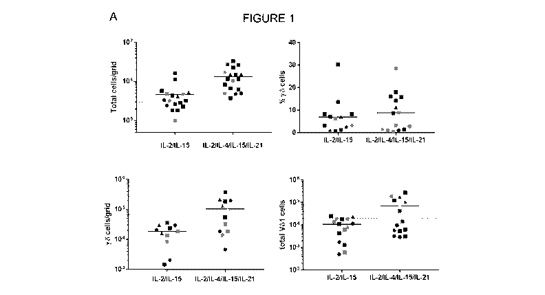
Figure 1: A: Total cell yield and the proportion of y6 T cells and Vol cells
was determined for
isolation methods using 2 cytokines (IL-2 and IL-15) and 4 cytokines (IL-2, IL-
4, IL-15 and IL-21).
B: Proportion of ye T cells and VO1 cells obtained using the 4 cytokine
method, shown as a
percentage compared to the 2 cytokine method.
Figure 2: A: Total cell yield was determined for isolation methods using 2
cytokines `20K' (IL-2
and IL-15), 3 cytokines `3CK' ((IL-2, IL-15 and IL-21) and 4 cytokines `40K'
(IL-2, IL-4, IL-15 and
IL-21) in AIM-V medium + 5% serum replacement in G-REX6. B: Proportion of yo T
cells, and C:
proportion of V61 cells of y6 T cells, also shown.
Figure 3: The phenotype of V61 cells isolated using the 2CK and 4CK method in
AIM-V with 5%
human AB serum in 24 well plates was analysed by measuring percentage TIGIT
and CD27
expression.
Figure 4: The phenotype of V61 cells isolated using the 20K, 30K and 4CK
method in (AIM-V
medium + 5% serum replacement in G-REX6 was analysed by measuring, A:
percentage 0D27
expression, and B: percentage TIGIT expression.
Figure 5: Initial testing comparing total cell yield from 3mm punch biopsies
and standard skin
mincing methods.
Figure 6: y6 cell yield from isolated punch biopsies of varying sizes in AIM-V
with 5% human AB
serum in 24 well plates compared to a minced scalpel sampled control.
CA 03117895 2021-04-27
WO 2020/095058
PCT/GB2019/053164
4
Figure 7: Total cell yield per biopsy (top graph) and per plate (bottom graph)
using different
culturing vessels with AIM-V with 5% human AB serum.
Figure 8: CD27 expression levels in cells isolated using 2 cytokine, 3
cytokine and 4 cytokine
isolation protocol.
Figure 9: The phenotype of V61 cells isolated using the 2CK and 4CK method in
G-REX6 in media
containing 10% human AB serum (left hand results of graph) or 5% serum
replacement (right hand
results of graph) was analysed by measuring A: percentage CD27 expression, and
B: percentage
TIGIT expression. C: PD-1 expression was also measured on ap T cells (CD3+,
pany6- cells)
isolated using 2CK and 4CK method in media containing 10% human AB serum.
Figure 10: Graphs showing comparison of different media used during isolation
methods, as
described in Example 6.
Figure 11: Comparison of total cell yield following 2 week (top graph) or 3
week (bottom graph)
isolation in AIM-V with the indicated serum supplement in G-REX6.
Figure 12: Total cell yield and proportion of V61 cells isolated using AIM-V
media containing 5%
serum replacement (SR) versus human AB serum (AB) at 5% or 10% in G-REX6.
Figure 13: Distribution of cell types in populations isolated using the A: 2CK
or B: 4CK method,
followed by expansion using the 4CK method.
Figure 14: Analysis of the expression of various markers of V61 cells isolated
using the 2CK or
4CK method, followed by expansion using the 4CK method.
Figure 15: Total numbers of y6 cells and V61 cells isolated using the 2CK or
4CK method, followed
by expansion using the 4CK method.
DETAILED DESCRIPTION OF THE INVENTION
According to a first aspect of the invention, there is provided a method for
the isolation of
lymphocytes from a non-haematopoietic tissue sample, said method comprising
the steps of:
(i) culturing the non-haematopoietic tissue sample in the presence of:
(a) Interleukin-2 (IL-2) or Interleukin-9 (IL-9);
(b) Interleukin-15 (IL-15); and
(c) Interleukin-21 (IL-21); and
CA 03117895 2021-04-27
WO 2020/095058
PCT/GB2019/053164
(ii) collecting a population of lymphocytes cultured from the non-
haematopoietic tissue
sample.
According to a further aspect of the invention, there is provided a method for
the isolation of y6 T
5 cells from a non-haematopoietic tissue sample comprising the steps of:
(i) culturing the non-haematopoietic tissue sample in the presence of:
(a) IL-2 or IL-9;
(b) IL-15; and
(c) IL-21; and
(ii) collecting a population of y6 T cells cultured from the non-
haematopoietic tissue sample.
References herein to "isolation" or "isolating" of cells, in particular of
lymphocytes and/or y6 T cells,
refer to methods or processes wherein cells are removed, separated, purified,
enriched or
otherwise taken out from a tissue or a pool of cells. It will be appreciated
that such references
include the terms "separated", "removed", "purified", "enriched" and the like.
Isolation of ye T cells
includes the isolation or separation of cells from an intact non-
haematopoietic tissue sample or
from the stromal cells of the non-haematopoietic tissue (e.g. fibroblasts or
epithelial cells). Such
isolation may alternatively or additionally comprise the isolation or
separation of y6 T cells from
other haematopoietic cells (e.g. ap T cells or other lymphocytes). Isolation
may be for a defined
period of time, for example starting from the time the tissue explant or
biopsy is placed in the
isolation culture and ending when the cells are collected from culture, such
as by centrifugation or
other means for transferring the isolated cell population to expansion culture
or used for other
purposes, or the original tissue explant or biopsy is removed from the
culture. The isolation step
may be for at least about three days to about 45 days. In one embodiment, the
isolation step is for
at least about 10 days to at least 28 days. In a further embodiment, the
isolation step is for at least
14 days to at least 21 days. The isolation step may therefore be for at least
three days, four days,
five days, six days, seven days, eight days, nine days, ten days, 11 days, 12
days, 13 days, 14
days, 15 days, 16 days, 17 days, 18 days, 19 days, 20 days, 21 days, 22 days,
23 days, 24 days,
25 days, 26 days, 27 days, 28 days, 29 days, 30 days, 31 days, 32 days, about
35 days, about 40
days, or about 45 days. It can be appreciated that although isolate cell
proliferation may not be
substantial during this isolation step, it is not necessarily absent. Indeed
for someone skilled in the
art it is recognized that isolated cells may also start to divide to generate
a plurality of such cells
within the isolation vessel containing the tissue and/or scaffold.
Thus, references herein to "isolated y6 T cells", "isolated y6 T cell
population", "isolated population
of y6 T cells", "separated y6 T cells", "separated y6 T cell population" or
"separated population of
y6 T cells" will be appreciated to refer to haematopoietic cells or a
population of haematopoietic
cells including y6 cells that have been isolated, separated, removed, purified
or enriched from a
CA 03117895 2021-04-27
WO 2020/095058
PCT/GB2019/053164
6
non-haematopoietic tissue sample of origin such that the cells are out of
substantial contact with
non-haematopoietic cells or cells contained within the intact non-
haematopoietic tissue. Likewise,
references herein to an "isolated or separated population of Vol T cells"
refer to haematopoietic
cells including V61 T cells that have been isolated, separated, removed,
purified or enriched from
non-haematopoietic tissue sample of origin such that the cells are out of
substantial contact with
non-haematopoietic cells or cells contained within the intact non-
haematopoietic tissue. Therefore,
isolation or separation refers to the isolation, separation, removal,
purification or enrichment of
haematopoietic cells (e.g. y6 T cells or other lymphocytes) from non-
haematopoietic cells (e.g.
stromal cells, fibroblasts and/or epithelial cells).
Methods of isolation of y6 T cells as defined herein may comprise disruption
of the tissue (e.g.
mincing) followed by the separation of y6 T cells from other cell types.
Preferably, methods of
isolation of y6 T cells as defined herein may comprise "crawl-out" of y6 T
cells and other cell types
from an intact non-haematopoietic tissue sample or tissue matrix of the
explant or biopsy, wherein
the tissue resident lymphocytes physically separate from the tissue matrix
without requiring the
disruption of the tissue matrix. By maintaining the integrity of the tissue
matrix, it has been
surprisingly found that the tissue resident lymphocytes preferentially egress
from the tissue matrix
with little or no egress of inhibitory cell types such as fibroblasts, which
are retained in the explant
or biopsy which can then be easily removed at the end of isolation. Thus, in
some embodiments,
the use of an intact non-haematopoietic tissue sample or tissue matrix leads
to a low number of
fibroblasts being released from the tissue into the culture. Such "crawl-out"
methods utilising intact
non-haematopoietic tissue or tissue matrix have the advantage of reducing the
need for excess
processing of the non-haematopoietic tissue sample or tissue matrix, maintain
the structural
integrity of the non-haematopoietic tissue or tissue matrix and may provide
the unexpected
advantage of delivering higher isolated cell yields.
Thus the methods of isolation of non-haematopoietic tissue derived lymphocytes
as defined herein
include methods for isolating non-haematopoietic tissue derived lymphocytes
from an intact biopsy
or explant of non-haematopoietic tissue. Such an intact biopsy or explant is
one wherein the
structural integrity of the biopsy or explant has not been deliberately
disrupted within the perimeter
of the excision removing the biopsy or explant from the tissue sample. Such an
intact biopsy or
explant will have the three dimensional structure largely maintained except
for minor disruption
caused by handling. This intact biopsy or explant therefore has not been
mechanically disrupted,
such as by mincing or chopping, nor chemically enzymatically disrupted, for
example. However,
disrupted tissue may be used in the isolation methods of the present
invention. In one embodiment,
the isolated lymphocyte is an a13 T cell. In an alternative embodiment the
isolated lymphocyte is a
y6 T cell. In another embodiment, the isolated lymphocyte is an NK cell. It
can be appreciated that
more than one type of lymphocyte may be isolated from the same isolation step.
CA 03117895 2021-04-27
WO 2020/095058
PCT/GB2019/053164
7
Methods of isolation of y6 T cells utilising "crawl-out" or e.g. methods as
defined herein, may
include the culturing of the cells and/or non-haematopoietic tissue sample in
the presence of
cytokines and/or chemokines sufficient to induce the isolation or separation
of y6 T cells and/or
other lymphocytes as defined herein. Thus, in one embodiment of the present
invention, isolation
of yo T cells from non-haematopoietic tissue sample comprises culturing the
non-haematopoietic
tissue sample in the presence of IL-2, IL-15 and IL-21. In an alternative
embodiment, isolation of
y6 T cells from non-haematopoietic tissue sample comprises culturing the non-
haematopoietic
tissue sample in the presence of IL-9, IL-15 and IL-21.
In one embodiment, the isolation of ya T cells according to the first aspect
of the invention further
comprises culturing the non-haematopoietic tissue sample in the presence of
Interleukin-4 (IL-4).
Thus, in a further embodiment, the non-haematopoietic tissue sample is
cultured in the presence
of IL-2, IL-15, IL-21 and IL-4. In an alternative further embodiment, the non-
haematopoietic tissue
sample is cultured in the presence of IL-9, IL-15, IL-21 and IL-4.
As used herein, "IL-2" refers to native or recombinant IL-2 or a variant
thereof that acts as an
agonist for one or more IL-2 receptor (IL-2R) subunits (e.g. mutants, muteins,
analogues, subunits,
receptor complexes, fragments, isoforms, and peptidomimetics thereof). Such
agents can support
proliferation of an IL-2-dependent cell line, CTLL-2 (33; American Type
Culture Collection
(ATCCC) TIB 214). Mature human IL-2 occurs as a 133 amino acid sequence (less
the signal
peptide, consisting of an additional 20 N-terminal amino acids), as described
in Fujita, et al.
Ce// 1986. 46.3:401-407. An IL-2 mutein is a polypeptide wherein specific
substitutions to the
Interleukin-2 protein have been made while retaining the ability to bind IL-
2R, such as those
described in US 2014/0046026. The IL-2 muteins can be characterized by amino
acid insertions,
deletions, substitutions and modifications at one or more sites in or at the
other residues of the
native IL-2 polypeptide chain. In accordance with this disclosure any such
insertions, deletions,
substitutions and modifications result in an IL-2 mutein that retains the IL-
2R6 binding activity.
Exemplary muteins can include substitutions of 1, 2, 3,4, 5,6, 7, 8, 9, 10 or
more amino acids.
Nucleic acid encoding human IL-2 can be obtained by conventional procedures
such as
polymerase chain reaction (PCR). The amino acid sequence of human IL-2 (Gene
ID 3558) is
found in Genbank under accession locator NP_000577.2 GI: 28178861. The murine
(Mus
muscu/us) IL-2 amino acid sequence (Gene ID 16183) is found in Genbank under
accession
locator NP_032392.1 GI: 7110653.
IL-2 can also refer to IL-2 derived from a variety of mammalian species,
including, for example,
human, simian, bovine, porcine, equine, and murine. Variants may comprise
conservatively
CA 03117895 2021-04-27
WO 2020/095058
PCT/GB2019/053164
8
substituted sequences, meaning that a given amino acid residue is replaced by
a residue having
similar physiochemical characteristics. Examples of conservative substitutions
include substitution
of one aliphatic residue for another, such as Ile, Val, Leu, or Ala for one
another, or substitutions
of one polar residue for another, such as between Lys and Arg; Glu and Asp; or
Gin and Asn.
Other such conservative substitutions, for example, substitutions of entire
regions having similar
hydrophobicity characteristics, are well known. Naturally occurring IL-2
variants are also
encompassed by the invention. Examples of such variants are proteins that
result from alternate
mRNA splicing events or from proteolytic cleavage of the IL-2 protein, wherein
the IL-2 binding
property is retained. Alternate splicing of mRNA may yield a truncated but
biologically active IL-2
protein. Variations attributable to proteolysis include, for example,
differences in the N- or C-
termini upon expression in different types of host cells, due to proteolytic
removal of one or more
terminal amino acids from the IL-2 protein (generally from 1-10 amino acids).
In some
embodiments, the terminus or interior of the protein can be modified to alter
its physical properties,
for example, with a chemical group such as polyethylene glycol (Yang, etal.
Cancer 1995. 76:
687-694). In some embodiments, the terminus or interior of the protein can be
modified with
additional amino acids (Clark-Lewis, etal. PNAS 1993. 90:3574-3577).
As used herein, "IL-15" refers to native or recombinant IL-15 or a variant
thereof that acts as an
agonist for one or more IL-15 receptor (IL-15R) subunits (e.g. mutants,
muteins, analogues,
subunits, receptor complexes, fragments, isoforms, and peptidomimetics
thereof). IL-15, like IL-2,
is a known T-cell growth factor that can support proliferation of an IL-2-
dependent cell line, CTLL-
2. IL-15 was first reported by Grabstein, et al. (Grabstein, etal. Science
1994.264.5161: 965-969)
as a 114-amino acid mature protein. The term "IL-15," as used herein, means
native or
recombinant IL-15 and muteins, analogs, subunits thereof, or complexes thereof
(e.g. receptor
complexes, e.g. sushi peptides, as described in WO 2007/046006), and each of
which can
stimulate proliferation of CTLL-2 cells. In the CTLL-2 proliferation assays,
supernatants of cells
transfected with recombinantly expressed precursor and in-frame fusions of
mature forms of IL-15
can induce CTLL-2 cell proliferation.
Human IL-15 can be obtained according to the procedures described by
Grabstein, et al.
(Grabstein, et al. Science 1994. 264.5161: 965-969) or by conventional
procedures such as
polymerase chain reaction (PCR). A deposit of human IL-15 cDNA was made with
the ATCCO on
Feb. 19, 1993 and assigned accession number 69245.
The amino acid sequence of human IL-15 (Gene ID 3600) is found in Genbank
under accession
locator NP000576.1 GI: 10835153 (isoform 1) and NP_751915.1 GI: 26787986
(isoform 2). The
murine (Mus muscu/us) IL-15 amino acid sequence (Gene ID 16168) is found in
Genbank under
accession locator NP_001241676.1 GI: 363000984.
CA 03117895 2021-04-27
WO 2020/095058
PCT/GB2019/053164
9
IL-15 can also refer to IL-15 derived from a variety of mammalian species,
including, for example,
human, simian, bovine, porcine, equine, and murine. An IL-15 "mutein" or
"variant", as referred to
herein, is a polypeptide substantially homologous to a sequence of a native
mammalian IL-15 but
that has an amino acid sequence different from a native mammalian IL-15
polypeptide because of
an amino acid deletion, insertion or substitution. Variants may comprise
conservatively substituted
sequences, meaning that a given amino acid residue is replaced by a residue
having similar
physiochemical characteristics. Examples of conservative substitutions include
substitution of one
aliphatic residue for another, such as Ile, Val, Leu, or Ala for one another,
or substitutions of one
polar, residue for another, such as between Lys and Arg; Glu and Asp; or Gln
and Asn. Other such
conservative substitutions, for example, substitutions of entire regions
having similar
hydrophobicity characteristics, are well known. Naturally occurring IL-15
variants are also
encompassed by the invention. Examples of such variants are proteins that
result from alternate
mRNA splicing events or from proteolytic cleavage of the IL-15 protein,
wherein the IL-15 binding
property is retained. Alternate splicing of mRNA may yield a truncated but
biologically active IL-
15 protein. Variations attributable to proteolysis include, for example,
differences in the N- or C-
termini upon expression in different types of host cells, due to proteolytic
removal of one or more
terminal amino acids from the IL-15 protein (generally from 1-10 amino acids).
In some
embodiments, the terminus of the protein can be modified to alter its physical
properties, for
example, with a chemical group such as polyethylene glycol (Yang, et al.
Cancer 1995. 76:687-
694). In some embodiments, the terminus or interior of the protein can be
modified with additional
amino acids (Clark-Lewis, etal. PNAS 1993. 90:3574-3577).
As used herein, "IL-4" refers to native or recombinant IL-4 or a variant
thereof that acts as an
agonist for one or more IL-4 receptor (IL-4R) subunits (e.g. mutants, muteins,
analogues, subunits,
receptor complexes, fragments, isoforms, and peptidomimetics thereof). Such
agents can support
differentiation of naïve helper T cells (Th0 cells) to Th2 cells. Mature human
IL-4 occurs as a 129
amino acid sequence (less the signal peptide, consisting of an additional 24 N-
terminal amino
acids). An IL-4 mutein is a polypeptide wherein specific substitutions to the
Interleukin-4 protein
have been made while retaining the ability to bind IL-4Ra, such as those
described in US Patent
No. 6,313,272. The IL-4 muteins can be characterized by amino acid insertions,
deletions,
substitutions and modifications at one or more sites in or at the other
residues of the native IL-4
polypeptide chain. In accordance with this disclosure any such insertions,
deletions, substitutions
and modifications result in an IL-4 mutein that retains the IL-2Ra binding
activity. Exemplary
muteins can include substitutions of 1,2, 3, 4, 5,6, 7, 8, 9, 10 or more amino
acids.
Nucleic acid encoding human IL-4 can be obtained by conventional procedures
such as
polymerase chain reaction (PCR). The amino acid sequence of human IL-4 (Gene
ID 3565) is
CA 03117895 2021-04-27
WO 2020/095058
PCT/GB2019/053164
found in Genbank under accession locator NG_023252. The murine (Mus muscu/us)
IL-4 amino
acid sequence (Gene ID 16189) is found in Genbank under accession locator
NC_000077.6.
IL-4 can also refer to IL-4 derived from a variety of mammalian species,
including, for example,
5 human, simian, bovine, porcine, equine, and murine. Variants may comprise
conservatively
substituted sequences, meaning that a given amino acid residue is replaced by
a residue having
similar physiochemical characteristics. Examples of conservative substitutions
include substitution
of one aliphatic residue for another, such as Ile, Val, Leu, or Ala for one
another, or substitutions
of one polar residue for another, such as between Lys and Arg; Glu and Asp; or
Gin and Asn.
10 Other such conservative substitutions, for example, substitutions of
entire regions having similar
hydrophobicity characteristics, are well known. Naturally occurring IL-4
variants are also
encompassed by the invention. Examples of such variants are proteins that
result from alternate
mRNA splicing events or from proteolytic cleavage of the IL-4 protein, wherein
the IL-4 binding
property is retained. Alternate splicing of mRNA may yield a truncated but
biologically active IL-4
protein. Variations attributable to proteolysis include, for example,
differences in the N- or C-
termini upon expression in different types of host cells, due to proteolytic
removal of one or more
terminal amino acids from the IL-4 protein (generally from 1-10 amino acids).
In some
embodiments, the terminus of the protein can be modified to alter its physical
properties, for
example, with a chemical group such as polyethylene glycol (Yang, et al.
Cancer 1995. 76:687-
694). In some embodiments, the terminus or interior of the protein can be
modified with additional
amino acids (Clark-Lewis, etal. PNAS 1993. 90:3574-3577).
As used herein, "IL-21" refers to native or recombinant IL-21 or a variant
thereof that acts as an
agonist for one or more IL-21 receptor (IL-21R) subunits (e.g. mutants,
muteins, analogues,
subunits, receptor complexes, fragments, isoforms, and peptidomimetics
thereof). Such agents
can support proliferation of natural killer (NK) and cytotoxic (CD8+) T cells.
Mature human IL-21
occurs as a 133 amino acid sequence (less the signal peptide, consisting of an
additional 22 N-
terminal amino acids). An IL-21 mutein is a polypeptide wherein specific
substitutions to the
Interleukin-21 protein have been made while retaining the ability to bind IL-
21Ra, such as those
described in US Patent No. 9,388,241. The IL-21 muteins can be characterized
by amino acid
insertions, deletions, substitutions and modifications at one or more sites in
or at the other residues
of the native IL-21 polypeptide chain. In accordance with this disclosure any
such insertions,
deletions, substitutions and modifications result in an IL-21 mutein that
retains the IL-21R binding
activity. Exemplary muteins can include substitutions of 1, 2, 3, 4, 5, 6, 7,
8, 9, 10 or more amino
acids.
Nucleic acid encoding human IL-21 can be obtained by conventional procedures
such as
polymerase chain reaction (PCR). The amino acid sequence of human IL-21 (Gene
ID 59067) is
CA 03117895 2021-04-27
WO 2020/095058
PCT/GB2019/053164
11
found in Genbank under accession locator NC_000004.12. The murine (Mus
muscu/us) IL-21
amino acid sequence (Gene ID 60505) is found in Genbank under accession
locator
NC_000069.6.
IL-21 can also refer to IL-21 derived from a variety of mammalian species,
including, for example,
human, simian, bovine, porcine, equine, and murine. Variants may comprise
conservatively
substituted sequences, meaning that a given amino acid residue is replaced by
a residue having
similar physiochemical characteristics. Examples of conservative substitutions
include substitution
of one aliphatic residue for another, such as Ile, Val, Leu, or Ala for one
another, or substitutions
of one polar residue for another, such as between Lys and Arg; Glu and Asp; or
Gin and Asn.
Other such conservative substitutions, for example, substitutions of entire
regions having similar
hydrophobicity characteristics, are well known. Naturally occurring IL-21
variants are also
encompassed by the invention. Examples of such variants are proteins that
result from alternate
mRNA splicing events or from proteolytic cleavage of the IL-21 protein,
wherein the IL-21 binding
property is retained. Alternate splicing of mRNA may yield a truncated but
biologically active IL-
21 protein. Variations attributable to proteolysis include, for example,
differences in the N- or C-
termini upon expression in different types of host cells, due to proteolytic
removal of one or more
terminal amino acids from the IL-21 protein (generally from 1-10 amino acids).
In some
embodiments, the terminus of the protein can be modified to alter its physical
properties, for
example, with a chemical group such as polyethylene glycol (Yang, et al.
Cancer 1995. 76:687-
694). In some embodiments, the terminus or interior of the protein can be
modified with additional
amino acids (Clark-Lewis, etal. PNAS 1993. 90:3574-3577).
As used herein, "IL-9" refers to native or recombinant IL-9 or a variant
thereof that acts as an
agonist for one or more IL-9 receptor (IL-9R) subunits (e.g. mutants, muteins,
analogues, subunits,
receptor complexes, fragments, isoforms, and peptidomimetics thereof). Mature
human IL-9
occurs as a 144 amino acid sequence. An IL-9 mutein is a polypeptide wherein
specific
substitutions to the Interleukin-9 protein have been made while retaining the
ability to bind IL-9R.
IL-9 muteins can be characterized by amino acid insertions, deletions,
substitutions and
modifications at one or more sites in or at the other residues of the native
IL-9 polypeptide chain.
In accordance with this disclosure any such insertions, deletions,
substitutions and modifications
result in an IL-9 mutein that retains the IL-9R binding activity. Exemplary
muteins can include
substitutions of 1, 2, 3, 4, 5, 6, 7, 8, 9, 10 or more amino acids.
Nucleic acid encoding human IL-9 can be obtained by conventional procedures
such as
polymerase chain reaction (PCR). The amino acid sequence of human IL-9 is
given by UniProtKB
P15248.
CA 03117895 2021-04-27
WO 2020/095058
PCT/GB2019/053164
12
IL-9 can also refer to IL-9 derived from a variety of mammalian species,
including, for example,
human, simian, bovine, porcine, equine, and murine. Variants may comprise
conservatively
substituted sequences, meaning that a given amino acid residue is replaced by
a residue having
similar physiochemical characteristics. Examples of conservative substitutions
include substitution
of one aliphatic residue for another, such as Ile, Val, Leu, or Ala for one
another, or substitutions
of one polar residue for another, such as between Lys and Arg; Glu and Asp; or
Gin and Asn.
Other such conservative substitutions, for example, substitutions of entire
regions having similar
hydrophobicity characteristics, are well known. Naturally occurring IL-9
variants are also
encompassed by the invention. Examples of such variants are proteins that
result from alternate
mRNA splicing events or from proteolytic cleavage of the IL-9 protein, wherein
the IL-9 binding
property is retained. Alternate splicing of mRNA may yield a truncated but
biologically active IL-9
protein. Variations attributable to proteolysis include, for example,
differences in the N- or C-
termini upon expression in different types of host cells, due to proteolytic
removal of one or more
terminal amino acids from the IL-9 protein (generally from 1-10 amino acids).
In some
embodiments, the terminus of the protein can be modified to alter its physical
properties, for
example, with a chemical group such as polyethylene glycol (Yang, et al.
Cancer 1995. 76:687-
694). In some embodiments, the terminus or interior of the protein can be
modified with additional
amino acids (Clark-Lewis, etal. PNAS 1993. 90:3574-3577).
In certain embodiments, the methods defined herein include IL-2 typically at a
concentration of at
least 10 IU/mL, such as at least 100 IU/mL (e.g., from 10 IU/mL to 1,000
IU/mL, from 20 IU/mL to
800 IU/mL, from 25 IU/mL to 750 IU/mL, from 30 IU/mL to 700 IU/mL, from 40
IU/mL to 600 IU/mL,
from 50 IU/mL to 500 IU/mL, from 75 IU/mL to 250 IU/mL, or from 100 IU/mL to
200 IU/mL, e.g.,
from 10 IU/mL to 20 IU/mL, from 20 IU/mL to 30 IU/mL, from 30 IU/mL to 40
IU/mL, from 40 IU/mL
to 50 IU/mL, from 50 IU/mL to 75 IU/mL, from 75 IU/mL to 100 IU/mL, from 100
IU/mL to 150
IU/mL, from 150 IU/mL to 200 IU/mL, from 200 IU/mL to 500 IU/mL, or from 500
IU/mL to 1,000
IU/mL). In
certain embodiments, the methods defined herein include IL-2 typically at a
concentration of less than 1,000 IU/mL, such as less than 500 IU/mL. In some
embodiments, the
methods include IL-2 at a concentration of about 100 IU/mL.
In further embodiments, the methods defined herein include IL-15 typically at
a concentration of at
least 0.1 ng/mL, such as at least 10 ng/mL (e.g., from 0.1 ng/mL to 10,000
ng/mL, from 1.0 ng/mL
to 1,000 ng/mL, from 5 ng/mL to 800 ng/mL, from 10 ng/mL to 750 ng/mL, from 20
ng/mL to 500
ng/mL, from 50 ng/mL to 400 ng/mL, or from 100 ng/mL to 250 ng/mL, e.g., from
0.1 ng/mL to 1.0
ng/mL, from 1.0 ng/mL to 5.0 ng/mL, from 5.0 ng/mL to 10 ng/mL, from 10 ng/mL
to 20 ng/mL,
from 20 ng/mL to 100 ng/mL, from 20 ng/mL to 50 ng/mL, from 40 ng/mL to 70
ng/mL, from 50
ng/mL to 100 ng/mL, from 50 ng/mL to 60 ng/mL, from 100 ng/mL to 200 ng/mL,
from 200 ng/mL
to 500 ng/mL, or from 500 ng/mL to 1,000 ng/mL). In further embodiments, the
methods defined
CA 03117895 2021-04-27
WO 2020/095058
PCT/GB2019/053164
13
herein include IL-15 typically at a concentration of less than 500 ng/mL, such
as less 100 ng/mL.
In some embodiments, the methods include IL-15 at a concentration of about 50
ng/mL.
In some embodiments, the isolation of y6 T cells from the non-haematopoietic
tissue sample
includes culture in the presence of both IL-2 and IL-15, each at any of the
concentrations listed
above. In some cases, the concentration of IL-2 is about 100 IU/mL, and the
concentration of IL-
is 55 ng/mL.
In further embodiments, the methods defined herein include IL-21 typically at
a concentration of at
10 least 0.1 ng/mL, such as at least 1.0 ng/mL (e.g., from 0.1 ng/mL to
1,000 ng/mL, from 1.0 ng/mL
to 100 ng/mL, from 1.0 ng/mL to 50 ng/mL, from 2 ng/mL to 50 ng/mL, from 3
ng/mL to 10 ng/mL,
from 4 ng/mL to 8 ng/mL, from 5 ng/mL to 10 ng/mL, from 6 ng/mL to 8 ng/mL,
e.g., from 0.1 ng/mL
to 10 ng/mL, from 1.0 ng/mL to 5 ng/mL, from 1.0 ng/mL to 10 ng/mL, from 1.0
ng/mL to 20 ng/mL).
In further embodiments, the methods defined herein include IL-21 typically at
a concentration of
15 less than 100 ng/mL, such as less 50 ng/mL. In some embodiments, the
methods include IL-21 at
a concentration of about 6 ng/mL, such as about 6.25 ng/mL.
In further embodiments, the methods defined herein include IL-4 typically at a
concentration of at
least 0.1 ng/mL, such as at least 10 ng/mL (e.g., from 0.1 ng/mL to 1,000
ng/mL, from 1.0 ng/mL
to 100 ng/mL, from 1.0 ng/mL to 50 ng/mL, from 2 ng/mL to 50 ng/mL, from 3
ng/mL to 40 ng/mL,
from 4 ng/mL to 30 ng/mL, from 5 ng/mL to 20 ng/mL, from 10 ng/mL to 20 ng/mL,
e.g., from 0.1
ng/mL to 50 ng/mL, from 1.0 ng/mL to 25 ng/mL, from 5 ng/mL to 25 ng/mL). In
further
embodiments, the methods defined herein include IL-4 typically at a
concentration of less than 100
ng/mL, such as less 50 ng/mL, in particular less than 20 ng/mL. In some
embodiments, the
methods include IL-4 at a concentration of about 15 ng/mL.
References herein to "non-haematopoietic tissues" or "non-haematopoietic
tissue sample" include
skin (e.g. human skin) and gut (e.g. human gut). Non-haematopoietic tissue is
a tissue other than
blood, bone marrow, or thymus tissue. In one embodiment, the non-
haematopoietic tissue sample
is skin (e.g. human skin). In a further embodiment, the non-haematopoietic
tissue sample is gut
or gastrointestinal tract (e.g. human gut or human gastrointestinal tract). In
some embodiments,
the lymphocytes and/or y6 T cells are not obtained from particular types of
samples of biological
fluids, such as blood or synovial fluid. In some embodiments, the non-
haematopoietic tissue
sample from which the lymphocytes and/or y6 T cells are isolated according to
the methods defined
herein is skin (e.g. human skin), which can be obtained by methods known in
the art. Alternatively,
the methods of isolation of lymphocytes and/or yo T cells provided herein can
be applied to the
gastrointestinal tract (e.g. colon or gut), mammary gland, lung, prostate,
liver, spleen, pancreas,
uterus, vagina and other cutaneous, mucosal or serous membranes. The
lymphocytes and/or yo
CA 03117895 2021-04-27
WO 2020/095058
PCT/GB2019/053164
14
T cells may also be resident in human cancer tissue samples, e.g. tumours of
the breast or
prostate. In some embodiments, the lymphocytes and/or y6 T cells may be from
human cancer
tissue samples (e.g. solid tumour tissues). In other embodiments, the
lymphocytes and/or y6 T
cells may be from non-haematopoietic tissue sample other than human cancer
tissue (e.g. a tissue
without a substantial number of tumour cells). For example, the lymphocytes
and/or y6 T cells
may be from a region of skin (e.g. healthy skin) separate from a nearby or
adjacent cancer tissue.
Thus, in some embodiments, the y6 T cells are not obtained from human cancer
tissue. In further
embodiments, the lymphocytes are not obtained from a human cancer tissue.
In one embodiment the non-haematopoietic tissue sample of the methods defined
herein has been
obtained from a human. In an alternative embodiment, the non-haematopoietic
tissue sample of
the methods defined herein has been obtained from a non-human animal subject.
Methods for obtaining such tissues are known in the art. Examples of such
methods include
scalpel explant or punch biopsy and may vary in size according to the method.
In some
embodiments, the non-haematopoietic tissue sample is obtained by punch biopsy.
In some embodiments of the present invention, the non-haematopoietic tissue
sample is an intact
biopsy. References herein to "intact" biopsy or "explant" include tissue and
tissue sample that is
not substantially disrupted, or not disrupted, such that the structural
integrity of the biopsy or
explant has not been deliberately disrupted within the perimeter of the
excision removing the
biopsy or explant from the tissue sample. Such an intact biopsy or explant
will have the three
dimensional structure largely maintained except for minor disruption caused by
handling. This
intact biopsy or explant therefore has not been mechanically disrupted, such
as by mincing or
chopping, nor chemically enzymatically disrupted, for example. An intact
biopsy or intact tissue
sample may comprise the whole tissue, the complete tissue, a portion of the
tissue or all elements
of said tissue. For example, in one embodiment the intact biopsy comprises all
layers of the skin.
In a further embodiment, the biopsy comprises the epidermal and dermal layers
of the skin. It will
be appreciated that in such embodiments wherein the biopsy is intact,
separation and distinction
between such layers is maintained. Thus, references herein to "intact"
additionally include biopsies
of full thickness of the non-haematopoietic tissue sample.
Thus, in one particular embodiment of the present invention, the non-
haematopoietic tissue sample
is not minced. In further embodiments, the intact biopsy is a punch biopsy. In
a yet further
embodiment, the intact biopsy is obtained by punch biopsy. Embodiments
presented herein where
the non-haematopoietic tissue sample is an intact biopsy provide the
surprising advantage of
obtaining high numbers of isolated or separated cells from non-minced and/or
intact non-
haematopoietic tissue sample. Furthermore, cells obtained from non-minced
and/or intact non-
CA 03117895 2021-04-27
WO 2020/095058
PCT/GB2019/053164
haematopoietic tissue sample according to the methods defined herein, as
demonstrated herein,
may retain a phenotype useful for subsequent expansion and/or engineering
methods known in
the art.
5 In a further embodiment the intact biopsy is skin (e.g. human skin) or
the intact biopsy is gut (e.g.
human gut). In one embodiment, the non-haematopoietic tissue sample has a
minimum cross-
section of at least 1mm. It will be understood that "minimum cross-section"
refers to the minimum
or shortest length measured through the centroid of the tissue sample. It will
be further understood
that "maximum cross-section" refers to the maximum or longest length measured
through the
10 centroid of the tissue sample. The term "centroid" as used herein is the
average or mean position
of all points of the tissue sample. It will be appreciated that, according to
further embodiments, the
non-haematopoietic tissue sample has a minimum cross-section of at least 2mm,
at least 3mm, at
least 4mm, at least at least 5mm, at least 6mm, at least 7mm or at least 8mm.
In further
embodiments, the non-haematopoietic tissue sample has a minimum cross section
of 8mm or less,
15 7mm or less, 6mm or less, 5mm or less, 4mm or less, 3mm or less or 2mm
or less. In one
embodiment, the non-haematopoietic tissue sample has a minimum cross-section
of between
1mm and 8mm (inclusive), such as between 2mm and 4mm. In one particular
embodiment, the
non-haematopoietic tissue sample has a minimum cross-section of about 3mm. In
one particular
embodiment, the non-haematopoietic tissue sample has a cross-section of about
3mm. It will be
appreciated that, according to further embodiments, the non-haematopoietic
tissue sample has a
maximum cross-section of at least 2mm, at least 3mm, at least 4mm, at least at
least 5mm, at least
6mm, at least 7mm or at least 8mm. In further embodiments, the non-
haematopoietic tissue sample
has a maximum cross section of 8mm or less, 7mm or less, 6mm or less, 5mm or
less, 4mm or
less, 3mm or less or 2mm or less. In one embodiment, the non-haematopoietic
tissue sample has
a maximum cross-section of between lmm and 8mm (inclusive), such as between
2mm and 4mm.
In one particular embodiment, the non-haematopoietic tissue sample has a
maximum cross-
section of about 3mm.
According to further embodiments, the non-haematopoietic tissue sample has a
minimum cross-
sectional area of at least 1mm2. It will be understood that "minimum cross-
sectional area" refers to
the area of the smallest cross-section measured about the centroid of the
tissue sample. It will be
further understood that "maximum cross-sectional area" refers to the area of
the largest cross-
section measured about the centroid of the tissue sample. The term "centroid"
as used herein is
the average or mean position of all points of the tissue sample. In a further
embodiment, the non-
haematopoietic tissue sample has a minimum cross-sectional area of at least
2mm2, at least 3mm2,
at least 4mm2, at least 5mm2, at least 6mm2, at least 7mm2, at least 8mm2, at
least 9mm2 or at
least 10mm2. In further embodiments, the non-haematopoietic tissue sample has
a minimum
cross-sectional area of 50mm2 or less, 40mm2 or less, 30mm2 or less, 25mm2 or
less, 20mm2 or
CA 03117895 2021-04-27
WO 2020/095058
PCT/GB2019/053164
16
less, 15mm2 or less, 10mm2 or less or 8mm2 or less. In one embodiment, the non-
haematopoietic
tissue sample has a minimum cross-sectional area of between 1mm2 and 50mm2,
such as between
3mm2 and 12mm2. In one particular embodiment, the non-haematopoietic tissue
sample has a
minimum cross-sectional area of about 7mm2. In a further embodiment, the non-
haematopoietic
tissue sample has a maximum cross-sectional area of at least 2mm2, at least
3mm2, at least 4mm2,
at least 5mm2, at least 6mm2, at least 7mm2, at least 8mm2, at least 9mm2 or
at least 10mm2. In
further embodiments, the non-haematopoietic tissue sample has a maximum cross-
sectional area
of 50mm2 or less, 40mm2 or less, 30mm2 or less, 25mm2 or less, 20mm2 or less,
15mm2 or less,
10mm2 or less or 8mm2 or less. In one embodiment, the non-haematopoietic
tissue sample has a
maximum cross-sectional area of between 1mm2 and 50mm2, such as between 3mm2
and 12mm2.
In one particular embodiment, the non-haematopoietic tissue sample has a
maximum cross-
sectional area of about 7mm2.
According to further embodiments, the non-haematopoietic tissue sample has a
volume of at least
5mm3. In a further embodiment, the non-haematopoietic tissue sample has a
volume of at least
8nnm3, at least 10nnm3, at least 15nnm3, at least 20nnm3, at least 25nnm3, at
least 30nnm3, at least
35mm3, at least 40mm3, at least 50mm3, or at least 60mm3. In further
embodiments, the non-
haematopoietic tissue sample has a volume of 250mm3 or less, 200mm3 or less,
such as 180mm3
or less, 1600mm3or less, 140mm3 or less, 120mm3 or less, 100mm3 or less, 80mm3
or less, 60mm3
or less, 50mm3 or less or 40mm3 or less. In one embodiment, the non-
haematopoietic tissue
sample has volume of between 5mm3 and 250mm3, such as between 15mm3 and 65mm3.
In one
particular embodiment, the non-haematopoietic tissue sample has a volume of
about 35mm3.
In one embodiment, the non-haematopoietic tissue sample is a punch biopsy. A
punch biopsy
may be of any shape, though is conveniently of circular cross-section and
suitably is at least 1mm
in diameter. In yet further embodiments, the non-haematopoietic tissue sample
comprises a punch
biopsy at least 2mm in diameter, such as at least 3mm in diameter, at least
4mm in diameter, at
least 5mm in diameter, at least 6mm in diameter, at least 7mm in diameter or
at least 8mm in
diameter. In further embodiments, the non-haematopoietic tissue sample
comprises a punch
biopsy 8mm or less in diameter, such as 7mm or less in diameter, 6mm or less
in diameter, 5mm
or less in diameter or 3mm or less in diameter. In one embodiment, the non-
haematopoietic tissue
sample comprises a punch biopsy of between 1mm and 8mm in diameter, such as
between 2mm
and 4mm in diameter. In a particular embodiment, the non-haematopoietic tissue
sample
comprises a punch biopsy of 3mm in diameter.
In certain embodiments, the non-haematopoietic tissue sample comprises a
biopsy (e.g. a punch
biopsy, in particular a punch biopsy of circular cross-section) according to
the sizes, areas,
volumes and/or diameters defined above and the maximum depth is determined by
the site from
CA 03117895 2021-04-27
WO 2020/095058
PCT/GB2019/053164
17
which the biopsy is obtained (although the depth may be reduced). In one
embodiment, the biopsy
is a skin biopsy and comprises the epidermal and dermal layers. In a further
embodiment, the
biopsy does not substantially comprise the subcutaneous fat. Thus, in one
embodiment, the biopsy
comprises epidermal and dermal layers and does not substantially comprise a
layer of
subcutaneous fat. In a further embodiment, the biopsy comprises no
subcutaneous fat.
Alternatively, the subcutaneous fat is not removed, therefore is present (or
at least partially
present) in the biopsy. Thus, in a yet further embodiment, the biopsy consists
of epidermal and
dermal layers. In one embodiment, the biopsy comprises the full thickness of
the non-
haematopoietic tissue sample.
Methods of the present invention comprise culturing non-haematopoietic tissue
sample as defined
herein. References herein to "culturing" include the addition of cells and/or
a non-haematopoietic
tissue sample, including isolated, separated, removed, purified or enriched
cells from non-
haematopoietic tissue sample, to media comprising growth factors and/or
essential nutrients
required and/or preferred by the cells and/or non-haematopoietic tissue
sample. It will be
appreciated that such culture conditions may be adapted according to the cells
or cell population
to be isolated from the non-haematopoietic tissue sample according to the
invention or may be
adapted according to the cells or cell population to be isolated and expanded
from the non-
haematopoietic tissue sample.
In certain embodiments, culturing of the non-haematopoietic tissue sample is
for a duration of time
sufficient for the isolation of y6 T cells from the non-haematopoietic tissue
sample. In alternative
embodiments, the culturing of non-haematopoietic tissue sample is for a
duration of time sufficient
for the isolation of lymphocytes other than y6 T cells from the non-
haematopoietic tissue sample
(e.g. ap T cells and/or NK (natural killer) cells). In certain embodiments,
the duration of culture
according to the methods defined herein is at least 14 days. In certain
embodiments, the duration
of culture according to the methods defined herein is less than 45 days, such
as less than 30 days,
such as less than 25 days. In a further embodiment, the duration of culture
according to the
methods defined herein is between 14 days and 35 days, such as between 14 days
and 21 days.
In a yet further embodiment, the duration of culture according to the methods
defined herein is
about 21 days.
In particular embodiments of the present invention, the lymphocytes and/or y6
T cells isolated
according to methods as defined herein are collected from the culture of non-
haematopoietic tissue
sample after culturing of the non-haematopoietic tissue sample. Collection of
the lymphocytes
and/or y6 T cells as defined herein may include the physical collection of
lymphocytes and/or y6 T
cells from the culture, isolation of the lymphocytes and/or y6 T cells from
other lymphocytes (e.g.
ap T cells, y6 T cells and/or NK cells) or isolation and/or separation of the
lymphocytes and/or yO T
CA 03117895 2021-04-27
WO 2020/095058
PCT/GB2019/053164
18
cells from stromal cells (e.g. fibroblasts). In one embodiment, lymphocytes
and/or y6 T cells are
collected by mechanical means (e.g. pipetting). In a further embodiment,
lymphocytes and/or yo
T cells are collected by means of magnetic separation and/or labelling. In a
yet further
embodiment, the lymphocytes and/or yO T cells are collected by flow cytometric
techniques such
.. as FACS. Thus, in certain embodiments, the y6 T cells are collected by
means of specific labelling
the yo T cells. In further embodiments, the lymphocytes are collected by means
of specific labelling
of the lymphocytes to distinguish them from other cells within the culture. It
will be appreciated
that such collection of lymphocytes and/or yo T cells may include the physical
removal from the
culture of the non-haematopoietic tissue sample, transfer to a separate
culture vessel or to
separate or different culture conditions.
It will be appreciated that such collecting of lymphocytes and/or ye T cells
is performed after a
duration of time sufficient to achieve an isolated population of lymphocytes
and/or yO T cells from
the non-haematopoietic tissue sample. In certain embodiments, the lymphocytes
and/or y6 T cells
are collected after at least one week, at least 10 days, at least 11 days, at
least 12 days, at least
13 days or at least 14 days of culturing of the non-haematopoietic tissue
sample. Suitably, the
lymphocytes and/or yo T cells are collected after 40 days or less, such as 38
days or less, 36 days
or less, 34 days or less, 32 days or less, 30 days or less, 28 days or less,
26 days or less or 24
days or less. In one embodiment, the lymphocytes and/or y6 T cells are
collected after at least 14
.. days of culturing of the non-haematopoietic tissue sample. In a further
embodiment, the
lymphocytes and/or yO T cells are collected after 14 to 21 days of culturing
of the non-
haematopoietic tissue sample.
In certain embodiments of the present invention, the non-haematopoietic tissue
sample is cultured
in media which is substantially free of serum (e.g. serum-free media or media
containing a serum-
replacement (SR)). Thus, in one embodiment, the non-haematopoietic tissue
sample is cultured
in serum-free media. Such serum free medium may also include serum replacement
medium,
where the serum replacement is based on chemically defined components to avoid
the use of
human or animal derived serum. In an alternative embodiment, the non-
haematopoietic tissue
sample is cultured in media which contains serum (e.g. human AB serum or fetal
bovine serum
(FBS)). In one embodiment, the non-haematopoietic tissue sample is cultured in
media which
contains serum-replacement. In one embodiment, the non-haematopoietic tissue
sample is
cultured in media which contains no animal-derived products.
It will be appreciated that embodiments according to the invention wherein the
non-haematopoietic
tissue sample is cultured in serum-free media have the advantage of avoiding
issues with filtration,
precipitation, contamination and supply of serum. Furthermore, animal derived
products are not
favoured for use in clinical grade manufacturing of human therapeutics. As can
be seen herein,
CA 03117895 2021-04-27
WO 2020/095058
PCT/GB2019/053164
19
the inventors have also surprisingly found that the use of serum-free media
for the isolation of cells,
particularly V61 y6 cells, substantially increases the number of cells
obtained from non-
haematopoietic tissue sample compared to the use of media containing AB serum.
In particular,
isolation of y6 T cells from non-haematopoietic tissue sample cultured in
serum-free media
increases the yield of V61 cells.
In one embodiment, the methods as defined herein are performed in an isolation
vessel. Reference
to an "isolation vessel" refers to a vessel comprising the non-haematopoietic
tissue sample for
separation of the lymphocytes and/or y6 T cells, optionally further comprising
a synthetic scaffold.
It will be noted that the isolation vessel may be used just for the isolation
method and not for the
further expansion steps.
In one embodiment, the methods as defined herein are performed in a vessel
(e.g. an isolation
vessel) comprising a gas permeable material. Such materials are permeable to
gases such as
oxygen, carbon dioxide and/or nitrogen to allow gaseous exchange between the
contents of the
vessel and the surrounding atmosphere. It will be appreciated that references
herein to "vessel"
include culture dishes, culture plates, single-well dishes, multi-well dishes,
multi-well plates, flasks,
multi-layer flasks, bottles (such as roller bottles), bioreactors, bags, tubes
and the like. Such
vessels are known in the art for use in methods involving expansion of non-
adherent cells and
other lymphocytes. However, as shown herein, vessels comprising a gas
permeable material also
surprisingly find utility in the isolation of y6 T cells which are considered
as usually being adherent.
The use of such vessels for culturing was found to greatly increase the yield
of isolated y6 T cells
from non-haematopoietic tissue sample. Such vessels were also found to
preferentially support y6
T cells and other lymphocytes over fibroblasts and other stromal cells (e.g.
epithelial cells),
including adherent cell-types. Thus, in one embodiment, the vessels comprising
a gas permeable
material as defined herein preferentially support y6 T cells and other
lymphocytes (e.g. a13 T cells
and/or NK cells). In a further embodiment, fibroblasts and/or other stromal
cells (e.g. epithelial
cells) are absent from cultures performed in vessels comprising a gas
permeable material.
Such vessels comprising gas permeable materials may additionally comprise a
gas permeable
material that is non-porous. Thus, in one embodiment, the gas permeable
material in non-porous.
In some embodiments, the gas permeable material is a membrane film such as
silicone,
fluoroethylene polypropylene, polyolefin, or ethylene vinyl acetate copolymer.
Furthermore, such
vessels may comprise only a portion of gas permeable material, gas permeable
membrane film or
non-porous gas permeable material. Thus, according to a yet further
embodiment, the vessel
includes a top, a bottom and at least one sidewall, wherein at least part of
the said vessel bottom
comprises a gas permeable material that is in a substantially horizontal plane
when said top is
above said bottom. In one embodiment, the vessel includes a top, a bottom, and
at least one
CA 03117895 2021-04-27
WO 2020/095058
PCT/GB2019/053164
sidewall, wherein at least a part of said bottom comprises the gas permeable
material that is in a
horizontal plane when said top is above said bottom. In a further embodiment,
the vessel includes
a top, a bottom and at least one sidewall, wherein the said at least one
sidewall comprises a gas
permeable material which may be in a vertical plane when said top is above
said bottom, or may
5 be a horizonal plane when said top is not above said bottom. It will be
appreciated that in such
embodiments, only a portion of said bottom or said side wall may comprise a
gas permeable
material. Alternatively, the entire of said bottom or entire of said sidewall
may comprise a gas
permeable material. In a yet further embodiment, said top of said vessel
comprising a gas
permeable material may be sealed, for example by utilisation of an 0-ring.
Such embodiments will
10 be appreciated to prevent spillage or reduce evaporation of the vessel
contents. Thus, in certain
embodiments, the vessel comprises a liquid sealed container comprising a gas
permeable material
to allow gas exchange. In alternative embodiments, said top of said vessel
comprising a gas
permeable material is in the horizonal plane and above said bottom and is not
sealed. Thus, in
certain embodiments, said top is configured to allow gas exchange from the top
of the vessel. In
15 further embodiments, said bottom of the gas permeable container is
configured to allow gas
exchange from the bottom of the vessel. In a yet further embodiment, said
vessel comprising a
gas permeable material may be a liquid sealed container and further comprise
inlet and outlet ports
or tubes. Thus, in certain embodiments, the vessel comprising a gas permeable
material includes
a top, a bottom and optionally at least one sidewall, wherein at least a part
of said top and said
20 bottom comprise a gas permeable material and, if present, at least part
of the at least one sidewall
comprises a gas permeable material. Example vessels are described in
W02005035728 and
US9255243 which are herein incorporated by reference. These vessels are also
commercially
available, such as the G-REX cell culture devices provided by Wilson Wolf
Manufacturing, such
as the G-REX6 well-plate, G-REX24 well-plate and the G-REX10 vessel.
In one embodiment, the non-haematopoietic tissue sample is placed on a
synthetic scaffold. As
used herein, a "synthetic scaffold," "scaffold," and "grid" are used
interchangeably and refer to a
non-native three-dimensional structure suitable to support cell growth. A non-
haematopoietic
tissue sample may be either placed on or adhered to a synthetic scaffold to
facilitate lymphocyte
egress from the explant onto the scaffold. Synthetic scaffolds may be
constructed from natural
and/or synthetic materials such as polymers (e.g. natural or synthetic
polymers, such as poly vinyl
pyrolidones, polymethylmethacrylate, methyl cellulose, polystyrene,
polypropylene, polyurethane),
ceramics (e.g. tricalcium phosphate, calcium aluminate, calcium
hydroxyapatite), or metals (e.g.
tantalum, titanium, platinum and metals in the same element group as platinum,
niobium, hafnium,
tungsten and combinations of alloys thereof). In one embodiment of the present
invention, the
synthetic scaffold is tantalum coated. Biological factors (e.g. collagens
(such as collagen I or
collagen II), fibronectins, laminins, integrins, angiogenic factors, anti-
inflammatory factors,
glycosaminoglycans, vitrogens, antibodies and fragments thereof, cytokines
(e.g. IL-2, IL-15, IL-4,
CA 03117895 2021-04-27
WO 2020/095058
PCT/GB2019/053164
21
IL-21, IL9 and combinations thereof) may be coated onto the scaffold surface,
encapsulated within
the scaffold material or added to the media to enhance cell adhesion,
migration, survival, or
proliferation, according to methods known in the art. This and other methods
can be used to isolate
lymphocytes from a number of other non-haematopoietic tissue types, e.g. skin,
gut, prostate and
breast.
In one embodiment, the non-haematopoietic tissue sample is placed on a
synthetic scaffold inside
the vessel used to isolate lymphocytes from the non-haematopoietic tissue
sample. In a further
embodiment, the synthetic scaffold is configured to facilitate lymphocyte
and/or y6 T cell egress
from the non-haematopoietic tissue sample to the bottom of the vessel. Such an
embodiment has
the advantage of allowing the isolation and/or separation of lymphocytes (e.g.
y5 T cells, a13 T cells
and/or NK cells) from the non-haematopoietic tissue sample and/or stromal
cells (e.g. fibroblasts
and/or epithelial cells). Furthermore, such embodiments allow the collection
of lymphocytes (e.g.
yO T cells, a13 T cells and/or NK cells) from the non-haematopoietic tissue
sample to the bottom of
the culture vessel. In a particular embodiment, the synthetic scaffold is
configured to facilitate the
egress of yO T cells from the non-haennatopoietic tissue sample. In a further
embodiment, the
synthetic scaffold is configured to facilitate the egress of lymphocytes, such
as ap T cells and/or
NK cells from the non-haematopoietic tissue sample.
Thus, in one aspect of the methods defined herein, the synthetic scaffold is
configured to facilitate
lymphocyte egress from the non-haematopoietic tissue sample to the bottom of
the culture vessel.
In a further aspect of the methods defined herein, synthetic scaffold is
configured to facilitate y5 T
cell egress from the non-haematopoietic tissue sample to the bottom of the
vessel.
The methods of the present invention provide a total cell yield far greater
than previously described.
In one embodiment, the total isolated cell number is at least 106 cells/cm2,
at least 2x10⁶ cells/cm2,
at least 5x10⁶ cells/cm2, at least 10x106 cells/cm2, at least 20x106
cells/cm2, at least 30x106
cells/cm2, at least 40x106 cells/cm2, at least 50x106 cells/cm2, at least
60x106 cells/cm2, at least
70x106 cells/cm2, at least 80x106 cells/cm2, at least 90x106 cells/cm2, at
least 100x106 cells/cm2,
at least 150x106 cells/cm2, at least 200x106 cells/cm2 of the tissue sample.
In a specific
embodiment, the total isolated cell number is at least at least 50x106
cells/cm2. In another
embodiment, the total isolated cell number is at least at least 100x106
cells/cm2.
y5 T cells that are dominant in the blood are primarily V52 T cells, while the
y5 T cells that are
dominant in the non-haematopoietic tissues are primarily VO1 T cells, such
that V51 T cells
comprise about 70-80% of the non-haematopoietic tissue-resident yo T cell
population. However,
some V62 T cells are also found in non-haematopoietic tissues, e.g. in the
gut, where they can
comprise about 10-20% of yo T cells. Some yo T cells that are resident in non-
haematopoietic
CA 03117895 2021-04-27
WO 2020/095058
PCT/GB2019/053164
22
tissues express neither VO1 nor V62 TCR and have been referred to herein as
double negative
(DN) y6 T cells. These ON y6 T cells are likely to be mostly V63-expressing
with a minority of V65-
expressing T cells. Therefore, the y6 T cells that are ordinarily resident in
non-haematopoietic
tissues and that are isolated by the method of the invention are preferably
non-V62 T cells, e.g.
V61 T cells, with the inclusion of a smaller amount of ON y6 T cells.
Thus, in one preferred embodiment, the y6 T cells isolated by the methods
defined herein comprise
a population of V61 T cells. In one embodiment, the y6 T cells isolated by the
methods defined
herein comprise a population of ON y6 T cells. In one embodiment, the y6 T
cells isolated by the
methods defined herein comprise a population of V63 T cells. In one
embodiment, the yi5 T cells
isolated by the methods defined herein comprise a population of V65 T cells.
y6 T cells may also be defined by the type of y chain that they express. In a
further embodiment,
the y6 T cells isolated by the methods defined herein comprise a population of
Vy4 T cells. Most
often, Vy4 T cells are obtained from gut tissue samples.
Methods of isolation provide an isolated population of y6 T cells that is
greater in number than a
reference population (e.g. at least 2-fold in number, at least 3-fold in
number, at least 4-fold in
number, at least 5-fold in number, at least 6-fold in number, at least 7-fold
in number, at least 8-
fold in number, at least 9-fold in number, at least 10-fold in number, at
least 15-fold in number, at
least 20-fold in number, at least 25-fold in number, at least 30-fold in
number, at least 35-fold in
number, at least 40-fold in number, at least 50-fold in number, at least 60-
fold in number, at least
70-fold in number, at least 80-fold in number, at least 90-fold in number, at
least 100-fold in number,
at least 200-fold in number, at least 300-fold in number, at least 400-fold in
number, at least 500-
.. fold in number, at least 600-fold in number, at least 700-fold in number,
at least 800-fold in number,
at least 900-fold in number, at least 1,000-fold in number at least 5,000-fold
in number, at least
10,000-fold in number).
In some embodiments, the population of y6 T cells isolated according to
methods of the invention
.. has a low proportion of cells expressing TIGIT. For example, the isolated
population of y6 T cells
may have a frequency of TIGIT+ cells of less than 90%, less than 80%, less
than 70%, less than
60%, less than 50%, less than 40%, less than 30%, less than 20% or less than
10%. Alternatively,
the isolated population of y6 T cells may have a frequency of TIGIT+ cells of
about 90%, about
80%, about 70%, about 60%, about 50%, about 40%, about 30%, about 20% or about
10%. In
.. certain embodiments, the isolated population of y6 T cells has a frequency
of TIGIT+ cells of less
than 80%. Thus, in one embodiment, the isolated population of y6 T cells has a
frequency of
TIGIT+ cells of about 70%. In a further embodiment, the isolated population of
y6 T cells has a
frequency of TIGIT+ cells of less than 60%. In a yet further embodiment, the
isolated population
CA 03117895 2021-04-27
WO 2020/095058
PCT/GB2019/053164
23
of y6 T cells has a frequency of TIGIT+ cells of about 30%. Thus, in one
embodiment the isolated
y6 T cells do not substantially express TIGIT.
In some embodiments, the isolated population of V61 T cells has a low
frequency of TIGIT+ cells.
For example, the isolated population of V61 T cells may have a frequency of
TIGIT+ cells than
other populations of V61 T cells of less than 90%, less than 80%, less than
70%, less than 60%,
less than 50%, less than 40%, less than 30%, less than 20% or less than 10%.
Alternatively, the
isolated population of V61 T cells may have a frequency of TIGIT+ cells of
about 90%, about 80%,
about 70%, about 60%, about 50%, about 40%, about 30%, about 20% or about 10%.
In certain
embodiments, the isolated population of V61 T cells has a frequency of TIGIT+
cells of less than
80%. Thus, in one embodiment, the isolated population of V61 T cells has a
frequency of TIGIT+
cells of about 70%. In a further embodiment, the isolated population of V61 T
cells has a frequency
of TIGIT+ cells of less than 60%. In a yet further embodiment, the isolated
population of V61 T
cells has a frequency of TIGIT+ cells of about 30%. Thus, in one embodiment
the isolated V51 T
cells do not substantially express TIGIT.
In some embodiments, the population of y6 T cells isolated according to the
methods of the
invention expresses CD27. For example, the isolated population of y6 T cells
may have a
frequency of CD27+ cells of greater than 10%, greater than 20%, greater than
30%, greater than
40%, greater than 50%, greater than 60%, greater than 70%, greater than 80% or
greater than
90%. Alternatively, the isolated population of y6 T cells may have a frequency
of CD27+ cells of
about 10%, about 20%, about 30%, about 40%, about 50%, about 60%, about 70%,
about 80% or
about 90%. In certain embodiments, the isolated population of y6 T cells has a
frequency of CD27+
cells of greater than 10%. Thus, in one embodiment, the isolated population of
y6 T cells has a
frequency of CD27+ cells of about 20%. In a further embodiment, the isolated
population of yO T
cells has a frequency of CD27+ cells greater than 20%. In one embodiment, the
isolated
population of yo T cells has a frequency of CD27+ cells of about 20%.
In some embodiments, the isolated population of V61 T cells expresses CD27. In
a further
embodiment, the isolated y6 T cells express CD27. In some embodiments, the
isolated population
of V61 T cells has a frequency of CO27+ cells of greater than 10%, greater
than 20%, greater than
30%, greater than 40%, greater than 50%, greater than 60%, greater than 70%,
greater than 80%
or greater than 90%. Alternatively, the isolated population of y6 T cells may
have a frequency of
CD27+ cells of about 10%, about 20%, about 30%, about 40%, about 50%, about
60%, about
70%, about 80% or about 90%. In certain embodiments, the isolated population
of V61 T cells has
a frequency of CD27+ cells of greater than 10%. Thus, in one embodiment, the
isolated population
of V61 T cells has a frequency of CD27+ cells of about 20 /o. In a further
embodiment, the isolated
CA 03117895 2021-04-27
WO 2020/095058
PCT/GB2019/053164
24
population of V61 T cells has a frequency of CD27+ cells greater than 20%. In
one embodiment,
the isolated population of V61 T cells has a frequency of CO27+ cells of about
20%.
In some embodiments of any of the preceding aspects, the isolated population
of y6 T cells has a
greater surface expression of one or more of the markers selected from the
group consisting of
00124, CD215, CD360, CTLA4, CD1b, BTLA, CD39, CD45RA, Fas Ligand, CD25, ICAM-
1,
0031, KLRG1, CD30, and CD2, relative to a reference population (e.g. relative
to a population of
y6 T cells isolated using alternative methods). Additionally or alternatively,
the isolated population
of y6 T cells may have a greater frequency of cells expressing one or more of
the markers selected
from the group consisting of 0D124, 0D215, CD360, CTLA4, CD1b, BTLA, CD39,
CD45RA, Fas
Ligand, CO25, ICAM-1, CD31, KLRG1, CD30, and CD2, relative to a reference
population. In
particular, the markers are selected from CD45RA and CO25. In some
embodiments, the isolated
population of y6 T cells has a lower surface expression of one or more of the
markers selected
from the group consisting of NKp44, NKp46, ICAM-2, CD70, 0D28, 00103, NKp30,
LAG3, CCR4,
0069, PD-1, and CD64, relative to a reference population. Additionally or
alternatively, the
isolated population of y6 T cells may have a lower frequency of cells
expressing one or more of
the markers selected from the group consisting of NKp44, NKp46, ICAM-2, 0070,
CD28, 0D103,
NKp30, LAG3, CCR4, CD69, PD-1, and CD64, relative to a reference population.
In some embodiments, the isolated population of V61 T cells has a greater
surface expression of
one or more of the markers selected from the group consisting of CD124, CD215,
CD360, CTLA4,
CD1b, BTLA, 0D39, CD45RA, Fas Ligand, CD25, ICAM-1, 0D31, KLRG1, CD30, and
002,
relative to a reference population. In some embodiments, the isolated
population of y6 T cells has
a greater frequency of cells expressing one or more of the markers selected
from the group
consisting of CD124, CO215, C0360, CTLA4, CD1b, BTLA, 0D39, CD45RA, Fas
Ligand, 0D25,
ICAM-1, 0031, KLRG1, CD30, and CD2, relative to a reference. In some
embodiments, the
isolated population of y6 T cells has a lower surface expression of one or
more of the markers
selected from the group consisting of NKp44, NKp46, ICAM-2, 0070, 0D28, CD103,
NKp30,
LAG3, CCR4, 0069, PD-1, and 0D64, relative to a reference population. In other
embodiments,
the isolated population of y6 T cells has a lower frequency of cells
expressing one or more of the
markers selected from the group consisting of NKp44, NKp46, ICAM-2, CD70,
CD28, CD103,
NKp30, LAG3, CCR4, CD69, PD-1, and 0D64, relative to a reference population.
Upon isolation from non-haematopoietic tissue (e.g. skin), the y6 T cells will
generally be part of a
.. larger population of lymphocytes containing, for example, a13 T cells, B
cells, and natural killer (NK)
cells. In some embodiments, 1c)/0-10% of the isolated population of
lymphocytes are y6 T cells
(e.g. 1-10% of the isolated population of skin-derived lymphocytes are y6 T
cells). In most cases,
the yO T cell population (e.g. skin-derived y6 T cell population) will include
a large population of
CA 03117895 2021-04-27
WO 2020/095058
PCT/GB2019/053164
V61 T cells. In some embodiments, 1-10% of the isolated population of
lymphocytes (e.g. skin-
derived lymphocytes) are Vb1 T cells (e.g. Vb1 T cells may represent over 50%,
over 60%, over
70%, over 80%, or over 90% of the population of an isolated population y6 T
cells). In some
instances, less than 10% of the isolated population of yO T cells are V62 T
cells (e.g. less than
5 10% of the isolated population of skin-derived yO T cells are V02 T
cells).
Non-VO1 T cells or non-ON T cells, such as VO2 T cells, a13 T cells, B cells,
or NK cells, may be
removed from the isolated population of the y6 T cells (e.g. prior to, during,
or after an expansion
step).
Isolated yb T cells (e.g. yb T cells isolated from skin, e.g. VO1 T cells
isolated from skin) have a
distinct phenotype from corresponding haematopoietic tissue-derived cells
(e.g. blood-derived yb
T cells and/or blood-derived V62 T cells). For example, the isolated
population of y6 T cells may
express a higher level of CCR3, CCR4, CCR7, CCR8, or C0103 than a reference
population, e.g.
a TCR activated population of non-haematopoietic tissue-resident yo T cells or
a corresponding
population of haematopoietic tissue-derived cells (e.g. blood-derived yb T
cells and/or blood-
derived V62 T cells). In some embodiments, the isolated population of yb T
cells includes at least
5%, 10%, 15%, 20%, 25%, 30%, 40%, 50%, 60%, 70%, 80%, 90% or more CCR3 +
cells; at least
5%, 10%, 15%, 20%, 25%, 30%, 40%, 50%, 60%, 70%, 80%, 90% or more CCR4 +
cells; at least
5%, 10%, 15%, 20%, 25%, 30%, 40%, 50%, 60%, 70%, 80%, 90% or more CCR7 E
cells; at least
5%, 10%, 15%, 20%, 25%, 30%, 40%, 50%, 60%, 70%, 80%, 90% or more CCR8 +
cells; and/or
at least 5%, 10%, 15%, 20%, 25%, 30%, 40%, 50%, 60%, 70%, 80%, 90% or more
CD103+ cells.
The isolated population of yi5 T cells may express one or more, two or more,
three or more, four
or more, five or more, or all six of CCR3, CCR4, CCR7, CCR8, or C0103.
In some embodiments, the isolated population of y6 T cells (e.g. skin-derived
yb T cells and/or
skin-derived VO1 T cells) expresses a higher level of NKGD2, CD56, CD69,
and/or TIM3 than a
reference population, e.g. a TCR activated population of non-haematopoietic
tissue-resident yi5 T
cells and/or a corresponding population of haematopoietic tissue-derived cells
(e.g. blood-derived
y6 T cells and/or blood-derived VO2 T cells). In some embodiments, the
isolated population of yb
T cells includes at least 5%, 10%, 15%, 20%, 25%, 30%, 40%, 50%, 60%, 70%,
80%, 90% or
more NKGD2 + cells; at least 5%, 10%, 15%, 20%, 25%, 30%, 40%, 50%, 60%, 70%,
80%, 90%
or more CD56+ cells; at least 5%, 10%, 15%, 20%, 25%, 30%, 40%, 50%, 60%, 70%,
80%, 90%
or more CD69+ cells; and/or at least 5%, 10%, 15%, 20%, 25%, 30%, 40%, 50%,
60%, 70%, 80%,
90% or more TIM3+ cells. The isolated population of yi5 T cells may express
one or more, two or
more, three or more, four or more, or all five of NKGD2, CD56, C069, and/or
1IM3.
CA 03117895 2021-04-27
WO 2020/095058
PCT/GB2019/053164
26
The isolated population of non-haematopoietic tissue-derived y6 T cells (e.g.
skin-derived y6 T
cells and/or skin-derived V61 T cells) can also be characterised by function.
Functional assays
known in the art can be performed to determine the functional differences
between any non-
haematopoietic tissue-derived cell of the invention (e.g. an isolated
population of y6 T cells, skin-
derived V61 T cells, or an expanded population of y6 T cells and/or skin-
derived V61 T cells) and
a reference cell (e.g. a TCR activated population of non-haematopoietic tissue-
resident y6 T cells
or a corresponding population of haematopoietic tissue-derived cells, e.g.
blood-derived y6 T cells
and/or blood-derived V62 T cells). Such assays may include proliferation
assays, cytotoxicity
assays, binding assays, assays the measure persistence and/or location, etc.
Thus, in one aspect of the invention, the methods as defined herein for
isolating a lymphocyte
and/or y6 T cell population yields a population comprising a surface phenotype
consistent with a
non-exhausted lymphocyte and/or y6 T cell population.
According to one aspect of the invention, there is provided an isolated
population of lymphocytes
(e.g. skin-derived a6 T cells and/or NK cells) obtained by any of the methods
defined herein.
According to one aspect of the invention, there is provided an isolated
population of lymphocytes
(e.g. skin-derived a6 T cells and/or NK cells) obtainable by any of the
methods defined herein.
According to a further aspect of the invention, there is provided an isolated
population of y6 T cells
obtained by any of the methods defined herein.
According to a further aspect of the invention, there is provided an isolated
population of y6 T cells
obtainable by any of the methods defined herein.
In one embodiment, the isolated population comprises greater than 5% y6 T
cells, such as between
7% and 12% y6 T cells. In one embodiment, the isolated population comprises
VO1 cells, wherein
less than 50%, such as less than 40% of the V61 cells express TIGIT. In one
embodiment, the
isolated population comprises V61 cells, wherein more than 50%, such as more
than 60% of the
V61 cells express CD27.
The isolated non-haematopoietic tissue-resident lymphocytes may be suitable
for use without
further expansion, or they may be expanded in a further step.
In certain embodiments, the invention features methods of expanding non-
haematopoietic tissue-
resident lymphocytes and/or y6 T cells (e.g. skin-derived ap T cells, NK
cells, y6 T cells and/or
non-V52 T cells, such as V61 T cells and/or DN T cells). These methods may be
carried out in
CA 03117895 2021-04-27
WO 2020/095058
PCT/GB2019/053164
27
vitro. In some embodiments, the y6 T cells are expanded from a population of
y6 T cells that has
been isolated from non-haematopoietic tissue sample according to methods
defined herein. In
general, non-haematopoietic tissue-resident y6 T cells are capable of
spontaneously expanding
upon removal of physical contact with stromal cells (e.g. skin fibroblasts).
The methods defined
__ herein can be used to induce such separation, resulting in de-repression of
the y6 T cells to trigger
expansion. In certain embodiments, lymphocytes (e.g. skin-derived ap T cells
and/or NK cells,
gut-derived ap T cells and/or NK cells) are expanded from a population of
lymphocytes that has
been isolated from non-haematopoietic tissue sample according to the methods
defined herein.
As used herein, references to "expanded" or "expanded population of
lymphocytes and/or y6 T
cells" includes populations of cells which are larger or contain a larger
number of cells than a non-
expanded population. Such populations may be large in number, small in number
or a mixed
population with the expansion of a proportion or particular cell type within
the population. It will be
appreciated that the term "expansion step" refers to processes which result in
expansion or an
expanded population. Thus, expansion or an expanded population may be larger
in number or
contain a larger number of cells compared to a population which has not had an
expansion step
performed or prior to any expansion step. It will be further appreciated that
any numbers indicated
herein to indicate expansion (e.g. fold-increase or fold-expansion) are
illustrative of an increase in
the number or size of a population of cells or the number of cells and are
indicative of the amount
of expansion.
Thus, in one embodiment, the y6 T cells isolated according to methods of the
invention are
expanded. Such expansion may comprise culturing the y6 T cells in the presence
of IL-2, IL-15
and IL-21, optionally including IL-4. Alternatively, expansion may comprise
culturing the y6 T cells
in the presence of IL-9, IL-15 and IL-21, optionally including IL-4. It will
be appreciated that any
expansion step is performed for a duration of time effective to produce an
expanded population of
lymphocytes and/or y6 T cells. In one embodiment, a duration of time effective
to produce an
expanded population of lymphocytes and/or y6 T cells is at least 5 days. Thus,
in one embodiment,
expansion comprises culturing the yO T cells in the presence of IL-2, IL-15
and IL-21 for at least 5
days in amounts effective to produce an expanded population of yo T cells. In
a further
embodiment, expansion comprises culturing the y6 T cells in the presence of IL-
2, IL-15, IL-21 and
IL-4 for at least 5 days in amounts effective to produce an expanded
population of y6 T cells. In a
yet further embodiment, expansion comprises culturing the y6 T cells in the
presence of IL-9, IL-
15 and IL-21 for at least 5 days in amounts effective to produce an expanded
population of yo T
cells. In one embodiment, expansion comprises culturing the y6 T cells in the
presence of IL-9,
IL-15, IL-21 and IL-4 for at least 5 days in amounts effective to produce an
expanded population
of y6 T cells.
CA 03117895 2021-04-27
WO 2020/095058
PCT/GB2019/053164
28
In further embodiments, expansion comprises culturing the lymphocytes and/or
ye T cells for a
duration (e.g. at least 5 days, at least 6 days, at least 7 days, at least 8
days, at least 9 days, at
least 10 days, at least 11 days, at least 12 days, at least 13 days, at least
14 days, at least 21
days, at least 28 days, or longer, e.g. from 5 days to 40 days, from 7 days to
35 days, from 14 days
to 28 days, or about 21 days) in an amount effective to produce an expanded
population of yO T
cells. In some embodiments, the lymphocytes and/or y6 T cells are expanded in
culture for a
period of several hours (e.g. about 2, 3, 4, 5, 6, 7, 8, 9, 10, 15, 18, or 21
hours) to about 35 days
(e.g. 1, 2, 3, 4, 5, 6, 7, 8, 9, 10, 11, 12, 13, 14, 15, 16, 17, 18, 19, 20,
21, 22, 23, 24, 25, 26, 27,
28, 29, 30, 31, 32, 33, 34, or 35 days). In one embodiment, the lymphocytes
and/or y6 T cells are
expanded fora period of 14 to 21 days. Thus, including an isolation culture
period (e.g. of 1 to 40
days, such as 14 to 21 days), the isolation and expansion steps, in some
embodiments, can last
between 28 and 56 days, or about 41 days.
In further embodiments, expansion comprises culturing the y6 T cells for at
least 5 days, at least 6
days, at least 7 days, at least 8 days, at least 9 days, at least 10 days, at
least 11 days, at least 12
days, at least 13 days, at least 14 days, at least 21 days, at least 28 days,
or longer, e.g. from 5
days to 40 days, from 7 days to 35 days, from 14 days 28 days, or about 21
days. In one
embodiment, the expansion step comprises culturing the y6 T cells for at least
10, 15 or 20 days
to produce an expanded population. In one embodiment, the expansion step
comprises culturing
the y6 T cells between 5 and 25 days, such as between 14 and 21 days. In a
further embodiment,
the expansion step comprises culturing the y6 T cells for about 20 days.
In some embodiments, the typical amount of IL-2 effective to produce an
expanded population of
y6 T cells is from 1 IU/mL to 2,000 IU/mL (e.g. from 5 IU/mL to 1,000 IU/mL,
from 10 IU/mL to 500
IU/mL, from 20 IU/mL to 400 IU/mL, from 50 IU/mL to 250 IU/mL, or about 100
IU/mL, e.g. from 5
IU/mL to 10 IU/mL, from 10 IU/mL to 20 IU/mL, from 20 IU/mL to 30 IU/mL, from
30 IU/mL to 40
IU/mL, from 40 IU/mL to 50 IU/mL, from 50 IU/mL to 60 IU/mL, from 60 IU/mL to
70 IU/mL, from
70 IU/mL to 80 IU/mL, from 80 IU/mL to 90 IU/mL, from 90 IU/mL to 100 IU/mL,
from 100 IU/mL to
120 IU/mL, from 120 IU/mL to 140 IU/mL, from 140 IU/mL to 150 IU/mL, from 150
IU/mL to 175
IU/mL, from 175 IU/mL to 200 IU/mL, from 200 IU/mL to 300 IU/mL, from 300
IU/mL to 400 IU/mL,
from 400 IU/mL to 500 IU/mL, from 500 IU/mL to 1,000 IU/mL, from 1,000 IU/mL
to 1,500 IU/mL,
from 1,500 IU/mL to 2,000 IU/mL, or greater). In some embodiments, the amount
of IL-2 effective
to produce an expanded population of y6 T cells is about 100 IU/mL.
In some embodiments, the typical amount of IL-15 effective to produce an
expanded population of
y6 T cells (e.g. skin-derived y6 T cells and/or non-V62 T cells, such as Vol T
cells and/or DN T
cells) is at least 0.1 ng/mL (e.g. from 0.1 ng/mL to 10,000 ng/mL, from 1.0
ng/mL to 1,000 ng/mL,
from 5 ng/mL to 800 ng/mL, from 10 ng/mL to 750 ng/mL, from 20 ng/mL to 500
ng/mL, from 50
CA 03117895 2021-04-27
WO 2020/095058
PCT/GB2019/053164
29
ng/mL to 400 ng/mL, or from 100 ng/mL to 250 ng/mL, e.g., from 0.1 ng/mL to
1.0 ng/mL, from 1.0
ng/mL to 5.0 ng/mL, from 5.0 ng/mL to 10 ng/mL, from 10 ng/mL to 20 ng/mL,
from 20 ng/mL to
50 ng/mL, from 50 ng/mL to 100 ng/mL, from 100 ng/mL to 200 ng/mL, from 200
ng/mL to 500
ng/mL, or from 500 ng/mL to 1,000 ng/mL). In some embodiments, the amount of
IL-15 effective
to produce an expanded population of y6 T cells is about 10 ng/mL.
In some embodiments, the typical amount of IL-21 effective to produce an
expanded population of
y6 T cells (e.g. skin-derived y6 T cells and/or non-V62 T cells, such as Vol T
cells and/or DN T
cells) is at least 0.1 ng/mL, such as at least 1.0 ng/mL (e.g., from 0.1 ng/mL
to 1,000 ng/mL, from
1.0 ng/mL to 100 ng/mL, from 1.0 ng/mL to 50 ng/mL, from 2 ng/mL to 50 ng/mL,
from 3 ng/mL to
10 ng/mL, from 4 ng/mL to 8 ng/mL, from 5 ng/mL to 10 ng/mL, from 6 ng/mL to 8
ng/mL, e.g.,
from 0.1 ng/mL to 10 ng/mL, from 1.0 ng/mL to 5 ng/mL, from 1.0 ng/mL to 10
ng/mL, from 1.0
ng/mL to 20 ng/mL). In further embodiments, the amount of IL-21 is typically
at a concentration of
less than 100 ng/mL, such as less 50 ng/mL. In some embodiments, the methods
include IL-21 at
a concentration of about 6 ng/mL, such as about 6.25 ng/mL.
In further embodiments, the methods defined herein include IL-4 typically at a
concentration of at
least 0.1 ng/mL, such as at least 10 ng/mL (e.g., from 0.1 ng/mL to 1,000
ng/mL, from 1.0 ng/mL
to 100 ng/mL, from 1.0 ng/mL to 50 ng/mL, from 2 ng/mL to 50 ng/mL, from 3
ng/mL to 40 ng/mL,
from 4 ng/mL to 30 ng/mL, from 5 ng/mL to 20 ng/mL, from 10 ng/mL to 20 ng/mL,
e.g., from 0.1
ng/mL to 50 ng/mL, from 1.0 ng/mL to 25 ng/mL, from 5 ng/mL to 25 ng/mL). In
further
embodiments, the methods defined herein include IL-4 typically at a
concentration of less than 100
ng/mL, such as less 50 ng/mL, in particular less than 20 ng/mL. In some
embodiments, the
methods include IL-4 at a concentration of about 15 ng/mL.
Substitution or addition of other factors in the expansion culture of non-
haematopoietic tissue-
resident lymphocytes and/or y6 T cells is also provided herein. For example,
in some
embodiments, any one or more factors selected from the group consisting of IL-
4, IL-6, IL-7, IL-8,
IL-9, IL-12, IL-18, IL-33, IGF-1, IL-1p, human platelet lysate (HPL), and
stromal cell-derived factor-
1 (SDF-1) is include in addition to, or in substitution of, any one of IL-2
and IL-15. Such additional
or alternative factors for the expansion of lymphocytes such as a13 T cells or
NK cells are known in
the art. In one embodiment, such factors are used in the expansion which
selectively promote the
expansion of y6 T cells. In a further embodiment such factors are used in the
expansion which
selectively promote the expansion of lymphocytes such as af3 T cells and/or NK
cells.
It will be understood that the amount of each of the above cytokines required
to produce an
expanded population of y6 T cells will depend of the concentrations of one or
more of the other
cytokines. For example, if the concentration of IL-2 is increased or
decreased, the concentration
CA 03117895 2021-04-27
WO 2020/095058
PCT/GB2019/053164
of IL-15 may be accordingly decreased or increased, respectively. As noted
above, the amount
effective to produce an expanded population refers herein to composite effect
of all factors on cell
expansion.
5
Methods of expansion provide an expanded population of y6 T cells that is
greater in number than
a reference population. In some embodiments, the expanded population of yo T
cells is greater in
number than the isolated population of y6 T cells prior to the expansion step
(e.g. at least 2-fold in
number, at least 3-fold in number, at least 4-fold in number, at least 5-fold
in number, at least 6-
fold in number, at least 7-fold in number, at least 8-fold in number, at least
9-fold in number, at
10 least
10-fold in number, at least 15-fold in number, at least 20-fold in number, at
least 25-fold in
number, at least 30-fold in number, at least 35-fold in number, at least 40-
fold in number, at least
50-fold in number, at least 60-fold in number, at least 70-fold in number, at
least 80-fold in number,
at least 90-fold in number, at least 100-fold in number, at least 200-fold in
number, at least 300-
fold in number, at least 400-fold in number, at least 500-fold in number, at
least 600-fold in number,
15 at
least 700-fold in number, at least 800-fold in number, at least 900-fold in
number, at least 1,000-
fold in number at least 5,000-fold in number, at least 10,000-fold in number,
or more relative to the
isolated population of y6 T cells prior to the expansion step).
In one embodiment, the expansion step comprises culturing the isolated y6 T
cells in the absence
20 of
substantial stromal cell contact. In a further embodiment, the expansion step
comprises culturing
the isolated y6 T cells in the absence of substantial fibroblast cell contact.
In further embodiments, the expansion step further comprises culturing the
isolated y6 T cells in
the presence of IL-4. Therefore, in one embodiment, expansion comprises
culturing the isolated
25 y6 T
cells in the presence of IL-2, IL-15, IL-4 and IL-21. Alternatively, expansion
may comprise
culturing the isolated y6 T cells in the presence of IL-9, IL-15, IL-4 and IL-
21.
It will be appreciated that methods of expansion defined herein also apply to
the expansion of other
lymphocytes (e.g. ap T cells and/or NK cells). In such embodiments, the
expansion step comprises
30
culturing the isolated lymphocytes in the presence of the relevant growth
factors and/or nutrients
(e.g. cytokines and/or chemokines) to produce an expanded population of
lymphocytes (e.g. ap T
cells and/or NK cells).
In one embodiment, the methods of expanding a population of y6 T cells as
defined herein
comprise culturing the yo T cells or other lymphocytes in serum-free medium.
In a further
embodiment, the methods of expanding a population of y6 T cells as defined
herein comprise
culturing the y6 T cells in medium containing serum-replacement. It will be
therefore appreciated
CA 03117895 2021-04-27
WO 2020/095058
PCT/GB2019/053164
31
that such expansion of y6 T cells in a serum-free or serum-replacement
containing medium will
achieve similar advantages to those described above.
in some embodiments, no substantial TCR pathway activation is present during
the expansion step
(e.g. no exogenous TCR pathway activators are included in the culture). In one
embodiment, the
expansion step comprises the absence of exogenous TCR pathway agonists.
Further, provided
herein are methods of expanding y6 T cells isolated according to the methods
defined herein,
wherein said expansion methods do not involve contact with feeder cells,
tumour cells, and/or
antigen-presenting cells. Thus, in a further embodiment of the methods defined
herein, the
expansion of y6 T cells comprises culturing the y6 T cells in the absence of
substantial stromal cell
contact.
Also provided is a means to produce large populations of non-haematopoietic
tissue-derived y6 T
cells (e.g. skin-derived y5 T cells and/or non-V62 T cells, such as Vol T
cells and/or DN T cells)
at high rates (e.g. by removing stromal cell contact and/or TCR stimulation,
or by culturing in the
presence of an effective amount of factors). In some embodiments, the
expansion step described
herein expands the y6 T cells at a low population doubling time, which is
given by the following
equation:
duration * log(2)
DoublingTime =
log(FinalConcentration)¨log(InitialConcentration)
Given the information provided herein, a skilled artisan will recognize that
the invention provides
methods of expanding non-haematopoietic tissue-derived y6 T cells (e.g. skin-
derived y6 T cells
and/or non-N/62 T cells, such as V61 T cells and/or DN T cells) at a
population doubling time of
less than 5 days (e.g. less than 4.5 days, less than 4.0 days, less than 3.9
days, less than 3.8
days, less than 3.7 days, less than 3.6 days, less than 3.5 days, less than
3.4 days, less than 3.3
days, less than 3.2 days, less than 3.1 days, less than 3.0 days, less than
2.9 days, less than 2.8
days, less than 2.7 days, less than 2.6 days, less than 2.5 days, less than
2.4 days, less than 2.3
days, less than 2.2 days, less than 2.1 days, less than 2.0 days, less than 46
hours, less than 42
hours, less than 38 hours, less than 35 hours, less than 32 hours).
In some embodiments, within 7 days of culture, the expanded population of y6 T
cells (e.g. the
expanded population of N/61 T cells and/or DN T cells) comprises at least 10-
fold the number of y6
T cells relative to the isolated population of y6 T cells prior to expansion
(e.g. at least 20-fold, at
least 30-fold, at least 40-fold, at least 50-fold, at least 60-fold, at least
70-fold, at least 80-fold, at
least 90-fold, at least 100-fold, at least 150-fold, at least 200-fold, at
least 300-fold, at least 400-
fold, at least 500-fold, at least 600-fold, at least 700-fold, at least 800-
fold, at least 900-fold, at least
1,000-fold, at least 2,000-fold, at least 3,000-fold, at least 4,000-fold, at
least 5,000-fold, at least
6,000-fold, at least 7,000-fold, or at least 8,000-fold the number of y6 T
cells relative to the isolated
CA 03117895 2021-04-27
WO 2020/095058
PCT/GB2019/053164
32
population of y6 T cells prior to expansion). In some embodiments, within 14
days of culture, the
expanded population of y6 T cells (e.g. the expanded population of V61 T cells
and/or DN T cells)
comprises at least 20-fold the number of yb T cells relative to the isolated
population of y6 T cells
prior to expansion (e.g. at least 30-fold, at least 40-fold, at least 50-fold,
at least 60-fold, at least
70-fold, at least 80-fold, at least 90-fold, at least 100-fold, at least 150-
fold, at least 200-fold, at
least 300-fold, at least 400-fold, at least 500-fold, at least 600-fold, at
least 700-fold, at least 800-
fold, at least 900-fold, at least 1,000-fold, at least 2,000-fold, at least
3,000-fold, at least 4,000-fold,
at least 5,000-fold, at least 6,000-fold, at least 7,000-fold, at least 8,000-
fold, at least 9,000-fold,
or at least 10,000-fold the number of y6 T cells relative to the isolated
population of y6 T cells prior
to expansion). In some embodiments, within 21 days of culture, the expanded
population of y6 T
cells (e.g. the expanded population of V61 T cells and/or DN T cells)
comprises at least 50-fold the
number of y6 T cells relative to the isolated population of y6 T cells prior
to expansion (e.g. at least
60-fold, at least 70-fold, at least 80-fold, at least 90-fold, at least 100-
fold, at least 150-fold, at least
200-fold, at least 300-fold, at least 400-fold, at least 500-fold, at least
600-fold, at least 700-fold, at
least 800-fold, at least 900-fold, at least 1,000-fold, at least 2,000-fold,
at least 3,000-fold, at least
4,000-fold, at least 5,000-fold, at least 6,000-fold, at least 7,000-fold, at
least 8,000-fold, at least
9,000-fold, or least 10,000-fold the number of y6 T cells relative to the
isolated population of y6 T
cells prior to expansion). In some embodiments, within 28 days of culture, the
expanded population
of y6 T cells (e.g. the expanded population of V61 T cells and/or DN T cells)
comprises at least
100-fold the number of y6 T cells relative to the isolated population of y6 T
cells prior to expansion
(e.g. at least 110-fold, at least 120-fold, at least 130-fold, at least 140-
fold, at least 150-fold, at
least 200-fold, at least 300-fold, at least 400-fold, at least 500-fold, at
least 600-fold, at least 700-
fold, at least 800-fold, at least 900-fold, at least 1,000-fold, at least
2,000-fold, at least 3,000-fold,
at least 4,000-fold, at least 5,000-fold, at least 6,000-fold, at least 7,000-
fold, at least 8,000-fold, at
.. least 9,000-fold, at least 10,000-fold, at least 12,000-fold, or at least
15,000-fold the number of ya
T cells relative to the isolated population of y6 T cells prior to expansion).
Non-haematopoietic tissue-derived y6 T cells (e.g. skin-derived y5 T cells
and/or non-V62 T cells,
such as Vol T cells and/or DN T cells) expanded by the methods provided herein
can have a
phenotype well-suited for anti-tumor efficacy. In some embodiments, the
expanded population of
y6 T cells (e.g. skin-derived V61 T cells) has a greater mean expression of
CD27 than a reference
population (e.g. the isolated population of y6 T cells prior to the expansion
step). In some
embodiments, the expanded population of y6 T cells has a mean expression of
CO27 that is at
least 2-fold relative to the isolated population of y6 T cells (e.g. at least
3-fold, at least 4-fold, at
least 5-fold, at least 6-fold, at least 7-fold, at least 8-fold, at least 9-
fold, at least 10-fold, at least
15-fold, at least 20-fold, at least 25-fold, at least 30-fold, at least 40-
fold, at least 50-fold, at least
60-fold, at least 70-fold, at least 80-fold, at least 90-fold, at least 100-
fold, at least 150-fold, at least
200-fold, at least 300-fold, at least 400-fold, at least 500-fold, at least
600-fold, at least 700-fold, at
CA 03117895 2021-04-27
WO 2020/095058
PCT/GB2019/053164
33
least 800-fold, at least 900-fold, at least 1,000-fold, at least 5,000-fold,
at least 10,000-fold, at least
20,000-fold, or more, relative to the isolated population of y6 T cells).
A distinct portion of the expanded population of y6 T cells (e.g. skin-derived
y6 T cells and/or non-
V62 T cells, such as N/61 T cells and/or DN T cells) may upregulate 0D27,
while another portion
is CD27I0w or CD275eg3tive. In this case, the frequency of CD27P s1t1ve cells
in the expanded
population relative to the isolated population of y6 T cells may be greater.
For example, the
expanded population of y6 T cells may have at least a 5% greater frequency of
CD27P0sit1ve cells
relative to that of the isolated population of y6 T cells prior to expansion
(e.g. at least a 10%, at
least a 15%, at least a 20%, at least a 25%, at least a 30%, at least a 35%,
at least a 40%, at least
a 45%, at least a 50%, at least a 60%, at least a 70%, at least an 80%, at
least a 90%, or up to
100% greater frequency of CD27P0s1t1ve cells relative to that of the isolated
population of y6 T cells
prior to expansion). In some embodiments, the number of CD27P0s1t1ve cells in
the expanded
population relative to the isolated population of y6 T cells may be increased.
For example, the
expanded population of y6 T cells may have at least 2-fold the number of
CD27P061t1ve cells relative
to the isolated population of y6 T cells prior to expansion. The expanded
population of y6 T cells
may have a frequency of CD27+ cells of greater than 10%, greater than 20%,
greater than 30%,
greater than 40%, greater than 50%, greater than 60%, greater than 70%,
greater than 80% or
greater than 90%. Alternatively, the expanded population of y6 T cells may
have a frequency of
0027+ cells of about 10%, about 20%, about 30%, about 40%, about 50%, about
60%, about
70%, about 80% or about 90%. In certain embodiments, the expanded population
of y6 T cells
has a frequency of CD27+ cells of greater than 50%.
Methods of expansion as provided herein, in some embodiments, yield an
expanded population
non-haematopoietic tissue-derived y6 T cells (e.g. skin-derived y6 T cells
and/or non-N/62 T cells,
such as V61 T cells and/or DN T cells) having a low expression of TIGIT,
relative to a reference
population (e.g. the isolated population of y6 T cells prior to the expansion
step). In some
embodiments, the expanded population of y6 T cells has a lower mean expression
of TIGIT than
a reference population (e.g. the isolated population of y6 T cells prior to
the expansion step). In
some embodiments, the expanded population of y6 T cells has a mean expression
of TIGIT that
is at least 10% less than the isolated population of y6 T cells (e.g. at least
20% less, at least 30%
less, at least 40% less, at least 50% less, at least 60% less, at least 70%
less, at least 80% less,
at least 90% less, or up to 100% less than the isolated population of y6 T
cells). The expanded
population of y6 T cells may have a frequency of TIGIT+ cells of less than
90%, less than 80%,
less than 70%, less than 60%, less than 50%, less than 40%, less than 30%,
less than 20% or less
than 10%. Alternatively, the expanded population of y6 T cells may have a
frequency of TIGIT+
cells of about 90%, about 80%, about 70%, about 60%, about 50%, about 40%,
about 30%, about
CA 03117895 2021-04-27
WO 2020/095058
PCT/GB2019/053164
34
20% or about 10%. In certain embodiments, the isolated population of yO T
cells has a frequency
of TIGIT+ cells of less than 80%.
In some embodiments, the expanded population of y6 T cells (e.g. skin-derived
y6 T cells or non-
V62 T cells, such as Vol T cells and/or DN T cells) has a high number or
frequency of CD27+ cells
and a low frequency of TIGIT+ cells. In some embodiments, the expanded
population of ye T cells
has a high frequency of CD27+TIGIT- cells relative to a reference population
(e.g. relative to an
isolated population of yo T cells prior to expansion). For instance, the
expanded population of yo
T cells may have at least a 5% greater frequency of CD27+ TIGIT- cells
relative to that of the
.. isolated population of y6 T cells prior to expansion (e.g. at least a 10%,
at least a 15%, at least a
20%, at least a 25%, at least a 30%, at least a 35%, at least a 40%, at least
a 45%, at least a 50%,
at least a 60%, at least a 70%, at least an 80%, at least a 90%, or up to 100%
greater frequency
of CD27+ TIGIT- cells relative to that of the isolated population of y6 T
cells prior to expansion). In
some embodiments, the number of CD27+ TIGIT- cells in the expanded population
relative to the
isolated population of y6 T cells may be increased. For example, the expanded
population of yO
T cells may have at least 2-fold the number of CD27+ TIGIT- cells relative to
the isolated population
of y6 T cells prior to expansion (e.g. at least a 10%, at least a 15%, at
least a 20%, at least a 25%,
at least a 30%, at least a 35%, at least a 40%, at least a 45%, at least a
50%, at least a 60%, at
least a 70%, at least an 80%, at least a 90%, or up to 100% greater frequency
of CD27+TIGIT-
cells relative to that of the isolated population of y6 T cells prior to
expansion).
In some instances, the mean expression of TIGIT on a population of CD27+ y6 T
cells in an
expanded population of y6 T cells (e.g. skin-derived y6 T cells and/or non-V62
T cells, such as
V61 T cells and/or DN T cells) is low relative to a reference population. In
some embodiments, the
expanded population of CD27+ y6 T cells has a lower mean expression of TIGIT
than a reference
population (e.g. the isolated population of CD27+ y6 T cells prior to the
expansion step). In some
embodiments, the expanded population of CD27+ y6 T cells has a mean expression
of TIGIT that
is at least 10% less than the isolated population of CD27+ y6 T cells (e.g. at
least 20% less, at least
30% less, at least 40% less, at least 50% less, at least 60% less, at least
70% less, at least 80%
less, at least 90% less, or up to 100% less than the isolated population of
CD27+ y6 T cells).
Additionally or alternatively, the median expression of CD27 on a population
of TIGIT- y6 T cells in
an expanded population of y6 T cells (e.g. skin-derived y6 T cells and/or non-
V62 T cells, such as
V61 T cells and/or DN T cells) is high relative to a reference population. For
example, the
expanded population of TIGIT- y6 T cells may have at least a 5% greater
frequency of CD27+ cells
relative to that of the isolated population of TIGIT- y6 T cells prior to
expansion (e.g. at least a 10%,
at least a 15%, at least a 20%, at least a 25%, at least a 30%, at least a
35%, at least a 40%, at
least a 45%, at least a 50%, at least a 60%, at least a 70%, at least an 80%,
at least a 90%, or up
CA 03117895 2021-04-27
WO 2020/095058
PCT/GB2019/053164
to 100% greater frequency of CD27+ cells relative to that of the isolated
population of TIGIT- ye T
cells prior to expansion). In some embodiments, the number of CD27+ cells in
the expanded
population relative to the isolated population of TIGIT- y6 T cells may be
increased. For example,
the expanded population of TIGIT- yO T cells may have at least 2-fold the
number of CD27+ cells
5 relative to the isolated population of TIGIT- yO T cells prior to
expansion (e.g. at least a 10%, at
least a 15%, at least a 20%, at least a 25%, at least a 30%, at least a 35%,
at least a 40%, at least
a 45%, at least a 50%, at least a 60%, at least a 70%, at least an 80%, at
least a 90%, or up to
100% greater frequency of CD27+ cells relative to that of the isolated
population of TIGIT- yO T
cells prior to expansion).
An increase or decrease in expression of other markers can be additionally or
alternatively used
to characterize one or more expanded populations of non-haematopoietic tissue-
derived y6 T cells
(e.g. skin-derived y6 T cells and/or non-V52 T cells, such as Vb1 T cells
and/or DN T cells),
including 0D124, 00215, CD360, CTLA4, CD1b, BTLA, 0D39, CD45RA, Fas Ligand,
CD25,
ICAM-1, 0031, KLRG1, 0030, CD2, NKp44, NKp46, ICAM-2, CD70, 0D28, CD103,
NKp30,
LAG3, CCR4, C069, PD-1, and CD64. In some instances, the expanded population
of yO T cells
(e.g. skin-derived yO T cells and/or non-V02 T cells, such as V61 T cells
and/or DN T cells) has a
greater mean expression of one or more of the markers selected from the group
consisting of
00124, 0D215, 00360, CTLA4, CD1b, BTLA, 0D39, CD45RA, Fas Ligand, CD25, ICAM-
1,
0031, KLRG1, CD30, and CD2, relative to the isolated population of ye T cells,
e.g. prior to
expansion. Additionally or alternatively, the expanded population of yb T
cells may have a greater
frequency of cells expressing one or more of the markers selected from the
group consisting of
00124, CO215, 0D360, CTLA4, CD1b, BTLA, 0D39, CD45RA, Fas Ligand, CO25, ICAM-
1,
0031, KLRG1, 0030, and CD2, relative to the isolated population of yO T cells.
In some
embodiments, the expanded population of yO T cells has a lower mean expression
of one or more
of the markers selected from the group consisting of NKp44, NKp46, ICAM-2,
0D70, CO28,
00103, NKp30, LAG3, CCR4, 0069, PD-1, and CD64, relative to the isolated
population of yO T
cells. The expanded population may similarly have a lower frequency of cells
expressing one or
more of the markers selected from the group consisting of NKp44, NKp46, ICAM-
2, 0070, CO28,
00103, NKp30, LAG3, CCR4, 0069, PD-1, and CD64, relative to the isolated
population of yO T
cells.
A non-haematopoietic tissue-resident yb T cell produced by the method of the
invention may thus
have one or more of the following properties: (i) displays the phenotype
0069h1gh, TIM3h1gh and
002810w/absent; (ii) upregulates of one or more of CCR3, 0D39, CD11b, and CD9;
(iii) produces IFN-
y in response to an NKG2D ligand in the absence of TCR agonists; (iv) produces
IL-13 in the
absence of TCR agonists; (v) produces one or more of IFN-y, TNF-a and GM-CSF
in response to
TCR activation; (vi) produces no or substantially no IL-17 in response to TCR
activation; (vii) grows
CA 03117895 2021-04-27
WO 2020/095058
PCT/GB2019/053164
36
in culture medium containing IL-2 without additional growth factors; (viii)
displays a cytotoxic T cell
response in the absence of TCR agonists; and/or (ix) displays selective
cytotoxicity for tumor cells
over normal cells.
In some instances, a non-haematopoietic tissue-resident yO T cell produced by
the methods of the
invention produces IL-13 in the absence of TCR agonists and/or produces IFN-y
in response to an
NKG2D ligand in the absence of TCR agonists.
Numerous basal culture media suitable for use in the proliferation of yi5 T
cells are available, in
particular medium, such as AIM-V, Iscoves medium and RPMI-1640 (Life
Technologies). The
medium may be supplemented with other media factors as defined herein, such as
serum, serum
proteins and selective agents, such as antibiotics. For example, in some
embodiments, RPMI-
1640 medium containing 2 mM glutamine, 10% FBS, 10 mM HEPES, pH 7.2, 1%
penicillin-
streptomycin, sodium pyruvate (1 mM; Life Technologies), non-essential amino
acids (e.g. 100 pM
Gly, Ala, Asn, Asp, Glu, Pro and Ser; 1X MEM non-essential amino acids (Life
Technologies)), and
10 pl/L p-nnercaptoethanol. In an alternative embodiment, AI M-V medium may be
supplemented
with CTS Immune serum replacement and amphotericin B. In certain embodiments
as defined
herein, the media may be further supplemented with IL-2 and IL-15.
Conveniently, cells are
cultured at 37 C in a humidified atmosphere containing 5% CO2 in a suitable
culture medium during
isolation and/or expansion.
According to a further aspect of the invention there is provided a method for
the isolation and
expansion of lymphocytes from a non-haematopoietic tissue sample comprising
the steps of:
(i) isolating a population of lymphocytes from the non-haematopoietic tissue
sample
according to the method defined herein; and
(ii) further culturing said population of lymphocytes (such as for at least 5
days) to produce an
expanded population of lymphocytes.
In one embodiment, the lymphocytes comprise ap T cells. Therefore, according
to a further aspect
of the invention there is provided a method for the isolation and expansion of
ap T cells from a
non-haematopoietic tissue sample comprising the steps of:
(i) isolating a population of ap T cells from the non-haematopoietic tissue
sample according
to the method defined herein; and
(ii) further culturing said population of ap T cells (such as for at least 5
days) to produce an
expanded population of ap T cells.
Culturing in step (ii) may be by selective expansion, such as by choosing
culturing conditions where
ap T cells are preferentially expanded over other cells types present in the
isolated population in
CA 03117895 2021-04-27
WO 2020/095058
PCT/GB2019/053164
37
step (i). Alternatively, the expansion conditions are not selective and
culturing in step (ii) may be
followed by depletion of non-target cells (e.g. cells other than ap T cells).
Alternatively, the
expansion conditions are not selective and depletion of non-target cells (e.g.
cells other than ap T
cells) occurs prior to culturing in step (ii). It is noted that the objective
of these embodiments is to
expand the total number of ap T cells while also increasing their proportion
in the population.
In one embodiment, the lymphocytes comprise NK cells. Therefore, according to
a further aspect
of the invention there is provided a method for the isolation and expansion of
NK cells from a non-
haematopoietic tissue sample comprising the steps of:
(i) isolating a population of NK cells from the non-haematopoietic tissue
sample according to
the method defined herein; and
(ii) further culturing said population of NK cells (such as for at least 5
days) to produce an
expanded population of NK cells.
Culturing in step (ii) may be by selective expansion, such as by choosing
culturing conditions where
NK cells are preferentially expanded over other cells types present in the
isolated population in
step (i). Alternatively, the expansion conditions are not selective and
culturing in step (ii) may be
followed by depletion of non-target cells (e.g. cells other than NK cells).
Alternatively, the
expansion conditions are not selective and depletion of non-target cells (e.g.
cells other than NK
cells) occurs prior to culturing in step (ii). It is noted that the objective
of these embodiments is to
expand the total number of NK cells while also increasing their proportion in
the population.
In one embodiment, the lymphocytes comprise y6 T cells. Therefore, according
to a further aspect
of the invention there is provided a method for the isolation and expansion of
y6 T cells from a non-
haematopoietic tissue sample comprising the steps of:
(i) isolating a population of y6 T cells from the non-haematopoietic tissue
sample according
to the method defined herein; and
(ii) further culturing said population of y6 T cells (such as for at least 5
days) to produce an
expanded population of yO T cells.
Culturing in step (ii) may be by selective expansion, such as by choosing
culturing conditions where
y6 T cells are preferentially expanded over other cells types present in the
isolated population in
step (i). Alternatively, the expansion conditions are not selective and
culturing in step (ii) may be
followed by depletion of non-target cells (e.g. cells other than y6 T cells).
Alternatively, the
expansion conditions are not selective and depletion of non-target cells (e.g.
cells other than y6 T
cells) occurs prior to culturing in step (ii). It is noted that the objective
of these embodiments is to
expand the total number of y6 T cells while also increasing their proportion
in the population.
CA 03117895 2021-04-27
WO 2020/095058
PCT/GB2019/053164
38
Thus, according to a further aspect of the invention, there is provided a
method for the isolation
and expansion of y6 T cells from a non-haematopoietic tissue sample comprising
the steps of:
(i) isolating a population of y6 T cells from a non-haematopoietic
tissue sample according to
the method defined herein; and
(ii) culturing said population of y6 T cells in the presence of:
(a) IL-2 or IL-9;
(b) 1L15; and
(c) IL-21
for at least 5 days in amounts effective to produce an expanded population of
y6 T cells.
In certain embodiments of this aspect of the invention, culturing said
population of y6 T cells further
comprises the presence of IL-4. Thus, in a further aspect of the invention,
there is provided a
method for the isolation and expansion of y6 T cells from a non-haematopoietic
tissue sample
comprising the steps of:
(i) isolating a population of y6 T cells from a non-haematopoietic tissue
sample according to
the method defined herein; and
(ii) culturing said population of y6 T cells in the presence of:
(a) IL-2 or IL-9;
(b) 1L15; and
(c) IL-21; and
(d) IL-4
for at least 5 days in amounts effective to produce an expanded population of
y6 T cells.
According to one aspect of the invention, there is provided an expanded
population of isolated
lymphocytes (e.g. skin-derived 138 T cells and/or NK cells) obtained by any of
the methods defined
herein.
According to a further aspect of the invention, there is provided an expanded
population of isolated
lymphocytes cells obtainable by any of the methods defined herein.
According to a yet further aspect of the invention, there is provided an
expanded population of
isolated y6 T cells obtained by any of the methods defined herein.
According to a yet further aspect of the invention, there is provided an
expanded population of
isolated y6 T cells obtainable by any of the methods defined herein.
In one embodiment, the isolated population comprises greater than 50% y6 T
cells, such as greater
that 75% yo T cells, in particular greater that 85% yo T cells. In one
embodiment, the isolated
CA 03117895 2021-04-27
WO 2020/095058
PCT/GB2019/053164
39
population comprises Vol cells, wherein less than 50%, such as less than 25%
of the V61 cells
express TIGIT. In one embodiment, the isolated population comprises V61 cells,
wherein more
than 50%, such as more than 60% of the V61 cells express CO27.
The lymphocytes and/or y6 T cells obtained by the method of the invention may
be used as a
medicament, for example for adoptive T cell therapy. This involves the
transfer of lymphocytes
and/or y6 T cells obtained by the method of the invention into a patient. The
therapy may be
autologous, i.e. the y6 T cells may be transferred back into the same patient
from which they were
obtained, or the therapy may be allogeneic, i.e. the y6 T cells from one
person may be transferred
into a different patient. In instances involving allogeneic transfer, the y6 T
cells may be
substantially free of ap T cells. For example, a13 T cells may be depleted
from the y6 T cell
population, e.g., after expansion, using any suitable means known in the art
(e.g., by negative
selection, e.g., using magnetic beads). A method of treatment may include;
providing a sample of
non-haematopoietic tissue obtained from a donor individual; culturing the y5 T
cells from the
sample as described above to produce an expanded population; and administering
the expanded
population of y6 T cells to a recipient individual.
The patient or subject to be treated is preferably a human cancer patient
(e.g., a human cancer
patient being treated for a solid tumor) or a virus-infected patient (e.g., a
CMV-infected or HIV
infected patient). In some instances, the patient has and/or is being treated
for a solid tumor.
Because they are normally resident in non-haematopoietic tissues, tissue-
resident V61 T and DN
y6 T cells are also more likely to home to and be retained within tumor masses
than their systemic
blood-resident counterparts and adoptive transfer of these cells is likely to
be more effective at
targeting solid tumors and potentially other non-haematopoietic tissue-
associated
immunopathologies.
As y6 T cells are non-MHC restricted, they do not recognize a host into which
they are transferred
as foreign, which means that they are less likely to cause graft-versus-host
disease. This means
that they can be used "off the shelf" and transferred into any recipient,
e.g., for allogeneic adoptive
T cell therapy.
Non-haematopoietic tissue-resident yo T cells obtained by methods of the
invention express
NKG2D and respond to a NKG2D ligand (e.g. MICA), which is strongly associated
with malignancy.
They also express a cytotoxic profile in the absence of any activation and are
therefore likely to be
effective at killing tumor cells. For example, the non-haematopoietic tissue-
resident y6 T cells
obtained as described herein may express one or more, preferably all of I FN-
y, TNF-a, GM-CSF,
CCL4, IL-13, Granulysin, Granzyme A and B, and Perforin in the absence of any
activation. IL-
17A may not be expressed.
CA 03117895 2021-04-27
WO 2020/095058
PCT/GB2019/053164
The findings reported herein therefore provide compelling evidence for the
practicality and
suitability for the clinical application of the non-haematopoietic tissue-
resident yo T cells obtained
by the method of the invention as an "off-the-shelf" immunotherapeutic
reagent. These cells
5 possess innate-like killing, have no MHC restriction and display improved
homing to and/or
retention within tumors than do other T cells.
In some embodiments, a method of treatment of an individual with a tumor in a
non-haematopoietic
tissue may include; providing a sample of said non-haematopoietic tissue
obtained from a donor
10 individual, culturing the y6 T cells from the sample as described above
to produce an expanded
population, and; administering the expanded population of y6 T cells to the
individual with the
tumor.
Pharmaceutical compositions may include expanded non-haematopoietic tissue-
resident y6 T
15 cells as described herein in combination with one or more
pharmaceutically or physiologically
acceptable carrier, diluents, or excipients. Such compositions may include
buffers such as neutral
buffered saline, phosphate buffered saline and the like; carbohydrates such as
glucose, mannose,
sucrose or dextrans, mannitol; proteins; polypeptides or amino acids such as
glycine; antioxidants;
chelating agents such as EDTA or glutathione; adjuvants (e.g., aluminum
hydroxide); and
20 preservatives. Cryopreservation solutions which may be used in the
pharmaceutical compositions
of the invention include, for example, DMSO. Compositions can be formulated,
e.g., for
intravenous administration.
In one embodiment, the pharmaceutical composition is substantially free of,
e.g., there are no
25 detectable levels of a contaminant, e.g., of endotoxin or mycoplasma.
In some instances, a therapeutically effective amount of expanded y6 T cells
obtained by the any
of the methods described above can be administered in a therapeutically
effective amount to a
subject (e.g., for treatment of cancer, e.g. for treatment of a solid tumor).
In some cases, the
30 therapeutically effective amount of expanded y6 T cells (e.g., skin-
derived y6 T cells and/or non-
V62 T cells, e.g., V61 T cells and/or DN T cells) is less than 10 x 1 012
cells per dose (e.g., less
than 9 x 1 012 cells per dose, less than 8 x 1 012 cells per dose, less than 7
x 1 012 cells per dose,
less than 6 x 1 012 cells per dose, less than 5 x 1 012 cells per dose, less
than 4 x 1 012 cells per
dose, less than 3 x 1 012 cells per dose, less than 2 x 1 012 cells per dose,
less than 1 x 1 012 cells
35 per dose, less than 9 x 1 011 cells per dose, less than 8 x 1 011 cells
per dose, less than 7 x 1 011
cells per dose, less than 6 x 1 011 cells per dose, less than 5 x 10¹¹ cells
per dose, less than 4 x
1 011 cells per dose, less than 3 x 1 011 cells per dose, less than 2 x 1 011
cells per dose, less than 1
x 1 011 cells per dose, less than 9 x 10¹⁰ cells per dose, less than 7.5 x
1010 cells per dose, less
CA 03117895 2021-04-27
WO 2020/095058
PCT/GB2019/053164
41
than 5 x 1 019 cells per dose, less than 2.5 x 10¹⁹ cells per dose, less than
1 x 1 019 cells per dose,
less than 7.5 x 10 cells per dose, less than 5 x 10 cells per dose, less
than 2.5 x 10⁹ cells per
dose, less than 1 x 10 cells per dose, less than 7.5 x 10⁸ cells per dose,
less than 5 x 10⁸ cells
per dose, less than 2,5 x 10⁸ cells per dose, less than 1 x 10⁸ cells per
dose, less than 7.5 x l0
cells per dose, less than 5 x 10⁷ cells per dose, less than 2,5 x 10⁷ cells
per dose, less than 1 x
107 cells per dose, less than 7.5 x 10⁶ cells per dose, less than 5 x 10⁶
cells per dose, less than
2,5 x 10⁶ cells per dose, less than 1 x 10⁶ cells per dose, less than 7.5 x
105 cells per dose, less
than 5 x 10⁵ cells per dose, less than 2,5 x 10⁵ cells per dose, or less than
1 x 10⁵ cells per dose).
In some embodiments, the therapeutically effective amount of expanded yb T
cells (e.g., skin-
derived yb T cells and/or non-V02 T cells, e.g., Vol T cells and/or DN T
cells) is less than 10 x
1012 cells over the course of treatment (e.g., less than 9 x 1 012 cells, less
than 8 x 1 012 cells, less
than 7 x 1 012 cells, less than 6 x 1 012 cells, less than 5 x 1 012 cells,
less than 4 x 1 012 cells, less
than 3 x 1 012 cells, less than 2 x 1 012 cells, less than 1 x 1 012 cells,
less than 9 x 1 011 cells, less
than 8 x 1 011 cells, less than 7 x 1 011 cells, less than 6 x 1 011 cells,
less than 5 x 1 011 cells, less
than 4 x 1 011 cells, less than 3 x 10¹¹ cells, less than 2 x 1 011 cells,
less than 1 x 1 011 cells, less
than 9 x 10¹⁰ cells, less than 7.5 x 1 010 cells, less than 5 x 1 019 cells,
less than 2.5 x 1 019 cells,
less than 1 x 1 019 cells, less than 7.5 x 10 cells, less than 5 x 10⁹ cells,
less than 2.5 x 10 cells,
less than 1 x 10 cells, less than 7.5 x 10⁸ cells, less than 5 x 10⁸ cells,
less than 2,5 x 10⁸ cells,
less than 1 x 10⁸ cells, less than 7.5 x l07 cells, less than 5 x l07 cells,
less than 2,5 x l07 cells,
less than 1 x l07 cells, less than 7.5 x 10⁶ cells, less than 5 x 10⁶ cells,
less than 2,5 x 10⁶ cells,
less than 1 x 10⁶ cells, less than 7.5 x 10⁵ cells, less than 5 x 10⁵ cells,
less than 2,5 x 10⁵ cells,
or less than 1 x 10⁵ cells over the course of treatment).
In some embodiments, a dose of expanded non-haematopoietic tissue-resident yb
T cells as
described herein comprises about 1 x 10⁶, 1.1 x 106,2 x 106, 3.6 x 1 06, 5 x
106, 1 x 10, 1.8x 10,
2 x 10, 5 x I0, 1 x 1 08, 2 x 10⁸, or 5 x 10⁸ cells/kg. In some embodiments, a
dose of expanded
non-haematopoietic tissue-resident yb T cells (e.g., skin-derived yo T cells
and/or non-V02 T cells,
e.g., V61 T cells and/or DN T cells) comprises at least about 1 x 10⁶, 1.1 x 1
06, 2 x 10⁶, 3.6 x 10⁶,
5 x 10⁶, 1 x 10, 1.8 x 10⁷, 2 x 10⁷, 5 x l0, 1 x 10⁸, 2 x 10⁸, or 5 x 10⁸
cells/kg. In some
embodiments, a dose of expanded non-haematopoietic tissue-resident y6 T cells
(e.g., skin-
derived yo T cells and/or non-V02 T cells, e.g., Vb1 T cells and/or DN T
cells) comprises up to
about 1 x 10⁶, 1.1 x 10⁶, 2 x 10⁶, 3.6 x 10⁶, 5 x 10⁶, 1 x 10⁷, 1.8 x 10⁷, 2 x
107, 5 x 10⁷, 1 x 10⁸, 2 x
108, 0r5 x 108 cells/kg. In some embodiments, a dose of expanded non-
haematopoietic tissue-
resident yo T cells (e.g., skin-derived yo T cells and/or non-V02 T cells,
e.g., VO1 T cells and/or
DN T cells) comprises about 1.1 x 10⁶- 1.8 x I07 cells/kg. In some
embodiments, a dose of
expanded non-haematopoietic tissue-resident y6 T cells (e.g., skin-derived y6
T cells and/or non-
V02 T cells, e.g., V61 T cells and/or DN T cells) comprises about 1 x l0, 2 x
l0, 5 x 10, 1 x 10⁸,
2 x 10⁸, 5 x 10⁸, 1 x 10⁹, 2 x 10⁹, or 5 x 10 cells. In some embodiments, a
dose of expanded non-
CA 03117895 2021-04-27
WO 2020/095058
PCT/GB2019/053164
42
haematopoietic tissue-resident y6 T cells (e.g., skin-derived y6 T cells
and/or non-V62 T cells, e.g.,
V61 T cells and/or DN T cells) comprises at least about 1 x 10⁷, 2 x 10⁷, 5 x
107, 1 x 10⁸, 2 x 10⁸,
x 108, 1 x 10⁹, 2 x 10⁹, or 5 x 10⁹ cells. In some embodiments, a dose of
expanded non-
haematopoietic tissue-resident y6 T cells (e.g., skin-derived y6 T cells
and/or non-V62 T cells, e.g.,
5 V61 T
cells and/or DN T cells) comprises up to about 1 x 10⁷, 2 x 10⁷, 5 x 10⁷, 1 x
108, 2 x 10⁸, 5
x 108, 1 x 10⁹, 2 x 10⁹, or 5 x 10⁹ cells.
In one embodiment, the subject is administered 104 to 105 expanded non-
haematopoietic tissue-
resident y6 T cells (e.g., skin-derived y6 T cells and/or non-V62 T cells,
e.g., V61 T cells and/or
DN T cells) per kg body weight of the subject. In one embodiment, the subject
receives an initial
administration of a population of non-haematopoietic tissue-resident y6 T
cells (e.g., an initial
administration of 104 to 106 yo T cells per kg body weight of the subject,
e.g., 104 to 105 yo T cells
per kg body weight of the subject), and one or more (e.g., 2, 3, 4, or 5)
subsequent administrations
of expanded non-haematopoietic tissue-resident y6 T cells (e.g., one or more
subsequent
administration of 104 to 106 expanded non-haematopoietic tissue-resident y6 T
cells per kg body
weight of the subject, e.g., 104 to 105 expanded non-haennatopoietic tissue-
resident y6 T cells per
kg body weight of the subject). In one embodiment, the one or more subsequent
administrations
are administered less than 15 days, e.g., 14, 13, 12, 11, 10, 9, 8, 7, 6, 5,
4, 3, or 2 days after the
previous administration, e.g., less than 4, 3, or 2 days after the previous
administration. In one
embodiment, the subject receives a total of about 106 y6 T cells per kg body
weight of the subject
over the course of at least three administrations of a population of y6 T
cells, e.g., the subject
receives an initial dose of 1 x 10⁵ y6 T cells, a second administration of 3 x
105yO T cells, and a
third administration of 6 x 10⁵ y6 T cells, and, e.g., each administration is
administered less than
4, 3, 0r2 days after the previous administration.
The non-haematopoietic tissue-resident y6 T cells obtained by the method of
the invention may
also be gene engineered for enhanced therapeutic properties, such as for CAR-T
therapy. This
involves the generation of engineered T cell receptors (TCRs) to re-program
the T cell with a new
specificity, e.g. the specificity of a monoclonal antibody. The engineered TCR
may make the T
cells specific for malignant cells and therefore useful for cancer
immunotherapy. For example, the
T cells may recognize cancer cells expressing a tumor antigen, such as a tumor
associated antigen
that is not expressed by normal somatic cells from the subject tissue. Thus,
the CAR-modified T
cells may be used for adoptive T cell therapy of, for example, cancer
patients.
The use of blood-resident y6 T cells for CAR has been described. However, non-
haematopoietic
tissue-resident yi5 T cells obtained by the method of the invention are likely
to be particularly good
vehicles for CAR-T approaches, as they can be transduced with chimeric antigen-
specific TCRs
while retaining their innate-like capabilities of recognizing transformed
cells, and are likely to have
CA 03117895 2021-04-27
WO 2020/095058
PCT/GB2019/053164
43
better tumor penetration and retention capabilities than either blood-resident
y6 T cells or
conventional, systemic ap T cells. Furthermore, their lack of MHC dependent
antigen presentation
reduces the potential for graft-versus-host disease and permits them to target
tumors expressing
low levels of MHC. Likewise, their non-reliance upon conventional co-
stimulation, for example via
engagement of CD28 enhances the targeting of tumors expressing low levels of
ligands for co-
stimulatory receptors.
In some embodiments, one or more additional therapeutic agents can be
administered to the
subject. The additional therapeutic agent may be selected from the group
consisting of an
immunotherapeutic agent, a cytotoxic agent, a growth inhibitory agent, a
radiation therapy agent,
an anti-angiogenic agent, or a combination of two or more agents thereof. The
additional
therapeutic agent may be administered concurrently with, prior to, or after
administration of the
expanded y6 T cells. The additional therapeutic agent may be an
immunotherapeutic agent, which
may act on a target within the subject's body (e.g., the subject's own immune
system) and/or on
the transferred y6 T cells.
The administration of the compositions may be carried out in any convenient
manner. The
compositions described herein may be administered to a patient
transarterially, subcutaneously,
intradermally, intratumorally, intranodally, intramedullary, intramuscularly,
by intravenous injection,
or intraperitoneally, e.g., by intradermal or subcutaneous injection. The
compositions of non-
haematopoietic tissue-resident y6 T cells may be injected directly into a
tumor, lymph node, or site
of infection.
It will be understood that all embodiments described herein may be applied to
all aspects of the
invention.
As used herein, the term "about" when used herein includes up to and including
10% greater and
up to and including 10% lower than the value specified, suitably up to and
including 5% greater
and up to and including 5% lower than the value specified, especially the
value specified. The
term "between", includes the values of the specified boundaries.
Certain aspects and embodiments of the invention will now be illustrated by
way of example and
with reference to the figures described above.
CA 03117895 2021-04-27
WO 2020/095058
PCT/GB2019/053164
44
EXAMPLES
EXAMPLE 1. Analytical methods
Unless otherwise stated, the following methods were utilized to generate the
results of the
subsequent examples.
Flow cytometry
Flow cytometry was performed using the following antibody-fluorochrome
conjugates: Ki-
67-BV421, CD3-BV510, V61-PeVio770, TIM-3-PE, CD9-PE, CCR3-BV421, and CD39-
BV421.
Samples were also stained for viability using eFluor770NIR. Commercial
antibodies were
purchased from Biolegend or Miltenyi. Viability dye (near IR)was from
eBioscience. Ki-67 staining
was performed on cells fixed and permeabilized using the Foxp3 staining buffer
set (eBioscience).
Once each experiment was finished, the cell population was washed in PBS and
split in half. Cells
were stained with eFluor770 NIR for viability and washed, followed by staining
with TrueStain
(Biolegend) to avoid unspecific binding of staining antibodies. Half of the
sample was stained for
the indicated surface markers, and the other half was stained for lineage
markers only (CD3, V61)
and with the equivalent isotype control for the surface markers used. The
matched mouse isotype
antibody conjugated to the same fluorochrome was used at the same
concentration. Isotype
controls bind to no known human antigen and therefor indicate unspecific
binding or false positives.
Histograms are shown in comparison to its corresponding isotype control or,
where indicated. Data
summaries indicate the percentage of cells that stained positive for the
indicated marker compared
and thus at a level higher than the isotype. Flow cytometry data analysis was
performed on
FLOWJO (Version 10.1).
Initial and final phenotypes of each cell population, including expression of
CD27 and
TIGIT, was also determined using mean fluorescence intensity (MFI).
Population analysis
Skin resident lymphocytes were isolated using the methods described herein.
Within
C045+ cells, anti-CD3 was used to stain for T cells and anti-CD56 antibody to
identify NK cells,
CD3- CD56+, respectively. Within CD3+ cells, antibodies against pan y6 T cell
receptorwere used
to identify skin-resident y6 T cells, and anti-CD8a to identify proportions of
conventional CD4 and
CD8 positive ap T cells within the CO3+, pan y6 TCR- gate.
Determining total cell number
Total cell numbers were generated using an NC-250 Nucleocounter (Chemometec,
Copenhagen Denmark) and manufacturer's instructions.
CA 03117895 2021-04-27
WO 2020/095058
PCT/GB2019/053164
EXAMPLE 2. Isolation of lymphocytes from human skin samples
A three-dimensional skin explant protocol was established and is described
herein.
Tantalum coated reticulated vitreous carbon scaffolds (also called grids)
(Ultramet, California,
USA) or equivalent having dimensions of 20 mm x 1.5 mm, were autoclaved and
washed then fully
5 submerged in PBS prior to use.
Complete isolation medium was prepared containing 1L of AIM-V media (Gibco,
Life
Technologies), 50mL of CTS Immune Serum Replacement (Life Technologies), human
recombinant IL-2 (Miltenyi Biotech, Cat no 130-097-746) and human recombinant
IL-15 (Miltenyi
Biotech, Cat no 130-095-766), also including human recombinant IL-21 (Miltenyi
Biotech, Cat no
10 130-095-784) for the 3 cytokine (3CK) measurements, and human
recombinant IL-4 (Miltenyi
Biotech, Cat no 130-093-922) at the concentrations described below for the 4
cytokine (4CK)
measurements. For the first 7 days of culture, complete isolation medium
containing 10mL of
Amphotericin B (250pg/mL, Life Technologies) was used ("+AMP"). Target final
concentration of
cytokines in complete isolation media is as follows:
Table 1: Final concentration of cytokines in complete isolation media
Cytokine Target Final Concentration
lx MEDIA
IL-2 20 pg/L
(20 ng/ml)
(>100 IU/m1)
IL-4 15 pg/L
(15 ng/ml)
(>75 !Wm!)
IL-15 55 pg/L
(55 ng/ml)
(>275 IU/m1)
IL-21 6.25 pg/L
(6.25 ng/ml)
(0.125 IU/m1)
Samples of adult human skin were obtained, shipped and processed within 48
hours of
collection. Excess subcutaneous fat and hair was removed from the samples with
a scalpel and
forceps. Skin samples were placed epidermal side facing upwards, and a punch
biopsy of the
appropriate size was used to cut the skin, holding the skin around the biopsy
with sterile forceps.
Three biopsies, epidermal side up, were spaced evenly and attached to the
surface of one
tantalum coated carbon grid. Using sterile forceps, the grid was transferred
into a tissue culture
vessel with a gas permeable membrane such as the well of a G-REX6 well plate
(Wilson Wolf
Manufacturing) containing 30mL of complete isolation medium (+AMP), or into a
G-REX100
bioreactor (Wilson Wolf Manufacturing) containing 300mL of complete isolation
medium (+AMP).
One grid is placed into each well of the G-REX6 well plate, three grids into
the G-REX10 bioreactor
or ten grids are placed into the G-REX100 bioreactor. Alternatively the
biopsies may be cultured
in conventional 24 well plates. Cultures were incubated at 37 C in a 5% CO2
incubator.
CA 03117895 2021-04-27
WO 2020/095058
PCT/GB2019/053164
46
Unless otherwise noted, media was changed every 7 days by gently aspirating
the upper
media and replacing with 2X complete isolation medium (without AMP), trying
not to disturb the
cells at the bottom of the plate or bioreactor.
To isolate the lymphocytes, the grids with skin were removed from the G-REX6
well plate
or G-REX10 or G-REX100 bioreactor and discarded for disposal. Cells present at
the bottom of
the plate or bioreactor were resuspended, transferred into 500mL centrifuge
tubes and then
centrifuged (e.g. 300g for 10 minutes).
When cell counts were required, lymphocytes were counted at this stage as
described in
Example 1. Results from an exemplary study are shown in Table 2:
Table 2. Isolated lymphocyte yields per donor.
Mean Value (5 donors)
2CK 4CK
Total lymphocytes (per grid) 3.50E+07 3.75E+07
Total y6 (per grid) 3.53E+06 8.30E+06
Total Vol (per grid) 2.58E+06 5.88E+06
EXAMPLE 3. Use of additional cytokines in isolation step
Use of additional cytokines were tested during the isolation stage. A 3
cytokine isolation
method (i.e. IL-2, IL-15 and IL-21) and a 4 cytokine isolation method (i.e. IL-
2, IL-15, IL-21 and IL-
4) were tested and directly compared with the 2 cytokine IL-
2 and IL-15) isolation method.
Skin samples were prepared as described in Example 2.
Total cell yield and the proportion of y6 T cells and VO1 cells was determined
as described
in Example 1. Results are shown in FIG. 1. The use of 4 cytokines in isolation
was shown to
improve the cell yield and increase the number of yO T cells and VO1 cells
isolated. Results
presented in FIG. 2 also show that 3 cytokines can be used to increase cell
yield and the number
of y6 T cells and V61 cells isolated.
The phenotype of isolated V61 cells was analysed by measuring TIGIT and CD27
expression using the methods described in Example 1. VO1 cells with a low
TIGIT expression and
high CD27 expression are considered to have a desirable phenotype. Results are
shown in FIG.
3 and 4. Overall, there was a lower TIGIT and higher CD27 expression in cells
isolated using 4
cytokines and 3 cytokines compared to cells isolated using 2 cytokines.
EXAMPLE 4. Optimisation of punch biopsy size
Initial testing showed that 3mm punch biopsies outperformed standard skin
mincing
methods (FIG. 5).
Optimal punch biopsy size was investigated further by testing 1mm, 2mm, 3mm,
4mm and
8mm punch biopsy sizes and using a 2mm scalpel minced explant as a control.
Skin samples were
CA 03117895 2021-04-27
WO 2020/095058
PCT/GB2019/053164
47
prepared as described in Example 2. Each size was tested by attaching one
biopsy, epidermal
side up, to the surface of a carbon grid and placed in a well of a 24-well
plate (Corning). Each well
contained AIM-V 10% human AB serum + IL-2 and IL-15 at the concentrations
noted above, plus
standard concentrations of p-mercaptoethanol (2 ME) and
penicillin/streptomycin (P/S).
Biopsies were incubated at 37 C in a 5% CO2 incubator for 21 days prior to
cell harvest
and cell yield analysis, with media refreshed three times per week (half media
change).
Total cell yield was determined as described in Example 1. Results are shown
in Table 3.
The results show that biopsies with a 2-4mm diameter provide the highest cell
yield.
Table 3: Total cell yield obtained by biopsy type.
Explant size Number per Average yield Potential cell yield per
tissue per biopsy 2x5cm tissue
2mm Scalpel minced 250 3.5E+05 2.9E+07
explant
1mm in diameter punch 950 8.4E+05 7.9E+08
biopsy
2mm in diameter punch 240 1.2E+06 2.9E+08
biopsy
3nnnn in diameter punch 96 8.9E+05 8.6E+07
biopsy
4mm in diameter punch 60 4.3E+05 2.6E+07
biopsy
8mm in diameter punch 12 3.0E+04 3.5E+05
biopsy
The proportion of yo T cells present in the cell yield was determined as
described in
Example 1. Results are presented in FIG. 6. The results show that biopsies
with a 3nnm diameter
provide the highest yield of yb T cells.
EXAMPLE 5. Optimisation of isolation vessel
Isolation in 24 well plates was compared to using vessels comprising a gas
permeable
material, such as the G-REX6 well plate (Wilson Wolf Manufacturing). Skin
samples were prepared
as described in Example 2. Biopsies were attached, epidermal side up, to the
surface of a carbon
grid which was then placed into a well of a 24 well plate or a G-REX6 well
plate. 9mm grids were
used for the 24-well plate and 20mm grids were used for the G-REX6 well plate.
All samples were
plated in AIM-V 10 /0AB serum + P/S + 2ME + IL-2 and IL-15. For 24-well
plates, media was
refreshed three times per week. For G-REX6 well plates, only 1 media refresh
per week was
required. Biopsies were incubated at 37 C in a 5% CO2 incubator for 21 days
prior to cell yield
analysis.
Total cell yield per plate and per biopsy was determined as described in
Example 1.
Experiments showed that the G-REX6 well plate provided increased cell yield
per biopsy and per
plate when compared to the 24-well plates (FIG. 7 and Table 4). The G-REX6
well plate allowed
CA 03117895 2021-04-27
WO 2020/095058
PCT/GB2019/053164
48
an increased amount of tissue to be cultured (2.5 times more tissue compared
to a 24-well plate),
however they yielded a staggering increase of 25 times the number of cells.
Table 4. Total cell yield obtained by 24-well plate vs. G-REX6 well plate
Vessel Biopsies/pieces Biopsies per Grids
per Average cell Potential cell
per grid tissue tissue yield per grid yield
24 well plate 3-4 250 62-83 350,000
8.75.E+07
G-REX6 well 3 96 33 3.E+07 9.E+08
plate
Use of the G-REX vessel was tested with the 2 cytokine, 3 cytokine and 4
cytokine isolation
protocol. The phenotype of V61 cells was analysed by measuring TIG1T and CO27
expression
using the methods described in Example 1. PD-1 expression was measured on the
isolated ap T
cells (CD3+, pany5- cells). Results are shown in FIG. 8 and 9. These results
confirm V61 cells
isolated with 4 cytokines using the G-REX vessel had a lower TIG1T and higher
CD27 expression
compared to V61 cells isolated with 2 cytokines using the G-REX vessel, while
ap T cells isolated
in 4 cytokines had a lower PD-1 expression compared to ap T cells isolated in
2 cytokines.
EXAMPLE 6. Optimisation of isolation protocol
Use of 3mm punch biopsies were further tested to optimise the isolation
protocol. Skin
samples were prepared and obtained using a 3mm punch biopsy as described in
Example 2.
Comparison with different media was tested. Biopsies were placed on a grid and
cultured
in 24 well plates in either:
= AIM-V containing 5% human AB serum and 1L-2/1L-15 (2CK) or IL-2/IL-15/IL-
21/IL-
4 (40K); or
= SKIN-T containing 10% fetal calf serum (FCS) and IL-2/1L-15 (2CK) or IL-
2/1L-
15/1L-21/1L-4 (4CK).
Biopsies were incubated at 37 C in a 5% CO2 incubator for either 14 days (AI M-
V) or 21
days (SKIN-T) prior to cell yield analysis. Total cell yield per grid was
determined as described in
Example 1. Results are shown in FIG. 10. Isolation in AIM-V resulted in better
cell yield and overall
higher V61 cell numbers, even in a shorter period of time.
Duration of cell isolation was also tested. 3mm punch biopsies were placed on
a grid and
placed in a G-REX6 well plate or G-REX10 bioreactor as described in Example 2.
Biopsies were
cultured in AIM-V (containing 5% serum replacement (SR), 5% human AB serum or
a 5% SR/5 /0
AB "Blend") + 2ME + P/S +1L2/15, and incubated at 37 C in a 5% CO2 incubator
for either 14 or
21 days prior to cell yield analysis. Total cell yield per grid was determined
as described in Example
CA 03117895 2021-04-27
WO 2020/095058
PCT/GB2019/053164
49
1. Results are shown in FIG. 11. Isolation after 3 weeks improved cell yield
when compared to
isolation after 2 weeks, for all media types.
Use of serum replacement versus human AB serum (at 5% or 10%) was also tested.
Biopsies were incubated at 37 C in a 5% CO2 incubator for 21 days prior to
cell analysis. Total
cell yield per grid and % of V61 cells was measured as described in Example 1.
Results are shown
in FIG. 12. Improved cell yield and a higher proportion of V61 cells was
obtained using media
supplemented with 5% serum replacement compared to human AB serum.
EXAMPLE 7. Cell expansion
Once cells have been isolated using the protocols described above, they can be
expanded
using methods known in the art. For example, selective expansion of y6 T cells
can be achieved
using the method of expansion described in W02017072367.
Expansion of y6 T cells using additional cytokines was also tested. Skin
tissue lymphocytes
isolated using 2 cytokines (2CK) or 4 cytokines (4CK), as described in Example
2, were collected
after 21 days of culturing. The collected cells were cultured in TexMACs
(Miltenyi Biotech) media
containing 5% serum replacement and human recombinant IL-2, IL-4, IL-15 and IL-
21. Cell type
was analysed using FACS and as described in Example 1. Results are shown in
FIG. 13. The use
of 4 cytokines during isolation resulted in a larger population of y6 T cells
following expansion
compared to using 2 cytokines during isolation.
The phenotype of V61 cells was analysed by measuring the expression of various
markers
using the methods described in Example 1. Results are shown in FIG. 14. The
use of 4 cytokines
during isolation and then expansion, resulted in cells with a higher CD27
expression compared to
cells isolated with 2 cytokines.
Total numbers of y6 cells and V61 cells per grid were measured as described in
Example
1. Results are shown in FIG. 15. The use of 4 cytokines during isolation was
shown to increase
the overall yield of V61 cells after expansion.