Note : Les descriptions sont présentées dans la langue officielle dans laquelle elles ont été soumises.
CA 03128091 2021-07-28
WO 2020/161117
PCT/EP2020/052717
1
Method for controlling an optogenetic device using filtering and associated
devices
TECHNICAL FIELD OF THE INVENTION
The invention concerns a computed implemented method for controlling a device
adapted for projecting an image on an eye of a wearer. The wearer is intended
to wear
said device. The invention concerns the associated computer program product
and a
computer readable medium adapted to store the computer program product. The
invention also concerns a wearable device adapted for projecting the image.
BACKGROUND OF THE INVENTION
The retina is composed of photoreceptors, which are highly specialized neurons
that
are responsible for photosensitivity of the retina by phototransduction, i.e.
the conversion
of light into electrical and chemical signals that propagate a cascade of
events within the
visual system, ultimately generating a representation of world. In the
vertebrate retina,
phototransduction is initiated by activation of light-sensitive receptor
protein, rhodopsin.
Photoreceptor loss or degeneration, such as in case of retinitis pigmentosa
(RP) or
macular degeneration (MD), severely compromises, if not completely inhibits,
phototransduction of visual information within the retina. Loss of
photoreceptor cells
and/or loss of a photoreceptor cell function are the primary causes of
diminished visual
acuity, diminished light sensitivity, and blindness.
Several therapeutic approaches dedicated to retinal degenerative diseases are
currently in development, including gene therapy, stem cell therapy,
optogenetics, and
retinal prostheses.
For example it has been proposed to restore photosensitivity of the retina of
a
subject by controlling activity of defined populations of neurons without
affecting other
neurons in the brain by gene- and neuroengineering technology termed
optogenetics. In
contrast to traditional gene therapy that attempts to replace or repair a
defective gene or
bypass the genetic defect through correction of the protein deficiency or
dysfunction,
optogenetic approaches can be used to endow normally non-photosensitive cells
in the
retina with the ability to respond to light, thus restoring useful vision to
the patient. Unlike
retinal chip implants that provide extracellular electrical stimulation to
bipolar or ganglion
cells, optogenetics-based therapies stimulate the cells from inside the cell.
Optogenetics refers to the combination of genetics and optics to control well-
defined
events within specific cells of living tissue. Optogenetics consists in (i)
genetically
modifying target cells in order to render them sensitive to light by the
expression of
CA 03128091 2021-07-28
WO 2020/161117
PCT/EP2020/052717
2
exogenous photoreactive proteins in cellular membrane and (ii) providing
illuminating
device able to provide light to said photoreactive proteins.
Examples of exogenous photoreactive proteins are provided in W02007024391,
W02008022772 or W02009127705 which describe the use of opsin genes derived
from
plants and microbial organisms (e.g. archaebacteria, bacteria, and fungi)
encoding light-
activated ion channels and pumps (e.g. channelrhodopsin-2 [ChR2];
halorhodopsin
[NpHR]), engineered for expression in mammalian neurons and which can be
genetically
targeted into specific neural populations using viral vectors. When exposed to
light with
appropriate wavelength, action potentials can be triggered in opsin-expressing
neurons
conferring thereby light sensitivity to these cells. Similarly, W02013071231
discloses new
channelrhodopsins, Chronos and Chrimson, which have different activation
spectra from
one another and from the state of the art (e.g., ChR2/VChR1), and allow
multiple and
distinct wavelengths of light to be used to depolarize different sets of cells
in the same
tissue, by expressing channels with different activation spectra genetically
expressed in
different cells, and then illuminating the tissue with different colors of
light. The
photoreactive protein disclosed in W02017187272 is another alternative.
Optogenetics is an extremely powerful tool for selective neuronal
activation/inhibition
which can, for example, be used to restore neural functions in living animals,
including
humans, particularly in the eye.
Nevertheless, it has been shown that selected wavelengths of light shall be
close to
the optimal wavelengths of the photoreactive proteins and that these
photoreactive
proteins have a very low sensitivity to light. Therefore in order to obtain
minimum level of
protein activation by light, the intensity of light received by the target
cell or protein shall
be above a minimum value. As a consequence, an external device providing
sufficient
irradiance at the right wavelength is mandatory.
Alternatively, it has been proposed to restore at least partially vision in
these patients
with visual prosthesis systems. These systems are comprising a retina implant
and are
helpful tools for at least partially re-establishing a modest visual
perception and a sense of
orientation for blind and visually impaired users by exploiting said fact that
although parts
of the retinal tissue have degenerated most of the retina may remain intact
and may still
be stimulated directly by light dependent electrical stimuli. Typically,
retina implant is
implanted into the patient's eye, effecting electrical excitation of the
remaining neuronal
cells upon light stimulation. When being stimulated, these remaining neuronal
cells
convey the artificially induced electrical impulses to the visual part of the
brain through the
optic nerve.
CA 03128091 2021-07-28
WO 2020/161117
PCT/EP2020/052717
3
Retinal implants can be broadly divided into two categories: epi- and sub-
retinal. Epi-
retinal devices are placed on or near the inner surface of the retina, i.e.
the side of the
retina which is first exposed to incident light and along which the nerve
fibers of the
ganglion cells pass on their way to the optic nerve. Epi-retinal implants
typically comprise
a chip with a plurality of pixel elements capable of receiving an image
projected by an
extraocular device (typically a camera and a microelectronic circuit for
decoding incident
light) on the retina through the lens of the eye, for converting the image
into electrical
signals and for further conveying the signals into electrical stimuli via a
plurality of
stimulation electrodes to stimulate the retinal cells adjacent the chip, in
order to
reconstruct or improve vision of blind or partially blind patients. In
contrast, sub-retinal
devices are placed under the retina, between the retina and the underlying
retinal pigment
epithelium or other deeper tissues. Currently available sub-retinal
technologies rely on the
implantation of a single, rigid and typically planar chip. It has been further
shown that it is
desirable to be able to implant more than one chip in order to cover a large
visual field.
Retinal prostheses and optogenetic therapies rely on two main components. The
first component engineered on the retina provides light sensitivity by
providing a change of
membrane potential of target retina cells: it is the implant in retinal
prosthesis system or
the light-gated ion channel protein genetically introduced in the retinal
cells in optogenetic
therapies. A second component is required to encode visual information
(usually acquired
with a camera or array of photodiodes) and to translate it in an input signal
required by the
former component. In retinal prostheses, the input signal is an electrical
current delivered
by a matrix of active electrodes or a pulse of light capable of activating
passive
components. In optogenetic gene therapy, the input signal which is delivered
is a pulse of
light at the appropriate intensity and wavelength required to activate the
optogenetic
protein in a defined spatio-temporal manner.
Document WO 2014/030158 Al discloses an apparatus, a system and a method for
aiding the vision of visually impaired individuals having a retina with
reduced functionality.
Document US 8 956 396 B1 discloses improved prosthesis and method for
stimulating vision nerves to obtain a vision sensation that is useful for the
patient that has
lost vision due to age-related macular degeneration, retinis pigmentosa, and
other
diseases. This method uses infrared light to cause action potentials in the
retinal nerves
similar to those which results from rods and cones stimulated by visible light
in healthy
retinas. In some embodiments, it is provided a prosthesis that generates a
stimulation
pattern of infrared light from an external stimulator array through the eye
and focusing the
stimulation pattern of infrared light on the retina, especially the fovea.
None of these solutions provide the wearer with a perfect comfort and
perception.
CA 03128091 2021-07-28
WO 2020/161117
PCT/EP2020/052717
4
SUMMARY OF THE INVENTION
There is therefore a need for a method for projecting an image on the eye of
the
wearer, which enables to obtain an improved comfort and perception.
The specification describes a computer implemented method for controlling a
device
adapted for projecting an image on at least a part of an eye of a wearer of
said device, the
method comprising the steps of providing the direction gaze of an eye of the
wearer,
providing an initial image to be projected, determining at least a filter
depending from the
provided gaze direction, filtering the initial image using the determined
filter, and sending
a command to the device for projecting the filtered image in the eye.
Comparatively with prior art document WO 2014/030158 Al, the filter depends on
the gaze direction and not on the pupil position. Such pupil position is
sharply different
from the gaze direction. Thanks to the filtering with a filter which depends
on the provided
gaze direction, a more accurate filtered image of the environment that the
wearer is
actually looking at is obtained.
Moreover, comparatively with prior art document US 8 956 396 B1 which does not
use any step of filtering, the proposed filtering ensures that the filtered
image is projected
only on the part of the eye to be illuminated without illuminating other parts
of the eye
which must not be illuminated. The solution of the invention thus conforms to
safety
regulations and provides an improved comfort and perception for the wearer.
According to further aspects which are advantageous but not compulsory,
computer
implemented method for controlling a device might incorporate one or several
of the
following features, taken in any technically admissible combination:
- at the determining step, at least one of the characteristics of the
filter is determined,
the characteristic of the filter belonging to the group consisting of the
location of a
pattern of the filter, the size of a pattern of the filter, the shape of a
pattern of the
filter, and the values of a pattern of the filter.
- at the determining step, the determined filter depends from at least one
additional
parameter, the additional parameter belonging to the group consisting of
parameters
linked to a disease of the eye, parameters linked to an implant in the eye,
parameters linked to the eye, and parameters linked to the device used.
- at the determining step, the determined filter comprises at least a
pattern for which a
center is defined, the location of the center of a pattern of the filter being
a linear
function of the direction gaze. Such feature allows the filtered image to be
projected
substantially in real time. This enables to obtain a method which is easier to
implement. The computational time is thus reduced.
CA 03128091 2021-07-28
WO 2020/161117
PCT/EP2020/052717
- at the determining step, the determined filter comprises at least a
pattern, the shape
of the at least one pattern is chosen in the group consisting of a circle, a
ring, and a
polygon.
- at the determined step, the determined filter depends from at least one
parameter
5 selected in the group of a maximum light intensity for the part of the
eye and a
minimum light intensity for the part of the eye.
- the part of the eye comprises several portions to be illuminated by the
light beam
and at the determining step, the filter depends from at least one parameter
selected
in the group of a maximum light intensity for each portion of the part and a
minimum
light intensity for each portion of the part.
- the filter comprises at least a pattern and at the determining step, the
filter depends
from the shape of the part of the eye so that the shape of the at least one
pattern
depends from the shape of the part.
- the determined filter comprises at least a pattern, and, at the
determining step, the
determined filter depends from the location of the part of the eye so that the
location
of the at least one pattern depends from the location of the part.
- a pupil is defined for the eye, the pupil being defined by a relative
position of the
pupil and the light beam and a size of the pupil, and, at the determining
step, the
determined filter depends from at least one parameter selected in the group of
the
relative position of the pupil and the light beam and the size of the pupil.
- the steps of providing, the step of determining, the step of filtering
and the step of
sending are repeated with an interval of time inferior or equal to 50
milliseconds.
The interval of time corresponds substantially to the average between two
saccades
of the eye. Thanks to the repeating of the step of providing, the step of
determining
and the step of sending with the interval of time inferior or equal to 50
miliseconds,
the method allows for an update of the projected image in function of the gaze
direction of the wearer. Such update provides the wearer with a real time
perception
when exploring his environment.
- the part is the retina of the eye.
- the device is also adapted for projecting a light beam on at least a part of
an eye of
a wearer, the device having an optical module comprising a light source, a
pupil
being defined for the part of the eye, the method further comprising a step of
providing the size of the pupil, determining a command law of the radiant
power of
the light source, the command law being determined on the provided pupil size,
andsending the determined command law to the light source.
CA 03128091 2021-07-28
WO 2020/161117
PCT/EP2020/052717
6
- at the determining step, the command law provides with the variation of
the radiant
power of the light source with time.
- at the determining step, the command law depends from at least one
additional
parameter, the additional parameter belonging to the group consisting of
parameters
linked to a disease of the eye, parameters linked to an implant in the eye,
parameters linked to the eye, and parameters linked to the device used.
- at the step of determining, the command law further depends on the
provided size
and a provided relative position, the relative position being the position of
the pupil
with respect to the light beam.
- at the step of determining, the command law further depends from the size of
an
image to be projected by the light beam.
- the size of the pupil varies according to a size variation function and
at the step of
determining, the command law further depends from the size variation function.
- a maximum light intensity and a minimum light intensity are defined for
the part of
the eye and, at the step of determining, the command law further depends from
at
least one parameter selected in the group of the maximum light intensity and a
minimum light intensity.
- each one of the maximum light intensity and the minimum light intensity
varies
spatially in the part of the eye.
- a light dose is defined for the part of the eye and, at the step of
determining, the
command law further depends from the light dose.
- a predefined light intensity is defined for the part of the eye, the
command law
further depends from the predefined light intensity.
- a predefined light wavelength range is defined for the part of the eye,
the command
law further depends from the predefined light wavelength range.
- a maximum light intensity for the retina and a maximum light intensity
for the cornea
are defined, the command law further depends from at least one parameter
selected
in the group of the maximum light intensity for the retina and the maximum
light
intensity for the cornea.
The specification describes a computer implemented method for controlling a
device
adapted for projecting a light beam on at least a part of an eye of a wearer,
the device
having an optical module comprising a light source, a pupil being defined for
the part of
the eye, the method comprising a step of providing the size of the pupil,
determining a
command law of the radiant power of the light source, the command law being
determined
on the provided pupil size, and sending the determined command law to the
light source.
CA 03128091 2021-07-28
WO 2020/161117
PCT/EP2020/052717
7
According to further aspects which are advantageous but not compulsory, the
method for projecting might incorporate one or several of the following
features, taken in
any technically admissible combination:
- at the determining step, the command law provides with the variation of
the radiant
power of the light source with time.
- at the determining step, the command law depends from at least one
additional
parameter, the additional parameter belonging to the group consisting of
parameters
linked to a disease of the eye, parameters linked to an implant in the eye,
parameters linked to the eye, and parameters linked to the device used.
- at the step of determining, the command law further depends on the provided
size
and a provided relative position, the relative position being the position of
the pupil
with respect to the light beam.
- at the step of determining, the command law further depends from the size
of an
image to be projected by the light beam.
- the size of the pupil varies according to a size variation function and at
the step of
determining, the command law further depends from the size variation function.
- a maximum light intensity and a minimum light intensity are defined for
the part of
the eye and, at the step of determining, the command law further depends from
at
least one parameter selected in the group of the maximum light intensity and a
minimum light intensity.
- each one of the maximum light intensity and the minimum light intensity
varies
spatially in the part of the eye.
- a light dose is defined for the part of the eye and, at the step of
determining, the
command law further depends from the light dose.
- a predefined light intensity is defined for the part of the eye, the command
law
further depends from the predefined light intensity.
- a predefined light wavelength range is defined for the part of the eye,
the command
law further depends from the predefined light wavelength range.
- a maximum light intensity for the retina and a maximum light intensity
for the cornea
are defined, the command law further depends from at least one parameter
selected
in the group of the maximum light intensity for the retina and the maximum
light
intensity for the cornea.
- the device is also adapted for projecting an image on at least a part of
an eye of a
wearer of said device, the method further comprising the steps of providing
the
direction gaze of an eye of the wearer, providing an initial image to be
projected,
determining at least a filter depending from the provided gaze direction,
filtering the
CA 03128091 2021-07-28
WO 2020/161117
PCT/EP2020/052717
8
initial image using the determined filter, and sending a command to the device
for
projecting the filtered image in the eye.
- at the determining step, at least one of the characteristics of the
filter is determined,
the characteristic of the filter belonging to the group consisting of the
location of a
pattern of the filter, the size of a pattern of the filter, the shape of a
pattern of the
filter, and the values of a pattern of the filter.
- at the determining step, the determined filter depends from at least one
additional
parameter, the additional parameter belonging to the group consisting of
parameters
linked to a disease of the eye, parameters linked to an implant in the eye,
parameters linked to the eye, and parameters linked to the device used.
- at the determining step, the determined filter comprises at least a
pattern for which a
center is defined, the location of the center of a pattern of the filter being
a linear
function of the direction gaze.
- at the determining step, the determined filter comprises at least a
pattern, the shape
of the at least one pattern is chosen in the group consisting of a circle, a
ring, and a
polygon.
- at the determined step, the determined filter depends from at least one
parameter
selected in the group of a maximum light intensity for the part of the eye and
a
minimum light intensity for the part of the eye.
- the part of the eye comprises several portions to be illuminated by the
light beam
and at the determining step, the filter depends from at least one parameter
selected
in the group of a maximum light intensity for each portion of the part and a
minimum
light intensity for each portion of the part.
- the filter comprises at least a pattern and at the determining step, the
filter depends
from the shape of the part of the eye so that the shape of the at least one
pattern
depends from the shape of the part.
- the determined filter comprises at least a pattern, and, at the
determining step, the
determined filter depends from the location of the part of the eye so that the
location
of the at least one pattern depends from the location of the part.
- a pupil is defined for the eye, the pupil being defined by a relative
position of the
pupil and the light beam and a size of the pupil, and, at the determining
step, the
determined filter depends from at least one parameter selected in the group of
the
relative position of the pupil and the light beam and the size of the pupil.
- the steps of providing, the step of determining, the step of filtering
and the step of
sending are repeated with an interval of time inferior or equal to 50
milliseconds.
- the part is the retina of the eye.
CA 03128091 2021-07-28
WO 2020/161117
PCT/EP2020/052717
9
The specification also describes a computer program product comprising
instructions for carrying out the steps of a method as previously defined when
said
computer program product is executed on a suitable computer device.
The specification also proposes a computer readable medium having encoded
thereon a computer program product as defined above.
It is also proposed a wearable device adapted for projecting an image on at
least a
part of an eye of a wearer of said wearable device, the wearable device
comprising a
module adapted to provide the direction gaze of an eye of the wearer, a camera
providing
an initial image to be projected, a data processing unit adapted to determine
at least a
filter depending from the provided direction gaze and adapted to filter the
initial image
using the determined filter, and a command module adapted to send a command to
the
device for projecting the filtered image in the eye.
According to further aspects which are advantageous but not compulsory, the
wearable device is adapted for projecting a light beam on at least a part of
an eye of a
wearer of the wearable device, the wearable device having an optical module
comprising
a light source, a pupil being defined for the part of the eye, the wearable
device
comprising a module adapted to provide the size of the pupil, a data
processing unit
adapted to determine a command law of the radiant power of the light source,
the
command law being determined on the provided pupil size, and a command module
adapted to send the determined command law to the light source.
It is also described a wearable device adapted for projecting a light beam on
at least
a part of an eye of a wearer of the wearable device, the wearable device
having an optical
module comprising a light source, a pupil being defined for the part of the
eye, the
wearable device comprising a module adapted to provide the size of the pupil,
a data
processing unit adapted to determine a command law of the radiant power of the
light
source, the command law being determined on the provided pupil size, and a
command
module adapted to send the determined command law to the light source.
According to further aspects which are advantageous but not compulsory, the
wearable device is adapted for projecting an image on at least a part of an
eye of a
wearer of said wearable device, the wearable device comprising a module
adapted to
provide the direction gaze of an eye of the wearer, a camera providing an
initial image to
be projected, a data processing unit adapted to determine at least a filter
depending from
the provided direction gaze and adapted to filter the initial image using the
determined
filter, and a command module adapted to send a command to the device for
projecting the
filtered image in the eye.
CA 03128091 2021-07-28
WO 2020/161117
PCT/EP2020/052717
BRIEF DESCRIPTION OF THE DRAWINGS
The invention will be better understood on the basis of the following
description
which is given in correspondence with the annexed figures and as an
illustrative example,
without restricting the object of the invention. In the annexed figures:
5 - figure 1 shows schematically a system and a computer program product
whose
interaction enables to carry out a method for controlling a device for
projecting an
image and/or enables to carry out a method for controlling a device for
projecting a
light beam;
- figure 2 shows a view of an initial image;
10 - figures 3 to 9 show a filtered image in function of a direction of an
eye gaze;
- figure 10 shows a superposition between a cross-section of a light beam
and a
cross-section of a pupil of an eye;
- figures 11 to 14 show projected images at a retina of an eye for
different pupil
positions with respect to light beam; and
- figure 15 shows schematically a medical device adapted to be worn by a human
wearer adapted for projecting an image and/or a light beam.
DETAILED DESCRIPTION OF SOME EMBODIMENTS
Methods for controlling a device adapted to project an image on an eye of a
wearer
of the device are proposed.
Examples of devices are given in section 4.
In particular, a method relying on filtering and a method relying on
controlling the
radiant power of a light beam will be detailed.
Both methods can be computer-implemented. The associated system is presented
in section 1.
SECTION 1 ¨ SYSTEM ADAPTED TO IMPLEMENT METHODS FOR
CONTROLLING
A system 10 and a computer program product 12 are represented in figure 1. The
interaction between the computer program product 12 and the system 10 enables
to carry
out a method for controlling.
System 10 is a computer. In the present case, system 10 is a laptop.
More generally, system 10 is a computer or computing system, or similar
electronic
computing device adapted to manipulate and/or transform data represented as
physical,
such as electronic, quantities within the computing system's registers and/or
memories
into other data similarly represented as physical quantities within the
computing system's
CA 03128091 2021-07-28
WO 2020/161117
PCT/EP2020/052717
11
memories, registers or other such information storage, transmission or display
devices.
According to the present invention, those terms are synonyms or equivalents.
System 10 comprises a processor 14, a keyboard 22 and a display unit 24.
According to a variant, the system 10 is a miniaturized computer. In
comparison with
the system defined in section 2, the present system 10 does not have any
keyboard and
display unit.
The system 10 is for example a miniaturized electronic board containing a
processor, memories and fast computing capabilities such as direct memory
accesses
(whose acronym is DMA).
For example, the electronic board comprises a field-programmable gate array
(whose aconym is FPGA), a System On Chip (whose acronym is SoC) or an
application-
specific integrated circuit (whose acronym is ASIC).
For example, the methods implemented in the system are a real-time methods.
The processor 14 comprises a data-processing unit 16, memories 18 and a
reader 20. The reader 20 is adapted to read a computer readable medium.
The computer program product 12 comprises a computer readable medium.
The computer readable medium is a medium that can be read by the reader of the
processor. The computer readable medium is a medium suitable for storing
electronic
instructions, and capable of being coupled to a computer system bus.
Such computer readable storage medium is, for instance, a disk, a floppy
disks,
optical disks, CD-ROMs, magnetic-optical disks, read-only memories (ROMs),
random
access memories (RAMs) electrically programmable read-only memories (EPROMs),
electrically erasable and programmable read only memories (EEPROMs), magnetic
or
optical cards, or any other type of media suitable for storing electronic
instructions, and
capable of being coupled to a computer system bus.
A computer program is stored in the computer readable storage medium. The
computer program comprises one or more stored sequence of program
instructions.
The computer program is loadable into the data-processing unit 16 and adapted
to
cause execution of a method for controlling.
SECTION 2¨ METHOD FOR CONTROLLING WITH USING A FILTERING
The method comprises a step of providing the direction gaze of an eye 28
(shown in
figures 3 to 9) of the wearer, a step of providing an initial image 30 (shown
in figure 2) to
be projected, a step of determining a filter depending from the provided
direction gaze, a
step of filtering the initial image 30 using the determined filter and a step
of sending a
CA 03128091 2021-07-28
WO 2020/161117
PCT/EP2020/052717
12
command to a device for projecting the filtered image 36 (shown in figures 3
to 9) in the
eye 28.
In what follows, the determination of the filter is first described and then
the use of
the filter in the frame of the method is then described.
The determination of the filter can be construed as an optimization technique.
The
optimization technique is a technique that processes input parameters to
obtain output
parameters. The processing step is using an optimization formula.
The input parameters are to be chosen among several kinds of parameters which
are respectively linked to: the direction of gaze, a disease, an implant, a
view behavior of
the wearer and the device used.
The direction of gaze depends on the position of the pupil 32 of the eye 28
and the
anatomy of the eye 28.
The position of the pupil of the eye is, for example, the position of a center
of a
pupil 32 of the eye 28 of the wearer.
The position of the center of the pupil may be expressed by a couple of angles
in a
frame.
The parameters linked to the disease may intervene in so far as the kind of
disease
may determine the shape of the zone of the eye 28 to be illuminated.
Thus, the parameters linked to the disease may also be considered as
parameters
linked to the implant, parameters which are detailed in what follows.
The part of the eye 28 on which the filtered image 36 is intended to be
projected
comprises, for example, transfected cells of the retina and/or electronical
retinal implants.
The transfected cells of the retina and/or electronical retinal implants
define the
implant.
In the present description, the implant is a part of the retina having to be
stimulated.
The rest of the retina which does not comprise implants corresponds to a
healthy retina in
which the image does not have to be projected on.
The implant is characterized by several parameters.
The several parameters comprise a location, a shape and/or a size of the
implant
and the response of the implant to stimulation.
The filter comprises at least one pattern. The pattern is detailed in the rest
of the
description.
For the location of at least one pattern, the filter depends from the location
of the
part of the eye 28 so that the location of the at least one pattern depends
from the location
of the part of the eye 28.
The location of pattern can be defined as the location of a center of the
pattern.
CA 03128091 2021-07-28
WO 2020/161117
PCT/EP2020/052717
13
In a specific example, when the filter comprises at least a pattern, the
filter depends
from the shape of the part so that the shape of the at least one pattern
depends from the
shape of the part.
As an illustration, when the shape of the part is a polygon, a same polygon
shape for
at least one pattern is favorable.
The filter may further depend from the size of the implant. For example, the
size of
the at least one pattern depends on the size of the implant.
The response of the implant to stimulation encompasses several parameters.
For instance, a maximum light intensity for the part and a minimum light
intensity for
the part can be defined.
If relevant, in case the part of the eye 28 comprises several portions to be
illuminated by the light beam, the filter depends from at least one parameter
selected in
the group of a maximum light intensity for each portion of the part and a
minimum light
intensity for each portion of the part.
Parameters linked to the eye 28 may also be involved.
The eye 28 can be characterized by several factors.
Notably, the pupil 32 and the optical aberrations of the eye 28 are examples
of
parameters linked to the eye 28.
When a pupil 32 is defined for the eye 28, the pupil 32 can be characterized
by
several parameters among which the relative position of the pupil 32 and the
light beam
and the size of the pupil 32. In such case, the filter depends from at least
one parameter
selected in the group of the relative position of the pupil and the light beam
and the size of
the pupil.
The eye 28 is itself an optical system with optical aberrations, which include
aberrations like myopia, hypermyopia and astigmatism, and which include
diffraction that
depends on pupil size. Optical aberrations are also present in emmetropic eyes
and are
taken into account in photobiological and ophthalmologic standards. These
aberrations
might reduce the light intensity received by photoactivable proteins or
retinal implants.
The eye 28 has a transmittance (that differs among individuals) that affects
the light
intensity received by photoactivable proteins or retinal implants.
The parameters linked to the device which are detailed in section 4 notably
encompass several parameters such as the radiant power of the light source of
the
device, the characteristics of the projector system, the characteristics of
the light beam
exiting the device and the characteristics of the camera.
The parameters of the light source comprise the radiant power of the light
source.
CA 03128091 2021-07-28
WO 2020/161117
PCT/EP2020/052717
14
According to an example, when the light source comprises more than one light
element, the characteristics of the light source comprise the radiant power of
each light
element.
The characteristics of the projector system comprise optical parameters of the
optical system.
For example, the optical parameters comprise transmittance of the optical
system
and/or the homogeneity of the optical system, defined as the variation ofthe
radiant power
across the illuminated area. In the present example, the homogeneity of the
optical
system is such as the radiant power across the illuminated area is uniform.
The characteristics of the light beam comprise geometry data of the light beam
exiting the device.
For example, the geometry data comprises a size of the cross-section of the
light
beam.
For example, the geometry data of the emitted light beam comprises the shape
of
the cross-section of the light beam.
For example the shape of the cross-section of the light beam is a disc.
According to a particular example, the shape of the cross-section of the light
beam is
a ring.
By definition, the cross-section of the light beam corresponds to the
intersection of
the light beam with a plane normal to the general direction of emission of the
light beam.
The initial image 30 is for example intended to be captured by a camera.
An example of initial image 30 is represented on figure 2. The initial image
30
represents a cat 34.
The characteristics of the camera comprise a maximum value of latency of the
camera.
The latency of the camera corresponds to the time of acquisition of a new
initial
image 30 by the camera in response to a change in the direction of the gaze
provided.
The output parameters are parameters which enable to characterize the filter.
The output parameters comprise the size of the filter, the location of a
pattern of the
filter, the shape of a pattern of the filter and/or the size of the pattern of
the filter.
The filter is defined by a number of rows and by a number of columns of
transformation elements. In this case, the filter is a matrix of
transformation elements.
Each transformation element has a scalar value which is a multiplicative
factor called
gain factor.
At least one transformation element of the filter is intended to be applied to
a pixel of
the initial image 30 to transmit the pixel or to suppress the pixel in the
filtered image 36.
CA 03128091 2021-07-28
WO 2020/161117
PCT/EP2020/052717
The size of the filter corresponds to the total number of rows and columns of
the
transformation elements.
For example, the size of the filter is equal to the size of the initial image
30.
As mentioned above, the filter comprises at least one pattern.
5 The pattern of the filter is adapted to delimit at least one region of
interest in the
initial image 30.
The pattern is defined by a number of selected transformation elements.
The gain factors of the transformation elements of the pattern are different
from
zero. In other words the pattern is the part of the filter which has non-zero
values
10 transformation elements. For example, the gain factor of the
transformation elements of
the pattern are equal to one.
The gain factors of the transformation elements which do not belong to to the
pattern
are each equal to zero.
Thus, the transformation elements of the filter allow a pixel of the initial
image 30 to
15 be transmitted or not.
According to a particular example, the gain factors of transformation elements
are
applied to the pixels of the initial image 30 for compensating dishomogeities
in the optical
system.
According to a variant, the gain factors of the pattern comprise greyscale
values.
For example, the greyscale values of the transformation elements of the
pattern are
greater than zero and lower or equal to one.
The parameters of the filter comprise the location of the at least one
pattern, the
location of the pattern corresponds to the position of the pattern in the
filter.
For example a center of the pattern is defined as a center of gravity of the
pattern.
The center of the pattern corresponds to the position of a transformation
element located
at the center of gravity of the pattern.
The parameters of the filter comprise the shape of the pattern.
The shape of the pattern corresponds to the contour delimited by the
transformation
elements of the pattern.
The shape of the pattern is, for example, a circle.
According to another example, the shape of the pattern is a ring.
According to yet another example, the shape of the pattern is a polygon.
When there is only one pattern, the shape of the pattern is named the shape of
the
filter.
The parameters of the filter comprise the size of the pattern.
CA 03128091 2021-07-28
WO 2020/161117
PCT/EP2020/052717
16
For example, the size of the pattern corresponds to the normalized ratio of
the
number of transformation elements in the pattern on the total number of the
transformation elements of the filter.
According to another example of filter, the size of the pattern is defined by
the
geometrical properties of the pattern. For example, the geometrical properties
of the
pattern comprise, a radius, a diameter, a length and/or a width of the pattern
depending
on the shape of the pattern.
As a variant or in addition, the size of the pattern is defined with respect
to elements
of the initial image 30 such as the size of the initial image.
The optimization formula corresponds to a criteria, for instance, improving
such
value or imposing that such value be superior to a given threshold.
According to an example, a criteria is the comfort of the subject. The
optimization of
comfort consists in improving the comfort of the subject
The comfort of the subject is defined as photophobic reactions or interference
with
residual vision produced by the healthy retina.
However, any criteria may be considered.
When being used, the method comprises three principal steps.
The method comprises a step of provided input parameters defined above to the
processor.
The input parameters are stored in the memories 18 of the processor 14.
The method comprises a step of generating the filter on the basis of the input
parameters by the data processing unit 16.
The data processing unit 16 generates the filter according to the input
parameters.
The step of generating the filter comprises determining the location of the at
least
one pattern of the filter according to the direction of the eye gaze provided.
Thus, the location of the pattern depends on the eye gaze provided.
The method comprises a step of application of the generated filter to the
initial
image 30.
As shown in figures 3 to 9, filtered images 36 depend on the direction of the
eye
gaze. In particular, the location of the region of interest selected in the
initial image 30 by
the pattern of the filter depends on the direction of the eye gaze of the eye
28.
In figures 3 to 6, 8 and 9, the shape of the region of interest selected in
the initial
image 30 by the pattern is a circle whereas in figure 7, the shape of the
region of interest
is a ring in order to adapt the filtered image 36 to the part of the eye 28.
Moreover, by comparing figures 8 and 9, the size of the region of interest in
figure 8
is larger than the size of the region of interest in figure 9. The size of the
region of interest
CA 03128091 2021-07-28
WO 2020/161117
PCT/EP2020/052717
17
selected in the initial image 30 depends notably on the size of the pattern.
Moreover, the
light intensity of the filtered image 36 on figure 8 being delivered in the
eye 28 of the
wearer is greater than the light intensity of the filtered image 36 in figure
9. Thus, the light
intensity of the projected filtered image depends on the size of the pattern
of the filter. This
means that the size of the pattern is adapted to modulate the total amount of
light
delivered to the eye 28.
The same method is applied to each new initial image 30 acquired along time by
the
camera.
Advantageously, the steps of providing, the step of determining, the step of
filtering
and the step of sending are repeated with an interval of time inferior or
equal to 200
milliseconds. According to a particular exemple of the method, the interval of
time is
inferior or equal to 50 milliseconds.
According to a variant of the method, the filter is a mathematical function.
According to another example of the method, the step of determining comprises
determining at least one filter.
In this case, the method is able to determine at least one filter.
The filters are analogous to the patterns defined above. Each filter delimits
one
region of interest in the initial image 30.
The pixel of the initial image 30 located outside the filter are not
transmitted to form
the filtered image 36.
Thus, the location of the at least one filter depends at least on the eye
gaze.
SECTION 3 ¨ METHOD FOR CONTROLLING BY VARYING THE INTENSITY OF
THE LIGHT
The method for controlling a device is adapted for projecting a light beam 38
on at
least a part of an eye 28 of a wearer, the device having an optical module
comprising a
light source, a pupil 32 being defined for the part of the eye 28, the method
comprising a
step of providing the size of the pupil 32, determining a command law of the
radiant power
of the light source, the command law being determined on the provided pupil
size, and
sending the determined command law to the light source.
In what follows, the determination of the command law is first described and
then the
use of the command law in the frame of the method is then described.
The command law provides the radiant power of the light source.
The radiant power needed to guarantee a stable irradiance, also called light
intensity, on the part of the object whatever the diaphragm size of the object
may be
determined by calculus.
CA 03128091 2021-07-28
WO 2020/161117
PCT/EP2020/052717
18
Figure 10 shows a superposition between the cross-section of the light beam 38
exiting the optical module and the cross-section of the pupil 32.
The relative position between the light beam 38 and the pupil 32 are
identified is
expressed in a polar coordinate system having a center 0 comprising radial
coordinate ro
and the angular coordinate 80. The center 0 of the polar coordinate system is
confused
with the center of the light beam 38.
In a first approximation, the light intensity at the part of the eye 28 is
assumed to be
proportional to or a function of the radiant power of the light beam 38
exiting the optical
module and entering the eye 28 or said differently, the radiant power that
crosses the
pupil 32. To derive a law in the framework of this first approximation, three
cases can be
given, from the more simple to the more generic.
In a first case, which is a specific case in which the light beam 38 cross
section is a
homogenous disc bigger than the pupil 32 of diameter D, and in which the light
beam 38
and the pupil 32 are centered, the radiant power of the light beam 38 will be
adjusted as:
1
Radiant power oz ¨D2
In a second case which is a more general case, a light beam 38 with a cross-
section shape at the pupil plane is characterized by the cross-section f(r,e)
in polar
coordinates. If the light beam 38 and the pupil 32 are centered, the radiant
power writes:
1
Radiant power = ___________________________________________
ff f (r, 0) X 7 (m)rdrdO
With the rectangular function being defined as:
1
Th(x) = ( 1 if Ix' < ¨2
0 otherwise
In a third case, if the beam 38 and the pupil 32 are not centered, then if the
pupil
32 is displaced by the value (ro,e) in polar coordinates, the radiant power
writes:
1
Radiant power oz ____________________________________________________
If f (r, 0) X 7
(Jr2 +r02 ¨ 2rrocos(0 ¨ 00))
rdrd0
The command law used to stabilize the value of retinal irradiance can also be
determined through optical simulations. Images shown in figures 11 to 14 are
the results
of optical simulations using the optical system of the device GS030-MD-V1b
used in
clinical trials of the optogenetic therapy with a diaphragm diameter which is
equal to 4
CA 03128091 2021-07-28
WO 2020/161117
PCT/EP2020/052717
19
mm. The optical simulations directly compute the irradiance at the part of the
object. The
minimum, average and maximum irradiances are computed for a predetermined
subset of
(ro, D) in which D is equal to 4 mm, the values for other couples (ro, D) are
then
determined by extrapolation.
The results are disclosed in Table 1 below.
Reference of the Misalignement (ro in Power loss over the Maximum
irradiance
corresponding millimeters (mm)) whole image [%] [1016
photons/s/cm2]
figure
Figure 11 0 0 1.8
Figure 12 2 29 1.8
Figure 13 4 84 0.7
Figure 14 6 100 0
The command law used to stabilize the value of retinal irradiance can also be
determined using optical measurements. The radiant power is measured by
putting an
artificial pupil or diaphragm in front of the optical module with different
values of ro and
different values of diameter of the diaphragm. These values are put in a table
and all other
radiant powers are extrapolated from the measurements.
As before, the obtaining of the command law is construed as an optimization
process involving input parameters processed according to an optimization
formula to
obtain output parameters.
Only the main differences with the method of section 2 are highlighted in the
remainder of the description, many features being similar between the two
methods.
The input parameters are to be chosen among several kinds of parameters.
The parameters of the section 2 can also be used here for determining the
command law. The different parameters are not repeated in what follows.
Only the specific parameters of this embodiment are described in the
following.
The command law depends from the pupil size.
A front plane of the head of the wearer is defined. The pupil size of the eye
28 is the
diameter D of the pupil 28 determined in the front plane.
The pupil 32 can also be characterized by a plurality of other parameters,
such as
the relative position.
The command law further depends from the relative position being the position
of
the pupil 32 with respect to the light beam 38.
CA 03128091 2021-07-28
WO 2020/161117
PCT/EP2020/052717
The relative position of the pupil 32 with respect to the light beams 38
controls light
intensity of the light beam entering the eye 28.
According to other embodiments, one or more of the following dependencies can
be
advantageously used:
5 - the command law further depends from the size of the image to be
projected.
- the size of the pupil 32 varies according to a size variation function
and the
command law further depends from the size variation function.
- a maximum light intensity and a minimum light intensity are defined for a
part of the
retina and the command law further depends from at least one parameter
selected
10 from the group of the maximum light intensity and a minimum light
intensity.
- each one of the maximum light intensity and the minimum light intensity
varies
spatially in the part of the retina.
- a light dose is defined for the part of the retina and the command law
further
depends from the light dose. The dose (integrated retinal light intensity) is
defined
15 per period of 24h or 48h (these are standard intervals used in
phototoxicity
standards EN ISO 15004-2:2007 "Ophthalmic instruments ¨ Fundamental
requirements and test methods. Part 2: Light hazard protection", EN ISO 62471
"Photobiological safety of lamps and lamp systems", ANSI Z136-1:2014 "Safe Use
of
Lasers"). It is the product between the time of illumination during 24 or 48h
and the
20 light intensity at the retina. This time of illumination can correspond
either to the time
during which the full light beam 38 exiting the control module is ON or to the
time
during which which a given part of the retina is illuminated or the time
during which a
given pixel from the filtered image is ON.
- a predefined light intensity is defined for the part of the retina, the
command law
further depending from the predefined light intensity.
- a predefined light wavelength range is defined for the part of the
retina, the
command law further depending from the predefined light wavelength range.
- a maximum light intensity for the retina and a maximum light intensity
for a cornea
are defined, the command law further depending from the maximum light
intensity
for the retina and a maximum light intensity for the cornea.
As output parameters, the optimization technique will provide with the command
law
to apply that is the variation of radiant power with time.
This variation can be expressed in various ways.
For instance, only the value of changes and the instant of changes can be
provided.
Alternatively, the whole function of the radiant power with time can be given.
The optimization is very similar to the previous section.
CA 03128091 2021-07-28
WO 2020/161117
PCT/EP2020/052717
21
In use, when the command law is determined, the determined command law of the
light source is sent to the device for being used.
The process can be achieved in real time.
Both methods enable to obtain an improved comfort for the wearer.
In particular, comfort is provided by the fact that the subject can use eye
movements
to explore the scene, something which is not possible in current solutions, to
allow wearer
with a reduced stimulation area to explore a larger portion of the visual
field by using eye
movements improving perception.
It is to be noted that the methods disclosed in sections 2 and 3 may be
combined.
Thus, the filtered image 36 obtained according to the method of section 2 is
projected on the eye 28 with the controlled light beam 38 emitted from the
optical module
according to the method for controlling disclosed in section 3.
SECTION 4¨ DESCRIPTION OF A SPECIFIC DEVICE
An example of medical device 40 intended to be worn by a human wearer in the
use
of the previously described methods is given on figure 15.
For example, the medical device is a head-mounted equipment. The medical
device
is shaped similarly to a pair of glasses.
According to a variant, the medical device 40 comprise a head-mounted
equipement. In this case, an element or a part of the medical device 40 is
shaped similarly
to a pair of glasses.
According to specific embodiment, the electronic circuitry 46 of the medical
device 40 can be located in separate pocket unit.
The medical device comprises the system 10 disclosed in section 2 adapted to a
medical use.
Namely, the system 10 is a miniaturized computer as disclosed in section 1.
For example, the methods implemented in the medical device 40 is a real-time
method.
The medical device 40 further comprises a frame (not shown on the figures)
fixed to
two arms (not shown either on the figures) on two respective sides.
The medical device 40 comprises a frame fixed to two arms on two respective
sides.
The frame is a main body of the medical device.
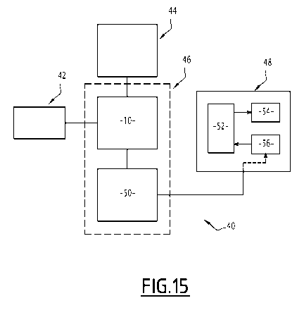
The medical device also comprises an eye tracker 42, a camera 44, an
electronic
circuitry 46 and an optical module 48 contained in the frame. The optical
module 48 forms
the projector system.
The camera 44 is adapted to capture the initial images 30.
CA 03128091 2021-07-28
WO 2020/161117
PCT/EP2020/052717
22
The eye tracker 42 is adapted to collect the data of the direction gaze. The
direction
gaze is, for example, determined either through a model of the eye 28 or
through
calibration. According to an example, the eye tracker 42 uses the center of
the pupil and
infrared light to gather light reflections from the cornea, and uses the
vector from the pupil
center to the corneal reflection to compute the gaze direction.
More precisely, the most commonly used technique is pupil centre corneal
reflection
(POOR). The basic concept is to use a light source to illuminate the eye
causing highly
visible reflections, and a camera to capture an image of the eye showing these
reflections.
The image captured by the camera is then used to identify the reflection of
the light source
on the cornea (glint) and in the pupil. One can then calculate a vector formed
by the angle
between the cornea and pupil reflections. The direction of this vector,
combined with other
geometrical features of the reflections, is then used to calculate the gaze
direction.
The direction gaze is the output of the eye tracker 42.
The eye tracker 42 is adapted to measure the size of the pupil 32.
The electronic circuitry 46 is a group of electronic components.
The electronic circuitry 46 includes the system 10 disclosed in section 2 and
a
command module 50.
The electronic circuitry 46 is configured to receive initial images 30 from
the camera
44 in the form of electronic data to issue commands to the optical module 48.
The other input parameters are pre-recorded in a memory of the system 10.
The system 10 generates the filter according to the method disclosed in
section 2.
Moreover, the system 10 is adapted to filter the initial image 30 with the
generated
filter.
The command module 50 is an electronic circuit including at least one
electronic
chip.
The command module is connected to the system 10 and to the optical module 48.
The optical module 48 is a light emitting device, adapted to illuminate the
eye of the
wearer with a controlled beam 38 of light exiting the optical module.
As mentioned above, the optical module 48 comprises a light source 52, an
optical
system 54 adapted to emit the light beam 38 reproducing the filtered image 36
and a
control module 56 adapted to control the radiant power of the light source 52.
The optical system 54 is a combination of optics adapted to reshape and
redirect
light emitted by the light source 52.
The optical system 54 is adapted to shape the light beam 38 emitted by the
light
source 52 in a controlled beam 38, and redirect a part of the controlled beam
on the
eye 28.
CA 03128091 2021-07-28
WO 2020/161117
PCT/EP2020/052717
23
The optical system 54 includes for example a collimator, a plurality of
mirrors, a
micro-mirror array, a photodiode and/or a liquid lens.
The light source 52 is composed of at least one light element generating
light.
Alternatively, the light source 52 comprises a light element generating light
and a light
transmitting element such as an optical fiber adapted to transmit light to the
optical
system 54. Thus, the light element generating light can be situated at a
greater distance
from the optical system 54 than in the case where the light source 52 includes
a light
element generating light without an optical fiber.
The command module 50 is adapted to send a command to the optical module 48
for projecting the filtered image 36 on the part of the eye 28. More
precisely, the command
module 50 is adapted to send a command to the control module 56 which in turn
commands the light source 52. The optical system 54 is adapted to receive the
light
emitted by the light source 52.
In particular, the light source 52 illuminates elements of the micromirror
matrix. Each
illuminated element of the micromirror matrix transmits a light which
corresponds to a
transmitted pixel in the filtered image 36.
According to a variant of the medical device, when the optical system does not
have
micromirror matrix, a discret element of the light source 52, such as Light-
Emitted Diode,
transmits a light which corresponds to a transmitted pixel in the filtered
image 36.
SECTION 5¨ APPLICATIONS
The method may be used in the field of vision restoration using vision
prostheses
such as retinal implants.
According to a specific embodiment, the method may be used in optogenetics.
The method of section 2 may be used for the subjects suffering of
photoreceptor
loss or degeneration, such as in case of retinitis pigmentosa (RP) or macular
degeneration (MD). As mentioned above, these affections diminish visual
acuity,
diminishes light sensitivity, or result in blindness of a part of the field of
view of the subject.
As explained above, some therapies consist in stimulating transfected cells of
the
retina and/or retinal implants with a light beam 38.
The part of the eye 28 on which the image is projected corresponds to portions
of
the retina of the eye 28.
The parts of the retina on which the image is intended to be projected
comprise
transfected cells of the retina and/or retinal implants having to be
stimulated.
The parts of the retina are stimulated by a light beam 38 reproducing the
filtered
image.
CA 03128091 2021-07-28
WO 2020/161117
PCT/EP2020/052717
24
The filtered image 36 rebuilds the lost field of view due to the photoreceptor
loss or
degeneration in function of the gaze direction of the subject.
The light beam 38 stimulating the parts of the retina is, for example,
obtained by the
method of section 3.
According to the method of section 3, the part of the retina is stimulated
with the
required light characteristics independent of the pupil size.
In such case, it is to be noted that thresholds of light intensity (maximum
and
minimum) are given by phototoxicity standards and are further analyzed in
literature
relevant to ophthalmology or to the application of light stimulation for an
optogenetic
therapy (Yan et al. 2016; Delori, Webb, and Sliney 2007; Sliney et al. 2005).
For example,
for a light with a wavelength of 595 nm,
o the maximum light intensity at the retina is 7 mW/mm2 (ISO 15004-2 2007;
ISO 62471 2006), and
o At the cornea (anterior segment), the maximum light intensity is 32 mW
over any 1 mm diameter disc (ISO 15004-2 2007).
In addition, the retinal radiant exposure limit when taking into account the
luminance
dose restriction is 6.6 J.cm-2 over 48 hours (luminance dose restriction, ANSI
Z136.1
2014).