Note : Les descriptions sont présentées dans la langue officielle dans laquelle elles ont été soumises.
CA 03136694 2021-10-12
WO 2020/210446
PCT/US2020/027406
LATERAL FLOW IMMUNOASSAY FOR MEASURING FUNCTIONAL Cl-
ESTERASE INHIBITOR (C1-INR) IN PLASMA SAMPLES
RELATED APPLICATIONS
This Application claims the benefit under 35 U.S.C. 119(e) of U.S.
provisional
application number 62/833,235, filed on April 12, 2019, and U.S. provisional
application
number 62/930,615, filed on November 5, 2019 the entire contents of each is
incorporated
herein by reference.
BACKGROUND
Cl-esterase inhibitor (also known as Cl-inhibitor or Cl-INH) is a protease
inhibitor
belonging to the serpin superfamily. Its main function is the inhibition of
the complement
system to prevent spontaneous activation. Cl-INH is also an endogenous
inhibitor for
plasma kallikrein (pKal). Auto somal dominant mutation in Cl-INH leads to
hereditary
angioedema (HAE), including types I and II HAE.
The currently available assays used to assess Cl-INH functional level measure
the
inhibition of Cis of the complement cascade by Cl-INH, utilizing either a
chromogenic assay
or a complex ELISA method. The chromogenic assay is generally considered
preferable but
both methods have limitations. The chromogenic assay is more likely to have an
occasional
false positive while the complex ELISA has a negative predictive value of only
62%.
It is of interest to develop newer assays and/or platforms for measuring
functional Cl-
INH involved in the HAE disease pathology.
SUMMARY
Provided herein are devices and methods for detecting functional Cl-INH (fC1-
INH)
in a qualitative and/or quantitative manner that is rapid and cost-effective.
Detecting fC1-
INH, in some embodiments, is achieved using a device configured for detecting
and/or
quantifying fC1-INH via a lateral flow immunoassay (LFA).
Accordingly, one aspect of the present disclosure provides a device for
detecting
and/or quantifying functional Cl-esterase inhibitor (fC1-INH), the device
comprising (i) a
conjugate pad comprising a first zone and a second zone, on which a first
agent and a second
agent are immobilized, respectively, and (ii) a membrane, which is in
communication with
the conjugate pad, wherein the membrane comprises a third zone, on which a
third agent is
immobilized. The first agent can be a functional Cl inhibitor (fC1-lNH)
binding agent or a
1
CA 03136694 2021-10-12
WO 2020/210446
PCT/US2020/027406
Cl inhibitor (C1-INTH) binding agent. The second agent and the third agent are
one of a
functional Cl inhibitor (fC1-INH) binding agent, a Cl inhibitor (C1-NH)
binding agent, and
a capture agent. The first agent, the second agent, and the third agents are
different from each
other. One of the fC1-INH binding agent, the Cl-INH binding agent, and the
capture agent is
conjugated to a detectable label, and one of the fC1-INH binding agent and the
C 1-INH
binding agent is conjugated to a docking agent. The detectable label and the
docking agent
are conjugated to a different agent. The conjugate pad further comprises a
fourth zone for
placing a biological sample, which flows through the device in the order of
the first zone, the
second zone, and the third zone. In some instances, the fourth zone for
placing the biological
.. sample may overlap with the second zone, on which the fC1-INH binding agent
may be
located.
In some embodiments, the first agent, the second agent, and the third agent
are the
Cl-INH binding agent, the fC1-INH binding agent, and the capture agent,
respectively. In
other embodiments, the first agent, the second agent, and the third agent are
the fC1-INH
binding agent, the Cl-INH binding agent, and the capture agent, respectively.
In some examples, the first agent is conjugated to a detectable label, the
second agent
is conjugated to a docking agent. In other examples, the first agent is
conjugated to a docking
agent, the second agent is conjugated to a detectable label.
In some embodiments, the fC1-INH binding agent can be an active form of Factor
XII
(FXIIa). Alternatively or in addition, the Cl-NH binding agent can be an
antibody that
binds Cl-INH. Further, the docking agent and the capture agent can be members
of a
receptor-ligand pair. For example, the receptor-ligand pair can comprise
biotin and avidin
(e.g., streptavidin or polystreptavidin).
In some embodiments, the detectable label may be europium, colloidal gold,
phycoerythrin, fluorescein, rhodamine, green fluorescent protein, quantum dot,
and
chromophore. In some embodiments, the detectable label is europium. In some
instances,
the detectable label may be attached to latex particles.
In one example, the first agent is the CI-NH binding agent located at the
first zone,
the second agent is the fC1-INH binding agent located at the second zone, and
the third agent
is the capture agent located at the third zone. The Cl-INH binding agent may
an antibody
binding to Cl-NH, which may be conjugated to a detectable label as disclosed
herein.
Alternatively or in addition, the fC1-INH binding agent may be FXIIa, which
may be
conjugated to a docking agent (e.g., biotin). Further, the capture agent can
be an avidin, such
as streptavidin or polystreptavidin.
2
CA 03136694 2021-10-12
WO 2020/210446
PCT/US2020/027406
In some embodiments, the device further comprises an absorbent pad in
communication with the membrane. The absorbent pad and the conjugate pad may
be
separated by the membrane. Alternatively or in addition, the device may
further comprise a
support member, on which the conjugate pad, the membrane, and/or the absorbent
pad is
mounted.
Further, the device may also comprise a housing. In some embodiments, the
housing
may comprise a first opening to form a buffer port, a second opening to form a
sample port,
and a third opening to form a test window. The sample port can be located
between the
buffer port and the test window. In some examples, the buffer port may align
with the first
zone, on which the Cl-INH binding agent is located, hi some examples, the
sample port may
align with the second zone, on which the fC1-INH binding agent is located. In
some
examples, the test window aligns with the third zone, on which the capture
agent is located.
In another aspect, the present disclosure provides methods for detecting
and/or
quantifying functional Cl-esterase inhibitor (fC1-INH) in a sample using any
of the LFA
devices disclosed herein. Such a method may comprise: (i) placing a sample in
the sample
port of the device described herein, (ii) placing a buffer in the buffer port
in the device,
wherein the buffer flows in the direction from the first zone to the third
zone; (iii) examining
a signal at the test window in the device, and (iv) determining presence or
measuring the
level of fC1-INH in the sample based on presence or intensity of the signal at
the test
window. In some embodiments, step (ii) is performed at least at least 5
minutes after step (i).
The fC1-INH binding agent can be immobilized at the second zone, which may
align with the
sample port.
Also provided herein are methods for detecting and/or quantifying functional
Cl-
esterase inhibitor (fC1-INH) in a sample, the method comprising (i) contacting
a sample with
an fC1-INH binding agent, a Cl-INH binding agent, and a capture agent to form
a complex,
wherein one of the fC1-INH binding agent and the Cl-INH agent is conjugated to
a docking
agent, which binds the capture agent, and wherein one of the fC1-INH binding
agent, the Cl-
INH agent, and the capture agent is conjugated to a detectable label, the
detectable label and
the docking agent being conjugated to a different agent; and (ii) detecting a
signal released
from the detectable label in the complex; wherein presence of the signal
released from the
detectable label in the complex indicates presence of fC1-INH in the sample.
Any of the fC1-
INH binding agent, the Cl-INH binding agent, the docking agent, the detectable
label, and
the capture agent as disclosed herein can be used in the methods disclosed
herein.
3
CA 03136694 2021-10-12
WO 2020/210446
PCT/US2020/027406
In some embodiments, step (i) can be performed by (a) incubating the sample
with the
fC1-INH binding agent for at least 5 minutes, and (b) contacting the sample
with the Cl-INH
binding agent and the capture agent. In other embodiments, step (i) is
performed by (a)
incubating the sample with the fC1-INH binding agent for at least 5 minutes to
form a first
complex, (b) contacting the first complex with the Cl-INH binding agent to
form a second
complex, and (c) contacting the second complex with the capture agent to form
the complex,
and wherein the capture agent is immobilized on a support member.
In any of the assay methods disclosed herein, the sample to be analyzed can be
a
biological sample obtained from a subject, for example, a serum sample, a
plasma sample, or
a blood sample (e.g., whole blood). In some embodiments, the subject can be a
human
patient suspected of having or at risk for a fC1-INH deficiency-mediated
disorder, which
includes, but is not limited to, hereditary angioedema (HAE), acquired
angioedema (AAE),
and a Cl-INH related immune disease. In some embodiments, the subject has a
symptom of
HAE. In some embodiments, the HAE is type I HAE or type II HAE. In other
embodiments,
the subject has no symptom of HAE, has no history of a symptom of HAE, or no
history of
HAE. In some embodiments, the subject is resistant to an anti-histamine
therapy, a
corticosteroid therapy, or both.
The details of several embodiments of the devices and methods described herein
are
set forth in the accompanying Figures and the Detailed Description. Other
features, objects,
and advantages of the devices and methods described herein will be apparent
from the
description and from the claims.
DETAILED DESCRIPTION OF THE DRAWINGS
Various aspects and embodiments will be described with reference to the
following
figures. The figures are not necessarily drawn to scale.
FIG. 1 is a schematic depiction of an exemplary lateral flow assay (LFA)
device 100
for detecting and/or quantifying functional Cl-esterase inhibitor (fC1-INH).
The LFA device
100 comprises a conjugate pad 200, a membrane 300, and optionally an absorbent
pad 400,
and a support member 500, in accordance with some embodiments of the
technology
described herein.
FIG. 2 is a schematic depiction of an exemplary LFA device 100 showing
exemplary
sizes of each components and exemplary sizes of overlap between two adjacent
components,
in accordance with some embodiments of the technology described herein.
4
CA 03136694 2021-10-12
WO 2020/210446
PCT/US2020/027406
FIG. 3A is a schematic depiction of a top view of an exemplary LFA device 100
for
detecting and/or quantifying fC1-INH. The LFA device 100 comprises a conjugate
pad 200,
which comprises a first zone 210 and a second zone 220, and a membrane 300
comprising a
third zone 230, in accordance with some embodiments of the technology
described herein.
FIG. 3B is a schematic depiction of a top view of an exemplary LFA device 100
for
detecting and/or quantifying fC1-INH. The LFA device 100 comprises a conjugate
pad 200,
which comprises a first zone 210 that overlaps with a fifth zone 250 for
placing a sample and
a second zone 220 that overlaps with a fourth zone 240 for placing a buffer,
and a membrane
300 comprising a third zone 230, in accordance with some embodiments of the
technology
described herein.
FIG. 4 is a schematic depiction of a top view of an exemplary LFA device 100
for
detecting and/or quantifying fC1-INH. The LFA device 100 further comprises a
housing
600, forming a buffer port 610, a sample port 620, and a test window 630, in
accordance with
some embodiments of the technology described herein. A first zone 210, a
second zone 220,
a third zone 230, a conjugate pad 200, a membrane 300, and an absorbent pad
400 are also
indicated.
FIG. 5 is an image of an exemplary LFA device, in accordance with some
embodiments of the technology described herein.
FIG. 6 is a graph showing a calibration curve generated from functional Cl-
esterase
inhibitor (fC1-INH) diluted into C 1-INH depleted plasma using the LFA device
disclosed
herein.
FIG. 7 is a graph of receiver operating curve (ROC) for diagnostic performance
based
on samples from control subjects and subjects with hereditary angioedema
(HAE), using the
LFA device disclosed herein.
FIG. 8 is a graph showing the level of functional Cl-INH in normal subjects
(Normal) and subjects with hereditary angioedema (HAE) using a chromogenic
assay
(Chrom) or the LFA device (LFA) described herein.
FIG. 9 is a graph showing the correlation between the level of functional Cl-
INH in
subjects with hereditary angioedema (HAE) as determined by a chromogenic assay
as
compared to the LFA device described herein.
FIG. 10A is a schematic showing collection of a blood sample using a sample
loop
and fingerstick.
FIG. 10B is a schematic showing addition of a sample loop filled with blood of
FIG.
10A to a SampleTainer Bottle.
5
CA 03136694 2021-10-12
WO 2020/210446
PCT/US2020/027406
FIG. 11A is a graph showing use of an anti-Cl-INH Fab conjugated to europium
nanoparticles under the indicated conditions.
FIG 11B is a graph showing use of an anti-C1-INH Fab conjugated to red gold
nanoparticles under the indicated conditions.
FIG. 12 is a graph showing results from polystreptavidin R compared to
streptavidin
as capture agents in the test line. R2 value indicates the fit to the model
line (dotted line).
FIG. 13 is a graph showing results from use of the indicated reagents as
capture
agents in the test line.
FIG. 14 is a graph showing results from the indicated anti-Cl-INH Fab or
antibody
clones as detection agents. For each antibody, columns refer to concentrations
of the
detection agent, from left to right, 1200 mU/mL, 600 mU/mL, 100 mU/mL, 0
mU/mL.
FIG. 15 is a graph showing results using a buffer containing inorganic
blocking agent
(IBA) or organic blocking agent (OBA) at various pH.
FIG. 16 is a graph showing results using a streptavidin test line (0.75 mg/mL
streptavidin) and the indicated concentrations of C 1-INH/CINRYZEO.
FIG. 17 is a schematic of exemplary methods described herein. A capture
protein,
such as streptavidin, interacts with its pair (e.g., biotin) conjugated to a
capture agent (e.g.,
biotinylated FXIIa) which binds to functional Cl-INH (fC1-INH, CINRYZEO).
Bound fC1-
INH is detected using a detection agent, such as an anti-C1-1NH antibody
conjugated to gold
particles (anti-Cl-INH Au conjugate).
DETAILED DESCRIPTION
The present disclosure is based, at least in part, on the development of
lateral flow
assay (LFA) methods and devices for measuring functional Cl-esterase inhibitor
(fC1-INH).
The LFA methods and devices are designed for specifically detecting presence
of fC1-INH
and/or measuring the level of fC1-INH in, e.g., a biological sample. Presence
and/or the level
of fC1-INH is often indicative of status of diseases associated with
biological pathways in
which Cl-INH plays a role. As such, the LFA methods and devices would be
particularly
useful in diagnosis and prognosis of diseases mediated by Cl-INH deficiency,
for example,
diseases mediated by the plasma kallikrein pathway (e.g., diseases such as
hereditary
angioedema) since fC1-INH is an inhibitor of the pKal pathway.
As used herein, "functional C 1-INH" or "fC1-INH" refers to the Cl-INH protein
in
the form that is capable of binding to a protein factor, to which C1-1NH binds
in nature and
6
CA 03136694 2021-10-12
WO 2020/210446
PCT/US2020/027406
exerts its biological activity. Such a protein factor includes, but is not
limited to, Cl s, Factor
XIIa (FXIIa), and plasma kallikrein (pKal). Detection of this subpopulation of
fC1-INH is
particularly helpful in assessing functional levels of Cl-INH in diseases such
as HAE.
HAE is a very rare and potentially life-threatening genetic condition that
occurs in
about 1 in 10,000 to 1 in 50,000 people. Symptoms include edema (swelling) in
various parts
of the body, including hands, feet, face and airway (throat). Patients often
suffer excruciating
abdominal pain, nausea, and vomiting caused by swelling in the intestinal
wall. Swelling of
the airway or throat is particularly dangerous; it can cause death by
asphyxiation. The three
specific blood tests required to confirm HAE Types I & II are ClINH antigen,
fClINH and
C4. Many diagnostic assays use outdated technologies and are not rapid or
standardized or
available throughout the world.
The rapid and sensitive LFA methods and devices disclosed herein can be
performed
in a physician's office lab for rapid diagnosis of HAE (e.g., Type I & II)
based on fClINH
levels. Such methods and devices would result in low cost consumables
reimbursed by
health insurance, high level of confidence in quantitative results, ease of
data interpretation
by physicians, and/or low level of need for confirmatory analysis. Such a
rapid analysis to
diagnose Type I or II HAE in the physician's office can expand screening for
HAE and
identify new HAE patients more quickly. Currently, global diagnosis rate for
HAE is only
40%; therefore, undiagnosed patients have high unmet need. Rapid test
availability on
common device platforms could expand recognition of HAE. Further, the rapid
test for
fClINH disclosed herein can help in monitoring the HAE disease progression or
response to
therapeutics in a timely fashion in clinical settings.
The LFA methods and devices disclosed herein involve a first binding agent
specific
to fC1-INH (a fC1-INH binding agent), a second binding agent specific to Cl-
INH (specific
to functional Cl-INH, non-functional Cl-INH, or both), and capture agent.
Either the fC1-
INH binding agent or the Cl-INH binding agent can be conjugated with a docking
agent
capable of binding to the capture agent. One of the fC1-INH binding agent, the
C1-INH
binding agent, and the capture agent can be conjugated with a detectable
label. Accordingly,
fC1-INH in a sample (e.g., a biological sample) can form a complex with the
fC1-INH
binding agent and the C1-INH binding agent. Such a complex can bound to the
capture agent
via interaction between the capture agent and the docking agent conjugated to
one of the Cl-
INH binding agent and the fC1-INH binding agent. Upon detection of a signal
(e.g., presence
or intensity) released from the detectable label, presence or the level of fC1-
INH in the
sample can be determined and/or quantified based on standards tested along
with. For
7
CA 03136694 2021-10-12
WO 2020/210446
PCT/US2020/027406
example, Cinryze , purified human plasma derived C llNH, at different levels
can be used as
standards to generate a standard curve to extrapolate and determine levels of
fClINH in the
sample as measured by any of the methods disclosed herein. The intensity of
the signal
released from the detectable label can be quantified to U/ml of fClINH in the
sample based
on the standards, and is indicative of the level of fC1-INH in the sample.
I. Components for Use in the Lateral Flow Assay (LFA)
The LFA methods and devices disclosed herein involve (i) a fC1-INH binding
agent,
(ii) a C1-1NH binding agent, one of which is conjugated to a docking agent,
and (iii) a
capture agent, which binds the docking agent. One of (i) ¨(iii) is conjugated
to a detectable
label.
(a) fC1-INH binding agent
A fC1-INH binding agent is a molecule (e.g., a protein or a fragment thereof
that
binds fC1-INH) that specifically binds a functional Cl-INH. A molecule is said
to exhibit
"specific binding" if it reacts more frequently, more rapidly, with greater
duration and/or with
greater affinity with a particular target (e.g., those disclosed herein) than
it does with
alternative targets, which can be an altered form of the particular target.
For example, a
molecule that specifically binds fC1-INH would react more frequently, more
rapidly, with
greater duration and/or with greater affinity to fC1-INH as compared with an
alternative
target, such as non-functional Cl-INH. "Specific binding" or "preferential
binding" does not
necessarily require (although it can include) exclusive binding.
In some instances, a protein or polypeptide capable of binding to fC1-INH in
nature,
or the binding fragment thereof, may be used as the fC1-INH binding agent. In
some
examples, the fC1-INH binding agent is FXII, for example, the active form of
FXII (FXIIa).
Factor XII is a serum glycoprotein that participates in the initiation of
blood coagulation,
fibrinolysis, and the generation of bradykinin and angiotensin. Prekallikrein
is cleaved by
Factor XII to form kallikrein, which then activates Factor XII resulting in
the formation of
Factor XIIa and Factor XII fragments (Factor XIIf) ("Histidine-rich
glycoprotein binds factor
XIIa with high affinity and inhibits contact-initiated coagulation"
Macquarrie, et al., Blood
117:4134-4141 2011). Cl inhibitor (C NINTH) has been shown to be an important
plasma
inhibitor of both Factor XIIa and Factor XIIf ("Effect of negatively charged
activating
compounds on inactivation of factor XIIa by Cl inhibitor" Pixley, et al., Arch
Biochem
Biophys 256(2):490-8 1987).
FXII proteins, including precursor forms, mature forms, and active forms
thereof,
8
CA 03136694 2021-10-12
WO 2020/210446
PCT/US2020/027406
were well known in the art. For example, the precursor protein sequence of
human Factor
XII and the active form thereof are provided under the GenBank Accession
Number:
NP_000496.2. FXII proteins of other species, e.g., non-human mammals, were
also known
in the art. Their structure information can be found in the art, for example,
identified from
publically available gene database, using the human FXII sequence as a query.
"Active" or "functional" Factor XII refers to a Factor XII polypeptide or
Factor XII
polypeptide fragment that retains a biological activity similar to, but may
not be necessarily
identical to, the naturally occurring Factor XII counterpart, including mature
forms. In some
embodiments, an active or functional Factor XII is a Factor XII polypeptide or
Factor XII
polypeptide fragment that binds to fC1-INH. In some embodiments, active or
functional
Factor XII is a Factor XIIa polypeptide or a Factor XIIa polypeptide fragment
that binds to
fC1-INH. In some embodiments, active or functional Factor XII is a Factor XIIf
polypeptide
or a Factor XIIf polypeptide fragment that binds to fC1-INH.
In other examples, the fC1-INH binding agent is plasma kallikrein (pKal), for
example, the catalytic fragment of a naturally-occurring pKal protein. Plasma
kallikrein is a
serine protease component of the contact system (Sainz I.M. et al., Thromb
Haemost 98, 77-
83, 2007). The contact system is activated by either factor XIIa upon exposure
to foreign or
negatively charged surfaces or on endothelial cell surfaces by
prolylcarboxypeptidases (Sainz
I.M. et al., Thromb Haemost 98, 77-83, 2007). Activation of plasma kallikrein
amplifies
intrinsic coagulation via its feedback activation of factor XII and enhances
inflammation via
the production of the proinflammatory nonapeptide bradykinin. As the primary
kininogenase
in the circulation, plasma kallikrein is largely responsible for the
generation of bradykinin in
the vasculature.
pKal proteins are well known in the art. Exemplary plasma kallikrein sequences
can
include human (Accession Number: NP_000883.2), mouse (Accession Number:
NP_032481.1), or rat (Accession Number: NP_036857.2) plasma kallikrein amino
acid
sequences.
"Active" or "functional" plasma kallikrein refers to a plasma kallikrein
polypeptide or
plasma kallikrein polypeptide fragment that retains a biological activity
(e.g., protease
activity) similar to, but may not be necessarily identical to, the naturally
occurring plasma
kallikrein counterpart, including mature forms. In some embodiments, an active
or functional
plasma kallikrein is a plasma kallikrein polypeptide or plasma kallikrein
polypeptide
fragment that binds to fC1-INH.
9
CA 03136694 2021-10-12
WO 2020/210446
PCT/US2020/027406
In yet other examples, the fC1-INH binding agent disclosed herein can be Cis
and
Clr, or active/functional fragments thereof. Cis and Clr are activated
homologous serine
proteases of the first component of complement (Cl). Both C is and Clr can
form a complex
with Cl-INH. Arlaud et al., (1993) Methods Enzymol. 223, 61-82. Cis is the
modular serine
protease, which executes the catalytic function of the Cl complex. Clr is an
enzyme that
activates Cis to its active form, by proteolytic cleavage.
Cis and Clr proteins are also well known in the art. For example, the
precursor
protein sequence of human Cis is provided under GenBank Accession Number:
NP 001725.1 and the human C lr is provided under GenBank Accession Number:
NP_001724.4.
"Active" or "functional" Cis and Clr refers to Cis and Clr polypeptide
fragment that
retains a biological activity similar to, but may not be necessarily identical
to, the naturally
occurring Cis and Clr counterparts, including mature forms, respectively. In
some
embodiments, an active or functional Cis or Clr fragment is the portion of a
Cis polypeptide
or Cis polypeptide that binds to fC1-INH.
Any of the fC1-INH binding agents may be produced via recombinant technology,
or
isolated from a suitable natural source.
(b) Cl-INH binding agent
The CI-INH binding agent for use in the LFA methods and devices disclosed
herein
can be any molecule (e.g., proteins or polypeptides) capable of binding to Cl-
INH. In some
instances, the Cl-INH binding agent is specific to fC1-INH. In other
instances, the C1-INH
binding agent cross-reacts with both functional and non-functional Cl-INH.
The CI-INH binding agent may be an antibody that binds Cl-INH. As used herein,
the term "antibody" refers to a protein that includes at least one
immunoglobulin variable
domain or immunoglobulin variable domain sequence. For example, an antibody
can include
a heavy (H) chain variable region (abbreviated herein as VH), and a light (L)
chain variable
region (abbreviated herein as VL). In another example, an antibody includes
two heavy (H)
chain variable regions and two light (L) chain variable regions. The term
"antibody"
encompasses antigen-binding fragments of antibodies (e.g., single chain
antibodies, Fab and
sFab fragments, F(ab')2, Fd fragments, Fv fragments, scFv, and domain
antibodies (dAb)
fragments (de Wildt et aL, Ear J Immunal. 1996; 26(3):629- 39.)) as well as
complete
antibodies. An antibody can have the structural features of IgA, IgG, IgE,
IgD, IgM (as well
as subtypes thereof).
CA 03136694 2021-10-12
WO 2020/210446
PCT/US2020/027406
The VH and VL regions can be further subdivided into regions of
hypervariability,
termed "complementarity determining regions" ("CDR"), interspersed with
regions that are
more conserved, termed "framework regions" ("FR"). The extent of the framework
region
and CDRs has been precisely defined (see, Kabat, E.A., et al. (1991) Sequences
of Proteins of
Immunological Interest, Fifth Edition, U.S. Department of Health and Human
Services, NIH
Publication No. 91-3242, and Chothia, C. et al., (1987) J. Mol. Biol. 196:901-
917, see also
www.hgmp.mrc.ac.uk). Kabat definitions are used herein. Each VH and VL is
typically
composed of three CDRs and four FRs, arranged from amino-terminus to carboxy-
terminus
in the following order: FR1, CDR1, FR2, CDR2, FR3, CDR3, FR4.
The VH or VL chain of the antibody can further include all or part of a heavy
or light
chain constant region, to thereby form a heavy or light immunoglobulin chain,
respectively.
In one embodiment, the antibody is a tetramer of two heavy immunoglobulin
chains and two
light immunoglobulin chains, wherein the heavy and light immunoglobulin chains
are
interconnected by, e.g., disulfide bonds. In IgGs, the heavy chain constant
region includes
three immunoglobulin domains, CH1, CH2 and CH3. The light chain constant
region
includes a CL domain. The variable region of the heavy and light chains
contains a binding
domain that interacts with an antigen. The constant regions of the antibodies
typically
mediate the binding of the antibody to host tissues or factors, including
various cells of the
immune system (e.g., effector cells) and the first component (C 1q) of the
classical
complement system. The light chains of the immunoglobulin may be of a kappa or
lambda
chain. In one embodiment, the antibody is glycosylated. An antibody can be
functional for
antibody-dependent cytotoxicity and/or complement-mediated cytotoxicity.
In some embodiments, the antibody that binds Cl-INH may specifically bind to
Cl-
INH, for example, an epitope of fC1-INH or an epitope shared by fC1-INH and
non-
functional Cl-INH. An antibody that "specifically binds" to an antigen or an
epitope is a
term well understood in the art, and methods to determine such specific
binding are also well
known in the art. An antibody is said to exhibit "specific binding" if it
reacts or associates
more frequently, more rapidly, with greater duration and/or with greater
affinity with a
particular target antigen than it does with alternative targets. An antibody
"specifically
binds" to a target antigen or epitope if it binds with greater affinity,
avidity, more readily,
and/or with greater duration than it binds to other substances. For example,
an antibody that
specifically (or preferentially) binds to an antigen (e.g., Cl-INH) or an
antigenic epitope
therein is an antibody that binds this target antigen with greater affinity,
avidity, more readily,
and/or with greater duration than it binds to other antigens or other epitopes
in the same
11
CA 03136694 2021-10-12
WO 2020/210446
PCT/US2020/027406
antigen. It is also understood by reading this definition that, for example,
an antibody that
specifically binds to a first target antigen may or may not specifically or
preferentially bind to
a second target antigen. As such, "specific binding" or "preferential binding"
does not
necessarily require (although it can include) exclusive binding. Generally,
but not necessarily,
reference to binding means preferential binding. In some examples, an antibody
that
"specifically binds" to a target antigen or an epitope thereof may not bind to
other antigens or
other epitopes in the same antigen.
The antibody binding to C 1-INH for use in the LFA methods and devices
disclosed
herein may have a suitable binding affinity for C1-INH or a suitable epitope
thereof. As used
herein, "binding affinity" refers to the apparent association constant or KA.
The KA is the
reciprocal of the dissociation constant (KD). The antibody described herein
may have a
binding affinity (KD) of at least 10-5, 10-6, 10-7, 10-8, 10, 10-10 M, or
lower. An increased
binding affinity corresponds to a decreased KD. Higher affinity binding of an
antibody for a
first antigen relative to a second antigen can be indicated by a higher KA (or
a smaller
numerical value KD) for binding the first antigen than the KA (or numerical
value KD) for
binding the second antigen. In such cases, the antibody has specificity for
the first antigen
relative to the second antigen. Differences in binding affinity (e.g., for
specificity or other
comparisons) can be at least 1.5, 2, 3, 4, 5, 10, 15, 20, 30, 40, 50, 60, 70,
80, 90, 100, 500,
1000, 10,000 or 105 fold. Binding affinity (or binding specificity) can be
determined by
conventional methods.
The antibody binding to Cl-INH may be a full-length antibody. Alternatively,
the
antibody is an antigen-binding fragment of a full length antibody. The term
"antigen-binding
fragment" of a full length antibody refers to one or more fragments of a full-
length antibody
that retain the ability to specifically bind to a target of interest. Examples
of binding
fragments encompassed within the term "antigen-binding fragment" of a full
length antibody
include (i) a Fab fragment, a monovalent fragment consisting of the VL, VH, CL
and CH1
domains; (ii) a F(abt)2 fragment, a bivalent fragment including two Fab
fragments linked by a
disulfide bridge at the hinge region; (iii) a Fd fragment consisting of the VH
and CH1
domains; (iv) a Fv fragment consisting of the VL and VH domains of a single
arm of an
antibody, (v) a dAb fragment (Ward et al., (1989) Nature 341:544-546), which
consists of a
VH domain; and (vi) an isolated complementarity determining region (CDR) that
retains
functionality. Furthermore, although the two domains of the Fv fragment, VL
and VH, are
coded for by separate genes, they can be joined, using recombinant methods, by
a synthetic
linker that enables them to be made as a single protein chain in which the VL
and VH regions
12
CA 03136694 2021-10-12
WO 2020/210446
PCT/US2020/027406
pair to form monovalent molecules known as single chain Fv (scFv). See, e.g.,
US patents
5,260,203, 4,946,778, and 4,881,175; Bird et al., (1988) Science 242:423-426;
and Huston et
al., (1988) Proc. Natl. Acad. Sci. USA 85:5879- 5883. Antibody fragments can
be obtained
using any appropriate technique including conventional techniques known to
those with skill
in the art.
Any of the antibodies binding to C 1-INH as described herein can be either
monoclonal or polyclonal. A "monoclonal antibody" refers to a homogenous
antibody
population and a "polyclonal antibody" refers to a heterogeneous antibody
population. These
two terms do not limit the source of an antibody or the manner in which it is
made.
The antibody binding to C 1-INH can be made by any methods known in the art.
See,
for example, Harlow and Lane, (1998) Antibodies: A Laboratory Manual, Cold
Spring
Harbor Laboratory, New York. In some embodiments, antibodies specific to Cl-
INH (e.g.,
human Cl-INH) can be made by the conventional hybridoma technology. In other
embodiments, antibodies specific to Cl-INH can be isolated from antibody
libraries
following conventional antibody library screening technology.
In some embodiments, the antibody is an antibody that specifically binds to
human
Cl-INH. In some embodiments, the antibodies are monoclonal antibodies that
specifically
bind to human CHNH. hi some embodiments, the antibodies are mouse monoclonal
antibodies that specifically bind to human C 1-INH, such as antibody clone
MMO6 or MMO3
(also referred to as 10995-MMO6 and 10995-MM03, respectively, from Sino
Biological Inc.).
In some embodiments, the antibodies are polyclonal antibodies that
specifically bind to
human Cl-INH. In some embodiments, the antibodies are rabbit polyclonal
antibodies that
specifically bind to human Cl-INH, such as antibody clone RPO1 or RPO2 (also
referred to as
10995-RP01 and 10995-RP02, respectively, from Sino Biological Inc.). In some
embodiments, the antibody is RP02.
(c) Docking-Capture agents
One of the fC1-INH binding agent and the Cl-INH binding agent is conjugated
with a
docketing agent, which is capable of binding to the capture agent also used in
the LFA
methods and devices as disclosed herein. In one embodiment, the docking agent
is
conjugated to the fC1-INH binding agent (e.g., FXIIa). In other embodiments,
the docking
agent can be conjugated to Cl-INH (e.g., an antibody binding to C 1-INH).
The docking agent and capture agent are members of a receptor-ligand pair,
which
refers to any pair of molecules capable of binding to each other to form a
complex. In one
13
CA 03136694 2021-10-12
WO 2020/210446
PCT/US2020/027406
example, the docking agent and capture agent are biotin and avidin,
respectively, or vice
versa. For example, the docking agent may be biotin, and the capture agent may
be
streptavidin or polystreptavidin.
(d) Detectable label
One of the fC1-INH binding agent, the Cl-INH binding agent, and the capture
agent
for use in the LFA methods and devices as disclosed herein can be conjugated
to a detectable
label. In some embodiments, the fC1-INH binding agent is conjugated to the
detectable
label. In other embodiments, the Cl-INH binding agent is conjugated to the
detectable label.
Alternatively, the capture agent is conjugated to the detectable label.
As used herein, a "detectable label" refers to any molecule that is capable of
releasing
a detectable signal, either directly or in directly. In some embodiments, the
detectable label
can be a fluorophore (e.g., fluorescein). As used herein, the term
"fluorophore" (also referred
to as ''fluorescent label" or "fluorescent dye") refers to moieties that
absorb light energy at a
defined excitation wavelength and emit light energy at a different wavelength.
Examples of fluorophores include, without limitation, xanthene derivatives
(e.g.,
fluorescein, rhodamine, Oregon green, eosin and Texas red), cyanine
derivatives (e.g.,
cyanine, indocarbocyanine, oxacarbocyanine, thiacarbocyanine and merocyanine),
naphthalene derivatives (e.g., dansyl and prodan derivatives), cournarin
derivatives,
oxadiazole derivatives (e.g., pyridyloxazole, nitrobenzoxadiazole and
benzoxadiazole),
pyrene derivatives (e.g., cascade blue), oxazine derivatives (e.g., Nile red,
Nile blue, cresyl
violet and oxazine 170), acridine derivatives (e.g., proflavin, acridine
orange and acridine
yellow), arylmethine derivatives (e.g., auramine, crystal violet and malachite
green),
tetrapyrrole derivatives (e.g., porphin, phthalocyanine and bilirubin), or
fluorescent proteins
(e.g., green fluorescent protein).
In some embodiments, the detectable lable is phycoerythrin.
In some embodiments, the detectable label is a chromophore (e.g., anthracene).
In
some embodiments, the detectable label is a semiconductor particle (e.g., a
quantum dot). In
some embodiments, the detectable label is attached to semiconductor particles
(e.g., quantum
dots). In some embodiments, the detectable label is europium. In some
embodiments, the
detectable label is colloidal gold. In some embodiments, the detectable label
is attached to
gold particles. In some embodiments, the detectable label is attached to red
gold particles. In
some embodiments, the detectable label is attached to latex particles. In some
embodiments,
the C 1-INH binding agent is antibody RPO2 conjugated to europium. In some
embodiments,
14
CA 03136694 2021-10-12
WO 2020/210446
PCT/US2020/027406
the Cl-INH binding agent is antibody RPO2 conjugated to gold particles. In
some
embodiments, the Cl-INH binding agent is antibody RPO2 conjugated to red gold
particles.
In some embodiments, the Cl-INH binding agent is antibody RPO2 conjugated to
latex
particles.
II. LFA Devices
In some aspects, the present disclosure provides a lateral flow assay (LFA)
device for
measuring fC1-INH in a sample containing such. Reference is now made to FIGs.
1-4,
which illustrate pictorially various embodiments of exemplary LFA devices
described herein.
As shown in FIG. 1, the device 100, in some embodiments, comprises a conjugate
pad 200, a membrane 300, and optionally an absorbent pad 400 and a support
member 500,
on which the conjugate pad 200, the membrane 300, and optionally the absorbent
pad 400 are
mounted.
The conjugate pad 200 is in communication with the membrane 300, either
directly or
via a linker. When the device contains the absorbent pad 400, the conjugate
pad 200 and the
absorbent pad 400 are separated by the membrane 300, which is in communication
with the
absorbent pad 400, either directly or in directly. In some embodiments, the
conjugate pad
200 overlaps with the membrane 300, e.g., by 2-6 mm, such as 3 mm as shown in
Fig. 2.
Alternatively or in addition, the membrane 300 overlaps with the absorbent pad
400 by, e.g.,
2-6 mm, such as 3 mm as shown in Fig. 2.
The specific characteristics and dimensions of the conjugate pad 200, the
membrane
300, the absorbent pad 400, and the support member 500 can be modified as
necessary to
achieve desired results. As shown in FIG. 2, in some embodiments, the support
membrane
has a length of 80 mm, which is the combined length of the conjugate pad (42
mm),
membrane (25 mm), and absorbent pad (19 mm) subtracted by the overlap of the
sample pad
and absorbent pad onto the membrane (3 mm, 3 mm).
As shown in FIG. 3A, a top view of device 100, the device may comprise various
zones (210, 220, 230) that are useful for, in some embodiments, immobilizing a
fC1-INH
binding agent, a Cl-INH binding agent, and a capture agent as those described
herein. In
some embodiments, the device 100 comprises a first zone 210 and a second zone
220, which
may be on the conjugate pad 200 and a third zone 230, which may be on the
membrane 300.
Each of the fC1-INH binding agent, the Cl-INH binding agent, and the capture
agent
may be immobilized on one of zones 210, 220, and 230 (which may be in any
order). Any of
these agents may be immobilized using any means known in the art. The agent
may be
CA 03136694 2021-10-12
WO 2020/210446
PCT/US2020/027406
immobilized on, or bound to, a surface of the conjugate pad 200 and/or the
membrane 300,
directly or indirectly. In some embodiments, the binding agent is immobilized
to a surface
via a covalent bond. In some embodiments, the binding agent is immobilized to
a surface via
a non-covalent bond. In some embodiments, the binding agent is immobilized to
a surface
via a linker. Examples of linkers, include, but are not limited to, carbon-
containing chains,
polyethylene glycol (PEG), nucleic acids, monosaccharide units, biotin,
avidin, and peptides.
In some embodiments, a fC1-INH binding agent such as FXIIa is immobilized on
the
first zone 210. The fC1-INH binding agent may be conjugated to a docking agent
such as
biotin. A Cl-INH binding agent such as an antibody binding to Cl-INH may be
immobilized
.. on the second zone 220. The Cl-INH binding agent may be conjugated to a
detectable label
such as those disclosed herein. A capture agent such as avidin (e.g.,
streptavidin) may be
immobilized on the third zone 230.
In some embodiments, a fC1-INH binding agent such as FXIIa is immobilized on
the
first zone 210. The fC1-INH binding agent may be conjugated to a detectable
label as those
described herein. A Cl-INH binding agent such as an antibody binding to C 1-
INH may be
immobilized on the second zone 220. The Cl-INH binding agent may be conjugated
to a
docking agent such as biotin. A capture agent such as avidin (e.g.,
streptavidin) may be
immobilized on the third zone 230.
In some embodiments, a C 1-INH binding agent such as an antibody binding to Cl-
INH can be immobilized on the first zone 210. The C 1-INH binding agent may be
conjugated to a docking agent such as biotin. A fC1-INH binding agent such as
FXIIa may
be immobilized on the second zone 220. The fC1-INH binding agent may be
conjugated to a
detectable label such as those described herein. A capture agent such as
avidin (e.g.,
streptavidin) can be immobilized on the third zone 230.
In some embodiments, a C 1-INH binding agent such as an antibody binding to Cl-
INH can be immobilized on the first zone 210. The C1-1NH binding agent may be
conjugated to a detectable label such as those described herein. A fC1-INH
binding agent
such as FXIIa may be immobilized on the second zone 220. The fC1-INH binding
agent may
be conjugated to a docking agent such as biotin. A capture agent such as
avidin (e.g.,
streptavidin) can be immobilized on the third zone 230.
In some embodiments, a fC1-INH binding agent such as FXIIa is immobilized on
the
first zone 210. The fC1-INH binding agent may be conjugated to a docking agent
such as
biotin. A capture agent such as avidin (e.g., streptavidin) may be immobilized
on the second
zone 220. The capture agent may be conjugated to a detectable label such as
those disclosed
16
CA 03136694 2021-10-12
WO 2020/210446
PCT/US2020/027406
herein. A C 1-INH binding agent such as an antibody binding to Cl-INH may be
immobilized on the third zone 230.
In some embodiments, a fC1-INH binding agent such as FXIIa is immobilized on
the
first zone 210. The fC1-INH binding agent may be conjugated to a detectable
label such as
those described herein. A capture agent such as avidin (e.g., streptavidin)
may be
immobilized on the second zone 220. A C1-INH binding agent such as an antibody
binding
to C 1-INH may be immobilized on the third zone 230. The Cl-INH binding agent
may be
conjugated to a docking agent such as biotin.
In some embodiments, a Cl-INH binding agent such as an antibody binding to Cl-
INH may be immobilized on the first zone 210. The C 1-INH binding agent may be
conjugated to a docking agent such as biotin. A capture agent such as avidin
(e.g.,
streptavidin) may be immobilized on the second zone 220. The capture agent may
be
conjugated to a detectable label such as those disclosed herein. A fC1-INH
binding agent
such as FXIIa may be immobilized on the third zone 230.
In some embodiments, a Cl-INH binding agent such as an antibody binding to Cl-
INH may be immobilized on the first zone 210. The Cl-INH binding agent may be
conjugated to a detectable label such as those described herein. A capture
agent such as
avidin (e.g., streptavidin) may be immobilized on the second zone 220. A fC1-
INH binding
agent such as FXIIa may be immobilized on the third zone 230. The fC1-INH
binding agent
may be conjugated to a docking agent such as biotin.
Any of the LFA devices disclosed herein may further comprise the 4t11 zone
240,
which may be for placing a sample such as those described herein, and
optionally the 5th zone
250, which may be for placing a buffer solution.
The 51h zone 250 may be located at one end of the device such that when a
buffer
solution is placed on the 5th zone 250, the buffer solution can flow through
the device from
the 1st zone 210 toward the 3rd zone 230. In some examples, the 5t1i zone 250
may overlap
with the Pt zone 210. See Fig. 3B. In this instance, a Cl-INH binding agent
such as an
antibody binding to the C1-INH may be immobilized on the 1st zone 210, which
overlaps
with the 5th zone 250. The C1-1NH binding agent may be conjugated to a
detectable label.
Alternatively or in addition, the 4th zone 240 may be located between the 5th
zone 250
and the 2nd zone 220. In some instances, the 4t1i zone 240 and the 2nd zone
220 may overlap.
See Fig. 3B. A fC1-INH binding agent such as FXIIa, which may be conjugated to
a docking
agent such as biotin, may be immobilized on the 2nd zone 220.
17
CA 03136694 2021-10-12
WO 2020/210446
PCT/US2020/027406
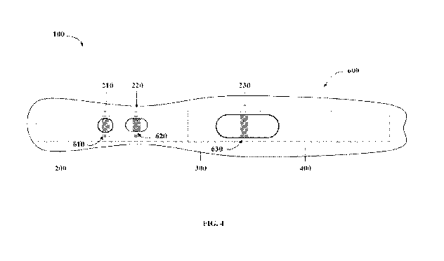
As shown in FIG. 4, the device 100, in some embodiments, may further comprise
a
housing 600, which may be removable. An image of a device as described herein
in a
housing is shown in FIG. 5. The housing 600 may be and configured to expose at
least a
portion of the conjugate pad 200 and the membrane 300 of the device 100. k
some
embodiments, the housing 600 comprises a first opening to form a buffer port
610, which
may align with the first zone 210. The housing 600 may further comprise a
second opening
to form a sample port 620, which may align with the second zone 220. Further,
the housing
600 may comprise a third opening to form a test window 630, which may align
with the third
zone 230.
In some embodiments, a C 1-INH binding agent such as an antibody binding to Cl-
INH is immobilized on the 1st zone 210, which aligns with the buffer port 610.
Fig. 4. The
Cl-INH binding agent may be conjugated to a detectable label such as those
described
herein. A fC1-1I\TH binding agent such as FXIIa may be immobilized on the 2nd
zone 220,
which may align with the sample port 620. The fC1-INH binding agent may be
conjugated to
a docking agent such as biotin. A capture agent such as avidin (e.g.,
streptavidin) may be
immobilized on the 3rd zone 230, which may align with the test window 630.
In some examples, a sample can be placed in the sample port 620, allowing
binding of
fC1-INH therein with FXIIa-biotin conjugate. A buffer solution can be placed
in the buffer
port 610, allowing migration of the Cl-INH binding agent on the 1st zone 210
toward the 2nd
zone 220 along with the buffer solution. When the Cl-INH binding agent is in
contact with
the fC1-INH-FXIIa complex at the 2nd zone 220, a complex of Cl-INH binding
agent/fC1-
INH/FXIIa-biotin is formed. This complex would migrate toward the 3rd zone 230
along
with the buffer solution and be captured at the 3rd zone 230 via the
interaction between biotin
and streptavidin at the 3rd zone 230. A signal released from the detectable
label conjugated to
the C 1-INH binding agent at the 3' zone 230, which align with the test window
630, can be
measured, which indicates presence or level of fC1-INH in the sample.
Alternatively or in addition, the housing 600 may be clear to facilitate
visualization of
the sample port 610 and/or the buffer port 620 and/or the test window 630. In
some
embodiments, a portion of the housing is clear. In some embodiments, the whole
housing
600 is clear.
The housing 600, in some embodiments, comprises a label to facilitate
identification
of a sample or a result. In some embodiments, the housing 600 comprises one or
more labels
to facilitate identification of a result in a test window 630. In some
embodiments, the one or
more labels identify a sample result.
18
CA 03136694 2021-10-12
WO 2020/210446
PCT/US2020/027406
It should be appreciated that various embodiments of the device, including the
multiple components in the device (e.g., the conjugate pad, the membrane, the
absorbent pad,
and the support member) as described herein may be formed with suitable
materials, e.g.,
with any suitable conjugate pad, with any suitable membrane, with any suitable
absorbent
pad, with any suitable support member, with any suitable binding agent, and
with any
suitable combination thereof.
For example, the membrane 300 in an LFA device as disclosed herein may be any
suitable membrane including, but is not limited to, a nitrocellulose membrane,
a nylon
membrane, a cellulose membrane, a polyvinylidine fluoride membrane, a
polycarbonate
membrane, a polypropylene membrane, a polyethylene membrane, a
polytetrafluoroethylene
membrane, and a poly-paraphenylene terephthalamide membrane. In some
embodiments, the
membrane is a nitrocellulose membrane.
Any suitable support member may be used in a device described herein. In some
embodiments, the support member comprises metal. In some embodiments, the
support
member comprises plastic. In some embodiments, the support member comprises
plastic
selected from the group consisting of styrene, polycarbonate, polypropylene,
polyethylene,
and polyvinyl chloride.
Any suitable pad may be used as the conjugate pad in a device described
herein. In
some embodiments, the conjugate pad comprises cellulose or glass fiber. In
some
embodiments, the absorbent pad comprises cellulose or glass fiber.
A device provided herein may further comprise a sample pad. In some
embodiments,
the sample pad comprises cellulose or glass fiber.
It should be appreciated that various embodiments of the present invention may
be
formed with one or more of the above-described features. The above aspects and
features of
the invention may be employed in any suitable combination as the present
invention is not
limited in this respect. It should also be appreciated that the drawings
illustrate various
components and features which may be incorporated into various embodiments of
the present
invention. For simplification, some of the drawings may illustrate more than
one optional
feature or component. However, the present invention is not limited to the
specific
embodiments disclosed in the drawings. It should be recognized that the
present invention
encompasses embodiments which may include only a portion of the components
illustrated in
any one drawing figure, and/or may also encompass embodiments combining
components
illustrated in different figures.
19
CA 03136694 2021-10-12
WO 2020/210446
PCT/US2020/027406
III. Measurement of Functional Cl-INH
Also provided herein are methods for detecting and/or quantifying functional
Cl-
esterase inhibitor (fC1-INH) in a sample. The assay methods disclosed herein
involve the use
of a fC1-INH binding agent, a Cl-INH binding agent, and a capture agent, which
are all
disclosed herein. One of the fC1-INH binding agent and the Cl-INH agent is
conjugated to a
docking agent, which binds the capture agent. One of the fC1-INH binding
agent, the Cl-
INH agent, and the capture agent is conjugated to a detectable label. The
detectable label and
the docking agent are conjugated to a different agent.
To perform the assay method disclosed herein, a sample suspected of containing
fC1-
INH can be brought in contact with the fC1-INH binding agent, the Cl-INH
binding agent,
and the capture agent under conditions allowing for formation of a complex
comprising the
fC1-INH, the fC1-INH binding agent, the Cl-INH binding agent, and the capture
agent (via
interaction with the docking agent conjugated to either the fC1-INH binding
agent or the Cl-
INH binding agent). Presence or level of the fC 1-INH in the sample can be
detected and/or
quantified by measuring a signal released from the detectable label, which can
be in
conjugation to any one of the fC1-INH binding agent, the Cl-INH binding agent,
and the
capture agent.
In some examples, the sample and the fC1-INH binding agent (e.g., FXIIa) can
be
incubated first for a suitable period (e.g., at least 5 minutes, such as 5-10
minutes) to allow
formation of a fC1-INH/FXIIa complex. The complex can then be incubated with a
Cl-INH
binding agent such as an antibody binding to Cl-INH to form a three-component
complex,
which can then be in contact with a capture agent that binds the docking agent
conjugated to
either the fC1-INH binding agent or the Cl-INH binding agent. A signal
released from the
detectable label conjugated to one component in the final complex can be
measured for
.. determining presence/absence and/or level of fC1-INH in the sample.
Methods for detecting and/or quantifying fC1-INH provided herein, in some
embodiments, comprise (i) contacting a sample with an fC1-INH binding agent, a
C 1-INH
binding agent, and a capture agent to form a complex, wherein one of the fC1-
INH binding
agent and the C 1-INH agent is conjugated to a docking agent, which binds the
capture agent,
and wherein one of the fC1-INH binding agent, the Cl-INH agent, and the
capture agent is
conjugated to a detectable label, the detectable label and the docking agent
being conjugated
to a different agent; and (ii) detecting a signal released from the detectable
label in the
complex; wherein presence of the signal released from the detectable label in
the complex
indicates presence of fC1-INH in the sample.
CA 03136694 2021-10-12
WO 2020/210446
PCT/US2020/027406
Methods described herein encompass a capture agent immobilized on any suitable
substrate in any suitable manner. Examples of substrates include, but are not
limited to,
beads, particles, slides, and multi-well plates. In some embodiments, the
capture agent is
bound covalently to the substrate. In some embodiments, the capture agent is
bound non-
covalently to the substrate. In some embodiments, the capture agent is bound
indirectly to
the substrate, e.g., through a linker.
In some embodiments, the assay methods disclosed herein can be carried out
using
any of the LEA devices disclosed herein. For example, a sample may be placed
in the sample
port 620 (Fig. 4) and a buffer solution may be placed in the buffer port 610.
The sample port
620 may be aligned with the 2nd zone 220. The buffer port 610 may be aligned
with the 1st
zone 210. The buffer solution would flow from, e.g., the 1st zone 210 toward
the 2nd zone
220 and the third zone 230, along with the sample and the fC1-INH binding
agent, the Cl-
INH binding agent, and/or the capture agent immobilized on the 1st zone 210
and the 2nd zone
220. This allows for the contact of the sample with the fC1-INH binding agent,
the Cl-INH
binding agent, and the capture agent when the buffer solution passes through
the Pt zone 210,
the 2nd zone 220, and the 3rd zone 230, such that a complex comprising the fC1-
1NH in the
sample, the fC1-INH binding agent, the Cl-INH binding agent, and the capture
agent can be
formed. Presence or the level of fC1-INH in the sample can be determined by
measuring a
signal released from the detectable label conjugated to one component in the
complex.
In one example, a method for detecting and/or quantifying fC1-1NH with a LFA
device comprising FXIIa-biotin and anti-Cl-INH antibody conjugated to europium
particles,
e.g., a device configured as shown in FIG. 4, is described for illustration
purposes only. In
this example, anti-Cl-INH antibody conjugated to europium particles functions
as a Cl-INH
binding agent conjugated to a detection label, and the anti-Cl-INH antibody
conjugate is
immobilized in the first zone 210. FXIIa conjugated to biotin functions as a
fC1-INH binding
agent conjugated to a docking agent, and FXIIa-biotin is immobilized in the
second zone 220.
To detect presence of fC1-INH in a sample, the sample is placed at the second
zone 220 on
the conjugate pad 200 via the sample port 620. When the sample contacts the
FXIIa-biotin in
the second zone 220, fC1-INH in the sample binds to FXIIa, thereby forming a
FXIIa-
biotin: fC1-INH complex.
As used herein, the term "contacts" refers to an exposure of a sample with one
or
more binding agents for a suitable period sufficient for the formation of a
complex with fC1-
INH and/or Cl-INH in the sample, if any. hi some embodiments, the sample
and/or buffer
21
CA 03136694 2021-10-12
WO 2020/210446
PCT/US2020/027406
contacts one or more binding agents via capillary action, in which a sample
and/or buffer
moves across a conjugate pad or a membrane.
A buffer may be placed at the first zone 210 on the conjugate pad 200 via the
buffer
port 610. The buffer may be placed at the first zone 210 any length of time
after placing the
sample at the second zone 220, e.g., the buffer may be placed at least 5
minutes after placing
the sample in the device. The buffer solubilizes the anti-Cl-INH antibody
conjugate, and
moves it along the conjugate pad 200 from the first zone 210 toward the
membrane 300 via
capillary action. When the buffer reaches the second zone 220, it contacts the
FXIIa-
biotin:fC1-INH complex, and the anti-Cl-INH antibody conjugate in the buffer
binds to fC1-
INH in complex with FX11a-biotin, thereby forming a "sandwich." In this
example, the fC1-
INH sandwich therefore comprises FXila conjugated to biotin bound to fC1-INH,
which is
bound by the anti-Cl-INH antibody conjugated to europium particles.
The buffer comprising the fC1-INH sandwich continues to move up the conjugate
pad
200 to the membrane 300, on which streptavidin is immobilized at the third
zone 230 (e.g., a
test line). In this example, streptavidin functions as the capture agent that
binds to the
docking agent, specifically biotin. When the buffer comes into contact with
streptavidin at
the third zone 230, biotin in the fC1-INH sandwich binds to streptavidin at
the third zone 230,
thereby capturing the fC1-INH sandwich. Presence of fC1-INH in the sample is
then
detected based on presence of signal from the europium particles at the third
zone 230 via test
window 630. Detection of the fCl-lNH sandwich is not limited to detection via
europium
particles. For example, presence of fC1-INH may be detected via a detectable
change in
color or pH. If fC1-INH is absent in the sample, no fC1-INH sandwich is
formed, and no
signal is detected at the third zone 230.
After moving into the third zone 230, the sample continues to move up the
membrane
300 into the absorbent pad 400, which acts as a wick to pull the sample
upward, thus
removing any background material from the third zone 230.
Methods provided herein encompass detecting and/or quantifying fC1-INH, or a
lack
thereof, in various samples. In some embodiments, the sample is a biological
sample
obtained from a subject. In some embodiments, the biological sample is a serum
sample, a
plasma sample, or a blood sample. In some embodiments, the sample is obtained
from a
subject suspected of having or at risk for a fC1-INH deficiency-mediated
disorder (e.g.,
HAE).
In some embodiments, the biological sample is a blood sample, e.g., a whole
blood,
obtained from a subject. Whole blood comprises red blood cells, white blood
cells, platelets,
22
CA 03136694 2021-10-12
WO 2020/210446
PCT/US2020/027406
and blood plasma. In some embodiments, a blood sample may be collected from
blood
vessels (e.g., capillaries, veins, and arteries). In some embodiments, a blood
sample may be
obtained by a fingerstick that produces drop(s) of blood. In some embodiments,
following
obtaining the blood sample, the blood samples are handled at 2-8 C. For plasma
or serum
preparation, the plasma or serum may be prepared after blood collection by
centrifugation.
Plasma and serum samples may be stored at -80 C prior to analysis.
In some embodiments, the methods and/or devices described herein may further
comprise a control line. As will be understood by one of ordinary skill in the
art, a control
line may be used to ensure that the method and/or device is functioning as
intended, e.g.,
detecting fC1-INH.
IV. Application of LFA Methods and Devices
Methods and devices described herein can be applied for evaluation of disease,
e.g.,
diagnosis or prognosis of a disease. Evaluation may include identifying a
subject as being at
risk for or having a disease as described herein, e.g., a fC1-INH deficiency-
mediated
disorder. Evaluation may also include monitoring treatment of a disease, such
as evaluating
the effectiveness of a treatment for a fC1-INH deficiency-mediated disorder.
Examples of
fC1-INH deficiency-mediated disorders include, but are not limited to,
hereditary
angioedema (e.g., type I and/or type II HAE), acquired angioedema (e.g., type
I and/or type II
AAE), Cl-INH deficiency related immune diseases (e.g., systemic lupus
erythematosus
(SLE), and C 1-INH deficiency related cancers (e.g., lymphoma).
In some embodiments, the methods and devices used herein are used to evaluate
whether a subject has or is risk for hereditary angioedema. In general, there
are different
types of hereditary angioedema, which exhibit similar inflammatory responses
but differ in
their etiology. For example, type 1 HAE is associated with functional but low
levels of Cl-
INH, whereas type II HAE is associated with non-functional Cl-INH that are
present at
normal concentrations.
A. Diagnosis
In some embodiments, the methods and devices described herein are used to
determine the level of fC1-INH in a biological sample (e.g., a serum sample or
a plasma
sample or a blood sample) collected from a subject (e.g., a human patient
suspected of having
a fC1-INH deficiency-mediated disorder such as HAE). The fC1-INH level is then
compared
to a reference value to determine whether the subject has or is at risk for
the fC1-INH
23
CA 03136694 2021-10-12
WO 2020/210446
PCT/US2020/027406
deficiency-mediated disorder. The reference value can be a control level of
fC1-INH capable
of binding to a fC1-INH binding agent as described herein (e.g., FXIIa). In
some
embodiments, the control level is a level of fC1-INH in a control sample that
is capable of
binding to a fC1-INTH binding agent. In some embodiments, a control sample is
obtained
from a healthy subject or population of healthy subjects. As used herein, a
healthy subject is
a subject that is apparently free of the fC1-INH deficiency-mediated disorder
at the time the
level of fC1-INH is measured or has no history of the disease.
The control level can also be a predetermined level. Such a predetermined
level can
represent the level of fC1-INH in a population of subjects that do not have or
are not at risk
.. for the fC1-INH deficiency-mediated disorder. The predetermined level can
take a variety of
forms. For example, it can be single cut-off value, such as a median or mean.
In some
embodiments, such a predetermined level can be established based upon
comparative groups,
such as where one defined group is known to have a target disease and another
defined group
is known to not have the target disease. Alternatively, the predetermined
level can be a
range, for example, a range representing the levels of fC1-INH in a control
population within
a predetermined percentile.
The control level as described herein can be determined by various methods. In
some
embodiments, the control level can be obtained by performing a known method.
In some
embodiments, the control level can be obtained by performing the same assay
used for
.. determining the level of fC1-INH in a sample from a subject. In some
embodiments, the
control level can be obtained by performing a method described herein. In some
embodiments, the control level can be obtained with a device described herein.
hi some
embodiments, the control level can be obtained from members of a control
population and the
results can be analyzed by, e.g., a computational program, to obtain the
control level (a
predetermined level) that represents the level of fC1-INH in the control
population.
By comparing the level of fC1-INH capable of binding to a fC1-INH binding
agent in
a sample obtained from a subject to the reference value as described herein,
it can be
determined as to whether the subject has or is at risk for a fC1-INH
deficiency-mediated
disease (e.g., HAE). For example, if the level of fC1-INH that binds to a fC1-
INH binding
.. agent of the subject deviates from the reference value (e.g., reduced as
compared to the
reference value), the candidate subject might be identified as having or at
risk for the fC1-
INH deficiency-mediated disease, e.g., HAE. The assay disclosed herein can be
used to
predetermine a cutoff value representing fC1-INH in normal subjects. Such a
cutoff value
can be used for determining whether a subject has or is at risk for a fC1-INH
deficiency-
24
CA 03136694 2021-10-12
WO 2020/210446
PCT/US2020/027406
mediated disease (e.g., HAE). In some instances, the level of fC1-INH in a
subject is lower
than the cutoff value may indicate disease risk or occurrence.
As used herein, "a decreased level or a level below a reference value" means
that the
level of fC1-INH that binds to a fC1-INH binding agent is lower than a
reference value, such
as a pre-determined threshold or a level of fC1-INH that binds to a fC1-INH
binding agent in
a control sample.
An decreased level of fC1-INH that binds to a fC1-INH binding agent includes a
fC1-
INH level that is, for example, 50%, 60%, 70%, 80%, 90%, 100%, 150%, 200%,
300%,
400%, 500% or more lower than a reference value. A decreased level of fC1-INH
that binds
to a fC1-INH binding agent also includes decreasing a phenomenon from a non-
zero state
(e.g., some or detectable fC1-INH that binds to a fC1-INH binding agent in a
sample) to a
zero state (e.g., no or undetectable fC1-INH that binds to a fC1-INH binding
agent in a
sample).
In some embodiments, the subject is a human patient having a symptom of a fC1-
INH
deficiency-mediated disease, e.g., those disclosed herein such as HAE. For
example, the
subject has edema, swelling wherein said swelling is completely or
predominantly peripheral;
hives; redness, pain, and swelling in the absence of evidence of infection;
non-histamine-
mediated edema, recurrent attacks of swelling, or a combination thereof. In
some
embodiments, the subject has no symptom of a fC1-INH deficiency-mediated
disease at the
time the sample is collected, has no history of a symptom of a fC1-INH
deficiency-mediated
disease, or no history of a fC1-INH deficiency-mediated disease such as HAE.
hi some
embodiments, the subject is resistant to an anti-histamine therapy, a
corticosteroid therapy, or
both.
Examples of fC1-INH deficiency-mediated diseases include, but are not limited
to,
non-histamine-dependent idiopathic angioedema, rheumatoid arthritis, Crohn's
disease,
lupus, Alzheimer's disease, septic shock, burn injury, brain
ischemiakeperfusion injury,
cerebral edema, diabetic retinopathy, diabetic nephropathy, macular edema,
vasculitis,
arterial or venous thrombosis, thrombosis associated with ventricular assist
devices or stents,
heparin-induced thrombocytopenia with thrombosis, thromboembolic disease, and
coronary
heart disease with unstable angina pectoris, edema, eye disease, gout,
intestinal bowel
disease, oral mucositis, neuropathic pain, inflammatory pain, spinal stenosis-
degenerative
spine disease, post-operative ileus, aortic aneurysm, osteoarthritis,
hereditary angioedema,
pulmonary embolism, stroke, head trauma or pen-tumor brain edema, sepsis,
acute middle
cerebral artery (MCA) ischemic event (stroke), restenosis (e.g., after
angioplasty), systemic
CA 03136694 2021-10-12
WO 2020/210446
PCT/US2020/027406
lupus erythematosis nephritis, an autoimmune disease, an inflammatory disease,
a
cardiovascular disease, a neurological disease, a disease associated with
protein misfolding, a
disease associated with angiogenesis, hypertensive nephropathy and diabetic
nephropathy,
allergic and respiratory diseases (e.g., anaphylaxis, asthma, chronic
obstructive pulmonary
disease, acute respiratory distress syndrome, cystic fibrosis, persistent,
rhinitis) and tissue
injuries (e.g., burn or chemical injury).
B. Evaluate Treatment Effectiveness
Methods and devices described herein can also be applied to evaluate the
effectiveness of a treatment for a fC1-INH deficiency-mediated disease (e.g.,
HAE). For
example, multiple biological samples (e.g., serum, plasma, or blood samples)
can be collected
from a subject to whom a treatment is performed either before and after the
treatment or
during the course of the treatment. The levels of fC1-INH can be measured by
any method
described herein. If the level of the fC1-INH increases after the treatment or
over the course
of the treatment (the level of fC1-INH in a later collected sample as compared
to that in an
earlier collected sample), remains the same or increases, it indicates that
the treatment is
effective.
If the subject is identified as not responsive to the treatment, a higher dose
and/or
frequency of dosage of the therapeutic agent are administered to the subject
identified. In
some embodiments, the dosage or frequency of dosage of the therapeutic agent
is maintained,
lowered, or ceased in a subject identified as responsive to the treatment or
not in need of
further treatment. Alternatively, a different treatment can be applied to the
subject who is
found as not responsive to the first treatment.
Therapeutic agents include, but are not limited to, kallikrein binding agents,
bradykinin B2 receptor antagonists, Cl-INH replacement agents, DX-2930, and
DX88 (see,
e.g., PCT Publication No. WO 2014/113701, which is incorporated herein by
reference in its
entirety).
EXAMPLES
In order that the devices and methods described herein may be more fully
understood,
the following examples are set forth. The examples described in this
application are offered
to illustrate the methods and compositions provided herein and are not to be
construed in any
way as limiting their scope.
26
CA 03136694 2021-10-12
WO 2020/210446
PCT/US2020/027406
Example 1: Preparation of a lateral flow assay (LFA) device for detecting
and/or
quantifying functional Cl-esterase inhibitor (fC1-INH)
Striping of membranes
FRONTLINE HRTM (BioDot) was used to stripe membranes. Front lines were
washed with 10 cycles of wash buffer (0.05% BIO-TERGE (Stepan Company) in
diH20),
and aligned so that the test line was dispensed 11 mm from the bottom of the
membrane.
Front lines were emptied and primed with 0.5 mg/mL polystreptavidin in 10 mM
phosphate,
pH 7.3, 0.5% sucrose. Test lines of polystreptavidin were striped onto
membranes (see, e.g.,
at a third zone 230 on a membrane 300 in FIG. 3A). Then, membranes were
labeled and
dried at 40 C for 30 minutes. Front lines were washed with 10 cycles of wash
buffer
following striping. Components for striping membranes are shown in Table 1.
Table 1. Components for striping of membranes.
Component Grade Vendor Part Number Amount per mL
0.5 M Sodium N/A DCN 10026 Variable
Phosphate, pH 7.3
Polystreptavidin N/A Biotez PolyStrept R
Sucrose ACS Sigma S5500 5 mg
Water (diH20) >18 M ohm Thermo >18 M ohm QS to final volume
0.05% BIO- N/A DCN N/A N/A
TERGEO
Membrane CN95 Sartorius 1UN95ER1000025NTB N/A
(25 mm x 300 mm)
Frontline Striper N/A BioDot XYZ 3210 N/A
Large Foil Bag IMPAK 125MF518 Large Foil Bag Large Foil Bag
Desiccants, 0.5 g IMPAK 39SG03 Desiccants, 0.5 g Desiccants, 0.5
g
Striping of conjugate pads
FRONTLINE HRTM (BioDot) was used to stripe anti-C1-INH Eu particle conjugates
and FXIIa-biotin onto the conjugation pad (Ahlstrom). Prior to striping
conjugate pads, anti-
C 1-INH Eu particle conjugates were diluted to 0.04% w/v into Eu Latex
Diluent, and FXIIa-
biotin was diluted to 2.4 iuM in FXIIa-biotin Diluent. Then, front lines were
washed with 10
cycles of wash buffer (0.05% BIO-TERGE (Stepan Company) in diH20). Front
lines were
27
CA 03136694 2021-10-12
WO 2020/210446
PCT/US2020/027406
aligned so that the anti-Cl-INH Eu particle conjugate was striped 15 mm from
the bottom of
the conjugate pad, and FXIIa-biotin conjugate was striped 8 mm from the top of
the
conjugate pad at a rate of 10 and 2.5 Vern, respectively. Positions of the
anti-Cl-INH Eu
particle conjugate and FXIIa-biotin conjugate align with the buffer port and
sample port in
custom SLA housing cassettes, respectively.
Front lines were emptied and primed with either conjugate. Conjugates were
striped
onto the conjugate pads, which were then labeled and dried at 40 C for 30
minutes.
Conjugate pads were sealed and dessicated. Front lines were washed with 10
cycles of wash
buffer following striping. Components for striping conjugate pads are shown in
Table 2.
Components for Eu Latex Diluent and FXIIa-biotin Diluent are shown in Table 3
and Table
4, respectively.
Table 2. Components for striping of conjugate pads
Component Grade Vendor Part Number
Conjugate Pad Fiberglass Ablstrom 8951
(42 mm x 300 mm)
Anti-Cl-INH Eu Particles N/A DCN Prepared as in NBR 715-001
FXIIa-biotin N/A Enzyme Research HFXIIabiotin 3790
Labs
FXIIa-biotin Diluent: N/A DCN N/A
10 mM Tris pH 8,0.1%
Tween-20, 2% Casein,
10% Sucrose, 4%
Trehalose, 0.25% Green
Food Dye
Eu Latex Diluent: N/A DCN N/A
10 mM Tris pH 8,0.1%
Tween-20, 2% Casein,
10% Sucrose, 4%
Trehalose
Frontline Striper N/A BioDot XYZ 3210
Large Foil Bag N/A IMPAK 125MF518
Desiccants, 0.5 g N/A IMPAK 39SG03
28
CA 03136694 2021-10-12
WO 2020/210446
PCT/US2020/027406
Table 3. Components for Eu Latex Diluent
Component Grade Vendor Part Number Amount per
mL
Tris-HC1 Reagent Sigma T3253 N/A
6% Casein in 50 mM N/A DCN Prepared as N/A
Tris, pH 8,5, 7-Day 21N0V21085M
Cure
Tween-20 BioXtra Sigma P7949 10 1.11_,
Sucrose ACS Sigma S5500 100 mg
Trehalose ACS Fisher BP2687100 N/A
Water (diH20) 218 M ohm Thermo 218 M ohm QS to final
volume
Table 4. Components for FXIIa-biotin Diluent
Component Grade Vendor Part Number Amount per
........................................................... mL
Tris-HC1 Reagent Sigma T3253 N/A
6% Casein in 50 mM N/A DCN Prepared as N/A
Tris, pH 8,5, 7-Day 21N0V21085M
Cure
Tween-20 BioXtra Sigma P7949 10 1.11-
Sucrose ACS Sigma S5500 100 mg
Trehalose ACS Fisher HP2687100 N/A
Green Food Dye Food Grade Vons N/A 2.5 111_,
Water (diH20) 218 M ohm Thermo 218 M ohm QS to final
volume
Preparation of anti-Cl-INH Eu particle conjugate
To prepare the anti-C1-INH Eu particle conjugate, 0.1 mg of anti-C1-INH
antibody
was exchanged into 50 mM borate, pH 8 via ZEBATM spin columns (Thermo Fisher)
using
the manufacture's protocol. The concentration of the antibody was determined
by
absorbance (A280, 1 mm, 1 OD = 1.4 mg/mL). Stock latex was rotated for 10
minutes, and
then sonicated for 10-15 seconds using a microtip sonicator (setting 25).
Stock latex solution
(10%) was diluted to 1% in 0.1 M MES, pH 6.5, and microfuged for 10 minutes at
17,000 g.
Supernatant was removed, and the pellet was resuspended in 0.1 M MES, pH 6.5
with the
volume of buffer being equal to the starting volume of latex. The resulting
solution was
sonicated, microfuged, and resuspended as previously described.
To prepare 15 mg/mL of EDC in 0.1 M MES buffer, EDC was allowed to equilibrate
to room temperature, and weighted out. The EDC solution was prepared within 10
minutes
29
CA 03136694 2021-10-12
WO 2020/210446
PCT/US2020/027406
of using in the preparation of anti-Cl-INH Eu particle conjugates. EDC stock
powder was
desiccated and frozen at -20 C for long term storage.
To prepare 50 mg/mL of sulfo-NHS in 0.1 M MES buffer, sulfo-NHS was allowed to
equilibrate to room temperature, and weighted out. The sulfo-NHS solution was
prepared
within 10 minutes of using in the preparation of anti-CI-11\TH Eu particle
conjugates. The
sulfo-NHS solution was activated by incubation for 30 minutes on a shaker at
1000 rpm. The
solution was microfuged for 8 minutes at 17,000 g. Pellets were resuspended in
50 mM
borate buffer, vortexed, and sonicated. The volume of buffer was equal to the
starting
volume of latex solution (600 !AL). The solution was then microfuged,
resuspended in borate
.. buffer (300 aL), vortexed, and sonicated as previously described. Activated
particles were
aliquoted into 50 pt/tube, and appropriate amounts of buffer and protein
(e.g., 20 ag protein
(i.e., antibody)) were added to obtain 20:1 mass to mass ratio of particles to
protein. Tubes
were vortexed immediately after the addition of buffer and protein.
Tubes were incubated for 2 hours at room temperature on a shaker at 1,000 rpm.
Following incubation, 1 M ethanolamine at 10 FL/mL was added, and tubes were
incubated
for 30 minutes at room temperature on a shaker at 1,000 rpm. Tubes were
microfuged for 10
minutes at 17,000 g, and pellets were resuspended into 1% casein 7-day cured
and incubated
overnight with shaking. Following the overnight incubation, tubes were
microfuged, and
pellets were resuspended into 1% casein 7-day cured, vortex, and sonicated.
Particle
conjugates were then striped onto conjugate pads as described herein.
Components for anti-
Cl-INH Eu particle conjugates are shown in Table 5.
Table 5. Anti-C1-INH Eu particle conjugates
Component Grade Vendor Part Number Amount per
mL
Anti-Cl-INH antibody N/A Shire AbD28387.2 0.05 mg
Eu Latex Particles, 0.2 N/A Thermo 93470520010150 1 mg
lull
Conjugate Storage DCN DCN N/A 1 mL
Diluent: 10 mM Tris
pH 8, 1% Casein
0.1 M MES, pH 6.5 N/A DCN N/A N/A
0.1 M Borate, pH 8 N/A DCN 10061 N/A
Water (diH20) 218 M ohm Thermo 218 M ohm QS to final
volume
CA 03136694 2021-10-12
WO 2020/210446
PCT/US2020/027406
6% Casein in 50 mM N/A DCN Prepared as N/A
Tris, pH 8.5, 7-Day
21NOV2108SM
Cure
EDC N/A Thermo 22980 N/A
NHS N/A Thermo 24500 N/A
Ethanolamine N/A TC1 A0297 N/A
Scientific Centrifuge N/A Thermo Legend XTR N/A
Micro Plate Mixer N/A Scilogex 82200004SX N/A
Sonicator N/A Qsonica Q55 N/A
Laminating components onto backing card
To laminate components onto the 80 mm long backing card (DCN), the backing
sticker was cut at 39 mm from bottom of card using a straightedge razor blade.
The backing
stickers were removed from the 39 mm position on the top of the card. The
membrane was
adhered to the card at 39 mm from the bottom of the card. The absorbent pad
(Ahlstrom) was
adhered to the top of the card so that the pad overlapped 3 mm with top of the
membrane.
The remaining backing stickers were removed, and the conjugate pad was adhered
to the
bottom of the card so that the pad overlapped 3 mm with bottom of the
membrane.
Components and component order are shown in Table 6 and Table 7, respectively.
Table 6. Card components
Component Material Length Location from bottom of
(mm) strip (mm)
Membrane Sartorius CN95 25 mm 39 mm
Conjugate Pad Ahlstrom 8951 42 mm N/A
Wick Pad Ahlstrom 243 19 mm 61 mm
(Absorbent Pad)
Backing Card DCN P/N P12-651 80 mm N/A
(Support Membrane)
Location of Test Line 11 mm (center) from bottom edge of
membrane. 1 mm width in total.
Overall Strip Width 5.0 mm
31
CA 03136694 2021-10-12
WO 2020/210446
PCT/US2020/027406
Table 7. Lamination component order, orientation, and positions
Order Component Orientation Position from Overlap
bottom of Card onto
Membrane
1 Striped Membrane side up, test line closest to 39 mm
N/A
Membrane bottom of card
2 Absorbent Pad Smooth side down 61 mm 3 mm
3 Striped Eu conjugate closest to bottom of card N/A 3 mm
Conjugation Pad
Preparing test strips
Cards were placed into the Kinematic Cutter (Kinematic), and cards were cut
into 5.0
mm wide strips. Strips shorter than 5.0 mm or strips that had been marked in
pen during
striping were discarded. Cut strips were placed into foil bags with dessicant.
Foil bags were
sealed and stored in dessicated box until use.
Example 2: Use of LFA device for detecting functional Cl-esterase inhibitor
(fC1-INH)
in plasma samples from patients.
To perform a lateral flow assay (LFA) for determining fC1-INH concentration,
the
test strips prepared as described in Example 1 above were placed into a custom
stereolithography (S LA) cassette (see, e.g., FIG. 4). Standards and quality
controls were
prepared by spiking Cl-INH reference standard (lot #TCP103, Shire, a Takeda
Company)
into Cl-INH depleted human K3-EDTA plasma (prepared by Shire, a Takeda
company).
Control samples containing 0 mU/mL to 800 mU/mL of purified Cl-INH were used
to
generate a calibration curve. Control samples were diluted 1:20 in Cl-INH
depleted media.
The sample was added to the sample port and incubated for 5 minutes. Run
buffer (30 pL)
was added to the sample port, which allowed the sample to run onto the
membrane and up to
the test window. Run buffer (150 uL) was then added to the buffer port, and
the cassette was
incubated for an additional 20 minutes. The test strip was removed from the
cassette, and the
intensity of the test line area was measured using the Axxin AX-2X fluorescent
reader
(Axxin). An image of a test strip in a cassette is shown in FIG. 5. The
measured intensity of
the test line area was plotted against the Cl-INH concentration to generate
the calibration
curve shown in FIG. 6, and gave a R2 of 0.97.
Fifty normal K3-EDTA plasma samples were obtained commercially and fifty HAE
plasma samples were used with patient consent from SAHARA, a Phase-III,
randomized,
double-blind, placebo-controlled, two-period, three-sequence, partial
crossover study, that
32
CA 03136694 2021-10-12
WO 2020/210446
PCT/US2020/027406
evaluated the efficacy and safety of subcutaneous administration of 2000 IU of
Cl esterase
inhibitor [Human] liquid for injection for the prevention of angioedema
attacks in adolescents
and adults with HAE.
The calibration curve from FIG. 6 was then used to determine the fC1-INH
concentration in plasma samples from the control subjects and subjects having
hereditary
angioedema (HAE). Briefly, plasma samples from the subjects were diluted 1:20
in Cl-INH
depleted plasma. The diluted plasma sample (20 !IL) was added to the sample
port, and
incubated for 5 minutes. The fC1-INH concentrations determined using test
strips were
within 20% of concentration values determined by ELISA.
As shown in FIG. 8, Cl-INH levels were lower in HAE subjects as compared to
the
healthy controls in both methods. The measured average Cl-INH concentrations
were 1345
and 1089 mU/mL for healthy controls and 275 and 163 mU/mL for HAE subjects in
the
Chromogenic and LFA methods, respectively (Table 8). The SEM and 95%
confidence
intervals for the measurements are provided in Table 8. Cl-INH data for all
HAE subjects
.. obtained from the two methods correlated with each other with a R2 pf 0.86,
including two
HAE subjects who had Cl-INH concentrations in the healthy control range (FIG.
9). The
average ratios between fClINH measured normal controls and HAE subjects were
4.9 and
6.7 in chromogenic and LFA methods, respectively.
Receiver operating curve (ROC) for diagnostic performance based on samples
from
control subjects and subjects with HAE was 0.98 indicating that the Cl-IFIN
concentration
determined by LFA accurately discriminated between control and HAE subjects
(FIG. 7).
The ROC curve indicated that a Cl-INH cut point of 496 mU/mL yields a
sensitivity (true
positive rate) of 94% and specificity (false positive rate) of 96% (FIG. 7);
the false negative
and false positives are listed in Table 9.
As shown in Table 8 and FIGs. 8-9, results obtained using the LFA were
compared to
analysis using a chromogenic assay by ELISA. Briefly, the chromogenic method
directly
measures fC1-INH levels, involving Cis cleaving a synthetic substrate to form
a color
compound, where diminished color intensity demonstrates inhibition of Cls
enzymatic
activity. The chromogenic assay was qualified for precision, accuracy,
linearity and upper
and lower limits of quantitation. Cl-INH protein (2000 ILJ of Cl esterase
inhibitor [Human]
liquid for injection, Shire, a Takeda Company) was used to prepare three
quality controls as
well as standard curve with ten standard points ranging from 1000-1.95 mU/mL.
The highest
and lowest points of standard curve were used as the anchor points. In brief,
the K3-EDTA
plasma samples and reference protein were pre-incubated with recombinant human
33
CA 03136694 2021-10-12
WO 2020/210446
PCT/US2020/027406
complement component Cis protein (R&D Systems) for 30 minutes at room
temperature
(RT) in a polypropylene plate. The formed Cl-INH and Cis complex was diluted
1:5 in
assay buffer and mixed with substrate solution ((synthetic substrate with a
thiobenzyl ester
group, M-1300, Bachem) and 5,5'-Dithiobis(2-nitrobenzoic acid) (DTNB) # D-
8000,
Biosynth) and the reaction was allowed to incubate for 40 minutes at RT.
Absorbance was
recorded at 405 nm using SpectraMax M5 plate reader with SoftMax Pro software.
Table 8. Results that compare the Chromogenic Assay and LFA to determine fC1-
INH
concentration
fClINH measurement using fC1INH measurement using
Chromogenic Assay LFA
Normal Mean 1345 mU/mL 1089 mU/mL
Control n 50 50
Plasma SEM 36 mU/mL 38 mU/mL
Samples 95% 1272 - 1418 mU/mL 1011 ¨ 1166 mU/mL
Confidence
Interval
HAE Plasma Mean 275 mU/mL 163 mU/mL
Samples n 50 50
SEM 39 mU/mL 32 mU/mL
95% 196 ¨ 354 mU/mL 100 ¨ 227 mU/mL
Confidence
Interval
Table 9. Results that show false positives and negatives of control and HAE
samples.
LFA fC1-INH Cut Point = 496mU/mL
HAE Samples Normal Controls
LFA Positive for HAE 47 2
LFA Negative for HAE 3 48
Taken together, these results demonstrate that similar fC1-INH concentrations
were
detected in patient plasma samples by LFA and ELISA (referred to as the
chromogenic
assay). Therefore, the LFA and test strips described herein may be an
effective tool for
identifying patients having HAE based on the level of fCl-lNH in a plasma
sample from the
patient. The results obtained using the LFA described herein correlated with
the results from
the chromogenic assay for assessing fC1-INH.
The rapid and sensitive LFA methods and devices disclosed herein can be
performed
in a physician's office lab for rapid diagnosis of HAE (e.g., Type I & II)
based on fClINH
levels. Such methods and devices may result in low cost consumables reimbursed
by health
34
CA 03136694 2021-10-12
WO 2020/210446
PCT/US2020/027406
insurance, high level of confidence in quantitative results, ease of data
interpretation by
physicians, and/or low level of need for confirmatory analysis. Such a rapid
analysis to
diagnose Type I or II HAE in the physician's office can expand screening for
HAE and
identify new HAE patients more quickly. Currently, global diagnosis rate for
HAE is only
40%; therefore, undiagnosed patients have a high unmet need. Rapid test
availability on
common device platforms could expand recognition of HAE. Further, the rapid
test for
fClINH disclosed herein can help in monitoring the HAE disease progression or
response to
therapeutics in a timely fashion in clinical settings.
References
1. Maurer, M. et al. (2018) The international WAO/EAACI guideline for the
management of
hereditary angioedema - the 2017 revision and update. World Allergy
Organization Journal
2. Aabom, A. et al. (2017) Complement factor C4 activation in patients with
hereditary
angioedema. Clinical Biochemistry 50(15), 816-821.
3. Bork, K. and Davis-Lorton, M. (2013) Overview of hereditary angioedema
caused by Cl-
inhibitor deficiency: assessment and clinical management. Eur Ann Allergy Clin
Immunol 45
(1), 7-16.
4. Csuka, D. et al. (2017) The role of the complement system in hereditary
angioedema. Mol
Immunol 89, 59-68.
5. Li, H.H. et al. (2015) Comparison of chromogenic and ELISA functional Cl
inhibitor tests
in diagnosing hereditary angioedema. J Allergy Clin Immunol Pract 3 (2), 200-
5.
6. Campbell, R.L., Wagner, D.B., and O'Connel, J.P. (1987). Solid phase assay
with visual
readout. U.S. Patent No. 4,703,017.
7. Rosenstein, R.W. and Bloomster, T.G. (1989). Solid phase assay employing
capillary
flow. U.S. Patent No. 4,855,240.
8. May, K., Prior, M.E., and Richards, I. (1997). Capillary immunoassay and
device
therefore comprising mobilizable particulate labelled reagents. U.S. Patent
No. 5,622,871.
9. O'Farrell, B. (2009). Evolution in Lateral Flow-Based Immunoassay Systems.
In: Wong,
R.C. and Tse, H.Y. (eds.). Lateral Flow Immunoassay. Humana Press New York
(NY).
10. Zahedi R, Aulak KS, Eldering E, Davis AE 3rd (1996). Characterization of
Cl inhibitor-
Ta. A dysfunctional ClINH with deletion of lysine 251. J Biol Chem. 1996 Sep
27;271(39):24307-12
CA 03136694 2021-10-12
WO 2020/210446
PCT/US2020/027406
Example 3: Competitive binding assays demonstrate specificity of the lateral
flow assay
(LFA) device for detecting functional Cl-esterase inhibitor (fC1-INH).
The specificity of the lateral flow assay (LEA) device as described herein for
determining fC1-INH concentrations was examined by performing assays in the
presence of
different competitive binding proteins. Samples contained 100 mU/mL of
purified Cl-INH.
No competing protein was added to the control sample. The intensity of the
test line area was
measured, and the percent reduction in signal from that of the control
reaction was calculated
for each sample. For samples with competing proteins, test line (TL)
intensities were reduced
between 40% to 60% compared to the control sample (Table 10). TL intensity was
reduced
55% by the addition of biotin labeled BSA demonstrating that unrelated
proteins are not
detected in the assay (Table 10). TL intensities were reduced 61% and 48% for
unlabeled
FXIIa and unlabeled antibody, respectively, demonstrating specificity of FXIIa
and antibody
with Cl-INH for signal production (Table 10).
Table 10. Results of specificity testing
Biotinylated Signal
Labeled
Reduction
Test Line Binding Competing Protein from 100
Conjugate in TL
Protein mli/mL
Signal
None 2400 N/A
3 niVI BSA-biotin 1080 55%
AbD2838 Neutravidin-
50 nM FXIIa 200 nM FXIIa (no
7 Eu 930 61%
biotin)
900 nM AbD28387 1240 48%
AbD2838 Neutravidin- None 3700 N/A
50 nM FXIIa
4 Eu 900 nM AbD28384 2150 42%
Specificity of detection was further tested using Cl-INH protein that had been
denatured by heating. TL intensity was not reduced by heating at 40 C, but
heating at 53 C
reduced TL intensity to that of the background signal (Table 11).
Table 11. Results of specificity testing with heat treated Cl-INH
Biotinylated
Signal from Signal from
Labeled
Test Line Conjugate Binding Heat Treatment 1000 mU/mL
Depleted
Protein in Plasma
Plasma
None 5410 360
Neutravidin-
AbD28387 Eu 50 nM FXIIa 40 C for 120 min 7110
360
53 C for 120 min 370 420
36
CA 03136694 2021-10-12
WO 2020/210446
PCT/US2020/027406
Taken together, these results demonstrate that the LFA and test strips
described herein
are specific for detection of fC1-INH in plasma samples.
Example 4: Use of blood samples from patients to detect functional Cl-esterase
inhibitor (fC1-INH) in LFA device.
Briefly, a whole blood sample is collected by performing a fingerstick
according to
laboratory practices that would be evident to one of ordinary skill in the
art. After wiping
away the first blood droplet, a second blood droplet forms on the finger. A
sample loop is
used to contact the second blood droplet fill the loop with blood (FIG. 10A).
The loop is
then added to container, such as a SampleTainerC) bottle (FIG. 10B). With the
loop touching
the bottom of the bottle, the bottle is snapped and twisted to break the
bottom of the shaft into
the bottle. Finally, the cap of the bottle is replaced and the bottle is
shaken to mix. The
whole blood sample may be added to any of the devices for analysis.
Example 5: Selection of Reagents
Reagents were selected for use in the methods and/or with the devices
described
herein. Two initial detection agents were developed: one employing a europium
nanoparticle
conjugated to an anti-fC1-INH Fab (FIG. 11A) and one using a Red Gold
nanoparticle
conjugated to an anti-fC1-INH Fab (FIG. 11B).
A steep decrease in signal at the lower end of the dynamic range was observed,
suggesting that the system was reaching the maximum signal. In effort to
decrease the slope
of the relationship, different amounts of different reagents were evaluated.
Two reagents
were assessed as Test Lines: streptavidin and polymeric version of
streptavidin
(polystreptavidin R) printed at a concentration of 0.5 mg/mL. Stretptavidin
was found to
have higher signals as compared to polystreptavidin, whereas polystreptavidin
was found to
have more linearity (FIG. 12). Europium conjugates were adjusted to 0.05%
solids from
0.1%. The concentration of FXIIa used in the incubation step with Cl-
INH/CINRYZEC) was
reduced from 1 pmollpt to 0.5 pmol/iut to reduce the dynamic range of the
assay. This
resulted in an assay with a signal within a detectable range.
Different agents were also assessed for the test lines. Briefly, three
different test lines
were generated: human biotinylated FXIIa (B-HFXIIa) was mixed with
streptavidin, B-
HFXIIa was mixed with polystreptavidin, and HFXIIa (non-biotinylated) alone.
Use of
HFXIIa provided a positive, yet low, signal whereas B-HFXIIa and streptavidin
and B-
HPFX1Ia and polystreptavidin did not have any appreciable positive signal
(FIG. 13).
37
CA 03136694 2021-10-12
WO 2020/210446
PCT/US2020/027406
Additionally, anti-C1-INH antibodies were also evaluated for use in the
methods
described herein. Four europium conjugates (the anti-Cl-INH Fab as well as
anti-C MINH
antibodies RP01, RPO2, MMO3 and MMO6 antibodies from Sino Biological Inc.)
were
evaluated at four concentrations (1200 mU/mL, 600 mU/mL, 100 mU/mL, and 9
mU/mL)
(FIG. 14). The signal obtained using each of antibodies RP01, RPO2, MMO3 and
MMO6 was
enhanced as compared to the Fab conjugate. Polyclonal antibody RPO2 was
selected for
further analysis.
Buffer conditions were also evaluated. For example, a buffer with inorganic
buffer
agent (IBA) or a buffer with organic buffer agent (OBA) were each prepared at
different pH
from 7.0-9.5. The buffer with the IBA performed better (e.g., higher signal)
at the higher end
of the pH range (pH 9.5), whereas the buffer with the OBA performed better
(e.g., higher
signal) at physiological pH ranges (around 7-7.5) (FIG. 15).
Finally, the detectable agent conjugated to the anti-Cl-INH binding agent was
also
evaluated. In particular, antibody RPO2 was assessed using an europium
conjugate or a red
gold conjugate and compared to the anti-Cl-INH Fab. The Fab conjugates did not
result in
positive signals, whereas the RPO2 antibodies with red gold conjugates
resulted in positive
signal (FIG. 16).
OTHER EMBODIMENTS
All of the features disclosed in this specification may be combined in any
combination. Each feature disclosed in this specification may be replaced by
an alternative
feature serving the same, equivalent, or similar purpose. Thus, unless
expressly stated
otherwise, each feature disclosed is only an example of a generic series of
equivalent or
similar features. From the above description, one skilled in the art can
easily ascertain the
essential characteristics of the present disclosure, and without departing
from the spirit and
scope thereof, can make various changes and modifications of the present
disclosure to adapt
it to various usages and conditions. Thus, other embodiments are also within
the claims.
EQUIVALENTS AND SCOPE
Those skilled in the art will recognize, or be able to ascertain using no more
than
routine experimentation, many equivalents to the specific embodiments of the
present
disclosure described herein. The scope of the present disclosure is not
intended to be limited
to the above description, but rather is as set forth in the appended claims.
38
CA 03136694 2021-10-12
WO 2020/210446
PCT/US2020/027406
In the claims articles such as "a," "an," and "the" may mean one or more than
one
unless indicated to the contrary or otherwise evident from the context. Claims
or descriptions
that include "or" between one or more members of a group are considered
satisfied if one,
more than one, or all of the group members are present in, employed in, or
otherwise relevant
to a given product or process unless indicated to the contrary or otherwise
evident from the
context. The present disclosure includes embodiments in which exactly one
member of the
group is present in, employed in, or otherwise relevant to a given product or
process. The
present disclosure includes embodiments in which more than one, or all of the
group
members are present in, employed in, or otherwise relevant to a given product
or process.
Furthermore, the present disclosure encompasses all variations, combinations,
and
permutations in which one or more limitations, elements, clauses, and
descriptive terms from
one or more of the listed claims is introduced into another claim. For
example, any claim that
is dependent on another claim can be modified to include one or more
limitations found in
any other claim that is dependent on the same base claim. Where elements are
presented as
lists, e.g., in Markush group format, each subgroup of the elements is also
disclosed, and any
element(s) can be removed from the group. It should it be understood that, in
general, where
the present disclosure, or aspects of the present disclosure, is/are referred
to as comprising
particular elements and/or features, certain embodiments of the present
disclosure or aspects
of the present disclosure consist, or consist essentially of, such elements
and/or features. For
purposes of simplicity, those embodiments have not been specifically set forth
in haec verba
herein. It is also noted that the terms "comprising" and "containing" are
intended to be open
and permits the inclusion of additional elements or steps. Where ranges are
given, endpoints
are included. Furthermore, unless otherwise indicated or otherwise evident
from the context
and understanding of one of ordinary skill in the art, values that are
expressed as ranges can
assume any specific value or sub¨range within the stated ranges in different
embodiments of
the present disclosure, to the tenth of the unit of the lower limit of the
range, unless the
context clearly dictates otherwise.
This application refers to various issued patents, published patent
applications, journal
articles, and other publications, all of which are incorporated herein by
reference. If there is a
conflict between any of the incorporated references and the instant
specification, the
specification shall control. In addition, any particular embodiment of the
present disclosure
that falls within the prior art may be explicitly excluded from any one or
more of the claims.
Because such embodiments are deemed to be known to one of ordinary skill in
the art, they
may be excluded even if the exclusion is not set forth explicitly herein. Any
particular
39
CA 03136694 2021-10-12
WO 2020/210446
PCT/US2020/027406
embodiment of the present disclosure can be excluded from any claim, for any
reason,
whether or not related to the existence of prior art.
Those skilled in the art will recognize or be able to ascertain using no more
than
routine experimentation many equivalents to the specific embodiments described
herein. The
scope of the present embodiments described herein is not intended to be
limited to the above
Description, but rather is as set forth in the appended claims. Those of
ordinary skill in the
art will appreciate that various changes and modifications to this description
may be made
without departing from the spirit or scope of the present disclosure, as
defined in the
following claims.
40