Note : Les descriptions sont présentées dans la langue officielle dans laquelle elles ont été soumises.
CA 03143443 2021-12-14
WO 2020/254633
PCT/EP2020/067196
1
Liposomal doxorubicin formulation, method for producing a lipo-
somal doxorubicin formulation and use of a liposomal doxorubicin
formulation as a medicament
The present invention relates to a liposomal doxorubicin formu-
lation, a method for producing a liposomal doxorubicin formula-
tion and a liposomal doxorubicin formulation for the use as a
medicament.
A liposome is a spherical vesicle having at least one lipid bi-
layer. Liposomes may also be multivesicular liposomes in which
one vesicle contains one or more smaller vesicles. The liposome
has an aqueous solution core surrounded by a hydrophobic mem-
brane in the form of a lipid bilayer.
The use of liposomes for drug delivery has been proposed for a
variety of drugs, particularly those which are administered par-
enterally. Liposomes have the potential to provide controlled
"depot" release of the administered drug over an extended time
period, and to reduce side effects of the drug, by limiting the
concentration of free drug in the bloodstream. Liposomes can al-
so alter the tissue distribution and uptake of drugs, in a ther-
apeutically favorable way, and can increase the convenience of
therapy, by allowing less frequent drug administration. For ex-
ample, liposomes may transport encapsulated active components
directly to the disease site, including tumour cells and sites
of inflammation. The active component can be directly released
from the liposome at the treatment site. Thus, a lower dosage of
the active component is allowed, and side effects are in conse-
quence limited.
The liposomes may transfer active components to the site of ac-
tion. Since the liposomal membrane is structurally similar to
CA 03143443 2021-12-14
WO 2020/254633
PCT/EP2020/067196
2
biological membranes, the liposomes may merge with the cellular
membranes. Upon merging, the liposomal contents may be emptied
into the cell where the active component can act. The use of
liposomes as drug carrier system may reduce the side effects as-
sociated with the administration of the respective active compo-
nent and related to high systematic absorption of the active
component. The active component can be accumulated at the de-
sired target. The components of the liposome bilayer may be me-
tabolised in the liver and/or spleen.
The development of drug delivery systems to treat cancer is par-
ticularly important as agents in cancer treatment are often cy-
tostatic or cytotoxic. It is desirable to prevent their release
to healthy tissue.
Liposomal compositions have been used successfully to deliver
entrapped therapeutics. For example, Doxil (Caelyx in Europe)
is a PEGylated liposomal formulation including entrapped doxoru-
bicin used for treatment of cancer such as ovarian cancer. Weak
amphipathic bases like doxorubicin may be loaded into the lipo-
somes using a transmembrane ion gradient. (See, e.g., Nichols et
al. (1976) Biochim. Biophys. Acta 455:269-271; Cramer et al
(1977) Biochemical and Biophysical Research Communications
75(2):295-301). This loading method, generally referred to as
remote loading, typically involves a drug having an ionizable
amine group which is loaded by adding it to a suspension of lip-
osomes prepared to have a lower inside/higher outside ion gradi-
ent, often a pH gradient.
Once the liposomes have drug loaded PLD (PEGylated Liposomal
Doxorubicin) extravasated into interstitial tissues' fluids,
little is known of the processes determining drug release. It is
believed that gradual loss of the ammonium/proton gradients re-
CA 03143443 2021-12-14
WO 2020/254633
PCT/EP2020/067196
3
taming the drug, enzymatic breakdown of liposomal phospholipids
by phospholipases and/or endocytosis by scavenger macrophages
likely contribute to drug release. (Barenholz, (2012) J Control
Release. 160(2): 117-34).
Liposome-encapsulated doxorubicin has proven effective in the
treatment of cancer. However, tumor accumulation, cytotoxicity
and efficiency in tumor weight reduction could be further im-
proved.
Liposome-encapsulated doxorubicin has proven less cardio toxic
than un-encapsulated doxorubicin. However, liposome-encapsulated
doxorubicin as known in the art causes severe side effects, such
as palmar-plantar erythrodysesthesia (PPE), more commonly known
as hand-foot syndrome. (See, e.g., Gabizon et al (1994) Cancer
Research 54:987-992; Solomon et al. (2008) Clinical Lymphoma and
melanoma 1:21-32). PPE results in redness, tenderness, and peel-
ing of the skin that can be uncomfortable and even painful. In
clinical testing at 50 mg/m dosing every 4 weeks, 50.6% of pa-
tients treated with DoxilED developed hand-foot syndrome. The
prevalence of this side effect limits the DoxilED dose that can
be given as compared with free doxorubicin in the same treatment
regimen. Also, liposome-encapsulated doxorubicin as is known in
the art continues to cause hematologic side effects, such as
neutropenia.
Certain Doxorubicin-loaded liposomes are known from Hong et al.
(Clin Cancer Res (1999) 5:3645-3652) and US 9 895 313. However,
these liposomes were obtained by different manufacturing pro-
cesses, namely by a combination of thin-film hydration and ex-
trusion; and by mixing a lipid solution in a water-miscible or-
ganic solvent with an aqueous solution in a specially designed
multi-port mixing chamber, respectively. Comparative tests car-
CA 03143443 2021-12-14
WO 2020/254633
PCT/EP2020/067196
4
ried out according to Hong's instruction for the preparation of
liposomes showed that the indicated particle diameters of 65 to
75 nm could not be reproduced. Instead a particle size of about
114 nm was obtained. Moreover, both disclosures remain silent
about the circularity of the liposomes, their size distribution
or the shape and size of the doxorubicin crystals loaded there-
in. However, the instructions for the preparation of the studied
liposomes indicate an ammonium sulfate concentration of 250 mM,
which according to the publication of Wei et al. (Wei et al.
(2018) ACS Omega 3:2508-2517) points to liposomes with a pro-
nounced aspect ratio. According to the same publication, a mini-
mum intraliposomal ammonium sulfate concentration of 200 mM is
required to support the stable nanocrystallization in pegylated
liposomal doxorubicins (PLDs), whereby PLDs lacking such crys-
tals or comprising crystals of poor crystallinity show a quick,
biphasic release of doxorubicin.
Therefore, there remains a need for chemically and physically
stable liposomal formulations for delivering doxorubicin with
improved tumor accumulation, cytotoxicity and efficiency in tu-
mor weight reduction. Also there remains a need to reduce un-
wanted side effects such as PPE without compromising the thera-
peutic efficacy. Furthermore, there is a need for an efficient,
reliable and cost effective method for producing suitable lipo-
somal formulations.
It is thus an object of the present invention to address those
needs and to provide improved liposomal doxorubicin formulations
for the treatment of cancer. It is another object of the present
invention to provide a method of producing such liposomal doxo-
rubicin formulations and to provide the use of such liposomal
doxorubicin as a medicament.
CA 03143443 2021-12-14
WO 2020/254633
PCT/EP2020/067196
The problem has been solved by a liposomal doxorubicin formula-
tion, a method for producing liposomal doxorubicin formulations
and the use of a liposomal doxorubicin formulation as a medica-
ment, having the features according to the independent claims.
5
The invention relates to a liposomal doxorubicin formulation,
wherein the lipid bilayer of the liposomes comprises at least
- phosphatidylcholine, preferably 1,2-distearoyl-sn-glycero-3-
phosphocholine (DSPC);
- cholesterol;
- a polyethyleneglycol-lipid conjugate, preferably DSPE-PEG
2000;
wherein
- the liposomes have a mean diameter between 30 and 70 nm,
preferably between 40 and 65 nm, measured by DLS; or
- the liposomes have a mean diameter between 20 and 50 nm,
preferably between 30 and 40 nm, measured based on Cryo-TEM
acquired images.
By "liposomal doxorubicin formulation" is meant a composition
comprising doxorubicin encapsulated in liposomes. In particular,
the formulation comprises doxorubicin hydrochloride. Doxorubicin
is an anthracycline topoisomerase II inhibitor well known in the
art for the treatment of cancer. The chemical name is (8S,10S)-
10-[(3-amino-2,3,6-trideoxy-a-L-lyxo-hexopyranosyl)oxy]-8-
glycoly1-7,8,9,10-tetrahydro-6,8,11-trihydroxy-1-methoxy-5,12-
naphthacenedione hydrochloride. The diseases treatable with the
liposomal doxorubicin formulation are types of cancer, including
acute leukemias, breast cancer, Hodgkin disease, non-Hodgkin
lymphomas, and sarcomas. Particularly preferred, the medicament
is for treatment of metastatic breast cancer, advanced ovarian
cancer, Kaposi's sarcoma and multiple myeloma.
CA 03143443 2021-12-14
WO 2020/254633
PCT/EP2020/067196
6
A major factor which determines stability as well as, location
and rate of drug release from the liposome is the liposome lipid
membrane composition. Other factors are size and morphology of
the liposomes in the composition as well as morphology of doxo-
rubicin crystals encapsulated in the liposome.
For the purposes of this invention, phosphatidylcholine in the
lipid bilayer can be any of DDPC, DLPC, DMPC, DPPC, DSPC, DOPC,
POPC and DEPC or mixtures thereof. Most preferred is 1,2-
distearoyl-sn-glycero-3-phosphocholine (DSPC).
Caelyx /DoxilED is essentially based on fully hydrogenated soy
phosphatiylcholine HSPC. HSPC has the following structural for-
mula:
Formula 1
with m,n = 14 or 16. Being obtained from a natural product (soy-
abean), HSPC is structurally less homogeneous than a fully syn-
thetic molecule. HSPC is hence less apt for dense packing of the
fatty acid chains when arranged in the lipid bilayer and there-
fore less suitable to form liposomes of the desired size. The
same applies to liposomal formulations, where the amount of
phospholipids in the lipid bilayer is formed of mixtures of dif-
ferent phosphatidylcholines, for example of a mixture of 1,2-
distearoyl-sn-glycero-3-phosphocholine (DSPC) and 1,2-
dipalmitoyl-sn-glycero-3-phosphocholine (DPPC). It is therefore
particularly preferred for the purposes of the invention that
the total amount of phosphatidylcholine (PC) used for the lipid
CA 03143443 2021-12-14
WO 2020/254633
PCT/EP2020/067196
7
bilayer comprises at least 95 wt-%, preferably at least 99 wt-%
and more preferably 100 wt-% of DSPC.
Liposomes comprising cholesterol in addition to phosphatidylcho-
line have improved circulation lifetime, pharmacokinetics and
therapeutic characteristics. They are biocompatible and biode-
gradable.
The polyethylenglycol-lipid conjugate is preferably methoxypoly-
ethylene glycol (MPEG), more specifically N-(carbonyl-
methoxypolyethylene glycol 2000)-1,2-distearoyl-sn-glycero-3-
phosphoethanolamine sodium salt, MPEG2000-DSPE). Membrane sur-
face modifications based on polyethylene glycol (PEG)-conjugated
lipids are known to improve blood circulation capability, e.g.
by avoiding capture of the liposomes by phagocytic cells in the
liver and spleen.
The liposomes in the inventive liposomal doxorubicin formulation
have a mean diameter between 30 and 70 nm, preferably between 40
and 65 nm, measured by dynamic light scattering DLS and/or the
liposomes have a mean diameter between 20 and 50 nm, preferably
between 30 and 40 nm, measured by cryo-TEM.
"Measured by dynamic light scattering" (DLS) means that DLS was
performed on samples with a lipid concentration between 20 and
mg/ml, which were diluted 1/19 in PBS or MQ H20 to reach an
attenuation factor in the instrument of around 6. DLS was meas-
ured on a Malvern Zetasizer Nano device at 25 C and 0 scatter-
ing angle. Instrument control and data analysis were performed
30 with the Zetasizer software (version 7.11) from Malvern. Parti-
cle size (hydrodynamic diameter) was determined using the
Stokes-Einstein equation:
CA 03143443 2021-12-14
WO 2020/254633
PCT/EP2020/067196
8
kT
d(H)= ___________________________________________________________ (Eq. 1)
3740
where k is Bolzmann's constant; T is absolute temperature; 17 is
dispersant viscosity and D is diffusion coefficient. Viscosity
was determined with the Zetasizer software and was 0.8872 cP.
Dispersant refractive index was 1.330. D was obtained by fitting
the autocorrelation function with a suitable algorithm. Cumu-
lants analysis is a simple method of analysing the autocorrela-
tion function generated by a DLS experiment and produces the
mean particle size (Z-ave) and polydispersity index (PDI). The
calculation is defined in ISO 13321 (1996) and ISO 22412 (2008).
The first order result from a DLS experiment is an intensity
distribution of particle sizes. The intensity distribution is
naturally weighted according to the scattering intensity. The
size distribution is displayed as a plot of the relative inten-
sity of light scattered by particles (on the Y axis) versus var-
ious size classes (on the X axis) which are logarithmically
spaced. Clear disposable zeta cells with a pathlength of 10 mm
were used for the measurements.
"Measured based on Cryo-TEM acquired images" means that the sam-
ples were subject to cryogenic transmission electron microscopy.
The liposomal samples diluted as appropriate, vitrified and pre-
pared on-grid (Formvar and Carbon) with an acc. voltage of
200kV. Images were acquired with a cryoTEM JEOL JEM-2100F a
TVIPS TemCam F415MP camera at 20'000x; 40'000x; 80'000x mag-
nification. Particle identification and size determination were
performed by by semi-automated image processing using Vironova
Analyzer Software, Vironova, Sweden. Briefly, a series of random
images of the same magnification was imported. Only lipsosome
particles located entirely within the boundaries of the image
and with a distinct membrane were detected. The identified ob-
jects were analysed for spherical diameter, circularity, unila-
CA 03143443 2021-12-14
WO 2020/254633
PCT/EP2020/067196
9
mellarity. All images were batch-processed with identical
thresholds and settings, accumulating over 5 to 18 images for
each sample, corresponding 6 to a number of particle analysed of
1560 to1178. Mean values have a standard deviation of approx. 10
nm.
Usually but not necessarily, the liposomes in the inventive for-
mulation will fall into the numerical ranges of size measured by
both methods. The diameter size measured by Cryo-TEM is general-
ly lower than the diameter size measured by DLS. Among other
factors, this is due to the PEG-chains being not visible in
cryo-TEM images and the hydrodynamic radius having an impact on
DLS but not cryo-TEM imaging.
In contrast to the disclosures from the state of the art, it has
now surprisingly been found that the liposomal doxorubicin for-
mulations disclosed herein already exhibit stable and particu-
larly uniformly formed doxorubicin crystal fibres at signifi-
cantly lower intraliposomal ammonium sulfate concentrations.
Without wishing to be bound by theory, it is currently consid-
ered that a higher concentration of ammonium sulfate causes more
doxorubicin to be loadable, resulting in larger doxorubicin
crystals inside the liposome. Owing to their size and rigidity,
these doxorubicin crystals expand the interior of the liposome,
which ultimately leads to a significant deviation of the lipo-
some from the perfect spherical shape. The different expansion
of the particles is also manifested in the occurrence of a major
axis and a minor axis, respectively, which can for example be
observed in cryogenic transmission electron microscopy (cryo-
TEM) images of such pegylated liposomal doxorubicin particles.
Of course it also follows that such particles do not feature a
high degree of homogeneity, i.e. a narrow particle size distri-
CA 03143443 2021-12-14
WO 2020/254633
PCT/EP2020/067196
bution and / or a high degree of circularity. However, these
properties have a positive effect on the pharmacokinetics and
the side effect profile of the liposomal doxorubicin formula-
tions disclosed herein, as will be described in more detail be-
5 low.
It has successfully been shown that the inventive liposomes of
the said composition and the indicated size are more stable than
those known in the art. Liposomes of such small diameter are op-
10 sonized less rapidly and at a lower extent than their larger
counterparts and are cleared less rapidly by the reticuloendo-
thelial system. Also, larger liposomes are more likely to fuse
or interact with other liposomes or particles.
As a result, liposomal doxorubicin formulations according to the
invention have proven more effective in the prevention of tumor
growth. They provide for higher accumulation of doxorubicin in
tumors and are more readily accumulated and cleared in the liv-
er. They also have higher cytotoxicity in in vitro assays. Lipo-
somal doxorubicin formulations according to the invention show
less pronounced serum leakage compared to Doxil /Caelyx formu-
lations, while having good cellular uptake. Serum half-life in
humans is considerably higher than the one for the lipsomal dox-
orubicin formulations known in the art. The formulation provides
higher drug exposition for a given dose in the relevant target
area. Adverse effects such as PPE and neutropenia can signifi-
cantly be reduced by using the inventive formulation rather than
Doxil /Caelyx .
The advantages, improved efficacies and reductions in adverse
effects will be shown in the examples section hereinafter.
CA 03143443 2021-12-14
WO 2020/254633
PCT/EP2020/067196
11
It is preferred that in the liposomal doxorubicin formulation
according as describe above, wherein the lipid bilayer essen-
tially consists of synthetic phosphatidylcholine, preferably a
structurally uniform type of synthetic phosphatidylcholine, of
cholesterol and of DSPE-PEG. It is particularly preferred that
the lipid bilayer essentially consists of 1,2-distearoyl-sn-
glycero-3-phosphocholine, cholesterol and DSPE-PEG. In particu-
lar, at least 95 wt-%, preferably at least 99 wt-%, more prefer-
ably 100% of the lipid bilayer consists exclusively of synthetic
phosphatidylcholine, preferably a structurally uniform type of
synthetic phosphatidylcholine, of cholesterol and of DSPE-PEG.
As has been described above, homogeneous lipid bilayer composi-
tion is advantageous for dense packing of fatty acid moieties.
Homogeneous lipid bilayers hence improve stability of the vesi-
cles.
Preferably, the phosphatidylcholine to cholesterol weight ratio
in the lipid bilayer of the liposomes in formulation is from
50:50 to 70:30, preferably from 55:45 to 65:35, more preferably
60:40.
It is preferred that in the liposomal doxorubicin formulation
the liposomes have a mean relative circularity of at least 0.99,
measured based on Cryo-TEM acquired images, and where
- the 10th percentile is at least 0.98;
- preferably where the 5th percentile is at least 0.98,
- more preferably where the 5th percentile is at least 0.98
and the 2nd percentile is at least 0.96.
The vesicular morphology of the liposomes is determined as de-
scribed above (see "measured based on cryo-TEM acquired imag-
es"). Circularity is calculated as according to the following
formula:
CA 03143443 2021-12-14
WO 2020/254633
PCT/EP2020/067196
12
,4..r 'x Area
Circularity ¨ ________________________________________
A high mean circularity value of the liposomes in the formula-
tion further supports stability of the vesicles and reduces side
effects of the doxorubicin therapy such as PPE and neutropenia.
Without being bound to such theory, an ellipsoidal shape togeth-
er with the size and lipid composition may enable premature re-
lease of the drug product in non-targeted locations. Such ellip-
soidal shape is characteristic for the approved Caelyx /Doxil
drug products.
In a preferred embodiment, the liposomal doxorubicin formulation
has a polydispersity index 0.15, preferably
0.10, more pref-
erably
0.09, measured by DLS. Such liposomes are therefore es-
sentially monodisperse. Measurement is performed as described
above (see "Measured by dynamic light scattering"). A polydis-
persity index
0.15 is superior over the polydispersity indices
of liposomal formulations known in the art. Liposomal formula-
tions known in the art, available by extrusion, homogenization,
and sonication procedures, typically show polydispersity indices
of 0.2 to 0.4 (Gim Ming Ong et al., Evaluation of Extrusion
Technique for Nanosizing Liposomes, Pharmaceutics 2016 (8) 36,
p. 5). Essentially monodisperse liposomal formulations are bene-
ficial for reproducibility purposes, industrial scale production
and compliant with marketing authorization requirements.
Preferably, the liposomes are unilamellar and hold one inner
compartment. The liposomes of a liposomal formulation according
to the invention are preferably unilamellar to an extent of at
least 90%, more preferably to an extent of at least 97%. A homo-
geneous size, circularity and unilamellarity of the liposomal
CA 03143443 2021-12-14
WO 2020/254633
PCT/EP2020/067196
13
dispersion provides a controlled and industrially scalable manu-
facturing process.
In the formulation as described above, the polyethyleneglycol-
lipid conjugate, preferably DSPE-PEG, may be located essentially
exclusively on the outer layer of the lipid bilayer.
"Essentially exclusively on the outer layer" in the context of
the invention refers to an amount of polyethyleneglycol-lipid
conjugate, preferably DSPE-PEG, of less than 0.1 mol-%, prefera-
bly even less than 0.01 mol-% and more preferably of 0.0 mol-%
in the inner layer of the lipid bilayer of the liposomes in the
formulation. The essential absence of polyethyleneglycol-lipid
conjugate can be ensured by applying a post-modification PEGyla-
tion-method as described hereinafter.
PEG-lipids located on the inner surface of liposomes are inef-
fective, unnecessarily enlarge the size of liposomes, and their
hydrolysate may cause an increase in membrane permeability.
It is preferred that the relative amount of polyethyleneglycol-
lipid conjugate, preferably DPE-PEG, in the lipid bilayer is at
least 2 mol-%, preferably at least 3 mol-%, more preferably be-
tween 4 mol-% and 6 mol-%. With the inner layer being essential-
ly free from polyethyleneglycol-lipid conjugate, this results in
a relative amount of polyethyleneglycol-lipid conjugate, prefer-
ably DSPE-PEG, of up to 12 mol-% on the outer layer of the lipo-
somes in formulation. It is has proven effective to include a
high relative amount of polyethyleneglycol-lipid conjugate in
the outer surface in order to further improve blood circulation
stability and biocompatibility of the vesicles. Surface-modified
liposomes were found less likely to be metabolized or scavenged.
CA 03143443 2021-12-14
WO 2020/254633
PCT/EP2020/067196
14
It shall be noted that liposomal doxorubicin formulations with a
relatively high amount of polyethyleneglycol-lipid conjugate in
the lipid bilayer and where the polyethyleneglycol-lipid conju-
gate, preferably DSPE-PEG, is located essentially exclusively on
the outer layer of the lipid bilayer, constitute an advantageous
concept of its own, independent of size and morphology of lipo-
somes. This aspect therefore is applicable in combination with
the aforementioned description but also on its own.
An aspect of the invention therefore is directed to a liposomal
doxorubicin formulation, wherein the lipid bilayer of the lipo-
somes comprises at least
- phosphatidylcholine, preferably 1,2-distearoyl-sn-glycero-3-
phosphocholine (DSPC).
- cholesterol;
- a polyethyleneglycol-lipid conjugate, preferably DSPE-PEG
2000;
wherein the relative amount of polyethyleneglycol-lipid conju-
gate in the lipid bilayer is at least 2 mol-%, preferably at
least 3 mol-%, more preferably between 4 mol-% and 6 mol-%, and
wherein the polyethyleneglycol-lipid conjugate is located essen-
tially exclusively on the outer layer of the lipid bilayer.
Also preferred are liposomal doxorubicin formulations with a
relatively high amount of polyethyleneglycol-lipid conjugate in
the lipid bilayer, essentially exclusively on the outer layer of
the lipid bilayer, as described in the preceding paragraph, and
having, in addition, any other feature or combination of advan-
tageous features as described in this specification, in particu-
lar feature(s) regarding size and/or morphology of liposomes.
In a preferred embodiment of the invention, the mean diameter of
liposomes in a liposomal doxorubicin formulation according to
CA 03143443 2021-12-14
WO 2020/254633
PCT/EP2020/067196
the invention after 6 months, preferably after 12 months, of
storage-time from manufacturing is between 30 and 70 nm, prefer-
ably between 40 and 65 nm measured by dynamic light scattering;
and/or the mean diameter of the said liposomes is between 20 and
5 50 nm, preferably between 30 and 40 nm, measured based on cryo-
TEM acquired images.
It is particularly preferred that the mean diameter of the lipo-
somes in a formulation after 6 months, preferably after 12
10 months, from manufacturing is essentially the same as the mean
diameter of the liposomes in the formulation immediately after
manufacturing. The variation in diameter is not more than 5nm,
preferably not more than 2nm, particularly preferably not more
than 1nm over a 12-month period from manufacture, measured by
15 DLS. The variation in polydispersity index is not more than
0.05, preferably not more than 0.02, particularly preferably
not more than 0.01, measured by DLS.
The liposomes according to the invention are thus particularly
stable. The controllability and longevity of the size of lipo-
somes is beneficial for manufacturing, storage, shelf life and
patient safety proposes.
The formulation may have a drug to total lipid weight ratio from
0.01 to 0.10, preferably from 0.03 to 0.07. In comparison, the
overall lipid content in DoxilED is nearly 16mg/mL, and 2mg/mL is
the doxorubicin concentration. That results in a drug to lipid
ratio of 0.138:1 (wt:wt) for Doxil(D. It is an advantage of a
lower drug to lipid ratio that the liposomes can be made smaller
and more spherical. Such morphology further contributes to the
reduced adverse effects. Nevertheless, drug exposition of a pa-
tient at a given dose is maintained or even increased as will be
shown in the examples section.
CA 03143443 2021-12-14
WO 2020/254633
PCT/EP2020/067196
16
It is preferred that the encapsulated doxorubicin crystals have
a mean length of 15 to 40 nm, preferably 18 to 37 nm, more pref-
erably 25 to 35 nm, and/or a mean crystal width of 5 to 15 nm,
preferably 6 to 12 nm more preferably 7 to 11 nm. The crystal
length and width are measured manually from a set of high magni-
fication images obtained by cryo-TEM. The said dimensions are
small compared to the similar compositions known in the art.
It is further preferred that the encapsulated doxorubicin has a
mean number of fibres per liposome of 1 to 6, preferably 2 to 5,
more preferably 3 to 4. The number of fibres was determined man-
ually from a set of high magnification images obtained by Cryo-
TEM. The number of individual fibers (high density nodes) per
crystals could be derived. Note that the doxorubicin crystals
have a helical conformation and the number of individual fibers
per turn may vary. One measurement was taken per turn of the
doxorubicin crystal in order to provide an accurate representa-
tion. The said number is small compared to similar compositions
known in the art.
It is an advantage of the small dimensions of crystals and of
the low number of fibres per crystal that the liposomes can be
made smaller and more spherical. Such morphology further con-
tributes to the reduced adverse effects. Nevertheless, drug ex-
position of a patient at a given dose is maintained or even in-
creased as will be shown in the examples section.
It shall be noted that liposomal doxorubicin formulations with a
relatively small fiber width and length of encapsulated doxoru-
bicin crystals in the liposomes, constitute an advantageous con-
cept of its own, independent of size and morphology of lipo-
somes.
CA 03143443 2021-12-14
WO 2020/254633
PCT/EP2020/067196
17
An aspect of the invention, which is applicable in combination
with the aforementioned description but also on its own, there-
fore is directed to a liposomal doxorubicin formulation, wherein
the lipid bilayer of the liposomes comprises at least
- phosphatidylcholine, preferably 1,2-distearoyl-sn-glycero-3-
phosphocholine (DSPC).
- cholesterol;
- a polyethyleneglycol-lipid conjugate, preferably DSPE-PEG
2000;
wherein the encapsulated doxorubicin crystals have a mean fibre
width of 5 to 15 nm, preferably 6 to 12 nm more preferably 7 to
11 nm, and/or a mean fibre length of 15 to 40 nm, preferably 18
to 37 nm, more preferably 25 to 35 nm.
Also preferred are liposomal doxorubicin formulations with a
relatively small fiber width and length, as described in the
preceding paragraph, and having, in addition, any other feature
or combination of advantageous features as described in this
specification, in particular feature(s) regarding size and/or
morphology of liposomes.
It is preferred that the formulation is dispersed in HEPES buff-
ered solutions, preferably at a concentration of 10mM, thereby
providing a pH value of 6.8. The solution may contain 0.9% NaCl.
Liposomal formulations known in the art are dispersed in buffer
solutions based on histidine and sugar groups (myocet lactose,
doxil sucrose). In this embodiment, the formulation may be free
from sucrose, therefore reducing the risk of bioburden. Also
sterile filtration following manufacture is enabled. As will be
explained below, this aspect facilitates cost-effective produc-
tion.
CA 03143443 2021-12-14
WO 2020/254633
PCT/EP2020/067196
18
It may be possible to encapsulate active components other than
doxorubicin in the formulation. For example, it is possible to
comprise or encapsulate active components that show a synergis-
tic effect upon release. At least one active component can also
be comprised in the liposomal bilayer and another at least one
active component can be encapsulated in the same liposome. The
term "active component" may include pharmacologically active
drugs as well as pro-drugs. Pro-drugs are medications or com-
pounds that, after administration, are metabolized into pharma-
cologically active drugs.
The active component can be selected from the group consisting
of small or large organic or inorganic molecules, nucleic acids,
nucleic acids analogues and derivatives, peptides, peptidomimet-
ics, protein, antibodies and antigen binding fragments thereof,
monosaccharides, disaccharides, trisaccharides, oligosaccha-
rides, lipids, glycosaminoglycans, an extract made from biologi-
cal material, and any combination thereof.
The liposome itself can also be an active component, loaded and
unloaded.
According to another aspect of the invention, which is applica-
ble in combination with the aforementioned description but also
on its own, it is particularly advantageous to administer lipo-
somal docetaxel formulations together with liposomal doxorubicin
formulations. Docetaxel is a chemotherapy medication used in the
treatment of cancer, in particular breast cancer, non-small-cell
lung cancer, prostate cancer, gastric adenocarcinoma, head and
neck cancer. Docetaxel is traded under the name Taxotere, which
contains docetaxel as a trihydrate in solvent. The chemical name
of docetaxel is [(1S,2S,3R,4S,7R,9S,10S,12R,15S)-4-acetyloxy-
1,9,12-trihydroxy-15-[(2R,3S)-2-hydroxy-3-[(2-methylpropan-2-
CA 03143443 2021-12-14
WO 2020/254633
PCT/EP2020/067196
19
yl)oxycarbonylamino]-3-phenylpropanoyl]oxy-10,14,17,17-
tetramethy1-11-oxo-6-oxatetracyclo[11.3.1.03,10.04,7]heptadec-
13-en-2-yl] benzoate.
It has been suggested in the art to combine doxorubicin and
docetaxel administration for the treatment of certain kinds of
cancer. However, it has now been found by the inventors that
combined administration of liposomal doxorubicin formulation and
liposomal docetaxel formulation has positive effects. Further-
more, it has been found that when administering liposomal formu-
lations where the individual liposomes comprise both drug sub-
stances at the same time such effect is even more pronounced. In
such an embodiment, the individual liposome encapsulates hydro-
philic doxorubicin in its inner aqueous compartment, while
docetaxel - having hydrophobic properties - is located in be-
tween layers of the lipid bilayer.
It is therefore particularly preferred, if the liposomal doxoru-
bicin formulation comprises individual liposomes which
- in the aqueous inner compartment comprise doxorubicin; and
- in the lipid bilayer comprise docetaxel.
It is particularly preferred that the formulation essentially
consists of individual liposomes which in their aqueous inner
compartment comprise doxorubicin and in their lipid bilayer com-
prise docetaxel, i.e. where the amount of liposomes having com-
bined active substances is at least 80%, preferably at least
90%.
It is preferred that the liposomal doxorubicin formulation com-
prising individual liposomes which in the aqueous inner compart-
ment comprise doxorubicin and in the lipid bilayer comprise
docetaxel have the advantageous properties as described herein,
CA 03143443 2021-12-14
WO 2020/254633
PCT/EP2020/067196
i.e. size, circularity, bilayer composition, stability, surface
modification etc. It is also preferred that such liposomal doxo-
rubicin formulations are used as a medicament, preferably as a
medicament in the treatment of the medical conditions indicated
5 herein. However, it shall be noted that the combination of doxo-
rubicin and docetaxel in individual liposome is an advantageous
concept of its own right.
A further aspect of the invention is a method for producing lip-
10 osomes, preferably liposomes as previously described. Method
comprises the steps of:
a) providing phosphatidylcholine, preferably 1,2-distearoyl-
sn-glycero-3-phosphocholine (DSPC) and cholesterol in an
organic solvent,
15 b) adding an aqueous liquid,
c) enabling liposome formation by sonication,
d) optionally: separating liposomes by filtration,
e) modifying liposomes by PEGylation,
f) loading doxorubicin into the liposomes, preferably by re-
20 mote load technique;
characterized in that step c) is carried out such that
- the liposomes have a mean diameter between 30 and 70 nm,
preferably between 40 and 65 nm, measured by dynamic light
scattering; and/or
- the liposomes have a mean diameter between 20 and 50 nm,
preferably between 30 and 40 nm, measured by cryo-TEM.
By means of this method, liposomes with greatly improved stabil-
ity values, greatly improved bioavailability and reduced toxici-
ty can be obtained. The empty liposomes are manufactured in a
gentle sonication process, at which the lipids naturally turn
into liposomes. The liposomes remain stable even without the ad-
dition of stabilizers in their natural shape over a long period
CA 03143443 2021-12-14
WO 2020/254633
PCT/EP2020/067196
21
of time. Size distribution of the liposomes has been measured
over various points in time and essentially remains constant.
The small size also proves constant over time.
Preferably, the organic solvent used in step a) is chosen from
the group, consisting of: ethanol, methanol, chloroform and mix-
tures thereof. Most preferably, organic solvents with high de-
gree of purity are used, e.g. ethanol or methanol absolute
>99.99%. Even more preferably, no thin-film hydration is needed.
The used lipids show a good solubility in these organic sol-
vents. By using organic solvents with a high degree of purity
contamination of the liposomes with impurities is avoided.
The aqueous liquid used in step b) may be chosen from the group,
consisting of: water, aqueous buffer solution, aqueous glycine-
solution. Preferably, aqueous buffer solutions with a physiolog-
ical salt concentration, e.g. PBS (10mM phosphate, pH 7.2-7.4,
0.9 % NaCl) can be used. It is also possible to use the follow-
ing aqueous buffer solutions: 150m1V1 ammonium sulphate, 150nm
calcium acetate, 150m1V1 magnesium acetate, 150m1V1 manganese ace-
tate, 150m1V1 iron chloride, or 150m1V1 copper sulphate.
It is preferred to use aqueous ammonium sulfate solution, pref-
erably 140 to 160 mM aqueous ammonium sulfate solution, more
preferably 150 mM aqueous ammonium sulfate solution. Concentra-
tions in the indicated numerical ranges allow controlled remote
load in step f) and are suitable to deliver a liposomal doxoru-
bicin formulation with a drug to total lipid weight ratio is
from 0.01 to 0.1, preferably from 0.04 to 0.6. A physiological
salt concentration can be provided such that the interior of the
liposome resembles the physiological conditions in the body.
CA 03143443 2021-12-14
WO 2020/254633
PCT/EP2020/067196
22
The sonication in step c) is preferably performed with an ampli-
tude of at least 60 m and for at least 1 hour. The sonication
can be performed up to 24 hours.
The separation step d), if any, can be achieved by centrifuga-
tion; filtration; field flow fractionation (FFF); dialysis;
chromatographic methods, preferably gel-permeations-
chromatography.
The liposomes are separated from remaining substances of the
liquid mixture, such as organic solvent, salts and/or deter-
gents.
The liposome distribution is preferably at least 95% unilamellar
and preferably at least 97% unilamellar, more preferably at
least 98% unilamellar. Preferably, the liposomes have a mean
relative circularity of at least 0.99, measured by cryo-TEM,
where the 10th percentile is at least 0.98; preferably where the
5th percentile is at least 0.98. Even more preferably where the
5th percentile is at least 0.98 and the 2nd percentile is at
least 0.96. Both unilamellarity and circularity are measured as
described above. In liposomal formulations according to the in-
vention, the ratio of spherical liposomes to broken particles
and/or aggregates in weight-% is higher than 9:1, measured by
cryo-transmission electron microscopy.
In step e) the liposomal dispersion is modified by PEGylation.
By "PEGylation" is meant polyethylene glycol-modification which
is performed as a post-modification method. Traditionally, phos-
phatidylcholine, cholesterol and and PEG-lipid are dissolved in
the same mixture to yield crude liposomes which are downsized
later on by extrusion. This method is called the pre-
CA 03143443 2021-12-14
WO 2020/254633
PCT/EP2020/067196
23
modification method. In contrast, when applying the post-
modification method, bare liposomes composed of phosphatidylcho-
line and cholesterol are prepared in solvent and are then ex-
truded through suitable membrane(s). In a variant of the post-
modification method, PEG-derivatized phospholipids are added to
a dilute suspension of pre-formed liposomes at temperatures
close to the melting temperature of the liposome components.
This technique is also referred to as "post-insertion", wherein
the insertion of the PEG-derivatized phospholipids is mainly
driven by the hydrophobic interaction of the membrane lipids and
the hydrophobic part of the PEG-derivatized phospholipids. It is
preferred that the PEGylation is performed at 60 to 70 C, pref-
erably at 65 C. The method is described in detail in Nakamura,
K.; Comparative studies of polyethylene glycol-modified lipo-
somes prepared using different PEG-modification methods; Biochim
Biophys Acta, 1818 (2012) 2801-2807. It is preferred the DSPE-
MPEG2000 is used. In another variant of the post-modification
method, PEG-modification of liposome surfaces is achieved by co-
valent attachment of PEG moieties containing a reactive group
that can react with a complementary reactive group present on
the liposome constituents. A variety of different methods for
coupling moieties such as PEG to the surface of preformed lipo-
somes are known, including crosslinking of primary amines by
glutaraldehyde, carbonyl-amine bond formation, amide bond for-
mation by the reaction of activated esters with primary amine,
disulfide bond formation, thioester bond formation by the malei-
mide-thiol addition reaction, and hydrazine bond formation. In
addition, a variety of biorthogonal approaches such as those
usually summarized under the term "click" chemistry exist which
allow for chemoselectivity, mild reaction conditions in aqueous
media, and good yields with little or no by-products. Examples
of click chemistry reactions that have been exploited to modify
the surface of liposomes include copper(I)-catalyzed Huisgen
CA 03143443 2021-12-14
WO 2020/254633
PCT/EP2020/067196
24
1,3-dipolar cycloaddition (CuAAC), ring-strain promoted copper-
free click reaction, Staudinger ligation, and tetrazine/trans-
cyclooctene inverse electron demand Diels-Alder cycloaddition
(IEDDA), as described in detail in Nag, 0. K.; Surface Engineer-
ing of Liposomes for Stealth Behavior; Pharmaceutics; 5 (2013)
542-569. In both variants, only after the steps of liposome for-
mation/minimization is a PEG-lipid added, preferably in aqueous
solution. Hence, PEGylated liposomes are yielded wherein the
DSPE-PEG is located essentially exclusively on the outer layer
of the lipid bilayer.
The method has the advantage that small homogeneous liposomes
with a mean diameter of less than 50 nm and a higher degree of
circularity are yielded which have a higher tendency to be sta-
ble. A further relevant aspect of the present invention is re-
duced manufacturing costs reduced amount of manufacturing steps
which facilitates large-scale production.
In step f) doxorubicin is loaded into the liposomes by remote
load technique.
By "remote load" is meant an approach for loading doxorubicin
into the intraliposomal aqueous phase. Remote load is applied to
liposomes already formed. In such liposomal formulations, a
transmembrane gradient of ammonium sulphate is established where
the [(NH4)2SO4] concentration in the aqueous inner of the lipo-
some largely exceeds the [(NH4)2SO4] concentration in the outer
medium. The gradient serves as a driving force for transferring
amphipathic weak base drugs, such as doxorubicin, across the
liposome bilayer, where they form a crystalline-like precipi-
tate. The approach was first developed by Barenholz and is well
known in the art (see Barenholz, Y.C.; DoxilED ¨ The first FDA-
CA 03143443 2021-12-14
WO 2020/254633
PCT/EP2020/067196
approved nano-drug: Lessons learned; J. Control. Release 2012,
160, 117-134).
It is preferred that the method does not contain any extrusion
5 step and does not contain any thin film hydration step.
By "extrusion" is meant a conventional technique for the prepa-
ration of liposomes, where a liposomal formulation is passed
through a membrane of defined pore size. Extrusion processes
10 have been discussed in the art as being the method of choice for
liposome production (Gim Ming Ong et al., Evaluation of Extru-
sion Technique for Nanosizing Liposomes, Pharmaceutics 2016 (8)
36; Perrie et al., Manufacturing Methods for Liposome Adjuvants,
in; Vaccine Adjuvants: Methods and Protocols, Methods in Molecu-
15 lar Biology, vol. 1494, 2017). Extrusion steps are, however,
costly and deliver inferior vesicle morphology values leading to
lower quality liposomes.
By "thin-film hydration" is meant a conventional method for the
20 preparation of liposomes, involving the step of making a thin
lipid film, e.g. in a round-bottom flask, by removal of organic
solvent. Heterogeneous liposomes are then formed upon the addi-
tion and agitation of a dispersion medium. Thin film hydration
is typically followed by extrusion through polycarbonate mem-
25 branes in order to obtain more homogeneous formulations of
smaller liposomes. Thin-film hydration is even more costly than
extrusion.
It has been found that liposomal formulations produced by soni-
cation according to this invention are less polydisperse, more
stable and less prone to degradation than liposomes obtainable
by conventional techniques. Also, the liposomes have reduced di-
CA 03143443 2021-12-14
WO 2020/254633
PCT/EP2020/067196
26
ameters and a higher degree of circularity when compared to
those resulting from extrusion processes.
It is preferred that the steps of the method as described above
is followed by one more step g). Step g) comprises sterilisation
by filtration.
By "sterilisation by filtration" is meant that the finalized
liposomal formulation is passed through a sterile filter having
a pore size of 0.22 pm in order to retain potential impurities
and bacterial organisms.
Sterilisation by filtration allows GMP-compliant manufacturing
of the liposomal doxorubicin formulation where the steps prior
to sterile filtration do not necessarily need to be performed
under sterile conditions. This is particularly cost effective.
In order to allow sterilisation by filtration, it is however a
prerequisite that the formulation be dispersed in storage buffer
solution which is free from sugar groups, such as dispersion in
HEPES buffered solution.
The liposomal doxorubicin formulation as previously described
may be used as a medicament, in particular for use in the treat-
ment of cancer, more particular for use in the treatment of sol-
id tumors, metastatic breast cancer, advanced ovarian cancer,
Kaposi's sarcoma and multiple myeloma.
The liposomal doxorubicin formulation as previously described
may be used in particular in the treatment of uterine leiomyo-
sarcoma.
The liposomal doxorubicin formulation as previously described
may be used in particular in the treatment of adnexal skin can-
cer.
CA 03143443 2021-12-14
WO 2020/254633
PCT/EP2020/067196
27
The liposomal doxorubicin formulation in the treatment of cancer
may be administered intravenously.
For intravenous injection, the liposomes may be present in
solved or suspended form. The amount of formulation may be in
the range of 1 to 100 ml per m2 (body surface) and is dose de-
pendent. The injectable solution can comprise further ingredi-
ents, such as stabilising agents. It can also comprise physio-
logically compatible ingredients such as salt, in particular so-
dium chloride or alcohol, preferably ethanol.
A further aspect of the invention is liposomes as previously de-
scribed obtainable by a method as previously described.
The invention will be further explained by the following exam-
ples. The examples are not intended to limit the scope of the
invention in any way.
Figure la/lb: morphology and size distribution of liposomal dox-
orubicin formulation according to the invention
measured by cryo-TEM;
Figure 2a/2b: morphology and size distribution of Caelyx formu-
lation measured by cryo-TEM;
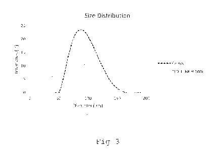
Figure 3: size distribution of different embodiments of the
liposomal doxorubicin formulation according to the
invention compared to Caelyx formulation measured
by DLS;
Figure 4a-4b: circularity distribution of the liposomal doxoru-
bicin formulation according to the invention (4a)
CA 03143443 2021-12-14
WO 2020/254633
PCT/EP2020/067196
28
compared to Caelyxe formulation (4b) measured by
cryo-TEM;
Fig 5a-5c: width measurements of the doxorubicin crystals ac-
cording to the invention (5a, 5b) compared to Cae-
lyx formulation (5c) measured by cryo-TEM;
Fig 6a-6c: length measurements of the doxorubicin crystals
according to the invention (6a, 6b) compared to
Caelyx formulation (5c) measured by cryo-TEM;
Fig 7a-7b: count of doxorubicin fibres per liposome, arranged
by classes; Fig 7a represents a doxorubicin formu-
lation according to the invention; Fig 7b a Cae-
lyx formulation, measured based on cryo-TEM imag-
ing;
Figure 8: doxorubicin accumulation in liver and tumor over
time for three compared administered formulations
CALYX, TLD-1, free DXR;
Figure 9: in vitro cytotoxicity study of three compared dox-
orubicin formulations CALYX, TLD-1, free DXR based
on MTS absorption;
Figure 10a-10c: in vitro cytotoxicity study of three compared
doxorubicin formulations CALYX, TLD-1, free DXR
based on luciferase luminescence;
Figure 11 results of comparative study on tumor growth (MDA-
MB231) over time under administration of liposomal
doxorubicin formulation according to Expl 1;
RECTIFIED SHEET (RULE 91) ISA/EP
CA 03143443 2021-12-14
WO 2020/254633
PCT/EP2020/067196
29
Figure 12a-b: results of comparative study on tumor growth
(A2780) over time under administration of liposo-
mal doxorubicin formulation according to Expl 1;
Figure 13: results of comparative study on tumor growth (411)
over time under administration of liposomal doxo-
rubicin formulation according to Expl 1;
Figures 14: results of in-vivo survival study in mice for
three compared doxorubicin formulations including
the one according to Expl 1.
Figures 15a-b: size and polydispersity results of stability meas-
urements over time (12 months) of liposomal doxo-
rubicin formulation according to Expl 1.
Example 1: Production of TLD-1
A 1,2-distearoyl-sn-glycero-3-phosphocholine and cholesterol
were provided in a 60:40 weight ratio and dissolved in ethanol
absolute >99.99%. The solution was hydrated in a 150m1V1 aqueous
solution of ammonium sulfate in sterile water at 68 C. The solu-
tion was sonicated with amplitude of 60pm for 24 hours to yield
crude liposomes. PSPE-MPEG2000 aqueous solution was then added
to the liposome suspension and heated to 65 C for 30 minutes to
yield PEGylated liposomes with the desired PEG-lipid amount of 5
mol%, corresponding to a PEG-lipid amount of 10 molt in the out-
er one of be lipid bilayer. Doxorubicin HC1 loading into lipo-
somes was performed to achieve a DXR/total lipid weight ratio of
0.05 by remote load technique. Unloaded DXR was removed by gray-
ity precipitation and filtration. The liposomal dispersion was
washed by tangential flow filtration and buffer exchange was
performed to achieve a dispersion of liposomes in 10mM HEPES-
buffered solution with 0.9 wt-% NaCl.
CA 03143443 2021-12-14
WO 2020/254633
PCT/EP2020/067196
The liposomal doxorubicin obtained as described in this example
may hereinafter be called "TLD", "TLD-1" o "Talidox".
Whenever free doxorubicin is applied as a comparative formula-
5 tion, this may in the Examples and Figures be referred to as
"Doxo", "DXR", "DX", "Doxorubicin".
Comparative Example
Commercially available Caelyx was purchased. For the cryo-TEM
10 measurements, Caelyx was diluted 10x in HEPES buffer (NaCl, pH
6.8).
Example 2: Size measurements
Size measurement of the liposomes obtained by the above method
15 was performed by cryo-TEM and DLS and the results compared to
corresponding measurements of commercially available Caelyx
formulation.
Figure la shows a high magnification (80'000x) representative
20 image of the formulation obtained according to Expl 1. Figure lb
shows the measured size distribution histogram. Figure 2a shows
a high magnification (80'000x) representative image of the com-
parative Caelyx formulation. Figure 2b shows the measured size
distribution histogram.
CryoTEM measurements were performed as follows: Liposomal sam-
ples according to Example 1 and comparative example were vitri-
fied. The samples were prepared on-grid (Formvar and Carbon)
with an acc. voltage of 200kV. Images were acquired with a cry-
oTEM JEOL JEM-2100F device and a TVIPS TemCam F415MP camera cam-
era at 40,000x magnification. Particle identification and size de-
termination were performed by by semi-automated image processing
using Vironova Analyzer Software, Vironova, Sweden. Briefly, a
CA 03143443 2021-12-14
WO 2020/254633
PCT/EP2020/067196
31
series of random images of the same magnification was imported.
Only liposome particles located entirely within the boundaries
of the image and with a distinct membrane were detected. The
identified objects were analyzed for spherical diameter, circu-
larity, unilamellarity. All images were batch-processed with
identical thresholds and settings, accumulating over 5 to 18 im-
ages for each sample, corresponding to a number of analyzed par-
ticles of 1560 to 1178. Mean values have a standard deviation of
approx. 10 nm.
Figure lb shows the size distribution of the liposomal formula-
tion according to Expl 1. The No. of images analyzed was 5. The
number of particles analyzed was 1560. The mean diameter was
35.61 nm and the standard deviation 7.42nm. The smallest diame-
ter measured was 24.81 nm, the largest diameter measured was
103.35 nm. Homogeneity Z-test gave a measure of the homogeneity
of the sampling of 1.01, indicating that all images included in
the analysis contained a population of particles with the same
means size.
Figure 2b shows the size distribution of the liposomal formula-
tion according to the comparative example. The No. of images an-
alysed was 18. The number of particles analysed was 1178. The
mean diameter was 70.26 nm and the standard deviation 13.41nm.
The smallest diameter measured was 32.52 nm, the largest diame-
ter measured was 159.09nm. Homogeneity Z-test gave a measure of
the homogeneity of the sampling of 3.15, indicating that not all
images included in the analysis contained a population of parti-
cles with the same means size.
The liposomal formulations according to Expl 1 further showed a
No. of broken particles <10%, and no particle aggregates nor
clusters in the cryoTEM analysis.
CA 03143443 2021-12-14
WO 2020/254633
PCT/EP2020/067196
32
Figure 3 shows a size of liposomal formulations according to
Expl. 1 and comparative example, measured by dynamic light scat-
tering (DLS). Both samples were diluted 10-fold in PBS or MQ H20
and measured on a Zetasizer device by Malvern at 25 C and 0
scattering angle. TLD-1 (according to Expl 1) had a mean diame-
ter of 60.5 ( 4.7 nm) nm and a polydispersity index of 0.084
0.038. Caelyx had a mean diameter of 85.0 nm in DLS-
measurements. It shall be noted that the values measured by dy-
namic light scattering are slightly higher than the values ob-
tainable by cryoTEM imaging due to the PEGylated surface not be-
ing detectable by cryoTEM, while it is included in DLS as part
of the hydrodynamic radius of liposomes.
Example 3: Circularity measurements
Circularity of the liposomal formulations according to Expl 1
and comparative Expl was measured by Cryo-TEM. The results are
presented in Fig 4a for Expl 1 and in 4b for comparative Expl.
Sample preparation and measurements were performed as described
earlier.
For Expl 1, the mean circularity of the particles was 0.99 with
a relative standard error of 0.03% and a mean standard deviation
of 0.01. The 50th percentile was measured 1.00, the 10th percen-
tile 0.98, the 5th percentile 0.98, and the 2nd percentile 0.96.
Homogeneity Z-test gave a measure of the homogeneity of the sam-
pling of 1.19, indicating that all images included in the analy-
sis contained a population of particles with the same means
size.
For the comparative example, the mean circularity of the parti-
cles was 0.99 with a relative standard error of 0.06% and a mean
standard deviation of 0.02. The 50th percentile was measured
CA 03143443 2021-12-14
WO 2020/254633
PCT/EP2020/067196
33
1.00, the 10th percentile 0.97, the 5th percentile 0.95, and the
2nd percentile 0.92. Homogeneity Z-test gave a measure of the ho-
mogeneity of the sampling of 6.10, indicating that not all imag-
es included in the analysis contained a population of particles
with the same means size.
Commercially available Caelyx hence shows a lower degree of
circularity of the liposomes in formulation. For example, 10% of
the liposomes in Caelyx have a circularity of only 0.97 and
lower.
The liposomal formulations according to Expl 1 further showed a
filling rate (filling with doxorubicin) of at least 80% and a
unilamellarity rate of 98% in the cryoTEM measurements.
Example 4: Crystal dimensions and number of fibres per crystal
Dimensions of the liposomal formulations according to Expl 1 and
comparative Expl were measured by Cryo-TEM. The results of the
width measurements are presented in Fig 5a and 5b for Expl 1 and
in Sc for the comparative Expl. The results of the length meas-
urements are presented in Fig 6a and 6b for Expl 1 and in 6c for
the comparative Example. Sample preparation and measurements
were performed as described earlier. The crystal length and
width were measured manually from a set of high magnification
images obtained by Cryo-TEM.
For Expl 1, the mean crystal width was 9.57 nm with a standard
deviation of 2.78 nm (No. of measurements: 140; 12 images ana-
lysed) and the mean crystal length was 27.36 nm with a standard
deviation of 9.15 nm (No. of measurements: 289; 5 images ana-
lysed). For the comparative example, the mean crystal width was
17.45 nm with a standard deviation of 4.60 nm (No. of measure-
ments: 60; 21 images analysed), and the mean crystal length was
CA 03143443 2021-12-14
WO 2020/254633
PCT/EP2020/067196
34
47.77 nm with a standard deviation of 15.33 nm (No. of measure-
ments: 105; 5 images analysed).
Amount of fibers per liposomes was determined from a set of high
magnification images obtained by Cryo-TEM. The number of indi-
vidual fibers (high density nodes) per liposome could be derived
manually. Since the doxorubicin crystals have a helical confor-
mation and the number of individual fibers per turn may vary,
one measurement was taken per turn, in order to provide an accu-
rate representation.
For Expl 1, the class ratios are displayed in Fig 7a. For the
comparative example, the class ratios are displayed in Fig 7b.
The x-Axis shows the class number (1 to 12), wherein the class
number indicates the number of individual fibers in the doxoru-
bicin crystal.
In Expl. 1, a number of 3 fibers per crystal was the most fre-
quent conformation. No crystals with 1, 7 or more individual fi-
bres were observed. The average distance between individual fi-
bres for all doxorubicin crystals in the dataset was measured to
2.6 nm.
In comparative Expl., a number of 7 fibers per crystal was the
most frequent conformation. No crystals with 1,2,3 and 12 or
more individual fibres were observed. The average distance be-
tween individual fibres for all doxorubicin crystals in the da-
taset was measured to 2.7 nm.
Commercially available Caelyx hence shows a lower degree of
circularity of the liposomes in formulation. For example, 10% of
the liposomes in Caelyx have a circularity of only 0.97 and
lower.
CA 03143443 2021-12-14
WO 2020/254633
PCT/EP2020/067196
The liposomal formulations according to Expl 1 further showed a
filling rate (filling with doxorubicin) of at least 80% and a
unilamellarity rate of 98% in the cryoTEM measurements.
5
Example 5: Tumor accumulation in mice
A liposomal doxorubicin formulation according to Expl 1 ("TLD-
1"), commercially available Caelyx ("CAELYX") and free doxoru-
bicin (Adriblastin; "free Doxorubicin") were administered to
10 mice (athymic Nude-Foxnlnu mice) in an amount of 3.5mg/kg. After
4h or 16h, the mice were sacrificed in order to detect the total
doxorubicin amount using HPLC analysis.
Figure 8 shows the doxorubicin accumulation in liver and tumor
15 over time for three compared administered formulations. Bars
represent mean and standard deviation (n=3).
TLD-1 accumulation was about 4x higher than accumulation of free
doxorubicin in the tumour and twice as high as for CAELYX . The
20 serum half-life up to 16 hrs is comparable between CAELYX and
TLD-1. Liver accumulation and clearance, however, is more effi-
cient for TLD-1.
Example 6: Cytotoxicity in vitro
25 In vitro cytotoxicity of TLD-1, CAELYX and free doxorubicin
("DX") was measured in A2780 cells seeded at 10'000 cells/ml in
96 wells plates (100 ml/well). 24 hrs after seeding, the cells
were treated with different concentrations of doxorubicin formu-
lations.
Figure 9 shows the result of added concentrations of 10, 50,
100, 500, 1000, 5000 and 10'000 ng/ml to the cells. 72 hrs after
treatment, MTS colorimetric assay components ((3-(4,5-
CA 03143443 2021-12-14
WO 2020/254633
PCT/EP2020/067196
36
dimethylthiazol-2-y1)-5-(3-carboxymethoxypheny1)-2-(4-
sulfopheny1)-2H-tetrazolium) "MIS") were added for colouring vi-
able cells. After another 3 hrs, absorbance was measured. As can
be seen, absorbance of the samples treated with TLD-1 was clear-
ly lower than absorbance of the samples treated with CAELYX for
any applied concentration. Therefore, cytotoxicity of TLD-1 is
higher and more similar to free doxorubicin.
Figures 10a to 10c show the result of added concentrations of 5,
50, 500, 5000, 25'000 and 50'000 ng/ml. Cells were treated in
the presence of a luciferase substrate and luminescence of the
viable cells measured over time (6, 24, 48, 73 hrs after treat-
ment). As can be seen from the charts, luminescence of the sam-
ples treated with TLD-1 was clearly lower than luminescence of
the samples treated with CAELYX after 48 hrs. The effect was
even more pronounced after 72 hrs. Therefore, cytotoxicity of
TLD-1 is higher and more similar to free doxorubicin.
Serum leakage studies: An experiment was performed to assess to
which extent TLD-1 and Caelyx release free doxorubicin into
RPMI (cell medium) +/- 10% FCS medium over time. Free
doxorubicin in said medium was measured after 72h incubation of
TLD-1 and CAELYX, respectively, in the RPMI medium +/- 10% FBS
at 37 in a metal beads bath, protected from light. Free and
liposomal doxorubicin were detected by HPLC size exclusion
chromatography at 478nm (hence avoiding background absorption
from proteins). Liposomal doxorubicin is complexed in aggregates
and thus appeares later than the free doxorubicin peak. The
latter was identified by comparison with values from a free DX
(adriblastin) control sample. A comparative analysis of the area
under the curve of the peaks (liposomal doxo vs free doxo) was
performed. Experiments revealed that both TLD-1 and Caelyx
remain stable when challenged at 37 for 72h in the medium used
CA 03143443 2021-12-14
WO 2020/254633
PCT/EP2020/067196
37
in the in-vitro experiments. The percentage of free DX in the
solution was below 4% for incubated TLD-1 and below 6% for
incubated Caelyx . In general, TLD-1 leakage was lower than
leakage of Caelyx. This indicates that the enhanced effect seen
in in-vitro cell toxicity assays is due to increased cellular
uptake rather than by leaking free doxorubicin into the medium.
Example 7: Tumor growth
In vivo effect on tumor growth was determined by administering
placebo formulations, TLD-1 and CAELYX to mice and by measuring
the effect on tumor size over time.
Figure 11 shows the result of such testing. Empty liposomes,
free doxorubicin, Caelyx and TLD-1 were administered on a regu-
lar basis to mice (5 mice/formulation) with injected MDA-MB231
cell lines. Each arrow indicates injection of a dose of 3.
5mg/kg body weight. The effect of TLD-1 (measured in tumor
weigth growth, pg) was clearly better than the effect of Caelyx
already 3 days after the start of the treatment and remained
substantial over 26 days.
Figure 12a and 12b show the result of another similar test set-
up. PBS, free doxorubicin, Caelyx and TLD-1 were administered
on a regular basis to mice (5 mice/formulation) with injected
A2780 cell lines. For Fig 12a, a formulation similar to the one
in Expl 1 was administered, however, unlike in Expl 1, soni-
cation was only performed under such conditions as to reach mean
liposomal diameter of 86.78 nm and a polydispersity index of
0.117 both measured by DLS. For Fig 12b, a formulation according
to Expl 1 was administered, liposomes having a mean diameter of
64.87 nm and a polydispersity index of 0.168, measured by DLS.
Each arrow indicates injection of a dose of 3.5 mg/kg body
weight. While in the test presented in Fig 12a, the effect on
CA 03143443 2021-12-14
WO 2020/254633
PCT/EP2020/067196
38
tumor growth of TLD-1 with a mean diameter outside of the speci-
fication (>70nm) was found to be inferior compared to treatment
with Caelyx , the effect of TLD-1 according to the specification
was clearly better than the effect of Caelyx already after 19
days after the start of the treatment and remained substantial
over another 12 days (measured in tumor weight growth, pg).
Figure 13 shows the result of another similar test setup. Sa-
line, Caelyx and TLD were administered on a regular basis to
mice with implanted 411 tumor (5 mice/formulation). The effect
of TLD-1 was clearly better than the effect of Caelyx 12 days
after the start of the treatment (measured in tumor size growth,
mm3).
Figures 14 shows the in-vivo survival study for mice treated
with saline, Caelyx or TLD-1 (according to Expl 1, 5
mice/formulation) respectively. 60% of mice treated with TLD-1
survived until day 30 and 40% of mice until day 34. In contrast,
100% of the mice of the group treated with Caelyx had died al-
ready on day 26.
Example 8: Other studies
Other comparative studies for a liposomal doxorubicin formula-
tion according to Expl 1 (TLD-1) and Caelyx were performed.
They included side-effect studies and efficacy studies in animal
models. For Expl 1, it also included serum half-life and Area
Under Curve (AUC) studies in humans, as well as side-effect
studies.
Serum half-life studies have been perfomed in human serum:
Caelyx has been documented to have a half-life of 74h in human
serum. Currently serum half-life of TLD-1 in human is estimated
from 5 patients and is about 100h. Moreover, the Area Under the
CA 03143443 2021-12-14
WO 2020/254633
PCT/EP2020/067196
39
Curve (AUC) of the serum half-life data of TLD-1 shows to be
larger than for a corresponding dose of Caelyx , which means
that higher drug exposition for a given dose is achieved. Drug
exposition of a patiend treated with TLD-1 (30mg/m2) is higher
than drug exposition of a patiend treated with Caelyx (37mg/m2)
despite the difference in dose.
Adverse effects studies have been performed in rats. Skin tox-
icity and in particular PPE (e.g. hand-foot-syndrome) was as-
sessed during toxicology studies conducted in rats. Skin toxici-
ty and in particular PPE were not promoted by the administration
of TLD-1, even at high concentration of 6mg/kg (male and female
data pooled together due to lack of statistically significant
difference). Similarly, neutropenia was assessed during toxicol-
ogy studies conducted in rats (by neutrophile count). Neutro-
phile count varied not significantly upon TLD-1 administration
even at high concentrations of 6mg/kg (male and female data
pooled together due to lack of statistically significant differ-
ence).
Example 9: Stability results
Figures 15a and 15b show the size and polydispersity stability
of liposomal formulations according to the invention over time,
measured by DLS. The liposomal formulations were obtained ac-
cording to the method described above (Expl 1). The liposomal
formulations were stored in HEPES buffered solution at a pH-
value of 6.5 to 6.8 and a temperature of 4 C. The variation in
size was not higher than 1 nm over a 12-months period from man-
ufacture. Variation in polydispersity index was not higher than
0.01, measured by DLS.
Example 10: Clinical trial results
CA 03143443 2021-12-14
WO 2020/254633
PCT/EP2020/067196
A clinical study is currently being conducted in which a liposo-
mal doxorubicin formulation according to Expl 1 (TLD-1) has so
far been used in twelve patients with advanced solid tumors
(Swiss Group for Clinical Cancer Research; Trial number: SAKK
5 65/16). The trial was designed as an open-label, single arm,
multicentre, first-in-human, phase-1 trial. The primary objec-
tive of this trial was to identify the maximum tolerated dose
(MTD) and the recommended phase 2 dose (RP2D) for TLD-1 in pa-
tients with advanced solid tumors. Further objectives of this
10 trial were to evaluate the safety, preliminary anti-tumor activ-
ity and pharmacokinetics of TLD-1.
The interim report of this study states that TLD-1 can be safely
administered up to a dose of 45 mg/m2 every 3 weeks in patients
15 with advanced, pretreated solid tumors. This dose is higher com-
pared to Caelyx@, where the MTD is 50 mg/m2 every 4 weeks. Fur-
thermore, the number and severity of undesired side effects of
TLD-1 was lower than with Caelyx . Specifically (TLD-1 vs.
Caelyx@), no clinically significant nausea (<8.3% vs. 38.5%),
20 vomiting (<8.3% vs. 24.3%), alopecia (0% vs. 13.4%), or cardiac
toxicity were observed while myelosuppression was rare and of
mild degree (8.3% vs. 25.6%). No unexpected toxicities were re-
ported.
25 Without being limited to this, it is hypothesized that the fewer
side effects observed with TLD-1 compared to conventional lipo-
somal formulations of doxorubicin, including Caelyx@, are due to
the comparatively small liposome size and the high degree of ho-
mogeneity of the doxorubicin-loaded liposomes administered to
30 the patients, in particular due to their pronounced circularity,
low polydispersity, and high degree of uniformity (length and
width) of the doxorubicin crystal fibres in the liposomes.