Note : Les descriptions sont présentées dans la langue officielle dans laquelle elles ont été soumises.
GALLBLADDER MODEL
[0001] This is a divisional application of co-pending Canadian
Application
No. 2,914,952, which entered the national phase in Canada on December 9, 2015
from
PCT Serial No. U52014/042998, having an international filing date of June 18,
2014.
Field of the Invention
[0002] This application relates to surgical training tools, and in
particular,
to simulated tissue structures and models for teaching and practicing surgical
procedures
involving a gallbladder.
Background of the Invention
[0003] A common treatment for gallstones and other gallbladder
conditions
is a cholecystectomy which is the surgical removal of the gallbladder from the
liver bed.
Laparoscopic cholecystectomy is the most common laparoscopic procedure and has
replaced open cholecystectomy as the first-choice of treatment for gallstones
and
inflammation of the gallbladder. Laparoscopic cholecystectomy advantageously
requires
smaller incisions, resulting in less pain, improved cosmetic results, quicker
healing, and
fewer complications such as infection and adhesions.
[0004] Laparoscopic cholecystectomy requires several small
incisions in
the abdomen to allow the insertion of trocars or small cylindrical tubes
approximately 5 to
millimeters in diameter through which surgical instruments and a laparoscope
are
placed into the abdominal cavity. The laparoscope illuminates the surgical
field and
sends a magnified image from inside the body to a video monitor giving the
surgeon a
close-up view of the organs and tissues. The surgeon watches the live video
feed and
performs the operation by manipulating the surgical instruments placed through
the
trocars.
[0005] In a laparoscopic cholecystectomy, a patient is placed in a
supine
position on the operating table and anesthetized. A scalpel can be used to
make a small
incision at the umbilicus. Using a trocar, the abdominal cavity is entered and
enlarged by
delivering carbon dioxide gas to insufflate the cavity to create a working
space inside the
patient's abdominal region. The trocar may include an inserted laparoscope for
1
Date Recue/Date Received 2022-05-17
observing the penetration, insertion, and insufflation of the abdominal space.
Additional
trocars are inserted at a location inferior to the ribs. Using the
laparoscope, the fundus of
the gallbladder, which is covered by the peritoneum, is identified, grasped
with a surgical
grasper extending through one of the trocars, and retracted. A second surgical
grasper
may be used to retract the rest of the gallbladder in a lateral direction to
expose Calot's
triangle. Calot's triangle is that portion of the gallbladder anatomy that is
bound by the
cystic duct, cystic artery, the hepatic duct and the border of the liver. The
surgeon
identifies the cystic duct and cystic artery. In this area, the underlying
structures are
carefully skeletonized from the peritoneum separating the peritoneum from the
both the
cystic duct and the cystic artery. A surgical clip applier is introduced
through one of the
trocars and clips are applied in two locations to both the cystic duct and the
cystic artery.
The cystic duct and the cystic artery are then divided with surgical scissors
between the
two locations of clips freeing the gallbladder for removal. The gallbladder is
dissected
from the bed of the liver and removed through one of the trocars. During
laparoscopic
cholecystectomy, complications may arise due to gallbladder perforation which
can occur
due to excessive traction during retraction or during dissection of the
gallbladder from the
liver bed or extraction from the abdomen. The outcome of laparoscopic
cholecystectomy
is greatly influenced by the training, experience and skill of the surgeon
performing the
procedure. In order for residents and surgeons to learn and practice these
surgical
techniques, a realistic, functional, and anatomically correct model for use in
a
laparoscopic training device is needed.
[0006] A gallbladder model is not only useful for training
residents and
surgeons in laparoscopic cholecystectomy, but also, desirable for training
residents and
surgeons in laparoscopic common bile duct exploration. The common bile duct is
a tube
that connects the liver, gallbladder and pancreas to the small intestine and
delivers fluid
to aid in digestion. Common bile duct exploration is a procedure used to see
if a
gallbladder stone or some other obstruction is blocking the flow of bile from
the
gallbladder or liver to the intestine which can cause jaundice. In a
laparoscopic common
bile duct exploration procedure, the abdominal cavity is approached as in a
cholecystectomy described above. The surgeon identifies the common bile duct
and a
small hemi-circumferential incision is made in the common bile duct. A
cholangiography
catheter is inserted into the insufflated abdominal cavity through one of the
trocars and
2
Date Recue/Date Received 2022-05-17
into the incision made in the common bile duct. Contrast media or radiopaque
fluid is
introduced into the cystic and common bile ducts and an X-ray is taken to
reveal the
location of any gallstones in the common bile duct. If there are gallstones,
the
obstructions will appear as discontinuities in the flow of contrast media. The
gallstones
are then surgically extracted.
[0007] In order to help patient outcomes and recoveries, surgeons
need a
way to practice laparoscopic cholecystectomies and common bile duct
explorations
outside of the operating room. The practice model needs to be anatomically
correct and
include all important landmarks normally seen during surgery in order to give
the surgeon
or resident the most realistic practice possible.
Summary of the Invention
[0008] According to one aspect of the invention, an anatomical
model for
surgical training is provided. The model includes a first layer having an
inner surface and
an outer surface. The first layer has a substantially uniform thickness
defined between
the inner surface and the outer surface. The first layer has a first perimeter
and is
configured to simulate at least a portion of a first anatomical structure. The
model
includes a second layer having an inner surface and an outer surface. The
second layer
has a thickness between the inner surface and the outer surface. The second
layer
defines a second perimeter and overlays the first layer such that the outer
surface of the
second layer faces the inner surface of the first layer. The model includes at
least one
second simulated anatomical structure which has a third perimeter around the
at least
one simulated anatomical structure. The at least one simulated anatomical
structure is
connected to the inner surface of the second layer. The outer surface of the
second
layer is connected to the inner surface of the first layer at least partially
around the
location of the at least one second simulated anatomical structure.
[0009] According to another aspect of the invention, an anatomical
model
for surgical training is provided. The model includes an anatomical portion
and a support
removably connectable to the anatomical portion. The anatomical portion
includes at
least a first layer having an inner surface and an outer surface
interconnected by a top
side and a bottom side and a left side and a right side. The first layer has a
thickness
defined between the inner surface and the outer surface. The first layer is
configured to
3
Date Recue/Date Received 2022-05-17
simulate at least a portion of a liver. The top side of the first layer has a
peak. The
model includes a simulated gallbladder positioned in the location of the peak
and facing
the inner surface of the first layer. The model includes a frame connected to
at least the
first layer. The frame has a first end interconnected to a second end by a
central portion.
The first end and the second end of the frame are removably connectable to the
support
to hold the anatomical portion in a substantially upright position. The frame
does not
extend into the location of the peak such that the first layer in the location
of the peak is
capable of flexing inwardly and outwardly relative to the frame.
[0010] According to another aspect of the invention, an anatomical
model
for surgical training is provided. The model includes an anatomical portion
having a first
layer. The first layer includes an inner surface and an outer surface
interconnected by a
top side and a bottom side and a left side and a right side. The first layer
has a thickness
defined between the inner surface and the outer surface. The first layer is
configured to
simulate at least one anatomical structure. The anatomical portion includes a
second
layer that includes at least one anatomical structure overlaying the first
layer. The
anatomical portion also includes a frame having a first end interconnected to
a second
end by a central portion. At least part of the frame is embedded within the
first layer with
the first end and the second end of the frame extending out from the first
layer. The
model includes a support to which the first end and the second end of the
frame are
removably connectable to the support to hold the anatomical portion in a
substantially
upright position with respect to a supporting surface.
[0011] According to another aspect of the invention, a surgical
simulation
system is provided. The system includes an anatomical model. The model
includes an
anatomical portion. The anatomical portion includes a first layer having an
inner surface
and an outer surface interconnected by a top side and a bottom side and a left
side and
a right side. The first layer has a substantially uniform thickness defined
between the
inner surface and the outer surface. The first layer is configured to simulate
at least one
anatomical structure and defines a substantially planar configuration. The
model
includes a second layer having a plurality of anatomical structures connected
to and
overlaying the inner surface of the first layer. A support is connectable to
the anatomical
portion and configured to hold the anatomical portion in a substantially
perpendicular
orientation with respect to a supporting surface. The system further includes
a surgical
4
Date Recue/Date Received 2022-05-17
training device. The surgical training device includes a base and a top cover
connected
to and spaced apart from the base to define a simulated insufflated internal
cavity
between the top cover and the base. The internal cavity is at least partially
obstructed
from direct observation by a user. The top cover includes an aperture or
penetrable
simulated tissue region. The top cover of the surgical training device is
angled to form
an acute angle with respect to a horizontal plane as measured from inside the
cavity.
The anatomical model is positioned inside the internal cavity a distance
opposite the
acute angle such that the inner surface of the first layer faces the acute
angle and the
aperture or penetrable simulated tissue region.
[0012]
According to another aspect of the invention, an anatomical model
for surgical training is provided. The model includes an anatomical portion.
The
anatomical portion includes a first layer having an inner surface and an outer
surface
interconnected by a top side, a bottom side, a left side and a right side. The
inner
surface is substantially planar and flat and the first layer defines a
thickness between the
inner surface and the outer surface. The first layer is configured to simulate
at least a
portion of a liver. The top side of the first layer has a peak. The anatomical
portion
includes a second layer having an inner surface and an outer surface
interconnected by
a top side, a bottom side, a left side and a right side. The second layer
overlays the first
layer such that the outer surface of the second layer faces the inner surface
of the first
layer. The outer surface of the second layer is connected to the inner surface
of the first
layer along at least part of a first perimeter. The second layer defines a
thickness
between the inner surface and the outer surface and the thickness of the
second layer is
smaller than the thickness of the first layer. The anatomical portion includes
a third layer
having at least one simulated anatomical structure. The at least one simulated
anatomical structure is connected to the inner surface of the second layer.
The
anatomical portion further includes a fourth layer having an inner surface and
an outer
surface interconnected by a top side, a bottom side, a left side and a right
side. The
fourth layer overlays the second layer and the third layer such that the outer
surface of
the fourth layer faces the inner surface of the second layer and the at least
one simulated
anatomical structure. The outer surface of the fourth layer is connected to
the inner
surface of the second layer along at least part of a second perimeter. The
fourth layer
defines a thickness between the inner surface and the outer surface and the
thickness of
Date Recue/Date Received 2022-05-17
the fourth layer is smaller than the thickness of the first layer. The
anatomical portion
further includes a frame at least partially embedded inside the first layer.
The model
includes a support connectable to the frame to hold the anatomical portion in
a
substantially upright position.
[0013] According to another aspect of the invention, a gallbladder
model is
provided. The model allows users to practice open and laparoscopic
cholecystectomies
and common bile duct explorations. The gallbladder model includes an
anatomical
portion connected to a support. The anatomical portion includes a liver layer,
a fascia
layer, a gallbladder layer, a peritoneum layer, and a frame connected together
and held
in an upright orientation by the support.
Brief Description of the Drawings
[0014] FIG. 1 is a top perspective view of an anatomical model
according
to the present invention.
[0015] FIG. 2 is an exploded, top perspective view of an anatomical
model
according to the present invention.
[0016] FIG. 3 is a side view of a liver layer of an anatomical
portion of the
anatomical model according to the present invention.
[0017] FIG. 4 is a partial side view of a prong of a frame of an
anatomical
portion of the anatomical model according to the present invention.
[0018] FIG. 5 is a side, cross-sectional view of a support for an
anatomical
portion of an anatomical model according to the present invention.
[0019] FIG. 6 is a top perspective view of a laparoscopic trainer
for use
with an anatomical model according to the present invention.
[0020] FIG. 7 is a top perspective view of a frame and support of
an
anatomical model according to the present invention.
Detailed Description of the Invention
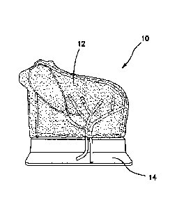
[0021] Turning now to FIG. 1, there is shown a gallbladder model 10
according to the present invention. The gallbladder model 10 includes an
anatomical
portion 12 removably connected to a support 14. The substantially planar
anatomical
portion 12 is maintained in an upright configuration by the support 14. In a
6
Date Recue/Date Received 2022-05-17
cholecystectomy, as described above in the background section of this
application, the
fundus of the gallbladder is visible and retracted. In doing so, the remainder
of the
gallbladder underlying the liver toward the posterior of the patient is
uncovered and made
visible along with the triangle of Calot in the insufflated cavity. This
retraction involves
lifting part of the lower or inferior portion of the right lobe of the liver.
With the liver and
gallbladder lying substantially in the X-Z plane or frontal plane of the
patient, and the
retraction lifting the liver and gallbladder substantially into the Y plane or
transverse
plane of the patient, the gallbladder model 10 of the present invention is a
substantial or
partial projection of at least a portion of the retracted liver and
gallbladder onto the X-Y
plane or transverse plane of a patient. Hence, the gallbladder model 10
represents a
substantial planar projection of a retracted liver and gallbladder in a
simulated insufflated
cavity. As such, the gallbladder model 10 configuration advantageously
provides a
surgical approach to a simulated gallbladder already in a retracted
perpendicular
orientation when viewed by the user approaching the gallbladder from the
location of the
umbilicus. Also, the gallbladder model 10 configuration permits practice by
the user
without requiring a second user to hold portions of the model with graspers in
a retracted
position and as such, the model 10 is advantageously designed to be used by
one
person at a time. Furthermore, in the model 10, only a portion of the liver is
simulated, in
particular, the right lobe of the liver. Together, with the right lobe, the
entirety of the
biliary structure including the gallbladder is included in the model.
[0022] Turning now to FIG. 2, there is shown an exploded view of
the
gallbladder model 10 comprising an anatomical portion 12 connected to a
support 14.
The anatomical portion 12 includes a liver layer 16, a fascia layer 18, a
gallbladder layer
20, a peritoneum layer 22, and a frame 24 connected together. Each layer will
now be
described in greater detail.
[0023] Still referencing FIG. 2, the liver layer or first layer 16
is molded
from silicone or thermoplastic elastomer that is dyed with a red color and
configured to
simulate a retracted portion of a liver. In particular, the liver layer 16 is
shaped to
represent a portion of the right lobe of a human liver that is retracted to
expose the
gallbladder and triangle of Calot. Referring to FIG. 3, the liver layer 16
includes a flat
planar inner surface 26 and a convex curved outer surface 28. The inner and
outer
surfaces 26 interconnect along four sides--a curved top side, a straight
bottom side, and
7
Date Recue/Date Received 2022-05-17
a left side and right side that interconnect the top and bottom sides. The
curved top side
includes a peak 30 near or at the left side of the model. The top side curves
downward
from the peak 30 to a lower portion that interconnects with the right side.
This peaked
shape resembles a substantially planar projection of a retracted right lobe of
a human
liver. The peak 30 has a longer length relative to other portions of the liver
layer 16. The
thickest portion of the liver layer 16 is approximately 0.5 inches and located
approximately at the middle. In one variation, the frame 24 is molded directly
into the
liver layer 16 such that at least a portion of the frame 24 resides inside the
liver layer 16
and a portion of the frame 24 resides outside of the liver layer 16 as shown
in FIG. 3.
The frame 24 will be described in greater detail below.
[0024] Still referencing FIG. 2, the fascia layer or second layer
18 is a thin
approximately 0.01-0.03 inches thick layer made of a thermoplastic elastomer
or silicone
that is partially translucent, clear or dyed with a slight yellow color. The
fascia layer 18
has the same peaked shape as the liver layer 16 and is sized and configured to
overlay
the liver layer 16. The fascia layer 18 has an inner surface and an outer
surface with the
outer surface overlaying a portion of the inner surface 26 of the liver layer
16. The fascia
layer 18 is attached to the liver layer 16 with adhesive that is placed at
least along the
perimeter such that the majority of the middle portion or portions interior
from the
perimeter of the fascia layer 18 are not attached to the liver layer 16, but
instead, are free
to remain mobile and separate away from the liver layer 16. While this fascia
layer 18
does not exist in real life, that is, there is no tissue layer located between
the gallbladder
and the liver, the gallbladder model 10 of the present invention includes a
fascia layer 18
which advantageously simulates the dissection and removal of the gallbladder
away from
the liver. This advantage will be described in greater detail below.
[0025] Still referencing FIG. 2, the gallbladder layer or third
layer 20
includes at least one body component. In FIG. 2, the at least one body
component is a
plurality of anatomical structures. For example, the gallbladder layer 20
includes a
gallbladder 32 connected to a cystic duct 34, a common hepatic duct 36
connected to a
common bile duct 38, a cystic artery 40, and a common hepatic artery 42
connected to
and branching into the right hepatic artery 44 and left hepatic artery 46. All
of these
anatomical structures are configured to simulate actual human anatomy and
arranged
within the gallbladder layer 20 in an anatomically correct fashion. The
gallbladder 32 is a
8
Date Recue/Date Received 2022-05-17
hollow bulbous structure molded out of silicone or other thermoplastic
material dyed with
a light green or yellow color to simulate bile. In another variation, the
gallbladder 32 is a
solid and not hollow structure. The cystic duct 34, common hepatic duct 36 and
common
bile duct 38 are also made of silicone or thermoplastic material that is dyed
with a light
green color. The cystic duct 34 is tubular in shape having a tapered end and a
diameter
of approximately 0.15-0.25 inches. In one variation, the cystic duct 34 has a
lumen with
a minimum inner diameter of 0.15 inches and a maximum outer diameter of 0.25
inches
making it small enough to clip and large enough to permit insertion of
catheter. In yet
another variation, the cystic duct 34 includes a lumen having an inner surface
that is
lubricated with lubricant. In yet another variation, the cystic duct 34 is
larger in outer
diameter relative to dimension of a real life cystic duct 34 to facility
training and insertion
of a catheter into the lumen. The common hepatic duct 36 and common bile duct
38 are
also tubular in shape having a diameter of approximately 0.15 inches. In one
variation,
the cystic duct 34, common hepatic duct 36 and common bile duct 38 are hollow
and in
another variation they are solid. The cystic artery 40, the common hepatic
artery 42, the
right hepatic artery 44 and the left hepatic artery 46 are made from silicone
or
thermoplastic material that is dyed a red color and molded into a tubular
shape having a
diameter of approximately 0.15 inches. In one variation, the cystic artery 40,
common
hepatic artery 42, the right hepatic artery 44 and the left hepatic artery 46
are hollow and
in another variation they are solid structures. The gallbladder layer 20 is
connected to
the fascia layer 18 with selectively-placed adhesive. The gallbladder layer 20
may be
formed from multiple pieces joined together or as a unit with no disconnects.
To form a
unitary gallbladder layer 20, the manufacturing process consists of a wax form
that is
dipped in molten plastic and melted out once the plastic has set.
[0026] In one variation, the gallbladder model 10 is configured
for
practicing bile duct exploration. In such a variation, the biliary structures
of the
gallbladder layer 20 are hollow and filled with fluid that resembles bile. An
exemplary
fluid is green-colored dishwashing liquid. The inner diameter of the hollow
biliary
structures is approximately 0.09 inches and the outer diameter is
approximately 0.15
inches. The gallbladder model 10 that is configured for biliary exploration
includes a
hollow gallbladder 32 filled with fluid that resembles bile. So that the
simulated bile fluid
is not lost, the free ends of the cystic duct 34, common hepatic duct 36, and
common bile
9
Date Recue/Date Received 2022-05-17
duct 38 are closed or capped with standard tubing caps, solid connectors or
barbed
connectors that retain fluid inside the ducts. If not molded as a single unit,
biliary
structures made of multiple tubular structures are connected together with
connectors.
For example, the junction between the common hepatic duct 36 and common bile
duct
38 is connected with a connector such as a Y-shaped split that permits fluid
to flow
therebetween. In one variation, the cystic duct 34 and the common bile duct 38
are
connected via a connector or molded as a unitary structure such that fluid is
allowed to
flow between the cystic duct 34 and the common bile duct 38. The employ of
connectors
is advantageous in that after practice scenarios in which the ducts are cut,
such as in a
cholecystectomy, the severed ducts are replaceable with new ducts that are
reconnected
at the same locations using the same connectors so that training scenarios can
be
repeated. In the gallbladder model 10 that is adapted for biliary duct
exploration, any
one or more of the gallbladder 32, bile duct 34, common hepatic duct 36, and
common
bile duct 38, may include one or more simulated gallstones (not shown). A
simulated
gallstone is a small bead-like structure made of plastic or other material.
The simulated
gallstones are placed inside the hollow space of the gallbladder 32 and/or
inside the
lumen of one or more of the cystic duct 34, common hepatic duct 36, and common
bile
duct 38. These simulated gallstones are shaped and configured such that they
are not
visible to the user when the model is received but become visible when a
syringe and/or
catheter is used to inject simulated contrast media fluid such as colored
water into one or
more of the ducts and the continuous flow of contrast media fluid is visibly
interrupted or
blocked by the gallstones as the simulated contrast media fluid fills the
biliary structures.
In another variation, a kit is provided that includes a syringe with which the
gallbladder 32
is injected with fluid and/or simulated gallstones. In another variation, the
gallbladder 32
is not filled with liquid but is filled with air which may be injectable into
the open cavity of
the gallbladder 32 with a syringe or other similar device. The cavity of the
gallbladder 32
may be pressurized to a pressure greater than ambient such that when the
gallbladder
32 is inadvertently punctured, as if by an improper surgical technique, the
gallbladder 32
noticeably deflates and as such provides a visual indication to the trainee.
In such a
variation, the gallbladder 32 has a wall thickness configured to permit
observation of
deflation of the gallbladder 32.
Date Recue/Date Received 2022-05-17
[0027] Still referencing FIG. 2, the peritoneum layer or fourth
layer 22 is a
thin layer approximately 0.01-0.03 inches thick made of a thermoplastic
elastomer or
silicone that is clear or partially translucent and/or dyed with a slightly
yellow color. The
peritoneum layer 22 is nearly identical to the fascia layer 18 and has the
same peaked
shape as the underlying fascia layer 18 and liver layer 16. The peritoneum
layer 22
includes an inner surface and an outer surface overlaying the gallbladder
layer 20 and
overlaying at least a portion of the inner surface of the second layer 18. In
one variation,
both the fascia layer 18 and the peritoneum layer 22 are each formed by
molding liquid
silicone on a layer of foam such as packaging foam or other spongiform
structure and
then peeled off the foam after it has set to impart at least one textured
surface to the
fascia and peritoneum layers 18, 22. The peritoneum layer 22 is sized and
configured to
overlay the gallbladder layer 20. The peritoneum layer 22 is attached to the
fascia layer
18 with adhesive that is placed in locations that are capable of direct
contact with the
fascia layer 18 without interference from the intervening gallbladder layer
20. Hence,
only portions of the peritoneum layer 22 are adhered to the fascia layer 18
and in one
variation, the peritoneum layer 22 is only adhered to the fascia layer 18 and
not to the
gallbladder layer 20. In another variation, portions of the peritoneum layer
22 are
adhered to portions of the gallbladder layer 20 as well as the fascia layer
18. In yet in
another variation, portions of the peritoneum layer 22 are adhered only to
portions of the
gallbladder layer 20. The layers are adhered with adhesive or by the inherent
tackiness
of the material composing the layers. In essence, the peritoneum layer 22 is
selectively
adhered to one or more of the underlying gallbladder layer 20 and fascia layer
18 with
adhesive.
[0028] Still referencing FIG. 2, the anatomical portion 12
includes a frame
24 that is configured to support the entire anatomical portion 12 in a
substantially upright
orientation with respect to a table top or other substantially flat surface
including an
organ-receiving tray or other surface inside a laparoscopic training
simulator. The frame
24 includes a left leg 48 and a right leg 50 interconnected by a central
portion 52. The
central portion 52 is curved and mimics the generally peaked-shape of the
other layers
16, 18, 22. The frame 24 is sized smaller than the liver, fascia and
peritoneum layers
16,18, 22. The frame 24 is made of rigid metal, plastic or other polymer or
material that
is capable and strong enough to support the layers of silicone and plastic
comprising the
11
Date Recue/Date Received 2022-05-17
anatomical portion 12 of the model 10 in an upright orientation. The left leg
49 is at or
adjacent to the peak and is approximately 3.5-4.0 inches long and the shorter
right leg 50
is approximately 2.5-3.0 inches long. The curved central portion 52 is
approximately 4.0-
4.5 inches long and follows the curvature of the layers 16, 18, 22. The
overall height of
the gallbladder model 10 is approximately 5-6 inches and the length of the
model 10 is
approximately 5-6 inches. The left leg 48 defines a left prong 54 at its free
end and the
right leg 50 defines a right prong 56 at the free end of the right leg 50. The
left and right
prongs 54, 56 extend beyond the anatomical portion 12 for insertion into a
support 14.
The cross-section of the frame 24 is substantially circular with a diameter of
approximately 0.15 inches with the prongs 54,56 having a slightly larger
diameter. Each
prong 54, 56 includes a curved, ball-shaped, or spherical-shaped or angled
detent 58 as
illustrated in FIG. 4 which shows a sectional view of a the left leg 48. The
prongs 54, 56
have angled distal tips. The frame 24 is connected to the anatomical portion
12 such
that the prongs 54, 56 protrude out from the layers for connection with the
support 14.
As described above, in one variation, the frame 24 is molded directly into the
liver layer
16 and is clear or transparent in color or substantially the same color as the
liver layer 16
in which it is embedded so that it is not readily visible to the user.
[0029] In another variation, the frame 24 does not have a peaked
portion
and is substantially U-shaped. As shown in FIG. 7, the central portion 52 of
the frame 24
is straight and does not follow the peaked-shaped of the other layers 16, 18,
22. This
variation provides less support to the other layers 16, 18, 22 in the location
of the peak
30 advantageously permitting all of these layers to be more flexible and to be
more easily
pushed distally or proximally relative to areas adjacent to the frame 24 to
practice the
retraction of the liver 16 from the gallbladder 32 while still providing
support to the overall
model 10 in the support 14. In this variation, both the right leg 50 and left
leg 48 are the
same length approximately 2.5-3.0 inches long instead of the left leg 48 in
the location of
the peak 30 being longer. The peak 30 formation in the layers 16, 18, 22
represents only
a portion of the liver, in particular, the right lobe of the liver with all of
the anatomical
structures of the gallbladder layer 20 being presented in the model 10.
[0030] With additional reference to FIG. 5, the support 14 is
configured to
connect with the anatomical portion 12 and hold the anatomical portion 12 in a
substantially upright orientation with respect to a table top or other
surface. The support
12
Date Recue/Date Received 2022-05-17
14 includes a base 60 interconnected with an upright portion 62. The upright
portion 62
includes at least two sockets 64 that are sized and configured to receive the
prongs 54,
56 of the frame 24. The upright portion 62 further includes a spring-biased
plunger 66 in
communication with each socket 64. To connect the anatomical portion 12 to the
support 14, the prongs 54, 56 are inserted into the sockets 64 of the support
14. The
angled distal tips of the prongs 54, 56 cam against the plungers 66 until they
snap into
the detents 58 on each prong 54, 56 to securely lock the anatomical portion 12
to the
support 14. The anatomical portion 12 may be removed from the support 14 by
releasing the plungers 66 from each detent 58 or by pulling with force such
that the
detent 58 cams against the plunger 66 moving it out of the way. The anatomical
portion
12 can be snapped into the support 14 or into sockets formed as a removable
part of a
larger anatomical model, organ tray or laparoscopic trainer. Any type of
connection fit is
within the scope of the present invention for connecting the anatomical
portion 12 to the
support 14 including left and right prongs 54, 56 that are split and splay
outwardly as
shown in FIG. 7. The prongs 54, 56 are further biased outwardly and ramped to
flex past
and snap behind a detent to secure the anatomical portion 12 to the support
14. To
remove the anatomical portion 12, the slit end of the prongs 54, 56 are
squeezed
together by a user from underneath the support 14 to permit the prongs 54, 56
to slide
past the detent. The frame 24 and the anatomical portion 12 are separated from
the
support 14.
[0031] The gallbladder model 10 can be used to practice open
procedures
that involve gallbladder anatomy. Also, the gallbladder model 10 is
particularly well
suited for practicing laparoscopic gallbladder procedures. To practice
laparoscopic
gallbladder procedures, the model 10 is placed inside a laparoscopic trainer
68 such as
the trainer 68 shown in FIG. 6 and described in co-pending U.S. Patent
Application Serial
No. 13/248,449 entitled "Portable laparoscopic trainer" and filed on September
29, 2011
by Pravong et al. to Applied Medical Resources Corporation and published as
U.S.
Patent Application Publication No. 2012/0082970.
[0032] Still referencing FIG. 6, the laparoscopic trainer 68
includes a top
cover 70 connected to a base 72 by a pair of legs 74 spacing the top cover 70
from the
base 72. The laparoscopic trainer 68 is configured to mimic the torso of a
patient such
as the abdominal region. The top cover 70 is representative of the anterior
surface of the
13
Date Recue/Date Received 2022-05-17
patient and the space between the top cover 70 and the base 72 is
representative of an
interior of the patient or body cavity where organs reside. The laparoscopic
trainer 68 is
a useful tool for teaching, practicing and demonstrating various surgical
procedures and
their related instruments in simulation of a patient. Surgical instruments are
inserted into
the cavity through pre-established apertures 76 in the top cover 48. These pre-
established apertures 76 may include seals that simulate trocars or may
include
simulated tissue region(s) that simulates the patient's skin and abdominal
wall portions.
Various tools and techniques may be used to penetrate the top cover 70 to
perform mock
procedures on model organs placed between the top cover 70 and the base 72
such as
the gallbladder model 10. When placed inside the cavity of the trainer 68, the
gallbladder
model 10 is generally obscured from the perspective of the user who can then
practice
performing surgical techniques laparoscopically by viewing the surgical site
indirectly via
a video feed displayed on a video monitor 78. The video display monitor 78 is
hinged to
the top cover 70 and is shown in an open orientation in FIG. 6. The video
monitor 78 is
connectable to a variety of visual systems for delivering an image to the
monitor 78. For
example, a laparoscope inserted through one of the pre-established apertures
76 or a
webcam located in the cavity and used to observe the simulated procedure can
be
connected to the video monitor 78 and/or a mobile computing device to provide
an image
to the user.
[0033] When assembled, the top cover 70 is positioned above the
base 72
with the legs 74 located substantially at the periphery and interconnected
between the
top cover 70 and base 72. The top cover 70 and base 72 are substantially the
same
shape and size and have substantially the same peripheral outline. The
laparoscopic
trainer 68 includes a top cover 48 that angulates with respect to the base 50.
The legs
52 are configured to permit the angle of the top cover 70 with respect to the
base 72 to
be adjusted. FIG. 6 illustrates the trainer 68 adjusted to an angulation of
approximately
30-45 degrees with respect to the base 72. The selected angulation of the top
cover 70
is locked by tightening thumbscrews provided on the legs 74. The angulation of
the top
cover 70 of the trainer 68 with respect to the base 72 is particularly
advantageous with
respect to accommodating the gallbladder model 10 of the present invention.
[0034] With the top cover 70 angled as shown in FIG. 6, the
gallbladder
model 10 is inserted into the cavity of the trainer 68 and positioned between
the top
14
Date Recue/Date Received 2022-05-17
cover 70 and base 72. With the gallbladder model 10 inserted into the trainer
68, the
peritoneum layer 20 faces the front of the trainer 68. In particular, the
inner surface of
the gallbladder model 10 substantially faces the apertures or tissue
simulation region 76.
The model 10 shares a vertical component with the top cover 70 in the angled
orientation. The top cover 70 is angled such that the top cover 70 is
positioned between
the user and the gallbladder model 10. The direction of approach by the user
is through
the apertures, or simulated tissue region(s) 76 in the top cover 70.
Instruments are
inserted through locations 76 in the top cover 70 to access the gallbladder
model 10 for
practicing surgical procedures. Also, a scope is inserted into the trainer
cavity between
the top cover 70 and base 72 via one of the apertures 76 to capture video
images of the
obscured gallbladder model 10 and display them to the user via the video
monitor 78.
[0035] Users practicing laparoscopic cholecystectomy will pass
other
instruments in addition to the scope into the cavity of the laparoscopic
trainer 68 to
access the gallbladder model 10 inside the trainer 68. Because the model 10
advantageously portrays a retracted gallbladder, the user is not required to
use surgical
graspers to retract the simulated liver, nor is it required to have an
assistant hold one or
more of the graspers to maintain the retracted position. Instead, the
gallbladder model
is designed to be used by one person.
[0036] In the practice of laparoscopic cholecystectomy, the user
will
practice identifying the triangle of Calot by using an inserted scope to view
an image on
the monitor 78. After the triangle of Calot is identified, the peritoneum
layer 22 is
dissected and the cystic duct 34 and cystic artery 40 are approached.
Advantageously,
because only select portions of the peritoneum layer 22 are adhered to the
underlying
layer 18 or layers 18 and 20, the cystic duct 34 and cystic artery 40 are
easily
skeleton ized or separated from the peritoneum layer 22. Also, because
portions of the
cystic duct 34 and cystic artery 40 and other elements of the gallbladder
layer 20 are
selectively attached to the underlying layer, they advantageously maintain
their
anatomical layout and are still relatively mobile as they would be in vivo.
The mobility of
the elements comprising the gallbladder layer 20 relative to the liver layer
16 or one or
more adjacent fascia or peritoneum layers 18, 22 is advantageously enhanced
not only
by the mere existence of such layers 18, 22 in the model 10 and the select
adhesion of
said gallbladder layer elements to one or more of the fascia layer 18 and
peritoneum
Date Recue/Date Received 2022-05-17
layer 22, but also, by mobility of the underlying fascia layer 18 which itself
is selectively
adhered to the underlying liver layer 16. Selective adherence of one layer to
an adjacent
layer typically results from the application of adhesive in pre-selected areas
and the
avoidance of adhesive in strategic areas of the anatomy that demand greater
mobility
and/or removal relative to the adjacent layer(s). With regards to the
gallbladder 32, the
gallbladder 32 is attached to the fascia layer 18 that is located above the
liver layer 16.
This allows the gallbladder 32 to be removed from the model 10 without
damaging the
liver layer 16 or only slightly damaging the liver layer 16 either of which is
a more realistic
outcome to the procedure. The liver is a vascular and sensitive structure and
removing
the gallbladder without taking too much of the liver is key to the success of
a
cholecystectomy and the model 10 advantageously allows realization of such
outcomes
in practice. While the fascia layer 18 does not exist in reality, it aids in
the simulation
because without the fascia layer 18, adhesive cannot be dissected in the same
manner
as the real-life connective tissue between the gallbladder and liver. In one
variation, the
outer surface of the peritoneum layer 22 is adhered to the gallbladder layer
20 with
adhesive. In the same variation, the peritoneum layer 22 is also adhered to
the inner
surface of the second layer 18 with adhesive only along at least part of the
perimeter.
Also, in the same variation, the outer surface of the second layer 18 is
adhered to the
inner surface of the liver layer 16 with adhesive only along at least part of
the perimeter.
As a result of this configuration, pulling of the peritoneum layer 22 will
result in the pulling
of the gallbladder layer 20 along with the peritoneum layer 22 and a resulting
tenting of
the combined peritoneum layer 22 and gallbladder layer 20 relative to the
second layer
18 and the liver layer 16 because the peritoneum layer 22 is attached to the
second layer
18 only at the perimeter and the second layer 18 is in turn attached to the
liver layer 16
only along at least part of the perimeter allowing for advantageous tenting
effect. In a
version of this variation, the gallbladder 32 is adhered to the inner surface
of the second
layer 32. Therefore, pulling of the gallbladder layer 20 and/or the peritoneum
layer 22
and/or gallbladder 32 in a direction substantially perpendicular to the layers
16, 18, 20,
22 or away from the liver layer 16 will result in a further tenting of the
second layer 18
relative to the liver layer 16 at the location of the gallbladder 32. Because
the layers 18,
22 are stretchy and selectively adhered as described, tenting of the layers
18, 20,22 will
readily occur. Hence, when the peritoneum layer 22 is pulled in a direction
away from
16
Date Recue/Date Received 2022-05-17
the liver layer 16 a first gap or pocket is formed between the peritoneum
layer 22 and the
fascia layer 18 by the tenting of the peritoneum layer 22 as a result of the
predetermined
and selective adherence. Also, a second gap or pocket is formed between the
fascia
layer 18 and the liver layer 16 as the fascia layer 18 tents with respect to
the liver layer
18 as the fascia layer 18 is pulled due to the predetermined and selective
adherence of
the gallbladder 32 to the second layer 18. Wherein the second gap or pocket is
smaller
than the first gap or pocket when the peritoneum layer 22 is pulled away from
the liver
layer 16. Also, the second layer 18 can be made slightly thicker than the
peritoneum
layer 22. The peritoneum layer 22 and the second layer 18 are thicker than the
liver
layer 16.
[0037] Prior to removal of the gallbladder 32, the user will
practice
introducing a surgical clip applier through one of the apertures 76 of the
trainer 68 and
applying clips in two locations to both the cystic duct 34 and the cystic
artery 40. The
vasculature and biliary structures are made of materials that allow the
simulated tissue
structures to function similarly to human anatomy and be pliable, dissectable,
and
withstand the application of real clips from a surgical clip applier such that
when the clips
are closed on the structures of the gallbladder layer 20, they do not sever
the structures.
The user then inserts laparoscopic scissors through one of the apertures 76
and cuts the
cystic duct 34 and the cystic artery 40 between the two locations of clips.
The
gallbladder 32 is then dissected from the bed of the liver and removed through
one of the
trocars inserted in one of the apertures 76. The gallbladder 32 is
advantageously
attached to the fascia layer 18 and not directly to the liver layer 16. The
presence of a
fascia layer 18 makes removal of the gallbladder 32 more realistic as
described above
providing a situs for incision.
[0038] The gallbladder model 10 is also useful for training
residents and
surgeons in laparoscopic common bile duct exploration. Common bile duct
exploration is
a procedure used to see if a gallbladder stone or some other obstruction is
blocking the
flow of bile from the gallbladder or liver to the intestine which can cause
jaundice. In the
practice of this procedure, the gallbladder model 10 is placed in the cavity
of the
laparoscopic trainer 68 and the abdominal cavity is approached as in a
cholecystectomy
described above with a scope inserted through one of the apertures 76 in the
laparoscopic trainer 68 and the resulting live image displayed on the video
monitor 78.
17
Date Recue/Date Received 2022-05-17
The user identifies the common bile duct 38 on the monitor 78. A bladed
instrument is
introduced into the cavity of the trainer 68 and a small hemicircumferential
incision is
made in the common bile duct 38. A cholangiography catheter (not shown) such
as the
AEROSTAT manufactured by Applied Medical Resources Corporation in California
is
inserted into the laparoscopic trainer 68 cavity through one of the apertures
76 and into
the incision made in the common bile duct 38. Instead of contrast media or
radiopaque
fluid, colored water is injected with a syringe into the proximal end of the
catheter and
allowed to flow into the cystic and common bile ducts 34, 38. The colored
water will fill
the one or more biliary structures allowing the simulated gallstones to be
seen. Hence,
in training for biliary duct exploration, no fluoroscopy is required to
identify the presence
of gallstones in the training procedure employing the gallbladder model 10 of
the present
invention. If there are gallstones, the obstructions will appear as
discontinuities in the
flow of colored water. The user can then practice locating the simulated
gallstones at the
location of fluid flow obstruction or color discontinuity. Once the simulated
gallstones are
located the user practices removing the gallstones from the hollow biliary
structures.
[0039] The present invention further includes a kit for practicing
common
bile duct exploration. A kit for common bile duct exploration comprises a
gallbladder
model 10 and a syringe of colored water. The kit further comprises a catheter
and/or a
plurality of simulated gallstones which can be inserted into the biliary
structures of the
gallbladder layer 20. The kit may further include replacement sections of any
one or
more ducts 34, 36, 38 and arteries 40, 42, 44, 46 and/or connectors. The
replacement
ducts have hollow lumens for practicing common bile duct exploration. Other
replacement ducts and/or arteries in the kit are solid diameter structures for
replacing
ducts and/or arteries that have been previously severed in practice of
previous
procedures.
[0040] The gallbladder model 10 of the present invention is
particularly
suited for laparoscopic procedures; however, the invention is not so limited
and the
gallbladder model of the present invention can be used in open surgical
procedures
equally effectively.
[0041] It is understood that various modifications may be made to
the
embodiments of the gallbladder model 10 disclosed herein. Therefore, the above
description should not be construed as limiting, but merely as
exemplifications of
18
Date Recue/Date Received 2022-05-17
preferred embodiments. Those skilled in the art will envision other
modifications within
the scope and spirit of the present disclosure.
19
Date Recue/Date Received 2022-05-17