Note : Les descriptions sont présentées dans la langue officielle dans laquelle elles ont été soumises.
WO 2021/150902
PCT/US2021/014628
TITLE
MRI-GUIDED ROBOTIC SYSTEMS AND METHODS FOR BIOPSY
CROSS-REFERENCE TO RELATED APPLICATION
[0001] This application claims the benefit of priority under 35 U.S.C.
119(e) to U.S.
Provisional Patent Application Serial No. 621965,070, titled GUIDED ROBOTIC
SYSTEM,
METHODS AND APPARATUS FOR BIOPSY, filed January 23, 2020, the entire
disclosure
of which is herein incorporated by reference.
BACKGROUND
[0002] Magnetic imaging, in particular, magnetic resonance imaging (MRI) is
ubiquitous in
modern medicine. While MRI remains one of the best imaging modalities to
perform
diagnostic scans for screening, planning biopsies and planning therapy, or
surgical
interventions, using a MRI system for guidance during an operation or a
procedure is
difficult, and in some cases, with very limited success, due to a variety of
issues. Some of
the issues stem from, for example, the strong magnetic field needed for
imaging in a MRI
system. In such cases, during magnetic resonance imaging, the strong magnetic
force from
large magnets inside the MRI system may damage surgical or diagnostic tools
that include a
metallic or any magnetizable part. In some cases, the strong magnetic field
may also
endanger the surgeon or medical personnel in the presence of the strong
magnetic field. If a
robot or a robotic system is used instead of a surgeon or medical personnel
for safety
reasons, the strong magnetic field may still interfere with the various
components of the
robot, including, for example, the control system or mechanism, or
interconnection joints of
conjoining robotic arms, and thus possibly causing the robot to malfunction
temporarily or
permanently. Therefore, there is a need for a robotic system that can operate
effectively and
accurately in conjunction with medical imaging apparatus, such as a MRI
system.
SUMMARY
[0003] In accordance with various embodiments, a guided robotic system is
provided. The
guided robotic system includes a magnetic imaging apparatus for continuously
acquiring
magnetic resonance images of a subject, a robotic arm, and a computer system
for
analyzing the magnetic resonance images and identifying a portion of the
subject, wherein
the magnetic resonance images are analyzed in real-time for guiding the
robotic arm to the
portion of the subject.
[0004] In accordance with various embodiments of the system, the robotic arm
is attached
to a component configured for drug delivery. In accordance with various
embodiments, the
robotic arm is configured for inserting a needle into the portion of the
subject for extracting a
- 1 -
CA 03165780 2022- 7- 22
WO 2021/150902
PCT/US2021/014628
specimen. in accordance with various embodiments, the robotic arm is
configured for
placing a stent into the portion of the subject. In accordance with various
embodiments, the
robotic arm is attached to a needle configured for removing a sample from the
portion of the
subject. In accordance with various embodiments, the robotic arm is configured
for
removing the identified portion by cutting the portion of the subject.
[0005] In accordance with various embodiments, the robotic arm is attached to
an end
effector containing a plurality of needles. In accordance with various
embodiments, the
robotic arm is attached to an end-effector configured for carrying one or more
stents. In
accordance with various embodiments, the robotic arm is attached to an end-
effector
configured for carrying one or more brachytherapy seeds.
[0006] In accordance with various embodiments, the robotic arm is configured
for
extracting a specimen for examination in a medical procedure from the list of
medical
procedures consisting of .transperineal biopsy, transperineal LDR
brachytherapy,
transperineal HDR brachytherapy, transperineal laser ablation, transperineal
cryoabiation,
transrectai HIFU, breast biopsies, deep brain stimulation (DBS), brain biopsy,
liver biopsy,
kidney biopsy, lung biopsy, coronary stent insertion, brain stent insertion,
and intensity
modulated radiation treatment guidance.
[0007] In accordance with various embodiments, a method of using a guided
robotic
system is provided. The method includes acquiring live magnetic resonance
images of a
subject, performing image analysis of the live magnetic resonance images to
continuously
identify a target portion of the subject, automatically guiding a robotic arm
towards an
identified target portion of the subject based on the live magnetic resonance
images, and
performing a procedure at the target portion of the subject.
[0008] In accordance with various embodiments of the method, acquired live
magnetic
resonance images are displayed within a graphical user interface (GUI) that
includes
functional buttons for controllino the procedure. In accordance with various
embodiments,
acquired live magnetic resonance images comprise a high resolution image
portion near a
needle inserted during the procedure and a lower resolution image portion
farther away from
the needle.
[0009] In accordance with various embodiments, the method further includes
correcting
acquired live magnetic resonance images for patient motion during the
performing of the
procedure. In accordance with various embodiments, the method further includes
correcting
acquired live magnetic resonance images for motion artifacts during insertion
of the needle.
In accordance with various embodiments, the method further includes overriding
existing
action to manually correct for the patient motion. In accordance with various
embodiments,
the method further includes manually advancing the robotic arm by controlling
the GUI using
a touch input, a mouse input or a joystick input. In accordance with various
embodiments,
- 2 -
CA 03165780 2022- 7- 22
WO 2021/150902
PCT/US2021/014628
the method further includes providing a needle attached to the robotic arm,
performing
automatic segmentation to capture the location of the needle, withdrawing the
needle, and
advancing the needle to a next target location,
[0010] In accordance with various embodiments of the method, the procedure
includes
one from the list of medical procedures consisting of transperineal biopsy,
transperineal t_DR
brachytherapy, transperineal HDR brachytherapy, transperineal laser ablation,
transperineal
cryoablation, transrectal HIFU, breast biopsies, deep brain stimulation (DBS),
brain biopsy,
liver biopsy, kidney biopsy, lung biopsy, coronary stent insertion, brain
stent insertion, and
intensity modulated radiation treatment guidance.
[0011] In accordance with various embodiments, a method of using a guided
robotic
system is provided. The method includes continuously acquiring magnetic
resonance
images of a subject, continuously identifying a target portion of the subject
in the magnetic
resonance images, guiding a needle attached to a robotic arm towards an
identified target
portion of the subject, wherein the magnetic resonance images are analyzed in
real-time for
guiding the needle to the target portion of the subject, and inserting the
needle to the target
portion of the subject and extracting a specimen,
[0012] In accordance with various embodiments of the method, continuously
acquired live
magnetic resonance images are displayed within a graphical user interface
(GUI) that
includes functional buttons for controlling during insertion of the needle. In
accordance with
various embodiments, continuously acquired live magnetic resonance images
comprise a
high resolution image portion near the needle and a lower resolution image
portion farther
away from the needle,
[0013] In accordance with various embodiments, the method further includes
automatically
correcting the continuously acquired live magnetic resonance images to
compensate for
motion blurring during insertion of the needle. In accordance with various
embodiments, the
method further includes automatically correcting a trajectory of the needle
during the
insertion based on corrected acquired live magnetic resonance images. In
accordance with
various embodiments, the method further includes overriding existing guided
trajectory to
manually correct for the motion blur. In accordance with various embodiments,
the method
further includes manually advancing the robotic arm by controlling the GUI
using a touch
input, a mouse input or a joystick input. In accordance with various
embodiments, the
method further includes performing automatic segmentation to capture the
location of the
needle, withdrawing the needle, and advancing the needle to a next target
location.
[0014] In accordance with various embodiments of the method, extracted
specimen is
examined in a medical procedure from the list consisting of transperineal
biopsy,
transperineal LDR brachytherapy, transperineal HDR brachytherapy,
transperineal laser
ablation, transperineal cryoablation, transrectal HIFU, breast biopsies, deep
brain stimulation
- 3 -
CA 03165780 2022- 7- 22
WO 2021/150902
PCT/US2021/014628
(DBS), brain biopsy, liver biopsy, kidney biopsy, lung biopsy, coronary stent
insertion, brain
stent insertion, and intensity modulated radiation treatment guidance.
[0016] In accordance with various embodiments, the guiding further includes
guiding
through a bore at the center of a magnetic imaging apparatus configured for
continuously
acquiring magnetic resonance images.
[0016] In accordance with various embodiments, a method of using a guided
system is
provided. The method includes acquiring live magnetic resonance images of a
subject,
continuously identifying a target portion of the subject in the live magnetic
resonance
images, guiding an end-effector attached to a mechanical arm towards an
identified target
portion of the subject, the end-effector carrying a plurality of needles, and
inserting the
plurality of needles one at a time at the target portion of the subject and
extracting a plurality
of specimens from the target portion of the subject.
[0017] In accordance with various embodiments of the method, acquired live
magnetic
resonance images are displayed within a graphical user interface (GUI) that
includes
functional buttons for controlling during insertion of the plurality of
needles. In accordance
with various embodiments, acquired live magnetic resonance images comprise a
high
resolution image portion near an inserted needle and a lower resolution image
portion farther
away from the inserted needle.
[0018] In accordance with various embodiments, the method further includes
automatically
correcting the acquired live magnetic resonance images to compensate for
motion blurring
during insertion of the plurality of needles. In accordance with various
embodiments, the
method further includes automatically correcting a trajectory of an inserted
needle during the
insertion based on corrected acquired live magnetic resonance images. In
accordance with
various embodiments, the method further includes overriding existing guided
trajectory to
manually correct for the motion blur. In accordance with various embodiments,
the method
further includes manually advancing the mechanical arm by controlling the GUI
using a
touch input, a mouse input or a joystick input. in accordance with various
embodiments, the
method further includes performing automatic segmentation to capture the
location of an
inserted needle, withdrawing the inserted needle, arid inserting a further
needle at a next
location.
[0019] In accordance with various embodiments of the method, extracted
specimens are
examined in one or more medical procedures from the list consisting of
transperineal biopsy,
transperineal LDR brachytherapy, transperineal HDR brachytherapy,
transperineal laser
ablation, transperineal cryoablation, transrectal HIFI.), breast biopsies,
deep brain stimulation
(DBS), brain biopsy, liver biopsy, kidney biopsy, lung biopsy, coronary stent
insertion, brain
stent insertion, and intensity modulated radiation treatment guidance.
- 4 -
CA 03165780 2022- 7- 22
WO 2021/150902
PCT/US20211014628
[0020] In accordance with various embodiments of the method, the guiding of
the end-
effector attached to the mechanical arm towards the identified target portion
of the subject
includes guiding through a bore at the center of a single-sided magnetic
imaging apparatus
configured for continuously acquiring magnetic resonance images.
[0021] In accordance with various embodiments, a guided robotic system is
provided. The
guided robotic system includes an imaging apparatus for real-time imaging of a
subject, a
computer system for analyzing images in real-time, and a robotic system for
guiding a
robotic arm based on real-time analysis of the images.
[0022] In accordance with various embodiments of the system, the robotic arm
is attached
to a component configured for drug delivery. In accordance with various
embodiments, the
robotic arm is configured for inserting a needle into the subject for
extracting a specimen. In
accordance with various embodiments, the robotic arm is configured for placing
a stent into
the subject. In accordance with various embodiments, the robotic arm is
attached to a
needle configured for removing a sample from the subject. In accordance with
various
embodiments, the robotic arm is attached to a component or a mechanism
configured to
provide ablation. In accordance with various embodiments, the robotic arrn is
attached to an
end-effector containing a plurality of needles. in accordance with various
embodiments, the
robotic arm is attached to an end-effector configured for carrying one or more
stents. In
accordance with various embodiments, the robotic arm is attached to an end-
effector
configured for carrying one or more brachytherapy seeds.
[0023] In accordance with various embodiments of the system, the robotic arm
is
configured for extracting a specimen for examination in a medical procedure
from the list of
medical procedures consisting of transperineal biopsy, transperineal LDR
brachytherapy,
transperineal HDR brachytherapy, transperineal laser ablation, transperineal
cryoablation,
transrectal HIFLI, breast biopsies, deep brain stimulation (DBS), brain
biopsy, liver biopsy,
kidney biopsy, lung biopsy, coronary stent insertion, brain stent insertion,
and intensity
modulated radiation treatment guidance.
[0024] In accordance with various embodiments of the system, the imaging
apparatus is a
single-sided magnetic resonance imaging apparatus having a bore at its center.
[0025] These and other aspects and implementations are discussed in detail
below. The
foregoing information and the following detailed description include
illustrative examples of
various aspects and implementations, and provide an overview or framework for
understanding the nature and character of the claimed aspects and
implementations. The
drawings provide illustration and a further understanding of the various
aspects and
implementations, and are incorporated in and constitute a part of this
specification.
- 5 -
CA 03165780 2022- 7- 22
WO 2021/150902
PCT/US2021/014628
BRIEF DESCRIPTION OF THE DRAWINGS
[0026] The novel features of the various aspects are set forth with
particularity in the
appended claims. The described aspects, however, both as to organization and
methods of
operation, may be best understood by reference to the following description,
taken in
conjunction with the accompanying drawings.
[0027] FIG, 1A is a schematic illustration of a guided robotic system,
according to various
aspects of the present disclosure.
[0028] FIG, 1B is a flowchart for a method of using a guided robotic system,
according to
various aspects of the present disclosure.
[0029] FIG, 2 is a graphical illustration another guided robotic system,
according to various
aspects of the present disclosure.
[0030] FIG, 3A is a schematic illustration of a graphical user interface of a
guided robotic
system, according to various aspects of the present disclosure.
[0031] FIG. 36 is a schematic illustration of a live view during imaging of a
guided robotic
system, according to various aspects of the present disclosure.
[0032] FIG. 4A is a schematic illustration showing a transverse image during a
planning
scan of a prostate sample, according to various aspects of the present
disclosure.
[0033] FIG. 46 is a schematic illustration showing a sagittal image during a
planning scan
of a prostate sample, according to various aspects of the present disclosure.
[0034] FIG. 40 is a schematic illustration showing a transverse image for a
biopsy plan
based on the planning scan illustrated in FIG. 4A, according to various
aspects of the
present disclosure.
[0035] FIG. 4D is a schematic illustration showing a sagittal image for a
biopsy plan based
on the planning scan illustrated in FIG. 4B, according to various aspects of
the present
disclosure.
[0036] FIG, 5A is a schematic illustration showing a transverse image for a
biopsy plan
that provides an extent of malignancy of a prostate sample, according to
various aspects of
the present disclosure.
[0037] FIG. 56 is a schematic illustration showing a sagittal image for a
biopsy plan that
provides an extent of malignancy of a prostate sample, according to various
aspects of the
present disclosure,
[0038] FIG, 5C is a schematic illustration showing a transverse image for a
low-dose
brachytherapy plan of a prostate sample, according to various aspects of the
present
disclosure.
[0039] FIG, 5D is a schematic illustration showing a sagittal image for a low-
dose
brachytherapy plan of a prostate sample, according to various aspects of the
present
disclosure.
- 6 -
CA 03165780 2022- 7- 22
WO 2021/150902
PCT/US2021/014628
[0040] FIG, 6A is a schematic illustration showing a transverse image without
a virtual grid
for a biopsy plan of a prostate sample, according to various aspects of the
present
disclosure.
[0041] FIG, 613 is a schematic illustration showing a sagittal image without a
virtual grid for
a biopsy plan of a prostate sample, according to various aspects of the
present disclosure.
[0042] FIG. 7 is a flowchart for a method of using a guided robotic system,
according to
various aspects of the present disclosure.
[0043] FIG. 8 is another flowchart for a method of using a guided robotic
system,
according to various aspects of the present disclosure.
[0044] FIG. 9 is another flowchart for a method of using a guided robotic
system,
according to various aspects of the present disclosure.
[0045] FIG. 10 is a schematic illustration of a magnetic resonance imaging
system,
according to various aspects of the present disclosure.
[0046] FIG. 11 is an exploded, perspective view of the magnetic resonance
imaging
system shown in Figure 10, according to various aspects of the present
disclosure.
[0047] FIG. 12 is an elevation view of the magnetic resonance imaging system
shown in
FIG. 10, according to various aspects of the present disclosure.
[0048] FIG. 13 is an elevation view of the magnetic resonance imaging system
shown in
FIG. 10, according to various aspects of the present disclosure.
[0048] FIG. 14 illustrates exemplary positioning of a patient for imaging by a
magnetic
resonance imaging system for certain surgical procedures and interventions,
according to
various aspects of the present disclosure.
[0050] The accompanying drawings are not intended to be drawn to scale.
Corresponding
reference characters indicate corresponding parts throughout the several
views. For
purposes of clarity, not every component may be labeled in every drawing. The
exemplifications set out herein illustrate certain embodiments of the
invention, in one form,
and such exemplifications are not to be construed as limiting the scope of the
invention in
any manner.
DETAILED DESCRIPTION
[0051] The following international patent applications are also incorporated
by reference
herein in their respective entireties]
International Application No. PCT/U52020/018352, titled SYSTEMS AND
METHODS FOR ULTRALOVV FIELD RELAXATION DISPERSION, filed February
14, 2020, now International Publication No. W02020/188233;
- 7 -
CA 03165780 2022- 7- 22
WO 2021/150902
PCT/US2021/014628
6 International Application No. PCTIUS2020/019530, titled
SYSTEMS AND
METHODS FOR PERFORMING MAGNETIC RESONANCE IMAGING, filed
February 24, 2020, now International Publication No. W02020/172673;
O International Application No. PCT/US2020/019524, titled PSEUDO-BIRDCAGE
COIL WITH VARIABLE TUNING AND APPLICATIONS THEREOF, filed February
24, 2020, now International Publication No. W02020/172672;
= International Application No. PCTIUS2020/024776, titled SINGLE-SIDED FAST
MRI
GRADIENT FIELD COILS AND APPLICATIONS THEREOF, filed March 25, 2020,
now International Publication No. W02020/198395;
= International Application No. PCT/US2020/024778, titled SYSTEMS AND
METHODS FOR VOLUMETRIC ACQUISITION IN A SINGLE-SIDED MRI SYSTEM,
filed March 25, 2020, now International Publication No. W02020/198396; and
= International Application No. PCT/1J52020/039667, SYSTEMS AND METHODS
FOR IMAGE RECONSTRUCTIONS IN MAGNETIC RESONANCE IMAGING, filed
June 25, 2020, now International Publication No. W02020/264194.
[0052] U.S. Patent Application No. 16/003,585, titled UNILATERAL MAGNETIC
RESONANCE IMAGING SYSTEM WITH APERTURE FOR INTERVENTIONS AND
METHODOLOGIES FOR OPERATING SAME, filed June 08, 2018, is incorporated by
reference herein in its entirety,
[0053] The following U.S. provisional patent applications are incorporated by
reference
herein in their respective entireties:
O U.S. Provisional Patent Application No. 62/979,332, titled SYSTEMS AND
METHODS FOR UTILIZING A RADIO FREQUENCY RECEIVE NETWORK FOR
SINGLE-SIDED MAGNETIC RESONANCE IMAGING, filed February 20, 2020;
cg' U.S. Provisional Patent Application No, 62/967,286, titled
SYSTEMS AND
METHODS FOR ADAPTING DRIVEN EQUILIBRIUM FOURiER TRANSFORM FOR
SINGLE-SIDED MRI, filed March 9, 2020; and
O U.S. Provisional Patent Application No. 62/987,292, tilled SYSTEMS AND
METHODS FOR LIMITING K-SPACE TRUNCATION IN A SINGLE-SIDED MRI
SCANNER, filed March 9, 2020.
[0054] Before explaining various aspects of an MRI-guided robotic system and
methods in
detail, it should be noted that the illustrative examples are not limited in
application or use to
the details of construction and arrangement of parts illustrated in the
accompanying
drawings and description. The illustrative examples may be implemented or
incorporated in
other aspects, variations, and modifications, and may be practiced or carried
out in various
ways. Further, unless otherwise indicated, the terms and expressions employed
herein have
been chosen for the purpose of describing the illustrative examples for the
convenience of
- 8 ¨
CA 03165780 2022- 7- 22
WO 2021/150902
PCT/US2021/014628
the reader and are not for the purpose of limitation thereof. Also, it will be
appreciated that
one or more of the following-described aspects, expressions of aspects, and/or
examples,
can he combined with any one or more of the other following-described aspeeis,
expressions
of aspects, and/or examples.
[0055] In some medical procedures, such as a prostate biopsy, it is typical
for the patient
to endure a lengthy procedure in an uncomfortable prone position, which often
includes
remaining motionless in one specific body position during the entire
procedure. In such long
procedures, if a metallic ferromagnetic needle is used for the biopsy with
guidance from an
MRI system, the needle may experience attraction force from the strong magnets
of the MRI
system, and thus may cause it to deviate from its path during the length of
the procedure.
Even in the case of using a non-magnetic needle, the local field distortions
can cause
distortions in the magnetic resonance images, and therefore, the image quality
surrounding
the needle may result in a poor quality. To avoid such distortions, pneumatic
robots with
complex compressed air mechanisms have been designed to work in conjunction
with
conventional MRI systems. Even then, access to target anatomy remains
challenging due to
the form factor of currently available MRI systems.
[0056] The various embodiments presented herein include improved MRI systems
that are
configured to use for guiding in medical procedures, including, for example,
robot-assisted,
invasive medical procedures. The technologies, methods and apparatuses
disclosed herein
relate to a guided robotic system using magnetic resonance imaging as a
guidance to
automatically guide a robot (generally referred to herein as "a robotic
system") in medical
procedures. In accordance with various embodiments, the disclosed technologies
combine
a robotic system with magnetic resonance imaging as guidance. In accordance
with various
embodiments, the robotic system disclosed herein is combined with other
suitable imaging
techniques, for example, optical, ultrasound, x-ray, laser, or any other
suitable diagnostic or
imaging methodologies.
[0057] In accordance with various embodiments, the guided robotic system
includes a
magnetic resonance imaging apparatus for real-time imaging of a subject, a
computer
system for analyzing images in real-time, and a robotic system for guiding a
robotic arm
based on real-time analysis of the images. In accordance with various
embodiments, a
method of using the guided robotic system can include acquiring live magnetic
resonance
images of a subject, analyzing the live magnetic resonance images to
continuously identify a
target portion of the subject, guiding a robotic arm towards an identified
target portion of the
subject based on the live magnetic resonance images, and performing a
procedure at the
target portion of the subject. The procedure, including any invasive
procedure, can include
for example, but not limited to, biopsy, or stent insertion.
- 9 -
CA 03165780 2022- 7- 22
WO 2021/150902
PCT/US2021/014628
[0058] FIG, 1A is a schematic illustration of a guided robotic system 100,
according to
various embodiments. The guided robotic system 100 includes an imaging
apparatus 120, a
computer system 140, and a robotic system 160. In accordance with various
embodiments,
the guided robotic system 100 optionally includes an operator 180.
[0059] In accordance with various embodiments as described herein, the imaging
apparatus 120 is a magnetic resonance imaging apparatus. In accordance with
various
embodiments as described herein, the imaging apparatus 120 is a single-sided
magnetic
resonance imaging apparatus. In accordance with various embodiments, the
imaging
apparatus 120 can be any imaging apparatus based on any other suitable
diagnostic or
imaging methodologies, including, but not limited to, for example, ultrasound,
x-ray, gamma
ray, ultraviolet, infrared, visible, laser, or visual guidance based on a
previously acquired
scan, a mixed or augmented reality based navigation system, etc. In accordance
with
various embodiments, a robot is used to replace a stereotactic frame that is
used for brain
procedures outside of magnetic resonance imaging (MRI). in such cases, a
procedure is
planned using magnetic resonance scan, and the frame is registered to the
magnetic
resonance image and the intervention is performed using the frame with or
without any
image guidance.
[0060] In accordance with various embodiments as described herein, the imaging
apparatus 120 is a low-field magnetic resonance imaging system that allows
placement of
robotic devices with adequate shielding in its vicinity. In accordance with
various
embodiments, the imaging apparatus 120 is configured to have a limited fringe
magnetic
field, and as a result, a robot or robotic arm can be placed in its vicinity
without damaging the
robot or the robotic arm. In accordance with various embodiments, the imaging
apparatus
120 is configured to be single-sided magnetic resonance imaging system. In
accordance
with various embodiments, the single-sided magnetic resonance imaging system
of the
imaging apparatus 120 has the imaging region (e.g., a target anatomical part
of the patient)
that is external to the magnet assembly. In accordance with various
embodiments, the
magnet assembly includes a single-sided gradient coil set comprising several
gradient
magnetic field spiral coils configured to work in a single-sided MRI system.
In accordance
with various embodiments, the single-sided MRI system of the imaging apparatus
120 is
configured so that the patient is covered on one side, but not completely
surrounded, by the
magnetic field producing materials and imaging system components. The single-
sided
configurations offer less restriction of patient movement while reducing
unnecessary burden
during situating and/or removing of the patient from the imaging apparatus
120. As such,
the patient would not feel entrapped inside the imaging apparatus 120 with the
placement of
a single-sided gradient coil set on only one side of the patient.
-10 -
CA 03165780 2022- 7- 22
WO 2021/150902
PCT/US2021/014628
[0061] In accordance with various embodiments as described herein, the imaging
apparatus 120 is configured to continuously acquire images of a patient (or
generally
referred to herein as a "subject"). In accordance with various embodiments as
described
herein, the imaging apparatus 120 is configured for continuous acquisition of
magnetic
resonance images of the subject. In accordance with various embodiments, the
imaging
apparatus 120 is configured for real-time or near-real0-time imaging of the
subject. In
accordance with various embodiments, the imaging apparatus 120 is configured
for
acquiring live images, magnetic resonance images or otherwise, of the subject.
[0062] In accordance with various embodiments as described herein, the
computer system
140 is coupled to the imaging apparatus 120. In accordance with various
embodiments, the
computer system 140 is configured for analyzing images automatically, or in
real-time, and
identifying a portion of the subject from the images. In accordance with
various
embodiments, the computer system 140 is configured for analyzing the magnetic
resonance
images from the imaging apparatus 120, and identifying a portion of the
subject from the
magnetic resonance images. In accordance with various embodiments, the
computer
system 140 is configured to continuously identify a target portion of the
subject in the live
images, magnetic resonance images or otherwise, received from the imaging
apparatus 120.
In accordance with various embodiments, the computer system 140 is configured
to analyze
images from the imaging apparatus 120 in real-time, or in near real-time, and
provide
guidance to the robotic system 160,
[0063] In accordance with various embodiments, the computer system 140 is
configured to
automatically analyze one or more images that are manually entered by a
physician or an
operator (and not acquired from the imaging apparatus 120), and then identify
a portion of
the subject from the analyzed images. In accordance with various embodiments,
the
computer system 140 is configured to identify a portion of the subject from
one or more
images that have been analyzed by a physician or an operator.
[0064] In accordance with various embodiments as described herein, the robotic
system
160 is coupled to the computer system 140. In accordance with various
embodiments, the
robotic system 160 is configured for guiding a robotic arm (or generally
referred to herein as
a "robotic system") based on guidance from the computer system 140. In
accordance with
various embodiments, the guidance includes, for example, executable
instructions, for the
robotic arm. In accordance with various embodiments, the executable
instructions include a
set of sequential motions for the robotic arm to maneuver. In accordance with
various
embodiments, the executable instructions result in guiding the robotic arm
towards an
identified target portion of the subject. In accordance with various
embodiments, the robotic
arm is configured to move based on instructions from the computer system 140.
- 11 -
CA 03165780 2022- 7- 22
WO 2021/150902
PCT/US2021/014628
[0065] In accordance with various embodiments, the robotic system 160 includes
a motion
controller and a robotic arm. In accordance with various embodiments, the
executable
instructions from the computer system 140 are received at the motion
controller for
executing the instructions that result in a set of sequential motions for the
robotic arm to
maneuver. In accordance with various embodiments, the executable instructions
result in
guiding the robotic arm towards an identified target portion of the subject.
In accordance
with various embodiments, the robotic arm is configured to move based on
instructions from
the motion controller. In accordance with various embodiments, the motion
controller of the
robotic system 160 resides on the computer system 140.
[0066] In accordance with various embodiments, the robotic system 160 is
configured for
guiding a robotic arm (also referred to herein as a "mechanical arm" or
"mechanical
member") towards an identified target portion of the subject based on real-
time analysis of
the acquired images, and for guiding the mechanical arm to the portion of the
subject. In
accordance with various embodiments, the robotic system 160 is configured for
automatically guiding a robotic arm towards the identified target portion of
the subject based
on analysis of the acquired images of the target portion of the subject by the
imaging
apparatus 120. In accordance with various embodiments, a real-time or near
real-time
operation of the guided robotic system 100 occurs automatically without any
further input
from the operator 180.
[0067] As shown in FIG. 1A, the guided robotic system 100 optionally includes
the
operator 180, in accordance with various embodiments. In accordance with
various
embodiments, the operator 180 intervenes during operation of the guided
robotic system 100
where an input or intervention is needed. In accordance with various
embodiments, an
intervention by the operator 180 occurs, for example, during image acquisition
at the imaging
apparatus 120, during analysis of acquired images at the computer system 140,
and/or
during guidance of the robotic system 160. In accordance with various
embodiments, the
operation 180 intervenes when an error occurs during operation of the guided
robotic system
100 or when a correction of course is needed during robotic manipulation.
(0068] FIG. 1B is a flowchart for a method S100 of using the guided robotic
system 100,
according to various embodiments. As shown in FIG, 16, the method S100
includes at step
5110 acquiring images of a subject. In accordance with various embodiments,
the acquiring
of the images of the subject includes acquiring one or more target anatomical
parts of the
subject or the patient. In accordance with various embodiments, the images are
acquired
from an imaging apparatus or an external source. The acquiring can be
performed by any
suitable imaging apparatuses or techniques based on including, but not limited
to, magnetic
imaging, magnetic resonance imaging, ultrasound, x-ray, gamma-ray,
ultraviolet, infrared,
visible, laser, or visual guidance based on a previously acquired scan, a
mixed or
- 12 -
CA 03165780 2022- 7- 22
WO 2021/150902
PCT/US2021/014628
augmented reality based navigation system, etc. In accordance with various
embodiments,
the images are acquired from an external source, such as a physician, a
patient, a user or
an operator.
[0069] As shown in FIG. 1B, the method 8100 includes at step S120
automatically
analyzing images to identify a target portion of the subject. In accordance
with various
embodiments, the acquired images are automatically uploaded into a computer
system,
such as the computer system 140, for analysis via one or more processes
including, but not
limited to, artificial intelligence (Al), machine learning, image or signal
denoising,
segmentation algorithms, objects and boundary identification, image
registration, adaptive
intensity correction, and pattern recognition, etc. In accordance with various
embodiments,
the acquired images are manually analyzed and entered by a physician or an
operator into a
computer system, such as the computer system 140, which is used to
automatically identify
a portion of the subject from the analyzed images.
[0070] At step S130, the method S100 includes automatically guiding (via
automatic
guidance) a robotic arm to an identified target portion of the subject based
on the image
analysis. in accordance with various embodiments, the automatic guidance
includes guiding
the robotic arm in real-time or near real-time based on analysis of
continuously acquired
images of the target portion of the subject. In accordance with various
embodiments, the
automatic guidance includes self-correction via image analysis. In accordance
with various
embodiments, the automatic guidance includes occasional interventions by a
physician or an
operator to correct the trajectory of the robotic arm based on acquired
images. In
accordance with various embodiments, the automatic guidance includes
occasional
interventions by a physician or an operator to alter the trajectory of the
robotic arm based on
acquired images in order to perform alternative or additional medical
procedures.
[0071] In accordance with various embodiments of the method 8100, the robotic
arm is
configured for movements in at least six degrees of freedom (DoF). In
accordance with
various embodiments, the robotic arm includes one or more mechanical arm
portions that
are connected in a configuration to allow the robotic arm to move, rotate, or
swivel in six
DoF. In accordance with various embodiments, the robotic arm is configured for
accessing
various anatomical parts of the subject.
[0072] In accordance with various embodiments, the robotic arm may have less
than six
DoF and three DoF may be sufficient for some scenarios such as transperineal
biopsies
where the robot only needs to move in plane (two DoF), and in and out of plan
along parallel
trajectories (one DoF). In accordance with various embodiments, one of two
more DoF may
be added to provide small rotations around x- and y- axis of the plane to
allow accessing
areas obscured or blocked by anatomical structures such as the pubic arch in
the case of
accessing the prostate.
-13 -
CA 03165780 2022- 7- 22
WO 2021/150902
PCT/US2021/014628
[0073] At step S140, the method S100 includes performing a procedure at the
target
portion of the subject. In accordance with various embodiments, the method
S100 includes
performing a suitable medical procedure including for example, but not limited
to,
transperineal biopsy, transperineal LDR brachytherapy, transperineal HDR
brachytherapy,
transperineal laser ablation, transperineal cryoablation, transrectai HIFU,
breast biopsies,
deep brain stimulation (DBS), brain biopsy, liver biopsy, kidney biopsy, lung
biopsy, coronary
stent insertion, brain stent insertion, and intensity modulated radiation
treatment guidance,
etc.
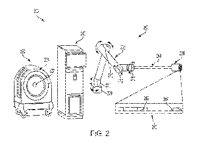
[0074] FIG. 2 is a graphical illustration of an example guided robotic system
200, in
accordance with various embodiments. As illustrated in FIG. 2, the guided
robotic system
200 includes a magnetic imaging apparatus 220, a computer system 240, and a
robotic
system 260. The guided robotic system 200 is similar in many aspects to the
robotic
system 100.
[0075] The example magnetic imaging apparatus 220 shown in FIG. 2 can include
a bore
222 (also referred to herein as "access pod") in the center of a single-sided
magnetic coil set
224 to provide access to one or more anatomical parts of a patient being
imaged during a
medical procedure. In accordance with various embodiments, the magnetic
imaging
apparatus 220 has a fixed field of view (FOV) relative to its mechanical
structure. In
accordance with various embodiments, the fixed FOV is defined as a cylindrical
volume with
about 4 inches in diameter and about 4 inches length, or a cubic volume with
sides of about
4 inches. In accordance with various embodiments, the fixed FOV ranges from
about 2
inches in diameter/sides to about 12 inches in diameter/sides. In some other
implementations, the FOV may be larger, such as for breast imaging
applications, where a
receive coil array (e.g. double receive coil) may cover a combined total of
about 18-24
inches side cubes/cylinders.
[0076] Within the defined fixed FOV, the robotic system 260 can be calibrated
to
determine a fixed frame of reference between the robotic system 260 and the
imaging FOV
of the magnetic imaging apparatus 220, according to some embodiments. This
calibration
can ensure the robotic system 260 is operationally coupled to the magnetic
imaging
apparatus 220 via the computer system 240.
[0077] The setup and calibration process can include setting up the robotic
system 260
and the magnetic imaging apparatus 220 for use together. In various instances,
set up
involves building an MR imaging phantom with at least four non-coplanar
markers, which are
easily identifiable in MR imaging.
[0078] To calibrate the system after set-up, the following steps can be
performed. First,
the phantom can be fixed rigidly in the field of view of the scanner and an
image can be
acquired. Second, the position of the marks can be recorded by visually
identifying them on
- 14 -
CA 03165780 2022- 7- 22
WO 2021/150902
PCT/US2021/014628
the image one at a time. This set of all points viewed on the image can be
called Point Set
AO (with dimensions Nx3, wherein N is the number of points identified). In
certain instances,
the identification can be done automatically by segmentation and/or
classification. Third, the
robot can be operated in free-drive mode and navigated to each Point Set AO.
The position
of the robot when the needle tip reaches each point in the set can be
recorded. This set of
all points recorded in robot coordinates can be called Point Set BO (also with
dimensions
Nx3). Fourth, the rigid linear least squares transformation that transforms BO
to AO can be
estimated (T: B0-->A0). This is the robot-to-image transform. The inverse of
this transform
is the image-to-robot transform.
[0079] To test the calibration based on the transform Tr the phantom can be
relocated to a
new position (e.g. shifted 1-2 cm in the X- and Y-directions) within the field
of view. The four
calibration steps above can be repeated to generate point Sets Al and Bl.
Then, previously-
estimated transform T can be applied to El to get T(B1) and the root mean
squared error
(RMSE) between T(B-1) and Al can be calculated. Finally, the RMSE can be
verified to
determine it is within an acceptable threshold and/or value.
[0080] As depicted in FIG. 2, the magnetic imaging apparatus 220 includes a
single bore
throughwhich a robotic arm can extend to reach a patient or target site. In
other instances,
the magnetic imaging apparatus 220 can include two or more access ports. Each
access
portion can provide access to the patient and/or surgical site. For example,
in instances of
multiple access ports, the multiple access ports can allow access from
different directions
and/or proximal locations.
[0081] Note that while FIG. 2 shows an example magnetic imaging apparatus 220
with a
bore 222 in the center of a single-sided magnetic coil set, this magnetic
imaging apparatus is
used for exemplary purposes only. The robotic arm 262 can be configured to
operate with
any magnetic imaging apparatus, or imaging apparatus generally (see above
discussion
related to imaging apparatus 120 for examples) regardless of the apparatus
design (e.g.,
standard MRI systems, single-sided MRI, or any other contemplated magnetic
imaging
apparatus or general imaging apparatus) as discussed herein.
[0082] Using a robot, instead of humans, for guiding tools for robot-assisted
medical
procedures can be a safer and more accurate approach in certain instances,
even given
some of the limitations of currently available imaging systems. These
limitations can stem,
for example, from the structural design and geometric architecture of, for
example, current
MR1 systems. For example, most, if not all, current MR1 systems in patient
care centers
utilize a magnet configuration where the patient typically lays inside a
gantry (scaffold) of the
MR1 machine during imaging. This arrangement of magnets to surround the
patient most
often prohibitively limits direct access to most anatomical parts of the
patient. Therefore.
MR1 systems (or imaging systems in general) that do not limit access to
various anatomical
-15 -
CA 03165780 2022- 7- 22
WO 2021/150902
PCT/US2021/014628
parts of the patient can further utilize the advantages of the robot, in
accordance with various
embodiments, especially to be able to use it as a guidance tool in medical
procedures. Such
Systems can therefore be additionally beneficial, particularly in robotic or
robot-assisted
invasive medical procedures, for targeting any anatomical parts of a patient,
without
constraints or iimitations resulting from the confining geometry of the
gantry, for example,
[0083] As shown, for example, in FIG. 2, the computer system 240 can be
coupled to the
magnetic imaging apparatus 220 and the robotic system 260, in accordance with
various
embodiments. Similar to FIG. 1, the computer system 240 can be configured for
analyzing
images acquired from the magnetic imaging apparatus 220 in real-time and
identifying
anatomical parts of the patient (or subject) from the acquired images. During
operation of a
medical procedure, for example, the magnetic imaging apparatus 220 is
configured to
acquire live (real-time) or near live (near real-time) images that may also
include surgical
device, such as a needle, a stent, or anything that is attached to the end of
the robotic
system 260, that is to be moved to the target anatomical parts of the patient
for the medical
procedure. Imaging the needle or the stent provides relative positioning of
the needle or the
stent with respect to the target portion of the anatomical parts of the
patient. For example,
during guidance of the robotic system 260 to insert the needle or the stent
within the FOV,
the plane of the acquired image containing the needle or the stent is
continuously monitored
rather than having to be determined manually. This provides advantages, for
example, for
having known the imaging plane containing the needle. In accordance with
various
embodiments, if the acquired images are not of sufficient quality to determine
relative
positioning of the needle with respect to the target portion of the anatomical
parts, a higher
resolution images can be acquired. In accordance with various embodiments, if
the acquired
images are of sufficient quality for determining relative positioning of the
needle with respect
to the target portion of the anatomical parts, a lower resolution images may
be taken at a
higher acquisition rate, which in turn provides real-time or near real-time
imaging capabilities
during operation of the medical procedure. In accordance with various
embodiments, the
image acquisition rate of the magnetic imaging apparatus 220 ranges from about
3-10
images per second to about one image per five minutes depending upon the
resolution. In
accordance with various embodiments, the image acquisition rate of the
magnetic imaging
apparatus 220 ranges is up to about 60 or 120 images per second.
[0084] In accordance with various embodiments, the robotic system 260 is
configured to
be placed outside the magnetic imaging apparatus 220. As shown in FIG. 2, the
robotic
system 260 can include a robotic arm 262 that is configured for movements in 6-
degrees of
freedom. In accordance with various embodiments, the robotic arm 262 includes
one or
more mechanical arm portions (also referred to herein as one or more
components),
including a hollow shaft 264 and an end-effector 266, that are connected in a
configuration to
-16 -
CA 03165780 2022- 7- 22
WO 2021/150902
PCT/US2021/014628
allow the robotic arm 262 to move, rotate, or swivel in 6-degrees of freedom
via one or more
motion controllers 270. The double-headed curved arrows signify rotational
motions
produced by the motion controllers 270, In accordance with various
embodiments, one or
more motion controllers 270 is an actuator, such as a mechanical actuator,
including but not
limited to servomotors. In accordance with various embodiments, one or more
motion
controllers 270 is an actuator, such as a pneumatic, spring-loaded,
mechanical, electrical
motor, piezoelectric actuator, or combinations thereof.
(0085] In accordance with various embodiments, the robotic arm 262 of the
robotic system
260 is configured for accessing various anatomical pads of interest through or
around the
magnetic imaging apparatus 220. In accordance with various embodiments, the
bore 222 in
the center of the magnetic imaging apparatus 220 is specifically designed to
provide access
to the robotic arm 262 of the robotic system 260 for operation at various
anatomical parts of
interest of the patient during a medical procedure. in accordance with various
embodiments,
the bore 222 in the center of the magnetic imaging apparatus 220 is designed
to account for
the size of the robotic arm 262. For example, the bore 222 defines a
circumference that is
configured to accommodate a robotic arm therethrough, such as the various
robotic arms
described herein. In accordance with various embodiments, the robotic arm 262
of the
robotic system 260 is configured for accessing various anatomical parts of the
patient from
around a side of the magnetic imaging apparatus 220. Magnetic imaging
apparatuses are
further described in U.S. Patent Application No. 16/003,585, titled UNILATERAL
MAGNETIC
RESONANCE IMAGING SYSTEM WITH APERTURE FOR INTERVENTIONS AND
METHODOLOGIES FOR OPERATING SAME, filed June 08, 2018, which is incorporated
by
reference herein in its entirety.
(0086] In accordance with various embodiments, the hollow shaft 264 provides
the
housing for the mechanism to actuate the end effector and may contain a long
screw drive,
shaft or another mechanism to provide the quick end effector action necessary
to take the
biopsy samples. Additionally, the hollow shaft may be able to store multiple
needles and/or
sampled cores.
(0087] In accordance with various embodiments, the end-effector 266 is
attached to one
end of the robotic arm 262, as illustrated in FIG. 2. In accordance with
various
embodiments, the end-effector 266 includes a mechanism, an actuator, a housing
or
configuration to store or carry one or more needles 280, and/or insert the one
or more
needles 280, or a housing or configuration to store, carry and/or insert one
or more stents or
brachytherapy seeds. In accordance with various embodiments, the end-effector
266
includes a mechanism to insert the needles 280 to obtain a biopsy sample, a
component or
a mechanism to provide ablation, or a component or a mechanism to perform
brachytherapy,
among many other suitable medical procedures (also referred to herein as
interventions). In
-17 -
CA 03165780 2022- 7- 22
WO 2021/150902
PCT/US2021/014628
accordance with various embodiments, the needles 280 are used for extracting a
specimen,
wherein the specimen can be attached to a needle 280, drawn into a needle 280,
or via any
other mechanism for which the specimen can be extracted using a noodle 280. In
accordance with various embodiments, the end-effector 266 has a minimal
mechanical or
pneumatic control to select the needle 280 to be inserted. In accordance with
various
embodiments, the motion or movement of the robotic arm 262 inserts or
withdraws the
needle 280.
[0088] In accordance with various embodiments, the one or more mechanical arm
portions
of the robotic arm 262, including the hollow shaft 264 and the end-effector
266, are made of
non-magnetic materials and do not include any electrical components, such as
for example,
servomotors for motion control. In such a configuration, all the degrees of
motion, such as
servomotors, for the robotic system 260 can remain outside the bore 222 on one
side of the
magnetic imaging apparatus 220 facing away from the patient. This
configuration allows
safe storage of the robotic system 260 away from the magnets of the magnetic
imaging
apparatus 220. Wth this configuration, in accordance with various embodiments,
the robotic
system 260 can extend using the one or more mechanical arm portions of the
robotic arm
262 to reach across to the target portions of the patient through the bore
222. In accordance
with various embodiments, the robotic system 260 can extend using the one or
more
mechanical arm portions of the robotic arm 262 to reach the target portions of
the patient
around the magnetic imaging apparatus 220, instead of through the bore 222.
The
configuration for reaching around is suitable for extremities or breast
biopsies, where a
needle (attached to the end-effector of the robotic arm 262) can be inserted
from the side of
the patient in an orthogonal direction. In accordance with various
embodiments, the needle
is inserted in an imaging plane and the needle trajectory is calibrated to lie
in the imaging
plane.
[0089] In accordance with various embodiments, the needles 280 include any non-
magnetic material, such as titanium, non-magnetic stainless steel, ceramics,
etc. In certain
instances, the needles 280 can be entirely non-magnetic to reduce interference
with the
magnetic imaging apparatus.
[0090] In accordance with various embodiments, image distortion can occur
locally when a
magnetic stainless steel needle is used, which is a common practice in certain
instances. If
there is distortion due to using a magnetic needle or other magnetic surgical
device, then the
distortion can be removed with image processing. A benefit of using a non-
magnetic needle
is that it would not cause distortion to the image. In accordance with various
embodiments,
the needle 280, such as a biopsy needle, includes an outer cylindrical sleeve
282 and an
inner core 284, as shown in FIG. 2. The inner sleeve has a recessed region for
containing
the sampled tissue. For example, during a medical procedure or intervention,
the inner
- 18 -
CA 03165780 2022- 7- 22
WO 2021/150902
PCT/US2021/014628
sleeve cuts through the tissue first, with tissue setting into the recess. In
such cases, the
outer sleeve follows shortly and cuts the tissue so that a sample of tissue
remains in the
recess.
[0091] In accordance with various embodiments, a hollow needle is used to
place a stent
or brachytherapy seeds. In accordance with various embodiments, the hollow
needle
includes an outer sleeve and an inner needle that pushes out the stentlseeds
at the
appropriate locations.
[0092] In accordance with various embodiments, the needles 280 include gauge
sizes
ranging from 12G to 18G, including 10G, 12G, 14G, 16G, and 18G. In accordance
with
various embodiments, the needles 280 are sized 16G to 18G for biopsy, and 10G
for
brachytherapy or ablation. In accordance with various embodiments, the needles
280 have
a range of lengths for prostate procedures between about 15 cm and 25 cm.
[0093] In accordance with various embodiments, the magnetic imaging apparatus
220 is a
low-field magnetic imaging system with a fixed geometry. During operation of
such low-field
magnetic imaging system, sufficiently low-field magnet may not interfere with
the shielded
robotic servo-motors. However, the presence and operation of these components
may
interfere with the magnetic field produced by the magnetic imaging apparatus
220 during
operation. To eliminate or reduce potential interference during magnetic
imaging, the robotic
system 260 is configured with the robotic arm 262 that can be extended via the
one or more
mechanical arm portions, including the hollow shaft 264 and the end-effector
266 through
the bore 222 of the magnetic imaging apparatus 220. In such instances, the
entire robotic
tool can be distal to the bore 220 and outside the magnetic imaging apparatus
220 during a
surgical procedure. In accordance with various embodiments, the magnetic
imaging
apparatus is designed to have a cylindrical region that is aligned with the
bore 222 and has
lower magnetic interference than other regions within the imaging zone. For
example, the
robotic tool can be positioned far enough from the coils and in the region of
the imaging zone
with the weakest magnetic field, gradient field, and/or RF field, Such a
cylindrical region can
be where the robotic arm 262 extends into and operates in various aspects. To
further
reduce or avoid potential magnetic interferences from the robotic system 260,
all or most of
the components of the robotic arm 262 can be constructed from non-magnetic
material. In
accordance with various embodiments, the magnetic imaging apparatus 220 is
Kept close to
the patient and away from sources of magnetic interference. For example, the
motors for
the robotic arm and/or robotic tool can be positioned outside of the bore 222.
In such
instances, referring to FIG. 14, the patient is proximate to the magnetic
imaging apparatus
220, and the magnetic imaging apparatus is between the patient and the robotic
system. A
distal portion of the robotic arm can reach through the magnetic imaging
apparatus 220 to
reach the patient. In accordance with various embodiments, active noise
cancellation
- 19 -
CA 03165780 2022- 7- 22
WO 2021/150902
PCT/US2021/014628
techniques can be used to sense the noise generated from the motors and then
remove it
from the acquired MRI signals. In accordance with various embodiments, signal
processing
can be used to remove any noise generated from the motors. For example, to
remove the
noise generated by the motors, the MRI signal can be combined with a motor
noise removal
signal that is actively generated to produce a noiseless MRI signal. Low-field
magnetic
imaging systems are further described in International Application No.
PCT/US2020/018352,
titled SYSTEMS AND METHODS FOR ULTRALOW FIELD RELAXATION DISPERSION,
filed February 14, 2020, now International Publication No. W02020/172673,
168233, which
is herein incorporated by reference in its entirety.
[0094] FIG. 3A is a schematic illustration of a graphical user interface (GUI)
300 for an
example guided robotic system, according to various embodiments. As shown in
FIG. 3A,
the GUI 300 includes a left panel 310, a middle panel 320, and a right panel
340. The GUI
300 shown in FIG. 3A is for illustrative purposes, and thus is a non-limiting
example user
interface. As a non-limiting example, the GUI 300 is configured for use in the
invasive
operating procedure, a robotic transperineal prostate biopsy.
[0095] As illustrated in FIG. 3A, the left panel 310 shows a plurality of
buttons for robotic
control. In accordance with various embodiments, the buttons are operated or
activated by
capacitive touching, a mouse input, or joystick input by an operator. In
accordance with
various embodiments, the left panel 310 includes touch-screen controls for
controlling the
robot and for various imaging adjustments. In accordance with various
embodiments, the
left panel 310 includes controls for overriding previous inputs, including
certain user actions,
for example, but not limited to, changing previous trajectory of the needle
movement. In
accordance with various embodiments, the left panel 310 may include a button
for motion
correction during live scans of the subject.
[0096] The middle panel 320 includes a live guidance view showing live images
320 (the
term "live" also refers to herein as "continuously captured" or "continuously
acquired") of a
portion of a target 330 (e.g., prostate 330), a current needle position 324, a
current needle
trajectory 326, and a target sample location 328 within the prostate 330.
During a surgical
procedure and/or intervention, "live" images are obtained intraoperatively. In
accordance
with various embodiments, the middle panel 320 shows live scanned images being
acquired,
which include the current needle position 324, the needle trajectory 326, and
the target 330
automatically identified from the scan. As the needle advances into the field
of view shown
on the live guidance view in the middle panel 320, the lives images 320
continuously display
the current location of the needle, i.e, updated current needle position 324.
In the
background (e.g., processing behind the scene), this view is continuously
registered with the
corresponding view from a pre--procedure image to compensate for the motion,
according to
some implementations. For example, every time there is a scan, a new image is
produced
- 20 -
CA 03165780 2022- 7- 22
WO 2021/150902
PCT/US2021/014628
and re-registered with the corresponding view from a pre-procedure image to
compensate
for any movement.
(00971 As shown in FIG. 3A, the right panel 340 includes various views of the
planning
scan, including for example, a transverse view 342, a sagittal view 344, and a
three-
dimensional (3D) view 346. The transverse view 342 shows a slice from the
planning scan
containing the target 330, In accordance with various embodiments, a virtual
grid 345 is
used to show evenly spaced potential needle locations, which are shown in the
transverse
view 342 as hollow circles. The sagittal view 344 shows the sagittal image
containing the
target 330. In accordance with various embodiments, the virtual grid 345 is
used to show
evenly spaced potential needle locations, which are shown in the sagittal view
344 as
horizontal lines. According to some embodiments, the lines represent the
potential needle
trajectories due to the transperineal approach along the transverse direction.
The 3D view
346 displays a cross-sectional view from the planning image based on the
current needle
location 324 and updates the graphic on the GUI (e.g. GUI 300) as the needle
is advanced
distally.
[00981 FIG. 36 is a schematic illustration of a live view 350 during imaging
of a guided
robotic system, according to various embodiments. As illustrated in FIG. 36,
the live view
350 is in the x-y-z coordinate system, designated by a dotted cube along the
x, y, and z
axes. The imaging needs to be acquired only in the plane in which the needle
is expected,
which is represented by imaging plane 370 in the middle of the field of view
360 in the FIG.
3B. In accordance with various embodiments, the live view 350 has a built-in z-
gradient and
can excite one or more slabs of varying thickness within the field of view
360. An alternative
embodiment, may not have the z axis gradient built-in. In accordance with
various
embodiments, the x- and y-gradients are embedded as phase-encodes for the
imaging in the
imaging plane 370 containing the needle. The spatial localization of points
within the field of
view 360 are determined by a combination of x and y phase encodes, and
transmit
frequency band corresponding to the z-gradient. These slabs are different from
the
conventional image slices and through image reconstruction, and therefore, can
be broken
into a number of slices.
[0099] In accordance with various embodiments, slice interleaving is utilized
in which the
system can excite the entire field of view by multiplexing excitation of
different slabs within
the field of view 360 to completely cover the entire field of view 360 by
transmitting and
receiving different bandwidths at different time intervals within the pulse
sequence. Based
on only y-phase encodes (due to the z-phase being built in the system) can
produce a two-
dimensional cross-sectional image containing the needle at a fast speed. For
example,
utilizing slice interleaving in a single dimension (e.g. the needle
trajectory) can be done at a
high resolution and fast rate. In accordance with various embodiments, there
is virtually no
- 21 -
CA 03165780 2022- 7- 22
WO 2021/150902
PCT/US2021/014628
acquisition and computing cost associated with obtaining a thick slab, while
using only y-
phase encodes using slice interleaving approach, where the sampling is done
only in one
dimension.
[0100] As illustrated in FIG, 3B, the live view 350 is configured to show a
projected needle
trajectory 380 within the imaging plane 370. According to some
implementations, the needle
is advancing in the positive z-direction in the x-y-z coordinate system as
illustrated in FIG.
3B. In accordance with various embodiments, the whole volume is imaged at a
lower detail
and then the region around the needle trajectory 380 is imaged in finer detail
during live
guidance to show accurate positioning of the needle. In accordance with
various
embodiments, a hybrid image that contains a higher resolution portion of the
image closer to
the needle and a lower resolution image portion elsewhere in the image may be
sufficient.
The hybrid imaging approach can offer further improvements in imaging
acquisition time, i.e.,
faster imaging, while maintaining sufficient details in the area that is
needed to determine
accurate positioning of the needle with respect to the position of the target
330.
[0101] Additional trade-offs between image acquisition rates versus resolution
of the
acquired images may be achieved by suitable optimization techniques using
hardware
and/or software approaches, such as compressed sensing using k-space under-
sampling,
parallel imaging, and multi-slice image acquisition. These techniques aim to
speed up image
acquisition with the typical cost of image signal to noise ratio. They
leverage data
symmetries and data compression techniques to acquire the minimal amount of
data
necessary to reconstruct the image.
[0102] Some target anatomies such as the prostate present unique challenges
for needle
guided interventions. The prostate, for example, is surrounded by soft tissue
and is prone to
movement as a result of any pressures from transrectal transducers or needle
entering into
the prostate. For example, as the needle is inserted into the prostate, the
prostate may be
pushed away and upon insertion, the gland may settle back into its original or
into some
other location. Similarly, when withdrawing the needle, the gland may continue
to push back
and change its location. This becomes particularly problematic when one is
trying to use a
rigid frame of reference with the robot as the registration between the
anatomy and the
imaging may become erroneous.
[0103] In accordance with various embodiments, a motion correction method is
used to
dynamically estimate the motion using image similarity metrics between the
live image and
the corresponding cross-section from the planning image. In accordance with
various
embodiments, this is further enhanced by motion detection and correction in
the k-space
itself. Correction in k-space ensures that the reconstructed image does not
have motion
artifacts, whereas the image-based registration minimizes the error caused by
motion in
accurate placement of the robot. For example, gross patient motion and
localized gland
- 22 -
CA 03165780 2022- 7- 22
WO 2021/150902
PCT/US2021/014628
deformations can be separated by using magnetic resonance visible fiducial
markers and
corrected for separately. Motion can be determined from comparing frames of
IVIRI images,
for example. The measured motion is applied to the robot frame of reference,
which is
known by the robot, and the target anatomy and the robot maintain their
correspondence.
For example, the measured motion is applied to update the target anatomy and
the robot
frame of reference to allow the robot to move in the correct path relative to
the target
anatomy. Fiduciary markers can also be used to determine correspondence.
[0104] For guided robotic procedure or intervention, magnetic imaging scans
are taken for
the target anatomy for planning the procedure. These scans (planning scan) may
include
magnetic (e.g., magnetic resonance) image scans using one or more contrast
types. The
images may manually or automatically be classified into suspected malignancies
for biopsies
and into the malignancy extents for an image-guided therapy. In accordance
with various
embodiments, the image guided procedure may be performed immediately alter the
planning
images are acquired, i.e., live imaging, or at a later time. In accordance
with various
embodiments for the procedure to be performed at a later time, a pre-procedure
anatomical
scan is performed to map the planning image into the current frame of
reference. The
following figures illustrate various embodiments of the procedures that
utilize guided robotic
procedure.
[0105] FIGS. 4A, 48, 4C, 4D, 5A, 5B, 5C, 5D show different views (e.g.
transverse and
sagittal views) for a virtual template-based or grid-based approach.
[0106] FIG. 4A is a schematic illustration showing a transverse view 400a
during a
planning scan of a prostate sample 430, according to various embodiments. FIG.
4B is a
schematic illustration showing a sagittal view 400b of the prostate sample
430, in
accordance with various embodiments. As illustrated in FIGS. 4A and 48, the
planning
image of the prostate sample 430 can be marked to show a suspected region 435
(e.g.,
possibly malignant or confirmed malignant) in both transverse view 400a and
sagittal view
400b. Also shown in FIGS. 4A and 48 is a virtual template grid 445, which is
illustrated as
evenly spaced hollow dots for potential needle locations in FIG. 4A and
straight lines for
potential needle trajectories in FIG, 48. In accordance with various
embodiments, the
spacing of the hollow dots can vary based on desired needle locations or
trajectories.
[0107] FIG. 4C is a schematic illustration showing a transverse view 400c for
a biopsy plan
based on the planning scan illustrated in FIG. 4A, according to various
embodiments. FIG.
4D is a schematic illustration showing a sagittal view 400d for the biopsy
plan based on the
planning scan of the prostate sample 430 illustrated in FIG. 48. As
illustrated in FIG. 4C,
four locations (e.g. four filled dots) of the grid 445 where a biopsy sample
can be obtained
from the suspected region 435 are shown at sample locations 455. Similarly,
FIG. 4D shows
two straight trajectory lines of the grid 445 in the sagittal view 400d that
correspond to the
- 23 -
CA 03165780 2022- 7- 22
WO 2021/150902
PCT/US2021/014628
sample locations 455, which encompass the suspected region 435. The biopsy
plan,
illustrated by filled circles and straight lines cover the entire target
suspected region 435.
[0108] FIGS. 5A and 58 respectively illustrate a transverse view 500a and a
sagittal view
500b of a prognosis plan to determine extents of the malignancies by placing a
bounding
box of a prostate sample 530, according to various embodiments. As illustrated
in FIGS. 5A
and 58, the prognosis plan for the prostate sample 530 is marked to show a
suspected
region 535 (e.g., possibly malignant or confirmed malignant) in both
transverse view 500a
and sagittal view 500b. Also shown in FIGS. 5A and 58 is a virtual template
grid 545, which
is illustrated as evenly spaced hollow dots for potential needle locations in
FIG. 5A and
straight lines for potential needle trajectories in FIG. 58. The prognosis
plan of the prostate
sample 530 as illustrated in FIGS. 5A and 58 provides the bounding box,
illustrated by
sample locations 555, which are shown as sixteen filled dots in FIG. 5A and
four filled lines
in FIG, 58. The sample locations 555 encompass a suspected or known malignancy
in the
suspected region 535 to determine the extent of the prognosis. This prognosis
may be used
for determining the disease management pathway, such as active surveillance,
or type and
extent of the procedure.
[0109] FIG. 5C is a schematic illustration showing a transverse view 500c for
a low-dose
brachytherapy plan of a prostate sample 560, according to various embodiments.
FIG. 5D
shows a schematic illustration of a sagittal view 500d, in accordance with
various
embodiments. The low-dose brachytherapy plan for the prostate sample 560 shown
in
FIGS. 5C and 5D is illustrated by sample locations 565, which are shown as
filled dots within
the entire prostate sample 560 in FIG. 5C and dashed lines within the entire
prostate sample
560 in FIG. 5D. In accordance with various embodiments, the low-dose
brachytherapy may
be used to treat the prostate sample 560 as illustrated in FIGS. 5C and 5D. In
some other
embodiments, the low-dose brachytherapy may be used to treat a portion of the
prostate
sample 560.
[0110] FIG, GA is a schematic illustration showing a transverse view 600a fora
biopsy plan
of a prostate sample 630 without a virtual grid, according to various
embodiments. FIG. 68
shows a schematic illustration of a sagittal view 600b for the biopsy plan of
the prostate
sample 630, in accordance with various embodiments. As illustrated in FIGS.
6.A and 68,
the biopsy plan for the prostate sample 630 is marked to show a suspected
region 635 (e.g.,
possibly malignant or confirmed malignant) in both transverse view 600a and
sagittal view
600b. As illustrated, the biopsy plan for the prostate sample 630 shown in
FIGS. 6A and 6B
are not restricted by the grid for needle insertion. The biopsy plan of the
prostate sample
630 as illustrated in FIGS. 6A and 6B includes some selected sample locations
655 in the
suspected region 635. The sample locations 655 are shown as 3 filled dots in
FIG. 6A and
a filled line in FIG. 6B.
- 24 -
CA 03165780 2022- 7- 22
WO 2021/150902
PCT/US2021/014628
[0111] In accordance with various embodiments at described herein, the robotic
system
260 of FIG. 2 can be used for any of the procedures or interventions
illustrated and
described with respect to FIGS, 4A, 4B, 40, 4D, 5A, 5B, 50, 5D, SA, and 6D. As
shown in
FIG, 2, the robotic system 260 includes the robotic arm 262 configured for
movements in 6-
degrees of freedom. After calibration at an origin within the imaging field of
view shown, for
example, in FIGS. 3A or 38, the robotic arm 262 can move the needle tip to any
point in the
x-y plane, for example, as shown in FIG. 38. In accordance with various
embodiments, the
robotic arm 262 can align to any point in the grid pattern of the overlaid
virtual template, for
example, as shown in FIGS. 4A, 40, 5A or 50, or any other location within the
field of view in
a template-free approach, for example, as shown in FIG. 6A. in accordance with
various
embodiments, the needle insertion is performed by advancing the robotic arm
262 along the
z-direction, as shown in FIG. 38, or from left to right or vice versa as shown
in FIGS. 4B, 4D,
513, 5D, or 68.
[0112] As the described above, in accordance with various embodiments, the
robotic arm
262 is configured to include one or more motion controllers 270, such as an
actuator, or the
end-effector 266 at the end of the arm that can hold one or more short
needles. In
accordance with various embodiments, the actuator 270 is completely mechanical
and is
triggered by a servo motor near the base of the robotic system 260. In
accordance with
various embodiments, the actuator is a pneumatic actuator for positioning the
needle within
a plane. In accordance with various embodiments, the actuator is pneumatically
controlled
and other components in the entire robotic system 260 is mechanically
controlled by one or
more servo motors. In accordance with various embodiments, almost all
components of the
robotic system 260, including the actuator is mechanically controlled by one
or more servo
motors.
[0113] In accordance with various embodiments, the robotic arm 262 of the
robotic system
260 is constrained to move along parallel lines, such as for example, in
transperineal
prostate procedures. In accordance with various embodiments, an additional
degree of
freedom (in addition to the 6-degrees of freedom) include angular motions so
that the needle
attached to the robotic arm 262 is maneuvered past the pubic arch area, such
as in case of
an enlarged prostate gland. In accordance with various embodiments, the
robotic system
260 is constrained to move so as to maintain an external Remote Center of
Motion (RCM)
such that it can approach the same location inside the body through various
trajectories. In
an RCM model, the robotic mechanism moves in such a fashion that the tool
actuated by it
always has the trajectory passing through a fixed point relative to the
robotic mechanism.
For example, for a minimally invasive single port intervention, the RCM may be
kept fixed at
the entry port into the body and the robotic mechanism may enter it through
different angles
to advance the tool to different locations inside the body. In accordance with
various
- 25 -
CA 03165780 2022- 7- 22
WO 2021/150902
PCT/US2021/014628
embodiments, the RCM center may be on surface of the patient's body to
facilitate
sampling/treating multiple location through only one access puncturelport.
[0114] In accordance with various embodiments, an operator may preload
multiple
needles 280 within the actuator within if a biopsy plan is already determined.
In such cases,
the biopsy plan includes obtaining sample specimens from all planned locations
as
described with respect to, for example, FIGS. 40, 4D, 5A, 58, 6A, 68. In such
implementation, the robotic arm 262 is configured to acquire multiple samples
at once using
multiple needles 280. In such a case, the prostate sample may need to be kept
in place by
using a trans-urethral tube (not shown). It may also be sufficient to only
track one of the
needles 280 since the prostate gland movement is only along the needle
insertion direction
and will be equal for all needles being inserted at the same time. As such, a
motion
correction for a central needle may be sufficient to correct for motion for
all the needles 280.
[0115] In accordance with various embodiments, the needles 280 may be inserted
one at
a time, In such configuration, a pre-determined order of needle insertion is
used along with
the optimized sampling scheme such that the effect of the needle insertion on
imaging of the
next target locations is minimized. In this implementation, while the actuator
does not hold
all the needles for insertion at the same time, it holds several needles in a
cartridge or end-
effector 266 to insert and withdraw one needle at a time. This is done to
avoid withdrawing
the entire robotic arm 262 across the bore 222.
[0116] In accordance with various embodiments, the needle 280 has an RF coil
or
metamaterial attached to the needle. In accordance with various embodiments,
the RF coil
or metamaterial is configured to couple to a receive coil chain of the
magnetic imaging
apparatus 220. This implementation would allow for wireless coupling and the
transfer of
information to the receive coil network to be digitized by the computer. In
accordance with
various embodiments, the attached RF coil or metamaterial can increase signal
transduction
from the tissue surrounding the needle during the insertion, which, in turn,
improves the
image quality acquired during the scan by the magnetic imaging apparatus 220.
[0117] In accordance with various embodiments, the guided robotic system 200
used for
guided robotic procedures, such as those described with respect to, for
example, FIGS. 4A,
48, 40, 4D, 5A, 58, 50, 5D, SA, and 613, includes an additional configuration
that utilizes a
nuclear magnetic resonance (NMR) analysis network. In accordance with various
embodiments, the NMR analysis network on the robotic system 260 is configured
to utilize a
higher magnetic field within the bore 222 to perform spectral analysis on the
collected biopsy
specimens. Since it is known that different tissue types have different NMR
spectrums, the
type and amount of tissues in each specimen can be characterized quickly, soon
after the
specimens are acquired. In accordance with various embodiments, the additional
NMR
information collected and analyzed from the specimens can be used for real-
time feedback
- 26 -
CA 03165780 2022- 7- 22
WO 2021/150902
PCT/US2021/014628
on the tissue type, providing additional information that may be relevant to
the pathology of
the biopsy core.
[0118] In accordance with various embodiments, the guided robotic system 200
used for
guided robotic procedures, such as those described with respect to, for
example, FIGS. 4A,
48, 40, 4D, 5A, 58, 50, 5D, 6A, and 6D, includes an additional configuration
that utilizes
ultrasound for guidance. In accordance with various embodiments; an apparatus
for
acquiring ultrasound is external to the guided robotic system 200. In
accordance with
various embodiments, the apparatus for acquiring ultrasound is integrated in
the guided
robotic system 200, for example, integrated to the robotic arm 262, near the
end-effector
266. In accordance with various embodiments, the guided robotic system 200
supplemented with ultrasound can improve magnetic guidance providing faster
imaging
updates or for localizing the veins and arteries within the subject during the
operation or
intervention,
[0119] Additional medical procedures, operations, or interventions where the
guided
robotic system 100 or the guided robotic system 200 uses magnetic imaging
technique can
include, but not limited to, for example, Transperineal biopsies,
Transperineal LDR
brachytherapy, Transperineal HDR braohytherapy, Transperineal laser ablation,
Transperineal cryoablation, and Transreetal HIFU.
[0120] For Transrectal HIFU, in accordance with various embodiments, the
robotic system
260 is used to turn the transrectal HIFU transducer about its axis. In
accordance with
various embodiments, an operator or a physician inserts the transducer first,
then moves the
patient in the magnetic imaging field of view. in these implementations, the
robotic arm 262
is configured to approach through the bore 222 of the magnetic imaging
apparatus 220 and
latch into the transducer or its holder.
[0121] Additional medical procedures, operations, or interventions where the
guided
robotic system 100 or the guided robotic system 200 uses magnetic imaging
technique
include breast biopsies. in breast biopsies, the procedure is similar to the
prostate biopsy
although the direction of insertion maybe different. For example, the robotic
system 260
used for breast biopsy can be configured to extend using the one or more
mechanical arm
portions of the robotic arm 262 to reach the target portions of the breast
around the magnetic
imaging apparatus 220, instead of through the bore 222. The configuration is
suitable for
breast biopsy particular, where the needle 280 is inserted from the side of
the breast.
[0122] Additional medical procedures, operations, or interventions where the
guided
robotic system 100 or the guided robotic system 200 uses magnetic imaging
technique
include deep brain stimulation (DBS). For DBS, the planning beforehand, e.g.
before the
procedure or intervention, is done to ensure that the needle trajectory does
not go through
any critical structure. In accordance with various embodiments, the critical
structures are
- 27 -
CA 03165780 2022- 7- 22
WO 2021/150902
PCT/US2021/014628
segmented, identified, or marked beforehand either automatically or manually.
These
structures can then be overlaid on the live image during the procedure. During
live
guidance, the image will be acquired to ensure that the needle 280 is inserted
to the
accurate location under direct visualization such that no critical structures
are damaged or
violated. In accordance with various embodiments, to minimize the complexity,
a RCM
model may be used once an entry point is selected for the entry into the
brain.
[0123] Additional medical procedures, operations, or interventions where the
guided
robotic system 100 or the guided robotic system 200 uses magnetic imaging
technique
include brain biopsies. In accordance with various embodiments, brain biopsies
are
conducted using the projected needle trajectory, which is displayed to the
operator on live
guidance panel, for example, as shown in FIG. 3A. Upon reviewing the
information on live
guidance panel of the middle panel 320, that operator can decide to initiate
insertion of the
needle. In accordance with various embodiments; the guided robotic system 200
is
configured to record the target location of the brain, in accordance with
various
embodiments, the operator reviews the acquired images of the target location
of the brain
and enter the pathology findings for each findings along with their respective
location within
the images.
[0124] Additional medical procedures, operations, or interventions where the
guided
robotic system 100 or the guided robotic system 200 uses magnetic imaging
technique
include liver and kidney biopsies. In accordance with various embodiments,
liver and kidney
biopsies include insertion at one entry point to obtain the specimen. In
accordance with
various embodiments, to minimize the complexity, a remote center or motion
(RCM) model
may be used once an entry point is selected for the entry into the brain.
[0125] Additional medical procedures, operations, or interventions where the
guided
robotic system 100 or the guided robotic system 200 uses magnetic imaging
technique
include lung biopsies. In accordance with various embodiments, lung biopsies
include
insertion of a tube through the trachea utilizing a robotic system.
[0126] In accordance with various embodiments as described herein, the guided
robotic
system 100 or the guided robotic system 200 can be utilized for medical
procedures,
operations, or interventions for insertion of a stent, for example, a coronary
stent or brain
stent. In accordance with various embodiments as described herein, the guided
robotic
system 100 or the guided robotic system 200 can be used for intensity
modulated radiation
treatment guidance.
[0127] FIG. 7 is a flowchart for an example method S200 of using the guided
robotic
system 200, according to various embodiments. As shown in FIG. 7, the method
S200
includes at step S210 acquiring magnetic resonance images of a subject. In
accordance
with various embodiments, the acquiring of the images of the subject includes
acquiring one
- 28 -
CA 03165780 2022- 7- 22
WO 2021/150902
PCT/US2021/014628
or more target anatomical parts of the subject or the patient. in accordance
with various
embodiments, the magnetic resonance images are acquired from a magnetic
resonance
imaging apparatus, such as the magnetic imaging apparatus 200 or an external
source. In
accordance with various embodiments, the magnetic resonance images are
acquired from
an external source, such as a physician, a patient, a user or an operator.
[0128] As shown in FIG. 7, the method S200 includes at step S220 performing
image
analysis of the live magnetic resonance images to continuously identify a
target portion of
the subject. In accordance with various embodiments, the acquired magnetic
resonance
images are automatically uploaded into a computer system, such as the computer
system
240, for analysis via one or more processes including, but not limited to,
artificial intelligence
(Al), machine learning, image or signal denoising, segmentation algorithms,
objects and
boundary identification, image registration, adaptive intensity correction,
and pattern
recognition, etc. In accordance with various embodiments, the acquired
magnetic
resonance images are manually analyzed and entered by a physician or an
operator into a
computer system, such as the computer system 240, which is used to
automatically identify
a portion of the subject from the analyzed images.
[0129] At step S230, the method S200 includes automatically guiding (via
automatic
guidance) a robotic arm, such as the robotic arm 262, to an identified target
portion of the
subject based on the live magnetic resonance images. In accordance with
various
embodiments, the automatic guidance includes guiding the robotic arm in real-
time or near
real-time based on analysis of continuously acquired magnetic resonance images
of the
target portion of the subject. In accordance with various embodiments, the
automatic
guidance includes self-correction via image analysis. In accordance with
various
embodiments, the automatic guidance includes occasional interventions by a
physician or an
operator to correct the trajectory of the robotic arm based on acquired
magnetic resonance
images. In accordance with various embodiments, the automatic guidance
includes
occasional interventions by a physician or an operator to alter the trajectory
of the robotic
arm based on acquired magnetic resonance images in order to perform
alternative or
additional medical procedures.
[0130] In accordance with various embodiments of the method S200, the robotic
arm is
configured for movements in 6-degrees of freedom, such as the robotic arm 262.
In
accordance with various embodiments, the robotic arm includes one or more
mechanical
arm portions that are connected in a configuration to allow the robotic arm to
move, rotate, or
swivel in 6-degrees of freedom. in accordance with various embodiments, the
robotic arm is
configured for accessing various anatomical parts of the subject.
[0131] In accordance with various embodiments of the method S200, no critical
structures
are damaged during the needle insertion by the robotic arm 262. For example,
in
- 29 -
CA 03165780 2022- 7- 22
WO 2021/150902
PCT/US2021/014628
accordance with various embodiments of a prostate biopsy, the guided robotic
system 200 is
configured such that the needle, which is attached to the robotic arm 262
being inserted into
the target portion of the subject, avoids passing through urethra or into the
bladder. In
accordance with various embodiments of brachytherapy, the needle does not
penetrate
beyond the prostate into the bladder and does not ablate the rectum or the
bladder.
[0132] In accordance with various embodiments the method 5200, acquired live
magnetic
resonance images are displayed within a graphical user interface (GUI) that
includes
functional buttons for controlling the procedure. In accordance with various
embodiments,
acquired live magnetic resonance images comprise a high resolution image
portion near a
needle inserted during the procedure and a lower resolution image portion
farther away from
the needle. In accordance with various embodiments, the method 8200 further
includes
correcting acquired live magnetic resonance images for motion during the
performing of the
procedure. In accordance with various embodiments, the method S200 further
includes
correcting acquired live magnetic resonance images for motion during insertion
of the
needle. In accordance with various embodiments, the method 8200 further
includes
overriding existing action to manually correct for the motion. in accordance
with various
embodiments, the method 8200 further includes manually advancing the robotic
arm by
controlling the GUI using a touch input, a mouse input or a joystick input. In
accordance with
various embodiments, the method 5200 further includes performing automatic
segmentation
to capture the location of the needle after extracting the specimen,
withdrawing the needle,
and advancing the needle to a next target location.
[0133] At step 5240, the method S200 includes performing a procedure at the
target
portion of the subject. In accordance with various embodiments, the method
S200 includes
performing a suitable medical procedure including for example, but not limited
to,
transperineal biopsy, transperineal LDR brachytherapy, transperineal HDR
brachytherapy,
transperineal laser ablation, transperineal cryoablation, transrectal HIFU,
breast biopsies,
deep brain stimulation (DBS), brain biopsy, liver biopsy, kidney biopsy, lung
biopsy, coronary
stent insertion, brain stent insertion, and intensity modulated radiation
treatment guidance,
etc. In accordance with various embodiments, performing a procedure includes
extracting a
specimen, for example, for biopsy.
[0134] FIG, 8 is a flowchart for an example method 6300 of using the guided
robotic
system 200, according to various embodiments. As shown in FIG. 8, the method
8300
includes at step 8310 continuously acquiring magnetic resonance images of a
subject. In
accordance with various embodiments, the acquiring of the images of the
subject includes
acquiring one or more target anatomical parts of the subject or the patient.
In accordance
with various embodiments, the magnetic resonance images are acquired from a
magnetic
resonance imaging apparatus, such as the magnetic imaging apparatus 100 or
200, or an
- 30 -
CA 03165780 2022- 7- 22
WO 2021/150902
PCT/US2021/014628
external source. In accordance with various embodiments, the magnetic
resonance images
are acquired .from an external source, such as a physician, a patient, a user
or an operator.
[0136] At step 8320, the method 3300 includes continuously identifying a
target portion of
the subject in the magnetic resonance images. In accordance with various
embodiments,
the acquired magnetic resonance images are automatically uploaded into a
computer
system, such as the computer system 240, for analysis via one or more
processes including,
but not limited to, artificial intelligence (Al), before identification of the
target portion. In
accordance with various embodiments, the acquired magnetic resonance images
are
manually analyzed and entered by a physician or an operator into a computer
system, such
as the computer system 240, which is used to automatically identify a portion
of the subject
from the analyzed images.
[0136] At step 8330, the method S300 includes guiding a robotic arm, such as
the robotic
arm 262, towards an identified target portion of the subject, wherein the
magnetic resonance
images are analyzed in real-time for guiding the robotic arm to the portion of
the subject. In
accordance with various embodiments, the continuously acquired magnetic
resonance
images are analyzed in real-time or near real-time to continuously identify
the target portion
of the subject. In accordance with various embodiments, the guiding of the
robotic arm
includes self-correction via image analysis. In accordance with various
embodiments, the
guiding of the robotic arm includes occasional interventions by a physician or
an operator to
correct the trajectory of the robotic arm based on the continuously acquired
magnetic
resonance images. In accordance with various embodiments, the guiding of the
robotic arm
includes occasional interventions by a physician or an operator to alter the
trajectory of the
robotic arm based on the continuously acquired magnetic resonance images in
order to
perform alternative or additional medical procedures.
[0137] At step S340, the method 8300 includes inserting the needle to the
target portion of
the subject and extracting a specimen. During the insertion, no critical
structures are
damaged during the needle insertion by the robotic arm 262. For example, in
accordance
with various embodiments of a prostate biopsy, the guided robotic system 200
is configured
such that the needle, which is attached to the robotic arm 262 being inserted
into the target
portion of the subject, avoids passing through the urethra or into the
bladder. In accordance
with various embodiments of brachytherapy, the needle does not penetrate
beyond the
prostate into bladder and does not ablate rectum or bladder.
[0138] In accordance with various embodiments of the method 8300, continuously
acquired live magnetic resonance images are displayed within a graphical user
interface
(GUI) that includes functional buttons for controlling during insertion of the
needle. In
accordance with various embodiments, continuously acquired live magnetic
resonance
images comprise a high resolution image portion near the needle and a lower
resolution
- 31 -
CA 03165780 2022- 7- 22
WO 2021/150902
PCT/US2021/014628
image portion farther away from the needle. In accordance with various
embodiments, the
method S300 further includes automatically correcting the continuously
acquired live
magnetic resonance ima,ges to compensate for motion blurring during insertion
of the
needle. In accordance with various embodiments, the method 8300 further
includes
automatically correcting a trajectory of the needle during the insertion based
on corrected
acquired live magnetic resonance images. In accordance with various
embodiments, the
method S300 further includes overriding existing guided trajectory to manually
correct for the
motion blur. In accordance with various embodiments, the method S300 further
includes
manually advancing the robotic arm by controlling the GUI using a touch input,
a mouse
input or a joystick input. In accordance with various embodiments, the method
8300 further
includes performing automatic segmentation to capture the location of the
needle after
extracting the specimen, withdrawing the needle; and advancing the needle to a
next target
location. In accordance with various embodiments, the guiding of the needle
attached to the
robotic arm towards the identified target portion of the subject includes
guiding through a
bore at the center of a magnetic imaging apparatus configured for continuously
acquiring
magnetic resonance images.
[0139] In accordance with various embodiments of step S340, the extracted
specimen is
for analysis in a medical procedure, such as for example, but not limited to,
transperineal
biopsy, transperineal LDR brachytherapy, transperineal HDR brachytherapy,
transperineal
laser ablation, transperineal cryoabiation, transrectal HIFU, breast biopsies,
deep brain
stimulation (DBS), brain biopsy, liver biopsy, kidney biopsy, lung biopsy,
coronary stent
insertion, brain stent insertion, and intensity modulated radiation treatment
guidance, etc.
[0140] FIG. 9 is a flowchart for an example method 8400 of using the guided
robotic
system 200, according to various embodiments. As shown in FIG. 9, the method
8400
includes at step 8410 acquiring live magnetic resonance images of a subject.
In accordance
with various embodiments, the acquiring of the live magnetic resonance images
of the
subject includes acquiring one or more target anatomical parts of the subject
or the patient.
In accordance with various embodiments, the live magnetic resonance images are
acquired
from a magnetic resonance imaging apparatus, such as the magnetic imaging
apparatus
100 or 200.
[0141] At step $420, the method $400 includes continuously identifying a
target portion of
the subject in the live magnetic resonance images. In accordance with various
embodiments, the acquired live magnetic resonance images are automatically
uploaded into
a computer system, such as the computer system 240, for analysis via one or
more
processes including, but not limited to, artificial intelligence (Al), before
identification of the
target portion. in accordance with various embodiments, the acquired live
magnetic
resonance images are manually analyzed and entered by a physician or an
operator into a
- 32 -
CA 03165780 2022- 7- 22
WO 2021/150902
PCT/US2021/014628
computer system, such as the computer system 240, which is used to
automatically identify
a portion of the subject from the analyzed images.
[0142] At step 8430, the method 8300 includes guiding an end-effector attached
to a
mechanical arm towards an identified target portion of the subject. in
accordance with
various e.rnbodiments, the end-effector is configured to carry a plurality of
needles.
[0143] At step 8440, the method 8300 includes inserting the plurality of
needles one at a
time or sequentially at the target portion of the subject and extracting a
plurality of
specimens from the target portion of the subject. In accordance with various
embodiments
of the step 8440, no critical structures of the subject are damaged during the
needle
insertion by the robotic arm 262. For example, in accordance with various
embodiments of a
prostate biopsy, the guided robotic system 200 is configured such that the
needle, which is
attached to the robotic arm 262 being inserted into the target portion of the
subject, avoids
passing through the urethra or into the bladder. In accordance with various
embodiments of
brachytherapy, the needle does not penetrate beyond the prostate into bladder
and does not
ablate rectum or bladder.
[0144] In accordance with various embodiments of the method 3400; acquired
live
magnetic resonance images are displayed within a graphical user interface
(GUI) that
includes functional buttons for controlling during insertion of the plurality
of needles. In
accordance with various embodiments, acquired live magnetic resonance images
comprise
a high resolution image portion near an inserted needle and a lower resolution
image portion
farther away from the inserted needle. In accordance with various embodiments,
the
method 8400 further includes automatically correcting the acquired live
magnetic resonance
images to compensate for motion blurring during insertion of the plurality of
needles. In
accordance with various embodiments, the method 8400 further includes
automatically
correcting a trajectory of an inserted needle during the insertion based on
corrected acquired
live magnetic resonance images. In accordance with various embodiments, the
method
8400 further includes overriding existing guided trajectory to manually
correct for the motion
blur. In accordance with various embodiments, the method 3400 further includes
manually
advancing the mechanical arm by controlling the GUI using a touch input, a
mouse input or a
joystick input. In accordance with various embodiments, the method S400
further includes
performing automatic segmentation to capture the location of an inserted
needle after
extracting the specimen, withdrawing the inserted needle, and inserting a
further needle at a
next location. In accordance with various embodiments, the guiding of the end-
effector
attached to the mechanical arm towards the identified target portion of the
subject includes
guiding through a bore at the center of a sindle-sided magnetic imaging
apparatus
configured for continuously acquiring magnetic resonance images.
- 33 -
CA 03165780 2022- 7- 22
WO 2021/150902
PCT/US2021/014628
[0145] In accordance with various embodiments of the step S440, the plurality
of extracted
specimens are for analyzed in one or more medical procedures, such as for
example, but
not limited to, transperineal biopsy, transperineal LDR brachytherapy,
transperineal HDR
brachytherapy, transperineal laser ablation, transperineal cryoablation,
transrectal HIFU,
breast biopsies, deep brain stimulation (DBS), brain biopsy, liver biopsy,
kidney biopsy, lung
biopsy, coronary stent insertion, brain stent insertion, and intensity
modulated radiation
treatment guidance, etc.
[0146] FIGS. 10-14 depict an magnetic resonance imaging system 700. As shown
in
FIGS. 10 and 11, the magnetic resonance imaging system 700 includes a housing
720. The
housing 720 includes a front surface 725. in accordance with various
embodiments, the front
surface 725 can be a concave front surface. In accordance with various
embodiments, the
front surface 725 can be a recessed front surface.
[0147] As shown in FIGS. 10 and 11, the housing 720 includes a permanent
magnet 730,
a radio frequency transmit coil 740, a gradient coil set 750, an electromagnet
760, and a
radio frequency receive coil 770. As shown in FIGS. 12 and 13, the permanent
magnet 730
can include a plurality of magnets disposed in an array configuration. The
plurality of
magnets forming the permanent magnet 730 are illustrated to cover an entire
surface as
shown in the front elevation view of FIG. 12 and illustrated as bars in a
horizontal direction
as shown in the side election view of FIG. 13. Referring primarily to FIG. 10,
the main
permanent magnet array can include at least one access aperture 735 for
accessing the
patient from multiple sides of the system.
[0148] In accordance with various embodiments, the permanent magnet 730
provides a
static magnetic field in a region of interest 790. In accordance with various
embodiments, the
permanent magnet 730 can include a plurality of cylindrical permanent magnets
in parallel
configuration as shown in FIGS. 12 and 13. In accordance with various
embodiments, the
permanent magnet 730 can include any suitable magnetic materials, including
but not
limited, to rare-earth based magnetic materials, such as for example, Nd-based
magnetic
materials, and the like. As shown in FIG. 10, the main permanent magnet can
include at
least one access aperture 735 for accessing the patient from the opposite side
of the system
through the body of the magnetic imaging system 700.
[0149] In accordance with various embodiments, using the magnetic resonance
imaging
system illustrated in FIG. 14, a patient can be positioned in any number of
different positions
depending on the type of anatomical scan desired. FIG. 14 illustrates an
example position
for when the abdomen-region is scanned. The patient can be laid on a surface
at a lateral
position. As illustrated, for the abdominal scan, a patient can be positioned
to lay sideways
facing the bore, with the arm closest to the table stretched out and the other
at the side of
the body. The abdomen region can be positioned such that it is directly in
front of the bore. A
- 34 -
CA 03165780 2022- 7- 22
WO 2021/150902
PCT/US2021/014628
robotic system can be placed on the other side of the magnetic resonance
imaging system,
such that the robotic system is away from the patient. A robotic arm of the
robotic system
can reach through the access aperture in the magnetic resonance imaging system
to
perform a procedure on the patient. In this example setup, there is the
robotic system, then
the magnetic resonance imaging system, and then the patient. This example
setup keeps
the patient close to the magnetic resonance imaging system and only allows the
arm of the
robotic system to reach toward the patient through an access aperture, which
keeps the
motors of the robotic system away from the magnetic resonance imaging system
reducing
interference with the magnetic resonance imaging. In other instances, the arm
of the robotic
system may reach around the side of the magnetic resonance imaging system to
reach the
patient. In both instances, the magnetic resonance imaging system is
intermediate the
patient and proximal portion of the robotic arm.
Examples
[0150] Example 1 - A guided robotic system, comprising: a magnetic imaging
apparatus
for continuously acquiring magnetic resonance images of a subject; a robotic
arm, and a
computer system for analyzing the magnetic resonance images and identifying a
portion of
the subject, wherein the magnetic resonance images are analyzed in real-time
for guiding
the robotic arm to the portion of the subject.
[0151] Example 2 The system of Example 1, wherein the robotic arm is attached
to a
component configured for drug delivery.
[0152] Example 3 - The system of any one of Examples 1 and 2, wherein the
robotic arm
is configured for inserting a needle into the portion of the subject for
extracting a specimen.
[0153] Example 4 - The system of any one of Examples, 1, 2, and 3, wherein the
robotic
arm is configured for placing a stent into the portion of the subject.
[0154] Example 5 - The system of any one of Examples 1, 2, 3, and 4, wherein
the robotic
arm is attached to a needle configured for removing a sample from the portion
of the subject.
[0155] Example 6 - The system of any one of Examples 1, 2, 3, 4, and 5,
wherein the
robotic arm is configured for removing the identified portion by cutting the
portion of the
subject.
[0156] Example 7 - The system of any one of Examples 1, 2, 3, 4, 5, arid 6,
wherein the
robotic arm is attached to an end-effector containing a plurality of needles.
[0157] Example 8 - The system of any one of Examples 1, 2, 3, 4, 5, 6, and 7,
wherein the
robotic arm is attached to an end-effector configured for carrying one or more
stents.
[0158] Example 9 - The system of any one of Examples 1, 2, 3, 4, 5, 6, 7, and
8, wherein
the robotic arm is attached to an end-effector configured for carrying one or
more
brachytherapy seeds.
- 35 -
CA 03165780 2022- 7- 22
WO 2021/150902
PCT/US2021/014628
[0159] Example 10- The system of any one of Examples 1, 2, 3, 4, 5, 6, 7, 8,
and 9,
wherein the robotic arm is configured for extracting a specimen for
examination in a medical
procedure from the list of medical procedures consisting of transperineal
biopsy,
transperineal LDR brachytherapy, transperineal HDR brachytherapy,
transperineal laser
ablation, transperineal cryoablation, transrectal HIFU, breast biopsies, deep
brain stimulation
(DBS), brain biopsy, liver biopsy, kidney biopsy, lung biopsy, coronary stent
insertion, brain
stent insertion, and intensity modulated radiation treatment guidance.
[0160] Example 11 - A method of using a guided robotic system, the method
comprising:
acquiring live magnetic resonance images of a subject; performing image
analysis of the live
magnetic resonance images to continuously identify a target portion of the
subject;
automatically guiding a robotic arm towards an identified target portion of
the subject based
on the live magnetic resonance images; and performing a procedure at the
target portion of
the subject.
[0161] Example 12 - The method of Example 11, wherein acquired live magnetic
resonance images are displayed within a graphical user interface (GUI) that
includes
functional buttons for controlling the procedure.
[0162] Example 13 - The method of any one of Examples 11 and 12, wherein
acquired live
magnetic resonance images comprise a high resolution image portion near a
needle inserted
during the procedure and a lower resolution image portion farther away from
the needle.
[0163] Example 14- The method of any one of Examples 11, 12, and 13, further
comprising: correcting acquired live magnetic resonance images for patient
motion during
the performing of the procedure.
[0164] Example '15- The method of any one of Examples 11, 12,13, and 14,
further
comprising: correcting acquired live magnetic resonance images for motion
artifacts during
insertion of the needle.
[0165] Example 16 The method of any one of Examples 11, 12, 13, 14, and 15,
further
comprising: overriding existing action to manually correct for the patient
motion.
[0166] Example 17 - The method of any one of Examples 11, 12, 13, 14, 15, and
16,
further comprising: manually advancing the robotic arm by controlling the GUI
using a touch
input, a mouse input or a joystick input.
[0167] Example 18 - The method of any one of Examples 11, 12, 13, 14, 15, 16,
and 17,
further comprising: providing a needle attached to the robotic arm, performing
automatic
segmentation to capture location of the needle; withdrawing the needle; and
advancing the
needle to a next target location.
[0168] Example 19 - The method of any one of Examples 11, 12, 13, 14,15, 16,
17, and
18, wherein the procedure includes one from the list of medical procedures
consisting of
transperineal biopsy, transperineal LDR brachytherapy, transperineal HDR
brachytherapy,
- 36 -
CA 03165780 2022- 7- 22
WO 2021/150902
PCT/US2021/014628
transperineal laser ablation, transperineal cryoablation, .transrectal HIFU,
breast biopsies,
deep brain stimulation (DBS), brain biopsy, liver biopsy, kidney biopsy, lung
biopsy, coronary
stent insertion, brain stent insertion, and intensity modulated radiation
treatment guidance.
[0169] Example 20 - A method of using a guided robotic system, the method
comprising:
continuously acquiring magnetic resonance images of a subject; continuously
identifying a
target portion of the subject in the magnetic resonance images; guiding a
needle attached to
a robotic arm towards an identified target portion of the subject, wherein the
magnetic
resonance images are analyzed in real-time for guiding the needle to the
target portion of the
subject, and inserting the needle to the target portion of the subject and
extracting a
specimen.
[0170] Example 21 - The method of Example 20, wherein continuously acquired
live
magnetic resonance images are displayed within a graphical user interface
(GUI) that
includes functional buttons for controlling during insertion of the needle.
[0171] Example 22 - The method of any one of Examples 20 and 21, wherein
continuously
acquired live magnetic resonance images comprise a high resolution image
portion near the
needle and a lower resolution image portion farther away from the needle.
[0172] Example 23 - The method of any one of Examples 20, 21, and 22, further
comprising: automatically correcting the continuously acquired live magnetic
resonance
images to compensate for motion blurring during insertion of the needle.
[0173] Example 24 - The method of Example 23, further comprising:
automatically
correcting a trajectory of the needle during the insertion based on corrected
acquired live
magnetic resonance images.
[0174] Example 25 - The method of Example 23, further comprising: overriding
existing
guided trajectory to manually correct for the motion blur,
[0175] Example 26 - The method of any one of Examples 20, 21, 22, 23, 24, and
25,
further comprising: manually advancing the robotic arm by controlling the GUI
using a touch
input, a mouse input or a joystick input.
[0176] Example 27 - The method of any one of Examples 20, 21, 22, 23, 24, 25,
and 26,
further comprising: performing automatic segmentation to capture location of
the needle;
withdrawing the needle; and advancing the needle to a next target location.
[0177] Example 28 - The method of any one of Examples 20, 21, 22, 23, 24, 25,
26, and
27, wherein extracted specimen is examined in a medical procedure from the
list consisting
of transperineal biopsy, transperineal LDR brachytherapy, transperineal HDR
brachytherapy,
transperineal laser ablation, transperineal cryoablation, transrectal HIFU,
breast biopsies,
deep brain stimulation (DBS), brain biopsy, liver biopsy, kidney biopsy, lung
biopsy, coronary
stent insertion, brain stent insertion, and intensity modulated radiation
treatment guidance.
- 37 -
CA 03165780 2022- 7- 22
WO 2021/150902
PCT/US2021/014628
[0178] Example 29- The method of any one of Examples 20, 21, 22, 23, 24, 25,
26, 27,
and 28, wherein the guiding further includes guiding the needle through a bore
at the center
of a magnetic imaging apparatus configured for continuously acquiring magnetic
resonance
images.
[0179] Example 30 - A method of using a guided system, the method comprising:
acquiring live magnetic resonance images of a subject; continuously
identifying a target
portion of the subject in the live magnetic resonance images; guiding an end-
effector
attached to a mechanical arm towards an identified target portion of the
subject, the end-
effector carrying a plurality of needles; and inserting the plurality of
needles one at a time at
the target portion of the subject and extracting a plurality of specimens from
the target
portion of the subject.
[0180] Example 31 - The method of Example 30, wherein acquired live magnetic
resonance images are displayed within a graphical user interface (GUI) that
includes
functional buttons for controlling during insertion of the plurality of
needles.
[0181] Example 32 - The method of any one of Examples 30 and 31, wherein
acquired live
magnetic; resonance images comprise a high resolution image portion near an
inserted
needle and a lower resolution image portion farther away from the inserted
needle.
[0182] Example 33 - The method of any one of Examples 30, 31, and 32, further
comprising: automatically correcting the acquired live magnetic resonance
images to
compensate for motion blurring during insertion of the plurality of needles,
[0183] Example 34 - The method of Example 33, further comprising:
automatically
correcting a trajectory of an inserted needle during the insertion based on
corrected acquired
live magnetic resonance images.
[0184] Example 35 - The method of any one of Examples 30, 31, 32, 33, and 34,
further
comprising: overriding existing guided trajectory to manually correct for the
motion blur.
[0185] Example 36 - The method of any one of Examples 30, 31, 32, 33, 34, and
35,
further comprising: manually advancing the mechanical arm by controlling the
GUI using a
touch input, a mouse input or a joystick input.
[0186] Example 37 - The method of any one of Examples 30, 31, 32, 33, 34, 35,
and 36,
further comprising: performing automatic segmentation to capture location of
an inserted
needle; withdrawing the inserted needle; and inserting a further needle at a
next location.
[0187] Example 38 - The method of any one of Examples 30, 31, 32, 33, 34, 35,
36, and
37, wherein extracted specimens are examined in one or more medical procedures
from the
list consisting of transperineal biopsy, transperineal LDR brachytherapy,
transperineal HDR
brachytherapy, transperineal laser ablation, transperineal cryoablation,
transrectal HIFU,
breast biopsies, deep brain stimulation (DES), brain biopsy, liver biopsy,
kidney biopsy, lung
- 38 -
CA 03165780 2022- 7- 22
WO 2021/150902
PCT/US2021/014628
biopsy, coronary stent insertion, brain stent insertion, and intensity
modulated radiation
treatment guidance.
[0188] Example 39 The method of any one of Examples 30, 31, 32, 33, 34, 35,
36, 37,
and 38, wherein the guiding of the end-effector attached to the mechanical arm
towards the
identified target portion of the subject includes guiding through a bore at
the center of a
single-sided magnetic imaging apparatus configured for continuously acquiring
magnetic
resonance images.
[0189] Example 40 - A guided robotic system, comprising an imaging apparatus
for real-
time imaging of a subject; a computer system for analyzing images in real-
time; and a
robotic system comprising a robotic arm, wherein the robotic system is
configured to guide
the robotic arm during a surgical procedure based on real-time analysis of the
images, and
wherein the robotic arm comprises: a proximal end and a distal end configured
to hold a
robotic surgical tool, wherein the imaging apparatus is positioned
intermediate the proximal
end of the robotic arm n and the subject during the surgical procedure,
[0190] Example 41 - The system of Example 40, wherein the distal end of the
robotic arm
is attached to a component configured for drug delivery.
[0191] Example 42 - The system of any one of Examples 40 and 41, wherein the
distal
end of the robotic arm is configured for inserting a needle into the subject
for extracting a
specimen,
[0192] Example 43 - The system of any one of Examples 40, 41, and 42, wherein
the
robotic arm is configured for placing a stent into the subject.
[0193] Example 44 - The system of any one of Examples 40, 41, 42, and 43,
wherein the
robotic arm is attached to a needle configured for removing a sample from the
subject.
[0194] Example 45 - The system of any one of Examples 40, 41, 42, 43, and 44,
wherein
the robotic arm is attached to an ablation tool.
[0195] Example 46 - The system of any one of Examples 40, 41, 42, 43, 44, and
45,
wherein the distal end of the robotic arm is attached to an end-effector
containing a plurality
of needles.
[0196] Example 47 - The system of any one of Examples 40, 41, 42, 43, 44, 45,
and 46,
wherein the distal end of the robotic arm is attached to an end-effector
configured for
carrying one or more sients,
[0197] Example 48 - The system of any one of Examples 40, 41, 42, 43, 44, 45,
46, and
47, wherein the distal end of the robotic arm is attached to an end-effector
configured for
carrying one or more brachytherapy seeds.
[0198] Example 49 - The system of any one of Examples 40, 41, 42, 43, 44, 45,
46, 47,
and 48, wherein the robotic arm is configured for extracting a specimen for
examination in a
medical procedure from the list of medical procedures consisting of
transperineal biopsy,
- 39 -
CA 03165780 2022- 7- 22
WO 2021/150902
PCT/US2021/014628
transperineal LDR brachytherapy, .transperineal HDR brachytherapy,
transperineal laser
ablation, transperineal ciyoablation, transrectal HIFU, breast biopsies, deep
brain stimulation
(DBS), brain biopsy, liver biopsy, kidney biopsy, lung biopsy, coronary stern
t insertion, brain
stent insertion, and intensity modulated radiation treatment guidance.
[0199] Example 50 - The system of any one of Examples 40, 41, 42, 43, 44, 45,
46, 47,
48, and 49, wherein the robotic arm is configured to extend through a bore in
the imaging
apparatus to position the distal end of the robotic arm proximate to the
subject.
[0200] Example 51 - The system of any one of Examples 40, 41, 42, 43, 44, 45,
46, 47,
48, 49, and 50, wherein the robotic arm comprises a motor, and wherein the
imaging
apparatus comprises an active noise cancellation module configured to: detect
noise
generated by the motor; and remove detected noise from the acquired signals.
[0201] Example 52 - The system of any one of Examples 1 2, 3, 4, 5, 6, 7, 8,
9, 10, 11
12, 13, 14, 15, 16, 17, 18, 19, 20, 21, 22, 23, 24, 25, 26, 27, 28, 29, 30,
31, 32, 33, 34, 35,
36, 37, 38, 39, 40, 41, 42, 43, 44, 45, 45, 47, 48, 49, 50, and 51, wherein
the imaging
apparatus is a single-sided magnetic resonance imaging apparatus having a bore
at its
center.
[0202] While several forms have been illustrated and described, it is not the
intention of
Applicant to restrict or limit the scope of the appended claims to such
detail. Numerous
modifications, variations, changes, substitutions, combinations, and
equivalents to those
forms may be implemented and will occur to those skilled in the art without
departing from
the scope of the present disclosure. Moreover, the structure of each element
associated with
the described forms can be alternatively described as a means for providing
the function
performed by the element. Also, where materials are disclosed for certain
components, other
materials may be used. it is therefore to be understood that the foregoing
description and the
appended claims are intended to cover all such modifications, combinations,
and variations
as falling within the scope of the disclosed forms. The appended claims are
intended to
cover all such modifications, variations, changes, substitutions,
modifications, and
equivalents.
[0203] The foregoing detailed description has set forth various forms of the
devices and/or
processes via the use of block diagrams, flowcharts, and/or examples. Insofar
as such block
diagrams, flowcharts, and/or examples contain one or more functions and/or
operations, it
will be understood by those within the art that each function and/or operation
within such
block diagrams, flowcharts, and/or examples can be implemented, individually
and/or
collectively, by a wide range of hardware, software, firmware, or virtually
any combination
thereof. Those skilled in the art will recognize that some aspects of the
forms disclosed
herein, in whole or in part, can be equivalently implemented in integrated
circuits, as one or
more computer programs running on one or more computers (e.g., as one or more
programs
- 40 -
CA 03165780 2022- 7- 22
WO 2021/150902
PCT/US2021/014628
running on one or more computer systems), as one or more programs running on
one or
more processors (e.g., as one or more programs running on one or more
microprocessors),
as firmware, or as virtually any combination thereof, and that designing the
circuitry and/or
writing the code for the software and or firmware would be well within the
skill of one of skill
in the art in light of this disclosure. In addition, those skilled in the art
will appreciate that the
mechanisms of the subject matter described herein are capable of being
distributed as one
or more program products in a variety of forms, and that an illustrative form
of the subject
matter described herein applies regardless of the particular type of signal
bearing medium
used to actually carry out the distribution.
[0204] Instructions used to program logic to perform various disclosed aspects
can be
stored within a memory in the system, such as dynamic random access memory
(DRAM),
cache, flash memory, or other storage. Furthermore, the instructions can be
distributed via a
network or by way of other computer readable media. Thus a machine-readable
medium
may include any mechanism for storing or transmitting information in a form
readable by a
machine (e.g., a computer), but is not limited to, floppy diskettes, optical
disks, compact disc,
read-only memory (CD-ROMs), and magneto-optical disks, read-only memory
(ROMs),
random access memory (RAM), erasable programmable read-only memory (EPROM),
electrically erasable programmable read-only memory (EEPROM), magnetic or
optical
cards, flash memory, or a tangible, machine-readable storage used in the
transmission of
information over the Internet via electrical, optical, acoustical or other
forms of propagated
signals (e.g., carrier waves, infrared signals, digital signals, etc.).
Accordingly, the non-
transitory computer-readable medium includes any type of tangible machine-
readable
medium suitable for storing or transmitting electronic instructions or
information in a form
readable by a machine (e.g., a computer).
[0205] As used in any aspect herein, the term "control circuit" may refer to,
for example,
hardwired circuitry, programmable circuitry (e.g., a computer processor
including one or
more individual instruction processing cores, processing unit, processor,
microcontroller,
microcontroller unit, controller, digital signal processor (DSP), programmable
logic device
(PL.D), programmable logic array (PLA), or field programmable gate array
(FPGA)), state
machine circuitry, firmware that stores instructions executed by programmable
circuitry, and
any combination thereof. The control circuit may, collectively or
individually, be embodied as
circuitry that forms part of a larger system, for example, an integrated
circuit (IC), an
application-specific integrated circuit (ASIC), a system on-chip (SoC),
desktop computers,
laptop computers, tablet computers, servers, smart phones, etc. Accordingly,
as used herein
"control circuit" includes, but is not limited to, electrical circuitry having
at least one discrete
electrical circuit, electrical circuitry having at least one integrated
circuit, electrical circuitry
having at least one application specific integrated circuit, electrical
circuitry forming a general
- 41 -
CA 03165780 2022- 7- 22
WO 2021/150902
PCT/US2021/014628
purpose computing device configured by a computer program (e.g., a general
purpose
computer configured by a computer program which at least partially carries out
processes
and/or devices described herein, or a microprocessor configured by a computer
program
which at least partially carries out processes and/or devices described
herein), electrical
circuitry forming a memory device (e.g., forms of random access memory),
and/or electrical
circuitry forming a communications device (e.g,, a modem, communications
switch, or
optical-electrical equipment). Those having skill in the art will recognize
that the subject
matter described herein may be implemented in an analog or digital fashion or
some
combination thereof.
[0206] As used in any aspect herein, the term "logic" may refer to an app,
software,
firmware and/or circuitry configured to perform any of the aforementioned
operations.
Software may be embodied as a software package, code, instructions,
instruction sets
and/or data recorded on non-transitory computer readable storage medium.
Firmware may
be embodied as code, instructions or instruction sets and/or data that are
hard-coded (e.g.,
nonvolatile) in memory devices.
[0207] As used in any aspect herein, the terms "component," "system," "module"
and the
like can refer to a computer-related entity, either hardware, a combination of
hardware and
software, software, or software in execution.
[0208] As used in any aspect herein, an "algorithm" refers to a self-
consistent sequence of
steps leading to a desired result, where a "step" refers to a manipulation of
physical
quantities and/or logic states which may, though need not necessarily, take
the form of
electrical or magnetic signals capable of being stored, transferred, combined,
compared, and
otherwise manipulated. It is common usage to refer to these signals as bits,
values,
elements, symbols, characters, terms, numbers, or the like. These and similar
terms may be
associated with the appropriate physical quantities and are merely convenient
labels applied
to these quantities and/or states.
[0208] A network may include a packet switched network. The communication
devices
may be capable of communicating with each other using a selected packet
switched network
communications protocol. One example communications protocol may include an
Ethernet
communications protocol which may be capable permitting communication using a
Transmission Control Protocol/Internet Protocol (TCP/IP). The Ethernet
protocol may comply
or be compatible with the Ethernet standard published by the Institute of
Electrical and
Electronics Engineers (IEEE) titled "IEEE 802.3 Standard", published in
December, 2008
and/or later versions of this standard. Alternatively or additionally, the
communication
devices may be capable of communicating with each other using an X.25
communications
protocol. The X.25 communications protocol may comply or be compatible with a
standard
promulgated by the International Telecommunication Union-Telecommunication
- 42 -
CA 03165780 2022- 7- 22
WO 2021/150902
PCT/US2021/014628
Standardization Sector (ITU-T). Alternatively or additionally, the
communication devices may
be capable of communicating with each other using a frame relay communications
protocol.
The frame relay communications protocol may comply or be compatible with a
standard
promulgated by Consultative Committee for International Telegraph and
Telephone (CCITT)
and/or the American National Standards Institute (ANSI). Alternatively or
additionally, the
transceivers may be capable of communicating with each other using an
Asynchronous
Transfer Mode (ATM) communications protocol. The ATM communications protocol
may
comply or be compatible with an ATM standard published by the ATM Forum titled
"ATM-
MPLS Network Interworking 2.0" published August 2001, and/or later versions of
this
standard. Of course, different and/or after-developed connection-oriented
network
communication protocols are equally contemplated herein.
[0210] Unless specifically stated otherwise as apparent from the foregoing
disclosure, it is
appreciated that, throughout the foregoing disclosure, discussions using terms
such as
"processing," "computing," "calculating," "determining," "displaying," or the
like, refer to the
action and processes of a computer system, or similar electronic computing
device, that
manipulates and transforms data represented as physical (electronic)
quantities within the
computer system's registers and memories into other data similarly represented
as physical
quantities within the computer system memories or registers or other such
information
storage, transmission or display devices.
[0211] One or more components may be referred to herein as "configured to,"
"configurable to," "operable/operative to," "adapted/adaptable," "able to,"
"conformable/conformed to," etc. Those skilled in the art will recognize that
"configured to"
can generally encompass active-state components and/or inactive-state
components and/or
standby-state components, unless context requires otherwise.
[0212] The terms "proximal" and "distal" are used herein with reference to a
clinician
manipulating the handle portion, or housing, of the surgical instrument. The
term "proximal"
refers to the portion closest to the clinician and/or to the robotic arm and
the term "distal"
refers to the portion located away from the clinician and/or from the robotic
arm. It will be
further appreciated that, for convenience and clarity, spatial terms such as
"vertical",
"horizontal", "up", and "down" may be used herein with respect to the
drawings. However,
robotic surgical tools are used in many orientations and positions, and these
terms are not
intended to be limiting and/or absolute.
[0213] Those skilled in the art will recognize that, in general, terms used
herein, and
especially in the appended claims (e.g., bodies of the appended claims) are
generally
intended as "open" terms (e.g., the term "including" should be interpreted as
"including but
not limited to," the term "having" should be interpreted as "having at least,"
the term
"includes" should be interpreted as "includes but is not limited to," etc.).
It will be further
- 43 -
CA 03165780 2022- 7- 22
WO 2021/150902
PCT/US2021/014628
understood by those within the art that if a specific number of an introduced
claim recitation
is intended, such an intent will be explicitly recited in the claim, and in
the absence of such
recitation no such intent is present. For example, as an aid to understanding,
the following
appended claims may contain usage of the introductory phrases "at least one
and "one or
more" to introduce claim recitations. However, the use of such phrases should
not be
construed to imply that the introduction of a claim recitation by the
indefinite articles "a" or
"an" limits any particular claim containing such introduced claim recitation
to claims
containing only one such recitation, even when the same claim includes the
introductory
phrases "one or more' or "at least one" and indefinite articles such as "a" or
"an" (e.q., "a"
and/or "an" should typically be interpreted to mean "at least one" or "one or
more"); the same
holds true for the use of definite articles used to introduce claim
recitations.
[0214] In addition, even if a specific number of an introduced claim
recitation is explicitly
recited, those skilled in the art will recognize that such recitation should
typically be
interpreted to mean at least the recited number (e.g., the bare recitation of
"two recitations,"
without other modifiers, typically means at least two recitations, or two or
more recitations).
Furthermore, in those instances where a convention analogous to "at least one
of A, B, and
C, etc." is used, in general such a construction is intended in the sense one
having skill in
the art would understand the convention (e.g., "a system having at least one
of A, B, and C"
would include but not be limited to systems that have A alone, B alone, C
alone, A and B
together, A and C together, B and C together, and/or A, B, and C together,
etc.). In those
instances where a convention analogous to "at least one of A, B, or C, etc."
is used, in
general such a construction is intended in the sense one having skill in the
art would
understand the convention (e.g., "a system having at least one of A, B, or C"
would include
but not be limited to systems that have A alone, B alone, C alone, A and B
together, A and C
together, B and C together, and/or A, B, and C together, etc.). It will be
further understood by
those within the art that typically a disjunctive word and/or phrase
presenting two or more
alternative terms, whether in the description, claims, or drawings, should be
understood to
contemplate the possibilities of including one of the terms, either of the
terms, or both terms
unless context dictates otherwise. For example, the phrase "A or B" will be
typically
understood to include the possibilities of "A" or "B" or "A and B."
[0215] With respect to the appended claims, those skilled in the art will
appreciate that
recited operations therein may generally be performed in any order. Also,
although various
operational flow diagrams are presented in a sequence(s), it should be
understood that the
various operations may be performed in other orders than those which are
illustrated, or may
be performed concurrently. Examples of such alternate orderings may include
overlapping,
interleaved, interrupted, reordered, incremental, preparatory, supplemental,
simultaneous,
reverse, or other variant orderings, unless context dictates otherwise.
Furthermore, terms
- 44 -
CA 03165780 2022- 7- 22
WO 2021/150902
PCT/US2021/014628
like "responsive to. "related to," or other past-tense adjectives are
generally not intended to
exclude such variants, unless context dictates otherwise.
[0216] It is worthy to note that any reference to "one aspect," "an aspect,"
"an
exemplification," one exemplification," and the like means that a particular
feature, structure,
or characteristic described in connection with the aspect is included in at
least one aspect.
Thus, appearances of the phrases "in one aspect," "in an aspect," "in an
exemplification,"
and "in one exemplification" in various places throughout the specification
are not
necessarily all referring to the same aspect, Furthermore, the particular
features, structures
or characteristics may be combined in any suitable manner in one or more
aspects.
[0217] Any patent application, patent, non-patent publication, or other
disclosure material
referred to in this specification and/or listed in any Application Data Sheet
is incorporated by
reference herein, to the extent that the incorporated materials is not
inconsistent herewith.
As such, and to the extent necessary, the disclosure as explicitly set forth
herein supersedes
any conflicting material incorporated herein by reference. Any material, or
portion thereof,
that is said to be incorporated by reference herein, but which conflicts with
existing
definitions, statements, or other disclosure material set forth herein will
only be incorporated
to the extent that no conflict arises between that incorporated material and
the existing
disclosure material.
[0218] In summary, numerous benefits have been described which result from
employing
the concepts described herein. The foregoing description of the one or more
forms has been
presented for purposes of illustration and description. It is not intended to
be exhaustive or
limiting to the precise form disclosed. Modifications or variations are
possible in light of the
above teachings. The one or more forms were chosen and described in order to
illustrate
principles and practical application to thereby enable one of ordinary skill
in the art to utilize
the various forms and with various modifications as are suited to the
particular use
contemplated. It is intended that the claims submitted herewith define the
overall scope.
- 45 -
CA 03165780 2022- 7- 22