Note : Les descriptions sont présentées dans la langue officielle dans laquelle elles ont été soumises.
CA 03168658 2022-07-19
WO 2021/150479 PCT/US2021/013887
SYSTEMS FOR TREATING TISSUE
FIELD OF THE DISCLOSURE
[0001] The present disclosure generally relates to systems and methods for
treating
cellulite.
BACKGROUND OF THE DISCLOSURE
[0002] There is a continuing need for an effective approach to treating
cellulite, also
known as gynoid lipodystrophy, nodular liposclerosis, edematofibrosclerotic
panniculopathy, panniculosis, adiposis edematosa, demopanniculosis deformans
or
status protrusus cutis. Moreover, there is a need for proactive treatment
modalities that
prevent future or reoccurrence of cellulite and which are easy and effective
to use.
[0003] It has been reported that more than 85% of women have cellulite thus
suggesting that cellulite is a physiologic rather than pathologic condition.
The
existence of fat in the reticular dermis alone is not thought to cause
cellulite. Cellulite
can be described as the herniation of subcutaneous fat within fibrous
connective tissue
that is expressed as dimpling of the skin. This fat loading can lead to stress
on
connective tissue located between fat lobulas. Such dimpling is more common in
women than men due to the orientation of subcutaneous fibrous structures
defining
chambers containing fat cells. In fact, it is this structure that is believed
to cause the
appearance of cellulite more than being overweight. Often, cellulite appears
on the
pelvic region including the buttocks, lower limbs and abdomen.
[0004] Subdermal fat layers below the epidermis are contained between
dermal layers
connected by septa which act as connective tissue between the dermal layers.
In men,
the septa are arranged more randomly and densely oriented in a more criss-
crossed
configuration while the septa in women are generally more parallel in
arrangement.
Also, men have thicker dermis and more angled septa relative to the skin
surface
whereas women have relatively thinner dermis which thins with age, and septa
that are
perpendicular to the skin surface. Moreover, women with cellulite have
exhibited
thickening of the septa in the regions of cellulite and tensioning of septa
highlights
cellulite. In women, fat storage in adipose tissue has a biological purpose in
that it is
1
CA 03168658 2022-07-19
WO 2021/150479 PCT/US2021/013887
maximized ensuring adequate caloric availability for pregnancy and lactation.
An
increase in fluid retention or proliferation of adipose tissue in such
subdermal fat layers
can further result in the appearance of cellulite where the septa is
maintaining a first
distance between dermal layers, thus creating dimples, whereas pockets between
septa
bulge. Over time, the septa may stretch, then eventually contract and harden
thus
retaining tissue layers at fixed distances, but pockets between such septa may
be
expanded thus adding to the appearance of cellulite.
[0005] Various approaches have been taken to treat or address cellulite.
Early
treatments involved attempts at increasing circulation and fat oxidation in
areas
exhibiting cellulite. Here, substances such as hyaluronic acid and
aminophylline were
injected in the target areas to reduce cellulite. Other approaches involved
electroporating the target areas followed by the application of mesotherapy,
or
applying dermological creams or other supplements to cellulite. These
approaches
could be supplemented by massage or massage was used alone for the purpose of
promoting increased fat reabsorption or drainage of fluids and toxins in the
treated
areas. Ultrasound has also been proposed to disrupt subcutaneous tissues and
fat and
has been used in combination with liposuction. Low acoustic pressure in
combination
with the infiltration of microbubbles has also been employed to reduce the
appearance
of cellulite, as has the use of other energies such as lasers and radio
frequency. Such
approaches have been characterized by limited or unpredictable results. More
recently,
the cutting of septa with blades or needles in the subdermal region has been
employed.
Prior approaches have been found to be labor intensive and very traumatic to
the tissue
leading to bleeding, bruising, tough tissue nodules, long, painful recoveries
and
inconsistent results.
[0006] Yet further approaches involved a minimally invasive subcutaneous
treatment
device involving a handpiece having a recessed area for receiving tissue, a
treatment
tool insertable into the recessed area, and a guidance track operably
connected to the
handpiece, wherein the guidance track is configured to constrain a portion of
the tool in
contact with the guidance track to move along a predetermined path to
cooperatively
move a distal end of the tool within the recessed area defined by the
predefined
path. However, various limitations are associated with this approach including
a lack
2
CA 03168658 2022-07-19
WO 2021/150479 PCT/US2021/013887
of automation, ability to isolate or verify disruption of target tissue,
device tracking and
visualization, and customization.
[0007] Accordingly, there is a need for additional strategies to effective
and efficient
approaches to treating, minimizing or eliminating cellulite with simple
systems that
minimize trauma. These approaches should be associated with predictable
results and
be relatively easy to employ.
[0008] The present disclosure addresses these and other needs.
3
CA 03168658 2022-07-19
WO 2021/150479 PCT/US2021/013887
SUMMARY OF THE DISCLOSURE
[0009] Briefly and in general terms, the present disclosure is directed
towards cellulite
treatment systems and methods involving an apparatus that facilitates and
methods
involving stretching, re-orienting, disrupting, cutting, slicing, and/or
tearing septum or
septa in a location of cellulite. In one aspect, the treatment approach
involves a tissue
cutting or slicing system.
[0010] In one or more embodiments, devices and methods are provided to
minimally
invasively create a limited planar dissection at a defined depth below the
dermis
through septa. In particular, the plane of dissection may be created generally
parallel to
and at a predefined depth below the dermis. Alternatively, a limited plane of
dissection
may be created at an angle or in a curved shape relative to the surface of the
dermis.
Additionally, the devices and methods are used to create a small tissue pocket
created
by the dissection by applying a vacuum assisted suction force to a handpiece
during the
performance of the dissection and lifting the skin after the dissection is
completed. The
small pocket can be filled with the patient's adipose tissue harvested from
other
locations or with other fillers or spacers. Alternatively, adhesive is used to
accomplish
lifting. Application of such forces on the skin during the dissection process
puts
traction on the underlying tissues, may better align the fibrous septa for
dissection with
the cutting tool, and allows for uniform and instantaneous separation of the
dissected
tissue layers. Additionally, devices and methods are disclosed for applying
lifting
forces to a skin area overlying treated subcutaneous tissue after creation of
the
dissection. A range of approaches are provided that are capable of creating
dissections
and/or achieving coagulation and hemostasis or a combination of these
treatment
modalities, thereby enabling tailoring the treatment to the individual
patient.
[0011] In one or more of the disclosed approaches, structure is provided to
selectively
protect tissue from cutting structure so as to improve incision site healing.
Moreover,
in one or more embodiments, methods are provided to reduce bruising from
suction or
other tissue lifting approaches such as providing custom shaped handpieces and
decreasing or monitoring tissued lifting structures. Additionally, there are
provided
4
CA 03168658 2022-07-19
WO 2021/150479
PCT/US2021/013887
structure and approaches that reduce pain and recovery by treating more
localized and
focused areas, being able to verify that target septa is treated and by
decreasing
variability of results and operator intervention times through automation.
[0012] In one embodiment, a cellulite treatment device is mounted at a
distal end
portion of a shaft and is sized and shaped to be advanced between tissue
layers. In one
particular aspect, fibrous septa that connect superior and inferior fascia
plateaus within
skin can be crossed with the treatment device using one or more of an array of
tools to
engage, and depending on the tool used and force applied by the user, stretch,
re-orient,
tear, disrupt, cut or slice septa. By doing so, the target subcutaneous
connective tissue
associated with the surface defect can be directly modified with minimal
impact to
surrounding blood vessels and lymphatic system and fat can be more evenly
distributed
and skin can assume a smoother appearance.
[0013] In one or more aspects, a cellulite treatment system embodies a tool
facilitating
an ability to reach and treat all target cellulite appearance areas through a
single or a
limited number of entries through the skin. In certain aspects, such tool is
sized,
shaped and configured (e.g. less than or equal to about two-three millimeters
diameter
and having a blunt dissection tip) to be placed within and advanced between
tissue
layers. Entry points through the skin such as high on the hip under where a
bikini or
underwear strap would be and along creases or transitions between buttocks and
thighs
are employed. Identification and assessment of target septa is accomplished by
pushing, pulling or otherwise tensioning septa in areas believed to be
associated with
the expression of cellulite on the outside of skin. It has been recognized
that septa
causing a dimple or depression are located at various angles and locations
relative to
the dimple or depression observed on the skin and are not necessarily directly
below
such expressions of cellulite, and the treatment system and method is
configured to
identify the septa responsible for the appearance of cellulite and target
treatment on
those septa and leave adjacent septa, blood vessels, etc. intact. Moreover, a
range such
as a small subset or a larger number of septa can be the structure causing a
particular
depression or dimple.
[0014] In one
method, anesthetic is injected into the treatment site transcutaneously or
subcutaneously, a cellulite treatment system is inserted subcutaneously across
the
CA 03168658 2022-07-19
WO 2021/150479
PCT/US2021/013887
treatment site and used to identify the septa responsible for a depression or
dimple by
pushing or pulling on various septa to cause a depression in the skin in the
target area,
and a cutting or slicing device or septa disruption structure is placed
subcutaneously at
the treatment site and employed to engage and cut or slice or break the septa
tissue.
Remote imaging or ultrasonic or fluoroscopic energy can be employed to observe
the
procedure. A resizing or alternative configuration of the treatment structure
can be
employed to complete the treatment of a particular area. The treatment device
is then
repositioned to treat additional areas. The treatment device can be configured
to treat a
plurality of areas simultaneously or in succession without removing from the
patient or
a spot treatment approach can be taken. Additionally, through one or more
entry
points, various treatment trajectories are directed and in certain
applications a steerable
introducer is used to access treatment areas. Further, anti-inflammatory,
collagenase,
deoxycholic acid, salicylic acid, glycolic acid, hyaluronic acid or cellulite
treatment
medicants can be employed at the interventional site separately or directly by
the
interventional device or other procedural instrumentation. Aspects of the
current
approach include specific identification of the septa responsible for the
cellulite
appearance, severing or separation of those septa, confirmation intra-
operatively of the
separation of those septa was accomplished and the prevention of the re-
appearance of
the cellulite.
[0015] In one approach, the treatment device comprises a handpiece
including structure
defining a recessed area, one or more conduits extending through a side of the
recessed
area, a tool configured to at least partially extend through the one or more
conduits and
into the recessed area and a guidance track operably connected to the
handpiece,
wherein the guidance track is configured to constrain a portion of the tool in
contact
with the guidance track to move along a predetermined path to cooperatively
move a
distal end of the tool within the recessed area in a plane substantially
parallel to a top of
the handpiece and within a region of a predetermined shape defined by the
predefined
path.
[0016] In one aspect, the handpiece conforms to a patient's anatomy. In an
additional or
alternative embodiment, the path taken by the treatment device within tissue
is guided
by a controller in communication with a tool control mechanism and is
programmable
6
CA 03168658 2022-07-19
WO 2021/150479 PCT/US2021/013887
for a specific patient's treatment regimen. Transillumination is employed to
provide
the operator with information regarding the positioning of the treatment
device within
tissue. The device further comprises an entry disposed on an inner side of the
conduit
and facing the recessed area, the entry hole defining a tool pivot point when
a distal
end of the tool is inserted through the conduit and into the recessed area. In
some
aspects, the device may also comprise a platform operatively connected to the
handpiece, wherein the platform includes the guidance track, and a guide pin
operably
connected to the tool, said guide pin slidably engaging the guidance track
such that the
tool is constrained to move in accordance with the predetermined path. The
tool further
comprises a cutting blade and in one specific aspect, a reciprocating motor
coupled to
the cutting blade, the reciprocating motor reciprocating the cutting blade.
The tool may
further include a sleeve, wherein the cutting blade is at least partially
slidably disposed
within the sleeve. Also, a vacuum conduit extending through a side of the
perimeter
elevation to the recessed area. In some aspects the vacuum conduit can be
connected to
a vacuum device. The handpiece is configured to be adjustable and configured
to
change the distance between an inner side of the top of the handpiece and
changes a
volume of the recessed area when the top is adjusted.
[0017] Methods for performing subcutaneous surgery in a region underlying skin
tissue
having a deformity, involve or include positioning a handpiece having a
recessed area
over a section of skin, reducing air pressure or otherwise volume inside the
recessed
area to move a portion of the section of skin and a subcutaneous tissue into
the
recessed area, inserting a dissection tool through a conduit in the handpiece
and
through the section of skin into the subcutaneous tissue, and creating a
dissection in the
subcutaneous tissue, wherein the deformity is selected from for example, a
scar, a
wrinkle, and a surface irregularity resulting from liposuction, and following
creation of
the dissection an appearance of the deformity is improved.
[0018] In various aspects, the treatment device can include one or more of
projecting
linkages or energy transmitting structure for disrupting, cutting, slicing or
dissecting
tissue and/or controlling bleeding. In one particular approach, the treatment
device
includes a mechanical septa cutting element, such as a blade or sharpened
surface, that
7
CA 03168658 2022-07-19
WO 2021/150479 PCT/US2021/013887
cooperates with a septa hooking element to both hook then cut, slice, tear or
disrupt
septa. One or more of the septa hooking element and the septa cutting element
is
convertible from a hooking configuration to a cutting configuration and from a
cutting
configuration to a hooking configuration or to a stored configuration. In
another
particular approach, the treatment device is embodied in an elongate member
insertable
through the skin capable of expanding at least one region from a smaller state
to a
wider state, and when in the wider state is configurable to both hook and cut,
slice or
disrupt target septa. In one or more alternative or additional aspects,
cutting or
disruption is accomplished with electrical or thermal means such as mono-polar
or bi-
polar structures or a hot wire configured to address bleeding and ease
cutting.
[0019] The cellulite treatment system also involves in certain approaches,
illumination
such as a bright light configured at or emitted through a tip of treatment
structure or
placed along or at strategic locations along treatment structure for the
purposes of
tracking advancement of the tool to the treatment site and locating intra-
dermal
structures at the treatment site. In this way, direct observation of the
treatment device
by transillumination through the skin is provided and positioning and
performance
thereof subcutaneously is readily available to an operator.
[0020] Additionally, the disclosed devices and structures are employed for
body
sculpting, eliminating wrinkles, treating acne scars and/or repositioning
skin. Foam
fillers or spacers of varying lengths and other structures such as adipose
tissue or
subcutaneous attachment structures that are absorbable or permanent are used
to
accomplish such objectives.
[0021] These and other features of the disclosure will become apparent to
those persons
skilled in the art upon reading the details of the systems and methods as more
fully
described below.
8
CA 03168658 2022-07-19
WO 2021/150479 PCT/US2021/013887
BRIEF DESCRIPTION OF THE DRAWINGS
[0022] Figs. 1A-B depict a dissection device, including a handpiece and a
cutting tool.
[0023] Figs. 1C-H depict alternate approaches to handpieces and cutters of a
dissection device.
[0024] Figs. 2A-B depict a cut-away view and perspective view of the handpiece
of Fig. 1 in
conjunction with a cutting tool.
[0025] Figs. 3A-B depict a perspective view of the handpiece and motor
controlled cutting
mechanism.
[0026] Fig. 4A is an exploded view, depicting the motor controlled mechanism.
[0027] Figs. 4B-C, are top and side views, depicting a treatment system
incorporating a
camera.
[0028] Fig. 5 is a perspective view, depicting a tool control mechanism.
[0029] Figs. 6A-F depicts an alternate approach to a treatment system.
[0030] Figs. 7A-H are partial cross-sectional views, depicting approaches to
treating septa
below a skin surface.
[0031] Figs. 8A-D are side and top views partially in cross-section, depicting
an alternative
approach to transillumination.
[0032] Fig. 9 is a cross-sectional view, depicting a sensor apparatus for
determining positional
and depth information of a treatment device.
[0033] Figs. 10A-C are top views, depicting further approaches to cutters for
a treatment
system.
[0034] Figs. 11A-F are bottom and top views, depicting yet other further
approaches to cutters
for a treatment system.
[0035] Figs. 12A-C are perspective views, depicting a fluid delivery system.
9
CA 03168658 2022-07-19
WO 2021/150479 PCT/US2021/013887
DETAILED DESCRIPTION OF THE DISCLOSURE
[0036] Before the present systems and methods are described, it is to be
understood that this
disclosure is not limited to particular embodiments described, as such may, of
course,
vary. It is also to be understood that the terminology used herein is for the
purpose of
describing particular embodiments only, and is not intended to be limiting,
since the
scope of the present disclosure will be limited only by the appended claims.
[0037] Where a range of values is provided, it is understood that each
intervening value, to
the tenth of the unit of the lower limit unless the context clearly dictates
otherwise,
between the upper and lower limits of that range is also specifically
disclosed. Each
smaller range between any stated value or intervening value in a stated range
and any
other stated or intervening value in that stated range is encompassed within
the
disclosure. The upper and lower limits of these smaller ranges may
independently be
included or excluded in the range, and each range where either, neither or
both limits
are included in the smaller ranges is also encompassed within the disclosure,
subject to
any specifically excluded limit in the stated range. Where the stated range
includes one
or both of the limits, ranges excluding either or both of those included
limits are also
included in the disclosure.
[0038] Unless defined otherwise, all technical and scientific terms used
herein have the same
meaning as commonly understood by one of ordinary skill in the art to which
this
disclosure belongs. Although any methods and materials similar or equivalent
to those
described herein can be used in the practice or testing of the present
disclosure, the
preferred methods and materials are now described.
[0039] It must be noted that as used herein and in the appended claims, the
singular forms
"a", "an", and "the" include plural referents unless the context clearly
dictates
otherwise. Thus, for example, reference to "the system" includes reference to
one or
more systems and equivalents thereof known to those skilled in the art, and so
forth.
[0040] Various approaches have been previously disclosed to create a planar
dissection at a
defined depth below the dermis. In one approach, as described in U.S. Patent
No.
10,271,866, the entire contents of which are incorporated herein by reference,
a plane
CA 03168658 2022-07-19
WO 2021/150479 PCT/US2021/013887
of dissection may be created generally parallel to and at a predefined depth
below the
dermis or may be created at an angle or in a curved shape relative to the
surface of the
dermis. The disclosed device and methods are additionally described as being
used to
enlarge the tissue pocket created by the dissection by applying a vacuum
assisted
suction force during the performance of the dissection and lifting the skin
after the
dissection is completed. Application of vacuum on the skin during the
dissection
process may put traction on the underlying tissues, may better align the
fibrous septa
for dissection with the cutting tool, and may allow for uniform and
instantaneous
separation of the dissected tissue layers. In one aspect, vacuum can be
created
thermally such as accomplished in a cupping procedure where a flame or other
heat
source is employed. In an alternative novel approach, in addition to suction
or in place
thereof, adhesive is used to lift skin during the performance of dissection
and lifting the
skin after dissection is completed.
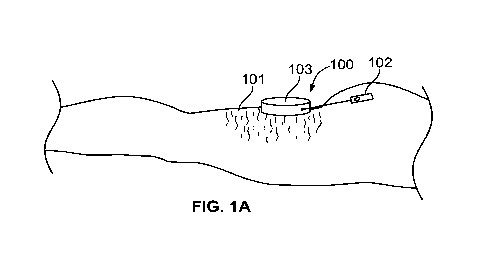
[0041] As illustrated by Figs. 1A-B, the prior approach describes utilizing a
handpiece 100
to capture and control a location of the skin, or dermis 101, as well as
precisely control
use of a cutting tool 102. The handpiece preferably has a top 103 and a
perimeter
elevation 104 that cooperatively define a recessed area 105 which can be
placed over
the patient's skin. By applying a force to the top of the handpiece or by a
vacuum
supplied to the handpiece, a portion of the dermis 101 can be moved into the
recessed
area to substantially fill the recessed area, thus capturing it within the
handpiece and
providing some control over the area of tissue captured. This allows a distal
portion of
cutting tool 102 or other suitable dissection device to be inserted through a
conduit 107
extending through a side of the perimeter elevation of the handpiece,
percutaneously
through the tissue disposed in the recessed area, and into the subcutaneous
tissues
encompassed by the recessed area of the handpiece. Cutting tool 102 is
maneuvered in
such a way as to cut a surgical lesion of a predetermined shape inside the
subcutaneous
tissues within the recessed area and parallel to the top of the handpiece. The
surgical
lesion (i.e., dissection) is targeted to be in a range from as shallow as at 1
mm to 2 mm
below the interface between the dermis and the shallow fat, to as deep as 20
mm below
the skin/fat interface. It is to be recognized that the cutting tool 102 can
in an
11
CA 03168658 2022-07-19
WO 2021/150479 PCT/US2021/013887
alternative or additional embodiment be configured to operate as a fluid
delivery
component such as for anesthesia.
[0042] With reference to Figs. 1C-G, in alternative approaches, the handpiece
can assume
other configurations and can be made from material that adapts to the shape of
the
tissue surface to which it is applied. For example as shown in Fig. 1C,
curving to match
the thigh to which it is applied. A kit of various sized and shaped handpieces
can be
provided for a particular treatment procedure and thus provide location
specific
structures. Additionally, by planning ahead, the handpiece can be formed of a
perimeter that can change shape for a specific procedure, such as being formed
from
telescoping, pivoting, adjustable or moldable, customized (e.g. 3D printed) or
segmented materials. For example, as shown in Fig. 1C, rather than defining a
circular
shape, the handpiece 120 is a generally rectangular structure that is bendable
to
conform to the tissue targeted for treatment. The handpiece can additionally
include a
plurality of conduits 122 through which a cutting tool or other treatment
device can be
inserted. In this way, the fractionalization of a targeted area can be
templatized. A
lower side of the handpiece 120 can be configured with an adhesive for lifting
tissue or
the handpiece 120 can be attached to suction for accomplishing lifting tissue.
Moreover, in another approach (Fig. 1D), the handpiece 130 defines a ring
structure
and includes a plurality of conduits 132 through which the cutting or other
tools can be
inserted to accomplish desired treatments within the ring structure itself or
within an
interior 134 of the ring 130. A clear lid is configured over the interior 134.
Suction
and/or adhesive can be employed to lift tissue when using the handpiece 130.
[0043] In yet a further approach (Figs. 1E-F), a handpiece assembly
embodies a ring-like
base 142 including a plurality of conduits 144 formed therein as well as a
spring-
loaded top 146 sized and shaped to be received within the base 142. A spring
147 is
configured between the top 146 and base 142. A lower platform 148 of the top
146 can
be equipped with an adhesive to engage and lift target tissue. When lifted, a
treatment
and/or cutting tool 107 is inserted through one or more of the conduits 144 to
treat
target septa. The extent to which the top 146 is lifted with respect to the
base 142 is
controlled by the springs 147. Various different heights between the top 146
and the
base 142 can be provided by structure such as that provided in a ball-point
pen.
12
CA 03168658 2022-07-19
WO 2021/150479
PCT/US2021/013887
[0044] With
reference now to Fig. 1G, there is shown a handpiece 150 having a curved
chamber 152 through which a cutting tool 107 can be inserted. To navigate
within the
curved chamber 152, the cutting tool 107 is equipped with one or more steering
wires
154 configured to deflect the cutting tool 107 to reside in various positions.
Such
steering wires 154 can be operator controlled or involve smart tensioning so
as to
counter bending forces and make a relatively small diameter device seem
stiffer than
would be provided by its structure alone. Here, the tensioning/steering wires
154 are
linked to the advancement of the device so that an appropriate amount of
curvature is
introduced based on the location of the cutter 107 within the chamber 152.
Moreover,
the stiffening of the device at the cutter 107 can be responsive to forces
experienced at
the cutter 107 such as those associated with engaging septa. For example, the
stiffness
of the assembly is increased or decreased as necessary, automatically or
manually,
when navigating through the chamber and when engaging septa targeted for
cutting. In
this way, the assembly also can be used to identify target tissues. As shown
in Fig. 1H,
the cutter 107 is provided with a steering sheath 156 the operation of which
can also be
operator manually controlled or automatically controlled by a computer. The
sheath
156 is configured to steer the cutter 107 and to create a relatively thin
device with an
active stiffness for cutting. It is to be recognized that such steering
functionality can be
incorporated into any of the disclosed cutting or treatment devices.
[0045] The handpiece and/or other components of the treatment system can
also be
custom fitted or manufactured or 3D printed to match the target treatment
surface of
the patient and/or a treatment plan developed for a patient. In this way, the
handpiece
for example can be best form fitted to a treatment surface so that an
effective seal is
made between the handpiece and tissue. Moreover, the number of incisions into
tissues
can therefore be minimized through such individualized treatment structures.
Various
sealing structures such as o-rings or flexible perimeters can also be provided
along the
surfaces where the treatment device engages tissues. In yet another approach
providing
for individualized treatment, the cutting tool can first be placed through
skin and within
the treatment site followed by applying suction or other means for lifting
tissue so that
treatment need not be constrained by the prior placement of the handpiece.
This
approach is aided by the transillumination structure disclosed below.
Moreover,
13
CA 03168658 2022-07-19
WO 2021/150479 PCT/US2021/013887
compression bands (not shown) can be applied at or near treatment sites to
cause
tension in uncompressed areas for the identification of target septa or to add
in
tensioning target septa for treatment. In this context, a physician presses or
a device is
used to push down on one area of skin and the displaced tissue causes an
increase in
tensioning of septa in nearby areas to thereby facilitate the identification
of cellulite,
and also to aid in accurately controlling treatment depth.
[0046] As shown in Fig. 2A, in previously disclosed approaches, a top wall
201 and
perimeter wall 202 define a tissue apposition surface (tissue facing surface)
203 facing
into recessed area 105. Tissue apposition surface 203 may be curved inward to
the
handpiece, or concave, or recessed, so that when handpiece 100 is disposed
against an
epidermis 204, further pressure against the handpiece 100 will cause the
handpiece to
encompass a subcutaneous level of tissue 205, particularly the subdermal
tissue layer
below the epidermis and dermis layers, wherein these layers will be positioned
within
recessed area 105. A tissue apposition surface 203 can include a perimeter
wall 202 as
a relatively small inner wall around the perimeter of recessed area 105 and
handpiece
100 may include a transparent cover 206 so that a physician can clearly see
and verify
that the dermis is properly positioned within the dissection region.
[0047] In use, the disclosed device 100 is pressed against the tissue to
move the
subcutaneous layer 205 into recessed area 105 and against tissue apposition
surface
203. In some embodiments, vacuum (suction) is used to enhance the capture of
the
tissue. As stated, in additional or alternative embodiments, adhesive is used
to capture
tissue. Where suction is employed, a vacuum source may be placed in fluid
connection
with handpiece 100 via an optional vacuum port 208 on handpiece 100. The
vacuum
source may include a vacuum pump in fluid communication with recessed area
105.
Vacuum pump supplies suction to the recessed area to pull tissue snugly and
securely
therein. In some embodiments, the vacuum pump is configured to communicate
with a
microprocessor and the graphical user interface to display a vacuum pressure.
The
system may further include a display indicating the elapsed amount of time
vacuum
was supplied to the handpiece by the vacuum pump. The vacuum pump may also
modulate the suction such that a higher suction force is applied initially to
pull the
tissue into the recess, and a somewhat lower suction force is used to
maintain/hold the
14
CA 03168658 2022-07-19
WO 2021/150479 PCT/US2021/013887
tissue in place thereafter. In other approaches, suction can be provided by a
syringe
configured to pull the desired vacuum on the skin to tension the septa or
otherwise lift
skin. Cupping structures or other vacuum providing bellows can also be
incorporated
into a suction device that provides the desired suction.
[0048] Suction applied at vacuum port 208 causes skin 101 to be pulled up
into contact
with apposition surface 203 of handpiece 100. By applying a sufficient suction
force, a
portion of epidermis 204 is pulled into the chamber of vacuum handpiece 100
and
conforms to inner recessed area 105. While the surface of the skin 204 is
tightly
positioned against top wall 201 and perimeter wall 202 of recessed area 105,
fat layer
205 (subcutaneous tissue) is also drawn into the chamber. A cutting tool 102
(e.g., a
cutting blade or RF probe, or needle), can be inserted through a conduit 213
in a side of
handpiece 100 and through entry hole 214, through the skin, and into the
subcutaneous
tissue. The blade can assume various configurations including curved and
angled
surfaces and profiles, as well as a serrated configuration of various sizes,
shapes and
lengths. Moreover, the blade assembly can be embodied one or a plurality of
horizontally extending blades that reciprocate longitudinally or
perpendicularly to the
direction the horizontally extending blades extend, and with respect to one or
more
stationary, or independently reciprocating, horizontally extending members or
blades
(such as in a clipper or hedge trimmer). Significantly, the handpiece enables
the cutting
tool to be consistently inserted at desired treatment depth 215. Handpiece 100
thus
provides for precise control of the depth of the dissection plane and allows
for cutting
and/or movement of tool 102 substantially parallel to the surface of the
tissue along a
plane 225 (Fig. 2B). Moreover, notably, use of an RF probe device or other
energy
emitting device in combination with or in place of a reciprocating blade lends
itself to
the electrocauterization of target tissue and septa.
[0049] A membrane 217 formed of a flexible and resilient material may also
be applied to
the perimeter wall (sidewall) across the proximal (away from the recessed
area) or
distal ends (closer to the recessed area) of the conduit 213 to minimize
vacuum leakage
there through. The membrane 217 preferably is sufficiently resilient to seal
around the
cutting tool as it pierces (self-sealing) therethrough and minimize vacuum
leakage.
Membrane 217 may be formed of silicone.
CA 03168658 2022-07-19
WO 2021/150479 PCT/US2021/013887
[0050] Conduit 213 is preferably located proximate a bottom edge 218 of
perimeter wall
(sidewall) 202 to allow a cutting tool or needle to be inserted into the
tissue (captured
in the recessed area) in a plane parallel to the dermis. Conduit 213 supplies
an angle of
penetration 219 so that the tool inserted through the conduit will penetrate
into tissue
disposed within the recessed area, and substantially parallel to the surface
of the tissue
and parallel to the surface of top wall 201 at depth 215. As depicted in Fig.
2B, entry
hole 214 is preferably disposed on an inner side of the conduit and facing the
recessed
area. Conduit 213 preferably widens outward toward an outer side of the
perimeter
elevation such that a distal end 222 of the cutting tool inserted through the
entry hole
moves in one direction 223 when a proximal end of the cutting tool outside the
conduit
moves in an opposite direction 224. Entry hole 214 thereby defines a cutting
tool pivot
point when a distal end 222 of cutting tool 102 is inserted through conduit
213 and into
recessed area 105, and the tool moves primarily in an x-y plane 225 parallel
to the top
surface of the handpiece. In an alternative approach, the cutting tool pivot
point is
configured to reside within tissue so as to minimize trauma to the tissue
insertion entry
point. In some embodiments entry hole 214 may include an optional locking
mechanism 226 that locks the tool in place upon insertion into the conduit. In
some
embodiments in which a vacuum is supplied to the recessed area, an optional
gasket or
seal 217 (not shown in Fig. 2B) may be placed within, in front of, behind, or
around
entry hole 214 to minimize vacuum leakage.
[0051] As depicted in FIGS. 3A and 3B, the previously disclosed dissection
system
includes a motor controlled cutting module 301 and a guidance track 302
operably
connected (i.e. physically and/or via sensing) to handpiece 100. In one
embodiment,
the motor module 301 is configured so that its operation is controlled by its
interaction
with the guidance track 302. That is, the motor commences
manipulation/reciprocation
of a cutting device only once the cutting device is placed within tissue and
at the
treatment site. The motor then ceases its action once the cutting device is
removed
from the treatment site within tissue. Thus, the tool insertion point is
protected from an
active, reciprocating or other blade by shutting the motor off In one
approach, the base
or guidance track can include a switch that automatically turns the device off
in the
event the cutting tool is removed from the guidance track or platform along
which the
16
CA 03168658 2022-07-19
WO 2021/150479 PCT/US2021/013887
treatment device is passed. The switch can be triggered in the pathway
provided by the
guidance track or platform such that the device will only turn on or off when
positioned
in a prescribed pathway or position. Where the device is not provided with a
guidance
track, the platform along which the device is advanced can be provided with
sensing
and control provided by a photo diode or a magnetic surface and tool sensing
arrangement. Accordingly, such a platform can be color coated and the photo
diode
responds to the color it passes over such as turning on when detecting green
and
turning off when detecting red. Colors or color intensities can also be
provided on a
platform for controlling speed of reciprocation or other action (opening
and/or closing)
of the cutting portion of a treatment device. Further, the cutter module
includes an
embodiment of cutting tool (a reciprocating cutting blade 303 disposed in a
retractable
and advanceable sleeve 304) and a housing 305 and a base 306. Guidance track
302 is
generally configured to constrain a portion of the cutting module guide pin
307 in
contact with the guidance track to move along a predetermined path. This guide
pin
307 can be utilized through its localization with respect to the guidance
track 302 to
control operation as stated above, that is, turning the motor on and off when
the cutting
tool 102 is positioned within tissue and at the interventional site. Thus, a
distal end of
the cutting tool, passing through entry hole 214, cooperatively moves within
recessed
area 105 in a plane substantially parallel to the top of the handpiece and
within a region
of a predetermined shape defined by the predefined path. Motor operation of
cutting
module 301 is preferably controlled manually by an electric switch or button
308, but
may also be activated by electrical or other contact means known in the art
within the
guidance track.
[0052] FIG. 4A depicts an exploded view of the previously disclosed cutting
module 301.
Cutter module 301 includes housing enclosure 305 and base 306, motor assembly
401
mounted on the base and enclosed by the housing, and a reciprocating cutting
blade
303 operably connected to motor assembly 401. Cutting blade 303 is slidably
disposed
within sleeve 304. Sleeve 304 minimizes the amount of tissue in direct contact
with the
shaft 402 of the cutting blade 303 to minimize drag and or tugging on the
tissue. Sleeve
304 also enables the isolation and/or capture of any fluid that may travel
along the shaft
of blade 303.
17
CA 03168658 2022-07-19
WO 2021/150479 PCT/US2021/013887
[0053] A motor assembly 401 is enclosed in enclosure 305 and base 306.
Sleeve 304 is
affixed at a distal end 403 of motor assembly 401. In one embodiment, motor
404 is a
DC motor which may incorporate a gear reduction. In the depicted embodiment, a
crank slider 405 converts motor rotation to cutter reciprocation. Motor 404,
within
enclosure 305 moves reciprocating cutter blade 303 within sleeve 304. As the
motor
turns, crank slider 405 moves cutter 303 back and forth within sleeve 304.
Cutter blade
303 may include a needle or a bayonet which may further include one or more
sharp
edges.
[0054] Sleeve 304 does not reciprocate and is typically comprised of a thin-
walled
polymer tube and is sterile for single patient use. In one particular
embodiment, the
sleeve 304 can be configured to be selectively translatable over the cutter
blade 303 so
that it can protect the tissue from the cutter blade 303 when desired, such as
when
entering tissue or between treatment sites (See Fig. 3A), and also at the
incision site
during cutting within tissue layers. Sleeve 304 and cutter blade 303 are
typically
disposable. Sleeve 304 may be affixed to cutter module 301 (and/or crank
slider 405)
by means of connection point 406. Connection point 406 may be a disposable
protective connector keeping cutter module 301 and gear motor assembly 401 in
fluid
isolation from sleeve 304 and cutting blade 303. In this manner, cutting blade
303 and
sleeve 304 could be disposed along with connection point 406 after each
procedure.
Correspondingly, cutting module 301 including motor assembly 401 and base 306
could be reused in subsequent procedures.
[0055] With reference again to Figs. 3A and 3B, the handpiece also
preferably includes a
platform 309 integral with or affixed to a proximal side of handpiece 100.
Platform 309
preferably includes guidance track 302, wherein guidance track 302 is used to
position,
guide, and support cutting module 301 by means of a guide pin 307. Guide pin
307
moves within and along the path of guidance track 301 to stabilize the cutter
module at
a proper position proximate to handpiece 100.
[0056] In this embodiment, guide pin 307 protrudes through base 306 of
cutter module
301, however, in other embodiments guide pin 307 may be part of base 306 or
cutting
module 301. The guide pin may serve dual purposes. Guide pin 307 serves to
guide the
disclosed cutting module embodiments to create a surgical lesion defined by
the path of
18
CA 03168658 2022-07-19
WO 2021/150479
PCT/US2021/013887
guidance track 302. Additionally, the guide pin may include a feature such as
an
enlarged head or the like which interacts with guidance track 302 and prevents
cutting
module 301 from being lifted off the platform 309 and/or supports cutting
module 301
at a predefined planar orientation relative to platform 309.
[0057] A physician treating the patient determines an instrument
insertion site and paths
that most efficiently treat cellulite with a minimal amount of insertion sites
and
instrument paths under the skin. Preferably, an instrument insertion site is
chosen that
is in a crease or fold of skin such as where the buttocks meets the thigh or
in the crease
between the two buttocks at a location that is not seen when the buttocks are
in natural
contact for improved cosmesis after the procedure healing period. In certain
patients,
the inner thigh is chosen as an insertion site as this location is less visual
as it heals.
Such treatment paths are selected by the operator or can be generated
automatically by
employing a computerized controller programmed to most efficiently address and
measure cellulite residing in a pre-defined treatment site. Thus, the
treatment device
can be programmed to take any possible or conceivable path or pattern of
treatment.
[0058] The computerized controller can be associated with a scanner or
camera that
identifies specific dimples and areas for treatment such as by employing laser
technology. In this regard, the computerized controller includes a program
specific to
cellulite treatment and is used in conjunction with an electronic and
mechanical device
and comprises or includes a non-transitory computer-readable storage medium
and a
computer-program mechanism embedded therein to both identify treatment areas
and
to plot primary and alternative approaches to treatments. In another
embodiment,
computerized visualization and treatment planning equipment is used to assist
the
physician in determining insertion site locations and paths to be taken to the
marked
targets.
[0059] In one approach (Figs. 4B-C), a treatment system 330 includes a
pressure gauge
332 associated with the system computer, the pressure gauge 332 being
operatively
associated with the performance of the treatment system such that treatment
can only
be performed when the system confirms that sufficient vacuum is being applied
to the
treatment area. Further, as shown in Figs. 4B-C, the treatment system 330
additionally
includes a downward facing digital camera 334 configured to see marks 335
associated
19
CA 03168658 2022-07-19
WO 2021/150479
PCT/US2021/013887
with pre-determined treatment locations viewable through a transparent
handpiece 100.
The camera 334 communicates with the system computer and the system computer
determines the treatment approach based upon the images provided by the
camera.
Accordingly, a user need only place the treatment system over a treatment site
marked
for treatment and instruct the system to begin operation, such as through the
pushing of
a button. Once the required suction forces are confirmed, the camera 334
communicates with the system computer to follow the plan of treatment.
Thereafter,
with or without further operator input, the system then automatically proceeds
with
treatment. The assembly is then moved to additional treatment sites as
necessary.
[0060] During treatment, the patient lies down on their stomach on the
treatment table.
Alternatively, because of the minimally invasiveness of the current approach,
a patient
can be treated while standing, particularly for a small number of treatment
targets, or
while standing and leaning forward on a support and alternatively between
standing
and leaning forward so that gravity can help identify and confirm treatment of
the
targeted septa. Moreover, a measurement device such as a camera and system
computer, creates a complete three-dimensional map of all cellulite relative
to normal
skin. By dating and comparing improvement of volume of divots or dimples
versus
normal idealized surfaces, the operator calculates total and local volume
benefits of
therapy and track improvement over time.
[0061] In one specific alternative approach, treatment can be directed at
various positions
about connecting tissue or septa. That is, septa can be engaged, stretched, re-
oriented,
torn, cut, sliced, ruptured or disrupted from various sides or angles
respecting septa and
the treatment target location. Thus, septa can be treated from superior,
inferior or
medial or lateral locations from the septa and treatment target location to
achieve the
best results. For example, in a particular situation, treatment can be most
effective
from a position superior on the patient above a particular connecting tissue
to take
advantage of gravity where treatment forces placed on the connecting tissue
coincide
with the direction of gravity or the direction that gravity most often works
on a
standing body, as it has been observed that cellulite is often most visible in
a standing
individual.
CA 03168658 2022-07-19
WO 2021/150479 PCT/US2021/013887
[0062] A force gauge (electronic or mechanical) can be provided to ensure that
a pre-
determined amount of force would be applied to the tissue when testing the
septa to
prevent over or under pulling. A treatment device capable of one or more of
engaging,
stretching, slicing, cutting or disrupting connective tissue is configured at
a distal end
portion of the device. All cutting means can be combined with or further
energized
with RF, a laser, ultrasonic or thermal energy to produce cutting and
coagulation
together or separately. In certain aspects, there can be a single entry site
or two entry
sites, one high on the hip and another along the crease or transition between
the
buttocks and thigh, or at the inner thigh. Such locations are characterized in
that they
can be easily hidden either naturally or by clothing. Treatment targets,
depressions and
dimples that have been marked on the skin surface while the patient is
standing often
go away when the patient lies down on their stomach because gravity acts on
the skin
and underlying connective tissue in a different direction such that the ink
mark is
apparent but the dimple or depression is not. Interventional devices are
configured
such that a user can approach a target location and first use the
interventional device to
push, pull or otherwise tension septa in a target area under the skin to
identify the
specific septa impacting the target location and/or which is the cause of the
expression
of cellulite. In other words, pulling or pushing on the septa under the skin
to find the
one(s) that create the dimple or depression in the skin surface. For some
treatment
targets, taking an approach from an entry located inferior the treatment
target,
advancing the end of the interventional device with a hooking and cutting
element
beyond the treatment target and then extending the hooking and cutting element
and
pulling inferiorly (effectively the "down" direction if the patient was
standing) can
provide a better approach in locating septa. For some treatment targets,
taking an
approach from an entry located superior the treatment target, advancing the
end of the
device with the hooking and cutting element collapsed beyond the treatment
target and
then pulling superiorly can provide an alternative effective approach (for
example, for
treatment targets on the leg, to re-create the dimple when the patient is
lying down).
One or more strain gauges can be incorporated within the treatment device to
help
identify target septa as well as to assess the progress and completion of
treating septa.
This facilitates targeting of key septa in a less impactful way, ideally
minimizing
21
CA 03168658 2022-07-19
WO 2021/150479
PCT/US2021/013887
bruising or other issues associated with cutting or disrupting a large area
around the
target. There are thus herein shown various approaches to treating cellulite
expressed
as dimples or depressions in the skin surface. Moreover, the handle portion
can be
employed to create an indentation in skin through which interventional devices
can be
inserted subcutaneously. A treatment regimen is selected for inserting
interventional
instruments based upon the subject's anatomy as it relates to the septa
connecting
tissue layers that define the chambers retaining fatty or other tissues. If
desired, while
anesthetic and/or sedation is taking effect, ultrasound can be used to assess
the
subcutaneous trajectory and depth of the various connective tissue bands
responsible
for the surface unevenness. The ultrasound evaluation can help with the
particular
trajectory selected for the desired depth. The ultrasound evaluation can also
help with
positioning the distal end portion of the treatment instrument strategically
at the
connection point between the connective tissue and the dermis or the facia.
[0063] In an additional or alternative embodiment, a tool control
mechanism 500 (See
Fig. 5) is provided which allows cutting tool or other tool appropriately
configured
device, to be controlled by a microprocessor. In one or more embodiments, the
microprocessor controls cutting device, with or without manual assistance of
the
operator, to precisely cut an area of tissue disposed within a recessed area
of the
handpiece. The area being cut is predetermined and programmed into the
microprocessor by the operator of the handpiece. Various areas and patterns of
treatment can be achieved as desired and required for a particular patient.
[0064] In one approach, the tool control mechanism 500 includes a motor
assembly 502
controlled by a programmable controller 504 and one or more of lateral and
axial
movement of the treatment device is controlled. The motor assembly 502 drives
a
shaft 506 configured to pass laterally through a bushing 508. An optical
encoder 510 is
configured axially within the bushing 508, and a clamp 512 is attached to the
optical
encoder 510. Both the optical encoder 510 and the clamp 512 are also
controlled by
the controller 504. Communication with the controller 504 can be wireless or
via a
hardwire connection with one or more components. The clamp 512, optical
encoder
510 and bushing 512 are aligned and include a through hole sized and shaped to
receive a shaft 506 of a cutter device, the shaft 506 being marked in a manner
to
22
CA 03168658 2022-07-19
WO 2021/150479
PCT/US2021/013887
communicate with the optical encoder 510. Here, the user will have control of
advancing the cutter or other interventional device within the tool control
mechanism
500 but a second automatically controlled motor (not shown) can be
incorporated into
the assembly to control longitudinal motion of the interventional device as
well. In
use, the controller 504 is programmed for treating a patient with a specific
treatment
regimen. Once the patient is prepared for the interventional treatment, the
user or
second motor will advance the interventional device within the bushing 508 and
the
controller 504 will turn the bushing 508 as directed by the treatment regimen
and based
upon the optical encoder readings. The controller 504 monitors the optical
encoder
510 as it identifies the shaft 506 markings to determine the depth the
interventional
device assumes. The controller 504 simultaneously controls the clamp 512 based
upon
the position of the shaft 506 and stage of the pre-programmed treatment. In
this way, a
controlled and precise treatment can be achieved by the tissue treatment
system.
[0065] Turning to Figs. 6A-F, another approach to a treatment system
550 is presented.
The treatment system 550 includes a base unit 552 and an elongate member 554
extending from the base unit. As best seen in Figs. 6B-C, the elongate member
554 is
equipped with a retractable sheath 556 selectively covering a cutting device
558. Here,
the cutting device 558 is shown as a double edged blade, but any of the
disclosed
embodiments of cutting structures can be used. Notably, as described herein,
the
sheath 556 can be operator or automatically computer controlled so that the
sheath 556
covers the cutting surface of the cutting device 558 when desired such as when
the
elongate member 554 is outside a patient's body and/or navigating within
tissue or
when not being employed for cutting. Accordingly, proximally extending wires
or
other elongate structures are attached to the sheath 556 to control its
positioning.
Housed within the base unit 552 is one or motor assemblies for controlling the
operation of the cutting device 558 and/or sheath 556. As shown schematically
in Fig.
6D, one embodiment of a motor assembly 557 for creating reciprocating motion
of the
cutting device 558 can be utilized. This motor assembly 557 can be battery
powered or
connected to an outside power source. In one aspect, engaging the motor
assembly 557
can simply cause the motor to spin and allow the cutter 558 to advance out of
the
sheath 556 to accomplish required cutting. The length of the exposed cutting
surface
23
CA 03168658 2022-07-19
WO 2021/150479 PCT/US2021/013887
of the cutting device 558 can be set by altering the oscillating structure of
the motor
assembly 557. In another aspect, an on button can be used to power the system,
releasing of the same would cause the system to power off. A treatment
platform 560
and a receptacle 562 are further provided, the receptacle being attached to a
suction
force or includes an adhesive for lifting tissue within the receptacle 562
(Figs. 6E-F).
Notably, the receptacle 562 can assume any one of a number of shapes and
configurations, and can include a seal (such as a membrane placed within the
receptacle 562) through which the cutting device 554 is inserted. The
receptacle can be
sized and shaped for use on the buttocks and/or the thigh, and can be formed
as a single
molded part with a co-molded or second molded material creating an elastomeric
edge
for flange for sealing and/or comfort. Additionally, the platform 560 is
characterized
by a smooth surface along which the base unit 552 can be slid. Here, no tracks
are
provided to constrain the movement of the base unit 552 and thus the cutting
device
558 can be moved as desired within a treatment site also without constraint,
and the
treatment site can be as large or small as is practical.
[0066] It has been recognized that various cutting devices can be employed
at a distal end
of a treatment device, and that there is a benefit to being able to track the
position of
the treatment device when placed within the patient. Such various cutting
devices can
be incorporated into the above treatment system and thus, can be reciprocated
by the
reciprocating motor, or the reciprocating action can be omitted and cutting
accomplished by the manipulation of the cutting device alone. In one aspect,
in order
to counter a natural damping that occurs in the superficial space and to
facilitate
controllable vibration of the cutting structure, the deployable cutting
structure is
provided with a resonate frequency being a multiplier above vibration
delivered in the
handle or base associated with the cutting structure.
[0067] In another aspect, a distal end portion of a cellulite treatment
assembly is inserted
through the skin and the tip is guided up into close proximity of the dermis
as the tip
can be tracked as it is advanced toward septa 650 (Fig. 7A). Given the
elasticity of
septa 650, the distance from the targeted treatment location to where the
treatment
assembly is inserted into the skin is preferably at least about 2 cm so that
there is
enough distance to pull and disrupt septa 650 and not have the tip of the
cellulite
24
CA 03168658 2022-07-19
WO 2021/150479 PCT/US2021/013887
treatment assembly exit the skin in the process. Additionally, a depth below
the skin
where septa 650 is preferably engaged (i.e., cut, sliced, torn, stretched, re-
oriented (e.g.
criss-crossing) or disrupted) is identified and determined. After determining
the
subcutaneous depth to be accessed for the cutting, slicing, tearing,
stretching, re-
orienting (e.g. criss-crossing) or disrupting of septum 650, the cellulite
treatment
assembly or other tool with a sharpened or blunt tip is inserted through the
skin,
advanced between subcutaneous tissue layers and toward septa 650. In one
approach, a
distal end portion of the cellulite treatment assembly is configured with an
illuminated
tip 652 with enough brightness to be seen through the skin. The intensity of
light
emitted by the tip 652 can be set to a specific constant level such that at
the preferred
depth below the skin for severing or otherwise engaging septa 650, the light
that
appears at the level of the skin as a circle or projection is of a pre-
determined size.
Thus, the treatment device is advanced to the target site. At the target site,
the user
adjusts the depth of the tip of the treatment tool such that the circle or
projection of
light is the pre-determined size. The septa 650 is tested and if confirmed as
a target for
treatment, the septa 650 is treated while maintaining the circle or projection
at the pre-
determined size. Notably, the diffusion and brightness of light can vary for
each patient
as the same can be affected by skin tone and body mass index. Thus,
calibration can be
conducted on a patient by patient basis based upon such factors at the
beginning or
prior to a procedure. The user can also use the size of the circle or
projection of light to
maintain the depth of the tip of the treatment tool as it is advanced under
the skin to the
treatment target. It is expected that the depth that these tools are advanced
will be
between about 3 and about 10 mm below the skin surface, but it is anticipated
that
lesser and greater depths may also be optimal for a particular subject. In any
event, the
depth selected is chosen for cutting, slicing, disrupting, tearing, stretching
or re-
orienting of the subject's septa 650. Moreover, in one embodiment, it is to be
appreciated that the device is formed from a substantially rigid material so
that a
consistent plane below the skin surface is accessed.
[0068] Using palpation, direct visualization (for example,
transillumination or
endoscopic) or non-invasive visualization (for example, ultrasound or
fluoroscopic) or
other means for determining the position of the interventional tool such as
markings
CA 03168658 2022-07-19
WO 2021/150479 PCT/US2021/013887
along the length of the instruments and its path within tissue, or providing
the
interventional instrumentation with radiopaque markers, the tool is placed at
a site
below where cellulite (for example a dimple) is seen on the subject's skin.
The
treatment device 655 is advanced through septa 650 and to where the treatment
device
is in a position best suited to accomplish the identification of target septa
and the
cellulite removal or minimization treatment. As shown in Figs. 7B-D, in one
approach,
the treatment device 655 is passed beyond septa 650, a hook is deployed and
then
pulled proximally to tension septa 650, such as by hooking the septa (Fig.
7E). In
particular, first hooking than cutting septa is advantageous when treating
cellulite. In
another approach, the treatment device is passed a few millimeters lateral,
preferably
about 1 to about 10 millimeters, more preferably about 3 to about 6
millimeters, and
beyond the target location, a hook is deployed and then moved toward the
target
followed by pulling proximally to hook and tension septa. During these and
other
steps, transillumination can be employed to track the treatment device and
guide the
procedure. The targeting of septa 650 is accomplished while using
transillumination to
see the location of the treatment device 655. In other approaches, a separate
device can
be employed to engage septa 650 to see if such septa are the source of a
dimple or
depression expressed on the outside of the skin. Such a secondary device can
be placed
remotely from the target (i.e. cellulite depression) and configured to be
capable of
applying tension to the surface of skin in a predetermined direction so as to
create the
effect of gravity and produce the visualization of the depressions while the
patient is in
a prone position (e.g. a broad region of adhesive attached to a spring
mechanism such
that a predetermined force would be applied relatively parallel to the surface
of the skin
in the direction the skin would move when standing in gravity). Using this
additional
device could further help the confirmation and location of depressions and
allow
confirmation that the treatment was effective. Also, in various approaches, a
portion of
the elongate member can be configured to transition from a smaller state to a
wider or
larger state, wherein in the wider or larger state a cutting surface (i.e.
sharpened edge or
energy) is presented to cut tissue, the device being sized and shaped to be
inserted
through the skin and engage one or more regions of septa subcutaneously.
[0069] It is noted that septa causing a dimple or depression may be coming
from various
26
CA 03168658 2022-07-19
WO 2021/150479 PCT/US2021/013887
angles and locations relative to the dimple or depression seen on the skin
rather than
being directly below the dimple or depression, and may be due to one or only a
few
septa or a large number of septa that remotely cause the depression or dimple.
Thus, so
engaging certain septa will be reflected in some change in the dimple or
depression on
the skin. A determination is made concerning the correspondence with targets
on the
skin and the dimples being formed or re-formed. If the initial septa 650 that
the user
presses on or pulls on using the tool do not recreate a dimple or depression
in the
targeted area, then the user releases those initial septa that were engaged
and
repositions the tool at different septa and presses on or pulls again. This is
repeated
until the septa responsible for a dimple or depression in the marked location
are
identified. Once proper septa are identified, the tool is manipulated to cut,
slice,
disrupt, re-orient, stretch or tear septum 650 connecting tissue layers. In
one approach,
a blade 656 is deployed and presented for treatment (Fig.7F).
[0070] After the proper septa have been cut, sliced, disrupted, stretched,
re-oriented or
torn, the treatment element is moved back to its initial collapsed
configuration. The
treatment element is then advanced beyond the marked treatment location, the
treatment element (e.g., hooking and cutting device) is deployed and then
pulled back
under the marked treatment location to confirm that all of the septa
responsible for
causing the marked dimple or depression have been separated intra-operatively.
If they
have not been, the tool is manipulated to cut, slice, disrupt, stretch, re-
orient or tear
additional septa. The steps are repeated until all of the septa responsible
for creating the
marked dimple or depression have been severed or sufficiently stretched and
the
dimple or depression cannot be re-created intra-operatively using the tool.
Such
manipulation results in selective rupture, tearing, cutting or slicing of
targeted septum
650, and the removal or minimization of dimples and the expression of
cellulite on skin
(Fig. 7G). Thereafter, the treatment element (e.g., hook and/or blade) is
retracted back
in (Fig. 7H partially collapsed) and the tool is removed from the site to be
withdrawn
from the body or repositioned in any direction along and within the target
tissue plane
to treat additional areas.
[0071] With reference to Figs. 8A-D, in additional or alternative
approaches, a second
light source 656 such as an LED (or other light source) is configured along
the cellulite
27
CA 03168658 2022-07-19
WO 2021/150479 PCT/US2021/013887
treatment assembly proximal the illuminated tip 652 or alternatively, at the
tip 652. In
various approaches, a light source such as an LED chip can be configured at
the tip of
or otherwise along the treatment device with an electrical wire running
proximally for
control by the operator, or the light source can be generated by a light fiber
extending
along the device or to the tip with the LED or light source is configured
within a
proximally located position such as a handle of the treatment device. By so
configuring
such light sources 652, 656, the depth of the cellulite treatment assembly
within tissue
can be assessed. As shown in Figs. 8A-B, when the cellulite treatment assembly
is
placed within a first relatively shallow desired depth, the light sources 652,
656 appear
spaced and define discrete patterns when viewing the light sources via
transillumination through skin (Fig. 8B). When the cellulite treatment
assembly is
placed deeper within tissue (Figs. 8C-D), the light sources 652, 656 overlap
(Fig. 8D)
due to the natural dispersion of light emitted from the light sources 652,
656. An
operator of the treatment system can determine a depth of the cellulite
treatment
assembly by noting the discrete patterns of light or the degree of overlap of
light
overlap, the dispersion of light emitted and intensity of the light emitted
from the light
sources 652, 656. Thus, allowing the operator to guide the distal end of the
treatment
assembly to the desired treatment location while maintaining the desired depth
below
the skin. The light sources 652, 656 can also be of a different color to aid
in
determining the orientation of the cellulite treatment system within tissue
through
illumination. Moreover, the second light source 656 can emit a red color, for
example,
while the illuminated tip 652 can emit white light, while noting any variation
of colors
can also be employed. Also, the color of the light can change depending on the
configuration of the treatment device, such as for example, the device can
project a
white or first color when sheathed or stowed and change to another color or
second
color when a portion of the device is deployed or before and after use such as
when
tissue is cut. A strain gauge can be configured to communicate and cooperate
with the
light source to sense loads placed on the treatment device during treatment to
thereby
facilitate a change in color of the light source and to signal the progress or
completion
of targeted treatment. Additionally, the second light source 656 can be
employed via
transillumination through skin to locate the cellulite treatment system
relative to a
28
CA 03168658 2022-07-19
WO 2021/150479 PCT/US2021/013887
treatment target area. Another benefit of the second light source is that it
can indicate to
the user where the hook and blade are located relative to the target septa.
Also, as the
treatment tool is being pulled proximally through the treatment target area,
the
illuminated tip 652 lets the user know when the hook and blade have been
pulled
through the target area. It is further noted that the light sources 652, 656
can be
positioned at various alternative locations along a treatment device, and can
be spaced
from each other by various amounts. Also, the cellulite treatment system can
include
greater than two light sources of the same or dissimilar colors. In another
embodiment,
different colors of light can be used to indicate that the state of the distal
end of the
instrument. For example, red light is used to indicate the hook and blade are
inside the
instrument for advancing under the skin, white light is then used to indicate
the hook is
deployed, and red light is then used to indicate when the blade is deployed.
[0072] In another approach, as shown in Fig. 9, a sensor apparatus 702 that
communicates
with structure or a sensor on the treatment device 107 is employed to
determine the
position and depth of the treatment device 107 below the skin 108. The sensor
apparatus 702 can employ a magnetic field, metal detection or ultrasound to
accomplish gathering such positional and depth information on the treatment
device
107. This information can further be communicated directly to the operator via
a
display or through a controller associated with the treatment device.
[0073] After completing treatment of one target area, the procedure is
repeated to treat
other target areas. Accordingly, the same device can be employed to access
tissue
layers below other sites or depressions existing in skin. Notably, in one
embodiment,
the device is capable of anesthetic delivery as needed or desired when
progressing to
additional or new locations. There is thus provided a system configured to
treat all
target areas on the buttocks and thigh through a limited number of small entry
sites,
including through a single entry site on a patient's treated side. It is to be
recognized
that the system can further include structure permitting the assembly to be
steerable to
subcutaneous treatment sites. In such an embodiment, the device would be
configured
to define longitudinally flexible material, and the instrumentation would be
steered to
the desired position within tissue. Moreover, in certain applications, the
device has a
stiffness that varies along its length. In another embodiment, the treatment
device is
29
CA 03168658 2022-07-19
WO 2021/150479 PCT/US2021/013887
embodied in a deflectable catheter.
[0074] Additionally, or alternatively, in each disclosed embodiment,
illumination can be
via a lightguide from an external light source or via one or more LEDs
external or
internal the treatment device. Illumination aids the user both with locating
the
treatment device as well as proper depth placement as transillumination
decreases with
increasing tool depth. In one aspect, the amount of illumination is set to
ensure proper
depth of a treatment device or structure, the level of illumination targeted
being
adjusted for skin type, thickness, presence of fat and pigment. Once selected
or
targeted septa are cut, sliced or disrupted, in each of the disclosed
approaches, the
cellulite treatment device can be or is advanced or repositioned to treat
additional target
areas from the same or different skin insertion location.
[0075] Various approaches to laterally projectable tissue engaging and/or
cutting
structure can be employed. Here again, so engaging septa can confirm that the
septa
responsible for creating skin surface dimples or depressions is being targeted
as such
engagement with septa will be reflected in a physical change of the skin
surface.
Transillumination functionality is provided by a light at a terminal end of
the device, or
formed in the shaft proximal the terminal end allows for the dispersion of
light energy.
Actuation of the engaging and cutting structures can be accomplished through
the
manipulation of a proximally positioned lever or trigger connected to the same
via a
wire or longitudinally directed shaft (not shown). Once a desired area is
treated,
additional target areas can be addressed.
[0076] With reference to Figs. 10A-C, the cutting, slicing or disrupting
treatment assembly
is defined by a projecting linkage arrangement. A first link 800 includes a
blade 801
and is rotatably attached at one end to a second link 802. The opposite end of
the first
link 800 slides with respect to a longitudinal shaft 805 (shown as at least
partially
transparent). The shaft 805 defines a housing for supporting and containing
the linkage
arrangement. A second end of the second link 802 is rotationally affixed to a
distal
point on the shaft 805. A drive shaft or push rod 807 is rotatably or
pivotably attached
to the opposite end of the first link 800 and the second link 802 includes a
generally
triangular or pointed projection 808 that is sized and shaped to shield the
blade 801
from contacting tissue when the assembly is placed in a hooking configuration.
When
CA 03168658 2022-07-19
WO 2021/150479
PCT/US2021/013887
the push rod 807 is fully retracted (Fig. 10A), the blade 801 is sheathed
within the body
of the longitudinal shaft 805. It is noted that in the fully retracted
configuration that the
first and second links 800, 801 form an obtuse angle and the projection 808
extends a
relatively small distance from an opposite side of the longitudinal shaft.
When the push
rod 807 is advanced completely to a stop, the projection 808 contacts the push
rod 807
and the blade 801 is again protected by the projection 808 (Fig. 10B). It is
in this
configuration that the treatment device can be used to hook target septa and
to test
septa to determine if such septa is associated with the expression of
cellulite on a
patient's skin. Withdrawing the push rod 807 from its fully advanced position
and to a
partially withdrawn position (See Fig. 10C where the blade 801 is shown
transparent
for illustrative purposes), the blade 801 is exposed and presented for
engaging and
cutting, slicing or disrupting target septa. The treatment device also has a
blunt,
atraumatic tip 806 (such as a conical profile for example) that allows the
treatment
device to be advanced through the subcutaneous tissue with little trauma. In
all
embodiments, blunt tip 806 can house a light emitting diode, be a light
emitting diode
or house the end of a light fiber in order to facilitate transillumination
through the skin
for the user to use for guidance in knowing the location of the tip of the
treatment
device.
[0077] It is to be recognized that additionally or alternatively, the tip in
any of the disclosed
embodiments can be shaped so as to be characterized by or associated with a
low
introduction and advancement force through and within the patient's skin and
anatomy,
while also presenting a low likelihood of damaging tissue. Accordingly, the
tip can
assume bullet point or short dilator tip shapes, or can define a sharp profile
or a trocar-
type configuration for ease of advancement or tracking. Additionally, the tip
can be
retractable, reconfigurable or otherwise define a sharpened structure only
when the tip
is presented with a pre-determined level of resistance. In one particular
approach, a
spring loaded cover or shield is configured about the tip such that when
presented with
a defined resistance, the cover or shield is removed to expose a sharpened tip
configured to facilitate advancement of the treatment device or reduce the
force to
cross patient anatomy.
[0078] In one
or more approaches, the second link 802 includes a blade 801 that has a
31
CA 03168658 2022-07-19
WO 2021/150479 PCT/US2021/013887
sharpened protrusion 803, and the first link 800 functions as a blocker to
shield a main
portion of the blade 801 from contacting tissue when the treatment device is
in the
hooking configuration (See Figs. 11A-F). When the treatment device is in the
hooked
configuration, the sharpened protrusion 803 extends proximally from the pivot
between
the first link 800 and second link 802 so that as the treatment device is
pulled
proximally by the user, the pivot location, as the leading portion of the
device during
retraction, does not get snagged in tissue but rather slices through it so the
user can
hook and feel resistance of septa with the main portion of the first link 800.
Notably, in
a fully retracted position (Fig. 11A), the first and second links 800, 801
define an
obtuse angle and when the push rod 807 is advanced nearly completely (Fig.
11B), a
majority of the blade 801 is protected by the second link 802. As such,
structure is
presented in a hook-form both to encourage hook capture as well as provide a
portion
of unprotected blade 803 near the connection between the first and second 800,
802
links. Completely advancing the push rod 807 fully exposes the blade 801 for
cutting,
slicing or disrupting target septa (See Figs. 11C-D; Fig. 11D showing the
first blade as
transparent for illustrative purposes) as the treatment device is retracted
proximally by
the user.
[0079] In employing one or more of the disclosed embodiments in a
treatment
procedure, there is an expectation that there are instances where it is
preferable to not
disrupt a hooked septa, and in such a case it is desirable to release or
disengage the
hooked septa. In certain approaches, to release or disengage, the treatment
device
would be advanced or twisted away from the hooked septa. It is thus recognized
that a
challenge exists in that there may be additional septa or other tissue in the
area which
could be unintentionally re-engaged by the treatment device when it is in a
hooking
configuration, and stowing of the treatment device may be inhibited by
adjacent patient
anatomy. With reference to Figs. 11E-F, treatment devices that include a hinge
link
arrangement 800, 802 or similar structure that transition from a hooked
configuration
(Fig. 11E) toward a stowed configuration (Fig. 11F) by pivoting relative to
the
longitudinal shaft 805, benefit from the blocking link 800 (or similar
structure) moving
to push septa 650 or other tissue away from the treatment device as the
treatment
device is being sheathed or stowed. This action requires no additional
advancement of
32
CA 03168658 2022-07-19
WO 2021/150479 PCT/US2021/013887
the treatment device within patient anatomy and ensures that septa 650 or
other tissue
do not become undesirably entrapped. Moreover, when being stowed the links
800,
802 dislodge any tissue that might have become captured within the
longitudinal shaft
805 and the links 800, 802 ultimately occupy such spaces within the
longitudinal shaft
805.
[0080] The treatment tool embodiments of Figs. 7A to 11F can be used in
previously
described hand piece embodiments. The illuminated tip can be used in the
previously
described cutting tools 102.
[0081] Turning to FIGS. 12A-C, guidance track 302 is configured to provide
a controlled
delivery of treatment fluid through needle 1001. Needle 1001 may be a tube, an
hypodermic needle and may have a multitude of holes for increased lateral
fluid
dispersion. A supply tube 1002 provides fluid connection of needle 1001 with a
syringe
1003, syringe pump, roller pump or other injection mechanism known in the art.
In
certain embodiments, a needle control module 1004 is included to house needle
1001
and to provide support for movement along guidance track 302. It is to be
recognized
however that one or more of the alternative or additional components and
structures
disclosed herein can replace the guidance track, and various handpieces can be
combined with the needle assembly. As shown, movement of needle 1001 along
guidance track 302 provides delivery of the treatment fluid in precise
locations of the
dissection region and minimizes the amount of infusion fluid required for a
single
treatment and/or over multiple treatment sites. Needle control module 1004
preferably
includes a guide pin to be engaged into guidance track 302 of platform 309.
The guide
pin guides the needle/cannula to insure that the injectable fluid is injected
into the
tissue at the desire depth and desired locations within a predefined treatment
area
defined by the path of guidance track 302.
[0082] As best seen in Figs. 12B-C, in an alternative embodiment, structure
is provided for
receiving a light source such as a light fiber or an LED. In one approach
(Fig.12B),
extending along the needle 1001 is a lumen 1010 sized and shaped to receive a
light
source 1011. In another approach (Fig. 12C), a sleeve 1012 is configured about
the
needle 1001 and the sleeve includes a inner lumen or space for receiving the
light
source 1011. In this way, the position of the needle 1001 can be tracked
and/or
33
CA 03168658 2022-07-19
WO 2021/150479 PCT/US2021/013887
anatomy can be viewed during an interventional treatment procedure.
[0083] Accordingly, various approaches to cellulite treatment methods and
apparatus are
presented. The disclosed approaches are configured to provide an effective and
focused approach to treating, minimizing and preventing cellulite. The
disclosed
approaches can also be used to repair and reduce the appearance of cellulite
in a
targeted manner. Further, the disclosed proactive treatment modalities are
easy and
effective to use.
[0084] Some of the specific aspects of the present disclosure include one
or more of focal
treatment of just the septa responsible for causing dimples or depressions in
the skin;
minimizing bruising; accessing all treatment targets from limited,
cosmetically
acceptable entries; capture and retention of septa while separating the septa;
intra-
operative confirmation of treated target; needle-diameter sized tools for
small
openings; and transillumination identification of tool tip location.
[0085] While the present disclosure has been described with reference to
the specific
embodiments thereof, it should be understood by those skilled in the art that
various
changes may be made and equivalents may be substituted without departing from
the
true spirit and scope of the disclosure. In addition, many modifications may
be made
to adapt a particular situation, material, composition of matter, process,
process step or
steps, to the objective, spirit and scope of the present disclosure. All such
modifications are intended to be within the scope of the present disclosure.
34