Note : Les descriptions sont présentées dans la langue officielle dans laquelle elles ont été soumises.
CA 03168969 2022-07-25
WO 2021/148881
PCT/IB2021/000027
METHODS AND SYSTEMS FOR USING MULTI VIEW POSE ESTIMATION
CROSS REFERENCE TO RELATED APPLICATION
[0001] This is an international (PCT) patent application relating to and
claiming priority
to commonly-owned, co-pending U.S. Provisional Patent Application Serial No.
62/965,628,
filed on January 24, 2020 and entitled "METHODS AND SYSTEMS FOR USING MULTI
VIEW POSE ESTIMATION," the contents of which are incorporated herein by
reference in
their entirety.
FIELD OF THE INVENTION
[0002] The embodiments of the present invention relate to interventional
devices and
methods of use thereof
BACKGROUND OF INVENTION
[0003] Use of minimally invasive procedures such as endoscopic procedures,
video-
assisted thoracic surgery, or similar medical procedures can be used as
diagnostic tool for
suspicious lesions or as treatment means for cancerous tumors.
SUMMARY OF INVENTION
[0004] In some embodiments, the present invention provides a method,
comprising:
obtaining a first image from a first imaging modality,
extracting at least one element from the first image from the first imaging
modality,
wherein the at least one element comprises an airway, a blood vessel, a body
cavity, or any combination thereof;
obtaining, from a second imaging modality, at least (i) a first image of a
radiopaque
instrument in a first pose and (ii) a second image of the radiopaque
instrument in a
second pose,
CA 03168969 2022-07-25
WO 2021/148881
PCT/IB2021/000027
wherein the radiopaque instrument is in a body cavity of a patient;
generating at least two augmented bronchograms,
wherein a first augmented bronchogram corresponds to the first image of the
radiopaque instrument in the first pose, and
wherein a second augmented bronchogram corresponds to the second image of
the radiopaque instrument in the second pose,
determining mutual geometric constraints between:
(i) the first pose of the radiopaque instrument, and
(ii) the second pose of the radiopaque instrument,
estimating the first pose of the radiopaque instrument and the second pose of
the
radiopaque instrument by comparing the first pose of the radiopaque instrument
and
the second pose of the radiopaque instrument to the first image of the first
imaging
modality,
wherein the comparing is performed using:
(i) the first augmented bronchogram,
(ii) the second augmented bronchogram, and
(iii) the at least one element, and
wherein the estimated first pose of the radiopaque instrument and the
estimated second pose of the radiopaque instrument meets the determined mutual
geometric
constraints,
generating a third image; wherein the third image is an augmented image
derived
from the second imaging modality which highlights an area of interest,
wherein the area of interest is determined from data from the first imaging
modality.
2
CA 03168969 2022-07-25
WO 2021/148881
PCT/IB2021/000027
[0005] In some
embodiments, the at least one element from the first image from the
first imaging modality further comprises a rib, a vertebra, a diaphragm, or
any combination
thereof In some embodiments, the mutual geometric constraints are generated
by:
a. estimating a difference between (i) the first pose and (ii) the second pose
by
comparing the first image of the radiopaque instrument and the second image of
the
radiopaque instrument,
wherein the estimating is performed using a device comprising a protractor, an
accelerometer, a gyroscope, or any combination thereof, and wherein the device
is attached to the second imaging modality;
b. extracting a plurality of image features to estimate a relative pose
change,
wherein the plurality of image features comprise anatomical elements, non-
anatomical elements, or any combination thereof,
wherein the image features comprise: patches attached to a patient, radiopaque
markers positioned in a field of view of the second imaging modality, or any
combination thereof,
wherein the image features are visible on the first image of the radiopaque
instrument and the second image of the radiopaque instrument;
c. estimating a difference between (i) the first pose and (ii) the second pose
by using a
at least one camera,
wherein the camera comprises: a video camera, an infrared camera, a depth
camera, or any combination thereof,
wherein the camera is at a fixed location,
wherein the camera is configured to track at least one feature,
3
CA 03168969 2022-07-25
WO 2021/148881
PCT/IB2021/000027
wherein the at least one feature comprises: a marker attached the patient,
a marker attached to the second imaging modality, or any combination
thereof, and
tracking the at least one feature;
d. or any combination thereof
[0006] In some embodiments, the method further comprises: tracking the
radiopaque
instrument for: identifying a trajectory, and using the trajectory as a
further geometric
constraint, wherein the radiopaque instrument comprises an endoscope, an endo-
bronchial tool,
or a robotic arm.
[0007] In some embodiments, the present invention is a method, comprising:
generating a map of at least one body cavity of the patient,
wherein the map is generated using a first image from a first imaging
modality,
obtaining, from a second imaging modality, an image of a radiopaque instrument
comprising at least two attached markers,
wherein the at least two attached markers are separated by a known distance,
identifying a pose of the radiopaque instrument from the second imaging
modality
relative to a map of at least one body cavity of a patient,
identifying a first location of the first marker attached to the radiopaque
instrument on
the second image from the second imaging modality,
identifying a second location of the second marker attached to the radiopaque
instrument on the second image from the second imaging modality, and
measuring a distance between the first location of the first marker and the
second
location of the second marker,
projecting the known distance between the first marker and the second marker,
4
CA 03168969 2022-07-25
WO 2021/148881
PCT/IB2021/000027
comparing the measured distance with the projected known distance between the
first
marker and the second marker to identify a specific location of the radiopaque
instrument inside the at least one body cavity of the patient.
[0008] In some embodiments, the radiopaque instrument comprises an endoscope,
an endo-
bronchial tool, or a robotic arm.
[0009] In some embodiments, the method further comprises: identifying a depth
of the
radiopaque instrument by use of a trajectory of the radiopaque instrument.
[0010] In some embodiments, the first image from the first imaging modality is
a pre-operative
image. In some embodiments, the at least one image of the radiopaque
instrument from the
second imaging modality is an intra-operative image.
[0011] In some embodiments, the present invention is a method, comprising:
obtaining a first image from a first imaging modality,
extracting at least one element from the first image from the first imaging
modality,
wherein the at least one element comprises an airway, a blood vessel, a body
cavity or any combination thereof;
obtaining, from a second imaging modality, at least (i) a one image of a
radiopaque
instrument and (ii) another image of the radiopaque instrument in two
different poses of
second imaging modality
wherein the first image of the radiopaque instrument is captured at a first
pose
of second imaging modality,
wherein the second image of the radiopaque instrument is captured at a second
pose of second imaging modality, and
wherein the radiopaque instrument is in a body cavity of a patient;
generating at least two augmented bronchograms correspondent to each of two
poses
of the imaging device, wherein a first augmented bronchogram derived from the
first
CA 03168969 2022-07-25
WO 2021/148881
PCT/IB2021/000027
image of the radiopaque instrument and the second augmented bronchogram
derived
from the second image of the radiopaque instrument,
determining mutual geometric constraints between:
(i) the first pose of the second imaging modality, and
(ii) the second pose of the second imaging modality,
estimating the two poses of the second imaging modality relatively to the
first image
of the first imaging modality, using the correspondent augmented bronchogram
images and at
least one element extracted from the first image of the first imaging
modality;
wherein the two estimated poses satisfy the mutual geometric constrains.
generating a third image; wherein the third image is an augmented image
derived
from the second imaging modality highlighting the area of interest, based on
data sourced
from the first imaging modality.
[0012] In some embodiments, anatomical elements such as: a rib, a vertebra, a
diaphragm, or
any combination thereof, are extracted from the first imaging modality and
from the second
imaging modality.
[0013] In some embodiments, the mutual geometric constraints are generated by:
a. estimating a difference between (i) the first pose and (ii) the second pose
by
comparing the first image of the radiopaque instrument and the second image
of the radiopaque instrument,
wherein the estimating is performed using a device comprising a
protractor, an accelerometer, a gyroscope, or any combination thereof,
and wherein the device is attached to the second imaging modality;
b. extracting a plurality of image features to estimate a relative pose
change,
wherein the plurality of image features comprise anatomical elements,
non-anatomical elements, or any combination thereof,
6
CA 03168969 2022-07-25
WO 2021/148881
PCT/IB2021/000027
wherein the image features comprise: patches attached to a patient,
radiopaque markers positioned in a field of view of the second imaging
modality, or any combination thereof,
wherein the image features are visible on the first image of the
radiopaque instrument and the second image of the radiopaque instrument;
c. estimate a difference between (i) the first pose and (ii) the second pose
by
using a at least one camera,
wherein the camera comprises: a video camera, an infrared camera, a
depth camera, or any combination thereof,
wherein the camera is at a fixed location,
wherein the camera is configured to track at least one feature,
wherein the at least one feature comprises: a marker attached
the patient, a marker attached to the second imaging modality,
or any combination thereof, and
tracking the at least one feature;
d. or any combination thereof
[0014] In some embodiments, the method further comprises tracking the
radiopaque
instrument to identify a trajectory and using such trajectory as additional
geometric constrains,
wherein the radiopaque instrument comprises an endoscope, an endo-bronchial
tool, or a
robotic arm.
[0015] In some embodiments, the present invention is a method to identify the
true instrument
location inside the patient, comprising:
7
CA 03168969 2022-07-25
WO 2021/148881
PCT/IB2021/000027
using a map of at least one body cavity of a patient generated from a first
image of a
first imaging modality,
obtaining, from a second imaging modality, an image of the radiopaque
instrument
with at least two markers attached to it and having the defined distance
between them
that may be perceived from the image as located in at least two different body
cavities
inside the patient,
obtaining the pose of the second imaging modality relative to the map
identifying a first location of the first marker attached to the radiopaque
instrument on
the second image from the second imaging modality,
identifying a second location of the second marker attached to the radiopaque
instrument on the second image from the second imaging modality, and
measuring a distance between the first location of the first marker and the
second
location of the second marker.
projecting the known distance between markers on each of the perceived
location of
the radiopaque instrument using the pose of the second imaging modality
comparing the measured distance to each of projected distances between the two
markers to identify the true instrument location inside the body.
[0016] In some embodiments, the radiopaque instrument comprises an endoscope,
an endo-
bronchial tool, or a robotic arm.
[0017] In some embodiments, the method further comprises: identifying a depth
of the
radiopaque instrument by use of a trajectory of the radiopaque instrument.
[0018] In some embodiments, the first image from the first imaging modality is
a pre-operative
image. In some embodiments, the at least one image of the radiopaque
instrument from the
second imaging modality is an intra-operative image.
8
CA 03168969 2022-07-25
WO 2021/148881
PCT/IB2021/000027
[0019] In some embodiments, a method includes receiving a sequence of medical
images
captured by a medical imaging device while the medical imaging device is
rotated through a
rotation, wherein the sequence of medical images show an area of interest that
includes a
plurality of landmarks; determining a pose of each of a subset of the sequence
of medical
images in which the plurality of landmarks are visible; estimating a
trajectory of movement of
the medical imaging device based on the determined poses of the subset of the
sequence of
medical images and a trajectory constraint of the imaging device; determining
a pose of at
least one of the medical images in which the plurality of landmarks are at
least partially not
visible by extrapolating based on an assumption of continuity of movement of
the medical
imaging device; and determining a volumetric reconstruction for the area of
interest based at
least on (a) at least some of the poses of the subset of the sequence of
medical images in which
the plurality of landmarks are visible and (b) at least one of the poses of
the at least one of the
medical images in which the plurality of landmarks are at least partially not
visible.
[0020] In some embodiments, the poses of each of the subset of the sequence of
medical images
are determined based on 2D-3D correspondences between 3D positions of the
plurality of
landmarks and 2D positions of the plurality of landmarks as viewed in the
subset of the
sequence of medical images. In some embodiments, the 3D positions of the
plurality of
landmarks are determined based on at least one preoperative image. In some
embodiments,
the 3D positions of the plurality of landmarks are determined by application
of a structure from
motion technique.
[0021] In some embodiments, a method includes receiving a plurality of medical
images using
an imaging device mounted to a C-arm while the medical imaging device is
rotated through a
motion of the C-arm having a constrained trajectory, wherein at least some of
the plurality of
medical images include an area of interest; determining a pose of each of a
subset of the
plurality of medical images; calculating locations of a plurality of 3D
landmarks based on 2D
9
CA 03168969 2022-07-25
WO 2021/148881
PCT/IB2021/000027
locations of the 3D landmarks in the subset of the plurality of medical images
and based on the
determined poses of each of the subset of the plurality of medical images;
determining a pose
of a further one of the plurality of medical images in which at least some of
the 3D landmarks
are visible by determining an imaging device position and an imaging device
orientation based
at least on a known 3D-2D correspondence of the 3D landmark; and calculating a
volumetric
reconstruction of the area of interest based on at least the further one of
the plurality of medical
images and the pose of the further one of the plurality of medical images.
[0022] In some embodiments, the pose of each of the subset of the plurality of
medical images
is determined based at least on a pattern of radiopaque markers visible in the
subset of the
plurality of medical images. In some embodiments, the pose is further
determined based on
the constrained trajectory.
[0023] In some embodiments, a method includes receiving a sequence of medical
images
captured by a medical imaging device while the medical imaging device is
rotated through a
rotation, wherein the sequence of medical images show an area of interest
including a landmark
having a 3D shape; calculating a pose of each of at least some of the medical
images based on
at least 3D-2D correspondence of a 2D projection of the landmark in each of
the at least some
of the medical images; and calculating a volumetric reconstruction of the area
of interest based
on at least the at least some of the medical images and the calculated poses
of the at least some
of the medical images.
[0024] In some embodiments, the landmark is an anatomical landmark. In some
embodiments,
the 3D shape of the anatomical landmark is determined based at least on at
least one
preoperative image.
[0025] In some embodiments, the 3D shape of the landmark is determined based
at least on
applying a structure from motion technique to at least some of the sequence of
medical images.
CA 03168969 2022-07-25
WO 2021/148881
PCT/IB2021/000027
In some embodiments, the structure from motion technique is applied to all of
the sequence of
medical images.
[0026] In some embodiments, the pose is calculated for all of the sequence of
medical images.
[0027] In some embodiments, the sequence of images does not show a plurality
of radiopaque
markers.
[0028] In some embodiments, the calculating a pose of each of the at least
some of the medical
images is further based on a known trajectory of the rotation.
[0029] In some embodiments, the 3D shape of the landmark is determined based
on at least
one preoperative image and further based on applying a structure from motion
technique to at
least some of the sequence of medical images.
[0030] In some embodiments, the landmark is an instrument positioned within a
body of a
patient at the area of interest.
[0031] In some embodiments, the landmark is an object positioned proximate to
a body of a
patient and outside the body of the patient. In some embodiments, the object
is fixed to the
body of the patient.
BRIEF DESCRIPTION OF THE FIGURES
[0032] The
present invention will be further explained with reference to the attached
drawings, wherein like structures are referred to by like numerals throughout
the several views.
The drawings shown are not necessarily to scale, with emphasis instead
generally being placed
upon illustrating the principles of the present invention. Further, some
features may be
exaggerated to show details of particular components.
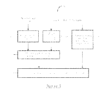
[0033] Figure 1
shows a block diagram of a multi-view pose estimation method used
in some embodiments of the method of the present invention.
11
CA 03168969 2022-07-25
WO 2021/148881
PCT/IB2021/000027
[0034] Figures
2, 3, and 4 show an exemplary embodiments of intraoperative images
used in the method of the present invention. Figures 2 and 3 illustrate a
fluoroscopic image
obtained from one specific pose. Figure 4 illustrates a fluoroscopic image
obtained in a
different pose, as compared to Figures 2 and 3, as a result of C-arm rotation.
The Bronchoscope
¨240, 340, 440, the instrument¨ 210, 310, 410, ribs -220, 320, 420 and body
boundary ¨230,
330, 430 are visible. The multi view pose estimation method uses the visible
elements in
Figures 2, 3, 4 as an input.
[0035] Figure 5
shows a schematic drawing of the structure of bronchial airways as
utilized in the method of the present invention. The airways centerlines are
represented by 530.
A catheter is inserted into the airways structure and imaged by a fluoroscopic
device with an
image plane 540. The catheter projection on the image is illustrated by the
curve 550 and the
radio opaque markers attached to it are projected into points G and F.
[0036] Figure 6
is an image of a bronchoscopic device tip attached to a bronchoscope,
in which the bronchoscope can be used in an embodiment of the method of the
present
invention.
[0037] Figure 7
is an illustration according to an embodiment of the method of the
present invention, where the illustration is of a fluoroscopic image of a
tracked scope (701)
used in a bronchoscopic procedure with an operational tool (702) that extends
from it. The
operational tool may contain radio opaque markers or unique pattern attached
to it.
[0038] Figure 8
is an illustration of epipolar geometry of two views according to an
embodiment of the method of the present invention, where the illustration is
of a pair of
fluoroscopic images containing a scope (801) used in a bronchoscopic procedure
with an
operational tool (802) that extends from it. The operational tool may contain
radio opaque
markers or unique pattern attached to it (points P1 and P2 are representing a
portion of such
pattern). The point P1 has a corresponding epipolar line Ll. The point PO
represents the tip of
12
CA 03168969 2022-07-25
WO 2021/148881
PCT/IB2021/000027
the scope and the point P3 represents the tip of the operational tool. 01 and
02 denote the focal
points of the corresponding views.
[0039] Figure 9 shows an exemplary method for 6-degree-of-freedom pose
estimation
from 3D-2D correspondences.
[0040] Figure 10A and 10B show poses of an X-ray imaging device mounted on
a C-
arm.
[0041] Figure 11 shows use of 3D landmarks use to estimate trajectory of a
C-arm.
[0042] Figure 12 shows a method for an algorithm to use a visible and known
set of
radiopaque markers to estimate a pose per each image frame.
[0043] Figure 13 shows a method for estimating 3D landmarks using a
structure from
motion approach without use of radiopaque markers.
[0044] Figure 14 shows a same feature point of an object visible in
multiple frames.
[0045] Figure 15 shows a same feature point of an object visible in
multiple frames.
[0046] Figure 16 shows a method for optimizing determination of location of
a feature
point of an object visible in multiple frames.
[0047] Figure 17 shows a process for determining a 3D image reconstruction
based on
a received sequence of 2D images.
[0048] Figure 18 shows a process for training an image-to-image translation
using
unaligned images.
[0049] Figure 19 shows training for a model for translation from domain C
to domain
B.
[0050] Figure 20 shows exemplary guidance for a user to position a
fluoroscope.
[0051] Figure 21 shows exemplary guidance for a user to position a
fluoroscope.
[0052] The figures constitute a part of this specification and include
illustrative
embodiments of the present invention and illustrate various objects and
features thereof
13
CA 03168969 2022-07-25
WO 2021/148881
PCT/IB2021/000027
Further, the figures are not necessarily to scale, some features may be
exaggerated to show
details of particular components. In addition, any measurements,
specifications and the like
shown in the figures are intended to be illustrative, and not restrictive.
Therefore, specific
structural and functional details disclosed herein are not to be interpreted
as limiting, but merely
as a representative basis for teaching one skilled in the art to variously
employ the present
invention.
DETAILED DESCRIPTION
[0053] Among
those benefits and improvements that have been disclosed, other objects
and advantages of this invention will become apparent from the following
description taken in
conjunction with the accompanying figures. Detailed embodiments of the present
invention
are disclosed herein; however, it is to be understood that the disclosed
embodiments are merely
illustrative of the invention that may be embodied in various forms. In
addition, each of the
examples given in connection with the various embodiments of the invention
which are
intended to be illustrative, and not restrictive.
[0054]
Throughout the specification and claims, the following terms take the meanings
explicitly associated herein, unless the context clearly dictates otherwise.
The phrases "in one
embodiment" and "in some embodiments" as used herein do not necessarily refer
to the same
embodiments, though it may. Furthermore, the phrases "in another embodiment"
and "in some
other embodiments" as used herein do not necessarily refer to a different
embodiment, although
it may. Thus, as described below, various embodiments of the invention may be
readily
combined, without departing from the scope or spirit of the invention.
[0055] In
addition, as used herein, the term "or" is an inclusive "or" operator, and is
equivalent to the term "and/or," unless the context clearly dictates
otherwise. The term "based
on" is not exclusive and allows for being based on additional factors not
described, unless the
14
CA 03168969 2022-07-25
WO 2021/148881
PCT/IB2021/000027
context clearly dictates otherwise. In addition, throughout the specification,
the meaning of
"a," "an," and "the" include plural references. The meaning of "in" includes
"in" and "on."
[0056] As used
herein, a "plurality" refers to more than one in number, e.g., but not
limited to, 2, 3, 4, 5, 6, 7, 8, 9, 10, etc. For example, a plurality of
images can be 2 images, 3
images, 4 images, 5 images, 6 images, 7 images, 8 images, 9 images, 10 images,
etc.
[0057] As used
herein, an "anatomical element" refers to a landmark, which can be,
e.g.: an area of interest, an incision point, a bifurcation, a blood vessel, a
bronchial airway, a
rib or an organ.
[0058] As used
herein, "geometrical constraints" or "geometric constraints" or "mutual
constraints" or "mutual geometric constraints" refer to a geometrical
relationship between
physical organs (e.g., at least two physical organs) in a subject's body which
construct a similar
geometric relationship within the subject between ribs, the boundary of the
body, etc. Such
geometrical relationships, as being observed through different imaging
modalities, either
remain unchanged or their relative movement can be neglected or quantified.
[0059] As used
herein, a "pose" refers to a set of six parameters that determine a relative
position and orientation of the intraoperative imaging device source as a
substitute to the optical
camera device. As a non-limiting example, a pose can be obtained as a
combination of relative
movements between the device, patient bed, and the patient. Another non-
limiting example of
such movement is the rotation of the intraoperative imaging device combined
with its
movement around the static patient bed with static patient on the bed.
[0060] As used
herein, a "position" refers to the location (that can be measured in any
coordinate system such as x, y, and z Cartesian coordinates) of any object,
including an imaging
device itself within a 3D space.
CA 03168969 2022-07-25
WO 2021/148881
PCT/IB2021/000027
[0061] As used
herein, an "orientation" refers the angles of the intraoperative imaging
device. As non-limiting examples, the intraoperative imaging device can be
oriented facing
upwards, downwards, or laterally.
[0062] As used
herein, a "pose estimation method" refers to a method to estimate the
parameters of a camera associated with a second imaging modality within the 3D
space of the
first imaging modality. A non-limiting example of such a method is to obtain
the parameters
of the intraoperative fluoroscopic camera within the 3D space of a
preoperative CT. A
mathematical model uses such estimated pose to project at least one 3D point
inside of a
preoperative computed tomography (CT) image to a corresponding 2D point inside
the
intraoperative X-ray image.
[0063] As used
herein, a "multi view pose estimation method" refers a method to
estimate to poses of at least two different poses of the intraoperative
imaging device. Where
the imaging device acquires image from the same scene/subject.
[0064] As used
herein, "relative angular difference" refers to the angular difference of
the between two poses of the imaging device caused by their relative angular
movement.
[0065] As used
herein, "relative pose difference" refers to both location and relative
angular difference between two poses of the imaging device caused by the
relative spatial
movement between the subject and the imaging device.
[0066] As used
herein, "epipolar distance" refers to a measurement of the distance
between a point and the epipolar line of the same point in another view. As
used herein, an
"epipolar line" refers to a calculation from an x, y vector or two-column
matrix of a point or
points in a view.
[0067] As used
herein, a "similarity measure" refers to a real-valued function that
quantifies the similarity between two objects.
[0068] In some embodiments, the present invention provides a method,
comprising:
16
CA 03168969 2022-07-25
WO 2021/148881
PCT/IB2021/000027
obtaining a first image from a first imaging modality,
extracting at least one element from the first image from the first imaging
modality,
wherein the at least one element comprises an airway, a blood vessel, a body
cavity, or any combination thereof;
obtaining, from a second imaging modality, at least (i) a first image of a
radiopaque
instrument in a first pose and (ii) a second image of the radiopaque
instrument in a
second pose,
wherein the radiopaque instrument is in a body cavity of a patient;
generating at least two augmented bronchograms,
wherein a first augmented bronchogram corresponds to the first image of the
radiopaque instrument in the first pose, and
wherein a second augmented bronchogram corresponds to the second image of
the radiopaque instrument in the second pose,
determining mutual geometric constraints between:
(i) the first pose of the radiopaque instrument, and
(ii) the second pose of the radiopaque instrument,
estimating the first pose of the radiopaque instrument and the second pose of
the
radiopaque instrument by comparing the first pose of the radiopaque instrument
and
the second pose of the radiopaque instrument to the first image of the first
imaging
modality,
wherein the comparing is performed using:
(i) the first augmented bronchogram,
(ii) the second augmented bronchogram, and
(iii) the at least one element, and
wherein the estimated first pose of the radiopaque instrument and the
17
CA 03168969 2022-07-25
WO 2021/148881
PCT/IB2021/000027
estimated second pose of the radiopaque instrument meets the determined mutual
geometric
constraints,
generating a third image; wherein the third image is an augmented image
derived
from the second imaging modality which highlights an area of interest,
wherein the area of interest is determined from data from the first imaging
modality.
[0069] In some
embodiments, the at least one element from the first image from the
first imaging modality further comprises a rib, a vertebra, a diaphragm, or
any combination
thereof In some embodiments, the mutual geometric constraints are generated
by:
a. estimating a difference between (i) the first pose and (ii) the second pose
by
comparing the first image of the radiopaque instrument and the second image of
the
radiopaque instrument,
wherein the estimating is performed using a device comprising a protractor, an
accelerometer, a gyroscope, or any combination thereof, and wherein the device
is attached to the second imaging modality;
b. extracting a plurality of image features to estimate a relative pose
change,
wherein the plurality of image features comprise anatomical elements, non-
anatomical elements, or any combination thereof,
wherein the image features comprise: patches attached to a patient, radiopaque
markers positioned in a field of view of the second imaging modality, or any
combination thereof,
wherein the image features are visible on the first image of the radiopaque
instrument and the second image of the radiopaque instrument;
c. estimating a difference between (i) the first pose and (ii) the second pose
by using a
at least one camera,
18
CA 03168969 2022-07-25
WO 2021/148881
PCT/IB2021/000027
wherein the camera comprises: a video camera, an infrared camera, a depth
camera, or any combination thereof,
wherein the camera is at a fixed location,
wherein the camera is configured to track at least one feature,
wherein the at least one feature comprises: a marker attached the patient,
a marker attached to the second imaging modality, or any combination
thereof, and
tracking the at least one feature;
d. or any combination thereof
[0070] In some embodiments, the method further comprises: tracking the
radiopaque
instrument for: identifying a trajectory, and using the trajectory as a
further geometric
constraint, wherein the radiopaque instrument comprises an endoscope, an endo-
bronchial tool,
or a robotic arm.
[0071] In some embodiments, the present invention is a method, comprising:
generating a map of at least one body cavity of the patient,
wherein the map is generated using a first image from a first imaging
modality,
obtaining, from a second imaging modality, an image of a radiopaque instrument
comprising at least two attached markers,
wherein the at least two attached markers are separated by a known distance,
identifying a pose of the radiopaque instrument from the second imaging
modality
relative to a map of at least one body cavity of a patient,
identifying a first location of the first marker attached to the radiopaque
instrument on
the second image from the second imaging modality,
19
CA 03168969 2022-07-25
WO 2021/148881
PCT/IB2021/000027
identifying a second location of the second marker attached to the radiopaque
instrument on the second image from the second imaging modality, and
measuring a distance between the first location of the first marker and the
second
location of the second marker,
projecting the known distance between the first marker and the second marker,
comparing the measured distance with the projected known distance between the
first
marker and the second marker to identify a specific location of the radiopaque
instrument inside the at least one body cavity of the patient.
It is possible that inferred 3d information from a single view is still
ambiguous and can fit the
tool into multiple locations inside the lungs. The occurrence of such
situations can be reduced
by analyzing the planned 3d path before the actual procedure and calculating
the most
optimal orientation of the fluoroscope to avoid the majority of ambiguities
during the
navigation. In some embodiments, the fluoroscope positioning is performed in
accordance
with the methods described in the claim 4 of the International Patent
Application No.
PCT/IB2015/00438, the contents of which are incorporated herein by reference
in their
entirety.
[0072]
[0073] In some embodiments, the radiopaque instrument comprises an endoscope,
an endo-
bronchial tool, or a robotic arm.
[0074] In some embodiments, the method further comprises: identifying a depth
of the
radiopaque instrument by use of a trajectory of the radiopaque instrument.
[0075] In some embodiments, the first image from the first imaging modality is
a pre-operative
image. In some embodiments, the at least one image of the radiopaque
instrument from the
second imaging modality is an intra-operative image.
[0076] In some embodiments, the present invention is a method, comprising:
CA 03168969 2022-07-25
WO 2021/148881
PCT/IB2021/000027
obtaining a first image from a first imaging modality,
extracting at least one element from the first image from the first imaging
modality,
wherein the at least one element comprises an airway, a blood vessel, a body
cavity or any combination thereof;
obtaining, from a second imaging modality, at least (i) a one image of a
radiopaque
instrument and (ii) another image of the radiopaque instrument in two
different poses of
second imaging modality
wherein the first image of the radiopaque instrument is captured at a first
pose
of second imaging modality,
wherein the second image of the radiopaque instrument is captured at a second
pose of second imaging modality, and
wherein the radiopaque instrument is in a body cavity of a patient;
generating at least two augmented bronchograms correspondent to each of two
poses
of the imaging device, wherein a first augmented bronchogram derived from the
first
image of the radiopaque instrument and the second augmented bronchogram
derived
from the second image of the radiopaque instrument,
determining mutual geometric constraints between:
(i) the first pose of the second imaging modality, and
(ii) the second pose of the second imaging modality,
estimating the two poses of the second imaging modality relatively to the
first image
of the first imaging modality, using the correspondent augmented bronchogram
images and at
least one element extracted from the first image of the first imaging
modality;
wherein the two estimated poses satisfy the mutual geometric constrains.
generating a third image; wherein the third image is an augmented image
derived
from the second imaging modality highlighting the area of interest, based on
data sourced
21
CA 03168969 2022-07-25
WO 2021/148881
PCT/IB2021/000027
from the first imaging modality.
[0077] During navigation of the endobronchial tool there is a need to verify
tool location in 3D
relatively to the target and other anatomical structures. After reaching some
location in the
lungs a physician may change the fluoroscope position while keeping the tool
at the same
location. Using these intraoperative images skilled in the art can reconstruct
the tool position
in 3d and show the physician the tool position in relation to the target in
3d.
[0078] In order to reconstruct the tool position in 3d it is required to pick
the corresponding
points on both views. The points can be special markers on the tool, or
identifiable points on
any instrument, for example, a tip of the tool, or a tip of the bronchoscope.
To achieve this,
epipolar lines can be used to find the correspondence between points. In
addition, epipolar
constraints can be used to filter false positive marker detections and also to
exclude markers
that don't have a corresponding pair due to marker miss-detection (see Figure
8).
[0079] (Epipolar is related to the geometry of the stereo vision, special area
of computational
geometry)
[0080] In some embodiments, the virtual markers can be generated on any
instrument, for
instance instruments not having visible radiopaque markers. It is performed
by: (1) selecting
any point on the instrument on the first image (2) calculating epipolar line
on the second image
using known geometric relation between both images; (3) intersecting epipolar
lines with the
known or instrument trajectory from the second image, giving a matching
virtual marker.
[0081] In some embodiments, the present invention is a method, comprising:
obtaining a first image from a first imaging modality,
extracting at least one element from the first image from the first imaging
modality,
wherein the at least one element comprises an airway, a blood vessel, a body
cavity or any combination thereof;
obtaining, from a second imaging modality, at least two images in two
different poses
22
CA 03168969 2022-07-25
WO 2021/148881
PCT/IB2021/000027
of second imaging modality of the same radiopaque instrument position for at
least one or
more different instrument positions,
wherein the radiopaque instrument is in a body cavity of a patient;
reconstructing the 3D trajectory of each instrument from the corresponding
multiple
images of the same instrument position in the reference coordinate system,
using
mutual geometric constraints between poses of the corresponding images;
estimating transformation between the reference coordinate system and the
image of
the first imaging modality by estimating the transform that fits reconstructed
3D
trajectories of positions of radiopaque instrument with the 3D trajectories
extracted
from the image of the first imaging modality;
generating a third image; wherein the third image is an augmented image
derived
from the second imaging modality with the known pose in a reference coordinate
system and
highlighting the area of interest, based on data sourced from the first
imaging modality using
the transformation between the reference coordinate system and the image of
the first
imaging modality.
[0082] In some embodiments, a method of collecting the images from different
poses of the
multiple radiopaque instrument positions, is comprising of: (1) positioning a
radiopaque
instrument in the first position; (2) taking an image of the second imaging
modality; (3) change
a pose of the second modality imaging device; (4) taking another image of the
second imaging
modality; (5) changing the radiopaque instrument position; (6) proceeding with
step 2, until
the desired number of unique radiopaque instrument positions is achieved.
[0083] In some embodiments, it is possible to reconstruct the location of any
element that can
be identified on at least two intraoperative images originated from two
different poses of the
imaging device. When each pose of the second imaging modality relatively to
the first image
of the first imaging modality is known, it is possible to show the element's
reconstructed 3D
23
CA 03168969 2022-07-25
WO 2021/148881
PCT/IB2021/000027
position with respect to any anatomical structure from the image of the first
imaging modality.
As an example of the usage of this technique can be a confirmation of 3D
positions of the
deployed fiducial markers relatively to the target.
[0084] In some embodiments, the present invention is a method, comprising:
obtaining a first image from a first imaging modality,
extracting at least one element from the first image from the first imaging
modality,
wherein the at least one element comprises an airway, a blood vessel, a body
cavity or any combination thereof;
obtaining, from a second imaging modality, at least (i) a one image of a
radiopaque
fiducials and (ii) another image of the radiopaque fiducials in two different
poses of second
imaging modality
wherein the first image of the radiopaque fiducials is captured at a first
pose of
second imaging modality,
wherein the second image of the radiopaque fiducials is captured at a second
pose of second imaging modality;
reconstructing the 3D position of radiopaque fiducials from two poses of the
imaging
device, using mutual geometric constraints between:
(i) the first pose of the second imaging modality, and
(ii) the second pose of the second imaging modality,
generating a third image showing the relative 3D position of the fiducials
relatively to
the area of interest, based on data sourced from the first imaging modality.
[0085] In some embodiments, anatomical elements such as: a rib, a vertebra, a
diaphragm, or
any combination thereof, are extracted from the first imaging modality and
from the second
imaging modality.
[0086] In some embodiments, the mutual geometric constraints are generated by:
24
CA 03168969 2022-07-25
WO 2021/148881
PCT/IB2021/000027
a. estimating a difference between (i) the first pose and (ii) the second pose
by
comparing the first image of the radiopaque instrument and the second image
of the radiopaque instrument,
wherein the estimating is performed using a device comprising a
protractor, an accelerometer, a gyroscope, or any combination thereof,
and wherein the device is attached to the second imaging modality;
b. extracting a plurality of image features to estimate a relative pose
change,
wherein the plurality of image features comprise anatomical elements,
non-anatomical elements, or any combination thereof,
wherein the image features comprise: patches attached to a patient,
radiopaque markers positioned in a field of view of the second imaging
modality, or any combination thereof,
wherein the image features are visible on the first image of the
radiopaque instrument and the second image of the radiopaque instrument;
c. estimate a difference between (i) the first pose and (ii) the second pose
by
using a at least one camera,
wherein the camera comprises: a video camera, an infrared camera, a
depth camera, or any combination thereof,
wherein the camera is at a fixed location,
wherein the camera is configured to track at least one feature,
wherein the at least one feature comprises: a marker attached
the patient, a marker attached to the second imaging modality,
or any combination thereof, and
CA 03168969 2022-07-25
WO 2021/148881
PCT/IB2021/000027
tracking the at least one feature;
d. or any combination thereof
[0087] In some embodiments, the method further comprises tracking the
radiopaque
instrument to identify a trajectory and using such trajectory as additional
geometric constrains,
wherein the radiopaque instrument comprises an endoscope, an endo-bronchial
tool, or a
robotic arm.
[0088] In some embodiments, the present invention is a method to identify the
true instrument
location inside the patient, comprising:
using a map of at least one body cavity of a patient generated from a first
image of a
first imaging modality,
obtaining, from a second imaging modality, an image of the radiopaque
instrument
with at least two markers attached to it and having the defined distance
between them
that may be perceived from the image as located in at least two different body
cavities
inside the patient,
obtaining the pose of the second imaging modality relative to the map
identifying a first location of the first marker attached to the radiopaque
instrument on
the second image from the second imaging modality,
identifying a second location of the second marker attached to the radiopaque
instrument on the second image from the second imaging modality, and
measuring a distance between the first location of the first marker and the
second
location of the second marker.
projecting the known distance between markers on each of the perceived
location of
the radiopaque instrument using the pose of the second imaging modality
26
CA 03168969 2022-07-25
WO 2021/148881
PCT/IB2021/000027
comparing the measured distance to each of projected distances between the two
markers to identify the true instrument location inside the body.
[0089] In some embodiments, the radiopaque instrument comprises an endoscope,
an endo-
bronchial tool, or a robotic arm.
[0090] In some embodiments, the method further comprises: identifying a depth
of the
radiopaque instrument by use of a trajectory of the radiopaque instrument.
[0091] In some embodiments, the first image from the first imaging modality is
a pre-operative
image. In some embodiments, the at least one image of the radiopaque
instrument from the
second imaging modality is an intra-operative image.
[0092] Multi view pose estimation
[0093] The application PCT/IB2015/000438 includes a description of a method
to
estimate the pose information (e.g., position, orientation) of a fluoroscope
device relative to a
patient during an endoscopic procedure, and is herein incorporated by
reference in its entirety.
PCT/IB15/002148 filed October 20, 2015 is also herein incorporated by
reference in its
entirety.
[0094] The present invention is a method which includes data extracted from
a set of
intra-operative images, where each of the images is acquired in at least one
(e.g., 1, 2, 3, 4, etc.)
unknown pose obtained from an imaging device. These images are used as input
for the pose
estimation method. As an exemplary embodiment, Figures 3, 4, 5, are examples
of a set of 3
Fluoroscopic images. The images in Figures 4 and 5 were acquired in the same
unknown pose
while the image in Figure 3 was acquired in a different unknown pose. This
set, for example,
may or may not contain additional known positional data related to the imaging
device. For
example, a set may contain positional data, such as C-arm location and
orientation, which can
be provided by a Fluoroscope or acquired through a measurement device attached
to the
Fluoroscope, such as protractor, accelerometer, gyroscope, etc.
27
CA 03168969 2022-07-25
WO 2021/148881
PCT/IB2021/000027
[0095] In some embodiments, anatomical elements are extracted from
additional
intraoperative images and these anatomical elements imply geometrical
constraints which can
be introduced into the pose estimation method. As a result, the number of
elements extracted
from a single intraoperative image can be reduced prior to using the pose
estimation method.
[0096] In some embodiments, the multi view pose estimation method further
includes
overlaying information sourced from a pre-operative modality over any image
from the set of
intraoperative images.
[0097] In some embodiments, a description of overlaying information sourced
from a
pre-operative modality over intraoperative images can be found in
PCT/IB2015/000438,which
is incorporated herein by reference in its entirety.
[0098] In some embodiments, the plurality of second imaging modalities
allow for
changing a Fluoroscope pose relatively to the patient (e.g., but not limited
to, a rotation or
linear movement of the Fluoroscope arm, patient bed rotation and movement,
patient relative
movement on the bed, or any combination of the above) to obtain the plurality
of images, where
the plurality of images are obtained from abovementioned relative poses of the
fluoroscopic
source as any combination of rotational and linear movement between the
patient and
Fluoroscopic device.
[0099] While a number of embodiments of the present invention have been
described,
it is understood that these embodiments are illustrative only, and not
restrictive, and that many
modifications may become apparent to those of ordinary skill in the art.
Further still, the
various steps may be carried out in any desired order (and any desired steps
may be added
and/or any desired steps may be eliminated).
[0100] Reference is now made to the following examples, which together with
the
above descriptions illustrate some embodiments of the invention in a non
limiting fashion.
[0101] Example: Minimally Invasive Pulmonary Procedure
28
CA 03168969 2022-07-25
WO 2021/148881
PCT/IB2021/000027
[0102] A non-
limiting exemplary embodiment of the present invention can be applied
to a minimally invasive pulmonary procedure, where endo-bronchial tools are
inserted into
bronchial airways of a patient through a working channel of the Bronchoscope
(see Figure 6).
Prior to commencing a diagnostic procedure, the physician performs a Setup
process, where
the physician places a catheter into several (e.g., 2, 3, 4, etc.) bronchial
airways around an area
of interest. The Fluoroscopic images are acquired for every location of the
endo-bronchial
catheter, as shown in Figures 2, 3, and 4. An example of the navigation system
used to perform
the pose estimation of the intra-operative Fluoroscopic device is described in
application
PCT/IB2015/00043 8, and the present method of the invention uses the extracted
elements (e.g.,
but not limited to, multiple catheter locations, rib anatomy, and a patient's
body boundary).
[0103] After
estimating the pose in the area of interest, pathways for inserting the
bronchoscope can be identified on a pre-procedure imaging modality, and can be
marked by
highlighting or overlaying information from a pre-operative image over the
intraoperative
Fluoroscopic image. After navigating the endo-bronchial catheter to the area
of interest, the
physician can rotate, change the zoom level, or shift the Fluoroscopic device
for, e.g., verifying
that the catheter is located in the area of interest. Typically, such pose
changes of the
Fluoroscopic device, as illustrated by Figure 4, would invalidate the
previously estimated pose
and require that the physician repeats the Setup process. However, since the
catheter is already
located inside the potential area of interest, repeating the Setup process
need not be performed.
[0104] Figure 4
shows an exemplary embodiment of the present invention, showing the
pose of the Fluoroscope angle being estimated using anatomical elements, which
were
extracted from Figures 2 and 3 (in which, e.g., Figures 2 and 3 show images
obtained from the
initial Setup process and the additional anatomical elements extracted from
image, such as
catheter location, ribs anatomy and body boundary). The pose can be changed
by, for example,
(1) moving the Fluoroscope (e.g., rotating the head around the c-arm), (2)
moving the
29
CA 03168969 2022-07-25
WO 2021/148881
PCT/IB2021/000027
Fluoroscope forward are backwards, or alternatively through the subject
position change or
either through the combination of both etc. In addition, the mutual geometric
constraints
between Figure 2 and Figure 4, such as positional data related to the imaging
device, can be
used in the estimation process.
[0105] Figure 1
is an exemplary embodiment of the present invention, and shows the
following:
[0106] I. The
component 120 extracts 3D anatomical elements, such as Bronchial
airways, ribs, diaphragm, from the preoperative image, such as, but not
limited to, CT,
magnetic resonance imaging (MRI), Positron emission tomography¨computed
tomography
(PET-CT), using an automatic or semi-automatic segmentation process, or any
combination
thereof Examples of automatic or semi-automatic segmentation processes are
described in
"Three-dimensional Human Airway Segmentation Methods for Clinical Virtual
Bronchoscopy", Atilla P. Kiraly, William E. Higgins, Geoffrey McLennan, Eric
A. Hoffman,
Joseph M. Reinhardt, which is hereby incorporated by reference in its
entirety.
[0107] II. The
component 130 extracts 2D anatomical elements (which are further
shown in Figure 4, such as Bronchial airways 410, ribs 420, body boundary 430
and
diaphragm) from a set of intraoperative images, such as, but not limited to,
Fluoroscopic
images, ultrasound images, etc.
[0108] III. The
component 140 calculates the mutual constraints between each subset
of the images in the set of intraoperative images, such as relative angular
difference, relative
pose difference, epipolar distance, etc.
[0109] In
another embodiment, the method includes estimating the mutual constraints
between each subset of the images in the set of intraoperative images. Non-
limiting examples
of such methods are: (1) the use of a measurement device attached to the
intraoperative imaging
device to estimate a relative pose change between at least two poses of a pair
of fluoroscopic
CA 03168969 2022-07-25
WO 2021/148881
PCT/IB2021/000027
images. (2) The extraction of image features, such as anatomical elements or
non-anatomical
elements including, but not limited to, patches (e.g., ECG patches) attached
to a patient or
radiopaque markers positioned inside the field of view of the intraoperative
imaging device,
that are visible on both images, and using these features to estimate the
relative pose change.
(3) The use of a set of cameras, such as video camera, infrared camera, depth
camera, or any
combination of those, attached to the specified location in the procedure
room, that tracks
features, such as patches attached to the patient or markers, markers attached
to imaging device,
etc. By tracking such features the component can estimate the imaging device
relative pose
change.
[0110] IV. The component 150 matches the 3D element generated from
preoperative
image to their corresponding 2D elements generated from intraoperative image.
For example,
matching a given 2D Bronchial airway extracted from Fluoroscopic image to the
set of 3D
airways extracted from the CT image.
[0111] V. The component 170 estimates the poses for the each of the images
in the set
of intra-operative images in the desired coordinate system, such as
preoperative image
coordinate system, operation environment related, coordinated system formed by
other
imaging or navigation device, etc.
[0112] The inputs to this component are as follows:
= 3D anatomical elements extracted from the patient preoperative image.
= 2D anatomical elements extracted from the set of intra-operative images.
As stated
herein, the images in the set can be sourced from the same or different
imaging device
poses.
= Mutual constraints between each subset of the images in the set of
intraoperative
images
[0113] The component 170 evaluates the pose for each image from the set of
intra-
operative images such that:
= The 2D extracted elements match the correspondent and projected 3D
anatomical
elements.
31
CA 03168969 2022-07-25
WO 2021/148881
PCT/IB2021/000027
= The mutual constraint conditions calculated by the component 140 apply
for the
estimated poses.
[0114] To match
the projected 3D elements, sourcing a preoperative image to the
correspondent 2D elements from an inter-operative image, a similarity measure,
such as a
distance metric, is needed. Such a distance metric provides a measure to
assess the distances
between the projected 3D elements and their correspondent 2D elements. For
example, a
Euclidian distance between 2 polylines (e.g., connected sequence of line
segments created as a
single object) can be used as a similarity measure between 3D projected
Bronchial airway
sourcing pre-operative image to 2D airway extracted from the intra-operative
image.
[0115]
Additionally, in an embodiment of the method of the present invention, the
method includes estimating a set of poses that correspond to a set of
intraoperative images by
identifying such poses which optimize a similarity measure, provided that the
mutual
constraints between the subset of images from intraoperative image set are
satisfied. The
optimization of the similarity measure can be referred to as a Least Squares
problem and can
be solved in several methods, e.g., (1) using the well-known bundle adjustment
algorithm
which implements an iterative minimization method for pose estimation, and
which is herein
incorporated by reference in its entirety: B. Triggs; P McLauchlan; R.
Hartley; A. Fitzgibbon
(1999) Bundle" ______________________________________________________
Adjustment A Modern Synthesis". ICCV 99: Proceedings of the
International Workshop on Vision Algorithms. Springer-Verlag, pp. 298-372, and
(2) using a
grid search method to scan the parameter space in search for optimal poses
that optimize the
similarity measure.
[0116] Markers
[0117] Radio-
opaque markers can be placed in predefined locations on the medical
instrument in order to recover 3D information about the instrument position.
Several pathways
of 3D structures of intra-body cavities, such as bronchial airways or blood
vessels, can be
32
CA 03168969 2022-07-25
WO 2021/148881
PCT/IB2021/000027
projected into similar 2D curves on the intraoperative image. The 3D
information obtained
with the markers may be used to differentiate between such pathways, as shown,
e.g., in
Application PCT/IB2015/000438.
[0118] In an exemplary embodiment of the present invention, as illustrated
by Figure
5, an instrument is imaged by an intraoperative device and projected to the
imaging plane 505.
It is unknown whether the instrument is placed inside airway 520 or airway 525
since both
airways are projected into the same curve on the imaging plane 505. In order
to differentiate
between airway 520 and airway 525, it is possible to use at least 2 radiopaque
markers attached
to the catheter having predefined distance "m" between the markers. In Figure
5, the markers
observed on the preoperative image are named "G" and "F".
[0119] The differentiation process between airway 520 and airway 525 can be
performed as follows:
[0120] (1) Project point F from intraoperative image on the potential
candidates of
correspondent airways 520, 525 to obtain A and B points.
[0121] (2) Project point G from intraoperative image on the potential
candidates of
correspondent airways 520, 525 to obtain points C and D.
[0122] (3) Measure the distance between pairs of projected markers IACI and
IBDI.
[0123] (4) Compare the distances IAC 1 on 520 and IBD 1 on 525 to the
distance m
predefined by tool manufacturer. Choose appropriate airway according to a
distance similarity.
[0124] Tracked Scope
[0125] As non-limiting examples, methods to register a patient CT scan with
a
Fluoroscopic device are disclosed herein. This method uses anatomical elements
detected both
in the Fluoroscopic image and in the CT scan as an input to a pose estimation
algorithm that
produces a Fluoroscopic device Pose (e.g., orientation and position) with
respect to the CT
scan. The following extends this method by adding 3D space trajectories,
corresponding to an
33
CA 03168969 2022-07-25
WO 2021/148881
PCT/IB2021/000027
endo-bronchial device position, to the inputs of the registration method.
These trajectories can
be acquired by several means, such as: attaching positional sensors along a
scope or by using
a robotic endoscopic arm. Such an endo-bronchial device will be referred from
now on as
Tracked Scope. The Tracked scope is used to guide operational tools that
extends from it to the
target area (see Figure 7). The diagnostic tools may be a catheter, forceps,
needle, etc. The
following describes how to use positional measurements acquired by the Tracked
scope to
improve the accuracy and robustness of the registration method shown herein.
[0126] In one
embodiment, the registration between Tracked Scope trajectories and
coordinate system of Fluoroscopic device is achieved through positioning of
the Tracked Scope
in various locations in space and applying a standard pose estimation
algorithm. See the
following paper for a reference to a pose estimation algorithm: F. Moreno-
Noguer, V. Lepetit
and P. Fua in the paper "EPnP: Efficient Perspective-n-Point Camera Pose
Estimation", which
is hereby incorporated by reference in its entirety.
[0127] The pose
estimation method disclosed herein is performed through estimating a
Pose in such way that selected elements in the CT scan are projected on their
corresponding
elements in the fluoroscopic image. In one embodiment of the present
invention, adding the
Tracked Scope trajectories as an input to the pose estimation method extends
this method.
These trajectories can be transformed into the Fluoroscopic device coordinate
system using the
methods herein. Once transformed to the Fluoroscopic device coordinate system,
the
trajectories serve as additional constraints to the pose estimation method,
since the estimated
pose is constrained by the condition that the trajectories must fit the
bronchial airways
segmented from the registered CT scan.
[0128] The
Fluoroscopic device estimated Pose may be used to project anatomical
elements from the pre-operative CT to the Fluoroscopic live video in order to
guide an
operational tool to a specified target inside the lung. Such anatomical
elements may be, but are
34
CA 03168969 2022-07-25
WO 2021/148881
PCT/IB2021/000027
not limited to,: a target lesion, a pathway to the lesion, etc. The projected
pathway to the target
lesion provides the physician with only two-dimensional information, resulting
in a depth
ambiguity, that is to say several airways segmented on CT may correspond to
the same
projection on the 2D Fluoroscopic image. It is important to correctly identify
the bronchial
airway on CT in which the operational tool is placed. One method used to
reduce such
ambiguity, described herein, is performed by using radiopaque markers placed
on the tool
providing depth information. In another embodiment of the present invention,
the Tracked
scope may be used to reduce such ambiguity since it provides the 3D position
inside the
bronchial airways. Having such approach applied to the brunching bronchial
tree, it allows
eliminating the potential ambiguity options until the Tracked Scope tip 701 on
Figure 7.
Assuming the operational tool 702 on Figure 7 does not have the 3D trajectory,
although the
abovementioned ambiguity may still happen for this portion of the tool 702,
such event is much
less probable to occur. Therefore this embodiment of present invention
improves the ability of
the method described herein to correctly identify the present tool's position.
[0129] Digital Computational Tomography (DCT)
[0130] In some embodiments, the tomography reconstruction from
intraoperative
images can be used for calculating the target position relatively to the
reference coordinate
system. A non-limiting example of such a reference coordinate system can be
defined by a jig
with radiopaque markers with known geometry, allowing to calculate a relative
pose of each
intraoperative image. Since each input frame of the tomographic
reconstructions has known
geometric relationship to a reference coordinate system, the position of the
target is also can
be positioned in the reference coordinate system. This allows to project a
target on further
fluoroscopic images. In some embodiments, the projected target position can be
compensated
for respiratory movement by tracking tissue in the region of the target. In
some embodiments,
the movement compensation is performed in accordance with the exemplary
methods described
CA 03168969 2022-07-25
WO 2021/148881
PCT/IB2021/000027
in International Patent Application No. PCT/IB2015/00438, the contents of
which are
incorporated herein by reference in their entirety.
[0131] A method
for augmenting target on intraoperative images using the C-arm based
CT and reference pose device, comprising of:
collecting multiple intraoperative images with known geometric relation to a
reference coordinate system;
reconstructing 3d volume;
marking the target area on the reconstructed volume;
projecting target on further intraoperative images with known geometric
relation to a reference coordinate system.
[0132] In other
embodiments, the tomography reconstructed volume can be registered
to the preoperative CT volume. Given the known position of the center of the
target, or
anatomical structures adjunctive to the target, such as blood vessels,
bronchial airways, or
airway bifurcations in the reconstructed volume and in the preoperative
volume, both volumes
can be initially aligned. In other embodiments, ribs extracted from both
volumes can be used
to find an alignment (e.g., an initial alignment). In some embodiments, a step
of finding the
correct rotation between the volumes the reconstructed position and trajectory
of the instrument
can be matched to all possible airway trajectories extracted from the CT. The
best match will
define the most optimal relative rotation between the volumes.
[0133] In some
embodiments, the tomography reconstructed volume can be registered
to the preoperative CT volume using at least 3 common anatomical landmarks
that can be
identified on both the tomography reconstructed volume and the preoperative CT
volume.
Examples of such anatomical landmarks can be airways bifurcations, blood
vessels.
36
CA 03168969 2022-07-25
WO 2021/148881
PCT/IB2021/000027
[0134] In some
embodiments, the tomography reconstructed volume can be registered
to the preoperative CT volume using image-based similarity methods such as
mutual
information.
[0135] In some
embodiments, the tomography reconstructed volume can be registered
to the preoperative CT volume using a combination of at least one common
anatomical
landmark (e.g., a 3D-to-3D constraint) between the tomography reconstructed
volume and the
preoperative CT volume and also at least one 3D-to-2D constraint (e.g., ribs
or a rib cage
boundary). In such embodiments, both type of constraints can be formulated as
an energy
function and minimized using standard optimization methods like gradient
descent.
[0136] In other
embodiments, the tomography reconstructed volumes from different
times of the same procedure can be registered together. Some application of
this could be
comparison of 2 images, transferring manual markings from one image to
another, showing
chronological 3D information.
[0137]
[0138] In other
embodiments, only partial information can be reconstructed from the
DCT because limited quality of fluoroscopic imaging, obstruction of the area
of interest by
other tissue, space limitations of the operational environment. In such cases
the corresponded
partial information can be identified between the partial 3d volume
reconstructed from
intraoperative imaging and preoperative CT. The two image sources can be fused
together to
form unified data set. The abovementioned dataset can be updated from time to
time with
additional intra procedure images.
[0139] In other
embodiments, the tomography reconstructed volume can be registered
to the REBUS reconstructed 3D target shape.
[0140] A method
for performing CT to fluoro registration using the tomography,
comprising of:
37
CA 03168969 2022-07-25
WO 2021/148881
PCT/IB2021/000027
Marking a target on the preoperative image and extracting a bronchial tree;
positioning an endoscopic instrument inside the target lobe of the lungs;
performing a tomography spin using c-arm while the tool is inside and stable;
marking the target and the instrument on the reconstructed volume;
aligning the preoperative and reconstructed volumes by the target position or
by
position of adjunctive anatomical structures;
for all possible airway trajectories extracted from the CT, calculate
the optimal rotation between the volumes that minimizes the distance between
the reconstructed trajectory of the instrument and each airway trajectory;
selecting the rotation corresponding to the minimal distance;
using the alignment between two volumes, enhancing the reconstructed volume
with the anatomical information originated in the preoperative volume;
highlighting the target area on further intraoperative images.
[0141] In other
embodiments, the quality of the digital tomosynthetis can be enhanced
by using the prior volume of the preoperative CT scan. Given the known coarse
registration
between the intraoperative images and preoperative CT scan, the relevant
region of interest can
be extracted from the volume of the preoperative CT scan. Adding constraints
to the well-
known reconstruction algorithm can significantly improve the reconstructed
image quality,
which is herein incorporated by reference in its entirety: Sechopoulos,
Ioannis (2013). "A
review of breast tomosynthesis. Part II. Image reconstruction, processing and
analysis, and
advanced applications". Medical Physics. 40 (1): 014302. As an example of such
a constraint,
the initial volume can be initialized with the extracted volume from the
preoperative CT.
[0142] In some
embodiments, a method of improving tomography reconstruction using
the prior volume of the preoperative CT scan, comprising:
38
CA 03168969 2022-07-25
WO 2021/148881
PCT/IB2021/000027
performing registration between the intraoperative images and preoperative CT
scan;
extracting the region of interest volume from the preoperative CT scan;
adding constraints to the well-known reconstruction algorithm;
reconstructing the image using the added constraints; and
using landmarks to estimate pose during tomography
[0143] In some
embodiments, in order to perform a tomography reconstruction,
multiple images of the same region from different poses are required.
[0144] In some
embodiments, pose estimation can be done using fixed pattern of the
3D radiopaque markers as described in International Pat. App. No.
PCT/IB17/01448, "Jigs for
use in medical imaging and methods for use thereof' (hereby incorporated by
reference herein).
For example, usage of such 3D patterns with radiopaque markers add a physical
limitation that
the said pattern has to be at least partially visible in the image frame
together with the region
of interest area of the patient.
[0145] For
example, one such C-arm based CT system is described in the Prior Art, the
US Patent Application for "C-arm computerized tomography system", published as
US
9044190B2. This application, generally uses a three-dimensional target
disposing in a fixed
position relative to the subject, and obtains a sequence of video images of a
region of interest
of a subject while the C-arm is moved manually or by a scanning motor. Images
from the video
sequence are analyzed to determine the pose of the C-arm relative to the
subject by analysis of
the image patterns of the target.
[0146] However,
this system is dependent on the three-dimensional target with opaque
markers that must be in the field of view for each frame in order to determine
its pose. This
requirement either significantly limits the imaging angles of a C-arm or,
alternatively, requires
positioning such three dimensional target (or the portion of the target) above
or around the
39
CA 03168969 2022-07-25
WO 2021/148881
PCT/IB2021/000027
patient which is a limiting factor from a clinical application perspective
since it is limiting the
approach to the patient or the movement of the C-Arm itself It is known that
the quality and
dimensionality of tomographic reconstruction among other factors depends on
the C-Arm
rotation angle. From the tomographic reconstruction quality perspective the C-
Arm rotation
angle range becomes critical for tomographic reconstruction of the small soft
tissue objects.
The non-limiting example representing such objects is soft-tissue lesion of 8-
15 mm size inside
the human lung.
[0147]
Therefore there is at least a need for a system to obtain wide-angle imaging
from
conventional C-arm fluoroscopic imaging systems, without the need to have a
limiting three
dimensional target (or its portion) with opaque markers in very imaged frame
in order to
determine the pose of the C-Arm suitable for every such frame of the C-Arm.
[0148] In some
embodiments of the present invention, the subject (patient) anatomy
can be used to extract a pose of every image using anatomical landmarks that
are already part
of the image. The non-limiting examples of such are ribs, lung diaphragm,
trachea and others.
This approach can be implemented by using 6-degree-of-freedom pose estimation
algorithms
from 3D-2D correspondences. Such methods are also described in this patent
disclose. See
Figure 9.
[0149] In some
embodiments, the C-Arm movement continuity the missing frame
poses can be extrapolated from the known frames. Alternatively, in such cases,
a hybrid
approach can be used through estimating a pose for a subset of frames through
a pattern of
radiopaque markers assuming that the pattern or its portion is visible for
such computation.
[0150] In some
embodiments, the present invention includes a pose estimation for
every frame from the known trajectory movement of the imaging device assuming
a trajectory
of an X-ray imaging device is known or can be extrapolated and bounded. The
non-limiting
example of the Figure 10A shows a pose of an X-ray imaging device mounted to a
C-arm and
CA 03168969 2022-07-25
WO 2021/148881
PCT/IB2021/000027
covering a pattern of radiopaque markers. A subset of all frames having a
pattern of radiopaque
markers is used to estimate a 3D trajectory of the imaging device. This
information is used to
limit the pose estimation of pose of Figure 10B to a specific 3D trajectory
significantly limiting
the solution search space.
[0151] In some
embodiments, after estimation of the 3D trajectory of a C-Arm
movement, such movement can be represented by a small number of variables. In
non-limiting
example drawn by the Figure Xl, the C-arm has such iso-center that 3D
trajectory can be
estimated using at least 2 known poses of a C-arm and it can be represented by
a single
parameter "t". For this case, having at least one known and visible 3D
landmark in the image
is sufficient to estimate the parameter "t" in the trajectory corresponding to
each pose of the C-
Arm. See Figure 11.
[0152] In some
embodiments, in order to estimate the 3D position of landmarks at least
two known poses of a C-arm are required using triangulation and assuming known
intrinsic
camera parameters. Additional poses can be used for more stable and robust
landmarks position
estimation.
[0153] In some
embodiments, the method of performing tomographic volume
reconstruction of the embodiment of the present invention, comprises:
Performing a rotation of an X-ray imaging device;
Estimating a trajectory of the X-ray imaging device movement using frames
wherein estimated 3D landmarks are visible by solving a camera position and
direction with
3D trajectory constraint and known 3D-2D correspond features;
Evaluation of the position on the trajectory of the frames where estimated 3D
landmarks are invisible or partially visible through extrapolation algorithm
based on an
assumption of continuity of movement;
41
CA 03168969 2022-07-25
WO 2021/148881
PCT/IB2021/000027
Estimating a pose of each frame by solving a camera position and direction
with
3D trajectory constraint and known 3D-2D correspond features; and
Calculating volumetric reconstruction for the area of interest.
[0154] In some
embodiments, the method of performing tomographic volume
reconstruction of the present invention comprises:
Performing a rotation of an X-ray imaging device;
Estimating a trajectory of the X-ray imaging device using frames wherein
pattern of radiopaque marker is visible and pose can be estimated;
Estimating a pose of frame where only estimated 3D landmarks are visible by
solving a camera position and direction with 3D trajectory constraint and
known 3D-2D
correspond features; and
Calculating volumetric reconstruction for the area of interest.
[0155] In some
embodiments, the present invention relates to a solution for the imaging
device pose estimation problem without having any 2D-3D corresponding features
(e.g. no
prior CT image is required). Camera calibration process can be applied online
or offline such
as described by Furukawa, Y. and Ponce, J., "Accurate camera calibration from
multi-view
stereo and bundle adjustment," International Journal of Computer Vision,
84(3), pp. 257-268
(2009) (which is incorporated herein by reference). Having a calibrated
camera, structure from
motion (SfM) technique can be applied to estimate the 3D structure of objects
visible on
multiple images. Such objects can be, but are not limited to, anatomical
objects such as ribs,
blood vessels, spine; instruments positioned inside a body such as
endobronchial tools, wires,
and sensors; or instruments positioned outside and proximate to a body, such
as attached to the
body; etc. In some embodiments, all cameras are solved together. Such
structure from motion
techniques are described in Torr, P.H. and Zisserman, A., "Feature based
methods for structure
42
CA 03168969 2022-07-25
WO 2021/148881
PCT/IB2021/000027
and motion estimation. In International workshop on vision algorithms" (pp.
278-294)
(September 1999), Springer, Berlin, Heidelberg (which is incorporated herein
by reference).
[0156] In some
embodiments, the present invention allows to overcome the limitation
of using a known pattern of 3D radiopaque markers through the combination of
target 3D
pattern and the 3D landmarks that are being estimated dynamically from the
time of the C-Arm
rotation aimed to acquire imaging sequence for tomographic reconstruction or
ever before such
rotation. The non-limiting examples of such landmark are represented through
objects either
inside the patient's body, such as markers on endobronchial tool, the tool
tip, etc, or objects
attached to the body exterior such as patches, wires etc.
[0157] In some
embodiments, the said 3D landmarks are estimated using prior art
tomography or stereo algorithms that utilize a visible and known set of
radiopaque markers to
estimate a pose per each image frame, as described in the Figure 12.
[0158] In some
embodiments, alternatively, the said 3D landmarks are estimated using
structure from motion (SfM) methods without relying on radiopaque markers in
the frame as
described in the Figure 13. In the next step additional 3D landmarks are
estimated. Poses for
frames without known 3D pattern of markers are estimated with the help of
estimated 3D
landmarks. Finally, the volumetric reconstruction is computed using the
sequence of all
available images.
[0159] In some
embodiments, the present invention is a method of reconstruction for
three dimensional volume from a sequence of X-ray images comprising:
Estimating three dimensional landmarks from at least two frames with a known
pose;
Using reconstructed landmarks to estimate a pose for other frames that do not
have a pattern of radiopaque markers in the image frame; and
Calculating volumetric Reconstruction using all frames.
43
CA 03168969 2022-07-25
WO 2021/148881
PCT/IB2021/000027
[0160] In some
embodiments, present invention is an iterative reconstruction method
that maximizes the output imaging quality by iteratively fine-tuning
reconstruction algorithm
input. As a non limiting example of image quality measurement might be
measuring image
sharpness. Because the sharpness is related to the contrast of an image and
thus the contrast
measure can be used as the sharpness or "auto-focus" function. The number of
such
measurements are defined in Groen, F., Young, I., and Ligthart, G., "A
comparison of different
focus functions for use in autofocus algorithms," Cytometry 6, 81-91 (1985).
As an example,
the value Oa) of the squared gradient focus measure for an image at area a is
given by:
[0161] 4k(a)=1 (f(x,y,z+1)-f(x,y,z))^2
[0162] Since
the area of interest should be roughly in the center of the reconstruction
volume, it makes sense to limit the calculation to a small rectangular region
in the center.
[0163] In some
embodiments, this can be formulated as an optimization problem and
solved using techniques like gradient descent.
Finetuning of poses happens by updating the function:
P8 I
7F1,Pn.), where F denotes the reconstruction function given poses pn and
then computing value of the sharpness function 4)0.
[0164] In some
embodiments, the present invention is an iterative pose alignment
method that improves the output imaging quality by iteratively fine-tuning
camera poses to
satisfy some geometric constraints. As a non-limiting example of such
constraints can be a
same feature point of an object visible in multiple frame and therefore have
to be in the
intersection of rays connecting that object and focal point of each camera
(see Figure 14).
44
CA 03168969 2022-07-25
WO 2021/148881
PCT/IB2021/000027
[0165] Initially, most of the times this is not the case because of the
inaccuracy in pose
estimation and also due to displacement of the object (for instance, because
of breathing).
Correcting the poses of cameras to satisfy the rays intersection constraint
will locally
compensate for pose determination errors and movement of the imaged area of
interest,
resulting in better reconstruction image quality. Examples of such feature
points could be the
tip of the instrument inside the patient, or opaque markers on the instrument,
etc.
[0166] In some embodiments, this process can be formulated as an
optimization
problem and may be solved using methods such as gradient descent. See Figure
16 for the
method. The cost function can be defined as a sum of squared distances between
object feature
point and a closest point on each ray (see Figure 15):
F (p) =
[0167] Fluoroscopy device positioning guidance
[0168] In some embodiments, each fluoroscope is calibrated before first
usage. In some
embodiments, a calibration process includes computing an actual fluoroscope
rotation axis,
registering preoperative and intraoperative imaging modalities, and displaying
a target on an
intraoperative image.
[0169] In some embodiments, before the C-arm rotation is started, the
fluoroscope is
positioned in a way that the target projected from preoperative image will
remain in the center
of the image during the entire C-Arm rotation.
CA 03168969 2022-07-25
WO 2021/148881
PCT/IB2021/000027
[0170] In some
embodiments, positioning the fluoroscope in such way that the target
will be in the center of the fluoroscopic image is not, in and of itself,
sufficient, as the
fluoroscope height is critical, while the rotation center is not always in the
middle of the image,
causing undesired target shift outside the image area during the C-Arm
rotation.
[0171] In some
embodiments, as the target location is known relative to the reference
system, an optimal 3D position of the C-Arm is calculated. In some
embodiments, optimizing
the 3D position of the C-Arm means minimizing the target's maximal distance
from the image
center during the C-Arm rotation.
[0172] In some
embodiments, to optimize the 3D position of the C-arm, a user first
takes a single fluoroscopic snapshot. In some embodiments, based on
calculations, the user is
instructed to move the fluoroscope in 3 axes: up-down, left-right (relative to
patient) and head-
feet (relative to patient). In some embodiments, the instructions guide the
fluoroscope towards
its optimal location. In some embodiments, the user moves the fluoroscope
according to the
instructions and then takes another snapshot for getting new instructions,
relative to the new
location. Figure 20 shows exemplary guidance that may be provided to the user
in accordance
with the above.
[0173] In some
embodiments, for each snapshot, the location quality is computed by
computing the percentage of the sweep (assuming +/- 30 degrees from AP) in
which the lesion
is entirely in the ROT, which is a small circle located in the image center.
[0174] In some
embodiments, an alternative way to communicate the instructions is to
display a static pattern and a similar dynamic pattern on an image, where the
static pattern
represents a desired location and the dynamic pattern represents a current
target. In such
embodiments, the user uses continuous fluoroscopic video and the dynamic
pattern moves
according to the fluoroscope movements. In some embodiments, the dynamic
pattern moves
in x and y axes according to fluoroscope's movements in the left-right and
head-feet axes, and
46
CA 03168969 2022-07-25
WO 2021/148881
PCT/IB2021/000027
the scale of the dynamic pattern changes according to fluoroscopy device
movement in the
vertical axis. In some embodiments, by aligning the dynamic and static
patterns, the user
properly positions the fluoroscopy device. Figure 21 shows exemplary static
and dynamic
patterns as discussed above.
[0175] Example
of Improved limited angle X-ray to CT reconstruction using
unsupervised deep learning models.
[0176] There
are different algorithms for 3D image reconstruction from 2D images,
that receives as an input a set of 2D images of an object with a camera pose
for every image
and calculates a 3D reconstruction of the object. Those algorithms provides
lower quality
results when the 2D images are from limited angles (angle range of less than
180 degrees for
X-rays) because of missing information. The proposed method results in
considerable 3D
image quality improvement in comparison to other methods that reconstruct the
3D image from
limited angle 2D images.
[0177] In some
embodiments, the present invention is an improved method that limited
angle x-ray to CT reconstruction using unsupervised deep learning models,
comprising:
Applying method of reconstruction low quality CT from x-ray images using
existing method;
Applying image translation algorithm from domain A to domain C; and
Applying image translation algorithm from domain C to domain B.
[0178] For
further discussion, the domains A, B and C will be used. Domain A, which
is defined as "low quality tomographic reconstruction" domain, domain B, which
is defined as
a CT scan domain, domain C, which is defined as "simulated low quality
tomographic
reconstruction" generated from the pre-procedure CT data.
[0179] In some
embodiments, section one can calculate pose for all the images, and
then to reconstruct a low quality 3D reconstruction, for example, by the
method "Using
47
CA 03168969 2022-07-25
WO 2021/148881
PCT/IB2021/000027
landmarks to estimate pose during tomography" described above, this method
translates the
2D images to low quality CT image, inside domain A.
[0180]
Continuing the last paragraph, the simulated low quality reconstruction, can
be
achieved by applying FP (forward projection) algorithm on a given CT, which
calculates the
intensity integrals along the selected CT axis and result in a simulated
series of 2D X-ray
images, the following step is applying method 1 from above, to reconstruct low
quality 3D
volume, for example by SIRT (Simultaneous Iterative Reconstruction Technique)
algorithm,
which iteratively reconstruct the volume, by starting with an initial guess of
the result
reconstruction, and iteratively applying FP and change the present
reconstruction result by it's
FP difference from the 2D images (https://tomroelandts.com/articles/the-sirt-
algorithm)
[0181] In some
embodiments, the domain translation model that is used to translate a
reconstruction from domain A to C, cannot be supervised (because the
simulation is aligned to
the CT, and there is not aligned CT for the 2D images). The simulated data can
be produced
by the method described above. It is possible to use Cycle-Consistent
Adversarial Networks
(Cycle Gan) to train the required model that translates reconstruction to his
aligned simulation.
The training of Cycle Gan, is done by combining adversarial loss, cycle loss,
and identity loss
(described at Jun-Yon Zhu, Taesung Park, Phillip Isola, and Alexei A Efros.
2017. Unpaired
image-to-image translation using cycle-consistent adversarial networks. In
Proceedings of the
IEEE international conference on computer vision. 2223--2232.) which allows to
train using
unaligned images, as described in Figure 18.
[0182] However,
in some embodiments, the translation model from domain C to
domain B can be supervised, because the creation of the simulation given a CT
is aligned to
the CT, by definition of the process. For example, i CNN-based neural network,
with perceptual
loss (as described in Justin Johnson, Alexandre Alahi, and Li Fei-Fei can be
used. Perceptual
48
CA 03168969 2022-07-25
WO 2021/148881
PCT/IB2021/000027
losses for real-time style transfer and super-resolution. In ECCV, 2016) and
L2 distance loss
to train such model, as described in Figure 19.
[0183] In some
embodiments, the combination of all the methods described above
appear in Figure 17, which describes the process of starting with sequence of
2D images, and
receives 3D image reconstruction
EQUIVALENTS
[0184] The
present invention provides among other things novel methods and
compositions for treating mild to moderate acute pain and/or inflammation.
While specific
embodiments of the subject invention have been discussed, the above
specification is
illustrative and not restrictive. Many variations of the invention will become
apparent to those
skilled in the art upon review of this specification. The full scope of the
invention should be
determined by reference to the claims, along with their full scope of
equivalents, and the
specification, along with such variations.
INCORPORATION BY REFERENCE
[0185] All
publications, patents and sequence database entries mentioned herein are
hereby incorporated by reference in their entireties as if each individual
publication or patent
was specifically and individually indicated to be incorporated by reference.
[0186] While a
number of embodiments of the present invention have been described,
it is understood that these embodiments are illustrative only, and not
restrictive, and that many
modifications may become apparent to those of ordinary skill in the art.
Further still, the
various steps may be carried out in any desired order (and any desired steps
may be added
and/or any desired steps may be eliminated).
49